influence of prolonged sitting and psychosocial stress on ...increased trunk flexion angles and led...
TRANSCRIPT

Influence of Prolonged Sitting and Psychosocial Stress on Lumbar
Spine Kinematics, Kinetics, Discomfort, and Muscle Fatigue
Bochen Jia
Dissertation submitted to the Faculty of Virginia Polytechnic Institute and State University
in partial fulfillment of the requirements for the degree of
Doctor of Philosophy
In
Industrial and Systems Engineering
Maury A. Nussbaum, Chair
Michael J. Agnew
Michael L. Madigan
Thurmon E. Lockhart
Miguel A. Perez
Kevin P. Granata
March 25, 2013
Blacksburg, Virginia
Keywords: Low back pain, prolonged sitting, psychosocial stress, muscle fatigue, viscoelastic
Influence of Prolonged Sitting and Psychosocial Stress on Lumbar Spine Kinematics, Kinetics, Discomfort, and Muscle Fatigue
Bochen Jia
ABSTRACT
Low back pain (LBP) is a common occupational problem and continues to be the leading cause of occupational disability. Among diverse known risk factors, sitting is commonly considered as an important exposure related to LBP. Both modern living and contemporary work involve increased sedentary lifestyles, including more frequent and prolonged sitting. At present, however, the causal role of sitting on LBP development is controversial due to the contribution of several moderating factors (e.g., task demands, duration of exposures, and presence of muscle fatigue). A few studies have assessed low back loads in seated postures, but none has investigated the effects of prolonged sitting or time-dependent variations on spinal structure and spinal loading. Adverse effects of muscle fatigue on low back pain are well documented, yet the specific relationship between muscle fatigue and sitting-related low back pain are not fully established. In addition to these fundamental limitations in our understanding of the physical consequences of sitting, there is also little evidence regarding the effects of task requirements on muscle fatigue and spine loading.
Therefore, the main objectives of this work were, in the context of sitting, to: 1) develop and evaluate a method to assess paraspinal muscle fatigue using electrical stimulation; 2) develop and evaluate a method (model) to quantify biomechanical loads on the lumbar spine in a seated posture; and 3) quantify the effects of prolonged seated tasks on low back loads, body discomfort, and localized muscle fatigue (LMF). The primary hypothesis was that exposure to sitting-related LBP risks is influenced by task requirements and sitting duration.
A muscle stimulation protocol was developed to measure stimulation responses in the lumbar extensors. A stimulation protocol, which included one conditioning train along with three 16-second stimulation train at 2 Hz, was recommended as appropriate to measure those muscles potentially fatigued during prolonged seated tasks. A three-dimensional, sitting-specific, fatigue-sensitive, time-dependent, electromyography (EMG)-based

iii
biomechanical model of the trunk was developed to investigate the effects of seated tasks and time-dependent variations on lumbosacral loading during sitting. Reasonable levels of correspondence were found between measured and predicted lumbosacral moments under a range of seated tasks. Lastly, the effects of prolonged sitting and psychosocial work stress on low back were quantitatively identified. Only prolonged sitting significantly increased trunk flexion angles and led to muscle fatigue. Relatively weak correlations were found between subjective and objective measures, though the two fatigue measurement methods (based on EMG and stimulated responses) showed a good level of correspondence.
Overall, this work provides a quantitative assessment of biomechanical exposures associated with seated tasks. The methods developed in this work make a contribution in terms of measurement/modeling approaches that can be used to assess LBP-relevant risks during prolonged sitting. The results of this work provide a better understanding of the effects of prolonged sitting on the risk of developing sitting-related LBP. Finally, results regarding the influences of prolonged sitting and psychosocial demands can be used to guide future job design.

iv
Acknowledgements
There are many people that I would like to thank for their help and support as I have
pursued this doctoral degree. First, I would like to heartily thank my advisor, Dr. Maury
Nussbaum, for his continuous guidance, support, and patience over the years. I would also
like to thank Dr. Michael Agnew for giving his precious advice regarding technical details of
my research. I would also like to express my gratitude to my committee members, Dr.
Miguel Perez, Dr. Thurmon Lockhart, and Dr. Michael Madigan for their comments,
perspectives, and feedback on my dissertation work. Their efforts helped improve the
quality of this work.
In addition, I would like to thank the colleagues and positive atmosphere of the Industrial
Ergonomics and Biomechanics Lab, in particular Nima Toosizadeh, Brad Hendershot,
Sunwook Kim, Ehsan Rashedi, Babak Bazrgari, for their help, advice, and pilot testing. I
am also thankful to Randy Waldron for building all of the equipment I used in my research
and Will Vest for his computer and technical support.
Finally, I would like to thank my family, and my love for your unconditional patience,
encouragement, and support throughout this journey. You all encouraged me to follow my
dream of pursuing a PhD and helping me get through the final push towards completing this
dissertation.
I offer my regards and blessings to all of those who supported me in any respect during the
completion of my dissertation work.

v
Table of Content
CHAPTER 1. INTRODUCTION .................................................................................................................... 1 REFERENCES .............................................................................................................................................. 5
CHAPTER 2. RELIABILITY OF A STIMULATION METHOD TO ASSESS THE CONTRACTILE STATUS OF THE LUMBAR EXTENSORS IN A SEATED POSTURE ....................................................................... 8
ABSTRACT .................................................................................................................................................. 8 2.1. INTRODUCTION ..................................................................................................................................... 9 2.2. METHODS .......................................................................................................................................... 11
2.2.1. Overview ................................................................................................................................... 11 2.2.2. Participants ............................................................................................................................... 12 2.2.3. Instrumentation ......................................................................................................................... 13 2.2.4. Experimental Procedures .......................................................................................................... 14 2.2.5. Data Processing and Analysis .................................................................................................. 18
2.3. RESULTS ............................................................................................................................................ 19 2.4. DISCUSSION ....................................................................................................................................... 23 REFERENCES ............................................................................................................................................ 29
CHAPTER 3. DEVELOPMENT AND EVALUATION OF AN EMG-BASED MODEL TO ESTIMATE LUMBOSACRAL LOADS DURING SEATED WORK ............................................................................... 34
ABSTRACT ................................................................................................................................................ 34 3.1. INTRODUCTION ................................................................................................................................... 35 3.2. METHOD ............................................................................................................................................ 37
3.2.1 Model and Procedural Overview ................................................................................................ 37 3.2.2 Structure of and Modifications to the Model............................................................................... 38 3.2.3. Participants and Experimental Procedure ................................................................................. 41 3.2.4. Model Implementation and Evaluation ...................................................................................... 45
3.3. RESULTS ............................................................................................................................................ 48 3.4. DISCUSSION ....................................................................................................................................... 52 REFERENCES ............................................................................................................................................ 58
CHAPTER 4. THE EFFECTS OF PROLONGED SITTING AND PSYCHOSOCIAL STRESS ON LOW BACK KINEMATICS, DISCOMFORT, AND LOCALIZED MUSCLE FATIGUE ....................................... 65
ABSTRACT ................................................................................................................................................ 65 4.1. INTRODUCTION ................................................................................................................................... 66 4.2. METHODS .......................................................................................................................................... 68
4.2.1. Participants ............................................................................................................................... 68 4.2.2. Experimental Design ................................................................................................................. 69 4.2.3. Instrumentation and Data Collection ......................................................................................... 70 4.2.4. Experimental Procedures .......................................................................................................... 72 4.2.5. Data Reduction and Processing ............................................................................................... 74 4.2.6. Statistical Analyses ................................................................................................................... 75
4.3. RESULTS ............................................................................................................................................ 76 4.4. DISCUSSION ....................................................................................................................................... 82 REFERENCES ............................................................................................................................................ 88
CHAPTER 5. CONCLUSIONS AND RECOMMENDATIONS ................................................................... 94 REFERENCE .............................................................................................................................................. 99
APPENDIX ................................................................................................................................................ 101

vi
List of figures FIGURE 2-1. ILLUSTRATION OF (A) EXPERIMENTAL FIXTURE SETUP DEMONSTRATING A PARTICIPANT IN AN UPRIGHT
SITTING POSTURE, AND (B) THE LOCATIONS OF DUAL-CHANNEL MUSCLE STIMULATION ELECTRODES (GREY) AND BIPOLAR EMG ELECTRODES (WHITE). .............................................................................................. 14
FIGURE 2-2. SCHEMATIC INDICATION OF STIMULATION PROCEDURES. (A) THREE STIMULATION FREQUENCIES IN SEPARATE TEST STAGES. (B) MUSCLE TWITCH PROTOCOL DURING ONE DATA COLLECTION STAGE. CONDITIONING: CONTINUOUS STIMULATION AT A GIVEN STIMULATION FREQUENCY TO REACH A PLATEAU IN TWITCH FORCE. TRAIN: STIMULATION AT EACH STIMULATION FREQUENCY WITH REST IN BETWEEN. (C) STIMULATION DURING ONE SAMPLING BLOCK. IN ADDITION, PARTICIPANTS WERE REPOSITIONED BETWEEN 2ND AND 3RD SAMPLING BLOCKS. .............................................................................................................. 17
FIGURE 2- 3. SAMPLE STIMULATION RESPONSES MEASURED IN RESPONSE TO THREE DIFFERENT STIMULATION FREQUENCIES. POSITIVE (+) VALUES REPRESENT FORCES RECORDED FROM THE LOAD CELL DURING TRUNK EXTENSION CAUSED BY MUSCLE STIMULATION. THE SECONDARY POSITIVE AND NEGATIVE (SMALLER) PEAKS, IN RESPONSE TO 2HZ STIMULATION, WERE PROBABLY DUE TO REFLEX OF TRUNK FLEXORS AND EXTENSORS, SIMILAR TO A DAMPED PENDULUM RESPONSE. ......................................................................................... 20
FIGURE 2- 4. MEAN STIMULATION RESPONSES (ERROR BARS INDICATED SDS) TO THE THREE STIMULATION FREQUENCIES DURING FOUR SAMPLING BLOCKS (A) AND THREE TRAINS (B). STIMULATION RESPONSES (FORCES) MEASURED AT 2 HZ WERE SIGNIFICANTLY (P<0.05) LARGER THAN THE OTHER TWO STIMULATION
FREQUENCIES IN ALL CASES. .................................................................................................................. 21
FIGURE 3- 1. ILLUSTRATION OF CALIBRATION FIXTURE SETUP (A), DEMONSTRATING A PARTICIPANT IN AN UPRIGHT
SITTING POSTURE, AND THE SITTING DEVICE AND ADJUSTABLE DESK SETUP (B) USED FOR EVALUATION AND FATIGUING TRIALS. ................................................................................................................................. 43
FIGURE 3- 2. ILLUSTRATION OF MEAN (SD) MUSCLE ACTIVATION LEVELS FOR THREE TRUNK EXTENSORS (MF, EL, AND ET) AND FLEXORS (IO, EO, AND RA) COLLECTED BILATERALLY (L/R) UNDER TWO LEVEL OF TIME PRESSURE. THE * SYMBOL INDICATES A SIGNIFICANT PAIRWISE DIFFERENCE. ........................................... 50
FIGURE 3- 3. REPRESENTATIVE RESULTS SHOWING PREDICTION ERRORS OVER TIME, BETWEEN MEASURED AND MODEL-PREDICTED LUMBOSACRAL SAGITTAL PLANE MOMENTS, BOTH WITH (ADJUSTED) AND WITHOUT (UNADJUSTED) A TIME-VARIABLE GAIN. .................................................................................................... 52
FIGURE 4- 1. METHODS USED TO OBTAIN SUBJECTIVE RATINGS OF PERCEIVED DISCOMFORT AND FATIGUE
(MODIFIED FROM KYUNG AND NUSSBAUM, 2008B) .................................................................................. 72
FIGURE 4- 2. ILLUSTRATION OF EXPERIMENT PROCEDURE. FR: FLEXION-RELAXATION MEASUREMENT; SR: STIMULATION-BASED FATIGUE MEASUREMENT; PD: PERCEIVED DISCOMFORT VIA SUBJECTIVE RATING. ....... 74
FIGURE 4- 3. SUMMARY OF MUSCLE ACTIVATION LEVELS (% OF MVC) BETWEEN THE TWO TASK CONDITIONS (LS = LOW PSYCHOSOCIAL STRESS, HS = HIGHER PSYCHOSOCIAL STRESS). ...................................................... 78
FIGURE 4- 4. ILLUSTRATION OF MEAN CHANGES (AFTER VS. BEFORE TASK) IN NORMALIZED FLEXION-RELAXATION ANGLES (FR), STIMULATION RESPONSES (SR), AND MEDIAN FREQUENCIES. ALL MEASURES WERE NORMALIZED TO INITIAL VALUES (I.E., 100% = NO CHANGE OVER TIME). THE * SYMBOL INDICATES A SIGNIFICANT EFFECT OF PROLONGED SITTING, AND ERROR BARS INDICATE STANDARD DEVIATIONS. ........... 79
vii
FIGURE 4- 5. MEAN (SD) VALUES OF PERCEIVED DISCOMFORT FOR EACH BODY PART. ALL PD SCORES SIGNIFICANTLY (P<0.01) INCREASED AT THE END OF THE TASK. ................................................................ 80
FIGURE 4- 6. RELATIONSHIPS BETWEEN SUBJECTIVE (PPC1) AND OBJECTIVE MEASURES [TRUNK ANGLE (A), EPC1 (B), AND STIMULATION RESPONSES (C)], AS WELL AS BETWEEN STIMULATION RESPONSES AND EPC1 (D). ...................................................................................................................................................... 82

viii
List of tables TABLE 2-1. ICC VALUES FOR THREE STIMULATION FREQUENCIES, DETERMINED WITHIN SAMPLING BLOCKS
(WITHIN-SB, FOR FOUR SAMPLING BLOCKS), BETWEEN SAMPLING BLOCKS (BETWEEN-SB, FOR THREE TRAINS), AND FOR MEAN TWITCH FORCES BETWEEN FOUR SAMPLING BLOCKS (BETWEEN-SB MEAN). ........ 23
TABLE 2- 2 ICC VALUES BEFORE AND AFTER REPOSITIONING A PARTICIPANT IN THE EXPERIMENTAL FIXTURE. .... 23
TABLE 4- 1. SUMMARY OF ANOVA RESULTS FOR EFFECTS OF PROLONGED SITTING AND PSYCHOSOCIAL STRESS. .................................................................................................................................................................... 77
TABLE 4- 2. PRINCIPAL COMPONENT (PC) COEFFICIENTS FOR EMG AND PD SCORES. ...................................... 81

1
Chapter 1. Introduction
Low back pain (LBP) is one of the most common societal health problems, causing
considerable disability, work absenteeism, and use of health services. LBP remains the
leading cause of disability among those under 45 year of age (Guo et al., 1999; BLS, 2009).
More than one-quarter of the working population is affected by LBP each year, with a total
of 222,290 cases reported in 2008 (BLS, 2008a). Annually, 40.2% of those with LBP have
persistent symptoms, 14.2% experience an aggravation of their symptoms, and 28.7% have
a recurrence within 6 months (Cassidy et al., 2005). Furthermore, LBP comprises about 40%
of all compensation claims in the United States (Lis et al., 2007) and 33% of total medical
costs (Pope et al., 2002).
Among the identified occupational risk factors for LBP, sitting is commonly considered
among these (Magora, 1972; Kelsey and White, 1980; Andersson, 1981; Wilder et al., 1988;
Chaffin et al., 1999; Vingard et al., 2000; Pope et al., 2002). Sitting is a complex behavior
that is affected by many moderating factors, such as sitting duration (Keyserling, 1991;
Pitman and Ntuen, 1996; Stricker et al., 2003) or psychological task demands (van Dieën et
al., 2001; McLean and Urquhart, 2002; Ellegast et al., 2007). Early work suggested that
both too much and too little sitting are related to an increased incidence of LBP, and which
can be described as a “U”-shaped relationship (Magora, 1972). Although studies have
been conducted to quantify the relationship between these moderating factors and risks of
sitting-related LBP, the specific mechanism(s) that underlie sitting-related LBP have not
been well studied (Hartvigsen et al., 2000; Leboeuf-Yde, 2004; Womersley and May, 2006;
Lis et al., 2007), and this lack has contributed to some controversy regarding the causal role

2
of sitting on LBP (Cholewicki and McGill, 1996; Lis et al., 2007). Existing inconsistencies
are likely due, at least in part, to the wide range of evaluation criteria used and the potential
insensitivity of associated methods employed.
The effects of prolonged sitting have been measured by monitoring participants’ muscle
activities, postures, perceived workloads and discomfort (van Dieën et al., 2001; Gregory et
al., 2006; Todd et al., 2007; Kingma and van Dieën, 2009), but spine kinetics during
prolonged sitting have not been well quantified (Callaghan and McGill, 2001). In addition,
an accurate assessment of spinal loading partitioning among passive and active
components of the trunk during prolonged sitting, such as in biomechanical models,
requires a realistic representation of time-dependent viscoelastic passive properties. The
viscoelastic properties of the spine have been extensively studied, by directly or indirectly
measuring the time-dependent creep and load-relaxation responses of the spine to applied
loading (Burns et al., 1984; Holmes and Hukins, 1996; Kurutz, 2006). However, viscoelastic
responses to prolonged sitting position have not been sufficiently described and it remains
unclear how spine loading may change as a result. Therefore, an effective method is
needed to assess the influence of prolonged sitting on spine kinetics.
Though low levels of muscle activity [<10% maximum voluntary contraction (MVC)] are
required, postural muscles, such as the lumbar erector spinae, are continuously activated to
stabilize the sitting posture (van Dieën et al., 2001). These continuous levels of muscles
activation can cause muscle fatigue in the low back area, and the accompanying aches and

3
cramping may further lead to low back pain (Kolich et al., 2001). However, measures of
muscle fatigue using EMG-based methods have shown some conflicting results. Öberg et
al. (1992; 1994) indicated that a muscle contraction at 15% MVC or higher is required to
detect fatigue-related changes in EMG signal during different working levels. McLean and
Goudy (2004) also indicated that during low level contractions, evidence of fatigue is
inconsistent between different muscle groups and across individuals. However, Søgaard et
al. (2003) and van Dieën et al. (2009) demonstrated the feasibility of using EMG to detect
muscle fatigue during low-level contractions (as low as 2% MVC), though their results were
derived based on 30-minute measurements and still showed a large inter-subject variation.
Therefore, work is needed to test the sensitivity of current EMG-based methods and to find
an efficient way (e.g., muscle stimulation) to detect muscle fatigue during prolonged low-
level exposures (e.g., during sitting).
Many tasks, such as computer-based typing that requires prolonged sitting, also involve
precision work, a high mental workload, a stressful working schedule, etc. The high
prevalence of musculoskeletal disorders in psychologically stressful but light physical work
has indicated that increased perceived workload plays an important role in the development
of LBP (Jensen et al., 1998). Psychosocial task demands (e.g., time pressure and high
mental workload) have been associated with the development of work-related
musculoskeletal disorders in the forearms (Jensen et al., 1998; Blangsted et al., 2004;
Hughes et al., 2007). Both physical and psychosocial task demands can substantially
influence trunk kinematics and increase muscle activity (Pitman and Ntuen, 1996; van
Dieën et al., 2001; Ellegast et al., 2007). Work duration, on the other hand, also has the

4
potential to affect physical and mental task performance (Pitman and Ntuen, 1996; Liao and
Drury, 2000). Psychosocial task demands seem to serve a mediator role with physical risk
factors (such as prolonged duration) and potentially increase the risk for LBP development
(Hughes et al., 2007). However, very few experimental studies have examined the
contribution of such factors to LBP in a seated posture.
The main objective of this work was to develop sensitive methods to detect muscle fatigue
and to quantify biomechanical loads in the spine during prolonged sitting. Additionally, the
effects of different psychosocial task demands on spinal loading and muscle fatigue were
also assessed. The primary hypothesis was that exposure to sitting-related LBP risks is
influenced by psychological task demands and sitting duration. In this work, LBP-relevant
risks were assessed using diverse approaches, including muscle activity and fatigue, trunk
kinematics, body discomfort, and spine kinetics (lumbosacral forces and moments).
The material presented in this dissertation is organized in five chapters. In chapter 1, an
introduction to the work was given, followed by a description of the context of the study, and
its rationale. In chapter 2, a stimulation method is developed and evaluated with a goal of
measuring muscle contractile status during seated postures. In chapter 3, the development
and evaluation of an EMG-driven biomechanical model for internal loads estimation is
presented. In chapter 4, an experimental study is described, to assess the effects of
prolonged sitting and psychosocial stress on low back kinematics, muscle activities, body
discomfort, and muscle fatigue. In chapter 5, research outcomes are summarized,

5
highlighting the major results obtained and important limitations of the current work. A
review of potential applications is also presented and areas described that should be
considered for further research in this field.
References
Andersson, G. B. J. (1981). "Epidemiologic Aspects on Low-Back Pain in Industry." Spine 6(1): 53-60.
Blangsted, A., K. Søgaard, et al. (2004). "The effect of physical and psychosocial loads on the trapezius muscle activity during computer keying tasks and rest periods." European Journal of Applied Physiology 91(2): 253-258.
BLS (2008a). Nonfatal occupational injuries and illnesses requiring days away from work in 2008. U. S. D. o. Labor, United States Department of Labor.
BLS (2009). Nonfatal occupational injuries and illnesses requiring days away from work in 2009. U. S. D. o. Labor, United States Department of Labor.
Burns, M. L., I. Kaleps, et al. (1984). "Analysis of compressive creep behavior of the vertebral unit subjected to a uniform axial loading using exact parametric solution equations of Kelvin-solid models--Part I. Human intervertebral joints." Journal of Biomechanics 17(2): 113-115, 117-130.
Callaghan, J. and S. McGill (2001). "Low back joint loading and kinematics during standing and unsupported sitting." Ergonomics 44: 280 - 294.
Cassidy, J. D. D. C. P. D., P. D. C. P. Cote, et al. (2005). "Incidence and Course of Low Back Pain Episodes in the General Population." Spine 30(24): 2817-2823.
Chaffin, D. B., G. Andersson, et al. (1999). Occupational biomechanics. New York, Wiley-Interscience Publication.
Cholewicki, J. and S. M. McGill (1996). "Mechanical stability of the in vivo lumbar spine: implications for injury and chronic low back pain." Clinical Biomechanics 11(1): 1-15.
Ellegast, R., R. Hamburger, et al. (2007). Effects of Using Dynamic Office Chairs on Posture and EMG in Standardized Office Tasks. Ergonomics and Health Aspects of Work with Computers. M. Dainoff, Springer Berlin / Heidelberg. 4566: 34-42.
Gregory, D. E., N. M. Dunk, et al. (2006). Stability ball versus office chair: comparison of muscle activation and lumbar spine posture during prolonged sitting. Human Factors. 48: 142-153.
Guo, H. R., S. Tanaka, et al. (1999). "Back pain prevalence in US industry and estimates of lost workdays." American Journal of Public Health 89(7): 1029-1035.
Hartvigsen, J., C. Leboeuf-Yde, et al. (2000). "Review Article: Is sitting-while-at-work associated with low back pain? A systematic, critical literature review." Scand J Public Health 28(3): 230-239.
Holmes, A. D. and D. W. L. Hukins (1996). "Analysis of load-relaxation in compressed segments of lumbar spine." Medical Engineering and Physics 18(2): 99-104.

6
Hughes, L. E., K. Babski-Reeves, et al. (2007). "Effects of psychosocial and individual factors on physiological risk factors for upper extremity musculoskeletal disorders while typing." Ergonomics 50(2): 261-274.
Jensen, C., V. Borg, et al. (1998). "Job demands, muscle activity and musculoskeletal symptoms in relation to work with the computer mouse." Scandinavian Journal of Work, Environment and Health 24(5): 418-424.
Kelsey, J. and A. White (1980). "Epidemiology and impact of low back pain." Spine 5: 133 - 142.
Keyserling, W. (1991). "Ergonomic job analysis: A structured approach for identifying risk factors associated with overexertion injuries and disorders." Applied occupational and environmental hygiene.
Kingma, I. and J. H. van Dieën (2009). "Static and dynamic postural loadings during computer work in females: Sitting on an office chair versus sitting on an exercise ball." Applied Ergonomics 40(2): 199-205.
Kolich, M., A. I. Mohamed, et al. (2001). "Electromyographic comparison of two lumbar support mechanisms intended for automotive seating applications." Proceedings of the Institution of Mechanical Engineers -- Part D -- Journal of Automobile Engineering; 2001, Vol. 215 Issue 7, p, 7p 215(7).
Kurutz, M. (2006). "In vivo age- and sex-related creep of human lumbar motion segments and discs in pure centric tension." Journal of Biomechanics 39(7): 1180-1190.
Leboeuf-Yde, C. (2004). "Back pain--individual and genetic factors." Journal of Electromyography and Kinesiology 14(1): 129-133.
Liao, M. H. and C. G. Drury (2000). "Posture, discomfort and performance in a VDT task." Ergonomics 43(3): 345-359.
Lis, A., K. Black, et al. (2007). "Association between sitting and occupational LBP." European Spine Journal 16(2): 283-298.
Magora, A. (1972). "Investigation of the relation between low back pain and occupation. 3. Physical requirements: sitting, standing and weight lifting." IMS Ind Med Surg 41(12): 5-9.
McLean, L. and N. Goudy (2004). "Neuromuscular response to sustained low-level muscle activation: within- and between-synergist substitution in the triceps surae muscles." European Journal of Applied Physiology 91(2): 204-216.
McLean, L. and N. Urquhart (2002). "The influence of psychological stressors on myoelectrical signal activity in the shoulder region during a data entry task." Work & Stress 16(2): 138-153.
Oberg, T., L. Sandsjo, et al. (1994). "Subjective and objective evaluation of shoulder muscle fatigue." Ergonomics 37(8): 1323-1333.
Oberg, T., L. Sandsjo, et al. (1992). "Electromyographic changes in work-related myalgia of the trapezius muscle." Eur J Appl Physiol Occup Physiol 65(3): 251-257.
Pitman, M. S. and C. A. Ntuen (1996). "The effect of prolonged sitting on mental task performance." Computers & Industrial Engineering 31(1-2): 499-502.
Pope, M. H., K. L. Goh, et al. (2002). "Spine ergonomics." Annu Rev Biomed Eng 4: 49-68. Søgaard, K., A. K. Blangsted, et al. (2003). "Evidence of long term muscle fatigue following
prolonged intermittent contractions based on mechano- and electromyograms." Journal of Electromyography and Kinesiology 13(5): 441-450.

7
Stricker, H., G. Colucci, et al. (2003). "The influence of a prolonged sitting position on the biochemical markers of coagulation activation in healthy subjects: evidence of reduced thrombin generation." J Thromb Haemost 1(2): 380-381.
Todd, A. I., A. I. Bennett, et al. (2007). "Physical implications of prolonged sitting in a confined posture - a literature review."
van Dieën, J. H., M. P. de Looze, et al. (2001). "Effects of dynamic office chairs on trunk kinematics, trunk extensor EMG and spinal shrinkage." Ergonomics 44(7): 739-750.
van Dieën, J. H., M. P. de Looze, et al. (2001). "Effects of dynamic office chairs on trunk kinematics, trunk extensor EMG and spinal shrinkage." Ergonomics 44: 739 - 750.
van Dieën, J. H., E. P. Westebring-van der Putten, et al. (2009). "Low-level activity of the trunk extensor muscles causes electromyographic manifestations of fatigue in absence of decreased oxygenation." Journal of Electromyography and Kinesiology 19(3): 398.
Vingard, E. M. D., L. P. Alfredsson, et al. (2000). "To What Extent Do Current and Past Physical and Psychosocial Occupational Factors Explain Care-Seeking for Low Back Pain in a Working Population?: Results from the Musculoskeletal Intervention Center-Norrtalje Study." Spine 25(4): 493-500.
Wilder, D. G., M. H. Pope, et al. (1988). "The Biomechanics of Lumbar Disc Herniation and the Effect of Overload and Instability." Journal of Spinal Disorders & Techniques 1(1): 16-32.
Womersley, L. and S. May (2006). "Sitting Posture of Subjects With Postural Backache." Journal of Manipulative and Physiological Therapeutics 29(3): 213-218.
8
Chapter 2. Reliability of a stimulation method to assess the contractile
status of the lumbar extensors in a seated posture
Abstract
The purpose of the present study was to develop and evaluate methods to assess stimulation responses of the lumbar extensors, as part of a longer-term goal of detecting fatigue during prolonged sitting. Three stimulation frequencies (2, 5, and 8 Hz) were tested in separate stages, which include three stimulation trains and four sampling blocks. Repeated Measures Analyses of Variance was used to determine if any significant differences in mean stimulation responses occurred with respect to stimulation frequency, sampling block, and stimulation train. Reliability of measured stimulation responses was assessed within and between sampling blocks using intraclass correlation coefficients (ICC). Stimulation frequencies significantly affected the stimulation responses and time-to-potentiation differed between the three stimulation frequencies, being highest for 2 Hz stimulation. All three stimulation frequencies resulted in excellent reliability within and between sampling blocks. Current protocol at 2 Hz is recommended as appropriate to measure the lumbar extensors status during prolonged sitting.
Keywords: Muscle stimulation; stimulation frequency; lumbar Extensors.

9
2.1. Introduction
Modern living increases the tendency to have a more sedentary lifestyle that involves
prolonged sitting (Ford, et al., 2005; Varo, et al., 2003). In the US, for example, an earlier
report indicated that more than 60% of adults are rarely physically active and 20% of all
adults are completely sedentary (US Department of Health and Human Services, 1996).
Current estimates are consistent in terms of leisure-time physical activity in the US (Pleis et
al. 2009), and more generally that less than one-third of adults worldwide are physically
active (Hallal et al., 2012). Recent evidence indicates that prolonged sitting postures
compromises blood flow, increases intradiscal loads, and reduces muscle oxygenation
(McLean and Urquhart, 2002; Vollestad, 1997). Such physiological changes can cause
lumbar extensor fatigue that in turn may contribute to low back pain (Dankaerts, et al., 2006;
Kolich, et al., 2001; NIOSH, 1997; O'Sullivan, et al., 2006; Womersley and May, 2006).
Postural muscles are continuously activated to stabilize the sitting posture, though relatively
low levels of muscle activity (< ~10% of maximal muscle capacity) are involved (van Dieën
et al. 2001; Mork and Westgaard 2005; Mork and Westgaard 2009). Further, both objective
and subjective signs of fatigue have been observed following 20 minutes or more of seated
exposures (Blangsted, et al., 2005; Hakala, et al., 2006; McLean and Goudy, 2004;
Sjøgaard, et al., 1986, 2003; van Dieën, et al., 2009).
Objective signs of muscle fatigue during sitting have typically been obtained from
electromyographic (EMG) measures, though the use of EMG in this context has associated
challenges and has provided some conflicting results. As noted, relatively low contraction
levels are involved, and these can be lower than that needed for EMG to detect fatigue-
related changes (Gamet, et al., 1993; Nagata, et al., 1990; Oberg, et al., 1994; Oberg, et al.,

10
1992; Sood, et al., 2007). McLean and Goudy (2004) also indicated that during low
contractions evidence of fatigue is inconsistent between different muscle groups and across
individuals. While Søgaard et al. (2003) and van Dieën et al. (2009) demonstrated the
feasibility of using EMG to detect muscle fatigue during low-level contractions (as low as 2%
MVC), their results showed large inter-subject variability.
A common alternative means to identify muscle fatigue is through use of external
stimulation to obtain stimulation responses. In this approach, fatigue is indicated when
stimulation responses fall significantly below initial, pre-fatigue levels (Binder-Macleod and
Snyder-Mackler, 1993; Edwards, et al., 1977; Johnson, 1998; Johnson, et al., 1995a).
Muscle fatigue has been measured by comparing the decrement of response ratio between
results to high frequency (50-100 Hz) stimulation and low frequency (1-20 Hz) stimulation
(Byström and Kilbom, 1991; Davies and White, 1982; Edwards, et al., 1977). Others have
measured muscle fatigue using a single stimulation frequency (Cooper, et al., 1988;
Johnson, et al., 1995a). While a variety of stimulation protocols have been used, a greater
loss in stimulation response can occur when evoked by low frequency stimulation (Cooper,
et al., 1988; Fitch and McComas, 1985). Low frequency stimulation can also be
advantageous since it is less likely to cause muscle fatigue or discomfort (Johnson, et al.,
1995a). Low frequency muscle stimulation has been shown to be an effective method to
assess muscle fatigue (Johnson, et al., 1995a) and has been successfully applied to the
forearm muscles (Johnson, et al., 1995b; 1996; Mork and Westgaard, 2005). Yet, it
remains unclear whether this approach would be similarly successful for assessing fatigue
in the trunk musculature.

11
The current work is part of broader effort to use muscle stimulation (low frequency,
specifically) to detect lumbar extensors fatigue during prolonged sitting. In this context, the
present study was conducted to develop a reliable muscle stimulation protocol to measure
stimulation responses in the lumbar extensors, specifically those muscles potentially
fatigued during prolonged seated tasks. Several stimulation parameters and conditions can
substantially affect stimulation response, thereby influencing the reliability of stimulation
response measurements. During prolonged stimulation, stimulation responses undergo a
gradual potentiation (Desmedt and Hainaut, 1968), which, if not accounted for, can mask or
confound other effects. For example, an increase in stimulation response amplitude due to
potentiation and a decrease due to fatigue can occur simultaneously (Rassier and
MacIntosh, 2000) and has been observed during low-frequency fatigue caused by sustained
low-level loading (Fowles and Green, 2003). Potentiated stimulation responses may also
be a more sensitive index of contractile fatigue than un-potentiated stimulation responses
(Kufel, et al., 2002). The relationship between stimulation response generation and
excitation is also dependent on stimulation frequency, which can also influence the pattern
of potentiation (Rassier and MacIntosh, 2000). In the current study, we evaluated the
effects of several stimulation frequencies in terms of potentiation and the reliability of
evoked stimulation responses. Results from this exploratory work were intended to facilitate
the selection of stimulation protocols in future studies.
2.2. Methods
2.2.1. Overview
A stimulation protocol to assess lumbar extensor stimulation responses was developed.
Effective stimulation sites and stimulus intensity were identified for each participant since

12
these may vary between individuals due to anatomical differences. Successive muscle
stimulation can lead to a gradual increase in measured muscle responses, a phenomenon
known as muscle potentiation (Desmedt and Hainaut, 1968; Small and Stokes, 1992).
Since potentiation could lead to decreased reliability in stimulation responses, and/or mask
fatigue-related effects, it was considered important to control for these. To address this, a
conditioning train was completed to potentiate target muscles to a stable status, and was
done using each of three stimulation frequencies. Stimulation responses of the lumbar
extensors, represented by the forces measured from resulting (evoked) trunk extension,
were measured in response to each stimulation frequency using the same stimulation
protocol and via a load cell and fixture. Responses to the three stimulation frequencies
were compared to help identify an appropriate stimulation protocol.
2.2.2. Participants
Six participants (gender balanced) were recruited and completed an informed consent
process approved by the Virginia Tech Institutional Review Board. Their mean (SD) age,
height, body mass, and body mass index (BMI) were 28.2 (4.8) yrs, 170.3 (8.7) cm, 67.6
(13.2) kg, and 23.2 (3.2), respectively. Individuals with body mass index >30 were excluded
due to the potential difficulty in evoking reliable muscle contractions. All participants were
physically active and had no self-reported musculoskeletal or neurological diseases within
the past year that restricted their daily activities.

13
2.2.3. Instrumentation
The lumbar extensors were stimulated bilaterally using a dual-channel, current-controlled
muscle stimulator (Grass S88, AstroMed, Inc., West Warwick, RI) in series with a stimulus
isolation unit (SIU5, AstroMed, Inc., West Warwick, RI) and a constant current unit (CCU1,
AstroMed, Inc., West Warwick, RI). Two pairs of 7 cm diameter bipolar stimulating
electrodes (PALS® Platinum Model 879300, Axelgaard Manufacturing Co.Lltd., Fallbrook,
CA) were placed bilaterally over the lumbar extensors (see below, regarding stimulation
site). During the experiment, participants sat in an experimental fixture (Figure 2-1A), in
which motion of the pelvis was restrained using straps. An adjustable footrest was used to
position the lower limb with right angles at the knee and ankle. Participants were positioned
in a comfortable and relaxed upright sitting posture using a rigid rod connected to a chest
harness at the T8 level, and which contained an in-line load cell (Interface SM2000,
capacity = 2000 N, Scottsdale, AZ, USA). Stimulation responses were sampled at 1000Hz
from the load cell. To minimize voluntary muscle activity during stimulation response
measurements, the erector spinae were monitored using two pairs of surface EMG
electrodes that were placed bilaterally at the L3 level (Figure 2-1 B). EMG signals were
monitored visually throughout the experiments in real time. Muscle stimulation was started
only after the observed level of muscle activation was (qualitatively) stabilized, specifically
as reflected in no substantial increase/decrease in EMG levels.

14
(A) (B)
Figure 2-1. Illustration of (A) experimental fixture setup demonstrating a participant in an
upright sitting posture, and (B) the locations of dual-channel muscle stimulation electrodes
(grey) and bipolar EMG electrodes (white).
2.2.4. Experimental Procedures
Participants’ skin in the dorsal lumbar region was prepared following procedures as
described by Cram and Rommen (1989). As needed, the skin surface where the electrodes
would be placed was first shaved, and then skin was cleaned with alcohol and dried. Based
on earlier recommendations on initial electrodes locations and probing procedures (Baker,
et al., 1993; Vanoncini, et al., 2006; Johnson et al. 1995a), subsequent preparatory
procedures were completed to identify the most effective stimulation site for each individual.
Initially, negative electrodes were placed at the level of the iliac crest and positive
electrodes were placed at the level of the inferior margin of the rib cage (see Figure 1.1.B).
Chest harness
Connecting
Load cell

15
Stimulus intensity was then determined by increasing the stimulation current from 10 mA at
each stimulation frequency (see below), until discomfort was reported or up to a max of 40
mA, and it was subsequently reduced by 15%. Discomfort/pain is related to the stimulation
level, which is controlled by the pulse amplitude (current) and pulse duration. Measured
stimulation responses are often a nonlinear function of either pulse duration or stimulation
amplitude (Crago et al., 1980). During stimulation, it is important to minimize charge
transfer to prevent damage to muscle tissue. At a given force level, a high stimulation
amplitude requires less charge transfer per stimulation pulse, and charge transfer increases
with increasing stimulation duration (Crago et al., 1974; Crago et al., 1980). As a
compromise, we used a pulse duration of 0.3 msec. Current-controlled stimulation (at 150v)
was also chosen, as it was easier to control and may induce less discomfort compared to
voltage-controlled stimulation (Merletti, et al., 1992). After identifying the stimulus intensity,
the test muscles were “warmed up” using a 4-minute stimulation block at the stimulation
frequency. Finally, effective electrode sites were then determined by stimulating the muscle
at each stimulation frequency and probing (i.e., adjusting the positive electrode locations)
along superior-inferior direction until maximal evoked stimulation responses were measured
using the load cell.
Each participant then completed three data collection stages (Figure 2-2 A), where each
stage involved one of the three stimulation frequencies (2, 5, and 8 Hz), and a different
presentation order was pre-defined for each participant using a Latin Square and a 10-min
break was given after each stage. The same stimulation frequency and parameters were
applied in the conditioning trains and in the subsequent measurement trains. Within each

16
stage, stimulation responses were measured in four sampling blocks (Figure 2-2 B) with a
2-min rest in between. Within each sampling block, a conditioning train was applied (Figure
2-2 C) to first potentiate the muscles into a steady state (Desmedt and Hainaut, 1968;
Edwards, et al., 1977; Kufel, et al., 2002; Rassier and MacIntosh, 2000). Each conditioning
train was terminated when stimulation responses plateaued, determined in real-time by
using a one-second moving window mean of peak responses measured. This Time-to-
Potentiation (tpot) was recorded along with the potentiated stimulation responses (Fpot).
Immediately after the conditioning train, muscle stimulation responses were measured
during three 16-second stimulation trains (Figure 2-2 C). Short breaks were provided
between each train. To minimize potential effects of repositioning, however, participants
were not allowed to leave the experimental fixture. After the second sampling block,
participants were removed and repositioned in the experimental fixture. This was done to
determine whether there were significant differences in stimulation responses.

17
Con
ditio
ning 1 2 3
Time (mins)
TEST FREQUENCY 1 (STAGE 1)
0
Sampling block 1
Conditioning Train 1
Time (sec)
…Rest
10 sec
16-second
…
Train 2
Rest10 sec
16-second
…
Train 316-second
…0 sec
SAMPLING BLOCK 1
(A)
Break(2 min)
Trai
n
Con
ditio
ning 1 2 3
Sampling block 2Tr
ain
Con
ditio
ning 1 2 3
Sampling block 3
Break(2min)
Trai
n
Con
ditio
ning 1 2 3
Sampling block 4
Trai
n
Repositioning(2min)
Trai
n
Trai
n
Trai
n
Trai
n
Trai
n
Trai
n
Trai
n
Trai
n
tp minutes
(B)
Time (mins)
DATA COLLECTION STAGES
0
Stage 1Test frequency 1
Break10 mins
(C)
Sam
plin
g 1
Sam
plin
g 2
Sam
plin
g 3
Sam
plin
g 4
Stage 2Test frequency 2
Break10 minsSa
mpl
ing
1
Sam
plin
g 2
Sam
plin
g 3
Sam
plin
g 4
Stage 3Test frequency 3
Sam
plin
g 1
Sam
plin
g 2
Sam
plin
g 3
Sam
plin
g 4
Figure 2-2. Schematic indication of stimulation procedures. (A) Three stimulation
frequencies in separate test stages. (B) Muscle twitch protocol during one data collection
stage. Conditioning: continuous stimulation at a given stimulation frequency to reach a
plateau in twitch force. Train: stimulation at each stimulation frequency with rest in between.
(C) Stimulation during one sampling block. In addition, participants were repositioned
between 2nd and 3rd sampling blocks.

18
2.2.5. Data Processing and Analysis
Stimulation responses were band-pass filtered (1.5 - 10 Hz) to remove environmental and
breathing artifacts (Beers, 2003). Peak responses from each stimulation pulse, within each
train, were determined using Matlab software (MathWorksTM, Inc, 2010a). Mean stimulation
responses were the dependent measure in the Repeated Measures Analyses of Variance
(RANOVAs). These were obtained as the means, across all peak stimulation responses
recorded during the 16s block of each train, excluding the three largest and three smallest
values. Such a truncated (trimmed) mean was used as an estimate of central tendency, as
it is less sensitive to outliers (Rothenberg et al., 1966; Wilcox and Keselman, 2003).
RANOVAs were used to identify any significant differences in mean stimulation responses
with respect to stimulation frequency, sampling block, and stimulation train. Where relevant,
significant effects were further investigated using Tukey’s honestly significant difference
(HSD) post-hoc analyses. RANOVA was also used to identify any significant differences in
tpot related to stimulation frequency and sampling block. In all analyses, a p-value of <0.05
was considered statistically significant.
The reliability of stimulation responses was evaluated both within and between sampling
blocks for each of the three stimulation frequencies. To allow for comparisons of twitch
responses within and between participants, stimulation responses measured within each
conditioning train and twitch train were normalized to stimulation responses collected after
potentiation within the first sampling block. This approach was used, since Fpot was
expected to be the most stable response measured. Reliability was evaluated using
intraclass correlation coefficients (ICC), and was determined separately for each of the
19
three stimulation frequencies. Within sampling blocks (Within-SP), reliability was
determined by calculating ICC using data obtained from the three stimulation trains.
Reliability between sampling blocks (Between-SP) was evaluated by calculating ICC from
mean stimulation responses in each train and across all four sampling blocks. ICC was
also calculated using mean stimulation responses across all three trains to evaluate
reliability between sampling blocks (Between-SP Mean). Finally, ICC was used to assess
the reliability of stimulation responses before and after repositioning the participant in the
experimental fixture. ICCs were qualitatively interpreted (Cicchetti and Sparrow (1981) as
poor (0.00–0.39), fair (0.40–0.59), good (0.60–0.74), or excellent (0.75–1.00).
2.3. Results
Stimulation responses were successfully evoked using all three stimulation frequencies
(Figure 2-3). In general, trunk movements and stimulation responses in response to
stimulation at 2 Hz were more noticeable visually, compared to the other two frequencies.
Stimulation at 2 Hz resulted in the largest stimulation responses [mean (SD) = 30.8 (8.8) N],
whereas stimulation at 5 Hz [mean (SD) = 19.6 (3.3) N] and 8 Hz [mean (SD) = 19.7 (4.5) N]
yielded comparable but smaller responses. There was a significant effect of stimulation
frequency (F(2,175) = 134.9; p<0.01) on stimulation responses (Figure 2-4). No significant
main effects of stimulation train (F(2,175)=0.4; p=0.65) or sampling block (F(3,175)=0.9; p=0.34)
were evident, or any interaction effects of stimulation frequency, stimulation train, or
sampling block (p>0.91).

20
Figure 2- 3. Sample stimulation responses measured in response to three different stimulation frequencies. Positive (+) values represent forces recorded from the load cell during trunk extension caused by muscle stimulation. The secondary positive and negative (smaller) peaks, in response to 2Hz stimulation, were probably due to reflex of trunk flexors and extensors, similar to a damped pendulum response.
-20
0
20
40
Stim
ulat
ion
resp
onse
(N) 5 Hz
-20
0
20
40St
imul
atio
n re
spon
se (N
) 2 Hz
-20
0
20
40
0 0.5 1 1.5 2 2.5 3 3.5
Stim
ulat
ion
resp
onse
s (N
)
Time (s)
8 Hz

21
(A) Sampling Blocks
(B) Twitch Trains
Figure 2- 4. Mean stimulation responses (error bars indicated SDs) to the three stimulation
frequencies during four sampling blocks (A) and three trains (B). Stimulation responses
(forces) measured at 2 Hz were significantly (p<0.05) larger than the other two stimulation
frequencies in all cases.
0
10
20
30
40
50
1 2 3 4
Stim
ulat
ion
resp
onse
(N) 2Hz 5Hz 8Hz
0
10
20
30
40
50
1 2 3
Stim
ulat
ion
resp
onse
(N)

22
Stimulation frequency also significantly (F(2,60)=17.3; p<0.01) affected tpot, which was longest
with 2 Hz stimulation (mean = 261 sec), compared to 5 or 8 Hz (mean = 85 sec).
Differences in tpot between the four sampling blocks were not significant (F(3,60)=1.2; p=0.32),
and there was not a significant effect of the frequency x sampling block interaction (F(6,60) =
0.9, p = 0.45). Reliability of stimulation responses within each sampling block (Within-SP)
was in the “excellent” range (>0.85) for all combinations of stimulation frequency and
sampling block (Table 2-1). Qualitatively, reliability within sampling blocks was highest for
stimulation at 2 Hz and consistent across the four sampling blocks. Excellent reliability was
also evident between sampling blocks, with the highest levels found for stimulation at 8 Hz
and the lowest at 2 Hz. This same pattern was evident for the reliability of mean stimulation
responses between sampling blocks. Repositioning participants did not substantially
influence reliability (Table 2- 2). Specifically, ICCs in each of the three trains were excellent,
with one exception (2 Hz, train 3). These values were generally comparable with overall
reliability between sampling blocks (Table 2-1), both in terms of magnitude and the
influence of stimulation frequency.

23
Table 2-1. ICC values for three stimulation frequencies, determined within sampling blocks
(Within-SB, for four sampling blocks), between sampling blocks (Between-SB, for three
trains), and for mean twitch forces between four sampling blocks (Between-SB Mean).
Within-SB ICC Between-SB ICC Between-SB Mean
SB 1 SB 2 SB 3 SB 4 Train 1 Train 2 Train 3 2 Hz 0.96 0.93 0.91 0.96 0.84 0.83 0.81 0.84 5 Hz 0.89 0.94 0.85 0.89 0.88 0.91 0.93 0.91 8 Hz 0.91 0.89 0.94 0.84 0.96 0.95 0.89 0.96
Table 2- 2 ICC values before and after repositioning a participant in the experimental fixture.
Repositioning ICC Train 1 Train 2 Train 3 2 Hz 0.87 0.84 0.76 5 Hz 0.94 0.82 0.94 8 Hz 0.97 0.88 0.97
2.4. Discussion
The objective of this study was to develop a method to measure stimulation responses of
the lumbar trunk extensors in the context of seated postures. Muscle stimulation responses
were successfully generated across all participants in response to all three stimulation
frequencies. Stimulation responses collected at 2 Hz in particular resulted in a mean
response of 32 N, comparable to a value of 35 N in an earlier study (Vanoncini, et al., 2006).
Although the test condition was different than what was used here, the results from
Vanoncini (2006) are considered to at least provide relevant information regarding the
magnitude of stimulation responses in a seated posture. All three frequencies of stimulation
produced measureable responses to muscle contractions, and no substantial pain or skin

24
irritation was either noticed or reported. Rather, participants indicated only minor discomfort
caused by the stimulation, and some minor skin reddening was evident under the
stimulation electrodes, the latter considered a normal reaction to the long stimulation
sessions involved (Baker, et al., 1993).
As an important aspect of stimulation, muscle potentiation was addressed. Potentiation is
considered to be a result of phosphorylation of the myosin light chains, and depends on the
fiber type (Hanson, 1974; Houston, et al., 1985). Specifically, Type II fibers are selectively
and preferentially activated before Type I fibers by stimulation (Hanson, 1974; Sinacore, et
al., 1990). The lumbar extensors (e.g., the erector spinae and multifidus) have a high
proportion of Type I fibers. However, the percentage of Type II fibers can reach 35-50%
(Mannion, et al., 1997; Thorstensson and Carlson, 1987), which emphasizes the importance
of potentiating the muscle first while using stimulation to measure stimulation responses
from these muscles. In the current study, twitch potentiation was observed in all
participants within the first 85-261 seconds of the conditioning trails. Compared to the 2 Hz
stimulation frequency, less time was required to reach a steady state with 5 and 8 Hz
frequencies. This inverse relationship between time-to-potentiation and stimulation
frequency is consistent with existing studies of the knee extensors (Binder-Macleod, et al.,
2002; Eom, et al., 2002) and ulnar nerve (Griffin and Mettler, 2010). Eom et al., (2002) also
indicated in their studies that the degree of force enhancement during potentiation
decreased with the stimulation frequency, which was also observed in the current study.
During potentiation, substantial enhancements of stimulation responses were observed
using all three test frequencies; approximate increases were 50, 30, and 30% in response
to 2, 5, and 8 Hz stimulation, respectively. This again indicates the need for potentiation,

25
since measurements of stimulation responses without potentiation may introduce large
variability to measured stimulation responses (Eom, et al., 2002; Johnson, et al., 1995a).
As found here, muscle stimulation can lead to variable (increasing) magnitudes of
stimulation responses (30%~50%) before reaching a steady state. No consistent pattern of
potentiation in stimulated responses was found in the 2nd or 3rd trains within a sampling
block. In a few cases, though, a fast (within first 2-3 stimulation pulses) and slight increase
(≤ 5%) in stimulation responses was observed at the start of these trains. Any effects of
such increases were minimized, however, during calculation of mean stimulation responses
using truncated means. Since mean stimulation responses measured between several
sampling blocks (Figure 2- 4) were consistent, the conditioning train appeared successful at
achieving a steady state in terms of stimulation responses and it likely did not introduce any
muscle fatigue.
The reliability of evoked stimulation responses was assessed in several ways. In general,
all three stimulation frequencies exhibited excellent reliability in terms of stimulation
responses within and between sampling blocks. Among the three frequencies, within-
sampling block reliability for 2 Hz was somewhat higher than the other two, indicating that
potentiated twitches at 2 Hz were more stable in the three stimulation trains. However, in
comparison to 5 and 8 Hz stimulation, relatively larger trunk movements were observed in
response to the 2 Hz stimulation. Since the seated posture was not precisely controlled
between different sampling blocks, the sitting position at each sampling block may have
varied. Different sitting positions may result in different trunk movement pattern and such
differences may have contributed the somewhat lower reliability between different sampling
blocks. Since 2 Hz stimulation provide more stable and larger stimulation responses, it is
26
reasonable to expect more successful measurements in practical applications (e.g., to
assess muscle fatigue). Stimulation at 5 Hz and 8 Hz, in contrast, though it resulted in
higher ICC values between sampling blocks also yielded stimulation responses that were
relatively lower in magnitude and less stable over the three stimulation trains.
Three potential difficulties need to be acknowledged. First, the most effective location of the
stimulation electrode was achieved using protocols derived from earlier work (Baker, et al.,
1993; Vanoncini, et al., 2006). However, isolation of different muscle groups may not be
practically achievable. The lumbar extensors can be functionally divided into several
different muscle groups, such as the erector spinae and multifidus, with the muscles
structured in different layers making it difficult to identify that the exact target muscle group
has been stimulated in vivo. However, stimulation response measurements here were done
with participants sitting in the experimental fixture in a relatively controlled posture, which
should ensure that the same muscles were stimulated at a similar electrode location within
each sampling block. Therefore, the potential effects of changes in muscle ‘cross-talk’ and
muscle geometry were assumed to be minimal. In addition, unlike voluntary muscle
contraction, muscle contraction induced by stimulation does not follow the size principle,
and muscle fibers are instead recruited without obvious sequencing related to fiber types
(Gregory and Bickel, 2005; Kubiak, et al., 1987). Instead, both slow and fast muscle fibers
are non-selectively activated during muscle stimulation regardless of force levels (Gregory
and Bickel, 2005). As such, the potential effect of differing recruitment patterns can be
minimized through stimulation. Therefore, for future applications, it is reasonable to assume
that any measured changes in stimulation responses using these procedures are
(predominantly) the result of changes in muscle contractile status (e.g., muscle fatigue)

27
versus changes in muscle geometry and/or recruitment pattern. Further work will be
completed to quantify the potential effects of trunk flexor and extensor activities on
measured stimulation responses. In addition, it remains to determine whether stimulation
responses remain reliable over longer blocks of measurement. For example, there may be
important influences of diurnal, temperature, or electrode changes.
Second, the seated posture was not precisely controlled between sampling blocks, and the
exact postures may have varied between different sampling blocks. Large postural
changes could have introduced variability in measured stimulation responses. One reason
to select a sitting position for muscle stimulation is to match the conditions (e.g. posture)
during the actual tasks we plan in future studies (of seated work tasks). By using similar
conditions, physiological properties of muscles (e.g., muscle length) and levels of voluntary
contraction can be kept relatively consistent, and thereby help in distinguishing fatigue from
potential confounding influences. Therefore, the effect of sitting postures could perhaps be
minimized by controlling participant postures more precisely within and between different
sampling blocks. In addition, monitoring trunk angles (e.g., using a motion tracking system),
may facilitate methods to account for changes in measured stimulation responses caused
by postural differences.
Third, voluntary contraction levels of the trunk flexors and extensors, as postural muscles,
were controlled but not eliminated during muscle stimulation. Thus, the potential
coexistence of muscle stimulation and voluntary contraction may have had a net effect on
measured stimulation responses. Instead of pure twitch forces, the current methods
provide a measure of net responses from both muscle stimulation and voluntary contraction

28
of trunk flexors and extensors. Trunk extensors were monitored (real time EMG) throughout
the current study, to minimize the levels of voluntary contraction. Qualitatively, this
monitoring indicated relatively low and consistent levels of contraction during the sampling
blocks. In addition, the sitting posture adopted during the sampling blocks placed the
lumbar spine in flexion; this placed the lumbar motion segments beyond a neutral posture
and activation of the trunk extensors was thus predominant. Thus, while potential effects of
voluntary contraction were not eliminated, these were considered relatively small in the
current study. Further work will be completed to identify the effects of trunk posture on
measured stimulation responses and these results can be used to account for the effects of
postural changes on measured stimulation responses.
Our long-term goal is to use muscle stimulation for detecting lumbar extensors fatigue
during prolonged sitting. The developed method will be used to measure muscle fatigue in
a future study, specifically to assess situations involving prolonged sitting. At present
however, this approach may be limited to use in a laboratory environment, given the needs
for a special seat and instrumentation. The current results suggest that stimulation at 2 Hz
can provide suitable stimulation responses for this purpose. The higher responses captured
using 2 Hz are considered beneficial, to allow for better separation of the muscle responses
to stimulation from any background noise and/or potential voluntary muscle forces.
Stimulation at this frequency required reasonable time blocks for potentiation (~4 min), and
evoked stimulation responses had excellent reliability both within and between sampling
blocks. The current results indicated high levels of reliability within and between sampling
blocks and trains, While this suggests that only a single sampling train may be required,

29
multiple sampling trains are recommended in future work to enhance the stability of
obtained measures. .
References
Adams GR, Harris RT, Woodard D, Dudley GA. (1993). Mapping of electrical muscle
stimulation using MRI. J Appl Physiol. 74: 532–537. Baker, L., McNeal, D., Benton, L., Bowman, B., & Waters, R. (1993). Neuromuscular
electrical stimulation : a practical guide (3rd ed.). Downey, Calif.: Rancho Rehabilitation Engineering Program, Rancho Los Amigos Medical Center.
Beach, T. A., Mooney, S. K., & Callaghan, J. P. (2003). The effects of a continuous passive motion device on myoelectric activity of the erector spinae during prolonged sitting at a computer workstation. Work, 20(3), 237-244.
Beers, M. H. (2003). The Merck manual of medical information (2nd home ed.). Whitehouse Station, NJ: Merck Research Laboratories.
Binder-Macleod, S. A., & Snyder-Mackler, L. (1993). Muscle Fatigue: Clinical Implications for Fatigue Assessment and Neuromuscular Electrical Stimulation. Physical Theropy, 73(12), 902-910.
Binder-Macleod SA, Halden EE, Jungles KA. (1995). Effects of stimulation intensity on the physiological responses of human motor units. Medicine and Science in Sports and Exercise, 27:556 –565.
Binder-Macleod, S. A., Dean, J. C., & Ding, J. (2002). Electrical stimulation factors in potentiation of human quadriceps femoris. Muscle and Nerve, 25(2), 271-279.
Blangsted, A. K., Sjøaard, G., Madeleine, P., Olsen, H. B., & Søaard, K. (2005). Voluntary low-force contraction elicits prolonged low-frequency fatigue and changes in surface electromyography and mechanomyography. Journal of Electromyography and Kinesiology, 15(2), 138-148.
Byström, S., & Kilbom, Å. (1991). Electrical stimulation of human forearm extensor muscles as an indicator of handgrip fatigue and recovery. European Journal of Applied Physiology and Occupational Physiology, 62(5), 363-368.
Callaghan, J., & McGill, S. (2001). Low back joint loading and kinematics during standing and unsupported sitting. Ergonomics, 44, 280 - 294.
Chou, L. W., & Binder-Macleod, S. A. (2007). The effects of stimulation frequency and fatigue on the force-intensity relationship for human skeletal muscle. Clinical Neurophysiololgy, 118(6), 1387-1396.
Cicchetti, D. V., & Sparrow, S. A. (1981). Developing criteria for establishing interrater reliability of specific items: applications to assessment of adaptive behavior. American Journal of Mental Deficiency, 86(2), 127-137.
Cooper, R. G., Edwards, R. H., Gibson, H., & Stokes, M. J. (1988). Human muscle fatigue: frequency dependence of excitation and force generation. The Journal of Physiology, 397, 585-599.
Crago, P., Peckham, P., Mortimer, J., & Van Der Meulen, J. (1974). The choice of pulse duration for chronic electrical stimulation via surface, nerve, and intramuscular electrodes. Annals of Biomedical Engineering, 2(3), 252-264.

30
Crago, P. E., P. H. Peckham, et al. (1980). "Modulation of Muscle Force by Recruitment During Intramuscular Stimulation." Biomedical Engineering, IEEE Transactions on BME-27(12): 679-684.
Cram, J. R., & Rommen, D. (1989). Effects of skin preparation on data collected using an EMG muscle-scanning procedure. Applied Psychophysiology and Biofeedback, 14(1), 75-82.
Dankaerts, W. P., O'Sullivan, P. P., Burnett, A. P., & Straker, L. P. (2006). Differences in Sitting Postures are Associated With Nonspecific Chronic Low Back Pain Disorders When Patients Are Subclassified. Spine, 31(6), 698-704.
Davies, C. T., & White, M. J. (1982). Muscle weakness following dynamic exercise in humans. Journal of Applied Physiology, 53(1), 236-241.
Desmedt, J. E., & Hainaut, K. (1968). Kinetics of Myofilament Activation in Potentiated Contraction: Staircase Phenomenon in Human Skeletal Muscle. Nature, 217(5128), 529-532.
Edwards, R. H., Hill, D. K., Jones, D. A., & Merton, P. A. (1977). Fatigue of long duration in human skeletal muscle after exercise. The Journal of Physiology, 272(3), 769-778.
Eom, G., Watanabe, T., Hoshimiya, N., & Khang, G. (2002). Gradual potentiation of isometric muscle force during constant electrical stimulation. Medical and Biological Engineering and Computing, 40(1), 137-143.
Fitch, S., & McComas, A. (1985). Influence of human muscle length on fatigue. Journal of Physiology, 362, 205-213.
Ford, E. S., Kohl, H. W., Mokdad, A. H., & Ajani, U. A. (2005). Sedentary Behavior, Physical Activity, and the Metabolic Syndrome among U.S. Adults. Obesity, 13(3), 608-614.
Fowles, J. R., & Green, H. J. (2003). Coexistence of potentiation and low-frequency fatigue during voluntary exercise in human skeletal muscle. Canadian Journal of Physiology and Pharmacology, 81(12), 1092-1100.
Gamet, D., Duchene, J., Garapon-Bar, C., & Goubel, F. (1993). Surface electromyogram power spectrum in human quadriceps muscle during incremental exercise. Journal of Applied Physiology, 74(6), 2704-2710.
Gregory, C. M., & Bickel, C. S. (2005). Recruitment Patterns in Human Skeletal Muscle During Electrical Stimulation. Physical Therapy, 85(4), 358-364.
Griffin, L., & Mettler, J. A. (2010). What are the stimulation parameters that affect the extent of twitch force potentiation in the adductor pollicis muscle? European Journal of Applied Physiology, 110(6), 1235-1242.
Hakala, P. T., Rimpela, A. H., Saarni, L. A., & Salminen, J. J. (2006). Frequent computer-related activities increase the risk of neck-shoulder and low back pain in adolescents. European Journal of Public Health, 16(5), 536-541.
Hallal, P. C., L. B. Andersen, et al. (2012). "Global physical activity levels: surveillance progress, pitfalls, and prospects." Lancet 380(9838): 247-257.
Hanson, J. (1974). The Effects of Repetitive Stimulation on the Action Potential and the Twitch of Rat Muscle. Acta Physiologica Scandinavica, 90(2), 387-400.
Houston, M. E., Green, H. J., & Stull, J. T. (1985). Myosin light chain phosphorylation and isometric twitch potentiation in intact human muscle. Pflügers Archiv European Journal of Physiology, 403(4), 348-352.
Johnson, P. W., Lehman, S. L., & Rempel, D. M. (1995a). Measuring low frequency fatigue with 2 Hertz stimulation. I. Stimulus-related potentiation effects. Paper presented at the Engineering in Medicine and Biology Society, IEEE 17th Annual Conference.

31
Johnson, P., Lehman, S. L., & Rempel, D. M. (1995b). Measuring low frequency fatigue with 2 Hertz stimulation. II. Muscle fatigue results. Paper presented at the Engineering in Medicine and Biology Society, IEEE 17th Annual Conference.
Johnson, P. W., Lehman, S. L., & Rempel, D. M. (1996). Measuring muscle fatigue during computer mouse use. Paper presented at the Engineering in Medicine and Biology Society. Bridging Disciplines for Biomedicine. Proceedings of the 18th Annual International Conference of the IEEE.
Johnson, P. W. (1998). The development, characterization and implementation of a technique to measure muscle fatigue during computer use. University of California, Berkeley.
Kolich, M., Mohamed, A. I., & Taboun, S. M. (2001). Electromyographic comparison of two lumbar support mechanisms intended for automotive seating applications. Proceedings of the Institution of Mechanical Engineers. Part D, Journal of Automobile Engineering; 215(7)
Kubiak, R. J., Whitman, K. M., & Johnston, R. M. (1987). Changes in quadriceps femoris muscle strength using isometric exercise versus electrical stimulation. Journal of Orthopaedic and Sports Physical Therapy, 8(11), 537-541.
Kufel, T. J., Pineda, L. A., & Mador, M. J. (2002). Comparison of potentiated and unpotentiated twitches as an index of muscle fatigue. Muscle and Nerve, 25(3), 438-444.
Mannion, A. F., Dumas, G. A., Cooper, R. G., Espinosa, F. J., Faris, M. W., & Stevenson, J. M. (1997). Muscle fibre size and type distribution in thoracic and lumbar regions of erector spinae in healthy subjects without low back pain: normal values and sex differences. Journal of Anatomy, 190(4), 505-513.
McLean, L., & Goudy, N. (2004). Neuromuscular response to sustained low-level muscle activation: within- and between-synergist substitution in the triceps surae muscles. European Journal of Applied Physiology, 91(2), 204-216.
Madeleine, P., Jørgensen, L., Søgaard, K., Arendt-Nielsen, L., & Sjøgaard, G. (2002). Development of muscle fatigue as assessed by electromyography and mechanomyography during continuous and intermittent low-force contractions: effects of the feedback mode. European Journal of Applied Physiology, 87(1), 28-37.
Mannion, J., Bitto, T., Hammond, R., Rubinstein, N., & Stephenson, L. (1986). Histochemical and fatigue characteristics of conditioned canine latissimus dorsi muscle. Circulation Research, 58(2), 298-304.
Merletti, R., Knaflitz, M., & DeLuca, C. J. (1992). Electrically evoked myoelectric signals. Critical Reviews in Biomedical Engineering, 19(4), 293-340.
Mork, P. J., & Westgaard, R. H. (2005). Long-term electromyographic activity in upper trapezius and low back muscles of women with moderate physical activity. Journal of Applied Physiology, 99(2), 570-578.
Mork, P. J. and R. H. Westgaard (2009). "Back posture and low back muscle activity in female computer workers: A field study." Clinical Biomechanics 24(2): 169-175.
Nachemson, A. (1966). The load on lumbar disks in different positions of the body. Clinical Orthopaedics and Related Research, 45, 107-122.
Nachemson, A. L. (1981). Disc pressure measurements. Spine (Phila Pa 1976), 6(1), 93-97. Nagata, S., Arsenault, A. B., Gagnon, D., Smyth, G., & Mathieu, P. A. (1990). EMG power
spectrum as a measure of muscular fatigue at different levels of contraction. Medical & Biological Engineering & Computing, 28(4), 374-378.

32
NIOSH. (1997). Musculoskeletal Disorders and Workplace Factors: A Critical Review of Epidemiologic Evidence for Work-Related Musculoskeletal Disorders of the Neck, Upper Extremity, and Low Back.
O'Sullivan, P. B., Mitchell, T., Bulich, P., Waller, R., & Holte, J. (2006). The relationship beween posture and back muscle endurance in industrial workers with flexion-related low back pain. Manual Therapy, 11(4), 264-271.
Oberg, T., Sandsjo, L., & Kadefors, R. (1994). Subjective and objective evaluation of shoulder muscle fatigue. Ergonomics, 37(8), 1323-1333.
Oberg, T., Sandsjo, L., Kadefors, R., & Larsson, S. E. (1992). Electromyographic changes in work-related myalgia of the trapezius muscle. European Journal of Applied Physiology and Occupational Physiology, 65(3), 251-257.
Pleis, J. R., J. W. Lucas, et al. (2009). "Summary health statistics for U.S. adults: National Health Interview Survey, 2008." Vital and Health Statistics. Series 10: Data from the National Health Survey(242): 1-157.
Rassier, D. E., & MacIntosh, B. R. (2000). Coexistence of potentiation and fatigue in skeletal muscle. Brazilian Journal of Medical and Biological Research, 33, 499-508.
Ratkevičius, A., Skurvydas, A., Povilonis, E., Quistorff, B., & Lexell, J. (1998). Effects of contraction duration on low-frequency fatigue in voluntary and electrically induced exercise of quadriceps muscle in humans. European Journal of Applied Physiology and Occupational Physiology, 77(5), 462-468.
Rothenberg, T. J., Fisher, F. M., & Tilanus, C. B. (1964). A note on estimation from a Cauchy sample. Journal of the American Statistical Association, 59(306), 460-463.
Sinacore, D. R., Delitto, A., King, D. S., & Rose, S. J. (1990). Type II Fiber Activation with Electrical Stimulation: A Preliminary Report. Physical Therpy, 70(7), 416-422.
Sjøgaard, G., Kiens, B., Jørgensen, K., & Saltin, B. (1986). Intramuscular pressure, EMG and blood flow during low-level prolonged static contraction in man. Acta Physiologica Scandinavica, 128(3), 475-484.
Slade JM, Bickel CS, Warren GL, Dudley GA. (2003). Variable frequency trains enhance torque independent of stimulation amplitude. Acta Physiologica Scandinavica. 177:87–92.
Small, S. C. and M. J. Stokes (1992). "Stimulation frequency and force potentiation in the human adductor pollicis muscle." Eur J Appl Physiol Occup Physiol 65(3): 229-233.
Søgaard, K., Blangsted, A. K., Jørgensen, L. V., Madeleine, P., & Sjøgaard, G. (2003). Evidence of long term muscle fatigue following prolonged intermittent contractions based on mechano- and electromyograms. Journal of Electromyography and Kinesiology, 13(5), 441-450.
Sood, D., Nussbaum, M. A., & Hager, K. (2007). Fatigue during prolonged intermittent overhead work: reliability of measures and effects of working height. Ergonomics, 50(4), 497-513.
Thorstensson, A., & Carlson, H. (1987). Fibre types in human lumbar back muscles. Acta Physiologica Scandinavica, 131(2), 195-202.
US Department of Health and Human Services. (1996). Physical activity and health: a report of the Surgeon General. Atlanta, Georgia: US Department of Health and Human Services, Public Health Service, CDC, National Center for Chronic Disease Prevention and Health Promotion,
33
van Dieën, J. H., de Looze, M. P., & Hermans, V. (2001). Effects of dynamic office chairs on trunk kinematics, trunk extensor EMG and spinal shrinkage. Ergonomics, 44(7), 739-750.
van Dieën, J. H., Westebring-van der Putten, E. P., Kingma, I., & de Looze, M. P. (2009). Low-level activity of the trunk extensor muscles causes electromyographic manifestations of fatigue in absence of decreased oxygenation. Journal of Electromyography and Kinesiology, 19(3), 398.
Vanoncini, M., Holderbaum, W., & Andrews, B. J. (2006). Investigations on the biomechanics of the human trunk. Proceedings of the 24th IASTED international conference on Biomedical engineering,
Varo, J. J., Martinez-Gonzalez, M. A., de Irala-Estevez, J., Kearney, J., Gibney, M., & Martinez, J. A. (2003). Distribution and determinants of sedentary lifestyles in the European Union. International Journal of Epidemiololgy, 32(1), 138-146.
Vollestad, N. K. (1997). Measurement of human muscle fatigue. Journal of Neuroscience Methods, 74(2), 219-227.
Wilcox, R. R. and H. J. Keselman (2003). "Modern robust data analysis methods: measures of central tendency." Psychological Methods 8(3): 254-274.
Wilke, H.-J. P., Neef, P. M. D., Caimi, M. M. D., Hoogland, T. M. D., & Claes, L. E. P. (1999). New In Vivo Measurements of Pressures in the Intervertebral Disc in Daily Life. Spine, 24(8), 755-762.
Womersley, L., & May, S. (2006). Sitting Posture of Subjects With Postural Backache. Journal of Manipulative and Physiological Therapeutics, 29(3), 213-218.

34
Chapter 3. Development and Evaluation of an EMG-based Model to
Estimate Lumbosacral Loads during Seated Work
Abstract
Low back pain (LBP) is a common occupational problem and continues to be the leading cause of occupational disability. Among diverse known risk factors, sitting is commonly considered as an important exposure related to LBP risk, and modern living and contemporary work both involve increasing sedentary behaviors including more frequent and prolonged sitting. At present, however, the causal role of sitting on LBP development is controversial due to the variety of potential moderating factors. Specifically, a few studies have assessed lumbosacral loads in seated postures, but no sitting-specific model has been developed to investigate the effects of seated tasks and time-dependent variations on lumbosacral loading during sitting. Here, a three-dimensional, electromyography (EMG)-based biomechanical model of the trunk was developed to predict lumbosacral loads during a range of seated tasks. This model was a modification of an earlier approach, and specific modifications included a revised representation of lumbar muscle anatomy and viscoelastic soft-tissue properties, and a method to account for muscular fatigue during prolonged sitting. With these enhancements, the predictive ability of the model was assessed over a range of seated tasks that differed in terms of lumbar posture, time pressure, and mental workload. Predicted model parameters corresponded well with values reported earlier. Reasonable levels of correspondence were found between measured and predicted lumbosacral moments across all tested tasks. Physical exposures and injury risks related to seated work can potentially be estimated using this modeling approach, and which may facilitate future injury prevention strategies.
Key words: Sitting; Electromyography; Biomechanical model; Spine; Low back pain
35
3.1. Introduction
With increasing computer use and deskwork associated with most work and leisure
activities, sedentary lifestyle has become a new trend in modern living, including substantial
time in seated postures. Sitting has been often highlighted as a risk factor for low back pain
(LBP) in the literature (Liira et al., 1996; Chaffin et al., 1999; Johanning, 2000; Vingard et al.,
2000; Lee et al., 2001; Pope et al., 2002; Kopec et al., 2004; Corlett, 2006). Recent reviews,
however, have suggested a less clear relationship between LBP and sitting at work
(Hartvigsen et al., 2000; Hartvigsen et al., 2003; Leboeuf-Yde, 2004; Womersley and May,
2006; Lis et al., 2007; Bakker et al., 2009; Chen et al., 2009). One study (Hartvigsen et al.,
(2001) concluded that sedentary work was not a risk factor for LBP development, and might
even have a protective effect. Sitting is a complex activity that can be affected by many
moderating factors, such as sitting duration (Keyserling, 1991; Pitman and Ntuen, 1996;
Stricker et al., 2003) and task requirements (van Dieën et al., 2001; McLean and Urquhart,
2002; Ellegast et al., 2007), and this complexity may underlie some of the controversy
regarding the causal role of sitting on LBP (Cholewicki and McGill, 1996; Lis et al., 2007).
Several studies have been conducted to quantify the relationship between the noted
moderating factors and the risk of sitting-related LBP. Yet, relatively little evidence exists
regarding the specific mechanism(s) that might underlie this relationship. Several
laboratory studies have focused on biomechanical mechanisms to help explain the
association between sitting and LBP (Nachemson and Elfstrom, 1970; Magora, 1972; Wilke
et al., 1999; Callaghan and McGill, 2001). While providing some support for biomechanical
mechanisms, some of the results have been inconsistent. For example, in vivo

36
measurements of intradiscal pressure (IDP) during standing have been reported to be lower
than those experienced when sitting (Nachemson and Elfstrom, 1970; Andersson and
Ortengren, 1974; Sato et al., 1999), yet recent studies indicated lower pressures during
relaxed sitting compared to standing (Wilke et al., 1999; Rohlmann et al., 2001). Some of
these inconsistencies may have been due, at least in part, to differences in measuring
methods and technologies.
Disc compression forces have also been measured, both indirectly using stadiometric
methods (Eklund and Corlett, 1987; Althoff et al., 1992) and more directly using
instrumented implants (Rohlmann et al., 2001), to assess differences between a range of
sitting and standing postures. Compression forces measured using both methods were
lower in sitting postures than in upright standing. In contrast, Callaghan and McGill (2001)
estimated compression forces using a biomechanical model during standing and prolonged
sitting, and found substantially larger compression forces during prolonged sitting versus
standing. These inconsistentces may be due to the fact that most studies comparing
between sitting and standing required participants to perform standing/sitting tasks
alternately. Spine length during sitting has been shown to increase after a period of
standing, and which may lead to a decrease in lumbosacral loads while seated (Althoff et al.,
1992). Further, both muscle fatigue and viscoelastic responses of spine passive structures
(e.g., creep) could cause time-dependent changes in lumbosacral loads during relatively
prolonged seated exposures (Bigland-Ritchie et al., 1995; McLean et al., 1997; Keller and
Nathan, 1999; Beach et al., 2005). No existing study, to the author’s acknowledge, has
37
assessed lumbosacral loads during seated tasks while considering the noted potential
effects of muscle fatigue and viscoelastic behaviors.
Therefore, the purpose of this work was to develop a quantitative method to assess
biomechanical exposures, specifically lumbosacral forces (compression and shear) and
moments, during diverse seated tasks, and accounting specifically for time-dependent
tissue responses and potential muscle fatigue. To this end, an EMG-based model was
developed, by modifying an earlier model to be applicable to seated work under different
working conditions. This model was subsequently used to predict lumbosacral loads during
a range of experimental seated tasks. Including in this approach were distinct subject-
specific calibration and model evaluation components. A longer-term goal of this effort was
to facilitate estimates of physical exposures and potential LBP risks during seated work
tasks, and to aid in the development and evaluation of injury prevention strategies for such
tasks.
3.2. Method
3.2.1 Model and Procedural Overview
An EMG-based, three-dimensional biomechanical model of the trunk, suitable for
application to seated tasks, was developed here as a modification of versions reported
earlier (Nussbaum and Chaffin, 1998; Jia et al., 2011). As in these prior versions, the
model was “driven” using surface EMG, trunk kinematics, and individual anthropometry, and
was used to estimate lumbosacral (L5/S1) forces (compression and shear). In subsequent
sections, an overview of and modifications (from Jia et al., 2011 version) to the major model

38
components were presented. The three major model components are: 1) an anatomical
model, representing lumbar trunk muscle geometry; 2) an EMG-based model, describing
lumbar muscle force dynamics; and 3) a viscoelastic trunk stiffness model, characterizing
the time-dependent behavior of trunk passive tissues. The latter two components were
modified here, to address specific issues related to seated tasks. As in prior versions, when
implementing the model a set of participant-invariant model parameters were first obtained
from calibration procedures, and specific procedures were developed here, again to
address seated tasks. Subsequently, a range of lab-based seated tasks were completed,
the data from which were obtained to evaluate the current model performance both for
different types of sitting work and for relatively prolonged sitting in which muscle fatigue
occurs.
3.2.2 Structure of and Modifications to the Model
Anatomical model
The current anatomical model included six bilateral pairs of muscles, and was built using
the AnyBodyTM musculoskeletal modeling system (v5.0, repository 7, AnyBody Technology,
Aalborg, Denmark). In this, a total of 92 fascicles were included. Muscles crossing the
lower lumbar region were divided into two sets: 1) three flexors, including the internal
oblique (IO), external oblique (EO), and rectus abdominis (RA); and 2) three extensors,
including the erector spinae lumbar group (EL), erector spinae thoracis group (ET), and
multifidus (MF). Initial insertions and origins were defined according to earlier data (de Zee
et al., 2007), and were scaled using AnyBody™ based on individual stature and body mass.

39
EMG-based Muscle Model
Lumbosacral forces and moments were estimated as a function of normalized EMG, an
EMG-to-force relationship, maximum muscle stress (for flexors and extensors separately),
physiological cross-sectional area, electromechanical delay, and force-length relationships,
as described earlier (Jia et al., 2011). Three modifications were made to account for seated
conditions. First, the force-velocity relationship was excluded, since seated work is typically
static or quasi-static, and given that existing studies have shown little to no improvement in
the predictive ability of an EMG model in such conditions (McGill and Norman, 1986;
Granata and Marras, 1995; Nussbaum and Chaffin, 1998; Nussbaum et al., 2000). Second,
the time lag between muscle activation and force generation, also known as
electromechanical delay (EMD), was determined by temporally shifting all EMG signals 130
milliseconds with respect to kinematic and kinetic data, as described in previous studies
(van Dieën et al., 1991; Nussbaum and Chaffin, 1998). A fixed EMD was used, rather than
the “global time delay” in Jia et al., (2011), to avoid having to assume linearity of the EMG-
to-force relationship. Third, to account for the influence of muscle fatigue, a fatigue gain (G)
was added, similar to an earlier approach (Sparto and Parnianpour, 1998). G was modified
over time, based on assessing model prediction errors over time (see below for additional
details).
Trunk Stiffness Model
Forces and moments caused by deformations of passive lumbar tissues (at L5/S1) were
estimated by modeling trunk stiffness as a traditional 6x6 spine stiffness matrix as described

40
earlier (Jia et al., 2011). Given the substantial trunk tissue deformations that occur in
seated work over time, the model was updated to account for time-dependent viscoelastic
behaviors of the passive lumbar tissues, specifically as related to trunk flexion (Toosizadeh
et al., 2012). Viscoelastic behaviors of the lumbar were modeled as a three-parameter
standard linear solid (SLS) subjected to prolonged axial loads (Keller et al., 1987; Taylor et
al., 1990; Li et al., 1995; Keller and Nathan, 1999; Tsui et al., 2004; Brown et al., 2008;
Groth and Granata, 2008; Watters et al., 2008; Toosizadeh et al., 2010).
Within the noted 6x6 stiffness matrix, the first three diagonal components (k11-k33) were
modeled as a function of axial compressive loads, based on existing data (Stokes and
Gardner-Morse, 2003; Gardner-Morse and Stokes, 2004), and as described earlier (Jia et
al., 2011). The remaining three diagonal components (k44-k66) were represented as an SLS
model, �𝑘2�𝑘2𝑒
−�𝑘1+𝑘2𝑐 �𝑡+𝑘1�
𝑘1+𝑘2� using a Kelvin-Voigt body (k1, c) and a spring (k2), to account for
viscoelastic behaviors (Poynting and Thomson, 1934; Keller et al., 1987; Toosizadeh et al.,
2010). Using recent data (Toosizadeh et al., 2012), the coefficients k1, k2 and c were
obtained as:
𝑘1 = −15.48 + 5.62 × 𝐹𝑅% − 0.19 × 𝐹𝑅%2
𝑘2 = 23.57 + 0.42 × 𝐹𝑅%
𝑐 = −2954263 + 326024.5 × 𝐹𝑅% − 11247.6 × 𝐹𝑅%2 + 128.1 × 𝐹𝑅%3

41
where FR% represents the ratio of initial trunk flexion angle to the flexion-relaxation (FR)
angle. FR angle is identified by a relative silence of lumbar extensor muscles activity when
approaching the end range-of-motion in trunk flexion (Shirado et al., 1995; Callaghan and
Dunk, 2002; Solomonow et al., 2003), and was determined here using procedures
described earlier (Hendershot et al., 2011).
3.2.3. Participants and Experimental Procedure
Eight participants (gender balanced) completed the study, and were recruited from the
university population and surrounding community. Mean (SD) age, stature, and body mass
were 26.3 (3.7) yrs, 178.3 (4.7) cm, and 77.8 (12.4) kg, respectively, for males, and 26.5
(7.1) yrs, 163.9 (3.8) cm, and 61.4 (7.3) kg for females. Individuals with body mass index
(BMI) >30 were excluded, due to associated challenges in measuring muscle activity using
surface EMG. All participants reported being moderately physically active and having no
illnesses or injuries that restricted their daily activities within the past year. Participants
completed informed consent procedures approved by the Virginia Tech Institutional Review
Board.
Each participant completed a series of calibration, evaluation, and fatiguing trials. During
these, both trunk muscle activity (EMG) and kinematics were monitored. The bilateral
activity of three extensor (MF, EL and ET) and three flexor (IO, EO and RA) muscle groups
were monitored, using two telemetered EMG systems (TeleMyo 900, Noraxon, AZ). Pairs

42
of disposable bipolar Ag/AgCl electrodes (AccuSensor, Lynn Medical, MI), with a 2.5 cm
inter-electrode spacing, were placed bilaterally as described in existing studies (McGill,
1992; Mirka and Marras, 1993; Cholewicki and McGill, 1996; Potvin et al., 1996; Hides et al.,
2008): IO, 10 – 12 cm lateral to umbilicus and superior to the inguinal ligament; EO, mid-
point between 12th rib and anterior superior iliac spine; RA, 2 – 3 cm lateral to umbilicus; EL,
3 – 4 cm lateral to the spinous process at the L3 level, parallel to the muscle fibers; ET, 4-5
cm lateral to the spinous process at the T9/T10 level; MF, 1-2 cm lateral to the spinous
process at the L3/L4 level. The skin was initially abraded and cleaned with a mild alcohol
solution, and inter-electrode impedance was verified as < 10 KΩ. Trunk kinematics were
monitored using two inertial measurement units (IMUs, Xsens Inc., Los Angeles, CA),
placed at the T10 and L5/S1 levels.
Calibration Trials: Maximum Voluntary Contractions
Participants sat in a calibration fixture (Figure 3- 1 A), in which motion of the pelvis was
securely but comfortably restrained by straps. An adjustable footrest was used to orient the
lower limbs with right angles at the knees and ankles. Participants were set in an upright
sitting posture, using a rigid rod connected to a chest harness at the T8 level, and which
contained an in-line load cell (Interface SM2000, Scottsdale, AZ, USA). Participants
completed four sets of maximum voluntary contractions (MVCs), involving static trunk
flexion/extension and right/left lateral bending efforts. All MVCs consisted of ramp-up, hold,
and ramp-down segments. At least three replications of each effort were completed, with at
least one-minute of rest between each. Additional replications were completed if the third
yielded the largest force. Lumbosacral moments were estimated from the recorded forces
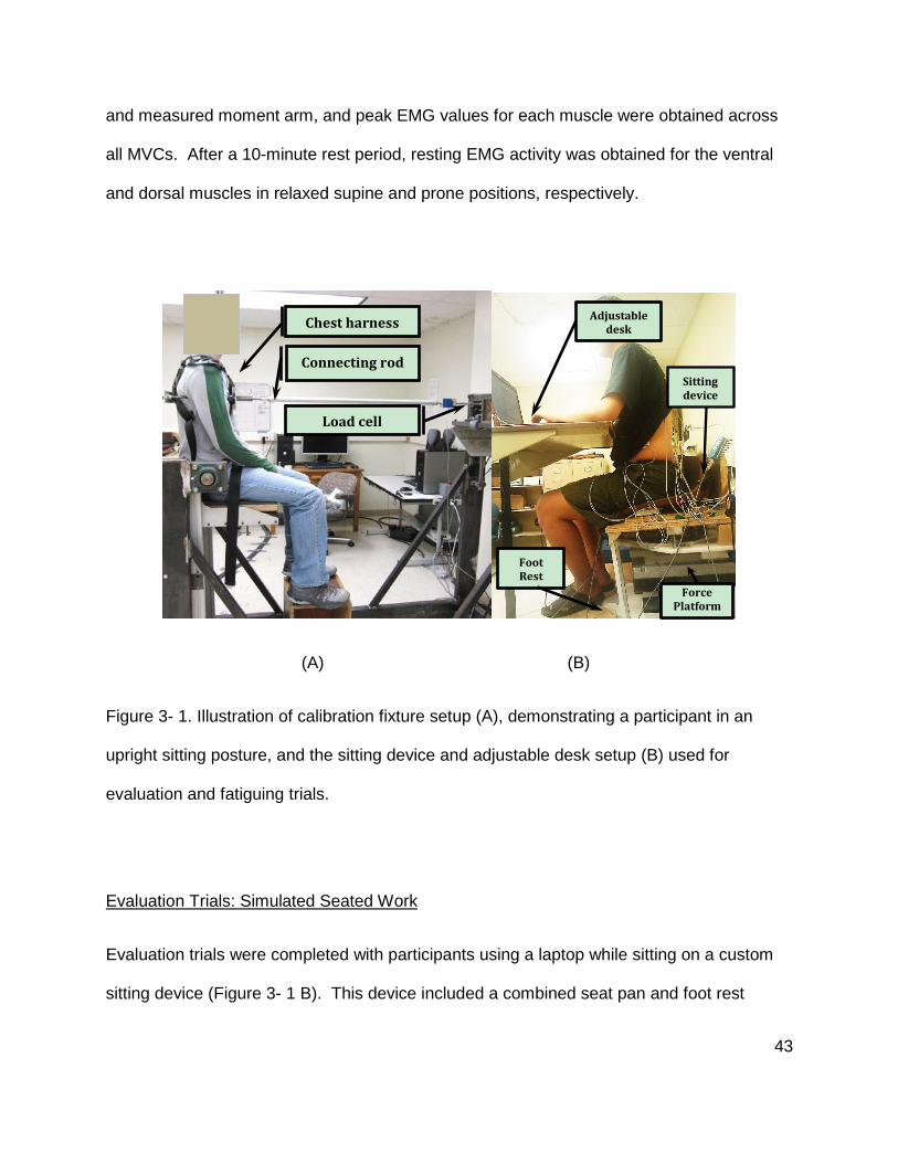
43
and measured moment arm, and peak EMG values for each muscle were obtained across
all MVCs. After a 10-minute rest period, resting EMG activity was obtained for the ventral
and dorsal muscles in relaxed supine and prone positions, respectively.
(A) (B)
Figure 3- 1. Illustration of calibration fixture setup (A), demonstrating a participant in an
upright sitting posture, and the sitting device and adjustable desk setup (B) used for
evaluation and fatiguing trials.
Evaluation Trials: Simulated Seated Work
Evaluation trials were completed with participants using a laptop while sitting on a custom
sitting device (Figure 3- 1 B). This device included a combined seat pan and foot rest
Chest harness
Connecting rod
Load cell
Adjustable desk
Sitting device
Force Platform
Foot Rest
44
structure that was mounted on a force platform (AMTI OR6-7-1000, Watertown,
Massachusetts). Footrest height was adjusted for each participant, so that both thighs were
horizontal. A height-adjustable desk was also used, and the desk, laptop placement, and
sitting device were adjusted according to workstation guidelines for each participant (HFES
and ANSI, 2007). Evaluation trials involved a simple typing task, and which was done in
several conditions described below. The specific task was adapted from a typing test
program (Typing test TQ, Giletech e.K., Germany), and participants were asked to duplicate
several paragraphs from this program using the laptop.
The typing task was done for two minutes in each of eight conditions, and which were
intended to represent a range of seated work, including distinct postures and levels of
muscle activity. The eight conditions included all combinations of two sitting postures, two
levels of mental workload, and two levels of time pressure. The two sitting postures were
lordosed (L) and kyphosed (K), and were used to represent relative extremes of common
postures adopted during seated tasks (Delleman et al., 2004; Vieira and Kumar, 2004; Pynt
et al., 2008). The lordosed posture was achieved by asking participants to sit in an
unsupported upright posture, with the locations of the desk and laptop were adjusted to
ensure the typing task could be completed without additional trunk movement. The
kyphosed posture was achieved by asking participants to sit in an unsupported slumped
posture, and with the same desk and laptop locations. The typing task alone was
considered to have relatively low mental workload (M0), and participants were asked to type
at a fast but comfortable pace with no additional time pressure (T0). A higher level of
mental workload (M1) was introduced by asking participants to mentally calculate the

45
product or summation of a pair of two-digit numbers displayed between paragraphs, similar
to the approach in earlier studies (Kirschbaum et al., 1992; Hughes et al., 2007; Wang et al.,
2011). Typing speed for each participant was measured after they completed an initial 1-
min typing task. To introduce time pressure (T1), participants were offered a $10 bonus, as
a stimulus, if they completed the experimental typing task at a rate ≥ 115% of their baseline
speed. All the tasks were completed with the same requirements on typing accuracy. The
order of presentation of the eight conditions was counterbalanced, using Latin Squares, and
two minutes of rest were provided between each.
Fatiguing Trials: Unsupported Upright Sitting
A rest period (~5 min) was provided after the evaluation trials, and each participant then
completed a single fatiguing trial involving unsupported sitting in the same device used in
the evaluation trials (Figure 1B). Participants were instructed to maintain an active, upright
sitting posture with lumbar lordosed (as in the “L” condition above), and this was held for 20
min or until a self-reported limit of endurance was reached.
3.2.4. Model Implementation and Evaluation
Calibration
Model parameters were specified for each participant, using data collected from the
calibration trials. Raw EMG signals were amplified, band-pass filtered (20–400 Hz), and
root-mean-squared converted (50 ms time constant) in hardware, then sampled at 1000 Hz.
Processed EMG signals were then normalized using individual maximal (from MVCs) and

46
resting EMG values (McGill, 1991; Mirka, 1991; Nussbaum and Chaffin, 1998; Marras et al.,
1999). Trunk kinematics (IMUs) and exerted forces (in-line load cell) were both sampled at
100 Hz, and both were subsequently low-pass filtered (4 Hz cutoff; 4th order Butterworth;
bidirectional). Maximum muscle stresses (for flexors and extensors) and EMG-force
relationships were then determined, using data from the MVCs, through an iterative
procedure that minimized errors in model-based predictions of flexion/extension (sagittal
plane) moments (Jia et al., 2011). This was done since both the MVC and evaluation tasks
were completed in largely symmetrical postures, for which the major trunk rotations and
largest lumbosacral loads were in the sagittal plane. Therefore, moments in sagittal plane
were selected to calibrate model parameters and evaluate model performance. Two
methods were used to assess the quality of model calibration, again focusing on sagittal
plane, lumbosacral moments, and were similar to earlier approaches (McGill, 1992; Granata
and Marras, 1995; Nussbaum and Chaffin, 1998; van Dieën and Kingma, 2005): 1)
coefficient of determination (r2), from a linear regression of measured vs. predicted
moments in the sagittal plane; and (b) average absolute errors (AAE) between measured
and predicted moments in the sagittal plane.
Evaluation
EMG and trunk kinematics were collected and processed as in calibration trials. Reaction
forces and moments were recorded (at 100 Hz) from the force platform and subsequently
low-pass filtered (4 Hz cutoff; 4th order Butterworth; bidirectional). The model, calibrated for
each participant, was then used to predict lumbosacral forces in each of the eight evaluation
trials. Model predictions in each evaluation trial were assessed, as for the calibration trials,

47
by comparing measured vs. predicted sagittal plane moments. The former were estimated
based on force platform moments and inverse dynamics analyses. As described above, r2
and AAE were used to quantify model performance for each trial. The cumulative
distribution function (CDF) of r2 was also derived to summarize results across all task
combinations.
Separate analyses of variance (ANOVAs) were used to determine whether model
performance (i.e., r2 and AAE in the sagittal plane) differed depending on posture, mental
workload, and time pressure. ANOVAs were also used to identify the effects of these same
factors on predicted muscle EMG, sagittal plane lumbosacral moment, and lumbosacral
forces (compression and shear). In these ANOVAs, within-trial mean values were the
dependent measures. Significance was concluded when p < 0.05, and statistical analyses
were performed with JMP™ (v9.0, SAS Institute Inc., Cary, NC). Summary results are
presented as means (SD).
Fatiguing Trials
Data were processed using the same methods as in the evaluation trials, and the model
was again evaluated by comparing measured and predicted lumbosacral moments (here, in
the sagittal plane only, since other moments were relatively quite small). Associated errors
in predicted lumbosacral moments were then assessed over the trial period (i.e., up to 20
min). If a temporal pattern in these errors was evident, it was presumed that this was
substantially a result of muscle fatigue, given that postures were controlled and since such
48
fatigue would alter the invariance of muscle stress and the nature of EMG-to-force
relationship (Sparto et al., 1998). From qualitative examinations, these temporal patterns
were mainly linear. Thus, linear regression was used to quantify the rate of change in
prediction errors, and a linear adjustment to the fatigue gain (G) was implemented as: G(t) =
1 + a(t). Here, the slope (a), was determined from regression analysis (minimizing total
errors between measured and predicted lumbosacral moments in the sagittal plane), and t
is time during the fatiguing trial. Coefficients of determination (r2) and average absolute
errors (AAE) were used to evaluate overall prediction errors (in the sagittal plane) with and
without adjustments for fatigue gain. Differences in model prediction errors (between
measured and predicted lumbosacral moments), with versus without adjustments to the
fatigue gain, were evaluated using a paired t test.
3.3. Results
Calibration
Calibration procedures resulted in predicted muscle stress values of 80 (25.10) N/cm2 for
the flexors and 86.7 (28.33) N/cm2 for the extensors. Calibrated models yielded a relatively
high correspondence between measured and predicted lumbosacral moments in the sagittal
plane, with AAE = 3.30 (1.40) Nm and r2 = 0.85 (0.06) across all MVC trials.
Evaluation
The CDF of r2 values — which assessed the correspondence between measured and
prediction sagittal plane lumbosacral moments — indicated that 45% of trials had r2 > 0.7.

49
Overall, sagittal plane moments had r2 values of 0.65 (0.30) and AAE of 5.90 (3.43) Nm.
Time pressure resulted in significantly (p = 0.03) higher AAE values in the sagittal plane
[6.80 (3.10) Nm] compared to when there was no time pressure [5.00 (3.60) Nm]. No
significant effects of posture or mental workload, or any interaction effects among posture,
mental workload, and time pressure, were found on model performance.
Within-trial mean muscle activation levels were generally lower than 10% MVC across the
six task conditions. Higher mean muscle activations were found in the left and right MF (p <
0.01 for both) in kyphosed posture. No differences in mean muscle activation levels were
found between the trials with different level of mental workloads. Higher time pressure, on
the other hand, resulted in higher mean muscle activation levels in several muscles (Figure
3- 2).

50
Figure 3- 2. Illustration of mean (SD) muscle activation levels for three trunk extensors (MF,
EL, and ET) and flexors (IO, EO, and RA) collected bilaterally (L/R) under two level of time
pressure. The * symbol indicates a significant pairwise difference.
All within-trial means of lumbosacral moments were flexion moments and all such means of
antero-posterior shear were anteriorly directed; as such, results are presented for
lumbosacral flexion moments and anterior shear. Across all trials mean anterior shear,
compression, and lateral shear forces, were 116.90 (75.60), 683.1 (180.80), and 10.2
(10.40) N, respectively. Predicted lateral bending, axial rotation, and flexion moments were
2.47 (2.60), 1.56 (1.48), and 19.80 (9.70) Nm, respectively. The absolute values of mean
lateral shear forces, lateral bending moments, and axial rotation moments, were presented
(assuming there is little functional difference between bilateral and biaxial directions,
respectively). There were significant main effects of posture and time pressure on flexion
moments and predicted lumbosacral forces. The kyphosed posture resulted in lower
0%
5%
10%
15%
LMF LEL LET LEO LIO LRA RMF REL RET REO RIO RRA
% M
VC
Muscle
T0 T1
*
*
*
*

51
anterior shear forces [34.90 (125.10) N vs. 128.80 (73.20) N] and higher flexion moments
[21.80 (8.90) Nm vs. 17.70 (10.70) Nm]. Including time pressure resulted in higher anterior
shear forces [104.60 (106.00) vs. 59.10 (115.10) N], larger compression forces [726.30
(173.80) vs. 639.90 (179.80) N], and higher flexion moments [21.4 (10.90) vs. 18.2 (8.00)
Nm]. No significant main effects of mental workload, or interaction effects of posture,
mental workload, and time pressure, were found on lumbosacral forces or flexion moments.
Fatigue
Adjusting the gain factor (over time) to account for fatigue generally improved the
correspondence between measured and predicted sagittal plane moments (representative
results shown in Figure 3- 3). Across all trials, the slope of the fatigue gain was -0.40 (0.30).
There were significantly (p < 0.01) larger prediction errors without adjustment of fatigue gain.
Using the model with adjustment yielded r2 values of 0.56 (0.33), which were higher than
values of 0.48 (0.38) without adjustment of fatigue gain. AAE was 5.40 (2.70) Nm vs. 0.60
(0.40) Nm with and without adjustment of fatigue gain, respectively.

52
Figure 3- 3. Representative results showing prediction errors over time, between measured
and model-predicted lumbosacral sagittal plane moments, both with (adjusted) and without
(unadjusted) a time-variable gain.
3.4. Discussion
While some recent evidence indicates that workers who mainly work in sitting postures are
more likely to experience LBP or exacerbate existing LBP (Dankaerts et al., 2006;
O'Sullivan et al., 2006; Womersley and May, 2006), it remains unclear regarding the
relationships between sitting and LBP and the specific mechanisms involved.
Biomechanical analyses provide a quantitative method to examine potential mechanism(s)
that might underlie this relationship. An accurate assessment of the partitioning of
biomechanical loading among passive and active components of the trunk during sitting is
needed, though, and requires representations of active and passive tissue properties and
G = 1 -0.1643Time -8
-6
-4
-2
0
2
4
0 5 10 15 20
Pred
ictio
n er
ror (
Nm
)
Time (min)
UnadjustedAdjusted

53
time-dependent viscoelastic properties. A model was presented here, to quantify
lumbosacral loads during a range of seated tasks. Along with detailed representations of
muscle anatomy and contraction dynamics, and participant-specific parameterization
(calibration), the current model also incorporated new components to account for time-
dependent viscoelastic behaviors of trunk tissues and muscle fatigue. The new model was
evaluated in terms of predictive ability, and overall exhibited reasonable levels of
correspondence between measured and predicted lumbosacral moments over the range of
seated tasks examined.
Model parameters were determined for each participant using a set of maximal efforts,
which was considered as a reasonable approach to estimate maximal muscle capacity
during sitting and also account for intra- and inter-individual variability (McGill, 1991; Mirka,
1991). Muscle stresses estimated here (range: SF=45-140 N/cm2; SE=35-115 N/cm2) were
comparable to ranges reported in earlier studies: SF= 56-145 N/cm2; SE= 43-123 N/cm2 in
Bochen et al. (2011); SF=40-140 N/cm2 and SE=37-105 N/cm2 in Nussbaum and Chaffin
(1998); S=37-93 N/cm2 in Granata and Marras (1995); S=29-108 N/cm2 in van Dieën and
Kingma (2005). The wide range of estimated muscle stress values across participants likely
indicates that the calibration procedures captured individual differences (e.g., PCSA and
maximal stress), which would otherwise be difficult to measure directly.
In the current model, viscoelastic responses of passive trunk structure were determined
using a time-dependent trunk stiffness model derived from recent empirical results
54
(Toosizadeh et al., 2012). Though not formally evaluated here, secondary analyses were
completed to evaluate the model performance with versus without this viscoelastic
component (i.e., comparing an elastic to a viscoelastic model). The results indicated a
slight, but not substantial, improvement when including the viscoelastic component. A lack
of more substantial differences is likely a result of the evaluation tasks lasting only 2 min.
The current conditions also involved relatively low levels of passive tissue loading, versus
previous reports of substantial viscoelastic responses during trunk flexion (Bazrgari et al.,
2011; Hendershot et al., 2011).
Mean (SD) values of r2 for sagittal plane moments during evaluation trials here [0.65 (0.30)]
were comparable with those reported in earlier studies: 0.76 (0.15) in Nussbaum and
Chaffin (1998) and 0.66 (0.20) in Kingma et al. (2001). Across all evaluation trials, the
current model yielded good levels of correspondence between measured and predicted
lumbosacral moments in the sagittal plane (45% of r2 > 0.7), and there were no significant
differences in this correspondence between tasks. Magnitudes of AAE here [5.9 (3.4) Nm]
were lower than those in earlier reports – 14.1(7.4) Nm in Nussbaum and Chaffin (1998)
and 56.5 (18.3) Nm in Kingma et al. (2001) – which may in part be due to the relatively low
loads in the current study. No significant differences were found in terms of model
prediction errors (AAE) between task conditions, except for the effects of time pressure.
Although AAE magnitudes were slightly larger (1.7 Nm) in trials including time pressure, this
difference was only ~ 8% of mean peak flexion moments.

55
In terms of predicted lumbosacral forces, the current model predicted relative smaller
compression forces compared to those in previous studies (Sato et al., 1999; Wilke et al.,
1999; Callaghan and McGill, 2001). Specifically, the mean compression forces predicted
here was 683.1 (180.8) N in a lordosed posture, which is lower than values of 800-1000 N
reported based on direct measurement in a upright sitting posture (Wilke et al., (1999); Sato
et al., (1999), and substantially lower than values of ~ 1400 N reported by Callaghan and
McGill (2001) using an EMG-based model. Two potential sources likely account for these
differences. First, compression forces-reported earlier were at the L4/L5 joint, and these
forces can be higher than at L5/S1 as assessed here (Stokes and Gardner-Morse, 1995).
Second, a portion of upper body mass in the current study was supported by the desk,
which likely resulted in lower compression forces vs. unsupported sitting. anterior shear
forces predicted here [116.90 (75.60) N], however, were comparable to values of 135 (200)
N reported by Callaghan and McGill (2001) .
Sitting posture and time pressure had significant effects on predicted lumbosacral loads.
Mean anterior shear forces were higher in the lordosed posture, and which likely resulted
from two sources. First, a larger portion of upper body weight, which can contribute to
anterior shear, was probably supported by the desk in the kyphosed posture (though not
directly measured, participants were observed to lean on the desk to a larger extent in this
posture). Second, the lumbosacral joint is rotated forward in the lordosed vs. kyphosed
posture. As such, gravitational loads make a larger contribution to anterior shear in a
lordotic posture. Including time pressure resulted in higher anterior shear and compression
forces, and higher flexion moments. In prior work, increased psychosocial stress, such as

56
higher time pressure, has been shown to increase muscle activation levels, especially
during situations involving low levels of physical demands (Marras et al., 2000; Hughes,
2004; Hughes et al., 2007). Consistent with this earlier evidence, a generalized increase in
muscle activity was observed when time pressure was included (Figure 3- 2), and which
likely accounts for the increased compression force. Specific causes of the larger anterior
shear and flexion moments with time pressure are more difficult to discern, but may have
involved changes in the patterns of muscle recruitment, postural adjustments, and/or task
performance.
In most previous EMG-based models, invariance of muscle stress and a constant EMG-to-
force relationship is assumed; either, however, may be violated when a muscle is fatigued.
To address this, particularly during prolonged sitting, potential muscle fatigue was
accounted for using a fatigue gain factor. Both linear and nonlinear EMG-to-force
relationships have been reported for trunk muscles (Stokes et al., 1987; Potvin, 1992; Dolan
and Adams, 1993; Potvin et al., 1996; Brown and McGill, 2008), and which especially
influences model predictions for muscle activation levels between 20 and 80% of MVC
(Potvin, 1992; Potvin et al., 1996). However, mean muscle activation levels here were
typically < 10% of MVC (Figure 2), consistent with previous findings (Mork and Westgaard,
2005; Mork and Westgaard, 2009). Therefore, only the assumption of invariance of muscle
stress was considered relevant in the current task conditions (prolonged sitting). Without an
adjustment for fatigue, increasing prediction errors were found over time here, typically
involving increasing overestimation of predicted moments, and consistent with existing
studies (Sparto et al., 1997; Sparto et al., 1998), that muscle contraction “efficiency”, or
57
force per unit EMG, declines with fatigue. Here, a simple linear correction was able to
substantially address the overestimation of predicted moments with fatigue. Future work,
though, may benefit from considering more complex time-dependent adjustments.
A few limitations in the present work should be acknowledged. First, all of the tasks
evaluated here involve sagittally-symmetric conditions and as a result the model evaluation
approach was only focused on the sagittal plane. Future work is needed to evaluate the
model performance under more complex task conditions that include a wider range of trunk
movements, especially those involve asymmetric postures such as use of a mouse or
reaching to the side. Second, the current work did not include a chair backrest, due to the
difficulties involved in tracking trunk posture (due to potential interference of a backrest) and
capturing forces exerted by/on a backrest. Further studies are needed to overcome these
limitations. Third, viscoelastic components were included in current model to account for
the contribution of time-dependent changes in spinal soft tissues. However, the current task
stress and sitting duration were both relatively low, which likely limited any observable effect
of the viscoelastic component on model performance. Additional testing is needed to
assess the potential benefits of the viscoelastic component, such as during more prolonged
sitting especially in kyphosed postures without desk. Fourth, fatigue was assessed only in a
limited aspect. Only a single gain factor was used for all muscles, which assumed that all
muscles had equal fatigue levels during a fatiguing trial. In future research, as noted earlier,
more complex fatiguing conditions should be used, and methods developed to account for
more complex patterns of muscle fatigue/recovery.

58
In summary, an EMG-based biomechanical model has been developed to predict
lumbosacral loads during various seated tasks. Through a calibration procedure, model
parameters were specified on an individual basis and used to estimate lumbosacral forces.
This model was assessed during a range of seated tasks, and demonstrated a reasonable
level of predictive ability. Subsequently, model outputs (e.g., lumbosacral loads) may be of
use as quantitative descriptions of physical demands and injury risks during more realistic
task conditions. Further research is still needed, however, to evaluate predictive ability and
to extend the model to more complex task conditions.
References
Althoff, I., P. Brinckmann, et al. (1992). "An Improved Method of Stature Measurement for Quantitative Determination of Spinal Loading: Application to Sitting Postures and Whole Body Vibration." Spine 17(6): 682-693.
Andersson, B. J. and R. Ortengren (1974). "Lumbar disc pressure and myoelectric back muscle activity during sitting. II. Studies on an office chair." Scandinavian Journal of Rehabilitation Medicine 6(3): 115-121.
Andersson, G. B. J. (1981). "Epidemiologic Aspects on Low-Back Pain in Industry." Spine 6(1): 53-60.
Bakker, E. W. P. P., A. P. P. Verhagen, et al. (2009). "Spinal Mechanical Load as a Risk Factor for Low Back Pain: A Systematic Review of Prospective Cohort Studies." Spine 34(8): E281-E293.
Bazrgari, B., B. Hendershot, et al. (2011). "Disturbance and recovery of trunk mechanical and neuromuscular behaviours following prolonged trunk flexion: influences of duration and external load on creep-induced effects." Ergonomics 54(11): 1043-1052.
Beach, T. A., R. J. Parkinson, et al. (2005). "Effects of prolonged sitting on the passive flexion stiffness of the in vivo lumbar spine." Spine J 5(2): 145-154.
Bigland-Ritchie, B., C. Rice, et al. (1995). "Task-dependent factors in fatigue of human voluntary contractions." Advances in Experimental Medicine and Biology 384: 361-380.
Blangsted, A., K. Søgaard, et al. (2004). "The effect of physical and psychosocial loads on the trapezius muscle activity during computer keying tasks and rest periods." European Journal of Applied Physiology 91(2): 253-258.
BLS (2008a). Nonfatal occupational injuries and illnesses requiring days away from work in 2008. U. S. D. o. Labor, United States Department of Labor.

59
BLS (2009). Nonfatal occupational injuries and illnesses requiring days away from work in 2009. U. S. D. o. Labor, United States Department of Labor.
Brown, K. R., P. Pollintine, et al. (2008). "Biomechanical implications of degenerative joint disease in the apophyseal joints of human thoracic and lumbar vertebrae." American Journal of Physical Anthropology 136(3): 318-326.
Brown, S. H. and S. M. McGill (2008). "Co-activation alters the linear versus non-linear impression of the EMG-torque relationship of trunk muscles." Journal of Biomechanics 41(3): 491-497.
Burns, M. L., I. Kaleps, et al. (1984). "Analysis of compressive creep behavior of the vertebral unit subjected to a uniform axial loading using exact parametric solution equations of Kelvin-solid models--Part I. Human intervertebral joints." Journal of Biomechanics 17(2): 113-115, 117-130.
Callaghan, J. and S. McGill (2001). "Low back joint loading and kinematics during standing and unsupported sitting." Ergonomics 44: 280 - 294.
Callaghan, J. P. and N. M. Dunk (2002). "Examination of the flexion relaxation phenomenon in erector spinae muscles during short duration slumped sitting." Clinical Biomechanics 17(5): 353-360.
Cassidy, J. D. D. C. P. D., P. D. C. P. Cote, et al. (2005). "Incidence and Course of Low Back Pain Episodes in the General Population." Spine 30(24): 2817-2823.
Chaffin, D. B., G. Andersson, et al. (1999). Occupational biomechanics. New York, Wiley-Interscience Publication.
Chen, S.-M., M.-F. Liu, et al. (2009). "Sedentary lifestyle as a risk factor for low back pain: a systematic review." International Archives of Occupational and Environmental Health 82(7): 797-806.
Cholewicki, J. and S. M. McGill (1996). "Mechanical stability of the in vivo lumbar spine: implications for injury and chronic low back pain." Clinical Biomechanics 11(1): 1-15.
Corlett, E. (2006). "Background to sitting at work: research-based requirements for the design of work seats." Ergonomics 49: 1538-1546.
Dankaerts, W. P., P. P. O'Sullivan, et al. (2006). "Differences in Sitting Postures are Associated With Nonspecific Chronic Low Back Pain Disorders When Patients Are Subclassified." Spine 31(6): 698-704.
de Zee, M., L. Hansen, et al. (2007). "A generic detailed rigid-body lumbar spine model." Journal of Biomechanics 40(6): 1219-1227.
Delleman, N. J., C. M. Haslegrave, et al. (2004). Working postures and movements : tools for evaluation and engineering. Boca Raton [Fla.], CRC Press.
Dolan, P. and M. A. Adams (1993). "The relationship between EMG activity and extensor moment generation in the erector spinae muscles during bending and lifting activities." J Biomech 26(4-5): 513-522.
Eklund, J. A. E. and E. N. Corlett (1987). "Evaluation of spinal loads and chair design in seated work tasks." Clinical Biomechanics 2(1): 27-33.
Ellegast, R., R. Hamburger, et al. (2007). Effects of Using Dynamic Office Chairs on Posture and EMG in Standardized Office Tasks. Ergonomics and Health Aspects of Work with Computers. M. Dainoff, Springer Berlin / Heidelberg. 4566: 34-42.
Gardner-Morse, M. G. and I. A. F. Stokes (2004). "Structural behavior of human lumbar spinal motion segments." Journal of Biomechanics 37(2): 205-212.
Granata, K. P. and W. S. Marras (1995). "An EMG-assisted model of trunk loading during free-dynamic lifting." Journal of Biomechanics 28(11): 1309-1317.

60
Gregory, D. E., N. M. Dunk, et al. (2006). Stability ball versus office chair: comparison of muscle activation and lumbar spine posture during prolonged sitting. Human Factors. 48: 142-153.
Groth, K. M. and K. P. Granata (2008). "The Viscoelastic Standard Nonlinear Solid Model: Predicting the Response of the Lumbar Intervertebral Disk to Low-Frequency Vibrations." Journal of Biomechanical Engineering 130(3): 031005-031006.
Guo, H. R., S. Tanaka, et al. (1999). "Back pain prevalence in US industry and estimates of lost workdays." American Journal of Public Health 89(7): 1029-1035.
Hartvigsen, J., K. O. Kyvik, et al. (2003). "Ambiguous relation between physical workload and low back pain: a twin control study." Occup Environ Med 60(2): 109-114.
Hartvigsen, J., C. Leboeuf-Yde, et al. (2000). "Review Article: Is sitting-while-at-work associated with low back pain? A systematic, critical literature review." Scand J Public Health 28(3): 230-239.
Hartvigsen, J. D. C. P., L. S. M. D. Bakketeig, et al. (2001). "The Association Between Physical Workload and Low Back Pain Clouded by the "Healthy Worker" Effect: Population-Based Cross-Sectional and 5-Year Prospective Questionnaire Study." Spine 26(16): 1788-1792.
Hendershot, B., B. Bazrgari, et al. (2011). "Disturbance and recovery of trunk stiffness and reflexive muscle responses following prolonged trunk flexion: Influences of flexion angle and duration." Clinical Biomechanics 26(3): 250-256.
HFES, H. F. a. E. S. and A. N. S. I. ANSI (2007). ANSI/HFES 100-2007 human factors engineering of computer workstations. Santa Monica, CA, Human Factors and Ergonomics Society.
Hides, J., C. Gilmore, et al. (2008). "Multifidus size and symmetry among chronic LBP and healthy asymptomatic subjects." Manual Therapy 13(1): 43-49.
Holmes, A. D. and D. W. L. Hukins (1996). "Analysis of load-relaxation in compressed segments of lumbar spine." Medical Engineering and Physics 18(2): 99-104.
Hughes, L. (2004). Effects of Time Pressure and Mental Workload on Physiological Risk Factors for Upper Extremity Musculoskeletal Disorders While Typing. Department of Industrial and System Engineering. Blacksburg, Virgnia Tech. M.S.
Hughes, L. E., K. Babski-Reeves, et al. (2007). "Effects of psychosocial and individual factors on physiological risk factors for upper extremity musculoskeletal disorders while typing." Ergonomics 50(2): 261-274.
Jensen, C., V. Borg, et al. (1998). "Job demands, muscle activity and musculoskeletal symptoms in relation to work with the computer mouse." Scandinavian Journal of Work, Environment and Health 24(5): 418-424.
Jia, B., S. Kim, et al. (2011). "An EMG-based model to estimate lumbar muscle forces and spinal loads during complex, high-effort tasks: Development and application to residential construction using prefabricated walls." International Journal of Industrial Ergonomics 41(5): 437-446.
Johanning, E. (2000). "Evaluation and management of occupational low back disorders." Am J Ind Med 37: 94 - 111.
Keller, T. S. and M. Nathan (1999). "Height Change Caused by Creep in Intervertebral Discs: A Sagittal Plane Model." Journal of Spinal Disorders and Techniques 12(4): 313-324.

61
Keller, T. S., D. M. Spengler, et al. (1987). "Mechanical behavior of the human lumbar spine. I. Creep analysis during static compressive loading." Journal of Orthopaedic Research 5(4): 467-478.
Kelsey, J. and A. White (1980). "Epidemiology and impact of low back pain." Spine 5: 133 - 142.
Keyserling, W. (1991). "Ergonomic job analysis: A structured approach for identifying risk factors associated with overexertion injuries and disorders." Applied occupational and environmental hygiene.
Kingma, I., C. T. M. Baten, et al. (2001). "Lumbar loading during lifting: a comparative study of three measurement techniques." Journal of Electromyography and Kinesiology 11(5): 337-345.
Kingma, I. and J. H. van Dieën (2009). "Static and dynamic postural loadings during computer work in females: Sitting on an office chair versus sitting on an exercise ball." Applied Ergonomics 40(2): 199-205.
Kirschbaum, C., S. Wust, et al. (1992). "Consistent sex differences in cortisol responses to psychological stress." Psychosomatic Medicine 54(6): 648-657.
Kolich, M., A. I. Mohamed, et al. (2001). "Electromyographic comparison of two lumbar support mechanisms intended for automotive seating applications." Proceedings of the Institution of Mechanical Engineers -- Part D -- Journal of Automobile Engineering; 2001, Vol. 215 Issue 7, p, 7p 215(7).
Kopec, J. A. M. D. P., E. C. B. Sayre, et al. (2004). "Predictors of Back Pain in a General Population Cohort." Spine 29(1): 70-77.
Kurutz, M. (2006). "In vivo age- and sex-related creep of human lumbar motion segments and discs in pure centric tension." Journal of Biomechanics 39(7): 1180-1190.
Leboeuf-Yde, C. (2004). "Back pain--individual and genetic factors." Journal of Electromyography and Kinesiology 14(1): 129-133.
Lee, P., A. Helewa, et al. (2001). "Low back pain: prevalence and risk factors in an industrial setting." The Journal of Rheumatology 28(2): 346-351.
Li, S., A. G. Patwardhan, et al. (1995). "Limitations of the standard linear solid model of intervertebral discs subject to prolonged loading and low-frequency vibration in axial compression." Journal of Biomechanics 28(7): 779-790.
Liao, M. H. and C. G. Drury (2000). "Posture, discomfort and performance in a VDT task." Ergonomics 43(3): 345-359.
Liira, J. P., H. S. Shannon, et al. (1996). "Long-term back problems and physical work exposures in the 1990 Ontario Health Survey." Am J Public Health 86(3): 382-387.
Lis, A., K. Black, et al. (2007). "Association between sitting and occupational LBP." European Spine Journal 16(2): 283-298.
Magora, A. (1972). "Investigation of the relation between low back pain and occupation. 3. Physical requirements: sitting, standing and weight lifting." IMS Ind Med Surg 41(12): 5-9.
Marras, W. S., K. G. Davis, et al. (2000). "The Influence of Psychosocial Stress, Gender, and Personality on Mechanical Loading of the Lumbar Spine." Spine 25(23): 3045-3054.
Marras, W. S., K. P. Granta, et al. (1999). "Variability in spine loading model performance." Clinical Biomechanics 14(8): 505-514.

62
McGill, S. M. (1991). "Electromyographic activity of the abdominal and low back musculature during the generation of isometric and dynamic axial trunk torque: Implications for lumbar mechanics." Journal of Orthopaedic Research 9(1): 91-103.
McGill, S. M. (1992). "A myoelectrically based dynamic three-dimensional model to predict loads on lumbar spine tissues during lateral bending." J Biomech 25(4): 395-414.
McGill, S. M. (1992). "A myoelectrically based dynamic three-dimensional model to predict loads on lumbar spine tissues during lateral bending." Journal of Biomechanics 25(4): 395-414.
McGill, S. M. and R. W. Norman (1986). "Partitioning of the L4/L5 dynamic moment into disc, ligamentous, and muscular components during lifting." 11: 666-677.
McLean, L. and N. Goudy (2004). "Neuromuscular response to sustained low-level muscle activation: within- and between-synergist substitution in the triceps surae muscles." European Journal of Applied Physiology 91(2): 204-216.
McLean, L., R. N. Scott, et al. (1997). Measurement of muscle fatigue in the cervical and lumbar regions during prolonged sitting. Engineering in Medicine and Biology Society, 1997. Proceedings of the 19th Annual International Conference of the IEEE.
McLean, L. and N. Urquhart (2002). "The influence of psychological stressors on myoelectrical signal activity in the shoulder region during a data entry task." Work & Stress 16(2): 138-153.
Mirka, G. A. (1991). "The quantification of EMG normalization error." Ergonomics 34(3): 343-352.
Mirka, G. A. and W. S. Marras (1993). "A Stochastic Model of Trunk Muscle Coactivation During Trunk Bending." Spine 18(11): 1396-1409.
Mork, P. J. and R. H. Westgaard (2005). "Long-term electromyographic activity in upper trapezius and low back muscles of women with moderate physical activity." Journal of Applied Physiology 99(2): 570-578.
Mork, P. J. and R. H. Westgaard (2009). "Back posture and low back muscle activity in female computer workers: A field study." Clinical Biomechanics 24(2): 169-175.
Nachemson, A. and G. Elfstrom (1970). "Intravital dynamic pressure measurements in lumbar discs. A study of common movements, maneuvers and exercises." Scand J Rehabil Med Suppl 1: 1-40.
Nussbaum, M. A. and D. B. Chaffin (1998). "Lumbar muscle force estimation using a subject-invariant 5-parameter EMG-based model." Journal of Biomechanics 31(7): 667-672.
Nussbaum, M. A., D. B. Chaffin, et al. (2000). "Motion times, hand forces, and trunk kinematics when using material handling manipulators in short-distance transfers of moderate mass objects." Applied Ergonomics 31(3): 227-237.
O'Sullivan, P. B., T. Mitchell, et al. (2006). "The relationship beween posture and back muscle endurance in industrial workers with flexion-related low back pain." Manual Therapy 11(4): 264-271.
Oberg, T., L. Sandsjo, et al. (1994). "Subjective and objective evaluation of shoulder muscle fatigue." Ergonomics 37(8): 1323-1333.
Oberg, T., L. Sandsjo, et al. (1992). "Electromyographic changes in work-related myalgia of the trapezius muscle." Eur J Appl Physiol Occup Physiol 65(3): 251-257.
Pitman, M. S. and C. A. Ntuen (1996). "The effect of prolonged sitting on mental task performance." Computers & Industrial Engineering 31(1-2): 499-502.
Pope, M. H., K. L. Goh, et al. (2002). "Spine ergonomics." Annu Rev Biomed Eng 4: 49-68.

63
Potvin, J. R. (1992). The influences of fatigue on hypothesized mechanisms of injury to the low back during repetitive lifting.
Potvin, J. R., R. W. Norman, et al. (1996). "Mechanically corrected EMG for the continuous estimation of erector spinae muscle loading during repetitive lifting." European Journal of Applied Physiology 74(1): 119-132.
Potvin, J. R., R. W. Norman, et al. (1996). "Mechanically corrected EMG for the continuous estimation of erector spinae muscle loading during repetitive lifting." Eur J Appl Physiol Occup Physiol 74(1-2): 119-132.
Poynting, J. H. and J. J. Thomson (1934). A text-book of physics. London,, C. Griffin & Company, Limited.
Pynt, J., M. G. Mackey, et al. (2008). "Kyphosed seated postures: extending concepts of postural health beyond the office." J Occup Rehabil 18(1): 35-45.
Rohlmann, A., L. E. Claes, et al. (2001). "Comparison of intradiscal pressures and spinal fixator loads for different body positions and exercises." Ergonomics 44(8): 781-794.
Sato, K., S. Kikuchi, et al. (1999). "In Vivo Intradiscal Pressure Measurement in Healthy Individuals and in Patients With Ongoing Back Problems." Spine 24(23): 2468.
Shirado, O., T. Ito, et al. (1995). "Flexion-relaxation phenomenon in the back muscles. A comparative study between healthy subjects and patients with chronic low back pain." American journal of physical medicine & rehabilitation / Association of Academic Physiatrists 74(2): 139-144.
Søgaard, K., A. K. Blangsted, et al. (2003). "Evidence of long term muscle fatigue following prolonged intermittent contractions based on mechano- and electromyograms." Journal of Electromyography and Kinesiology 13(5): 441-450.
Solomonow, M., R. V. Baratta, et al. (2003). "Flexion–relaxation response to static lumbar flexion in males and females." Clinical Biomechanics 18(4): 273-279.
Sparto, P. J. and M. Parnianpour (1998). "Estimation of trunk muscle forces and spinal loads during fatiguing repetitive trunk exertions." Spine (Phila Pa 1976) 23(23): 2563-2573.
Sparto, P. J., M. Parnianpour, et al. (1997). "Neuromuscular Trunk Performance and Spinal Loading During a Fatiguing Isometric Trunk Extension with Varying Torque Requirements." Journal of Spinal Disorders and Techniques 10(2): 145-156.
Sparto, P. J., M. Parnianpour, et al. (1998). "Effect of electromyogram-force relationships and method of gain estimation on the predictions of an electromyogram-driven model of spinal loading." Spine 23(4): 423-429.
Stokes, I., S. Rush, et al. (1987). "Trunk extensor EMG-torque relationship." Spine 12(8): 770.
Stokes, I. A. F. and M. Gardner-Morse (1995). "Lumbar spine maximum efforts and muscle recruitment patterns predicted by a model with multijoint muscles and joints with stiffness." Journal of Biomechanics 28(2): 173-175.
Stokes, I. A. F. and M. Gardner-Morse (2003). "Spinal stiffness increases with axial load: another stabilizing consequence of muscle action." Journal of Electromyography and Kinesiology 13(4): 397-402.
Stricker, H., G. Colucci, et al. (2003). "The influence of a prolonged sitting position on the biochemical markers of coagulation activation in healthy subjects: evidence of reduced thrombin generation." J Thromb Haemost 1(2): 380-381.
Taylor, D. C., J. D. Dalton, et al. (1990). "Viscoelastic properties of muscle-tendon units." The American Journal of Sports Medicine 18(3): 300-309.

64
Todd, A. I., A. I. Bennett, et al. (2007). "Physical implications of prolonged sitting in a confined posture - a literature review."
Toosizadeh, N., B. Bazrgari, et al. (2010). In vivo load-relaxation of the trunk with prolonged flexion. the 34th annual meeting of American Society of Biomechanics, Providence, Rhode Island.
Toosizadeh, N., M. A. Nussbaum, et al. (2012). "Load-Relaxation Properties of the Human Trunk in Response to Prolonged Flexion: Measuring and Modeling the Effect of Flexion Angle." PLoS ONE 7(11): e48625.
Tsui, C. P., C. Y. Tang, et al. (2004). "Active finite element analysis of skeletal muscle-tendon complex during isometric, shortening and lengthening contraction." Bio-Medical Materials and Engineering 14(3): 271-279.
van Dieën, J. H., M. P. de Looze, et al. (2001). "Effects of dynamic office chairs on trunk kinematics, trunk extensor EMG and spinal shrinkage." Ergonomics 44: 739 - 750.
van Dieën, J. H., M. P. de Looze, et al. (2001). "Effects of dynamic office chairs on trunk kinematics, trunk extensor EMG and spinal shrinkage." Ergonomics 44(7): 739-750.
van Dieën, J. H. and I. Kingma (2005). "Effects of antagonistic co-contraction on differences between electromyography based and optimization based estimates of spinal forces." Ergonomics 48(4): 411-426.
van Dieën, J. H., C. E. Thissen, et al. (1991). "The electro-mechanical delay of the erector spinae muscle: influence of rate of force development, fatigue and electrode location." European journal of applied physiology and occupational physiology 63(3-4): 216-222.
van Dieën, J. H., E. P. Westebring-van der Putten, et al. (2009). "Low-level activity of the trunk extensor muscles causes electromyographic manifestations of fatigue in absence of decreased oxygenation." Journal of Electromyography and Kinesiology 19(3): 398.
Vieira, E. and S. Kumar (2004). "Working Postures: A Literature Review." Journal of Occupational Rehabilitation 14(2): 143-159.
Vingard, E. M. D., L. P. Alfredsson, et al. (2000). "To What Extent Do Current and Past Physical and Psychosocial Occupational Factors Explain Care-Seeking for Low Back Pain in a Working Population?: Results from the Musculoskeletal Intervention Center-Norrtalje Study." Spine 25(4): 493-500.
Wang, Y., G. Szeto, et al. (2011). "Effects of physical and mental task demands on cervical and upper limb muscle activity and physiological responses during computer tasks and recovery periods." European Journal of Applied Physiology: 1-13.
Watters, W. C., 3rd, J. Baisden, et al. (2008). "Degenerative lumbar spinal stenosis: an evidence-based clinical guideline for the diagnosis and treatment of degenerative lumbar spinal stenosis." Spine Journal 8(2): 305-310.
Wilder, D. G., M. H. Pope, et al. (1988). "The Biomechanics of Lumbar Disc Herniation and the Effect of Overload and Instability." Journal of Spinal Disorders & Techniques 1(1): 16-32.
Wilke, H.-J. P., P. M. D. Neef, et al. (1999). "New In Vivo Measurements of Pressures in the Intervertebral Disc in Daily Life." Spine 24(8): 755-762.
Womersley, L. and S. May (2006). "Sitting Posture of Subjects With Postural Backache." Journal of Manipulative and Physiological Therapeutics 29(3): 213-218.

65
Chapter 4. The Effects of Prolonged Sitting and Psychosocial Stress
on Low Back Kinematics, Discomfort, and Localized Muscle Fatigue
Abstract
Changes in the nature of work have led to an increase in sedentary behavior. The effects of sitting duration and work stress on the low back, however, are not well documented, and quantifying these effects was the purpose of this study. Participants (n=14) completed prolonged (40-min) computer-based tasks, using a laptop without a desk. These were done under two levels of psychosocial stresses: low stress involved relaxed web browsing, whereas higher stress was induced through additional mental workload (mental calculation) and time pressure (fast typing speed) during a typing task. Both subjective (perceived discomfort) and objective measures obtained, the latter including lumbosacral kinematics, muscle activity, and measures of localized fatigue. Prolonged sitting significantly increased perceived discomfort and trunk flexion angles and led to muscle fatigue. No significant effects of psychosocial stress were found on any of the dependent measures. Relatively weak correlations were found between subjective and objective measures, though two fatigue measurement methods (based on EMG and stimulated responses) showed a good level of correspondence. This study provides a quantitative assessment of the effects of the prolonged sitting and psychosocial stress during a specific computer-based task, extending our understanding of sitting behaviors under different work conditions. Further research is needed to investigate more realistic occupational and non-occupational sitting conditions.
Key words: Prolonged sitting; Psychosocial stress; Perceived discomfort; Muscle Fatigue;
Lumbosacral kinematics/kinetics

66
4.1. Introduction
Sedentary lifestyle, which involves more prolonged sitting, becomes a new trend in
modern living (Varo et al., 2003; Ford et al., 2005). Recent estimates indicate that
more than half of all population do not maintain physical activity levels to achieve
health benefit (Blair et al., 1999; Pleis et al., 2009), and more than one-third of adults
worldwide are physically inactive (Hallal et al., 2012). Existing evidence suggests
that workers who mainly work in sitting postures are more likely to experience or
aggravate low back pain (LBP) (NIOSH, 1997; Dankaerts et al., 2006; O'Sullivan et
al., 2006; Womersley and May, 2006). Sitting is a complex behavior that is affected
by both the design of working environment and the tasks being performed
(Magnusson and Pope, 1998; Delleman et al., 2004). Moderating factors, such as
sitting duration (Keyserling, 1991; Pitman and Ntuen, 1996; Stricker et al., 2003), or
psychological task stress (McLean and Urquhart, 2002; Ellegast et al., 2007), likely
also play important roles in individual behaviors and responses (e.g., LBP) during
sitting.
Prolonged sitting often results in discomfort and/or pain in the lower back (Pope, 1991;
Fenety et al., 2000; Beach et al., 2005; Dunk and Callaghan, 2005; Gregory et al.,
2006). Both muscle fatigue and viscoelastic properties of spine passive structures
could cause time-dependent changes in lumbosacral loads during prolonged sitting
(Bigland-Ritchie et al., 1995; McLean et al., 1997; Keller and Nathan, 1999; Beach et
al., 2005). Muscle fatigue has been measured via muscle electromyography to
assess the effects of prolonged sitting (McLean et al., 1997; Seghers et al., 2003).

67
Likely due to potential instability of EMG median power frequency caused by
alterations in neuromuscular control strategies and geometric changes during low
level muscle contraction, previous evidence indicates that fatigue-related changes in
EMG signals can be detected consistently typically when muscle contraction level is
greater than 15% of maximal muscle capacity (Oberg et al., 1992; Oberg et al., 1994).
Typical activities generated by postural muscles during sitting, on the other hand, are
usually at relatively low contraction levels of < 10% of maximal muscle capacity (van
Dieën et al., 2001; Mork and Westgaard, 2005; Mork and Westgaard, 2009).
Viscoelastic changes in spine structures (i.e., creep) during prolonged sitting have
been investigated by measuring trunk flexion angles (Scannell and McGill, 2003;
Beach et al., 2005). No existing study, to the author’s knowledge, has quantified the
time-dependent changes in lumbosacral kinetic during prolonged sitting with
consideration of both potential effects of muscle fatigue and viscoelastic behaviors of
trunk.
Modern jobs that require prolonged sitting also can involve precision work, a high
mental workload, and a stressful working schedule. Psychosocial stress (e.g., time
pressure and high mental workload) has been found to affect not only trunk
kinematics but also to increase muscle activation levels (Pitman and Ntuen, 1996;
van Dieën et al., 2001; Ellegast et al., 2007; Hughes et al., 2007). The high
prevalence of musculoskeletal disorders in psychologically stressful, but light physical,
work has indicated that increased perceived workload plays an important role in the
development of LBP (Jensen et al., 1998; Davis and Heaney, 2000). More generally,

68
psychosocial stress has been related to the development of work-related
musculoskeletal disorders in upper extremities (Jensen et al., 1998; Hughes et al.,
2007), shoulder (McLean and Urquhart, 2002; Blangsted et al., 2004), and low back
(Marras et al., 2000). However, the effects of psychosocial stress on the low back
during prolonged sitting have not been quantified.
Therefore, this study was designed to determine the effects of prolonged sitting and
psychosocial stress associated with computer-based seated tasks on several
outcomes including discomfort, lumbosacral kinematics, muscle activity, and localized
muscle fatigue (LMF). Participants completed two computer-based seated tasks,
intended to represent some common activities performed in a seated posture. We
hypothesized that prolonged sitting and psychosocial stress would have significant
effects on both objective and subjective measures, and that there would be a
correspondence between these measurements. A longer-term goal of this effort was
to expand our understanding of the potential adverse consequences of prolonged
seated tasks and provide information to facilitate future practical interventions to
reduce sitting-related risks.
4.2. Methods
4.2.1. Participants
Fourteen participants (gender balanced) completed the experiment and were
recruited from the local student population and community. Mean (SD) age, body
mass, and stature were 24.5 (4.1) yrs, 174.5 (9.1) cm, and 72.3 (11.1) kg,

69
respectively. After a demonstration of the experimental protocols, all participants
completed an informed consent process approved by the Virginia Tech Institutional
Review Board. No participants had any self-reported musculoskeletal injuries or
neurological diseases within the past year, and individuals with body mass index >30
kg/m2 were excluded due to the potential difficulties in obtaining EMG measures and
evoking reliable muscle stimulation response.
4.2.2. Experimental Design
Participants completed two tasks in separate trials that involved prolonged (40 min)
use of a laptop in a seated posture without a desk, and which were done in two
conditions involving different levels of psychosocial stress (low vs. higher). While
some individuals often sit longer, a 40 min period was thought to represent a typical
uninterrupted period of laptop usage under non-desk condition. The low psychosocial
stress (LS) condition had participants engage in relaxed internet browsing, which
involved participants performing random internet “surfing” (e.g., reading news,
streaming video). A higher level of psychosocial stress (HS) was introduced by
asking participants to complete a typing task in a situation involving both additional
mental workload and time pressure (described in Chapter 3). During the typing task
participants duplicated several paragraphs from a typing test program (Typing test TQ,
Giletech e.K., Germany). To induce additional mental workload, pairs of two-digit
numbers were displayed between each paragraph of text, and participants were
asked to mentally calculate the product or summation of these paired numbers,
similar to existing approaches (Kirschbaum et al., 1992; Hughes et al., 2007; Wang et

70
al., 2011). To add time pressure, participants were asked to type as fast as they
could and they were offered a $10 bonus, as an incentive, if they completed the
typing task at a rate ≥ 115% of their baseline speed; this baseline was measured
during a 1-min typing task completed at an individual’s normal typing speed. The
order of stress conditions was counterbalanced across participants, and at least four
days were provided between each trial to minimize carry-over effects (Edwards et al.,
1977; Jones et al., 1989).
4.2.3. Instrumentation and Data Collection
Trunk angles (relative rotations of the trunk vs. the pelvis) and muscle EMG were
collected continuously during each trial, as described earlier (Chapter 3). Briefly, two
telemetered EMG systems (TeleMyo 900, Noraxon, AZ) were used to collected EMG
bilaterally from three lumbar extensors [erector spinae lumbar group (EL), erector
spinae thoracis group (ET), and multifidus (MF)], and three lumbar flexors [internal
oblique (IO), external oblique (EO), and rectus abdominis (RA)]. Raw EMG signals
were sampled at 1000 Hz and then amplified, band-pass filtered (20 - 400 Hz), and
root-mean-square (RMS) converted with a moving window of 50-millisecond duration
in hardware. Trunk angles were monitored using two inertial measurement units
(IMUs: Xsens Inc., Los Angeles, CA), fixed to the dorsal aspect of the trunk at the
T10 and S1 levels. Trunk angles were collected at 100 Hz and subsequently low-
pass filtered at 4 Hz (4th order Butterworth; bidirectional). All data were collected
simultaneously using LabVIEW software (version 8, National InstrumentsTM, Austin,
USA). RMS EMG signals were down sampled to 100 Hz to match the trunk angles.

71
Stimulated responses of the lumbar extensor muscles were measured (as an
indicator of fatigue), following the protocols described in Chapter 2. In brief, a dual-
channel system, consisting of a stimulator (Grass S88) in series with a stimulus
isolation unit (SIU5) and a constant current unit (CCU1; all from AstroMed, Inc., West
Warwick, RI) was used to stimulate the lumbar extensors bilaterally. After a
conditioning train, the stimulation responses were collected (at 1000 Hz) during three
16-second stimulation trains at 2 Hz.
Perceived discomfort (PD), for the whole body and eight local body parts (neck,
left/right shoulders, upper/lower back, buttocks and left/right thighs), was measured
using a modified Borg CR-10 and Body Part Discomfort Scale (Corlett and Bishop,
1976; Borg, 1990; Kyung et al., 2008a; Figure 4-1).

72
Figure 4- 1. Methods used to obtain subjective ratings of perceived discomfort and
fatigue (modified from Kyung and Nussbaum, 2008b)
4.2.4. Experimental Procedures
Each trial consisted of initial calibration procedures, a 40 min seated task, and
collection procedures of flexion-relaxation (FR) angle, stimulation responses, and
perceived discomfort (PD) before and after seated task (Figure 4- 2). The FR angle
of the trunk was collected. FR angle was measured by asking participants to bend
forward to reach their end range-of-motion in trunk flexion (Shirado et al., 1995;

73
Callaghan and Dunk, 2002; Solomonow et al., 2003), and then the FR angle was
determined by the relative silence of lumbar extensor muscle activity using
procedures described earlier (Hendershot et al., 2011). Subsequently, muscle
stimulation responses were collected after positioning the participant into a
customized sitting fixture and completing a conditioning train (Chapter 2).
Participants then completed a 40-min task in one of the two conditions. Each trial
involved participants sitting on a stool with no back rest or back support. The height
of the stool was adjusted for each participant, so that both thighs were horizontal and
with a 90 degree included knee angle. The stool height, laptop placement, and
participants’ postures were monitored and the same setup was used for the second
trial. Each participant performed the task with the laptop on their lap and in a
kyphosed posture, and this configuration was used to represent one of the more
common non-neutral sitting postures when using a laptop away from a desk (Moffet
et al., 2002; Asundi et al., 2010; Gold et al., 2012). To avoid potential confounding
from participants’ movements and the ambient environment, participants were
required to sit still but relaxed and were not allowed to communicate with the
experimenters for the duration of the task. At the beginning of the task, participants
were asked to rate their perceived discomfort. Right after the task trial, PD scores,
FR angles, and stimulation responses were again collected, in a consistent sequence.

74
time
FR FR
Start
Computer-based seated task
40 min
PD PD
SR SR
Figure 4- 2. Illustration of experiment procedure. FR: flexion-relaxation measurement;
SR: stimulation-based fatigue measurement; PD: perceived discomfort via subjective
rating.
4.2.5. Data Reduction and Processing
The first minute of each task was discarded since participants often needed this time
to obtain a stable posture. Mean (SD) values of trunk angles (flexion), and EMG MPF,
were obtained from the first five (i.e., 2 - 6 min) and the last five min of each task.
Only trunk flexion angles were analyzed, since both trials were completed largely in
symmetrical postures, for which the major trunk rotations were in the sagittal plane.
Therefore, trunk flexion angles in sagittal plane were selected to evaluate the effects
of prolonged sitting and psychosocial stress. FR angle, SR, and EMG MPFs were
normalized to initial values collected before each task to facilitate comparisons
between participants and different measures.

75
Localized muscle fatigue was assessed using two methods. The first method used
raw EMG obtained during the trials, from which the median power frequency (MPF) of
each EMG signal was obtained over 1-second windows. Mean EMG MPF obtained
from the first five and the last five min of each task were calculated and compared.
Changes between these mean values were used as a predictor of fatigue
development in these muscles. The second method used mean stimulation
responses to identify potential muscle fatigue. Muscle fatigue is identified when a
drop was found in measured stimulation responses before and after each trial, as
described in existing studies (Edwards et al., 1977; Binder-Macleod and Snyder-
Mackler, 1993; Johnson et al., 1995a). Stimulation responses were obtained as the
means, across all peak stimulation responses recorded during the 16s block of all
three trains, and the three largest and three smallest values were excluded to
minimize the influence of outliers on estimates of central tendency (Kogi and
Hakamada, 1962; Rothenberg et al., 1964).
4.2.6. Statistical Analyses
Multivariate analysis of variance (MANOVA, using Wilks’ Lambda) was used to
determine the effects of prolonged sitting (specifically, the first five vs. last five
minutes), psychosocial stress, and their interaction on the dependent measures as a
whole. Specific dependent measures were, FR angle, trunk flexion angle, stimulation
response, muscle EMG and MPF, and perceived discomforts. In the event of a
significant MANOVA effect, univariate ANOVAs were performed for each dependent
variable where relevant. Effects were considered significant when p<0.05. To
76
assess whether there was any underlying relationship among perceived discomforts
from the eight body segments, principal component analysis (PCA) was performed on
the PD scores. For the same reason, PCA analysis was also performed on all EMG
median frequencies. Correspondence between objective measures [trunk angle
(flexion), muscle EMG MPF (PC component) and stimulation response] and
subjective measures [perceived discomfort (PC component)] were evaluated using
bivariate coefficients of correlation (ρ). To assess the consistency between the two
current fatigue measurement methods, ρ values were also obtained between EMG
median frequencies (PC component) and stimulation responses.
4.3. Results
MANOVA and ANOVAs results are summarized in Table 4- 1. The former indicated
that both prolonged sitting (p<0.01) and psychosocial stress (p=0.04) had significant
effects on the set of dependent measures, but no significant interaction effect (p =
0.90). Subsequent univariate ANOVAs indicated that prolonged sitting significantly
affected, trunk flexion angles, stimulation responses, MPF from most muscles, and
perceived discomfort in all body regions evaluated. Psychosocial stress did not have
significant effects on any of the dependent measures.

77
Table 4- 1. Summary of ANOVA results for effects of prolonged sitting and
psychosocial stress.
Prolonged Sitting Psychosocial Stress
F(2,1057) p F(1,1057) p Flexion-relaxation angles 0.49 0.49 2.83 0.10
Trunk angle 8.32 <0.01 1.09 0.30 Stimulation Response 4.01 0.05 0.11 0.74
EM
G M
edia
n Fr
eque
ncy
LMF 2.16 0.15 0.07 0.79 LEL 7.65 <0.01 0.15 0.71 LET 5.36 0.02 1.02 0.32 LEO 5.46 0.03 0.11 0.75 LIO 6.51 0.02 0.25 0.62 LRA 2.71 0.12 2.59 0.12 RMF 10.58 <0.01 0.17 0.68 REL 6.25 0.02 3.11 0.09 RET 4.34 0.04 3.02 0.09 REO 0.03 0.86 0.13 0.73 RIO 6.38 0.02 0.94 0.34 RRA 5.10 0.03 1.23 0.74
Dis
com
fort
Neck 35.31 <0.01 1.01 0.32 L shoulder 22.59 <0.01 0.01 0.94 R shoulder 32.45 <0.01 0.09 0.77 Upper back 106.53 <0.01 1.94 0.17 Lower back 39.64 <0.01 0.03 0.86
Buttock 27.86 <0.01 0.32 0.57 L wrist 5.68 0.02 1.71 0.20 R wrist 11.85 <0.01 0.48 0.49 Whole body 104.07 <0.01 1.02 0.32
Across trials, mean levels of muscle activity were typically lower than 10% of MVC
and with the highest levels of activity found for the bilateral erector spinae thoracis.
No significant (p>0.10) differences on mean muscle activity levels were found
between two psychosocial stress conditions. For the extensor muscles, however,
activation levels were typically higher in the HS condition (Figure 4- 3). Activation

78
levels of trunk flexors, on the other hand, were generally lower in the HS condition
exception the left IO.
Figure 4- 3. Summary of muscle activation levels (% of MVC) between the two task
conditions (LS = low psychosocial stress, HS = higher psychosocial stress).
FR angle, muscle stimulation responses, and EMG MPF, and are summarized in
Figure 4- 4. Although not significant (p=0.44), FR angle generally increased at the
end of each task. Signs of muscle fatigue were found in both stimulation response
and EMG MPF measures. Prolonged sitting resulted in a significant (p=0.05)
decrease in stimulation responses and significant declines of MPF from all muscles
except the REO. No main or interaction effects of psychosocial stress effects were
found for stimulation response, EMG MPF, or FR angle. Though not significant, both
fatigue measurements indicated more fatigue in the HS condition.
0%
4%
8%
12%
16%
LMF LEL LET LEO LIO LRA RMF REL RET REO RIO RRA
Activ
atio
n Le
vel (
% M
VC)
Muscles
LS HS

79
Figure 4- 4. Illustration of mean changes (after vs. before task) in normalized flexion-
relaxation angles (FR), stimulation responses (SR), and median frequencies. All
measures were normalized to initial values (i.e., 100% = no change over time). The *
symbol indicates a significant effect of prolonged sitting, and error bars indicate
standard deviations.
Perceived discomfort results are summarized in Figure 4- 5. Perceived discomfort of
the whole body and each of the eight local body parts significantly (p<0.01) increased
by the end of the tasks. Largest mean perceived discomfort was found in upper back,
followed by lower back and whole body. No main or interaction effects of
psychosocial stress were found on perceived discomfort.
60%
80%
100%
120%
140%
FR SR LMF LEL LET LEO LIO LRA RMF REL RET REO RIO RRA
*
* *
* *
* * * *
*

80
Figure 4- 5. Mean (SD) values of perceived discomfort for each body part. All PD
scores significantly (p<0.01) increased at the end of the task.
For both EMG and PD scores, one dominant PC was found for each, and which
accounted for > 38% of the variance in EMG and > 67% of the variance in PD scores
(Table 4- 2). PC1 for both EMG MPF (EPC1) and PD scores (PPC1) can be roughly
considered as representing a mean across individual MPF and PD measures, in that
most of the coefficients have comparable magnitudes with the same sign. Therefore,
these two PCs were denoted as the components that represent overall measures of
EMG and PD.
00.5
11.5
22.5
33.5
4
neck
left
shou
lder
right
sho
ulde
r
uppe
rbac
k
low
er b
ack
butto
ck
left
wris
t
right
wris
t
who
le b
ody
Perc
eive
d di
scom
fort
Beginning End

81
Table 4- 2. Principal component (PC) coefficients for EMG and PD scores.
PD Neck Left
Shoulder Right
Shoulder Upper Back
Lower Back
Buttock Left Wrist
Right Wrist
Whole Body
PPC1 0.3 0.36 0.35 0.32 0.32 0.33 0.34 0.35 0.33
EMG LMF LEL LET LEO LIO LRA RMF REL RET REO RIO RRA
EPC1 0.15 0.37 0.12 0.37 0.19 0.40 0.27 0.35 0.17 0.20 0.31 0.38
As shown in Figure 4- 6, no strong or significant relationships were found between
objective and subjective measures. Although not significant, PPC1 was positively
related to trunk angle (flexion), compression forces, and flexion moments, and was
negatively related to stimulation responses and EPC1. A positive, though not
significant (ρ=0.23, p=0.08) relationship was found between EPC1 and stimulation
responses.

82
(A) (B)
(C) (D)
Figure 4- 6. Relationships between subjective (PPC1) and objective measures [trunk
angle (A), EPC1 (B), and stimulation responses (C)], as well as between stimulation
responses and EPC1 (D).
4.4. Discussion
While some moderating factors, such as prolonged sitting and psychosocial stress
could have a direct impact in individual behavior, these may also potentially increase
the risk for LBP development though few studies have investigated the effects of such
factors. The objective of this study was to determine the effects of prolonged sitting
0
4
8
12
-0.04 0 0.04 0.08
PPC
1
Trunk Angle (rad)
ρ = 0.13 p=0.33 ρ = -0.16 p = 0.23
0
4
8
12
2 4 6
PPC
1
EPC1
ρ = -0.11 p=0.40
0
4
8
12
10 30 50
PPC
1
Stimulation Responses (N)
ρ = 0.23 p=0.08
3
4
5
10 30 50
EPC
1
Stimulation Responses (N)

83
and psychosocial stress associated with computer-based seated tasks on trunk
kinematics, perceived discomfort, and localized muscle fatigue.
Only prolonged sitting had significant main effects on several of the dependent
measures (Table 4- 1), with no significant main or interactive effect of psychosocial
stress found. The latter suggests only additive effects of prolonged sitting, or that the
effects of prolonged sitting exposure are consistent across different levels of stress.
It may be that the levels of both factors examined here were relatively narrow, and
thus not able to capture potential interactive effects. It is also unclear if this lack of an
interaction effect will generalize to a larger range of exposure conditions. Previous
work indicated that increased psychosocial stress could contribute to increased
muscle activation, especially during conditions involving relatively low physical
demands (Marras et al., 2000; Hughes, 2004; Hughes et al., 2007), and such
increased muscle activity was found here (Figure 4- 4), though such difference were
not significant.
There were significant increases in trunk flexion angles at the end of both tasks.
Participants were asked to maintain a static, kyphosed sitting posture, with minimum
posture adjustments or in-chair movement. Both creep and fatigue in the low back
can develop with static trunk flexion (Kazarian, 1975; Keller et al., 1987; Adams and
Dolan, 1996; Kanlayanaphotporn et al., 2002), and creep may occur after only five
minutes of static flexion (Adams and Dolan, 1996; Kanlayanaphotporn et al., 2002).

84
FR angle is often used as an indicator of creep deformation in spinal structure under
flexion exposures, with results from such studies indicating that prolonged trunk
flexion increases FR angle (Olson et al., 2004; Olson et al., 2006; Shin et al., 2009).
Here, prolonged sitting resulted in an increase in FR angles in both stress condition
(Figure 4- 5), indicating likely creep deformation of the spine, and supporting that
observed increases in trunk angles were likely due to creep responses during
prolonged sitting.
Muscle fatigue was measured using both muscle stimulation and EMG MPF.
Prolonged sitting resulted in significant drops in stimulated responses and declines in
EMG MPF at the end of both task conditions, but no effects of psychosocial stress
were evident. Stimulation responses have been used to measure muscle fatigue
during sustained, low level muscle contractions (Johnson et al., 1995a; Johnson,
1998; Mork and Westgaard, 2005), with fatigue identified by a drop in measured
stimulation response (Kazarian, 1975; Edwards et al., 1977). Using this criterion,
muscle fatigue in trunk extensors was apparent as a result of 40 min seated task in a
kyphosed sitting posture. Muscle fatigue was also apparent from a significant decline
of EMG MPF, which was found for several flexor and extensor muscles. Some of the
inconsistencies between muscles may be attributed to functional differences between
the trunk flexors and extensors. During prolonged sitting, trunk extensors are usually
considered as postural muscles, and which are required to be continuously activated
to stabilize the sitting posture (van Dieën et al., 2001; Mork and Westgaard, 2005;
Mork and Westgaard, 2009). Therefore, the observed decline of EMG MPF among

85
three trunk extensors (EL, ET and MF) could be used as a good representation of
muscle fatigue caused by prolonged sitting. The decline of EMG MPF in the bilateral
both IO suggests that this muscle also plays an important role in stabilizing the trunk
posture during sitting, and this is consistent with existing evidence (Snijders et al.,
1995). Instead of testing the relationship between stimulation responses and each
individual EMG MPF, the correlation between the two muscle fatigue measurement
methods was instead completed using the first principal component for all EMG MPF.
Though only approaching significance EMG MPF was positively correlated with
stimulation responses (Figure 4- 6 F), and both measurements declined over time.
Consistency between these different measures supports that fatigue indeed
developed during the tasks, and that both approaches have potential value in
assessing muscle fatigue during prolonged sitting.
Perceived discomfort also significantly increased with prolonged sitting. A prolonged
static sitting posture, such as that imposed by the computer-based tasks in this study,
has been known to lead to discomfort of the neck, shoulders, and back (Grandjean et
al., 1983; Stokes et al., 1987; Gay et al., 2006), and to increase with time during
uninterrupted tasks over the working day (Marras et al., 1995; Ostensvik et al., 2009).
To minimize the potential confounding effects of in-chair movement, participants here
maintained a kyphosed sitting posture, and which can increase intradiscal pressures
(Wilcox and Keselman, 2003), stretch the posterior spine ligaments (Owen et al.,
2010), and restrict nutrient supplies to nerves (Hallal et al., 2012). These factors may
have contributed to the observed increases in discomfort.

86
While subjective measures (e.g., perceived discomfort) provided direct assessment of
sitting discomfort, objective measures can potentially provide unbiased estimations
that are related to sitting discomfort (Lee et al., 1993; Kyung et al., 2008a). Support
for objective measures, as useful additions to subjective measurements of sitting
discomfort, would be provided if a good correlation existed between these two types
of measures. Several studies have been completed to identify the relationship
between subjective measures (e.g., perceived discomfort) and a range of objective
measures (e.g., trunk posture, muscle EMG and pressure distribution; Eklund and
Corlett, 1987; Lee et al., 1993; Salewytsch and Callaghan, 1999; Vergara and Page,
2000). From these, pressure distribution was found to be most clearly related to
subjective measures (Thakurta et al., 1995; Vergara and Page, 2000; Kyung and
Nussbaum, 2008b).
In this study, the first principal component (PC1) of PD scores was used as a
representation of overall perceived discomfort across all several measured body parts
and the whole body, and correlations were obtained between this and several
objective measures (trunk angle, muscle EMG MPF and stimulation responses). No
significant correlations between subjective and objective measures were found.
Trunk angle was positively related to perceived discomfort, specifically with higher
trunk flexion angles related to higher perceived discomfort, and which was consistent
with previous evidence (Eklund and Corlett, 1987). Although not significant, declines
in both fatigue measures (EPC1 and stimulation response) were related to an

87
increase in perceived discomfort. This suggests that participants’ perceived higher
discomfort at least in part as a result of muscular fatigue, as has been described
earlier (Rohmert and Luczak, 1978; Zhang et al., 1996). Although no strong
association between subjective and objective measures was found in this study, the
objective measures used may still provide useful information, such as in future work
to predict discomfort without potential biases involved with subjective measures.
Two potential limitations of this work need to be acknowledged. First, the effects of
prolonged sitting and psychosocial stress were evaluated under a constrained sitting
condition. Each experimental task was completed with participants sitting in a single,
fixed posture, and with minimized in-chair moments. In a realistic sitting situation,
participants usually make several posture adjustments or take microbreaks to reduce
body discomfort (Salewytsch and Callaghan, 1999; McLean et al., 2001). Therefore,
the results from the current study may have limited ability to generalize to realistic use
of a laptop in a non-desk condition. Future work is needed to test the effects of
prolonged sitting and psychosocial stress under more complex task conditions that
include a wider range of trunk movements and sitting postures. Second, the duration
of the prolonged task was determined based on the results from our pilot studies,
where muscle fatigue was observed after 30 min of quiet sitting. There is a lack of
evidence, to the author’s knowledge, regarding typical exposure durations for laptop
use without a desk, so the applicability of the current work, in terms of the duration of
actual use, is unknown. Additional study is needed to identify the common exposure
durations of sitting under different conditions.

88
In summary, prolonged sitting was found to have important impacts on lumbosacral
kinematics, and to lead to the development of muscle fatigue and perceived
discomfort. The current findings suggest that individuals who sit for prolonged
periods can be at increased risk of injury and that this risk may also be higher with
increased psychosocial stress. These outcomes were evident after 40 min of sitting
under experimental conditions involving laptop use without a desk, which could be of
particular concern for some conditions of laptop use (e.g., in an airport). This study
highlighted the potential influence of sitting-related moderating factors on individual
behaviors and provided evidence to facilitate better understanding of sitting-related
risks. Further research is still needed, however, to assess the effects of prolonged
sitting and psychosocial stress in more complex occupational and non-occupational
sitting conditions.
References
Adams, M. A. and P. Dolan (1996). "Time-dependent changes in the lumbar spine's resistancc to bending." Clinical Biomechanics 11(4): 194-200.
Asundi, K., D. Odell, et al. (2010). "Notebook computer use on a desk, lap and lap support: Effects on posture, performance and comfort." Ergonomics 53(1): 74 - 82.
Beach, T. A., R. J. Parkinson, et al. (2005). "Effects of prolonged sitting on the passive flexion stiffness of the in vivo lumbar spine." Spine J 5(2): 145-154.
Bigland-Ritchie, B., C. Rice, et al. (1995). "Task-dependent factors in fatigue of human voluntary contractions." Advances in Experimental Medicine and Biology 384: 361-380.
Binder-Macleod, S. A. and L. Snyder-Mackler (1993). "Muscle Fatigue: Clinical Implications for Fatigue Assessment and Neuromuscular Electrical Stimulation." PHYS THER 73(12): 902-910.

89
Blair, S., N.,, A. Franks, L.,, et al. (1999). Physical activity and health: A report of surgeon general. U. S. D. o. H. a. H. Services, C. f. D. C. a. Prevention and N. C. f. C. D. P. a. H. Promotion. Atlanta, GA.
Blangsted, A., K. Søgaard, et al. (2004). "The effect of physical and psychosocial loads on the trapezius muscle activity during computer keying tasks and rest periods." European Journal of Applied Physiology 91(2): 253-258.
Borg, G. (1990). "Psychophysical scaling with applications in physical work and the perception of exertion." Scandinavian Journal of Work, Environment and Health 16 Suppl 1: 55-58.
Callaghan, J. P. and N. M. Dunk (2002). "Examination of the flexion relaxation phenomenon in erector spinae muscles during short duration slumped sitting." Clinical Biomechanics 17(5): 353-360.
Cholewicki, J., S. M. McGill, et al. (1995). "Comparison of muscle forces and joint load from an optimization and EMG assisted lumbar spine model: towards development of a hybrid approach." Journal of Biomechanics 28(3): 321-331.
Corlett, E. N. and R. P. Bishop (1976). "A Technique for Assessing Postural Discomfort." Ergonomics 19(2): 175-182.
Dankaerts, W. P., P. P. O'Sullivan, et al. (2006). "Differences in Sitting Postures are Associated With Nonspecific Chronic Low Back Pain Disorders When Patients Are Subclassified." Spine 31(6): 698-704.
Davis, K. G. and C. A. Heaney (2000). "The relationship between psychosocial work characteristics and low back pain: underlying methodological issues." Clinical Biomechanics 15(6): 389-406.
Delleman, N. J., C. M. Haslegrave, et al. (2004). Working postures and movements : tools for evaluation and engineering. Boca Raton [Fla.], CRC Press.
Dunk, N. M. and J. P. Callaghan (2005). "Gender-based differences in postural responses to seated exposures." Clinical Biomechanics 20(10): 1101-1110.
Edwards, R. H., D. K. Hill, et al. (1977). "Fatigue of long duration in human skeletal muscle after exercise." J Physiol 272(3): 769-778.
Eklund, J. A. E. and E. N. Corlett (1987). "Evaluation of spinal loads and chair design in seated work tasks." Clinical Biomechanics 2(1): 27-33.
Ellegast, R., R. Hamburger, et al. (2007). Effects of Using Dynamic Office Chairs on Posture and EMG in Standardized Office Tasks. Ergonomics and Health Aspects of Work with Computers. M. Dainoff, Springer Berlin / Heidelberg. 4566: 34-42.
Fenety, A., P., C. Putnam, et al. (2000). "In-chair movement: validity, reliability and implications for measuring sitting discomfort." Applied Ergonomics 31(4): 383-393.
Ford, E. S., H. W. Kohl, et al. (2005). "Sedentary Behavior, Physical Activity, and the Metabolic Syndrome among U.S. Adults." Obesity 13(3): 608-614.
Gay, R. E., B. Ilharreborde, et al. (2006). "Sagittal plane motion in the human lumbar spine: comparison of the in vitro quasistatic neutral zone and dynamic motion parameters." Clinical Biomechanics 21(9): 914-919.
Gold, J. E., J. B. Driban, et al. (2012). "Characterization of posture and comfort in laptop users in non-desk settings." Applied Ergonomics 43(2): 392-399.
Granata, K. P. and W. S. Marras (2000). "Cost–Benefit of Muscle Cocontraction in Protecting Against Spinal Instability." Spine 25(11): 1398-1404.

90
Grandjean, E., W. nting, et al. (1983). "VDT Workstation Design: Preferred Settings and Their Effects." Human Factors: The Journal of the Human Factors and Ergonomics Society 25: 161-175.
Gregory, D. E., N. M. Dunk, et al. (2006). Stability ball versus office chair: comparison of muscle activation and lumbar spine posture during prolonged sitting. Human Factors. 48: 142-153.
Hallal, P. C., L. B. Andersen, et al. (2012). "Global physical activity levels: surveillance progress, pitfalls, and prospects." Lancet 380(9838): 247-257.
Hendershot, B., B. Bazrgari, et al. (2011). "Disturbance and recovery of trunk stiffness and reflexive muscle responses following prolonged trunk flexion: Influences of flexion angle and duration." Clinical Biomechanics 26(3): 250-256.
Hughes, L. (2004). Effects of Time Pressure and Mental Workload on Physiological Risk Factors for Upper Extremity Musculoskeletal Disorders While Typing. Department of Industrial and System Engineering. Blacksburg, Virgnia Tech. M.S.
Hughes, L. E., K. Babski-Reeves, et al. (2007). "Effects of psychosocial and individual factors on physiological risk factors for upper extremity musculoskeletal disorders while typing." Ergonomics 50(2): 261-274.
Jensen, C., V. Borg, et al. (1998). "Job demands, muscle activity and musculoskeletal symptoms in relation to work with the computer mouse." Scandinavian Journal of Work, Environment and Health 24(5): 418-424.
Johnson, P. W. (1998). The development, characterization and implementation of a technique to measure muscle fatigue during computer use [PhD Dissertation], University of California, Berkeley.
Johnson, P. W., S. L. Lehman, et al. (1995a). Measuring low frequency fatigue with 2 Hertz stimulation. I. Stimulus-related potentiation effects. Engineering in Medicine and Biology Society, 1995., IEEE 17th Annual Conference.
Jones, D. A., D. J. Newham, et al. (1989). "Mechanical influences on long-lasting human muscle fatigue and delayed-onset pain." The Journal of Physiology 412(1): 415-427.
Kanlayanaphotporn, R., M. Williams, et al. (2002). "Reliability of the vertical spinal creep response measured in sitting (asymptomatic and low-back pain subjects)." Ergonomics 45(3): 240-247.
Kazarian, L. E. (1975). "Creep characteristics of the human spinal column." Orthopedic Clinics of North America 6(1): 3-18.
Keller, T. S. and M. Nathan (1999). "Height Change Caused by Creep in Intervertebral Discs: A Sagittal Plane Model." Journal of Spinal Disorders and Techniques 12(4): 313-324.
Keller, T. S., D. M. Spengler, et al. (1987). "Mechanical behavior of the human lumbar spine. I. Creep analysis during static compressive loading." Journal of Orthopaedic Research 5(4): 467-478.
Keyserling, W. (1991). "Ergonomic job analysis: A structured approach for identifying risk factors associated with overexertion injuries and disorders." Applied occupational and environmental hygiene.
Kirschbaum, C., S. Wust, et al. (1992). "Consistent sex differences in cortisol responses to psychological stress." Psychosomatic Medicine 54(6): 648-657.

91
Kogi, K. and T. Hakamada (1962). "[Frequency analysis of the surface electromyogram in muscle fatigue]." Rodo Kagaku 38: 519-528.
Kyung, G. and M. A. Nussbaum (2008b). "Driver sitting comfort and discomfort (part II): Relationships with and prediction from interface pressure." International Journal of Industrial Ergonomics 38(5-6): 526-538.
Kyung, G., M. A. Nussbaum, et al. (2008a). "Driver sitting comfort and discomfort (part I): Use of subjective ratings in discriminating car seats and correspondence among ratings." International Journal of Industrial Ergonomics 38(5-6): 516-525.
Lee, K. S., P. Ferraiuolo, et al. (1993). "Measuring Seat Comfort." SAE Technical Papers Series 930105.
Magnusson, M. L. and M. H. Pope (1998). "A review of the biomechanics and epidemiology of working postures." Journal of Sound and Vibration 215(4): 965-976.
Marras, W. S., K. G. Davis, et al. (2000). "The Influence of Psychosocial Stress, Gender, and Personality on Mechanical Loading of the Lumbar Spine." Spine 25(23): 3045-3054.
Marras, W. S., S. A. Lavender, et al. (1995). "Biomechanical risk factors for occupationally related low back disorders." Ergonomics 38(2): 377-410.
McLean, L., R. N. Scott, et al. (1997). Measurement of muscle fatigue in the cervical and lumbar regions during prolonged sitting. Engineering in Medicine and Biology Society, 1997. Proceedings of the 19th Annual International Conference of the IEEE.
McLean, L., M. Tingley, et al. (2001). "Computer terminal work and the benefit of microbreaks." Applied Ergonomics 32(3): 225-237.
McLean, L. and N. Urquhart (2002). "The influence of psychological stressors on myoelectrical signal activity in the shoulder region during a data entry task." Work & Stress 16(2): 138-153.
Moffet, H., M. Hagberg, et al. (2002). "Influence of laptop computer design and working position on physical exposure variables." Clinical Biomechanics 17(5): 368-375.
Mork, P. J. and R. H. Westgaard (2005). "Long-term electromyographic activity in upper trapezius and low back muscles of women with moderate physical activity." Journal of Applied Physiology 99(2): 570-578.
Mork, P. J. and R. H. Westgaard (2009). "Back posture and low back muscle activity in female computer workers: A field study." Clinical Biomechanics 24(2): 169-175.
NIOSH (1997). Musculoskeletal Disorders and Workplace Factors: A Critical Review of Epidemiologic Evidence for Work-Related Musculoskeletal Disorders of the Neck, Upper Extremity, and Low Back. T. N. I. f. O. S. a. Health, NIOSH Publication.
O'Sullivan, P. B., T. Mitchell, et al. (2006). "The relationship beween posture and back muscle endurance in industrial workers with flexion-related low back pain." Manual Therapy 11(4): 264-271.
Oberg, T., L. Sandsjo, et al. (1994). "Subjective and objective evaluation of shoulder muscle fatigue." Ergonomics 37(8): 1323-1333.
92
Oberg, T., L. Sandsjo, et al. (1992). "Electromyographic changes in work-related myalgia of the trapezius muscle." Eur J Appl Physiol Occup Physiol 65(3): 251-257.
Olson, M., M. Solomonow, et al. (2006). "Flexion-relaxation response to gravity." Journal of Biomechanics 39(14): 2545-2554.
Olson, M. W., L. Li, et al. (2004). "Flexion-relaxation response to cyclic lumbar flexion." Clinical Biomechanics 19(8): 769-776.
Ostensvik, T., K. B. Veiersted, et al. (2009). "A method to quantify frequency and duration of sustained low-level muscle activity as a risk factor for musculoskeletal discomfort." Journal of Electromyography and Kinesiology 19(2): 283-294.
Owen, N., G. N. Healy, et al. (2010). "Too much sitting: the population health science of sedentary behavior." Exercise and Sport Sciences Reviews 38(3): 105-113.
Parnianpour, M., M. Nordin, et al. (1988). "1988 Volvo award in biomechanics. The triaxial coupling of torque generation of trunk muscles during isometric exertions and the effect of fatiguing isoinertial movements on the motor output and movement patterns." Spine 13(9): 982-992.
Pitman, M. S. and C. A. Ntuen (1996). "The effect of prolonged sitting on mental task performance." Computers & Industrial Engineering 31(1-2): 499-502.
Pleis, J. R., J. W. Lucas, et al. (2009). "Summary health statistics for U.S. adults: National Health Interview Survey, 2008." Vital and Health Statistics. Series 10: Data from the National Health Survey(242): 1-157.
Pope, M. H. (1991). Occupational low back pain : assessment, treatment, and prevention. St. Louis, Mosby Year Book.
Rohmert, W. and H. Luczak (1978). "Ergonomics in the design and evaluation of a system for postal video letter coding." Applied Ergonomics 9.
Rothenberg, T. J., F. M. Fisher, et al. (1964). "A note on estimation from a Cauchy sample." Journal of the American Statistical Association 59(306).
Salewytsch, A. J. and J. P. Callaghan (1999). "Can quantified lumbar spine postures and trunk muscle activation levels predict discomfort during prolonged sitting?" the 31st Annual Conference of the ACE.
Scannell, J. P. and S. M. McGill (2003). "Lumbar posture - Should it, and can it, be modified? A study of passive tissue stiffness and lumbar position during activities of daily living." Physical Therapy 83(10): 907-917.
Seghers, J., A. Jochem, et al. (2003). "Posture, muscle activity and muscle fatigue in prolonged VDT work at different screen height settings." Ergonomics 46(7): 714.
Shin, G., C. D’Souza, et al. (2009). "Creep and Fatigue Development in the Low Back in Static Flexion." Spine 34(17): 1873-1878 1810.1097/BRS.1870b1013e3181aa1876a1855.
Shirado, O., T. Ito, et al. (1995). "Flexion-relaxation phenomenon in the back muscles. A comparative study between healthy subjects and patients with chronic low back pain." American journal of physical medicine & rehabilitation / Association of Academic Physiatrists 74(2): 139-144.
Snijders, C. J., M. P. Bakker, et al. (1995). "Oblique abdominal muscle activity in standing and in sitting on hard and soft seats." Clinical Biomechanics 10(2): 73-78.

93
Solomonow, M., R. V. Baratta, et al. (2003). "Flexion–relaxation response to static lumbar flexion in males and females." Clinical Biomechanics 18(4): 273-279.
Stokes, I., S. Rush, et al. (1987). "Trunk extensor EMG-torque relationship." Spine 12(8): 770.
Stricker, H., G. Colucci, et al. (2003). "The influence of a prolonged sitting position on the biochemical markers of coagulation activation in healthy subjects: evidence of reduced thrombin generation." J Thromb Haemost 1(2): 380-381.
Thakurta, K., D. Koester, et al. (1995). "Evaluating short and long term seating comfort." SAE Technical Paper Series 950144.
van Dieën, J. H., M. P. de Looze, et al. (2001). "Effects of dynamic office chairs on trunk kinematics, trunk extensor EMG and spinal shrinkage." Ergonomics 44: 739 - 750.
van Dieën, J. H., M. P. de Looze, et al. (2001). "Effects of dynamic office chairs on trunk kinematics, trunk extensor EMG and spinal shrinkage." Ergonomics 44(7): 739-750.
Varo, J. J., M. A. Martinez-Gonzalez, et al. (2003). "Distribution and determinants of sedentary lifestyles in the European Union." Int. J. Epidemiol. 32(1): 138-146.
Vergara, M. and Á. Page (2000). "System to measure the use of the backrest in sitting-posture office tasks." Applied Ergonomics 31(3): 247-254.
Wang, Y., G. Szeto, et al. (2011). "Effects of physical and mental task demands on cervical and upper limb muscle activity and physiological responses during computer tasks and recovery periods." European Journal of Applied Physiology: 1-13.
Wilcox, R. R. and H. J. Keselman (2003). "Modern robust data analysis methods: measures of central tendency." Psychological Methods 8(3): 254-274.
Womersley, L. and S. May (2006). "Sitting Posture of Subjects With Postural Backache." Journal of Manipulative and Physiological Therapeutics 29(3): 213-218.
Zhang, L., M. G. Helander, et al. (1996). "Identifying Factors of Comfort and Discomfort in Sitting." Human Factors: The Journal of the Human Factors and Ergonomics Society 38(3): 377-389.

94
Chapter 5. Conclusions and Recommendations
Sitting is a complex behavior, though outcomes of which are likely affected by a
number of potential moderating factors such as sitting duration or psychological task
stress (McLean and Urquhart, 2002; Ellegast et al., 2007). The relationship between
these moderating factors and risks of sitting-related LBP has been studied previously,
yet there remains little and inconsistent evidence regarding the specific mechanism(s)
that underlie sitting-related low back pain (Hartvigsen et al., 2000; Leboeuf-Yde, 2004;
Womersley and May, 2006; Lis et al., 2007). There is also a lack of effective
methods to assess the influence of prolonged seated tasks on spine loading or
muscle fatigue, two outcomes that are likely to reflect or influence low back pain. To
address these needs, three laboratory studies were completed to: 1) develop
methods that can be used to detect muscle contractile status responses in the lumbar
extensors, as part of a longer-term goal of detecting muscle fatigue during prolonged
sitting; 2) develop and evaluate a biomechanical model to assess lumbosacral loads
in a seated posture; and 3) quantify the effects of prolonged sitting and psychosocial
task demands on spinal loading and muscle fatigue.
Muscle contractile status
This work (Chapter 2) developed and assessed a new method to evaluate the status
of the lumbar extensor muscles during prolonged sitting conditions. A muscle
stimulation protocol was developed to measure stimulation responses in the lumbar
extensors, specifically those muscles potentially fatigued during prolonged seated

95
tasks. Results indicated that the protocol gives measures of muscle status with high
levels of reliability in a sitting posture. Further, a specific stimulation protocol was
recommended that includes one conditioning train along with three 16-second
stimulation train at 2 Hz.
For the torso muscles, EMG-based muscle fatigue measurements are typically used.
As there are debates and inconsistencies regarding the ability of traditional EMG-
based fatigue method at detecting muscle fatigue during low-level sustained muscle
activity, the developed stimulation-based fatigue method has the potential to resolve
these issues by detecting low-level sustained muscle activity and related muscle
fatigue during long-term exposures. This study successfully applied muscle
stimulation method to detect muscle contractile status in trunk extensors, which could
be further used as a method to detect muscle fatigue during prolonged sitting.
Assessing lumbosacral loads during sitting
To facilitate assessments of seated tasks and time-dependent variations in
lumbosacral loading during sitting, a new electromyography (EMG)-based
biomechanical model of the trunk was developed. Through calibration procedures,
several subject-invariant model parameters were identified, and which had a good
correspondence with values reported in earlier studies. Reasonable levels of
correspondence were found between measured and predicted lumbosacral moments
under a range of seated tasks, supporting the predictive ability of the model. Model

96
outputs may be of use as quantitative descriptions of physical demands and injury
risks during more realistic task conditions.
This study provided an innovative way to assess internal loads during prolonged
seated tasks. Due to the fact that the capability of the spinal structure to resist
prolonged loading is influenced by muscle fatigue and the viscoelastic properties of
the disc, ligaments, and muscle tissues (Li et al., 1995), it is important to model
potential time-dependent changes of spine structures such as during prolonged
seated tasks. Therefore, a 3D EMG-based spine model was developed, by
incorporating a trunk stiffness component to account for the viscoelastic changes of
the spine, with a goal of provide a more precise estimation of spine kinetics as well as
time-dependent spinal changes during prolonged seated tasks.
Effects of prolonged sitting and psychosocial stress
Prolonged sitting was found to increase trunk flexion angles, likely due to viscoelastic
deformation, and to lead to muscle fatigue, even though at low level muscle
contractions (<10% MVC), likely as a result of prolonged low-level muscle
contractions. Psychosocial stress, though, did not modify the influence of prolonged
sitting. Relatively weak correlations were found between subjective and objective
measures, though muscle fatigue was positively associated with perceived body
discomfort, suggesting the former is at least a component of the latter. A good level

97
of consistency was been found between two fatigue measurement methods,
supporting the value of both approaches.
This study provided a quantitative assessment on the potential role of prolonged
sitting and psychosocial task demands on the pathway from exposures to the
biomechanical risk factors to the development of LBP. While the main effects of
prolonged sitting and psychosocial task demands have been investigated
independently, the combined effects have received little attention. This study
quantitatively examined the pathway between psychosocial task stress and
physical/physiological implications during prolonged sitting. Furthermore, this study
also provided a more detailed assessment of the correspondence between
biomechanics (i.e., trunk kinematics and LMF) and perceptual responses (i.e.,
perceived discomfort and fatigue).
Future research directions
First, there are several limitations of this work that should be addressed in the future
research. During the development of the muscle stimulation method, there was
difficulty in identifying the exact target muscle group that has been stimulated. Future
work is needed to quantify the potential effects of various trunk muscle activities on
measured stimulation responses, and such results can help in identification the most
effective location of the stimulation electrodes. In addition, during muscle stimulation,
the potential coexistence of muscle stimulation and voluntary contraction may have a

98
net effect on measured stimulation responses. Further work should be completed to
identify the effects of trunk posture on measured stimulation responses, and these
results can be used to account for the effects of postural changes on measured
stimulation responses. Regarding EMG model development, model prediction
capability may be limited based on collection of data only from a set of symmetric
tasks completed in an unsupported sitting condition. Future work is needed to
evaluate the model performance under more complex task conditions, that include a
wider range of trunk movements, and a better representation of interaction between
the human body and a sitting device. In addition, the contributions of viscoelastic
components and muscle fatigue gain on model performance were not fully evaluated.
Further testing is needed to assess the potential benefits of the viscoelastic
component and muscle fatigue gain, such as during longer sitting periods and
including more complex fatiguing task conditions. In terms of evaluating the effects of
prolonged sitting and psychosocial stress on the low back, this work involved a
relatively constrained condition, including a restricted sitting position and single,
relatively short exposure duration. Future work could be completed to test the effects
of the prolonged sitting and psychosocial stress under more realistic task condition
with several exposure durations.
Second, to bring the current research to practice, a few application-oriented research
directions should be considered. The current work provided a quantitative
assessment of biomechanical exposures associated with seated tasks. By
incorporating more direct measures of time-dependent spinal loads, the results from

99
this work could contribute to refining existing ergonomic sitting or chair design
guidelines, to account for the effects of time-dependent changes in muscle and spine
structure on sitting behaviors. In addition, existing evidence has shown conflicting
results regarding the causal role of sitting on LBP development (Cholewicki and
McGill, 1996; Lis et al., 2007; Ramond et al., 2011). For example, some studies have
reported that people working in a sitting posture may have a lower risk of LBP due to
relative low physical requirement in sitting (Levangie, 1999; Vingard et al., 2000),
while other studies have indicated that exposure to sitting, especially along with other
moderating factors, such as, long duration, work stress, may lead to more severe
LBP (Lee et al., 2001; Lis et al., 2007). Therefore, by estimating time-dependent
spinal load change in a seated posture, the results from this work could further help
identify more precisely the effects of sitting on the development of LBP.
Reference
Cholewicki, J. and S. M. McGill (1996). "Mechanical stability of the in vivo lumbar spine: implications for injury and chronic low back pain." Clinical Biomechanics 11(1): 1-15.
Ellegast, R., R. Hamburger, et al. (2007). Effects of Using Dynamic Office Chairs on Posture and EMG in Standardized Office Tasks. Ergonomics and Health Aspects of Work with Computers. M. Dainoff, Springer Berlin / Heidelberg. 4566: 34-42.
Hartvigsen, J., C. Leboeuf-Yde, et al. (2000). "Review Article: Is sitting-while-at-work associated with low back pain? A systematic, critical literature review." Scand J Public Health 28(3): 230-239.
Leboeuf-Yde, C. (2004). "Back pain--individual and genetic factors." Journal of Electromyography and Kinesiology 14(1): 129-133.

100
Lee, P., A. Helewa, et al. (2001). "Low back pain: prevalence and risk factors in an industrial setting." The Journal of Rheumatology 28(2): 346-351.
Levangie, P. (1999). "Association of low back pain with selfreported risk factors among patients seeking physical therapy services." Phys Ther 79(8).
Lis, A., K. Black, et al. (2007). "Association between sitting and occupational LBP." European Spine Journal 16(2): 283-298.
McLean, L. and N. Urquhart (2002). "The influence of psychological stressors on myoelectrical signal activity in the shoulder region during a data entry task." Work & Stress 16(2): 138-153.
Ramond, A., C. Bouton, et al. (2011). "Psychosocial risk factors for chronic low back pain in primary care--a systematic review." Family Practice 28(1): 12-21.
Vingard, E. M. D., L. P. Alfredsson, et al. (2000). "To What Extent Do Current and Past Physical and Psychosocial Occupational Factors Explain Care-Seeking for Low Back Pain in a Working Population?: Results from the Musculoskeletal Intervention Center-Norrtalje Study." Spine 25(4): 493-500.
Womersley, L. and S. May (2006). "Sitting Posture of Subjects With Postural Backache." Journal of Manipulative and Physiological Therapeutics 29(3): 213-218.

101
Appendix
VIRGINIA POLYTECHNIC INSTITUTE AND STATE UNIVERSITY
Informed Consent for Participants
in Research Projects Involving Human Subjects
Title of Project: Influence of Prolonged Sitting and Psychosocial Stress on Lumbar Spine Loads, Discomfort, and Muscle Fatigue
Investigator:
Bochen Jia – (540) 239-7581 – Department of Industrial and Systems Engineering Maury A. Nussbaum, Ph.D. – (540) 231-6053 – Department of Industrial and Systems Engineering
Purpose
The purposes of this proposed project are, in the context of sitting, to quantify low back loads and localized muscle fatigue development, and to assess the effects of different seated tasks and a novel seating intervention.
Procedures
It is important for you to understand that we are not evaluating you or your performance in any way. You are helping us to collect data that will be used to estimate the physical demands along the presented tasks and improve the design of a seating intervention. Any tasks you perform, or opinions you have will only help us do a better job in this evaluation and design. Therefore, we ask that you perform normally and be as honest as possible. The information and feedback that you provide is very important to this project. The total experiment time will be approximately 3.5 hours.

102
During the course of this experiment you will be asked to perform the following tasks:
1) Read and sign an Informed Consent Form (this form).
2) Allow several measures to be obtained from you (e.g. height, weight, leg length, etc.).
3) Allow experimenters to place electromyography (EMG) and neuromuscular electrical stimulation (NMES) electrodes over low back region and place constrain harness about 10th thoracic vertebra (T10) level. Depending on your preference, a same- or opposite-gender experimenter will be applying the electrodes. The purpose and procedures involved with these are explained below.
4) Be instructed in seated tasks and techniques.
5) Complete a set of seated tasks, with and without using the prototype seating intervention device.
6) Complete customized questionnaire regarding body comfort and discomfort.
This experiment will take place within the Safety Engineering Laboratory, in the Department of Industrial and Systems Engineering. We may apply low frequency (<5Hz) artificial neuromuscular electrical stimulation to your low back muscles. This involves relatively brief and controlled electrical stimulation, but note that the applied stimulations are at relatively low levels. In addition, we may also apply quick but small perturbations to record the stiffness of your spine. Our experiment will quantify the physical demands at the lower back during prolonged seated tasks, with and without an intervention.
In order to estimation the physical demands at the lower back, we need to measure several things while you perform the tasks. We will monitor muscle activities through electromyography (EMG). This procedure involves measuring the electrical activity given off by muscles when they contract (similar to an EKG for your heart). While you do the seated tasks, we will also measure the position and orientation of you torso, using markers that we attach to your skin or clothing (using double sided tape). In addition, we will have you sit on a pressure mapping system that will record interface pressure at your buttocks position. Using these data, and the measures we take of your height and weight, we will use a biomechanical model to estimate the loads on your lower back and the presence of muscle fatigue.

103
Risks and Benefits
There are minimal risks to you as a participant in this study as follows. 1) You may experience minor muscle strain as a result of performing the experimental tasks. 2) You may experience some muscle soreness, 1-2 days after the experiment. 3) You may experience some discomfort as a results of the electrical stimulation. Participants in a study are considered volunteers, regardless of whether they receive payment for their participation. Under Commonwealth of Virginia law, workers compensation does not apply to volunteers. Appropriate health insurance is strongly recommended to cover these types of expenses.
This research project will help quantify the effects of prolonged seated work and a new chair design, which if effective may benefit people who sit for long periods (e.g., at work). While this research may yield such benefits, no promise or guarantee of benefits will be made to participants. Participants may contact the investigators listed at the end of Consent Form to inquire about the results and conclusions of this research.
Extent of Anonymity and Confidentiality
Your personal information and identity will be kept in the strictest of confidence. No names will appear on questionnaires or surveys, and a coding system will be used to associate your identity with questionnaire answers and data. The list associating names with answers will be destroyed one month after completion of data collection. Photographing might occur for assisting in the assessment of postures. However, any images used in documentation will have your face blacked out to maintain confidentiality. All information will be collected in a file and locked when not being used, and only the investigators have access to the data. It is possible that the Institutional Review Board (IRB) may view this study’s collected data for auditing purposes. The IRB is responsible for the oversight of the protection of human subjects involved in research.
Informed Consent
You will receive two informed consent forms to be signed before beginning the experiment; one for your record and one for the experimenter’s record.

104
Compensation
You will be compensated for your participation at a rate of $10 per hour. Compensation will be limited to time spent in the experimental session (e.g., you will not be compensated for your travel to or from the study). Your total payment will vary, depending on the duration required, but the total compensation will be approximately $35.
Freedom to Withdraw
You are free to withdraw from this study at any time without penalty or reason stated, and no penalty or withholding of compensation will occur for doing so. If you choose to withdraw, you will be compensated for the portion of time of the study for which you participated. Furthermore, you are free not to answer any question or respond to experimental situations without penalty. There may be circumstances under which the investigator may determine that the experiment should not be continued. In this case, you will be compensated for the portion of the project completed.
Approval of Research
The Department of Industrial and Systems Engineering has approved this research, as well as the Institutional Review Board (IRB) for Research Involving Human Participants at Virginia Tech.
Participant’s Acknowledgments
Check in the box if the statement is true:
I have U.S citizenship.
I am not under the influence of alcohol or drugs.

105
I have no current or recent (past year) musculoskeletal problems (the experimenter will discuss this with you).
Participant's Responsibilities
I voluntarily agree to participate in this study. I have the following responsibilities:
1. To read and understand the aforementioned instructions 2. To answer questions, surveys, etc. honestly and to the best of my ability 3. Be aware that I am free to ask questions at any point time
Participant's Permission
I have read and understand the Informed Consent and conditions of this research project. I have had all my questions answered. I hereby acknowledge the above and give my voluntary consent for participation in this project.
If I participate, I reserve the right to withdraw at any time without penalty. I agree to abide by the responsibilities noted above, to the best of my ability, or to inform the investigators if I am unable to comply with these.
Participant’s Signature Date
Experimenter’s Signature Date

106
Signature Page
I have read the description of this study and understand the nature of the research and my rights as a participant. I hereby consent to participate with the understanding that I may discontinue participation at any time if I choose to do so.
Participant’s Signature Date
Printed Name
The research team for this experiment is led by Dr. Maury Nussbaum. He may be contacted at the following address and phone number:
Dr. Maury A. Nussbaum
Professor
Department of Industrial and Systems Engineering
521 Whittemore Hall (0118)
Blacksburg, VA 24061
(540) 231-6053

107
In addition, if you have any detailed questions regarding your rights as participant in University Research, you may contact the following individual:
Dr. David Moore
Chair, Virginia Tech Institutional Review Board
for the Protection of Human Subjects
Office of Research Compliance
1880 Pratt Drive, Suite 2006 (0497)
Blacksburg, VA 24061
(540) 231-4991