influence of polymer structure and biodegradation on dna release from silk–elastinlike protein...
TRANSCRIPT
P
Is
DJa
b
c
d
e
a
ARRAA
KHCBEGN
1
ttSfoWot(e
m3f
0d
International Journal of Pharmaceutics 368 (2009) 215–219
Contents lists available at ScienceDirect
International Journal of Pharmaceutics
journa l homepage: www.e lsev ier .com/ locate / i jpharm
harmaceutical nanotechnology
nfluence of polymer structure and biodegradation on DNA release fromilk–elastinlike protein polymer hydrogels
avid Hwanga,1, Vikas Moolchandanib,1, Ramesh Dandub, Mohamed Haiderb,e,oseph Cappelloc, Hamidreza Ghandeharia,b,d,∗
Department of Bioengineering, University of Maryland, College Park, MD, USACenter for Nanomedicine & Cellular Delivery and Department of Pharmaceutical Sciences, University of Maryland, Baltimore, Baltimore, MD, USAProtein Polymer Technologies, Inc., San Diego, CA, USADepartments of Pharmaceutics & Pharmaceutical Chemistry and Bioengineering, Nano Institute of Utah, University of Utah, Salt Lake City, UT, USADepartment of Pharmaceutical Sciences, Cairo University, Cairo, Egypt
r t i c l e i n f o
rticle history:eceived 12 August 2008eceived in revised form 15 October 2008ccepted 16 October 2008vailable online 5 November 2008
eywords:
a b s t r a c t
Silk–elastinlike protein polymers (SELPs) of varying ratios and lengths of silk and elastin blocks capableof hydrogel formation were evaluated as matrices for controlled delivery of plasmid DNA. Influence ofpolymer structure, ionic strength of the media and gelation time on DNA release from two structurallyrelated hydrogels, SELP-47K and SELP-415K, was evaluated. The influence of elastase-induced degradationon the swelling behavior and DNA release from these hydrogels was investigated. Results indicate thatrelease is a function of polymer structure, concentration and cure time. SELP-415K which has twice the
ydrogelsontrolled releaseiodegradationlastaseene deliveryaked DNA
number of elastin units as that of SELP-47K demonstrated higher release than that of SELP-47K. DNA releasefrom these hydrogels is an inverse function of polymer concentration and cure time, with higher releaseobserved at lower polymer concentration and shorter cure time. Results indicate that ionic strength of themedia governs the rate of release. An increase in swelling ratio was observed in the presence of elastaseat 12 wt.% composition for both SELP analogs. Release in the presence of elastase was enhanced due toincreased swelling ratio and loss of hydrogel integrity. These studies allude to the utility of recombinant
mid D
t2
caaoof
techniques to control plas
. Introduction
Progress made in gene therapy is severely undermined byransient and low transfection efficiency of non-viral vectors andhe toxicity and immunogenicity of viral vectors (Verma andomia, 1997). Several systemic and localized gene delivery plat-orms have been explored to improve the duration and localizationf gene transfer (Shen et al., 2007; Cohen-Sacks et al., 2004;ang et al., 2004). Encapsulation of naked DNA, non-viral vectors,
r viral vectors in polymeric matrices provides several advan-ages. These include (i) protection from enzymatic degradation,ii) prolonged delivery, and (iii) efficient localization of transgenexpression thereby minimizing toxicity and maximizing therapeu-
∗ Corresponding author. Present address: Departments of Pharmaceutics & Phar-aceutical Chemistry and Bioengineering, Nano Institute of Utah, University of Utah,
83 Colorow Road, Room 343, Salt Lake City, UT 84108, USA. Tel.: +1 801 587 1566;ax: +1 801 585 0575.
E-mail address: [email protected] (H. Ghandehari).1 These authors contributed equally to this manuscript.
GpwaSc(sbaoo
378-5173/$ – see front matter © 2008 Elsevier B.V. All rights reserved.oi:10.1016/j.ijpharm.2008.10.021
NA release and biodegradation in SELP hydrogels.© 2008 Elsevier B.V. All rights reserved.
ic efficacy (Doukas et al., 2001; Wang et al., 2003; Wang et al.,005).
Both natural and synthetic polymers have been applied as matri-es for controlled gene delivery (Dang and Leong, 2006; Shen etl., 2007; Cohen-Sacks et al., 2004; Wang et al., 2005; Bellocq etl., 2004). Limited control over polymer sequence and structurer issues surrounding the biocompatibility and biodegradabilityf such polymers has reduced the viability of these delivery plat-orms for gene delivery applications (Van De Weert et al., 2000).enetically engineered polymers couple the versatility of biocom-atibility and biodegradability of natural polymers on one handith the high degree of control over polymer structure achiev-
ble by recombinant synthesis on the other (Haider et al., 2004).ilk–elastinlike protein polymers (SELPs) are a class of geneti-ally engineered polymers composed of repeating units of silk-likeGAGAGS) and elastin-like (GVGVP) peptide blocks in the polymer
tructure (Cappello et al., 1990). Silk provides crosslinking capa-ility and renders mechanical strength, while elastin enhancesqueous solubility. By carefully controlling the ratio and lengthf silk and elastin units using recombinant techniques, SELPsf diverse functionalities (stimuli-sensitive, biocompatible and
216 D. Hwang et al. / International Journal of P
Fl
be
wetiaHgfrpwocbCbamhaeipbnoearb
2
2
(bcpiamcipTM2
tfseuw
2
cwtmbostKw2
2
lverrTwsiutt
2
oHPhhydrogels for 1-week in 1X dPBS under mild agitation (120 rpm)at 37 ◦C. Fresh buffer was replaced daily throughout the wash-ing period. Hydrogels were equilibrated for 24 h in PBS or PBSwith 100 ng elastase. The weight equilibrium swelling ratio (q)was experimentally determined as a ratio of the weight of swollen
Table 1Polymer composition and concentration and elastase effects on swelling.
SELP analog SELP concentration (%) Release media
47K 4 PBS47K 8 PBS47K 12 PBS415K 12 PBS47K 4 PBS with 100 ng elastase47K 8 PBS with 100 ng elastase47K 12 PBS with 100 ng elastase
ig. 1. Amino acid sequences of (A) SELP-47K; (B) SELP-415K. Lys (K) residues under-ined. The elastin blocks are highlighted in gray. Head and tail sequences not shown.
iodegradable) can be produced (Megeed et al., 2002; Nagarsekart al., 2003; Haider et al., 2005).
SELPs with two or more silk units in the monomer repeat andith favorable silk to elastin (S:E) ratio tend to form hydrogels with
levation of temperature from room temperature to 37 ◦C. Solu-ions of two SELP analogs, 47K and 415K (Fig. 1), which undergorreversible sol-to-gel transition at 37 ◦C have been developed thatre capable of matrix-mediated gene delivery (Megeed et al., 2004;aider et al., 2005). SELP-47K has a higher S:E and forms hydro-els in the concentration range 4–12 wt.% where as SELP-415Korms hydrogels above 10 wt.% concentration due to its low S:Eatio (Haider et al., 2005). This allows the injection of aqueousolymer–DNA solutions that transform into solid hydrogel matricesithin minutes in the body, enabling temporal and spatial control
ver gene release. Controlled plasmid DNA delivery can be benefi-ial in tissue engineering, vaccine delivery and a number of otheriomedical applications (Doukas et al., 2001; Megeed et al., 2004).ontrolled release of DNA from SELP-47K hydrogels is shown toe a function of polymer concentration and cure time (Megeed etl., 2002). SELP-415K which has eight more elastin repeats in itsonomer sequence than that of SELP-47K has been shown to have
igher swelling ratio (Haider et al., 2005) but its DNA release char-cteristics are unknown. The influence of length and sequence oflastin units (SELP-415K) on interaction with DNA and elastase-nduced biodegradation in these polymers is also unexplored. Theresence of elastin blocks in SELPs that can potentially be degradedy the endogenous elastase (released from leukocytes) can alter theetwork properties of the formed matrix and thereby the releasef DNA from these systems. The current study reports the influ-nce of SELP structure, composition, ionic strength of the media,nd hydrogel cure time on controlled release of plasmid DNA. Alsoeported is the influence of elastase incorporation on the swellingehavior and DNA release from these matrices.
. Materials and methods
.1. Preparation of DNA-containing hydrogels
SELP-47K was provided by Protein Polymer Technologies, Inc.San Diego, CA). SELP-415K was biosynthesized and characterizedy procedures described previously (Haider et al., 2005). Leuko-yte elastase was purchased from Sigma (St. Louis, MO). PlasmidRL-CMV-luc 4.08 kbp (Promega, Madison, WI) was propagated
n Novablue Singles Competent Cells (Novagen, San Diego, CA),nd purified using an EndoFree Giga Kit (QIAGEN Sciences, Ger-antown, MD) according to manufacturer’s instructions. Syringes
ontaining frozen 12 wt.% (%,w/w) polymer solutions were thawed
n a beaker containing 500 ml of water for 5 min at room tem-erature. Polymer solutions and plasmid DNA were gently mixed.he volume of the mixture was adjusted by addition of PBS andilliQ ultrapurified water from Millipore (Billerica, MA) to yield50 �g/ml plasmid DNA in 11.5 wt.% polymer. The mixtures were
44444
harmaceutics 368 (2009) 215–219
hen transferred to disposable syringes (1 ml), incubated at 37 ◦Cor 4 h and 48 h, respectively. After the appropriate cure time, theyringe tip was cut off, and the DNA containing hydrogels werextruded and cut into 50 mm3 cylindrical discs (2.3 mm radius)sing razor blade for swelling and release studies. The hydrogelsere robust and did not deform.
.2. Influence of ionic strength on DNA release
DNA-containing hydrogel discs were placed in 4 ml glass vialsontaining 3 ml of the release buffer. PBS (10 mM, pH 7.4, 0.01%,/v NaN3) buffer with total ionic strength (�), adjusted with NaCl
o 0.016 M, 0.16 M, and 1.6 M, respectively were used as releaseedia. Vials containing hydrogels submerged in buffer were incu-
ated at 37 ◦C in a shaking (120 rpm) incubator for the durationf the experiments. At predetermined time points, the buffer wasampled and replaced with fresh buffer. The amount of DNA inhe release media was determined by Picogreen DNA Quantitationit (Molecular Probes, Eugene, OR) and cumulative released DNAas calculated by procedures described previously (Megeed et al.,002).
.3. Ionic interactions of SELP copolymers with plasmid DNA
The interaction of both polymers, 47K and 415K, containing oneysine residue per monomer repeat (Fig. 1) with plasmid DNA atarious ionic strengths was evaluated by a turbidity assay (Megeedt al., 2002). DNA:polymer complexes were formed at the chargeatios of 2:1, 1:1, and 1:2 assuming full ionization of the lysineesidues and DNA phosphates at pH 7.4 and room temperature.he upper limit concentration of polymer used in the complexesas 0.0094 �g/�L. Polymer solutions were added drop wise to
olutions containing 50 �g of plasmid pRL-CMV-luc in PBS withonic strengths adjusted to 0.016 M, and 0.16 M to a final vol-me of 137 �L. After incubation at room temperature for 30 minhe absorbance at 400 nm was determined by spectrophotometricechniques.
.4. Determination of swelling behavior
SELP-47K and 415K form hydrogels in the concentration rangef 4–12 wt.%, and 10–12 wt.%, respectively (Haider et al., 2005).ydrogels were prepared and allowed to reach equilibrium in aBS solution (Table 1). Soluble polymer fractions remaining in theydrogels post-gelation were removed by extensively washing the
15K 12 PBS with 100 ng elastase7K 4 PBS with 10 ng elastase7K 4 PBS with 50 ng elastase7K 4 PBS with 150 ng elastase7K 4 PBS with 200 ng elastase

al of Pharmaceutics 368 (2009) 215–219 217
hcaaewdfod
2p
hP(pet
atw
3
3
mtoudobiirc
pe
F1bad
F7r
hhtbditti
3
swhgasiscbo
D. Hwang et al. / International Journ
ydrogel (Ws) over the weight of dry hydrogel (Wd) using pro-edures described previously (Dinerman et al., 2002; Dandu etl., 2008). Briefly, hydrogels were removed from test conditionsfter 1-week and gently blotted with a lint-free wipe to removexcess solvent from surfaces prior to determining the swollen (wet)eight. Dry weight was determined by weighing the hydrogels afteresiccating the wet hydrogels using Drierite dessicant (Xenia, OH)or 5 days. Gel disks were then weighed and placed into a vacuumven at 30 ◦C for 24 h and then re-weighed to ensure no change inry hydrogel weight following vacuum drying.
.5. Evaluation of polymer degradation and DNA release in theresence of elastase
Hydrogels were prepared as described above. Cylindrical SELPydrogel analogs with 50 mm3 volume were placed in 1 mL PBS orBS containing physiological concentration of elastase (0.1 �g/mL)Hind et al., 1991; Morrison et al., 1999). Media was collected atredetermined time points, and replenished with fresh media atach time point. The DNA concentration was then determined usinghe Quanti-It Picogreen assay kit.
Statistical analysis. Single factor analysis of variance (ANOVA)nd student’s t-test were carried out at ˛ = 0.05 to determine sta-istically significant differences between test samples. All studiesere performed in triplicate.
. Results and discussion
.1. Effect of polymer structure and cure time on DNA release
Fig. 2 shows the release profile of plasmid DNA from both poly-er constructs at 4 h and 48 h cure times, respectively. At 4 h cure
ime cumulative release from 415K hydrogels was higher than thatf 47K up to day 28. Since there is an overall lower number of silknits in the polymer backbone than 47K, this is presumed to beue to the lower crosslinking density of 415K (Fig. 1). For both setsf hydrogels cured for 4 h, there was no significant DNA releaseeyond day 21 suggesting that the remainder of DNA is trapped
n the matrices. An increase in cure time from 4 h to 48 h resultedn lower cumulative release for both polymers. This difference inelease was more pronounced for hydrogels made from SELP-47K
ompared to SELP-415K.Previous studies suggest that an increase in cure time for botholymers results in a decrease in their degree of swelling (Haidert al., 2005). These physically cross linked hydrogels are known to
ig. 2. Influence of polymer structure and cure time (CT) on DNA release from2 wt.% SELP hydrogels at pH 7.4 and � = 0.16 M released over a 28-day period. Sym-ols represent SELP-415K at 4 h cure time (CT) (�) and 48 h CT (�), and SELP-47Kt 4 h CT (�) and 48 h CT (×), respectively. Each symbol represents mean ± standardeviation for n = 3 samples.
3p
aSaar4sfwatbt
3S
e
ig. 3. Influence of ionic strength on DNA release from SELP-415 K hydrogels at pH.4. Symbols represent release at 0.016 M (�), 0.16 M (�) and 1.6 M (�). Each symbolepresents mean ± standard deviation for n = 3 samples.
ave a fraction of soluble polymer that does not participate in theydrogel network (Dandu et al., 2008; Dinerman et al., 2002). Withime there increased chances for more silk units in the vicinity toe recruited at the crosslinking site allowing the formation of aenser network therefore increased crosslinking density, resulting
n decreased release. The reason for the pronounced effect of cureime for 47K is presumed to be the higher number of silk units inhe polymer backbone that allows a higher degree of inter-polymernteractions compared to 415K.
.2. Influence of ionic strength on DNA release
The release of DNA from hydrogels made from SELP-415Khowed a strong dependence on the ionic strength of the mediumith the highest release observed at 0.16 M (Fig. 3). Previously wead shown that an increase in ionic strength for SELP-415K hydro-els results in a decrease in their degree of swelling (Haider etl., 2005). The lower degree of swelling of SELP-415K at high ionictrength (1.6 M) explains the lower release of plasmid DNA at thisonic concentration compared to the physiologically relevant ionictrength of 0.16 M. At very low ionic strength (0.016 M) negativelyharged plasmid DNA interacts with lysine residues of the polymerackbone which results in no release consistent with our previousbservations (Megeed et al., 2002).
.3. Influence of polymer structure and ionic strength onolymer–DNA interaction
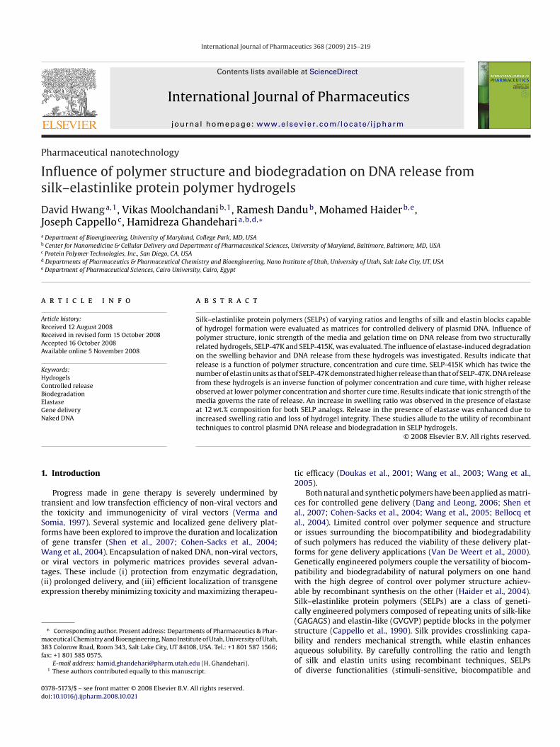
Recombinant techniques allow the introduction of functionalmino acid residues at precise locations in the polymer backbone.ELP-415K was designed such that it has a similar molecular weights SELP-47K but with longer elastin-like units. Consequently therere five more lysine residues in 47K compared to 415K. This couldeduce the interaction of negatively charged plasmid DNA with the15K polymer backbone. To evaluate this phenomenon we mea-ured the absorbance of DNA/polymer complexes at low non-gelorming concentrations. The absorbance of Polymer 47K mixtureith plasmid DNA showed a substantial increase when prepared
t low ionic strength compared to high ionic strength (Fig. 4). Mix-ures of SELP-415K and plasmid DNA showed lower turbidity atoth ionic strengths compared to their SELP-47K counterparts dueo the presence of lower number of lysine residues.
.4. Influence of elastase-induced biodegradation on swelling ofELP hydrogels
The elastin units in SELPs are susceptible to degradation byndogenous elastases providing a possible mechanism for their
218 D. Hwang et al. / International Journal of Pharmaceutics 368 (2009) 215–219
Fr4s
bbutittthcttauatllobwod
3
s
Ft(m
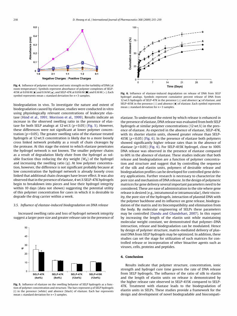
Fig. 6. Influence of elastase-induced degradation on release of DNA from SELPh1Sm
ethew4seDtrtobepmcri
ig. 4. Influence of polymer structure and ionic strength on the turbidity of DNA (atoom temperature): Symbols represent absorbance of polymer complexes of SELP-15K at 0.016 M (�) and 0.16 M (�), and SELP-47K at 0.016 M (�) and 0.16 M (×). Eachymbol represents mean ± standard deviation for n = 3 samples.
iodegradation in vivo. To investigate the nature and extent ofiodegradation caused by elastase, studies were conducted in vitrosing physiologically relevant concentrations of leukocyte elas-ase (Hind et al., 1991; Morrison et al., 1999). Results indicate anncrease in the observed swelling ratio in the presence of elas-ase for both SELP analogs at 12 wt.% (p < 0.05) (Fig. 5). However,hese differences were not significant at lower polymer concen-ration (p = 0.05). The greater swelling ratio of the elastase treatedydrogels at 12 wt.% concentration is likely due to a more looselyross linked network probably as a result of chain cleavages byhe protease. At this stage the extent to which elastase penetrateshe hydrogel network is not known. The smaller polymer chainss a result of degradation likely elute from the hydrogel as sol-ble fraction thus reducing the dry weight (Wd) of the hydrogelnd increasing the swelling ratio (q). At low polymer concentra-ion, however, the difference is not significant probably because atow concentration the hydrogel network is already loosely crossinked that additional chain cleavages have lesser effect. It was alsobserved that in the presence of elastase, 4 wt.% SELP-47K hydrogelsegin to breakdown into pieces and lose their hydrogel integrityithin 10 days (data not shown) suggesting the potential utility
f this polymer concentration for cases in which it is desirable toegrade the drug carrier within a week.
.5. Influence of elastase-induced biodegradation on DNA release
Increased swelling ratio and loss of hydrogel network integrityuggest a larger pore size and greater release rate in the presence of
ig. 5. Influence of elastase on the swelling behavior of SELP hydrogels as a func-ion of polymer concentration and structure. The bars represent q of SELP hydrogelsi) in the presence (white) and absence (black) of elastase. Each bar represents
ean ± standard deviation for n = 3 samples.
tdtmbmibmstv
4
sfat4ed
ydrogel analogs. Symbols represent cumulative percent release of DNA from2 wt.% hydrogels of SELP-47K in the presence (�) and absence (�) of elastase, andELP-415K in the presence (♦) and absence (�) of elastase. Each symbol representsean ± standard deviation for n = 3 samples.
lastase. To understand the extent by which release is enhanced inhe presence of elastase, DNA release was evaluated from both SELPydrogels at similar polymer concentrations (12 wt.%) in the pres-nce of elastase. As expected in the absence of elastase, SELP-47K,ith its shorter elastin units, showed greater release than SELP-
15K (p < 0.05) (Fig. 6). In the presence of elastase both polymershowed significantly higher release rates than in the absence oflastase (p < 0.05) (Fig. 6). For SELP-415K hydrogel, close to 100%NA release was observed in the presence of elastase compared
o 60% in the absence of elastase. These studies indicate that bothelease and biodegradation are a function of polymer concentra-ion and structure and suggest that by controlling the sequencef the silk and elastin units, polymers of desirable release andiodegradation profiles can be developed for controlled gene deliv-ry applications. Further research is necessary to characterize theore size and mechanism of DNA release. In the design of polymericatrices for gene delivery several important parameters need to be
onsidered. These are ease of administration to the site where geneelease is desired (e.g., intratumoral or intramuscular), their viscos-ty, the pore size of the hydrogels, interaction of plasmid DNA withhe polymer backbone and its influence on gene release, biodegra-ation of the matrix and its biocompatibility and elimination fromhe body. By molecular engineering of SELPs these parameters
ay be controlled (Dandu and Ghandehari, 2007). In this reporty increasing the length of the elastin unit while maintainingolecular weight constant, we demonstrated that polymer–DNA
nteraction, release and biodegradation can be modulated. Hencey design of polymer structure, matrix-mediated delivery of plas-id DNA from SELP hydrogels may be optimized. In addition, these
tudies can set the stage for utilization of such matrices for con-rolled release or incorporation of other bioactive agents such asiruses, cells, proteins and peptides.
. Conclusion
Results indicate that polymer structure, concentration, ionictrength and hydrogel cure time govern the rate of DNA releaserom SELP hydrogels. The influence of the ratio of silk to elastinnd the length of elastin units on release is demonstrated by
he higher release rate observed in SELP-415K compared to SELP-7K. Treatment with elastase leads to the biodegradation oflastin units in SELPs. These studies provide a framework for theesign and development of novel biodegradable and biocompati-
al of P
ba
A
R
B
C
C
D
D
D
D
D
H
H
H
M
M
M
N
S
V
V
W
Wang, Y., Li, C.Y., Yuan, F., 2004. Systemic virus dissemination during local gene
D. Hwang et al. / International Journ
le SELP-based matrix-mediated delivery systems for gene therapypplications.
cknowledgement
Funding for this study was provided under NIH R01 CA107621.
eferences
ellocq, N.C., Kang, D.W., Wang, X., Jensen, G.S., Pun, S.H., Schluep, T., Zepeda, M.L.,Davis, M.E., 2004. Synthetic biocompatible cyclodextrin-based constructs forlocal gene delivery to improve cutaneous wound healing. Bioconjug. Chem. 15,1201–1211.
appello, J., Crissman, J., Dorman, M., Mikolajczak, M., Textor, G., Marquet, M., Ferrari,F., 1990. Genetic engineering of structural protein polymers. Biotechnol. Prog. 6,198–202.
ohen-Sacks, H., Elazar, V., Gao, J., Golomb, A., Adwan, H., Korchov, N., Levy, R.J.,Berger, M.R., Golomb, G., 2004. Delivery and expression of pDNA embedded incollagen matrices. J. Control Release 95, 309–320.
andu, R., Cappello, J., Ghandehari, H., 2008. Characterization of structurally relatedadenovirus-laden silk-elastinlike hydrogels. J. Bioact. Compat. Polym. 23, 5–19.
andu, R., Ghandehari, H., 2007. Delivery of bioactive agents from recombinantpolymers. Prog. Polym. Sci. 32, 1008–1030.
ang, J.M., Leong, K.W., 2006. Natural polymers for gene delivery and tissue engi-neering. Gene Deliv. Tissue Eng. 58, 487–499.
inerman, A.A., Cappello, J., Ghandehari, H., Hoag, S.W., 2002. Swelling behavior of
a genetically engineered silk-elastinlike protein polymer hydrogel. Biomaterials23, 4203–4210.oukas, J., Chandler, L.A., Gonzalez, A.M., Gu, D., Hoganson, D.K., Ma, C., Nguyen,T., Printz, M.A., Nesbit, M., Herlyn, M., Crombleholme, T.M., Aukerman, S.L., Sos-nowski, B.A., Pierce, G.F., 2001. Matrix immobilization enhances the tissue repairactivity of growth factor gene therapy vectors. Hum. Gene Ther. 12, 783–798.
W
harmaceutics 368 (2009) 215–219 219
aider, M., Leung, V., Ferrari, F., Crissman, J., Powell, J., Cappello, J., Ghandehari, H.,2005. Molecular engineering of silk-elastinlike polymers for matrix-mediatedgene delivery: biosynthesis and characterization. Mol. Pharm. 2, 139–150.
aider, M., Megeed, Z., Ghandehari, H., 2004. Genetically engineered polymers:status and prospects for controlled release. J. Control Release 95, 1–26.
ind, C.R., Joyce, H., Tennent, G.A., Pepys, M.B., Pride, N.B., 1991. Plasma leukocyteelastase concentrations in smokers. J. Clin. Pathol. 44, 232–235.
egeed, Z., Cappello, J., Ghandehari, H., 2002. Controlled release of plasmidDNA from a genetically engineered silk-elastinlike hydrogel. Pharm. Res. 19,954–959.
egeed, Z., Haider, M., Li, D., O’Malley Jr., B.W., Cappello, J., Ghandehari, H., 2004. Invitro and in vivo evaluation of recombinant silk-elastinlike hydrogels for cancergene therapy. J. Control Release 94, 433–445.
orrison, H.M., Welgus, H.G., Owen, C.A., Stockley, R.A., Campbell, E.J., 1999. Interac-tion between leukocyte elastase and elastin: quantitative and catalytic analyses.Biochim. Biophys. Acta 1430, 179–190.
agarsekar, A., Crissman, J., Crissman, M., Ferrari, F., Cappello, J., Ghandehari, H.,2003. Genetic engineering of stimuli-sensitive silk–elastinlike protein blockcopolymers. Biomacromolecules 4, 602–607.
hen, X., Tong, H., Jiang, T., Zhu, Z., Wan, P., Hu, J., 2007. Homogeneous chi-tosan/carbonate apatite/citric acid nanocomposites prepared through a novelin situ precipitation method. Compos. Sci. Technol. 67, 2238–2245.
an De Weert, M., Hennink, W.E., Jiskoot, W., 2000. Protein instability in poly(lactic-co-glycolic acid) microparticles. Pharm. Res. 17, 1159–1167.
erma, I.M., Somia, N., 1997. Gene therapy – promises, problems and prospects.Nature 389, 239–242.
ang, Y., Hu, J.K., Krol, A., Li, Y.P., Li, C.Y., Yuan, F., 2003. Systemic dissemination ofviral vectors during intratumoral injection. Mol. Cancer Ther. 2, 1233–1242.
delivery in solid tumors and its control with an alginate solution. In: Interna-tional Conference of the IEEE Engineering in Medicine and Biology Society.IEEEEngineering in Medicine and Biology Society Conference, United States.
ang, Y., Liu, S., Li, C.Y., Yuan, F., 2005. A novel method for viral gene delivery in solidtumors. Cancer Res. 65, 7541–7545.