influence of chewing force on salivary stress journal...
TRANSCRIPT
Posted at the Institutional Resources for Unique Collection and Academic Archives at Tokyo Dental College,
Available from http://ir.tdc.ac.jp/
TitleInfluence of chewing force on salivary stress
markers as indicator of mental stress
Author(s)
AlternativeSoeda, R; Tasaka, A; Sakurai, K
Journal Journal of oral rehabilitation, 39(4): 261-269
URL http://hdl.handle.net/10130/2841
Right
This is the pre-peer reviewed version of the
following article: J Oral Rehabil. 2012
Apr;39(4):261-9, which has been published in final
form at http://dx.doi.org/10.1111/j.1365-
2842.2011.02264.x

1
Influence of Chewing Force on Salivary Stress Markers as Indicator of
Mental Stress
Ryohei SOEDA, Akinori TASAKA, Kaoru SAKURAI
Department of Removable Prosthodontics and Gerodontology, Tokyo Dental College, Chiba,
Japan
Running title: Influence of Chewing Force on Mental Stress
Keywords: chewing force, mental stress, cortisol, alpha-amylase, secretory immunoglobulin A

2
Correspondence: Dr. Ryohei SOEDA
Department of Removable Prosthodontics and Gerodontology, Tokyo Dental College,
1-2-2 Masago, Mihama-ku, Chiba City, Chiba, 261-8502, Japan
Phone: +81-43-270-3933/Fax: +81-43-270-3935
E-mail address: [email protected]

3
SUMMARY
The aim of the present study was to investigate the influence of chewing force on salivary stress
markers (alpha-amylase activity, salivary cortisol level, and secretory immunoglobulin A
secretion rate) as indicators of mental stress. Participants comprised 20 healthy men. The first set
of saliva specimens (S1) was collected at immediately after a 20-minute rest to evaluate stress
markers. As stress loading, the participants were required to perform arithmetic calculations for
20 min, after which the second set of saliva specimens (S2) was collected. Each participant was
then required to chew a piece of tasteless gum for 10 min, after which the third set of saliva
specimens (S3) was collected. After a 20-minute rest, the fourth set of saliva specimens (S4) was
collected. Weak, habitual, and strong chewing forces were assigned. Change rates of stress
markers between S2 and S3, and S2 and S4 were calculated. A significant difference was
observed in the change rate of cortisol levels between S2 and S3. Cortisol level decreased more
under strong chewing than under weak chewing. No significant differences were observed in the
change rate of amylase activity or s-IgA secretion rate among the 3 chewing forces. The results
suggest that differences in chewing force influence the salivary cortisol level of the 3 stress
markers, and that a strong chewing force induces a greater reduction in mental stress than a weak
one.
4
Introduction
Many reports have been published on mental stress reduction by chewing as reflected in
changes in sympathetic, endocrine, and immune system markers in saliva. Nakajo et al. reported
that alpha-amylase activity, a sympathetic nervous system stress marker, decreased with
chewing in a mentally stressful environment (1). Tahara et al. (2) and Scholey et al. (3)
reported that salivary cortisol levels, an endocrine system stress marker, decreased with chewing
after mental stress loading. Furthermore, Ishiyama et al. reported that s-IgA levels, an immune
system stress marker, decreased with chewing (4).
In terms of masticatory movement parameters, Tasaka et al. suggested that a fast chewing rate
induced a greater reduction in mental stress than a slow one (5). No studies, however, have
investigated the influence of other masticatory movement parameters such as chewing force,
chewing number, and chewing time on reduction of mental stress. Peter A et al. reported a
correlation between the mean amplitude electromyogram (EMG) of the masticatory muscle and
chewing force (6). Ruf S et al. reported that the mean amplitude EMG of the masticatory muscle
during chewing increased under mental stress (7). Niwa M et al. reported that chewing increased
activity in the prefrontal cortex, which is involved in stress control (8).
5
We hypothesized that chewing force as a masticatory movement parameter induced a
reduction in mental stress. The aim of the present study was to investigate the influence of
chewing force on salivary stress markers (sympathetic system alpha-amylase activity, endocrine
system salivary cortisol level, and immune system s-IgA secretion rate) as indicators of mental
stress.
Materials and methods
Participants
Twenty healthy men of between 23 and 30 years of age (average, 25.5 years) were recruited from
students and staff at Tokyo Dental College. All participants had complete natural dentition,
excluding the third molars, and were without subjective or objective abnormalities of the
stomatognathic system. None had any medical history of mental illness. All were non-smokers.
Written informed consent was obtained from all participants. The study was approved by the
Ethics Committee of Tokyo Dental College (#218).
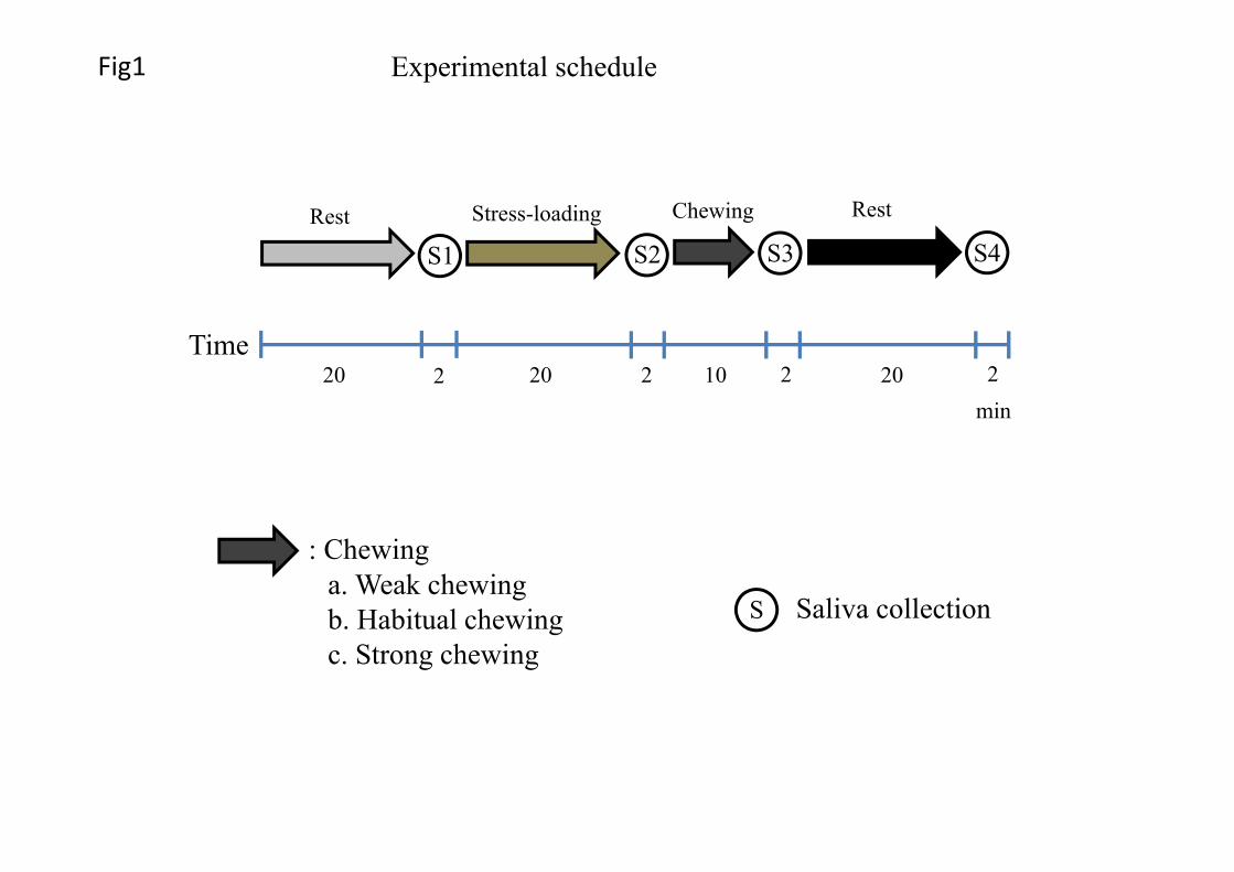
Experiment schedule

6
In consideration of the influence of circadian rhythm on salivary stress marker levels, the
experiments were performed between 2:00 PM and 7:00 PM. The participants were required to
refrain from eating, drinking (alchohol, caffeine), taking drug and exercising within 2 hr prior to
commencement of the experiments (9, 10) and were allowed 20 min in the experimental room to
rest and familiarize themselves with the environment before testing. Immediately after this, the
first set of saliva specimens (S1) was collected. As stress loading, participants were required to
perform arithmetic calculations (addition, subtraction, multiplication, division) for 20 min, after
which the second set of saliva specimens (S2) was collected. Each participant was then required
to chew a piece of tasteless gum base weighing 1.0 g (Lotte, Saitama, Japan) for 10 min. The
hardness of the gum base was 6.4×10³ Pa・s (soft type). After that, the third set of saliva
specimens (S3) was collected. After a 20-min rest, the fourth set of saliva specimens (S4) was
collected, completing the experiment (Fig. 1). The schedule of mental stress loading and rests
was determined based on previous reports (2, 5).
Weak, habitual, and strong chewing forces were applied. Participants were told to chew softly
for weak chewing, and strongly for strong chewing. Conscious chewing does not cause
discomfort or fatigue of the masticatory muscle. Therefore, a non-chewing condition was not
applied to avoid excessive stress.

7
The habitual chewing rate of each participant was determined with an electronic metronome.
The participants were instructed to maintain the same posture throughout the experiment. Each
participant was assigned all 3 chewing forces, each on a different days, also left the interval more
than a day. The 3 chewing forces were assigned in random order.
8
Measurements
In this study, salivary cortisol, alpha-amylase and s-IgA levels were selected as markers of
increase in stress. The cortisol and s-IgA saliva specimens were collected using a saliva sampling
device (Salivette, Sarsted, Rommelsdorf, Germany), keeping cotton rolls in the oral cavity for 2
min. In the laboratory, the samples were centrifuged at 3000 rpm for 20 min in a refrigerated
centrifuge. The supernatant of the collected whole saliva was frozen at -20. Salivary cortisol
levels (μg/dl) were determined with a radioimmunoassay kit (GammaCoat, Diasorin, Stillwater,
OK, USA) in accordance with the manufacturer’s instructions. The s-IgA levels (μg/dl) were
determined with an enzyme immunoassay kit (Poseidon2, Aloka, Tokyo, Japan). Alpha-amylase
saliva specimens were collected with an amylase monitor chip (Salivary amylase monitor chip,
Nipro, Osaka, Japan). The monitor chip was immersed in whole saliva under the tongue of the
participant for 30 sec.
Alpha-amylase activity (kU/l) was analyzed with a salivary amylase monitor (Salivary Amylase
Monitor, Nipro, Osaka, Japan). The s-IgA level (μg/dl) was corrected to the s-IgA secretion rate
(μg/min) by saliva flow rate for 2 min (dl). This was because, while salivary cortisol level and
alpha-amylase activity are not influenced by saliva flow rate, s-IgA level is (11-16).

9
This was because, while salivary cortisol level and alpha-amylase activity are not influenced by
saliva flow rate (11-14). S-IgA level is influenced (15, 16).
An EMG recording system (Muscle Tester ME3000P, Mega Electronics, Kuopio, Finland),
with a built-in 12-bit A/D converter was used to monitor chewing rates and determine
myoelectrical activity in the masseter muscle. For EMG of muscle activity, bipolar surface
electrodes (Blue Sensor P-00-S, Medicotest, Olstykke, Denmark) were positioned parallel to the
main direction of the muscle fibers on the maximal bulk of the bilateral masseter muscle, which
was determined by palpation while the participants clenched intermittently. Interelectrode
distance was 25 mm centre-to-centre. Reference (grounding) electrodes with integrated
preamplifiers were attached behind the ear. Prior to attachment of the electrodes and bioelectrical
measurement, the skin was thoroughly cleansed with a specific skin cleansing gel (Skin Pure,
Nihon Kohden, Tokyo, Japan) and ethanol-soaked gauze. Skin impedance between the electrodes
was lower than 8 kΩ. Sampling frequency was 1000 Hz at a sampling period of 0.1 sec.
The daily precision of the EMG was corrected by the mean amplitude EMG of the masseter
muscle under non-chewing conditions.
Statistical analysis

10
Alpha-amylase activity at S1 was regarded as signifying the baseline relaxed state. Participants
showing a lower alpha-amylase activity at S2 than at S1were regarded as being uninfluenced by
the mental stress loading applied in this study and excluded from the statistical analysis. Changes
in salivary stress markers (salivary cortisol level, alpha-amylase activity and s-IgA secretion rate)
between S2 and S3, and S2 and S4 were determined and compared. The mean amplitude and
integrated EMG of the masseter muscle over 10 min was calculated. Changes in salivary stress
markers and the mean amplitude and integrated EMG of the masseter muscle among the 3
chewing forces were compared using a one-way repeated measures analysis of variance
(ANOVA) and the post-hoc Bonferroni test at a significance level of 5% using statistical
software (SPSS 11.0J; SPSS, Chicago, IL, USA).
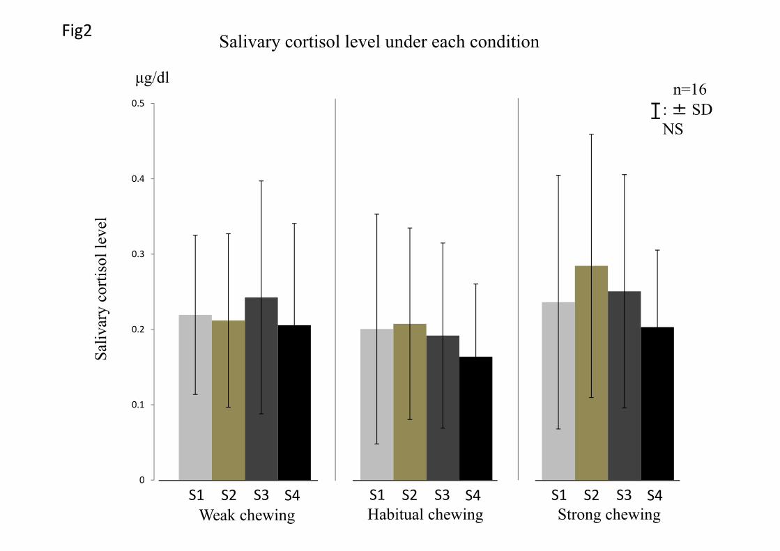
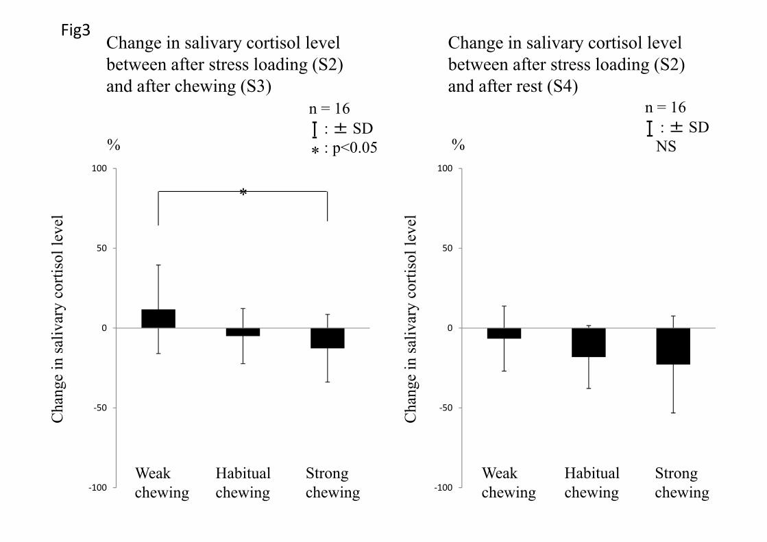
Results
Figure 2 shows changes in salivary cortisol level with time. Change in the salivary cortisol
level was expressed as a change rate, as individual differences exist in cortisol levels. The change
in the salivary cortisol level between S2 and S3 was significantly smaller with strong chewing
than with weak chewing. Only the change rate in salivary cortisol level with weak chewing
increased, suggesting an increase in mental stress. The change in salivary cortisol level between
11
S2 and S4 decreased under all chewing conditions, but no significant difference was observed
among the 3 chewing forces (Fig. 3).
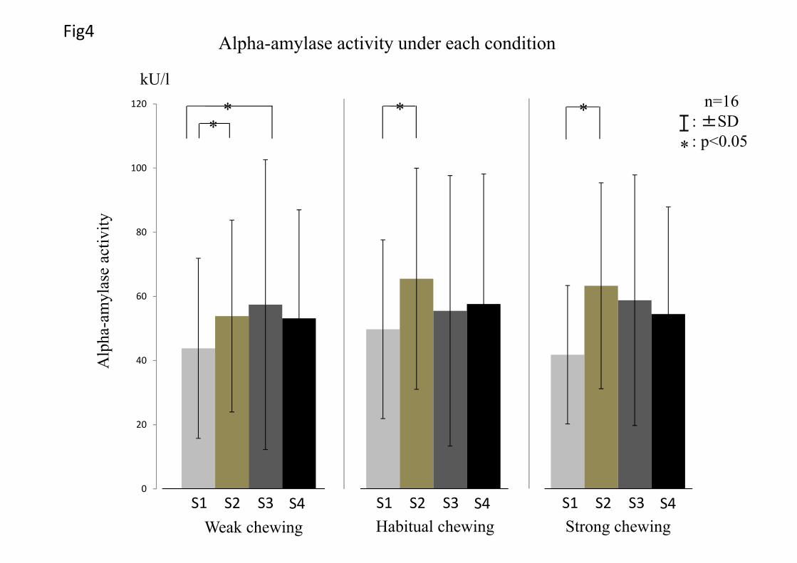
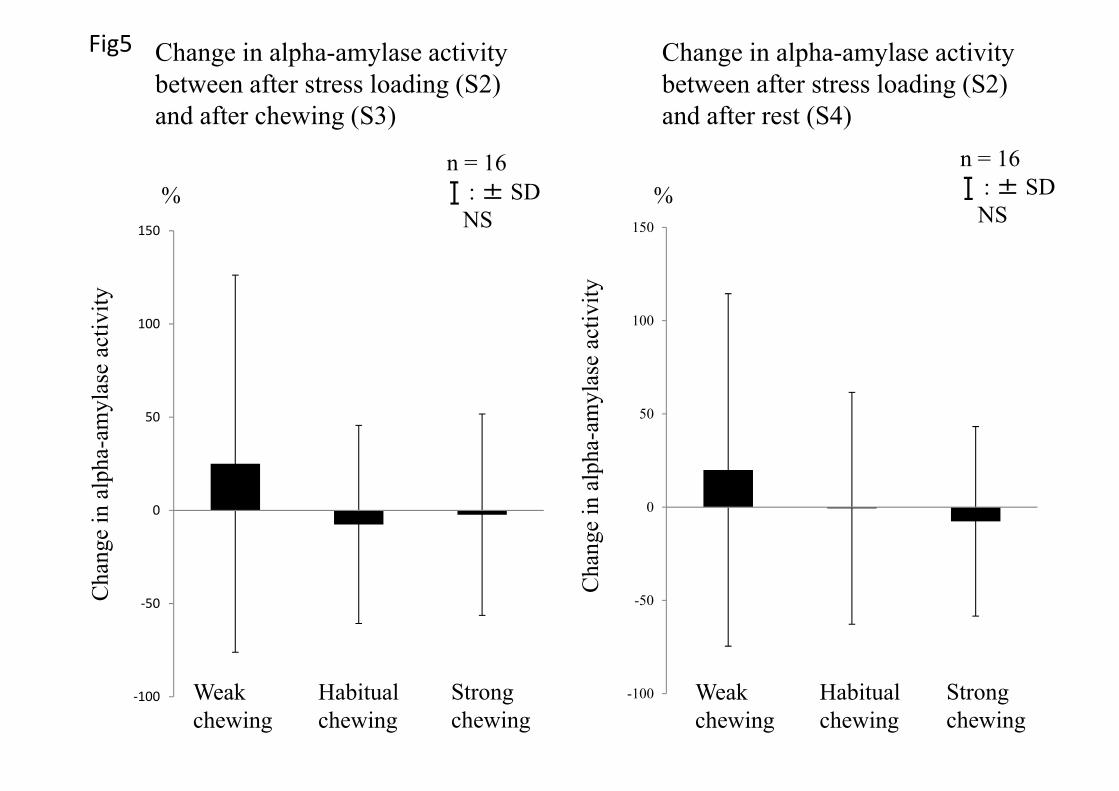
Figure 4 shows change in alpha-amylase activity with time. The mean change rates of alpha-
amylase activity between S2 and S3, and S2 and S4 decreased with strong chewing and increased
with weak chewing, but no significant difference was observed among the 3 chewing forces
(Figs. 5).
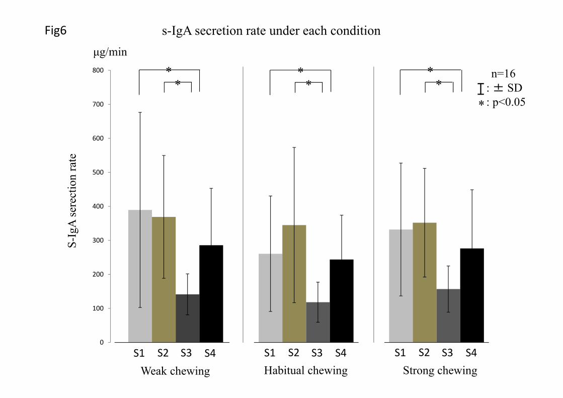
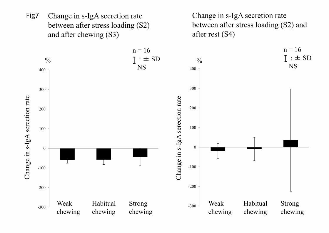
Figure 6 shows change in s-IgA secretion rate with time. The change in the s-IgA secretion
rate between S2 and S3 decreased under all chewing conditions. No significant difference was
observed among the 3 chewing forces. Change in the s-IgA secretion rate between S2 and S4
decreased with weak and habitual chewing and increased with strong chewing. No significant
differences were observed among the 3 chewing forces (Fig. 7).
The mean amplitude EMG of the masseter muscle showed significant differences among the 3
chewing rates (Fig. 8). Comparison of the mean amplitude EMG revealed that strong chewing
comprised 169% of habitual chewing, and weak chewing 25%. The integrated EMG of the
masseter muscle revealed significant differences among the 3 chewing rates (Fig. 9).
Discussion
12
Individual differences were observed in the level of chewing force required to fatigue each
participant in a pilot experiment. Therefore, chewing conditions were determined by the
participants themselves to avoid physical stress. The participants were instructed to adjust the
chewing force consciously. Mean amplitude EMG is considered to be associated with chewing
force. Significant differences in the mean amplitude EMG of the masseter muscle were observed
under all chewing conditions, thus confirming differences in chewing force and the validity of
setting different chewing forces as an experimental condition. Although chewing force can be
adjusted by changing the hardness of the gum base, this method was not applied in the present
study, as occlusal force differs among individuals and recognition of softness and change in gum
base hardness in itself may cause physical stress.
In the pilot study, change in cortisol level, alpha-amylase activity, and s-IgA secretion rate in
saliva after mental stress loading was compared between with and without chewing. The results
showed that stress markers in saliva were lower under chewing than non-chewing conditions.
This was consistent with the results of earlier reports (1-4). In the present study, taking stress into
consideration, the change rate was compared without applying a non-chewing condition (there
was a 10-min rest without chewing after stress loading).
13
The change rate in salivary cortisol level between S2 and S3 showed a significantly greater
decrease with strong chewing compared with weak chewing. This suggests that strong chewing
reduces mental stress more than weak chewing.
Stress includes both mental and physical stress. In the present study, arithmetic calculation
(addition, subtraction, multiplication, and division) was used to apply mental stress. It was
confirmed that alpha-amylase activity was higher at S2 (after mental stress loading) than at S1
(before mental stress loading). It has been reported that alpha-amylase, a sympathetic system
stress marker, reacts rapidly, and is therefore considered to be an effective mental stress marker
(17). Four of 20 participants in this study showed lower alpha-amylase activity at S2 than at S1,
and these were excluded from the statistical analysis.
Salivary cortisol level was endocrine system stress marker. In this study all participants were
male. Because, female participants need to control estradiol cycle (18). If it were not controlled ,
daily varience of cortisol level was large.
Salivary cortisol level did not increase at S2 compared with at S1 with weak chewing. We
believe that this was because salivary cortisol level increases at S1 before stress loading due to
stress anticipation ( 19, 20), and it takes 5-20 minutes for stress-induced blood cortisol to appear
14
in saliva (12, 21). S-IgA secretion rate and amylase activity are increase immediately after stress
loading.
There were reported about circadian rhythm. In cortisol level, circadian variability is low after
3:00 PM (22). In amylase activity, circadian variability is low between 4:00 and 5:00 PM (23). In
s-IgA secretion rate, circadian variability is low afternoon (24). Considering all of stress marker’s
condition, experimental schedule is severe. In this study, the experiment is performed between
2:00PM and 7:00PM.
And then, there were reported about stress marker reaction time. In cortisol levels, it takes 5-
20 minutes for increase after stress loading (12, 21). In amylase activity, it takes 1 minute for
increase after stress loading (25). In s-IgA secretion rate, immediately increase after stress
loading (26). Considering all of stress marker’s character, saliva was collected immediately after
task and each task was set enough long time in this study.
The cortisol-involving hypothalamic-pituitary-adrenal axis (HPA), the amylase-involving
sympatho-adrenal system (SAS), and the s-IgA-involving immune system react to mental stress.
However, it was reported that the s-IgA reaction under acute mental stress reflects SAS stress
reaction (27, 28). The s-IgA secretion rate was reported to increase by acute stress and decrease
by chronic stress (29, 30). In this study, it was anticipated that s-IgA secretion rate would

15
increase with acute stress and decrease with chewing. Previous reports have shown that mental
stress is reduced by chewing, as evidenced by a decrease in alpha-amylase activity, a sympathetic
nerve marker (1). A number of studies have reported that chewing activates the sympathetic
nerves (4, 31, 32). Meanwhile, other reports have demonstrated that chewing activates the
parasympathetic nerves (33). In this study, the results showed that using sympathetic nerve
activation as a marker of stress reduction was not consistent. Together with the results of the
earlier studies mentioned above, this suggests that the reaction changes under different chewing
conditions. Although we set the hardness of the sample food, chewing time, and chewing rate,
the change rate of alpha-amylase activity varied widely. It is considered difficult to evaluate
mental stress reduction based on SAS-related alpha-amylase and s-IgA activity. On the other
hand, change in HPA cortisol level showed no relationship with sympathetic nerve activity, and,
therefore, no correlation with change in alpha-amylase or s-IgA level (34, 35). This suggests that
chewing force specifically affects not the SAS, but the HPA stress reaction.
The integrated EMG of the bilateral masseter muscles (workload) showed a significant
difference between weak, habitual, and strong chewing conditions, suggesting that the workload
changed under different chewing forces. The relationship between the integrated EMG and
salivary cortisol level change rate was analyzed. A moderate negative correlation was observed
16
in the change rate of the salivary cortisol level and the integrated EMG between S2 and S3 (Fig.
13). The results showed that an increase in chewing force increased the workload and released
mental stress. Tasaka et al. reported that there was a correlation between the number of chewing
cycles and mental stress reduction (r = 0.49). This suggests that conscious strong chewing that
does not cause fatigue, and increasing the number of chewing cycles are effective for stress
reduction.
Conclusion
The results of the present study showed that chewing force affected an HPA stress marker,
salivary cortisol level, and that a strong chewing force was more effective than a weak one in
reducing mental stress.
Acknowledgments
We are grateful to the participants for their kind cooperation in this study. We would also like to
thank Dr Mutsumi Takagiwa (Associate Professor, Mathematics Laboratory, Tokyo Dental
College) for his help with the statistical analysis and Associate Professor Jeremy Williams,
Tokyo Dental College, for his assistance with the English of the manuscript. Lotte Co., Ltd.
17
(Saitama, Japan) are also acknowledged for their kind gifts of the test foods. This work was
supported by a grant for the Promotion and Mutual Aid Corporation for Private Schools of Japan.
Reference
(1) Nakajo N, Tomioka S, Eguchi S, Takaishi K, Cho G, Sato K. Gum Chewing may Attenuate
Salivary Alpha- Amylase of Psychological Stress Responses (in Japanese). J Jpn Dent Soc
Anesthesiol. 2007; 35: 346-353.
(2) Tahara Y, Sakurai K, Ando T. Influence of Chewing and Clenching on Salivary Cortisol
Levels as an Indicator of Stress. J Prosthodont. 2007; 16: 129-135.
(3) Scholey A, Haskell C, Robertson B, Kennedy D, Milne A, Wetherell M. Chewing gum
alleviates negative mood and reduces cortisol during acute laboratory psychological stress.
Physiol Behav. 2009; 97: 304-312.
(4) Ishiyama I, Suzuki M, Sato M, Nakamura T. Application of heart rate variability, salivary
constituents and electroencephalographic elements to evaluate function of the sympathetic or
parasympathetic nerves during masticatory exercise (in Japanese). J Masticat Health Soc.
2006; 16: 55–69.

18
(5) Tasaka A, Tahara Y, Sugiyama T, Sakurai K. Influence of Chewing Rate on Salivary Stress
Hormone Levels. J Jpn Prosthodont Soc. 2008; 52: 482-487.
(6) Peter A, Rer Nat, Jurgen Raum. Preconditions for Estimation of Masticatory Force from
Dynamic EMG and Isometric Bite Force-Activity Relations of Elevator Muscles. Int J
Prosthodont. 2001; 14: 563-569.
(7) Ruf S, Cecere F, Kupfer H. Stress-induced changes in the functional electromyographic
activity of the masticatory muscles. Acta Odontol Scand. 1997; 55: 44-48.
(8) Niwa M, Hiramatsu I, Nakata F, Hamaya C, Onogi N, Saito K. Functional significance of
stress-relieving act of chewing and it effect on brain activation by stress. Nihon Nouson
Igakukai Zasshi (JJRM) (in Japanese). 2005; 11: 661-666.
(9) Toda M, Morimoto K, Nagasawa S, Kitamura K. Effect of snack eating on sensitive salivary
stress markers cortisol and chromogranin A. Environ Health Prev Med. 2004; 9: 27–29.
(10) O’Connor PJ, Corrigan DL. Influence of short-term cycling on salivary cortisol levels. Med
Sci Sports Exerc. 1987; 19: 224–228.
(11) Vining RF, McGinley RA, Maksvytis JJ: Salivary cortisol: a better measure of adrenal
cortical function than serum cortisol. Ann Clin Biochem. 1983; 20: 329-335.
19
(12) Fukuda S, Morimoto K: Lifestyle, stress and cortisol response:II review. Environ Health
Prev Med. 2001; 6: 9-14.
(13) Rohleder N, Wolf JM, Maldonado EF, Kirschbaum C. The psychosocial stress-induced
increase in salivary alpha-amylase is independent of saliva flow rate. Psychophysiology. 2006;
43: 645-652.
(14) Takai N, Yamaguchi M, Aragaki T, Eto K, Uchihashi K, Nishikawa Y. Effect of
psychological stress on the salivary cortisol amd amylase levels in the healthy young adults.
Arch Oral Biol. 2004; 49: 963-968.
(15) Gallagher S, Phillips AC, Evans P, Der G, Hunt K, Carroll D. Caregiving is associated with
low secretion rates of immunoglobulin A in saliva. Brain, Behavior, and Immunity. 2008; 22:
565-572.
(16) Miletic ID, Schiffman SS, Miletic VD, Sattely-Miller EA. Salivary IgA Secretion Rate in
Young and Elderly Persons. Physiology & Behavior. 1996; 60: 243-248.
(17) Zeier H, Brauchli P, Joller-Jemelka HI. Effects of work demands on immunoglobulin A and
cortisol in air traffic controllers. Biol Psychol. 1996; 42: 413-423.(18) Kirschbaum C, Kudielka
20
BM, Gaab J, et al. Impact of gender, menstrual cycle phase, and oral contraceptives on the
activity of the hypothalamus-pituitary-adrenal axis. Psychosom Med 1999; 61(2): 154-162.
(19) Bassett JR, Marshall PM, Spillane R. The physiological measurement of acute stress (public
speaking) in bank employees. Int J Psychophysiol 1987; 5: 265–273.
(20) Miki K, Sudo A. Effects of mental task with noise exposure in men (in Japanese). Jpn J
Stress Sci 1995; 10: 81–85.
(21) Kudielka BM, Buske-Kirschbaum A, Hellhammer DH, Kirschbaum C. HPA axis responses
to laboratory psychosocial stress in healthy elderly adults, younger adults, and children: impact
of age and gender. Psychoneuroendcrinology. 2004; 29: 83-98.
(22) Izawa S, Sugaya N, Ogawa N, et al. Episodic stress associated with writing a graduation
thesis and free cortisol secretion after awakening. Int J Psychophysiol 2007; 64(2): 141-145
(23) Rohleder N, Natar UM, Wolf JM, et al. Psychosocial stress-induced activation of salivary
alpha-amylase: an indicator of sympathetic activity? Ann N Y Acad Sci 2004; 1032: 258-263.
(24) Hucklebridge F, Clow A, Evans P. The relationship between salivary secretory
immunoglobulin A and cortisol: neuroendocrine response to awakening and the diurnal cycle. Int
J Psychophysiol 1998; 31(1): 69-76.
21
(25) Ring C, Harison LK, Winzer A, et al. Secretory immunoglobulin A and cardiovascular
reactions to mental arithmetic, cold pressor, and exercise: effects of alpha-adrenergic blockade.
Psychophysiology 2000; 37(5): 634-643.
(26) Nater UM, Rohlder N, Gaab J, et al. Human salivary alpha-amylase reactivity in a
psychosocial stress paradigm. Int J Psychophysiol 2005; 55(3): 333-342.
(27) Allgrove JE, Gomes E, Hough J, Gleeson M. Effects of exercise intensity on saliva
antimicrobial proteins and markers of stress in active men. J Sports Sci. 2008; 26: 653-661.
(28) Evans P, Clow A, Hucklebridge F. Stress and the immunie system. Psychologist. 1997; 303-
307.
(29) Ring C, Drayson M, Walkey D.G, Dale S, Carroll D. Secretory immunoglobulin A reactions
to prolonged mental arithmetic stress: inter-session and intra-session reliability. Biol Psychol.
2002; 59: 1-13.
(30) Phillips A.C, Carroll D, Evans P, Bosch J.A, Clow A, Hucklebridge F, et al. Stressful life
events are associated with low secretion rates of immunoglobulin A in saliva in the middle aged
and elderly. Brain Behav Immun. 2006; 20: 191-197.
22
(31) Onozuka M, Fujita M, Watanabe K, Hirano Y, Niwa M, Nishiyama K. Mapping brain region
activity during chewing: a functional magnetic resonance imaging study. J Dent Res. 2002; 81:
743–746.
(32) Hasegawa Y, Ono T, Hori K, Nokubi T. Influence of human jaw movement on cerebral
blood flow. J Dent Res. 2007; 86: 64–68.
(33) Ohtsuka K, Kubo S, Takiguchi T, Ohkuma Y. The relaxing effect of gum chewing (in
Japanese). J Masticat Health Soc 1997; 7: 11–16.
(34) Susman EJ, Dockray S, Granger DA, Blades KT, Randazoo W, Heaton JA, et al. Cortisol
and alpha amylase reactivity and timing of puberty: Vulnerabilities for antisocial behaviour in
young adolescents. Psychoneuroendcrinology. 2010; 35: 557-569.
(35) Lester SR, Brown JR, Aycock JE, Grubbs SL, Johnson RB. Johnson. Use of Saliva for
Assessment of Stress and Its Effect on the Immune System Prior to Gross Anatomy Practical
Examinations. Anat Sci Educ. 2010; 3: 160-167.
Figure legend
23
Fig1
Experimental schedule . S was saliva collection timing .
Fig2
Salivary cortisol level under each condition. These were compared between saliva collection
timing in each condition using a one-way repeated measures analysis of variance (ANOVA) and
the post-hoc Bonferroni test at a significance level of 5%.
Fig3
Changes in salivary cortisol level between after stress loading (S2) and after chewing (S3), after
stress loading (S2) and after rest (S4). These were compared using a one-way repeated measures
analysis of variance (ANOVA) and the post-hoc Bonferroni test at a significance level of 5%.
Fig4
Alpha-amylase activity under each condition. These were compared between saliva collection
timing in each condition using a one-way repeated measures analysis of variance (ANOVA) and
the post-hoc Bonferroni test at a significance level of 5%.
Fig5
24
Changes in alpha-amylase activity between after stress loading (S2) and after chewing (S3), after
stress loading (S2) and after rest (S4). These were compared using a one-way repeated measures
analysis of variance (ANOVA) and the post-hoc Bonferroni test at a significance level of 5%.
Fig6
The s-IgA secretion rate under each condition. These were compared between saliva collection
timing in each condition using a one-way repeated measures analysis of variance (ANOVA) and
the post-hoc Bonferroni test at a significance level of 5%.
Fig7
Changes in s-IgA secretion rate between after stress loading (S2) and after chewing (S3), after
stress loading (S2) and after rest (S4). These were compared using a one-way repeated measures
analysis of variance (ANOVA) and the post-hoc Bonferroni test at a significance level of 5%.
Fig8
Mean amplitude EMG of masseter muscle for 10 minutes chewing. These were compared using a
one-way repeated measures analysis of variance (ANOVA) and the post-hoc Bonferroni test at a
significance level of 5%.
Fig9

25
Integrated EMG of masseer muscle for 10 minutes chewing. These were compared using a one-
way repeated measures analysis of variance (ANOVA) and the post-hoc Bonferroni test at a
significance level of 5%.
Fig10
Correlation between change in salivary cortisol levels between S2 and S3 and integrated EMG of
masseter muscle for 10 minutes. This was compared using Spearmans’ correlation.
20 20 2010
Rest RestChewingStress-loading
S1 S3S2 S4
Saliva collection
Time2 2 2 2
: Chewinga. Weak chewingb. Habitual chewingc. Strong chewing
S
min
Fig1 Experimental schedule
0
0.1
0.2
0.3
0.4
0.5n=16
: ± SDNS
Weak chewing Habitual chewing Strong chewing
Saliv
ary
corti
soll
evel
μg/dl
Fig2
S1 S3 S4 S2
Salivary cortisol level under each condition
S1 S3 S4 S2 S1 S3 S4 S2
‐100
‐50
0
50
100*
Change in salivary cortisol level between after stress loading (S2) and after rest (S4)
Cha
nge
in sa
livar
y co
rtiso
llev
el
Weak chewing
Habitual chewing
Strong chewing
n = 16: ± SD: p<0.05
*
‐100
‐50
0
50
100
%
Cha
nge
in sa
livar
y co
rtiso
llev
el
n = 16: ± SD
NS
Weak chewing
Habitual chewing
Strong chewing
Fig3Change in salivary cortisol level between after stress loading (S2) and after chewing (S3)
%
n=16: ±SD: p<0.05
0
20
40
60
80
100
120
Weak chewing Habitual chewing Strong chewing
Alp
ha-a
myl
ase
activ
itykU/l
Fig4Alpha-amylase activity under each condition
S1 S3 S4 S2 S1 S3 S4 S2 S1 S3 S4 S2
**
* *
*
‐100
‐50
0
50
100
150
%
Cha
nge
in a
lpha
-am
ylas
e ac
tivity
n = 16: ± SD
NS
Fig5
Weak chewing
Habitual chewing
Strong chewing
-100
-50
0
50
100
150
%
Cha
nge
in a
lpha
-am
ylas
e ac
tivity
n = 16: ± SD
NS
Weak chewing
Habitual chewing
Strong chewing
Change in alpha-amylase activity between after stress loading (S2) and after chewing (S3)
Change in alpha-amylase activity between after stress loading (S2) and after rest (S4)
0
100
200
300
400
500
600
700
800
Weak chewing Habitual chewing Strong chewing
Fig6
n=16: ± SD: p<0.05
S-Ig
Ase
rect
ion
rate
μg/min
s-IgA secretion rate under each condition
S1 S3 S4 S2 S1 S3 S4 S2 S1 S3 S4 S2
** *
**
*
*
-300
-200
-100
0
100
200
300
400
%
Cha
nge
in s-
IgA
sere
ctio
nra
te
n = 16: ± SD
NS
Fig7
Weak chewing
Habitual chewing
Strong chewing
-300
-200
-100
0
100
200
300
400
%
Cha
nge
in s-
IgA
sere
ctio
nra
te
n = 16: ± SD
NS
Weak chewing
Habitual chewing
Strong chewing
Change in s-IgA secretion rate between after stress loading (S2) and after chewing (S3)
Change in s-IgA secretion rate between after stress loading (S2) and after rest (S4)

0
0.1
0.2
0.3
0.4M
ean
ampl
itude
EM
G o
f mas
sete
rmV
Weak chewing Habitual chewing Strong chewing
** *
n = 16: ± SD: p<0.05*
Fig8 Mean amplitude EMG of masseter muscle over 10 min chewing

0
10
20
30
40
n = 16: ± SD: p<0.05
mVsM
ean
inte
grat
ed E
MG
of m
asse
ter
** *
Weak chewing Habitual chewing Strong chewing
*
Fig9 Integrated EMG of masseter muscle over 10 min chewing

-100
-80
-60
-40
-20
0
20
40
60
80
100
0 5 10 15 20 25 30 35 40 45 50
Mean integrated EMG of masseter over 10 min
S2-S
3 C
hang
e in
saliv
ary
corti
sol l
evel
s
r=-0.420P<0.01n=16
Fig10 Correlation between change in salivary cortisol levels between S2 and S3 and integrated EMG of masseter muscle over 10 min
: Weak chewing: Habitual chewing: Strong chewing
mVs
%Pearson’s correlation coefficient