influence of bruxism on survival of porcelain laminate veneersporcelain laminate veneers in patients...
TRANSCRIPT
Med Oral Patol Oral Cir Bucal. 2014 Sep 1;19 (5):e426-32. Bruxism and porcelain laminate veneers
e426
Journal section: Clinical and Experimental DentistryPublication Types: Research
Influence of bruxism on survival of porcelain laminate veneers
Maria Granell-Ruíz 1, Rubén Agustín-Panadero 1, Antonio Fons-Font 2, Juan-Luis Román-Rodríguez 1, María-Fernanda Solá-Ruíz 3
1 DDS,PhD. Associate Profesor, Oclusion and Prosthodontics, Department of Stomatology, University of Valencia, Valencia, Spain2 DDS,PhD,MD. Professor of Oclusion and Prosthodontics, Department of Stomatology, University of Valencia, Valencia, Spain3 MF,DDS,PhD,MD. Adjunct Lecturer, Department of Stomatology, University of Valencia, Valencia, Spain
Correspondence:Unidad de Prostodoncia y OclusiónEdificio Clínica OdontológicaC\ Gascó Oliag, Nº 146010 Valencia [email protected]
Received: 31/01/2013Accepted: 19/05/2013
AbstractObjectives: This study aims to determine whether bruxism and the use of occlusal splints affect the survival of porcelain laminate veneers in patients treated with this technique.Material and Methods: Restorations were made in 70 patients, including 30 patients with some type of parafunc-tional habit. A total of 323 veneers were placed, 170 in patients with bruxism activity, and the remaining 153 in patients without it. A clinical examination determined the presence or absence of ceramic failure (cracks, frac-tures and debonding) of the restorations; these incidents were analyzed for association with bruxism and the use of splints.Results: Analysis of the ceramic failures showed that of the 13 fractures and 29 debonding that were present in our study, 8 fractures and 22 debonding were related to the presence of bruxism.Conclusions: Porcelain laminate veneers are a predictable treatment option that provides excellent results, rec-ognizing a higher risk of failure in patients with bruxism activity. The use of occlusal splints reduces the risk of fractures.
Key words: Veneer, fracture, debonding, bruxism, occlusal splint.
Granell-Ruíz M, Agustín-Panadero R, Fons-Font A, Román-Rodríguez JL, Solá-Ruíz MF. Influence of bruxism on survival of porcelain laminate veneers. Med Oral Patol Oral Cir Bucal. 2014 Sep 1;19 (5):e426-32. http://www.medicinaoral.com/medoralfree01/v19i5/medoralv19i5p426.pdf
Article Number: 19097 http://www.medicinaoral.com/© Medicina Oral S. L. C.I.F. B 96689336 - pISSN 1698-4447 - eISSN: 1698-6946eMail: [email protected] Indexed in:
Science Citation Index ExpandedJournal Citation ReportsIndex Medicus, MEDLINE, PubMedScopus, Embase and Emcare Indice Médico Español
doi:10.4317/medoral.19097http://dx.doi.org/doi:10.4317/medoral.19097
IntroductionThe veneer restoration technique was developed in the mid nineteen-eighties in the United States, and later spread throughout the world. Bonding these fragile por-celain laminae securely to natural teeth has been a chal-lenge for our profession. Fortunately, these restorations
have proven to be one of the most successful techniques used in Restorative Dentistry (1).Porcelain laminate veneers represent a predictable re-storative solution for anterior teeth due to their excellent aesthetics as well as their durability and biocompatibi-lity (2). These restorations constitute an alternative to
Med Oral Patol Oral Cir Bucal. 2014 Sep 1;19 (5):e426-32. Bruxism and porcelain laminate veneers
e427
full-coverage restorations since they require minimal tooth preparation, there by maintaining the dental structure.Currently, porcelain laminate veneers are indicated for a wide range of situations and can be used to correct the shape and position of teeth, close diastema, replace old composite restorations, mask tooth discoloration (3), and to restore teeth following incisal abrasion and dental erosion. Some authors (4,5) suggest that bruxism constitutes a contraindication to these bonded restora-tions. Bruxism is generally recognized as non-function-al jaw movements, and is defined as a forcible clenching or grinding of the teeth, or a combination of both, and has long been regarded as a disorder requiring treatment (6). According to the American Academy of Orofacial Pain, bruxism is a diurnal or nocturnal parafunctional activity which includes clenching, bracing, gnashing and grinding of the teeth (7). Magne et al. report that the success rate for the veneer is reduced to 60% in patients with bruxism activity (8). This percentage is very similar to that obtained for metal-ceramic restora-tions in the same situation. The success rates may be increased if bruxism iscontrolled; therefore, a nocturnal and / or diurnal splint is recommended as a preventive measure to reduce the risk of failure, especially in these patients (4,9).The occlusal splint is generally used to treat muscle hy-peractivity. Studies carried out by various authors (10-13) show that these splints decrease bruxism activity generated during periods of stress; it is therefore ad-visable to use these devices in patients with suspected bruxism following prosthodontic treatment with either full coverage crowns or with laminate veneers.Restorations placed in patients presenting some type of bruxism activity should have a functional design, especially in situations where the patient has already lost some tooth structure and where these restorations provide the patient with a correct anterior and canine guidance (14). As with any technique, the use of porcelain veneers re-quires medium and long term studies to confirm their indications (4,9,15-20).These techniques have been used since 1985 at the Prosthodontics and Oclussion Teaching Unit of the Uni-versity of Valencia, School of Medicine and Dentistry, where to date a large number of patients have been treated with porcelain laminate veneers in response to aesthetic demands.We conducted a retrospective clinical study to review patients wearing porcelain laminate veneers. We ana-lyzed whether the presence of bruxims activity and the use of occlusal splints in our patients, affected the medium and long term survival of these treatments. To this end we developed a data collection methodology to provide reliable results able to withstand the usual sta-
tistical tests for these sample types and to be compared with results of other authors.
Material and MethodsThree hundred twenty-three porcelain laminate veneers were placed during a period of eight years, all fabricated with IPS-Empress ceramic (Ivoclar®, Schaan, Liechten-stein) in order to standardize the results and eliminate any variables that could arise from the use of different ceramics.At the time of the study, the 323 restorations studied had been placed in 70 patients with a duration ranging from 3 to 11 years. Of the patients studied, 24.3% (17) were male and 75.7% (53) were female, with a mean age of 46 years (range 18 to 74). Thirty of the 70 patients pre-sented bruxism activity, all patients with it, had to use occlusal splints (Hard acrylic), 15 complied with this requirement and 15 did not. The clinical diagnosis was made by clinical inspection of teeth of the consequenc-es of clenching or grinding activities were visible in the dentition and consistent with a bruxing habit.Of the 323 veneers, 124 (38.4%) were of simple design or window preparation, covering only the buccal sur-face (B) and 199 (61.6%) corresponded to those denomi-nated ‘functional’ (with incisal overlap), covering the incisal edge and part of the palatal/lingual tooth surface (F). Regarding location, 238 were placed in the maxi-llary arch and 85 in the mandibular arch. Of the ma-xillary restorations, 97 were on central incisors, 82 on lateral incisors, 49 on canines and 10 on premolars. Of the mandibular restorations, 31 were located on central incisors, 31 on lateral incisors, 19 on canines and 4 on premolars. One hundred seventy veneers were bonded in patients with bruxism activity and 153 in patients without it.This study focussed on the relationship between the dif-ferent ceramic failures and bruxism; therefore informa-tion was collected on the presence or absence of bru-xism activity and whether or not these patients had to use splints. These criteria provided us with 3 patients groups for the study:A- Patients without bruxism. This group included 40 patients, representing 57.1% of the total. These patients were restored with 153 veneers (65 conventional design and 88 functional).B- Patients with bruxism activity using splints properly. This group included 15 patients (21.4%) with 89 veneers (31 conventional and 58 functional).C- Patients with bruxism activity not using splints (they have it but they don t́ use it). This group included 15 patients (21.4%) with 81 veneers (28 conventional and 53 functional).Therefore, after placing the ceramic restorations, we checked occlusion properly, during maximum intercus-pation and during mandibular excursive movements.
Med Oral Patol Oral Cir Bucal. 2014 Sep 1;19 (5):e426-32. Bruxism and porcelain laminate veneers
e428
Patients who were bruxers were provided with hard acrylic resin occlusal guards to protect the definitive restorations during bruxing episodes.All of the patients were treated at the Prosthodontics and Occlusion Teaching Unit of the University of Valencia School of Medicine and Dentistry by a team that had followed the same method when placing the veneers. The statistical analysis focused on:An initial descriptive analysis containing the frequen-cies and percentages for the categorical variables in the study.A bivariate analysis, covering all the statistical compar-isons necessary to assess the relationship between frac-tures and debonding in patients with bruxism activity, and the use of a splint by these patients. These analyses were performed using nonparametric statistical tests given the categorical nature of the variables.The Pearson c2 test was used to test the association or dependence between two categorical variables, always provided that more than 5 cases were present in the con-tingency tables. Otherwise, and only for dichotomous variables, the Fisher’s exact test was used.A Kaplan-Meier Survival Analysis was used to study survival. As a comparative test the log-rank test was used (Kaplan-Meier, 1958).
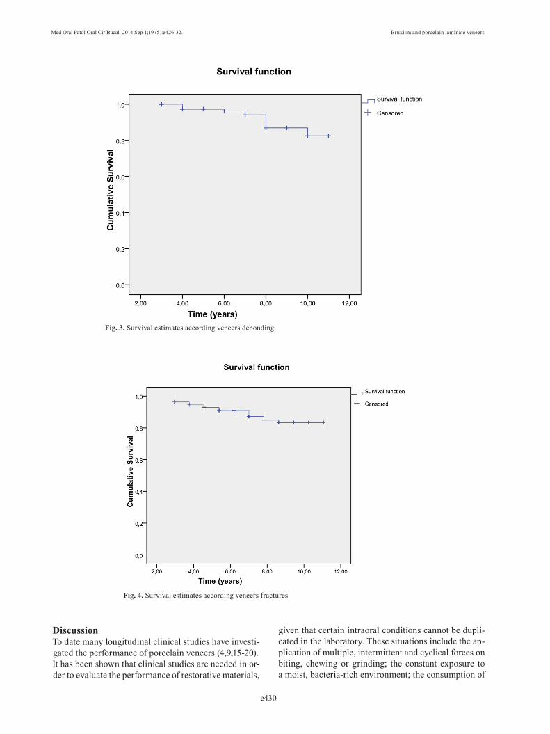
ResultsDuring the evaluation period, the results were:Ceramic failures: The survival of restorations in terms of their structural integrity is the most important fac-tor for both patients and professionals when deciding on this treatment option. Therefore, the analysis was made in terms of the presence or absence of the three most important aspects: cracks, fractures and debond-ing (Table 1). Cracks: At the time of the review no cracks were ob-Cracks: At the time of the review no cracks were ob-served. This does not mean that some of the fractures found had not initiated as a crack, which over time had developed into a fracture. Fractures: A total of 13 fractures were observed (4%). Eight appeared in patients with bruxism, and the re-maining 5 in patients without it.
Debonding: A total of 29 debonded restorations were observed, corresponding to 9% of the sample. Twenty-two were found in patients with bruxism, and the re-maining 7 in patients without it. By statistically relating ceramic failures with bruxism, a clear link can be seen. On one hand we can see that fractures, although more frequent in the presence of bruxism, are not statistically significant, given that 5 fractures appeared in patients without bruxism versus 8 fractures that occurred in patients with it (p = 0.511) (Chi2); in contrast, statistically significant differences were found when examining the correct use of splints in patients with bruxism, since of these 8 fractures, 1 occurred in a patient who did use a splint, and 7 in pa-tients who did not (p = 0.023) (Fisher). The figure be-low shows that a higher proportion of fractures were observed in patients with bruxism activity who did not use a splint (9%) than in those who used a splint pro-perly (1%) (Fig. 1).Regarding debonding, this was observed to be more frequent in patients with bruxism. Of the 29 debonded veneers, 22 were produced in these patients (p = 0.009) (Chi2), a clear statistically significant difference can be seen between the two groups of patients (with and without bruxism activity). The figure below illustrates the higher proportion of debonding in patients with bru-xism versus those without it (Fig. 2). Of the 22 debond-. 2). Of the 22 debond-2). Of the 22 debond-ed restorations in patients with bruxism, 12 appeared in patients using a splint and 10 in patients where splints were not used, without statistically significant diffe-rences (p = 0.825) (Chi2).Regarding design, there were no significant differences between the type of restoration used (conventional or functional) and the presence of bruxism activity (p = 0.151) (Chi2); although, in this study most patients with bruxism were fitted with functional restorations (F).The Kaplan-Meier curves (Kaplan-Meier, 1958) clearly show the survival of the restorations, indicating the probability that a restoration will remain in good condi-tion over time.This analysis considered the time in years during which the restoration remained in good condition or the time un-til deterioration. Two types of deterioration were consid-ered: debonding and fracture, in addition this deterioration was related to the presence or absence of bruxism.Fractures: The estimated survival table showed that the mean survival times were similar between patients with and without bruxism. Furthermore, the log-rank test confirmed that the survival curves were statistically equal (p = 0.519) (Fig. 3).Debonding: Although the estimated survival table in-dicated that the mean survival times were similar be-tween patients with and without bruxism, the log-rank test confirmed statistically significant differences in the survival curves (p = 0.008) (Fig. 4).
Presence of
Bruxism
Nº
patientsNº veneers Fractures Debonding
No 40 153 (65C-88F) 5 7
Yes (with splint) 15 89 (31C-58F) 1 12
Yes (without splint) 15 81 (28C-53F) 7 10
Total 70 323 13 29
Table 1. Distribution of veneers restorations. Frequency of frac-tures and debonding.

Med Oral Patol Oral Cir Bucal. 2014 Sep 1;19 (5):e426-32. Bruxism and porcelain laminate veneers
e429
Fig. 1. Percentage of veneers fractures and use of splint.
Fig. 2. Percentage of veneers debonding and patients with bruxism.
Med Oral Patol Oral Cir Bucal. 2014 Sep 1;19 (5):e426-32. Bruxism and porcelain laminate veneers
e430
DiscussionTo date many longitudinal clinical studies have investi-gated the performance of porcelain veneers (4,9,15-20).It has been shown that clinical studies are needed in or-der to evaluate the performance of restorative materials,
given that certain intraoral conditions cannot be dupli-cated in the laboratory. These situations include the ap-plication of multiple, intermittent and cyclical forces on biting, chewing or grinding; the constant exposure to a moist, bacteria-rich environment; the consumption of
Fig. 3. Survival estimates according veneers debonding.
Fig. 4. Survival estimates according veneers fractures.

Med Oral Patol Oral Cir Bucal. 2014 Sep 1;19 (5):e426-32. Bruxism and porcelain laminate veneers
e431
hot and cold liquids, as well as vigorous brushing. In vivo studies are therefore necessary to verify the ac-ceptability of a laminate veneer as a definitive restora-tive treatment. Retrospective studies can provide a reli-able picture of the clinical performance of materials and techniques.While numerous in vitro studies exist (21,22), these do not offer the same prognostic value or long-term pre-dictability of this treatment as studies in vivo. Although longitudinal clinical studies of longer than 5 years cer-tainly provide useful scientific data, they can sometimes become out of date due to the rapid and constant change in technology and materials. Thus, in vitro studies may have more impact, but have no greater utility.Discussion of resultsWith respect to ceramic failures, in the present study there were 13 fractures (4.0%), 29 debondings (9.0%) and no cracks.Cracks: The fact that in this study no cracks appeared in the restorations may be due to the use of high-strength porcelain (IPS-Empress). Magne et al. (8) in a clinical study used conventional feldspathic porcelain for the fabrication of the restorations; the authors observed 12% cracks, thus justifying the use of stronger por-celain. The majority of authors do not consider small cracks in restorations as failures (5,23).Fractures: We found 4% of fractures, data similar to those of Jordan et al. (15) and Calamia (24) with 3%, and Nordbø et al. (17) with 5%. The majority of clinical studies reviewed report a low incidence of fractures, for example Kinh et al. (23) 0%; and Peumans et al. (5) 1%. However, other authors indicate a much higher rate of fractures, Christensen et al. (9) reported 13% at 3 years and Walls (4) 14% at 5 years, arguing that the majority of their patients had a history of bruxism, and that they had used conventional feldspathic porcelain, which has a lower fracture strength than high-strength feldspathic restorations.In the present study it was observed that fractures oc-curred more frequently in patients with bruxism, and that not using a splint when required constitutes a risk factor for the presence of fractures.Debonding: there was a notably high percentage of debonding in this study (9%), a high proportion of which occurred in patients with bruxism. It was found that of the 22 debonded veneers in patients with brux-ism, 12 were related to patients who used an occlusal splint, while the remaining 10 were related to patients who did not use a splint; we therefore consider that the debonding was not so much related to the use or other-wise of a splint, but more to the existence of a history of bruxism, taking into account that these patients gener-ally wear the splint only at night, and it has been found that bruxism may be both diurnal and nocturnal (25).Some authors (16,18) with in vivo studies report high
rates of debonding in restorations due to the presence of composite reconstructions in teeth supporting this type of restoration. In these cases the adhesion is be-tween resin and resin, which reduces the bond strength between the porcelain veneer-tooth complexes.Some authors do not consider debonding as a failure, since the restoration is simply replaced. Fradeani et al. (19), report three cases of debonding in one of their studies, with no signs of internal damage or fracture; these were replaced, commenting that the debonding was most certainly due to an inappropriate adhesive technique.
Conclusions1. In this study, the presence of fractures and debonding in porcelain laminate veneers increases considerably in patients with bruxism. The probability of debonding is almost 3 times higher in patients with it.2. It was found that, the use of splints reduces the failure rate of porcelain laminate veneers in patients with brux-ism activity; the probability of fracture being 8 times greater in patients who are required to use a splint but do not.3. Longitudinal in vivo clinical studies are needed to evaluate the performance and predictability of restora-tive materials, since certain intraoral conditions cannot be reproduced in the laboratory.
References1. Chen W, Raigrodski AJ. A conservative approach for treating young adult patients with porcelain laminate veneers. J Esthet Restor Dent. 2008;20:223-238.2. Fons-Font A, Solá-Ruiz MF, Granell-Ruiz M, Labaig-Rueda C, Martínez-González A. A choice of ceramic for use in treatments with porcelain laminate veneers. Med Oral Patol Oral Cir Bucal. 2006;11:e297-302.3. Freire A, Archegas LR. Porcelain laminate veneer on a highly dis-coloured tooth: a case report. J Can Dent Assoc. 2010;76:a126.4. Walls AWG. The use of adhesively retained all-porcelain veneers during the management of fractured and worn anterior teeth: Part 2. Clinical result after five years follow-up. Br Dent J. 1995;178:337-340.5. Peumans M, Van Meerbeeck B, Yoshida Y, Lambrechts P, Van-herle G. Five-year clinical performance of porcelain veneers. Quin-tessence Int. 1998;29:211-221.6. Attanasio R. Intraoral orthotic Therapy. Dent Clin North Am. 1997;41:309-324.7. American Academy of Orofacial Pain. Okeson JP, ed. Orofacial Pain. Guidelines for assessment, diagnosis, and management. Chi-cago: Quintessence Publish Co; 1996. p. 49-73.8. Magne P, Perroud R, Hodges JS, Belser UC. Clinical performance of novel-design porcelain veneers for the recovery of coronal volume and length. Int J Periodontics Restorative Dent. 2000;20:440-57.9. Christensen GJ, Christensen RP. Clinical observations of porce-lain veneers: a three-year report. J Esthet Dent. 1991;3:174-9.10. Solberg WK, Clark GT, Rugh JD. Nocturnal electromyographic evaluation of bruxism patients undergoing short term splint therapy. J Oral Rehabil. 1975;2:215-23.11. Van Der Zaag J, Lobbezoo F, Wicks DJ, Visscher CM, Hamburger HL, Naeije M. Controlled assessment of the efficacy of occlusal sta-bilization splints on sleep bruxism. J Orofac Pain. 2005;19:151-158.
Med Oral Patol Oral Cir Bucal. 2014 Sep 1;19 (5):e426-32. Bruxism and porcelain laminate veneers
e432
12. Koyano K, Tsukiyama Y, Ichiki R, Kuwata T. Assessment of bruxism in clinic. J Oral Reabil. 2008;35:495-508.13. Lobbezoo F, van Der Zaag J, van Selms MKA, Hamburger HL. Principles for management of bruxism. J Oral Reabil. 2008;35:509-523.14. Highton R, Caputo AA, Mátyás J. A photoelastic study of stresses on porcelain laminate preparations. J Prosthet Dent. 1987;58:157-61.15. Jordan RE, Suzuki M, Senda A. Four year recall evaluation of labial porcelain veneer restoration. J Dent Res. 1989;68Special:544.16. Dunne SM, Millar BJ. A longitudinal study of the clinical per-formance of porcelain veneers. Br Dent J. 1993;175:317-21.17. Nordbø H, Rygh-Thoresen N, Henaug T. Clinical performance of porcelain laminate veneers without incisal overlapping: 3-year re-sults. J Dent. 1994;22:342-5.18. Shaini FJ, Shortall AC, Marquis PM. Clinical performance of porcelain laminate veneers. A retrospective evaluation over a period of 6.5 years. J Oral Rehabil. 1997;24:553-9.19. Fradeani M, Redemagni M, Corrado M. Porcelain laminate ve-neers: 6-to 12 year clinic evaluation- a restorative study. Int J Peri-odontics Restorative Dent. 2005;25:9-17.20. Granell-Ruiz M, Fons-Font A, Labaig-Rueda C, Martínez-González A, Román-Rodríguez JL, Solá-Ruiz MF. A clinical longi-tudinal study 323 porcelain laminate veneers. Period of study from 3 to 11 years. Med Oral Patol Oral Cir Bucal. 2010;15:e531-7. 21. Magne P, Kwon KR, Belser UC, Hodges JS, Douglas WH. Crack propensity of porcelain laminate veneers: A simulated operatory evaluation. J Prosthet Dent. 1999;81:327-34.22. Castelnuovo J, Tjan AH, Phillips K, Nicholls JI, Kois JC. Fracture load and mode of failure of ceramic veneers with different prepara-tions. J Prosthet Dent. 2000;83:171-80.23. Kihn PW, Barnes DM. The clinical longevity of porcelain ve-neers: A 48-month clinical evaluation. Jada. 1998;129:747-752.24. Calamia JR. Clinical evaluation of etched porcelain veneers. Am J Dent. 1989;2:9-15.25. Beier US, Kapferer I, Burtscher D. Clinical performance of porcelain laminate veneers for up to 20 years. Int J Prosthodont. 2012;25:79-85.