inflammation of the larynx - uomustansiriyah.edu.iq08_52... · severe toxaemia is rare. 8. ......
TRANSCRIPT
Inflammation of the larynx
Acute non-specific laryngitis Acute simple laryngitis in adults
An acute superficial inflammation of the laryngeal mucous membrane.
Aetiology
1. Infection. Laryngitis occurs either as a part of a generalized upper respiratory infection, or as a sudden localized laryngitis. These forms are commonly viral.
It is‘ more common in winter and early Spring and often occurs each year. Patient suffering from sinusitis, nasal obstruction from any cause, overuse Of the voice and indulgence in alcohol or tobacco are more prone .
2. Trauma due to vocal abuse and/or endoscopic manipulation. 3. Irritation from inhaled fumes or gas, including tobacco smoke.
Pathology
1. Infection of the mucosa is at first slight and generalized, and the vocal cords themselves may escape in the early stages. Later they become reddened.
2. Oedema follows and the whole laryngeal mucosa is usually symmetrically involved.
, 3. Sticky mucopurulent exudate may cover the surface
4. Slight abrasions are sometimes seen, but deep ulceration does not occur.
. 5. Purulent exudation occurs in the very severe forms with marked injection and oedema (‘septic laryngitis’)
. 6. Fibrinous laryngitis occurs in influenza, or in certain coccal infections. There are white deposits or plaques on the surfaces of the cords and upper laryngeal inlet. Minute erosions may be found; they heal in a few days. 7. Perichondritis may follow the purulent form. The causal organism is usually the haemolytic streptococcus.

Clinical features
1. Hoarseness. The patient may speak in a gruff or falsetto voice, or become quite aphonic. 2. Discomfort in the throat is common.
3. Pain is slight or absent except in very severe cases.
4. Dysphagia is usually slight, unless the epiglottis or arytenoids are markedly involved as after swallowing hot fluids.
5. Dyspnoea is absent, unless oedema occurs in severe cases.
6. Cough. If present is dry and irritating.
7. Generalized symptoms. Malaise and fever often occur before the local symptoms appear. Severe toxaemia is rare. 8. Symmetrical redness and/or sticky secretions on both vocal cords, at indirect laryngoscope.
Progress
The inflammation usually resolves in a few days. The hoarseness may persist for as long as 2 weeks after apparent resolution. A functional aphonia .may follow a simple acute laryngitis, especially in women. In severe cases the inflammation may Spread throughout the lungs, more commonly in aged patients.
Treatment
‘1~ Local
vocal rest is essential. A Quiet unforced whisper is allowed
steam inhalations with tinct benzoin co or menthol help to loosen viscid secretions
0 Aspirin for its anti-inflammatory effect may be so soothing
Warm applications to the neck. .
Codeine to suppress dry cough Is comforting.
2. General
Rest. In a room with an equable temperature. Sedatives may help
. Avoidance of alcohol and tobacco
Systemic antibiotics in cases of infective origin.
Acute simple laryngitis in children
Acute laryngitis is a more serious condition in children than in adults. The lymphatic drainage of the larynx is richer in the child and the submucosa is therefore more likely to swell. The neuromuscular mechanism is also more easily upset and spasm more easily provoked, Finally, the child is less able to expel the secretions by coughing
. Aetiology
1. Any upper respiratory infection. Viral or bacterial, may lead to an acute laryngitis. It is often associated with the common infectious diseases. 2. Trauma. To the pharyngeal or laryngeal mucosa may institute an attack.
Clinical features
The onset is usually preceded by a simple sore throat, with slight fever and irritating cough ' 1. Cough. A laryngeal spasm (‘false croup’) develops suddenly.
2. Dyspnoea, cyanosis and stridor. From laryngeal spasm and mucosal oedema, the latter sometimes extending to the subglottic space. ‘Laryngitis stridulosa ’ is the name given to the condition when stridor is present. It is inspiratory and indrawing of the supra- and infra-clavicular fossae may be present during the paroxysms of stridor.
3. Hoarseness. The voice is hoarse but strong unless the condition is very severe and exhausting, when it becomes weak.
Differential diagnosis
1. Laryngeal diphtheria in which the child is usually more ill but less febrile; the characteristic membrane can usuall be seen and the organism can be identified on culture
2.imapcted foreign body in the larynx
3. inhaled foreign body in the bronchus
4. Congenital laryngeal stridor, webs, cysts.
5. spasmodic laryngitis (spasmodic croup )
6.layrngeal edema from steam or corrosives
7.allergic laryngitis (Quincke’s oedema). Rarely
8. Laryngeal papillomatosis

Treatment
1, Rest in bed is essential . A sitting position should be adopted.
2. Systemic antibiotic , antibiotic are given in full doses to ill children and especially to infants.
3_ Systemic steroids in all severe cases
4. Humidification is helpful for the first day or two.
5. Oxygen May be essential; 80% helium 20% oxygen quarters the work of breathing
6. Nasotracheal tube may be inserted
7. Tracheostomy is indicated when the obstruction is severe. It is better done the too early than too late. Cardiac insufficiency may develop suddenly though it is less common than in diphtheria
8. Laryngotomy is quicker to perform and may thus be life-saving. It must be “El-a y replaced by a tracheostomy as soon as possible.
9. Intravenous fluids are sometimes necessary to combat dehydration.
After the acute phase has subsided it is necessary to attend to the general health and to remove infected tonsils, adenoids, or teeth

Acute laryngbtracheobronchitis
Aetiology Affects infants and young children. Causative organism .is usually parainfluenza virustype 1 or similar which shows seasonal and epidemic tendencies, Secondary bacterial infection by the third day of symptoms is common and makes the condition worse

clinical features Are those of an acute simple laryngitis as it occurs in children, but are much more severe.
1. Hard, dry, croupy cough and hoarseness. Occur after a cold or influenza.
2. Pyrexia. May be very high.
3. Dyspnoea and cyanosis. Often marked, from involvement of the trachea and bronchi.
4. Tenacious exudation and crusting. Characteristic,
5. Oedema of the larynx. Seen on layrngoscopy
6. Atelectasis. Caused by occlusion of the bronchi.
Differential diagnosis
1. Acute simple laryngitis which is less sever and produces no Physical Signs in the chest
2. Diphtheria which can be distinguished by bacteriological examination and there is no bleeding when the crust are removed in cases of acute laryngotracheobronchitis.
3. Bronchopneumonia in which there is usually no crusting
4. Bronchial foreign bodies espec1ally those of a vegetable nature.

Treatment
Should be in hospital.
1 Rest and reassurance are important.
2 Systemic antibiotic and antipyretic each degree Celsius rise in body temperature causes an Obligatory 10 % rise in metabolic rate.
3 Humidification of the inspired
4. Oxygen may have to be given ( by tent ) Heliox may be given also
5. Fluids must be given by mouth or IV to prevent dehydration
6.NG tube or tracheostomy may be needed
7. removal of secretion can be done by bronchoscopy or through the tracheostomy
Acute epiglottis
Definition
Special form of acute laryngitis, in- which the inflammatory changes affect mainly the loosely attached mucosa of the epiglottis.
Pathology
Localized oedema may obstruct the airway, especially in children. H.
influenza is the usual causal organism, B Haemolytic streptococci rarely.
Submucosal abscesses may form.
Clinical features
1. Dyspnoea maybe progressive and alarming, especially in children, in
whom death may occur within a few hours of onset.
2. Pain on swallowing is commoner than respiratory obstruction in adults.
Treatment
1. Constant supervision in hospital, when stridor is present
2. Throat swab and blood cultures before antibiotics 'started. Septicemia accompanies the laryngeal infection.
3. Intravenous antibiotics in high doses Chosen to kill H .influenza. type B which also causes the concomitant septicemia .
4. Endotracheal intubation. May prove extremely difficult.
5. Tracheostomy may become urgently necessary specially in, children.


Chronic non-specific laryngitis
This may follow an acute attack, but more often it arises insidiously due to
1. Faulty use of the voice, overstraining or excessive force, is the most important factor
2. Infection in teeth, tonsils and sinuses especially when they result in excessive hawking or coughing.
3. Excessive alcohol or tobacco.
4. Dust or irritant fumes.
Pathology
Hyperaemia of the vocal cords is generally marked.
Oedema may also be present even in the absence of hyperaemia.
Myositis occurs in the intrinsic muscles.
Excessive viscid secretions result from increased activity of the mucous glands.
The hyperaemic and oedematous stage often passes to a granular hypertrophic one, rarely to an atrophic one.
Clinical features
Hoarseness is intermittent at first and may become less marked after use of the voice.
Cough may be present. It is only slight and is dry and irritating, with constant hawking and clearing of the throat.
Soreness in the throat is a common complaint.
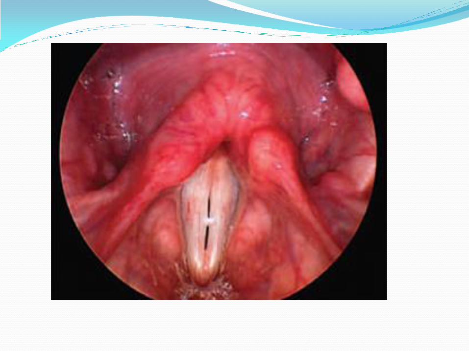
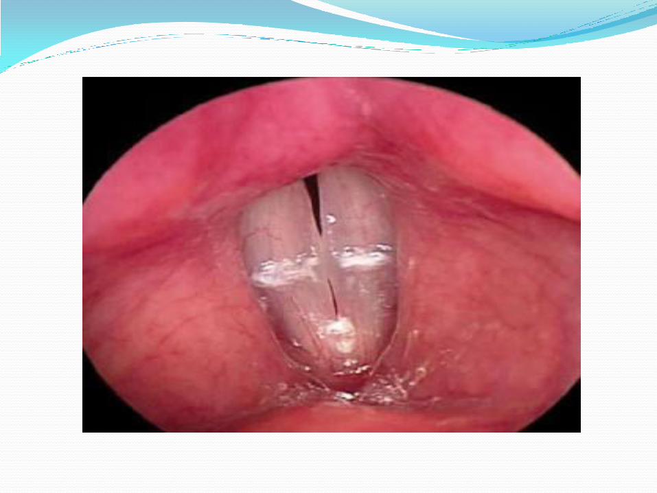
Laryngeal appearances
There are three types of chronic simple laryngitis:
1. Hyperaemic in which the cords are injected or dull pink and stiffened in appearance. There' may be some loss of adduction due to myositis . Flecks of viscid mucus may lie on the cords and in the interarytenoid space. In severe cases the cords are deep red in colour and appear rounded and ‘sausage- like’
2. Hypertrophic in which there is thickening of the tissues ,of the cords and also of the ventricular bands, arytenoids, interarytenoid space, and some of the subglottic region.
3. Oedematous in which the true cords are swollen and pale. The condition may be of long standing
In all types the larynx is nearly always affected bilaterally and symmetrically

Treatment
Vocal rest
Speech therapy may be supplement it and correct faulty use
Systemic antibiotic . when swabs show a significant growth of organism
Elimination of irritating factors such as dust and tobacco smoking
Carbocisteine ( a mucolytic ) may give symptomatic benefit
Stripping of the vocal cord is performed endoscopically in resistant cases of chronic edematous laryngitis
Atrophic laryngitis (laryngitis sicca)
An uncommon type of chronic laryngitis, usually associated with atrophic rhinitis and pharyngitis.
Aetiology
Aggravating factors include dusty atmospheres, industrial fumes and chronic infection in the paranasal sinuses. It appears to be more common in women.
Clinical features
Hoarseness and irritation in the throat both of which are improved temporarily by hawking and coughing up the crusts.
Dyspnea may be caused by large obstructing crusts.
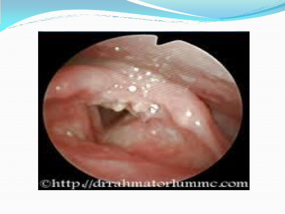
Dry and atrophic mucosa over the whole of the interior of the larynx. Crusts of varying sizes lie over the mucosa which may be excoriated when they are removed. They have a foul odour. The crusting usually extends into the trachea.


Treatment
Eradication of associated lesions in the nose and paranasal sinuses.
Change of atmospheric conditions may be beneficial.
Removal of crusts will afford some local relief.
This is aided by:
1. Inhalations of menthol, or oil of pine.
2. Carbocisteine by mouth.
3. laryngeal spray such as Benadryl or sodium bicarbonate
4. hormonal local application of hormone in oil with uncertain result

Tuberculous laryngitis
1. Acute miliary tuberculosis of the larynx
The rare laryngeal lesions are accompanied by lesions in the pharynx . tubercles appear on the swollen mucosa of the epiglottis and arytenoids these break down and form confluent grayish ulcers great pain is usually present The prognosis is serious and the treatment is that of the general infection.
2. Chronic tuberculosis of the larynx; laryngeal Phthisis
Aetiology
Infection in the larynx is almost always Secondary to a pulmonary lesions reports of primary infections are very rare and increasingly so with improved methods of diagnosis.
Most infections are Sputogenic, a few are hematogenous and very small number with lymph stream
Both sex equally affected
20 – 40 years are more usually affected
Pathology
With the common sputogenic type of infection the tubercle bacillus can infect the intact laryngeal mucosa especially that in the posterior third of the larynx infected sputum lies in the interarytenoid region . .
Progress of the disease leads to masses of granulation tissue and also to cellular swelling (a pseudo-oedema) Lesions are usually asymmetrical, except With interarytenmd granulations and the turban-like pseudo-oedema which involves the epiglottis, 'aryepiglottic folds, arytenoids, and ventricular bands.
The posterior half of the larynx is most frequently involved, but often one whole cord may be affected or the epiglottis, or the mucosa of the ventricle.
At a later stage there is perichondritis and cartilage necrosis

Clinical features
1. Weakness of the voice with periods of aphonia, is sometimes the earliest symptom
2. Hoarseness becomes evident in varying degrees. Even minima lesions can cause marked voice changes, leading to an erroneous. diagnoms of a functional condition. Complete aphonia is rare occasionally large lesions produce little hoarseness.
3. Cough is a prominent symptom.
4. Pain on swallowing is present if the laryngeal inlet is involved. It is a Common symptom in advanced lesions and it may sever
5. Referred otalgia is common.
6- Dypnea is rare unless perichondritis causes marked edema
7‘ Localized tenderness is rare unless perichonritis

8. laryngoscopic appearance Slight impairment of adduction may be an early Sign and IS Caused by a myositis .
.Marked injection of one of the vocal cord or may involve whole of the cord or the posterior part
.ulceration of the edge of the cord giving it mouse nibbled appearance
.granulations in the interarytenoid region or on the vocal process of the arytenoids cartilage this is the commonest tuberculous lesion and it is accompany by hidden indolent ulceration
. edema of the mucosa of the ventricles causing swelling which mimic prolapse
. psudoedema of the epiglottis and arytenoids ( turban larynx ) of pale susage like appearance
. subglottic infiltration and ulceration and granulation uncommon
. tuberculoma is very rare manifestation and it is small grey ( tumour like ) often associated with ulceration of base of epiglottis . vocal cord paralysis may occur at apical pulmonary disease more on the right side than the left

Diagnosis
1. chest radiography must always be taken when there is persistent laryngeal symptoms from any cause to exclude TB of the lung
2. sputum will usually contain the tubercle bacilli
3. biopsy when in doubt
Treatment
The most important of all is treating pulmonary disease
Antituberculous chemotherapy , Rifampicin , ethambutol , INAH , streptomycin , and PAS are used with dramatic response in local laryngeal lesion result
Vocal rest is still necessary
Smoking should be stopped
Nasal infection should be treated