infective endocarditis dipin.s junior resident internal medicine
TRANSCRIPT
INFECTIVE ENDOCARDITIS
Dipin.S Junior Resident
Internal medicine
Definition
• Infective endocarditis (IE) is a microbial infection of the endothelial surface of the heart or iatrogenic foreign bodies like prosthetic valves or other intracardiac devices
• Infective Endarteritis – AV shunts, Arterioarterial shunts, Coarctation of aorta
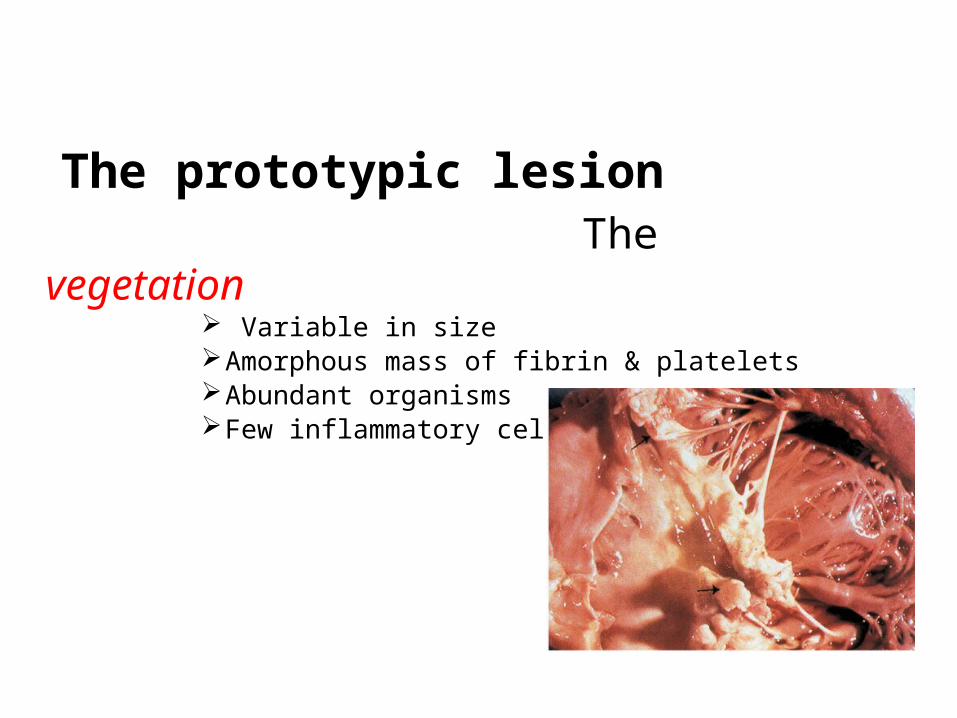
The prototypic lesion The vegetation
Variable in sizeAmorphous mass of fibrin & plateletsAbundant organismsFew inflammatory cells

Pathogenesis
Infective Endocarditis
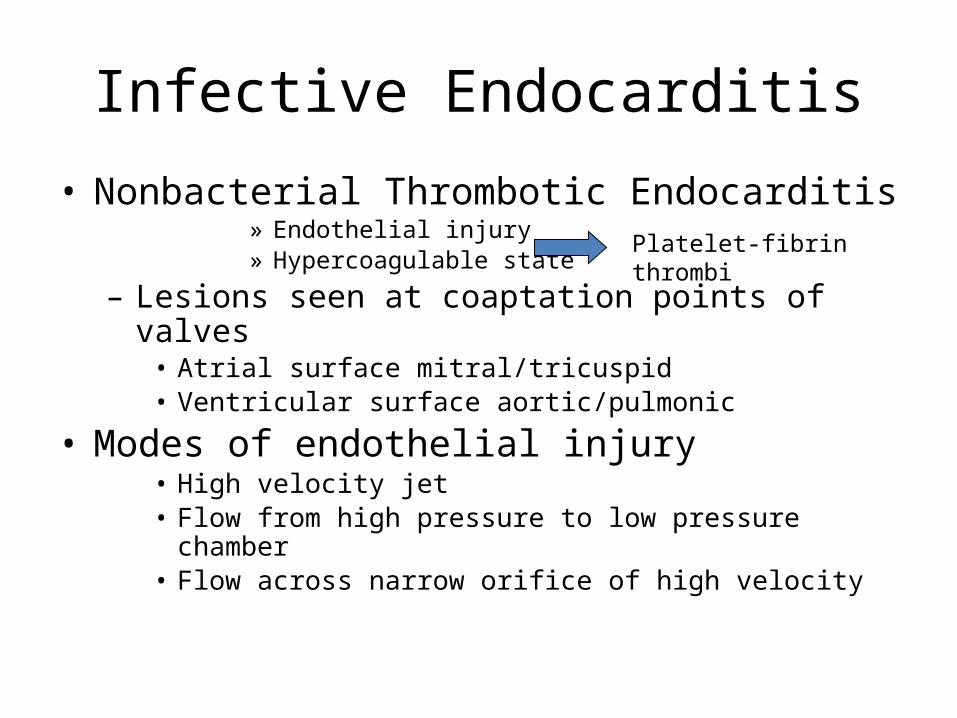
• Nonbacterial Thrombotic Endocarditis» Endothelial injury» Hypercoagulable state
– Lesions seen at coaptation points of valves• Atrial surface mitral/tricuspid• Ventricular surface aortic/pulmonic
• Modes of endothelial injury• High velocity jet• Flow from high pressure to low pressure chamber• Flow across narrow orifice of high velocity
Platelet-fibrin thrombi
Conversion of NBTE to BE
• Transient bacteremia– Traumatization of mucosal surface colonized
with bacteria (oral, GI)– Low grade, cleared in 15-30 minutes– Susceptibility to complement-mediated
bacterial killing• Leads to concept of prophylaxis
Infective Endocarditis
• Pathology– NVE infection is largely confined to leaflets– PVE infection commonly extends beyond valve
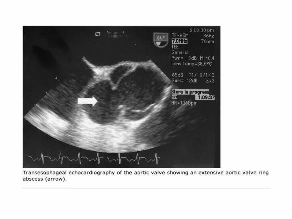
ring into annulus/periannular tissue• Ring abscesses• Septal abscesses• Fistulae• Prosthetic dehiscence
– Invasive infection more common in aortic position and if onset is early
– Bracht Wachter bodies

Epidemiology• 1.7 to 6.2 cases per 100,000 population per year
in US• The cumulative rate of prosthetic valve
endocarditis is 1.5 to 3.0% at 1 year after valve replacement.
• 3 to 6% at 5 years; the risk is greatest during the first 6 months after valve replacement.
• Men predominate in most case series, with male-to-female ratios ranging from 2:1 to 9:1

Risk Factors
• Structural heart disease– Rheumatic, congenital, aging– Prosthetic heart valves
• Injected drug use• Invasive procedures (Intracardiac pacemaker, ICD ,
AV Fistula)• Indwelling vascular devices• Other infection with bacteremia (e.g. pneumonia,
meningitis)• Immunocompromised states• History of infective endocarditis
Viridans Streptococci
• 30-65% of native valve endocarditis• Normal oral commensals• A group, composed of several species:– S. mitior, S. sanguis, S. mutans,etc.– Alpha-hemolytic, non-typable
• Typical agents of classic “SBE”
Strep. viridans

Other Streptococci
• S. bovis– Lancefield group D– Gut flora: associated with GI pathology
• S. pneumonia– 1-3% of cases of IE with predilection for AV– Usually, in those with immune suppression• DM and Alcoholism
• Group B Streptococci– Elderly with chronic disease

Enterococcus
• Normal inhabitant of GI tract.• Frequently encountered in UTIs.• Up to 40% of cases without identified
underlying predisposition to IE.
Difficult to treat due to drug resistance.

Staphylococci
• Coagulase Positive (Staph. aureus)– a major causative agent in all populations of IE– typically produces “acute” IE• fulminant, rapidly progressive with few immunologic
signs.• CNS complications in 30-50%
• Coagulase Negative (Staph. Epi)– Major cause of PVE. 3-8% of NVE.
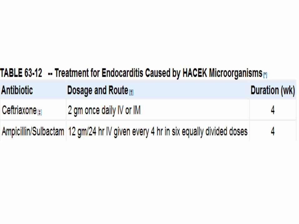
HACEK organisms
• Hemophilus, Actinobacillus, Cardiobacterium, Eikenella, Kingella
• Gram negative inhabitants of the upper airways.• Large vegetations, high likelihood of
embolization.• Slow growing: hold cultures for 3 weeks.• Traditionally sensitive to beta lactams, now some
produce beta lactamase.
Fungi• Commonly encountered agents:– Candida, Torulopsis, Aspergillus
• Predispositions– Prosthetic valves– IVDA– Immunosupression– Hyperalimentation– Prolonged antibiotic treatment
• Large vegetations and frequent embolic events.
Other Organisms
• Pseudomonas• Diphtheroids• Listeria• Bartonella• Coxiella,Legionella,salmonella,brucella• Chlamydia,Abiotropia• Bartonella ,tropheryma, streptobacillus

BCNE
• Blood culture sterile in 31 % (western data)• Sterile culture in India – 48 to 54 %• Blood culture is positive only on 67.7% of the
cases in recently published data from India
• Causes– Antibiotic therapy before blood culture– Fastidius or atypical organisms do not grow in
routine culture media– Fungal or viral Endocarditis

IV Drug Users
• Accounts for 25% of cases of IE in US.
• 5:1 male:female• Pre-existing valvular
diseases uncommon.• Variable microbiology.• Mortality<10%.
AV6%
MV24%
TV70%
Prosthetic Valve IE
• Affects 3% of prosthesis patients.– Highest risk in first 6 months post op.
• Accounts for 10-20% of all IE cases.• Increased risk in…– Males– Blacks– Multiple valve replacement
Prosthetic Valve IE
• “Early” (<2 months)-Staph epi• “Late” (after 2 months)- mimics NVE

Clinical features • High index of clinical suspicion is the cornerstone of early
diagnosis
• Symptoms– Fever, sweats, chills– Anorexia, malaise, weight loss
• Signs– Anemia (normochromic, normocytic)– Splenomegaly– Microscopic hematuria, proteinuria– New or changing heart murmur, CHF– Embolic or immunologic dermatologic signs– Hypergammaglobulinemia, elevated ESR, CRP, RF

CLINICAL MANIFESTATIONS
• Fever is the most common symptom and sign in patients with IE.
Fever may be absent or minimal in – elderly – in those with CHF,– severe debility, – chronic renal failure– NVE caused by coagulase-negative staphylococci

Cardiac murmur
• New changing regurgitant murmur is the hallmark of IE
• Murmurs are commonly not audible in – Tricuspid valve IE– Acute NVE due to S. aureus.
• Murmurs are heard in only 30 to 45 percent of patients on initial evaluation but are ultimately noted in 75 to 85 percent.
• Embolic phenomina include systemic,cerebral and pumonary emboli are common in >50 % cases.


Cardiac Pathologic Changes
• Vegetations on valve closure lines• Destruction and perforation of valve leaflet• Rupture of chordae tendinae, intraventricular
septum, papillary muscles• Valve ring abscess• Myocardial abscess• Conduction abnormalities

S. Aureus mitral valve vegetation, anterior leaflet
Pathologic Changes
• Kidney– Immune complex glomerulonephritis– Emboli with infarction, abscess
• Aortic mycotic aneurysms
Pathologic Changes
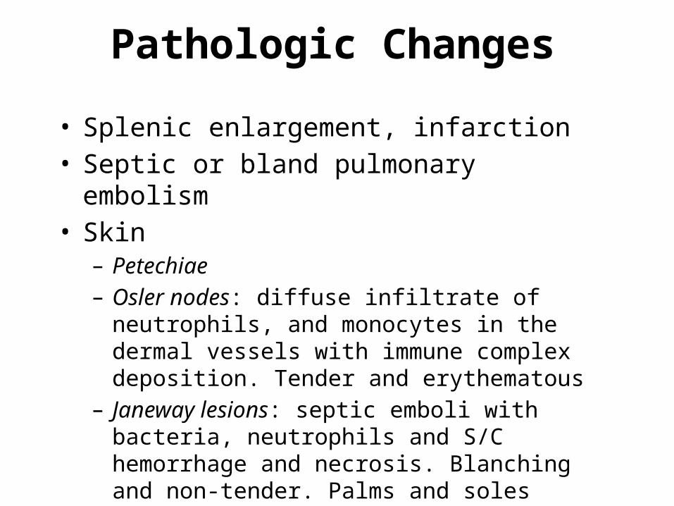
• Splenic enlargement, infarction• Septic or bland pulmonary embolism• Skin– Petechiae– Osler nodes: diffuse infiltrate of neutrophils, and
monocytes in the dermal vessels with immune complex deposition. Tender and erythematous
– Janeway lesions: septic emboli with bacteria, neutrophils and S/C hemorrhage and necrosis. Blanching and non-tender. Palms and soles

MUSCULOSKELETAL
• Vertebral osteomyelitis - rare. • Osteomyelitis -S. aureus endocarditis .• Acute septic arthritis
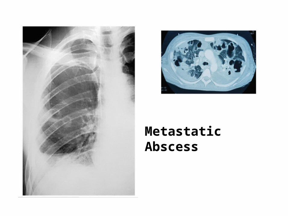
Metastatic Abscess

Splinter Hemorrhages
1. Nonspecific2. Nonblanching3. Linear reddish-brown lesions found under the nail bed4. Usually do NOT extend the entire length of the nail
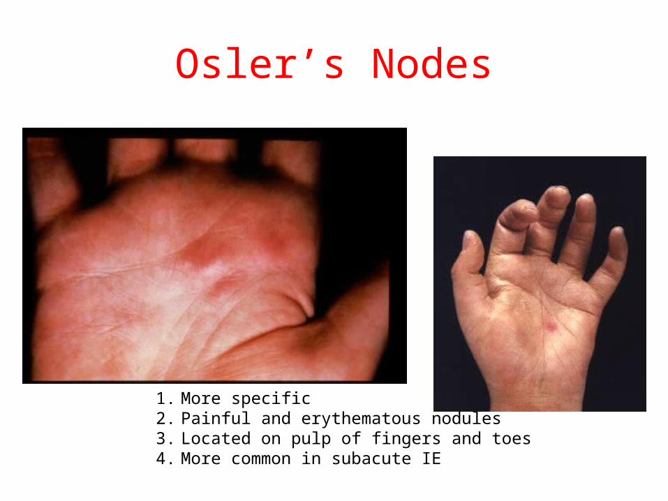
Osler’s Nodes
1. More specific2. Painful and erythematous nodules3. Located on pulp of fingers and toes4. More common in subacute IE
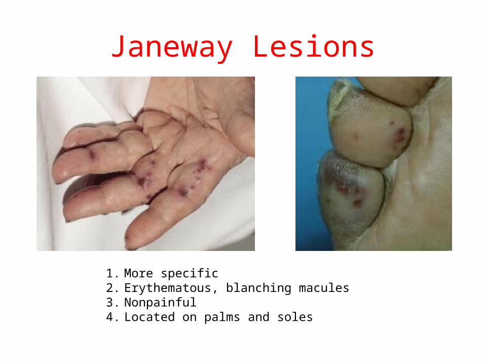
Janeway Lesions
1. More specific2. Erythematous, blanching macules 3. Nonpainful4. Located on palms and soles
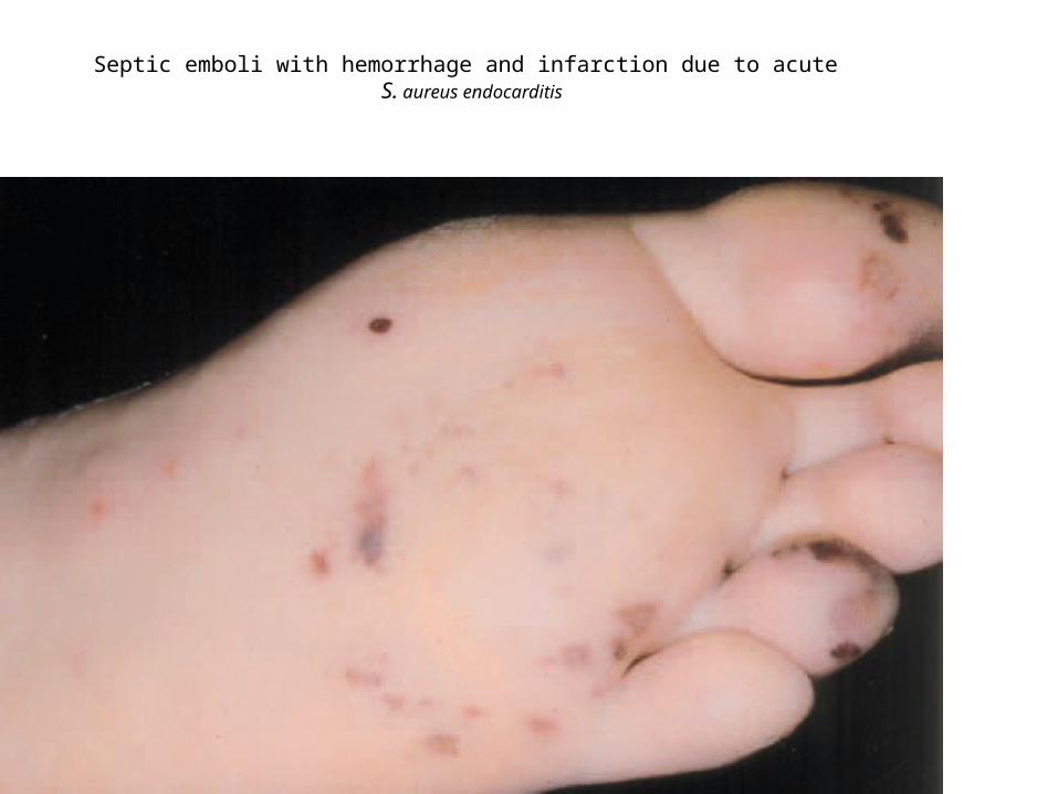
Septic emboli with hemorrhage and infarction due to acute S. aureus endocarditis

Roth spots
• Enlargement of the spleen is noted in 15 to 50 percent of patients and is more common in subacute IE of long duration.

Duke’s v/s modified dukes
• Duke’s sensitivity -76 % in western• Major deficiency is inability to diagnose BCNE• Major additions in modified dukes includes Q
fever serology, staphylococcal bacteremia in the absence of other primary focus as major criteria, serological evidence of other organism consistent with endocarditis as minor criteria.

Modified Duke Criteria

Two major criteria, OR One major and three minor criteria, ORFive minor criteria allows a clinicaldiagnosis of definite endocarditis.
Rejected IE
– If an alternative diagnosis is established, – If symptoms resolve and do not recur with
4 days of antibiotic therapy, or– If surgery or autopsy after 4 days of
antimicrobial therapy yields no histologic evidence of endocarditis.
Possible IE– Illnesses not classified as definite endocarditis or
rejected– When either one major and one minor criteria or
three minor criteria are identified.
St Thomas modifications From the Division of Infection, United Medical and Dental School,
St. Thomas’ Hospital, London, United KingdomLAMAS & EYKYN et al
• Inclusion of ESR , CRP, Presence of newly diagnosed clubbing, splenomegaly and microhematuria, as minor criteria
• Increases sensitivity by 10 %• More appropriate in Indian patients
St Thomas modifications From the Division of Infection, United Medical and Dental School,
St. Thomas’ Hospital, London, United Kingdom
• Pathologically proven yet culture negative Endocarditis– 21 % were classified definite by Original Duke’s– 32 % were definite by modified Duke’s– 62 % were definite by St Thomas modification
Blood Cultures in IE• 3 separate venepunctures during one hour
period at least 10 ml blood before giving antibiotics.
• Adherence to above practice yields positive culture in 90 %
• But at least 30 % are prescribed antibiotics before taking culture.
• Sterile culture ---western =2.5 to 31% ---Indian = 48 to 54 %

Blood Cultures
• MULTIPLE BLOOD CULTURES BEFORE EMPIRIC THERAPY
• If not critically ill– 3 blood cultures over 12-24 hour period– ? Delay therapy until diagnosis confirmed
• If critically ill– 3 blood cultures over one hour
• No more than 2 from same venepuncture• Relatively constant bacteremia
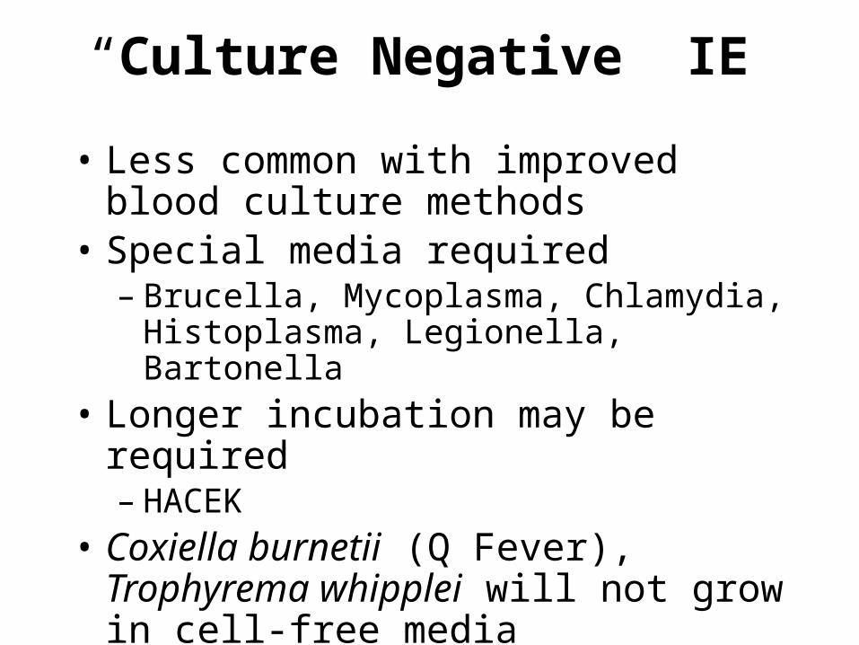
“Culture Negative” IE
• Less common with improved blood culture methods
• Special media required– Brucella, Mycoplasma, Chlamydia,
Histoplasma, Legionella, Bartonella• Longer incubation may be required– HACEK
• Coxiella burnetii (Q Fever), Trophyrema whipplei will not grow in cell-free media
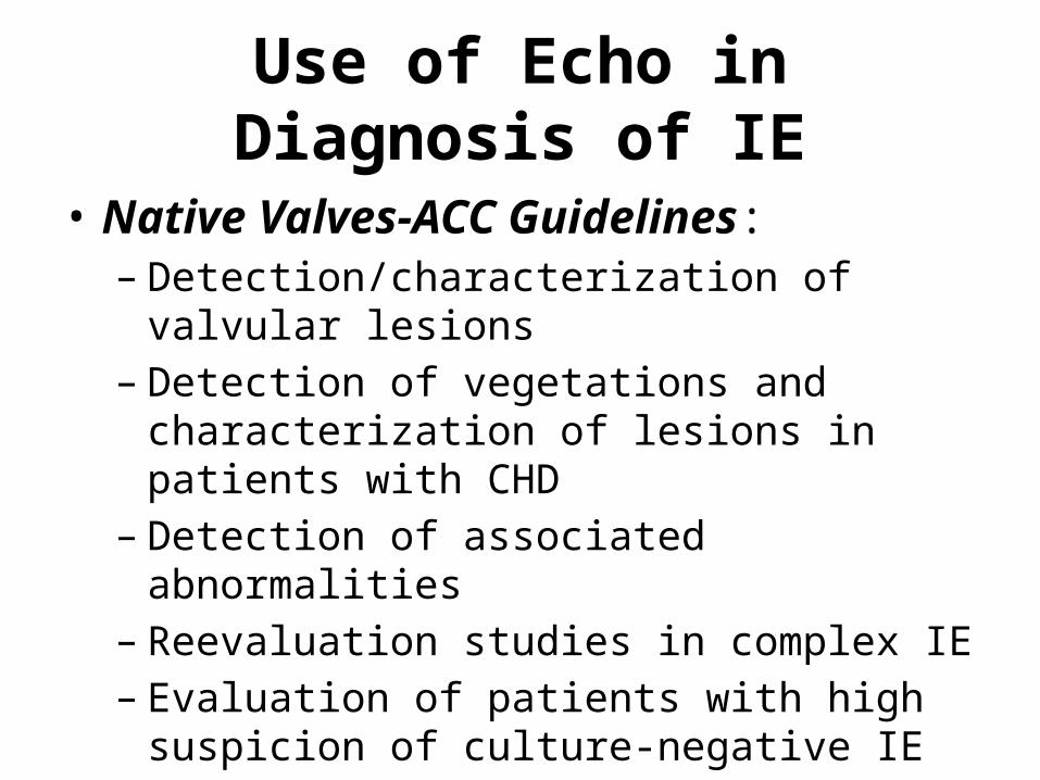
Use of Echo in Diagnosis of IE
• Native Valves-ACC Guidelines:– Detection/characterization of valvular lesions– Detection of vegetations and characterization of
lesions in patients with CHD– Detection of associated abnormalities– Reevaluation studies in complex IE– Evaluation of patients with high suspicion of
culture-negative IE
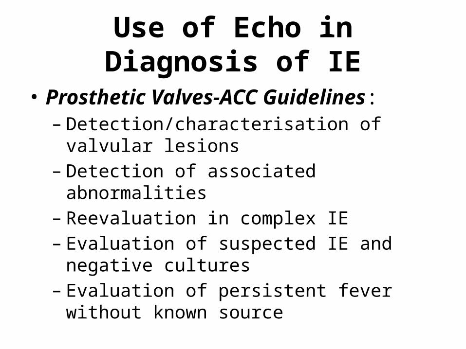
Use of Echo in Diagnosis of IE
• Prosthetic Valves-ACC Guidelines:– Detection/characterisation of valvular lesions– Detection of associated abnormalities– Reevaluation in complex IE– Evaluation of suspected IE and negative cultures– Evaluation of persistent fever without known
source

Typical echo features
• Oscillating intracardiac mass on a valve or supporting structure or device or in the path of a regurgitant stream
• Abscess• New partial dehiscence of prosthetic valve• New valvular regurgitation
• Echo is useful in predicting complications based on the size of the vegetation, mobility , extent,& consistency, either embolisation or destruction.
• Vegetations greater than 10 mm often embolise

TTE v/s TEE
• TTE – Initial echo. Sensitive in VSD and aortic valve repair. vegetation above AV or suture site
• TEE- - Can detect structure upto 1 mm– Pulmonic and prosthetic valve lesion aare better
visualised– Sensitivity 81- 100%– Specificity – 91 to 100%
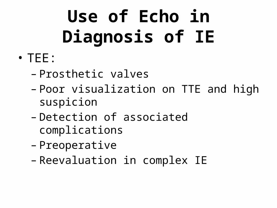
Use of Echo in Diagnosis of IE
• TEE:– Prosthetic valves– Poor visualization on TTE and high suspicion– Detection of associated complications– Preoperative– Reevaluation in complex IE
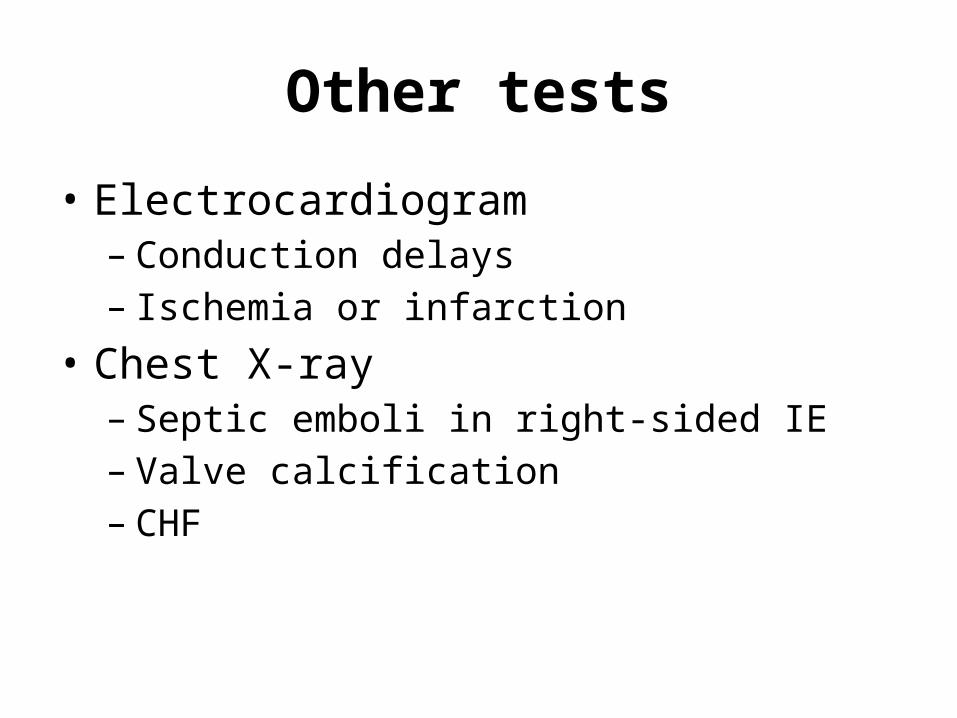
Other tests
• Electrocardiogram– Conduction delays– Ischemia or infarction
• Chest X-ray– Septic emboli in right-sided IE– Valve calcification– CHF
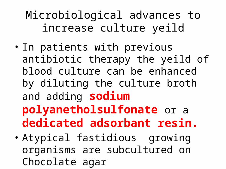
Microbiological advances to increase culture yeild
• In patients with previous antibiotic therapy the yeild of blood culture can be enhanced by diluting the culture broth and adding sodium polyanetholsulfonate or a dedicated adsorbant resin.
• Atypical fastidious growing organisms are subcultured on Chocolate agar
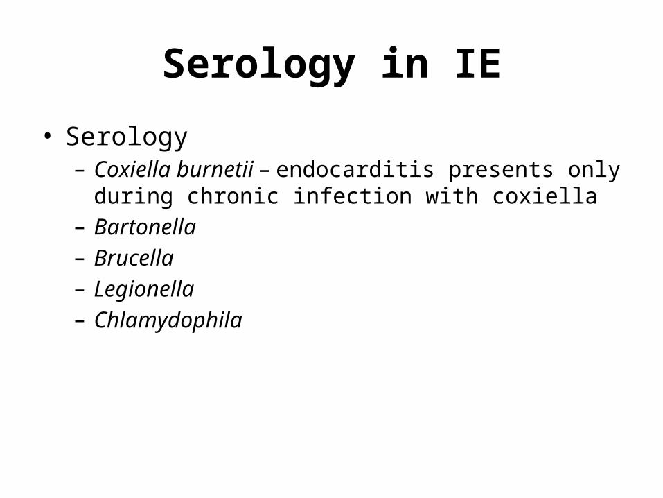
Serology in IE
• Serology– Coxiella burnetii – endocarditis presents only during
chronic infection with coxiella– Bartonella– Brucella– Legionella– Chlamydophila

Molecular diagnostic techniques in IE
• PCR• RT PCR• Proteomics ( protein signatures of the
organism used to identify the pathogens)
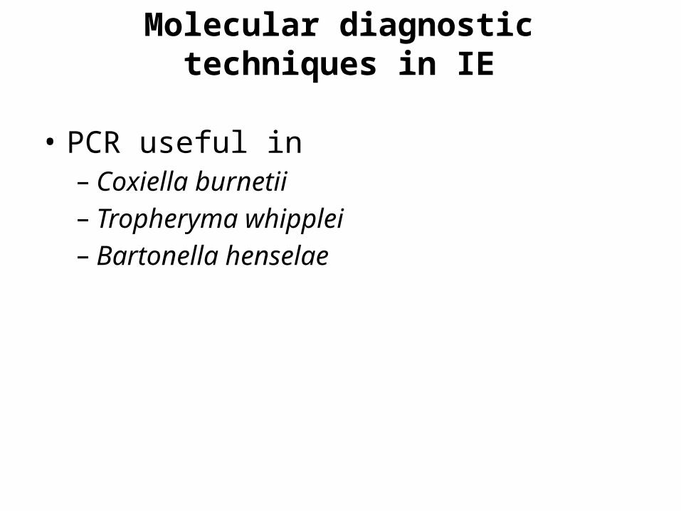
Molecular diagnostic techniques in IE
• PCR useful in– Coxiella burnetii– Tropheryma whipplei– Bartonella henselae

Procalcitonin
• 116 amino acid peptide• No known hormonal activity• Under normal metabolic conditions, PCT is only
present in the C cell of the thyroid gland• In sepsis it is released to circulation• Values exceeding 2.3 ng/ml in a suspected case
of IE has a sensitivity of 81 %& specificity of 85 %.
• More valuable than CRP in Sepsis
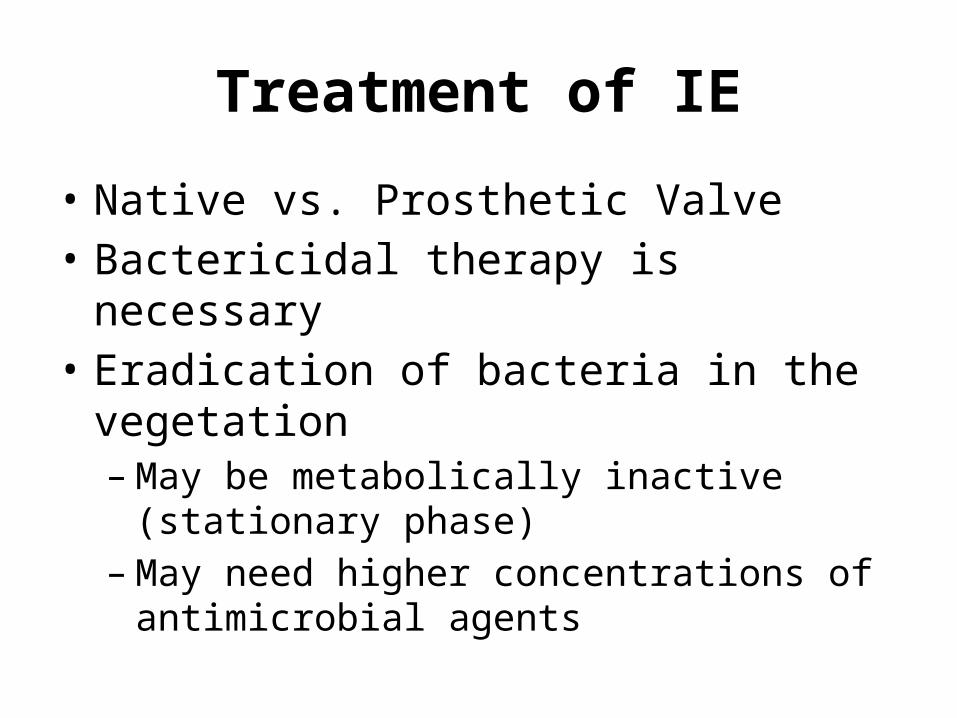
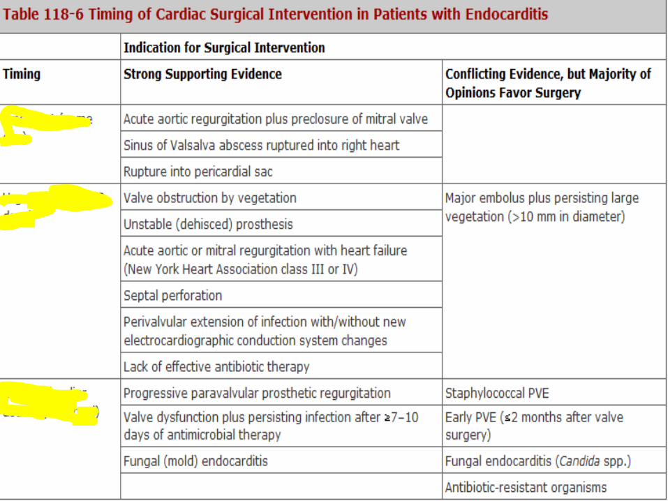
Treatment of IE
• Native vs. Prosthetic Valve• Bactericidal therapy is necessary• Eradication of bacteria in the vegetation– May be metabolically inactive (stationary phase)– May need higher concentrations of antimicrobial
agents
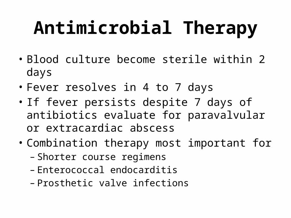
Antimicrobial Therapy
• Blood culture become sterile within 2 days• Fever resolves in 4 to 7 days• If fever persists despite 7 days of antibiotics
evaluate for paravalvular or extracardiac abscess
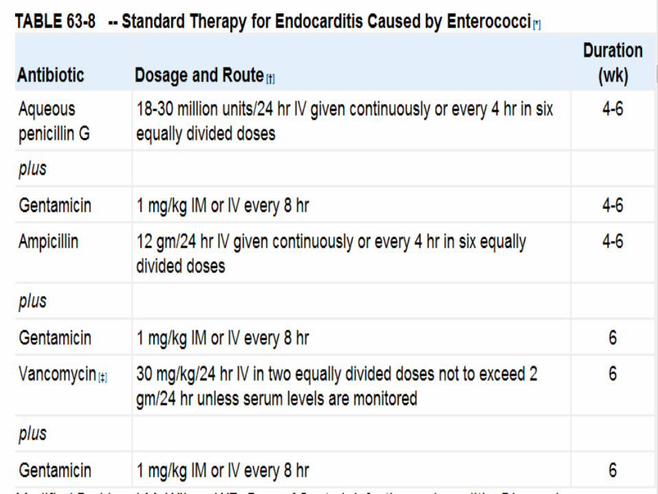
• Combination therapy most important for– Shorter course regimens– Enterococcal endocarditis– Prosthetic valve infections
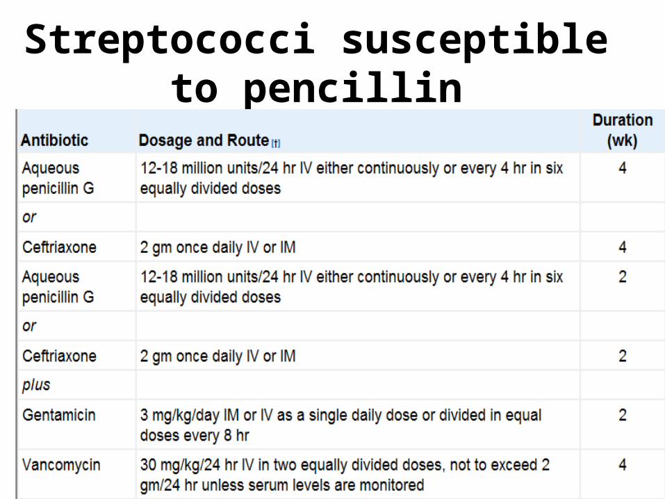
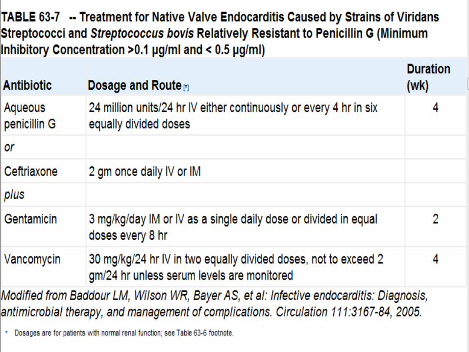
Streptococci susceptible to pencillin


NVE
• Fungal– Amphotericin– Fluconazole– Caspofungin, little data– Surgery usually necessary 1-2 weeks into
treatment
Prosthetic Valve IE
• Staphylococci most common– Coagulase negative staphylococci
• Enterococcus• Nutritonally variant streptococci• Fungi
OTHER ORGANISMS
Streptococcus pneumoniae• Penicillin if sensitive- can be treated with
intravenous penicillin• ceftriaxone (2 g/d as a single dose), or
cefotaxime (at a comparable dosage).
• Infection caused by penicillin resistant strains hould be treated with vancomycin.
• P. aeruginosa endocarditis is treated with an antipseudomonal penicillin (ticarcillin or piperacillin) and high doses of tobramycin (8 mg/kg per day in three divided doses).
• Endocarditis caused by Enterobacteriaceae is treated with a potent beta-lactam antibiotic plus an aminoglycoside.

• Corynebacterial endocarditis is treated with penicillin plus an aminoglycoside (if the organism is susceptible to the aminoglycoside) or with vancomycin
• Therapy for Candida endocarditis consists of amphotericin B plus flucytosine and early surgery; long-term suppression with fluconazole is also used.
Empirical Therapy
• Therapy without culture data (i.e., before culture results are known or when cultures are negative).
• For acute endocarditis in an injection drug user should cover methicillin-resistant S. aureus and gram-negative bacilli.
• The initiation of treatment with vancomycin plus gentamicin immediately after blood is obtained for cultures covers these as well as many other potential causes.

• In culture-negative -marantic endocarditis must be excluded and fastidious organisms sought serologically.
• In the absence of prior antibiotic therapy, it is unlikely to be due to S. aureus, coagulase-negative staphylococcal, or enterococcal.
• Blood culture–negative subacute native valve endocarditis is treated with ceftriaxone plus gentamicin.
• These two antimicrobials plus vancomycin should be used if prosthetic valves are involved.

• Serologic abnormalities (e.g., erythrocyte sedimentation rate, rheumatoid factor) resolve slowly and do not reflect response to treatment.
• Vegetations become smaller with effective therapy, but at 3 months after cure half are unchanged and 25% are slightly larger.

Prophylactic Therapy -- Current Scenario

1997 American Heart Assoc. Guidelines:Endocarditis Prophylaxis Recommended: High-risk category
Prosthetic cardiac valves, including bioprosthetic and homograft valves Previous bacterial endocarditis Complex cyanotic congenital heart disease (eg, single ventricle states, transposition of the great
arteries, tetralogy of Fallot) Surgically constructed systemic pulmonary shunts or conduits
Moderate-risk category Most other congenital cardiac malformations (other than above and below) Acquired valvar dysfunction (eg, rheumatic heart disease) Hypertrophic cardiomyopathy Mitral valve prolapse with valvar regurgitation and/or thickened leaflets
Endocarditis Prophylaxis Not Recommended: Negligible-risk category (no greater risk than the general population)
Isolated secundum atrial septal defect Surgical repair of atrial septal defect, ventricular septal defect, or patent ductus arteriosus (without residua beyond 6 mo) Previous coronary artery bypass graft surgery Mitral valve prolapse without valvar regurgitation1 Physiologic, functional, or innocent heart murmurs1 Previous Kawasaki disease without valvar dysfunction Previous rheumatic fever without valvar dysfunction Cardiac pacemakers (intravascular and epicardial) and implanted defibrillators
1997 AHA Guidelines
• Assumptions:– Bacteremia with organisms known to cause IE occurs in
assoc. with invasive dental/GI/GU procedures– Antibiotic prophylaxis was proven effective in animals– Antibiotic prophylaxis thought to be effective in human

Reasons for 2007 Revision
• IE more likely due to frequent exposure to random bacteremias from daily activities than from bacteremia during dental/GI/GU procedure
• Prophylaxis may prevent only small number of cases of IE, even if 100% effective
• Risk of antibiotic-assoc. adverse events exceeds the benefit, if any, from prophylaxis
• To reduce the risk of bacteremia from dental procedure: maintaining good oral health and hygiene is more important than Antibiotic prophylaxis

Frequency of Transient Bacteremia
• Tooth extraction 10-100%• Periodontal surgery 36-88%• Teeth cleaning 40%
• Tooth brushing, 20-68%• Using wooden toothpicks 20-40%• Chewing food 7-51%

Risk of IE from dental procedures?
• No prospective, randomized, placebo-controlled studies exist on efficacy of Antibiotic prophylaxis in preventing IE after dental procedure

2007: Who gets prophylaxis?
Only patients with the highest risk of adverse outcomes (heart failure, surgery, death) from endocarditis:
1. Prosthetic cardiac valve2. Previous IE3. Cardiac transplant recipients who develop
cardiac valvulopathy4. Congenital Heart Disease
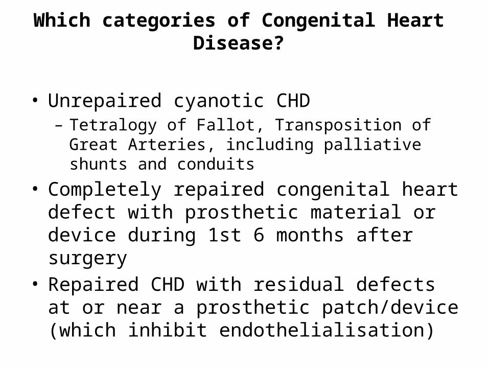
Which categories of Congenital Heart Disease?
• Unrepaired cyanotic CHD – Tetralogy of Fallot, Transposition of Great Arteries,
including palliative shunts and conduits
• Completely repaired congenital heart defect with prosthetic material or device during 1st 6 months after surgery
• Repaired CHD with residual defects at or near a prosthetic patch/device (which inhibit endothelialisation)
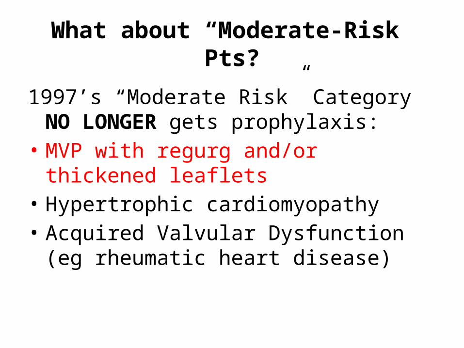
What about “Moderate-Risk” Pts?
1997’s “Moderate Risk” Category NO LONGER gets prophylaxis:
• MVP with regurg and/or thickened leaflets• Hypertrophic cardiomyopathy• Acquired Valvular Dysfunction (eg rheumatic
heart disease)

Dental Procedures
• “If it bleeds, give prophylaxis” • High-risk pts undergoing all dental procedures that
involve manipulation of gingival tissues OR periapical region of teeth OR perforation of oral mucosa– i.e. biopsies, suture removal, placing orthodontic bands
• NO PROPHYLAXIS:– Xray, anesthetic injections, fluoride treatments– Shedding of deciduous teeth– Placement/adjustment of removable prosthodontic or
orthodontic appliances

Prophylaxis for Dental Procedures• Goal: cover Strep Viridans• Single dose, 30-60 min prior to procedure
What about resistant Strep Viridans?
• Quinolones or IV Vancomycin not recommended for prophylaxis due to concern of creating new drug resistance
Respiratory Tract Procedures
• No published data linking resp tract procedures and IE....
• Consider prophylaxis for High-risk pts undergoing Invasive Procedure in resp tract with incision or biopsy of resp mucosa:
• Tonsillectomy• Adenoidectomy• Bronchoscopy WITH biopsy (not for BAL alone)• Resp tract procedure to drain abscess or empyema

GI/GU Procedures
• No published data linking GI/GU procedures and IE....
• NO prophylaxis for GI/GU procedures

Procedures on Infected Skin/Skin Structure, or Msk Tissue
In patients who are HIGH-risk for IE:• The antibiotic regimen given to treat the skin
or musculoskeletal infection should contain an Anti-staphylococcal Pencillin or cephalosporin
• If unable to take PO or Pencillin-allergic: Clindamycin or Vancomycin
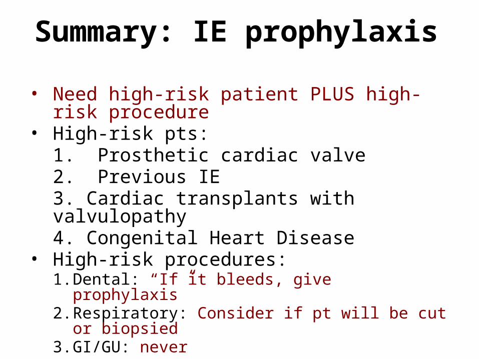
Summary: IE prophylaxis
• Need high-risk patient PLUS high-risk procedure• High-risk pts:
1. Prosthetic cardiac valve2. Previous IE3. Cardiac transplants with valvulopathy4. Congenital Heart Disease
• High-risk procedures:1. Dental: “If it bleeds, give prophylaxis”2. Respiratory: Consider if pt will be cut or biopsied3. GI/GU: never
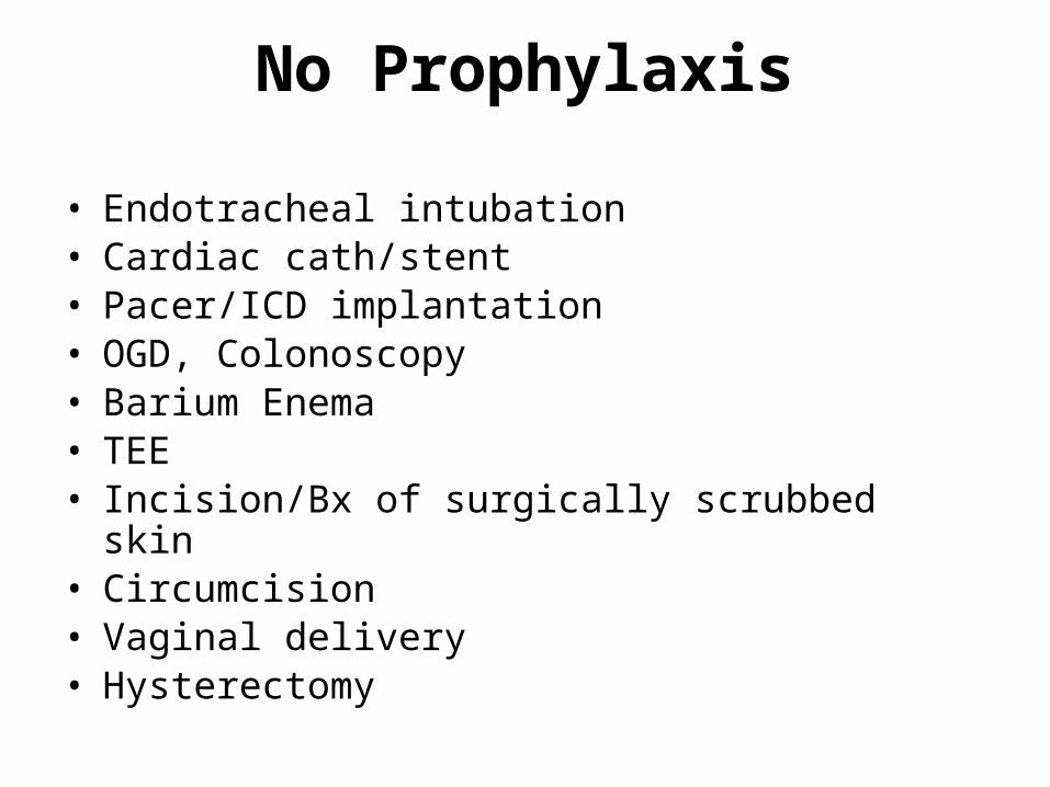
No Prophylaxis
• Endotracheal intubation• Cardiac cath/stent• Pacer/ICD implantation• OGD, Colonoscopy• Barium Enema• TEE• Incision/Bx of surgically scrubbed skin• Circumcision• Vaginal delivery• Hysterectomy
