infections of the central - download.e-bookshelf.de · kum thong wong, mbbs, mpath , frcpath , md...
TRANSCRIPT


Infections of the Central Nervous System


Infections of the Central Nervous System: Pathology and GeneticsEDITORS
FABRICE CHRÉTIEN, MD, PhDDepartment of Neuropathology, Sainte Anne Hospital, Paris;Paris University, Paris;Experimental Neuropathology Unit, Pasteur Institute, Paris, France
KUM THONG WONG, MBBS, MPath , FRCPath , MDDepartment of Pathology, Faculty of Medicine, University of Malaya, Kuala Lumpur, Malaysia
LEROY R . SHARER , MDDivision of Neuropathology, Department of Pathology, Immunology, and Laboratory Medicine, Rutgers New Jersey Medical School and University Hospital, Newark, NJ, USA
SERIES EDITORS
CATHERINE (KAT Y) KEOHANE, MB, B CH, BAO, FRCPath , FFPathDepartment of Pathology and School of Medicine, University College Cork, Brookfield Health Science Complex, Cork, Ireland
FR ANÇ OISE GR AY, MD, PhDRetired from Department of Pathology, Lariboisière Hospital, APHP, Paris;University Paris Diderot (Paris 7), Paris, France

This edition first published 2020© 2020 John Wiley & Sons Ltd
All rights reserved. No part of this publication may be reproduced, stored in a retrieval system, or transmitted, in any form or by any means, electronic, mechanical, photocopying, recording or otherwise, except as permitted by law. Advice on how to obtain permission to reuse material from this title is available at http://www.wiley.com/go/permissions.
The right of Fabrice Chrétien, Kum Thong Wong, Leroy R. Sharer, Catherine (Katy) Keohane and Françoise Gray to be identified as the author(s) of the editorial material in this work has been asserted in accordance with law.
Registered Office(s)John Wiley & Sons, Inc., 111 River Street, Hoboken, NJ 07030, USAJohn Wiley & Sons Ltd, The Atrium, Southern Gate, Chichester, West Sussex, PO19 8SQ, UK
Editorial Office9600 Garsington Road, Oxford, OX4 2DQ, UK
For details of our global editorial offices, customer services, and more information about Wiley products visit us at www.wiley.com.
Wiley also publishes its books in a variety of electronic formats and by print‐on‐demand. Some content that appears in standard print versions of this book may not be available in other formats.
Limit of Liability/Disclaimer of WarrantyThe contents of this work are intended to further general scientific research, understanding, and discussion only and are not intended and should not be relied upon as recommending or promoting scientific method, diagnosis, or treatment by physicians for any particular patient. In view of ongoing research, equipment modifications, changes in governmental regulations, and the constant flow of information relating to the use of medicines, equipment, and devices, the reader is urged to review and evaluate the information provided in the package insert or instructions for each medicine, equipment, or device for, among other things, any changes in the instructions or indication of usage and for added warnings and precautions. While the publisher and authors have used their best efforts in preparing this work, they make no representations or warranties with respect to the accuracy or completeness of the contents of this work and specifically disclaim all warranties, including without limitation any implied warranties of merchantability or fitness for a particular purpose. No warranty may be created or extended by sales representatives, written sales materials or promotional statements for this work. The fact that an organization, website, or product is referred to in this work as a citation and/or potential source of further information does not mean that the publisher and authors endorse the information or services the organization, website, or product may provide or recommendations it may make. This work is sold with the understanding that the publisher is not engaged in rendering professional services. The advice and strategies contained herein may not be suitable for your situation. You should consult with a specialist where appropriate. Further, readers should be aware that websites listed in this work may have changed or disappeared between when this work was written and when it is read. Neither the publisher nor authors shall be liable for any loss of profit or any other commercial damages, including but not limited to special, incidental, consequential, or other damages.
Library of Congress Cataloging‐in‐Publication Data
Names: Chret́ien, Fabrice, editor. | Wong, Kum Thong, editor. | Sharer, Leroy R., editor. | Keohane, Catherine, editor. | Gray, Franco̧ise, editor. | International Society of Neuropathology, issuing body.
Title: Infections of the central nervous system : pathology and genetics / edited by Fabrice Chret́ien, Kum Thong Wong, Leroy R. Sharer, Catherine (Katy) Keohane, Franco̧ise Gray.
Other titles: Infections of the central nervous system (Chre ́tien)Description: Hoboken, NJ : Wiley-Blackwell, 2020. | Includes bibliographical references and index. Identifiers: LCCN 2019033002 (print) | LCCN 2019033003 (ebook) | ISBN 9781119467762 (hardback) |
ISBN 9781119467700 (adobe pdf) | ISBN 9781119467793 (epub)Subjects: MESH: Central Nervous System Infections–pathology | Central Nervous System Infections–genetics Classification: LCC RC359.5 (print) | LCC RC359.5 (ebook) | NLM WL 301 | DDC 616.8–dc23 LC record available at https://lccn.loc.gov/2019033002LC ebook record available at https://lccn.loc.gov/2019033003
Cover Design: WileyCover Images: Logo used with permission of Pasteur Institute, Main image, 1st and 6th images on right courtesy of Françoise Gray; 2nd image on right courtesy of Michael Farrell; 3rd and 4th images courtesy of Catherine (Katy) Keohane; 5th and 7th images courtesy of Gregory Jouvion
Set in 9.5/12pt Minion by SPi Global, Pondicherry, India
10 9 8 7 6 5 4 3 2 1

v
List of Contributors, ix
1 Introduction and Classification of Infections of the CNS According to the Agent, 1Fabrice Chrétien, Kum Thong Wong, Leroy R. Sharer, Catherine (Katy) Keohane, and Françoise Gray
2 Sepsis‐Associated Encephalopathy, 11Franck Verdonk, Aurelien Mazeraud, Fabrice Chrétien, and Tarek Sharshar
3 Variation of CNS Infections According to the Host, 21Leroy R. Sharer, Catherine (Katy) Keohane, and Françoise Gray
4 Clinical Approach to the Adult Patient with CNS Infection, 29Michel Wolff, Romain Sonneville, and Tarek Sharshar
5 Herpes Simplex Virus Infections of the CNS, 43Bette K. Kleinschmidt‐DeMasters, Catherine (Katy) Keohane, and Françoise Gray
6 Varicella‐Zoster Virus and Epstein‐Barr Virus Infections of the CNS, 55Catherine (Katy) Keohane and Françoise Gray
7 Cytomegalovirus Infections of the CNS, 65Homa Adle‐Biassette and Natacha Teissier
8 Adenovirus Meningoencephalitis, 77Harry V. Vinters and Xinhai R. Zhang
9 Polyomavirus Infections of the CNS, 83Susan Morgello
10 Measles Virus Infection of the CNS, 95Catherine (Katy) Keohane, Leroy R. Sharer, and Françoise Gray
11 Rubella Virus, 105Bette K. Kleinschmidt‐DeMasters
12 Henipavirus Encephalitis, 113Kum Thong Wong and Kien Chai Ong
13 Rabies, 121Guilherme Dias de Melo, Perrine Parize, Grégory Jouvion, Laurent Dacheux, Fabrice Chrétien, and Hervé Bourhy
14 Flaviviruses 1: General Introduction and Tick‐Borne Encephalitis, 131Herbert Budka
15 Flaviviruses 2: West Nile, St. Louis Encephalitis, Murray Valley Encephalitis, Yellow Fever, and Dengue, 147Edward S. Johnson and Juan M. Bilbao
16 Flaviviruses 3: Zika Virus Infection of the CNS, 163Leila Chimelli
17 Flaviviruses 4: Japanese Encephalitis, 169Shankar Krishna Susarla, Anita Mahadevan, Bishan Radotra, Masaki Takao, and Kum Thong Wong
Contents

vi
Contents
18 CNS Disorders Caused by Hepatitis C and Hepatitis E Viruses, 177Melissa Umphlett, Clare Bryce, and Susan Morgello
19 Alphaviral Equine Encephalomyelitis (Eastern, Western, and Venezuelan), 183Kum Thong Wong
20 Chikungunya Virus, 189Cássia Shinotsuka, Michael Blatzer, Grégory Jouvion, and Fabrice Chrétien
21 Poliovirus Infection and Postpolio Syndrome, 195Catherine (Katy) Keohane, Leila Chimelli, and Aisling Ryan
22 Enterovirus A71 Infection, 205Kum Thong Wong, Kien Chai Ong, Thérèse Couderc, and Marc Lecuit
23 Human Immunodeficiency Virus Infection of the CNS, 215Françoise Gray and Leroy R. Sharer
24 HTLV‐1 and Neurological‐Associated Disease, 231Antoine Gessain, Olivier Cassar, and Philippe V. Afonso
25 Parechovirus A, 243Clayton A. Wiley
26 Acute Disseminated Encephalomyelitis and Acute Hemorrhagic Leukoencephalomyelitis, 251Romana Höftberger and Hans Lassmann
27 Miscellaneous Inflammatory Disorders of the CNS of Possible Infectious Origin, 259Mari Perez‐Rosendahl, Jamie Nakagiri, Xinhai R. Zhang, and Harry V. Vinters
28 Mycoplasmal and Rickettsial Infections of the CNS, 267Roy H. Rhodes
29 Pathogenesis and Pathophysiology of Bacterial Infections of the CNS, 279Loic Le Guennec and Sandrine Bourdoulous
30 Pyogenic Infections of the CNS 1: Acute Bacterial Meningitis, 295Loic Le Guennec and Sandrine Bourdoulous
31 Pyogenic Infections of the CNS 2: (Brain Abscess, Subdural Abscess or Empyema, Epidural Abscess, Septic Embolism, and Suppurative Intracranial Phlebitis), 309Arnault Tauziede‐Espariat, Alexandre Roux, Megan Still, Marc Zanello, Gilles Zah‐Bi, Ghazi Hmeydia, Catherine Oppenheim, Michel Wolff, Johan Pallud, and Fabrice Chrétien
32 Pyogenic Infections of the CNS 3: Following Neurosurgical Procedures, 319Alexandre Roux, Megan Still, Marc Zanello, Gilles Zah‐Bi, Arnault Tauziede‐Espariat, Ghazi Hmeydia, Catherine Oppenheim, Michel Wolff, Fabrice Chrétien, and Johan Pallud
33 CNS Involvement in Tropheryma whipplei Infection, 331Emmanuèle Lechapt‐Zalcman
34 Cerebral Actinomycosis, 337Arnault Tauziede‐Espariat
35 Cerebral Nocardiosis, 343Arnault Tauziede‐Espariat and Leroy R. Sharer
36 CNS Tuberculosis, 349Michael A. Farrell, Eoin R. Feeney, and Jane B. Cryan
37 Non‐Tuberculous Mycobacterial Infections, 357Leroy R. Sharer
38 Spirochetal Infections of the CNS, 363Françoise Gray and Catherine (Katy) Keohane
39 Neurobrucellosis, 379Marine Le Dudal, Fabrice Chrétien, and Grégory Jouvion
40 Legionellosis, 383Edward S. Johnson
41 Neurosarcoidosis, 393Michael A. Farrell and Alan Beausang
42 Hypertrophic Pachymeningitis, 403Françoise Gray and Leroy R. Sharer
43 Toxin‐Induced Neurological Diseases, 411Pierre L. Goossens, Cédric Thépenier, and Michel R. Popoff

vii
Contents
44 Fungal Infections of the CNS, 419Michael Blatzer, Fanny Lanternier, Jean‐Paul Latgé, Anne Beauvais, Stéphane Bretagne, Fabrice Chrétien, and Grégory Jouvion
45 Cerebral Malaria, 437Patrícia Reis, Vanessa Estato, and Hugo Caire de Castro Faria Neto
46 Toxoplasma Infection of the CNS, 449Stéphane Bretagne, Catherine (Katy) Keohane, and Homa Adle‐Biassette
47 Other Protozoal Infections, 463Leila Chimelli and Catherine (Katy) Keohane
48 Helminth Infections of the CNS, 475Marine Le Dudal, Stéphane Bretagne, David Hardy, Fabrice Chrétien, and Grégory Jouvion
Appendix: CASE EXAMPLE: Schistosoma mekongi Neuroschistosomiasis, 500Edward S. Johnson
49 Brain Myiasis, 503Arnault Tauziede‐Espariat
50 Emerging CNS Infections, 505Kum Thong Wong
Index, 515

ix
Homa Adle‐BiassetteDepartment of PathologyLariboisière HospitalAPHP, Paris;
Paris University, NeuroDiderot INSERM F-75019 Paris, France
Philippe V. AfonsoOncogenic Virus Epidemiology and Pathophysiology Department of Virology Pasteur Institute Paris, France
Alan BeausangDepartment of Neuropathology Beaumont Hospital Dublin, Ireland
Anne BeauvaisAspergillus Unit Pasteur Institute Paris, France
Juan M. BilbaoDepartment of Pathology University of Toronto Toronto, Ontario, Canada
Michael BlatzerAspergillus and Experimental Neuropathology Units Pasteur Institute Paris, France
Sandrine BourdoulousInserm, U1016, Institut Cochin, CNRS UMR8104, Paris University Paris, France
Hervé BourhyLyssavirus Epidemiology and Neuropathology Unit French National Reference Centre for Rabies WHO Collaborating Centre for Reference and Research on Rabies Pasteur Institute Paris, France
Stéphane BretagneSaint Louis Hospital APHP, Paris;
University Paris Diderot Sorbonne Paris Cité, Paris;
Molecular Mycology Unit French National Reference Centre for Invasive Mycoses and Antifungal Treatments Pasteur Institute Paris, France
List of Contributors

x
List of Contributors
Clare BryceDepartment of Pathology, Neurology, and Neuroscience Icahn School of Medicine Mount Sinai, New York NY, USA
Herbert BudkaInstitute of Neurology Medical University Vienna Vienna, Austria
Olivier CassarOncogenic Virus Epidemiology and Pathophysiology Department of Virology Pasteur Institute, Paris;
CNRS, UMR3569 Paris, France
Hugo Caire de Castro Faria NetoLaboratory of Immunopharmacology Oswaldo Cruz Institute, FIOCRUZ Rio de Janeiro, Brazil
Leila ChimelliLaboratory of Neuropathology and Molecular Genetics State Brain Institute Paulo Niemeyer Rio de Janeiro, Brazil;
Federal University of Rio de Janeiro (UFRJ), Rio de Janeiro, Brazil
Fabrice ChrétienDepartment of Neuropathology Sainte Anne Hospital Paris;
Paris University Paris;
Experimental Neuropathology Unit Pasteur Institute Paris, France
Thérèse CoudercBiology of Infection Unit Pasteur Institute Paris;
Inserm U1117 Paris, France
Jane B. CryanDepartment of Neuropathology Beaumont Hospital Dublin, Ireland
Laurent DacheuxLyssavirus Epidemiology and Neuropathology Unit French National Reference Centre for Rabies WHO Collaborating Centre for Reference and Research on Rabies Pasteur Institute Paris, France
Marine Le DudalExperimental Neuropathology Unit Pasteur Institute Paris;
Embryology, Histology and Pathology Unit The National Veterinary School of Alfort University Paris-Est Maison-Alfort, France
Vanessa EstatoLaboratory of Immunopharmacology Oswaldo Cruz Institute, FIOCRUZ Rio de Janeiro, Brazil
Michael A. FarrellDepartment of Neuropathology Beaumont Hospital Dublin, Ireland
Eoin R. FeeneyDepartment of Infectious Diseases St. Vincent’s Hospital Dublin, Ireland
Antoine GessainOncogenic Virus Epidemiology and Pathophysiology Department of Virology, Pasteur Institute Paris;
CNRS, UMR3569 Paris, France

xi
List of Contributors
Pierre L. GoossensExperimental Neuropathology Unit Pasteur Institute Paris;
Yersinia, Pasteur Institute Paris, France
Françoise GrayRetired from Department of Pathology Lariboisière Hospital APHP, Paris;
University Paris Diderot (Paris 7) Paris, France
Loic Le GuennecInserm, U1016, Institut Cochin CNRS, UMR8104 Paris University, Paris;
La Pitie‐Salpetriere Hospital APHP, Paris, France
David HardyExperimental Neuropathology Unit Pasteur Institute Paris, France
Romana HöftbergerInstitute of Neurology Medical University of Vienna Vienna, Austria
Ghazi HmeydiaDepartment of Neuropathology Sainte‐Anne Hospital Paris, France
Edward S. JohnsonDepartment of Laboratory Medicine and Pathology University of Alberta Edmonton, Alberta, Canada
Grégory JouvionExperimental Neuropathology Unit Pasteur Institute Paris, France
Catherine (Katy) KeohaneDepartment of Pathology and School of Medicine University College Cork Brookfield Health Science Complex Cork, Ireland
Bette K. Kleinschmidt‐DeMastersDepartment of Pathology, Neurosurgery, and Neurology University of Colorado School of Medicine Aurora, CO, USA
Fanny LanternierMolecular Mycology Unit French National Reference Centre for Invasive Mycoses and Antifungal Treatments Pasteur Institute, Paris;
Paris University, Paris;
Department of Infectious Diseases and Tropical Medicine Necker-Sick Children University Hospital and Imagine Institute APHP, Paris, France
Hans LassmannCenter for Brain Research Medical University of Vienna Vienna, Austria
Jean‐Paul LatgéAspergillus Unit Pasteur Institute Paris, France
Emmanuèle Lechapt‐ZalcmanDepartment of Neuropathology Sainte Anne Hospital Paris, France
Marc LecuitBiology of Infection Unit Pasteur Institute Paris;
Inserm U1117, Paris;
Department of Infectious Diseases and Tropical Medicine

xii
List of Contributors
Necker-Sick Children University Hospital and Imagine Institute APHP, Paris;
Paris University Paris, France
Anita MahadevanDepartment of Neuropathology National Institute of Mental Health and Neurosciences (NIMHANS) Bangalore, India
Aurelien MazeraudExperimental Neuropathology Unit Pasteur Institute, Paris;
Department of Anesthesiology and Intensive Care Sainte Anne Hospital Paris;
University Paris Diderot, Sorbonne Paris Cité APHP, Paris, France
Guilherme Dias de MeloLyssavirus Epidemiology and Neuropathology Unit French National Reference Centre for Rabies WHO Collaborating Centre for Reference and Research on Rabies Pasteur Institute Paris, France
Susan MorgelloDepartment of Pathology, Neurology, and Neuroscience Icahn School of Medicine Mount Sinai, New York NY, USA
Jamie NakagiriDepartment of Pathology and Laboratory Medicine UC Irvine Medical Center Orange, CA, USA
Kien Chai OngDepartment of Biomedical Science Faculty of Medicine University of Malaya Kuala Lumpur, Malaysia
Catherine OppenheimDepartment of Neuroradiology Sainte Anne Hospital Paris;
Paris University Paris;
Inserm U894, IMA‐Brain Psychiatry and Neurosciences Center Paris, France
Johan PalludDepartment of Neurosurgery Sainte Anne Hospital Paris;
Paris University Paris;
Inserm U894, IMA‐Brain Psychiatry and Neurosciences Center Paris, France
Perrine ParizeLyssavirus Epidemiology and Neuropathology Unit French National Reference Centre for Rabies WHO Collaborating Centre for Research on Rabies Pasteur Institute Paris, France
Mari Perez‐RosendahlDepartment of Pathology and Laboratory Medicine UC Irvine Medical Center Orange, CA, USA
Michel R. PopoffBacterial Toxins Unit Pasteur Institute Paris, France
Bishan RadotraDepartment of Histopathology Postgraduate Institute of Medical Education and Research Chandigarh, India
Patrícia ReisLaboratory of Immunopharmacology Oswaldo Cruz Institute, FIOCRUZ Rio de Janeiro, Brazil

xiii
List of Contributors
Roy H. RhodesSchool of Medicine Department of Pathology Louisiana State University New Orleans, LA, USA
Alexandre RouxDepartment of Neurosurgery Sainte Anne Hospital Paris;
Paris University Paris;
Inserm U894, IMA‐Brain Psychiatry and Neurosciences Center Paris, France
Aisling RyanDepartment of Neurology Cork University Hospital Cork, Ireland
Leroy R. SharerDivision of Neuropathology Department of Pathology, Immunology, and Laboratory Medicine Rutgers New Jersey Medical School and University Hospital Newark, NJ, USA
Tarek SharsharExperimental Neuropathology Unit Pasteur Institute, Paris;
Department of Anesthesiology and Intensive Care Sainte Anne Hospital Paris;
University Paris Diderot Sorbonne Paris Cité Paris, France
Cássia ShinotsukaExperimental Neuropathology Unit Pasteur Institute Paris, France
Romain SonnevilleDepartment of Anesthesiology and Intensive Care Saint Anne Hospital Paris;
Department of Intensive Care Medicine and Infectious Diseases Inserm U1148, Bichat‐Claude‐Bernard Hospital APHP, Paris;
University Paris Diderot Sorbonne Paris Cité Paris, France
Megan StillDepartment of Neurosurgery Sainte Anne HospitalParis, France
University of Texas Southwestern Medical Center Dallas, TX, USA
Shankar Krishna SusarlaDepartment of Neuropathology National Institute of Mental Health and Neurosciences (NIMHANS)Bangalore, India
Masaki TakaoDepartment of Neurology Saitama International Medical Center Saitama Medical University Hidaka, Japan
Arnault Tauziede‐EspariatDepartment of Neuropathology Sainte Anne Hospital Paris;
Paris University Paris, France
Natacha TeissierDepartment of Pediatric Otolaryngology Robert Debre Hospital APHP, Paris;
Paris University, NeuroDiderot INSERM F-75019 Paris, France
Cédric ThépenierExperimental Neuropathology Unit Pasteur Institute Paris, France

xiv
List of Contributors
Melissa UmphlettDepartment of Pathology, Neurology, and Neuroscience Icahn School of Medicine Mount Sinai, New York, NY, USA
Franck VerdonkExperimental Neuropathology Unit Pasteur InstituteParis;
Department of Anesthesiology and Intensive Care Saint Antoine Hospital Paris;
Sorbonne University Paris, France
Harry V. VintersDepartment of Pathology and Laboratory Medicine (Neuropathology) and Neurology David Geffen School of Medicine UCLA and Ronald Reagan UCLA Medical Center Los Angeles, CA, USA
Clayton A. WileyDivision of Neuropathology UPMC Presbyterian Hospital Pittsburgh, PA, USA
Michel WolffDepartment of Anesthesiology and Neurological Intensive Care Sainte Anne HospitalParis;
Department of Intensive Care Medicine and Infectious Diseases Bichat‐Claude‐Bernard Hospital APHP, Paris, France
Kum Thong WongDepartment of Pathology Faculty of Medicine University of Malaya Kuala Lumpur, Malaysia
Gilles Zah‐BiDepartment of Neurosurgery Sainte‐Anne Hospital Paris, France
Marc ZanelloDepartment of Neurosurgery Sainte Anne Hospital Paris;
Paris University Paris;
Inserm U894, IMA‐Brain Psychiatry and Neurosciences Center Paris, France
Xinhai R. ZhangDepartment of Pathology RUSH University Medical Center Chicago, IL, USA

Infections of the Central Nervous System: Pathology and Genetics, First Edition. Edited by Fabrice Chrétien, Kum Thong Wong, Leroy R. Sharer, Catherine (Katy) Keohane and Françoise Gray. © 2020 John Wiley & Sons Ltd. Published 2020 by John Wiley & Sons Ltd.
1
Introduction and Classification of Infections of the CNS According to the AgentFabrice Chrétien1,2,3, Kum Thong Wong4, Leroy R. Sharer5, Catherine (Katy) Keohane6, and Françoise Gray7,8
1 Department of Neuropathology, Sainte Anne Hospital, Paris, France2 Paris University, Paris, France3 Experimental Neuropathology Unit, Pasteur Institute, Paris, France4 Department of Pathology, Faculty of Medicine, University of Malaya, Kuala Lumpur, Malaysia5 Division of Neuroptathology, Department of Pathology, Immunology and Laboratory Medicine, Rutgers New Jersey Medical School and University Hospital, Newark, NJ, USA6 Deparment of Pathology and School of Medicine, University College Cork, Brookfield Health Science Complex, Cork, Ireland7 Retired from Department of Pathology, Lariboisière Hospital, APHP, Paris, France8 University Paris Diderot (Paris 7), Paris, France
Despite modern antimicrobial therapies and vac-cines, infections of the CNS still have an unaccept-ably high mortality and may generate permanent neurologic deficits in survivors. The bony struc-tures of the skull and vertebral column and the blood‐brain barrier (BBB) afford strong protection for the brain and spinal cord from invading patho-gens. However, once the pathogen enters the CNS, the host defense mechanisms are often inefficient in preventing severe, life‐threatening infections. The clinical severity of infection results from com-plex interactions between the host and the invad-ing pathogen, but it is clear that CNS infections differ fundamentally from those in other organs and are usually more serious, partly because of the CNS’s immunological privilege.
The growing numbers of patients with immune deficiency together with the increased displace-ment of people (i.e. migrants or international travelers) has expanded the spectrum of infectious
diseases and its range, making the pathological diagnosis of CNS infectious disease more difficult. On the other hand, important advances in molecu-lar medicine have improved our knowledge of the genetics of both the pathogens and the immuno-logical characteristics of the host.
For these reasons, the International Society of Neuropathology devotes a volume of the established series of Pathology and Genetics textbooks to the pathology and genetics of infections in the CNS.
This volume Infections of the Central Nervous System: Neuropathology and Genetics consists of 50 chapters describing the most important infec-tions involving the CNS. Numerous agents may infect the CNS, including viruses, bacteria, fungi, parasites, and arthropods (myiasis), and sequen-tial chapters describing them have been laid out in this order. An exceptional case of meningitis resulting from an alga Prototheca wickerhamii in a patient with severe immunodeficient acquired
1

Infections of the Central Nervous System
2
immunodeficiency syndrome (AIDS) has been reported [1] but does not warrant a special chapter.
This introductory chapter proposes a classifica-tion of the different pathogens affecting the CNS; Chapters 2–4 discuss inflammation and sepsis, genetic and other host variations in infections, and the clinical approach to CNS infectious diseases.
The section on viral infections starts with DNA viruses, beginning with the large herpesvirus group, including herpes simplex 1 and 2, varicella‐zoster and Epstein‐Barr viruses, and cytomegalovi-rus, followed by adenovirus and polyomavirus (Table 1.1). For the RNA viruses, there are chapters on measles, rubella, henipavirus, rabies, hepatitis C and E viruses, alphaviral equine encephalitis viruses, Chikungunya virus, poliovirus (including postpolio syndrome), enterovirus A71, human immunodeficiency virus (HIV), human T‐lymphocytic/ leukemia virus I, and parechovirus. The large important group of flaviviruses are dealt with in four chapters with a general introduction to flaviviral encephalitides and a description of tick‐borne encephalitis, followed by encephalitides caused by yellow fever, West Nile, St. Louis, dengue and Murray Valley encephalitis viruses, Zika virus, and Japanese encephalitis virus. Chapter 26 describes entities long known to be associated with viral infections (e.g. acute disseminated encephalo-myelitis); the subsequent chapter deals with encephalitides of uncertain origin but that may be associated with viral infection (e.g. Rasmussen encephalitis).
Descriptions of CNS bacterial infections (Table 1.2) include chapters on Mycoplasma and Rickettsia, pyo-genic bacteria, Actinomyces, Tropheryma whipplei (Whipple disease), Nocardia, Mycobacterium tuber-culosis, nontuberculous Mycobacteria, spirochetes, Brucella, and Legionella. In addition to a chapter on general pathogenesis and pathophysiology of bacte-rial infections, the different types of pyogenic bacteria are discussed according to the clinicopathological entities they commonly cause: acute meningitis and abscess or suppuration; additionally, a separate chap-ter is devoted to bacterial infections following neuro-surgical procedures. Disease entities such as neurosarcoidosis and chronic immunoglobulin G4 (IgG4) pachymeningitis are included because they are often a major differential diagnosis of bacterial
infections. Toxin‐induced neurological diseases are discussed in a separate chapter.
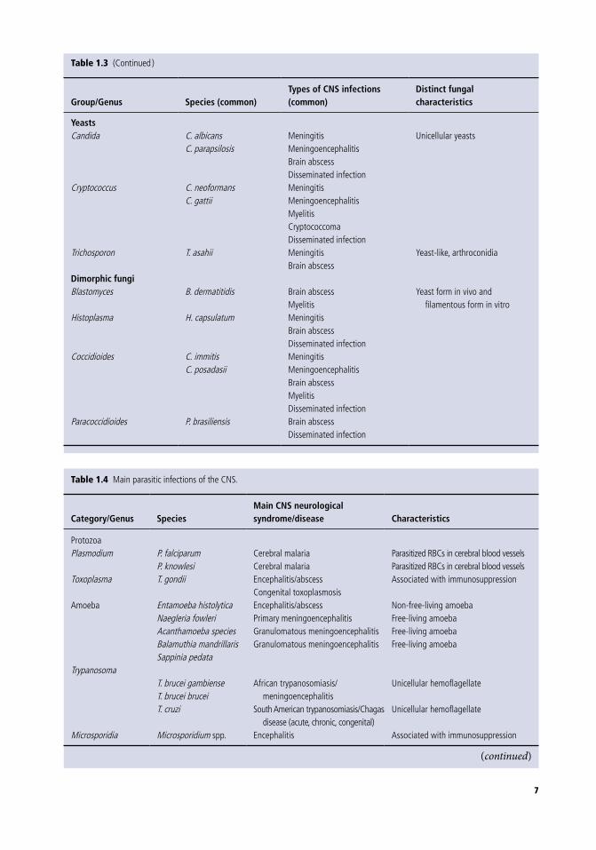
Numerous fungi (Table 1.3) cause CNS infec-tions; according to their morphology, fungal path-ogens can be divided into three major groups: molds, yeasts, and dimorphic fungi. This categori-zation does not relate to the taxonomic groups, but it may be helpful in guiding diagnosis because each category shares similar clinical features.
Parasitic infections of the CNS caused by protozoa include infections by Plasmodium, Toxoplasma, and other protozoa such as amebae, trypanosomes, Microsporidium, and Leishmania (Table 1.4). Helminthic cestodes (i.e. Taenia, Echinoccocus, Spirometra), trematodes (i.e. Schistosoma, Paragonimus), and nematodes (i.e. Angiostrongylus, Gnathostoma, Toxocara, Trichinella, filaria, Strongyloides) infections and CNS myiasis resulting from fly larvae infestation of the CNS are also described.
Many factors determine the incidence of different CNS infections in disparate geographical regions. Endemic and emerging infections in susceptible human populations depend on the dynamic interactions between (or changes in) microbe‐environment‐host factors. The increasing virulence of CNS pathogens because of antimicrobial resistance is of huge impor-tance, resulting in increased incidence of more severe infections. Environmental factors caused by poor sani-tation and hygiene and absence of vector control, and so on, enable diseases to spread rapidly. Host factors including changes in human behavior and demogra-phy, immunosuppression, genetic predisposition, and many others conspire to increase host susceptibility to CNS infections. In recent years, acquired immunosup-pression resulting from HIV infection or associated with therapeutic modalities, including newer immu-nomodulating treatments, has dramatically increased host susceptibility to numerous opportunistic agents. A separate chapter on emerging CNS infections discusses more details about this phenomenon.
This book aims to describe and illustrate the various lesions and entities that may be encoun-tered in CNS infections to help in pathological diagnosis. These include meningitis, abscess, encephalitis, myelitis, demyelination, vasculopa-thy, infarction, or combinations thereof (e.g. meningoencephalitis and encephalomyelitis). The

Introduction and Classification of Infections of the CNS According to the Agent Chapter 1
3
Table 1.1 Main viruses involving the CNS.
Virus Genus/SpeciesMain CNS neurological syndromesa Characteristicsb
DNA virusesHerpes virus Herpes simplex I, II
HHV‐1 and ‐2Encephalitis Virus targets neurons
Varicella‐zoster virusHHV‐3
MyelitisVasculitis/infarctsMeningoencephalitisEtc.
Virus targets neuroglial and Schwann cells, blood vessels.
Epstein‐Barr virusHHV‐4
MyelitisMeningoencephalitis
Associated with immunosuppression
CytomegalovirusHHV‐5
MyelitisVentriculoencephalitisCongenital neurological syndrome
Associated with immunosuppression
Adenovirus Adenovirus species Meningoencephalitis Associated with immunosuppressionPolyomavirus JC virus
BK virusProgressive multifocal
leukoencephalopathyVirus targets oligodendroglia mainly Associated with immunosuppression
RNA virusesMorbillivirus Measles virus (rubeola) Acute postinfectious encephalitis
MIBESSPE
Virus targets neuroglial cells and neurons in MIBE and SSPE
MIBE mainly associated with immunosuppressionSSPE associated with mutant virus
Rubivirus Rubella virus CRS(RE)
Cerebral blood vessel damage and microcephaly in CRS
Neuronal degeneration in REHenipavirus Hendra virus
Nipah virusAcute encephalitisRelapsing encephalitis
Virus targets cerebral blood vessels and neuroglial cells in acute encephalitis
Virus targets only neuroglial cells in relapsing encephalitis
Lyssavirus Rabies virus and other species
Encephalitis(Paralytic rabies; furious rabies)
Virus mainly targets neurons
Flavivirus Tick‐borne encephalitis virus
Encephalitis Virus targets neurons
West Nile virus Encephalitis Virus targets neuronsSt. Louis encephalitis
virusEncephalitis Virus targets neurons
Murray valley encephalitis virus
Encephalitis Virus targets neurons
Yellow fever virus Encephalopathy Mainly viscerotropicDengue virus Encephalopathy Mainly viscerotropicZika virus Congenital Zika syndrome Virus mainly targets neural progenitor cellsJapanese encephalitis
virusEncephalitis Virus targets neurons
Hepatitis C virus Neuropsychiatric‐related (parainfectious)
Meningoencephalitis/myelitis (rare)
Neurotropism unconfirmed
Hepevirus Hepatitis E virus Meningoencephalitis/myelitis (rare) Neurotropism unconfirmed
(continued)

Infections of the Central Nervous System
4
Table 1.1 (Continued)
Virus Genus/SpeciesMain CNS neurological syndromesa Characteristicsb
Alphavirus Eastern equine encephalitis virus
Western equine
encephalitis virus Venezuelan equine
encephalitis virus
Encephalitis Virus targets neurons
Chikungunya virus EncephalopathyEncephalitis
Specific neuropathology unknown
Enterovirus Enterovirus A71 Acute flaccid paralysisEncephalomyelitis
Virus targets mainly lower motor neurons
Poliovirus PoliomyelitisPolioencephalitisPostpolio syndrome
Virus targets mainly lower motor neurons
Retrovirus HIV HIV‐induced encephalitis and other encephalopathy/disorders
Opportunistic infections
Virus targets CD4 lymphocytes and macrophages/microglia
HTLV Tropical spastic paraparesis/HTLV‐1–associated myelopathy
Virus targets CD4 lymphocytes Pathogenesis may be associated with immune factors
Parechovirus Parechovirus A Encephalitis Virus targets cerebral blood vessels
a Commonly used terms for the neurological syndromes in the CNS; peripheral nervous system disease not included.b Selected characteristics only; cellular tropism is not well established in a significant number of viral CNS infections.CRS, congenital rubella syndrome; HHV, human herpes virus; HIV, human immunodeficiency virus; HTLV, human T cell leukemia/lymphoma virus; JC virus, John Cunningham virus; MIBE, measles inclusion body encephalitis; RE, rubella encephalitis; SSPE, subacute sclerosing panencephalitis.
Table 1.2 Mycoplasmal rickettsial and bacterial infections of the CNS.
Organisms Genus/Species Type of CNS infection Characteristics
Mycoplasma M. pneumoniae Encephalitis Intracellular bacteriaM. hominis Brain abscess
MeningitisIntracellular bacteria
Rickettsia R. rickettsii EncephalitisChronic leptomeningitisCerebral infarction
Intracellular bacteriaTick‐borne
R. conorii MeningoencephalitisCerebral infarctionPolyneuropathy and polyneuritis
Intracellular bacteriaTick‐borne
R. prowazekii MeningoencephalitisPeripheral neuropathyTransverse myelitis
Intracellular bacteriaLouse‐borne
R. typhi Meningoencephalitis Intracellular bacteriaFlea‐borne

Introduction and Classification of Infections of the CNS According to the Agent Chapter 1
5
Organisms Genus/Species Type of CNS infection Characteristics
Orientia O. tsutsugamushi MeningoencephalitisCerebral vein thrombosisGuillain‐Barré syndromeTransverse myelitisCranial neuropathyPolyneuropathy
Intracellular bacteriaMite‐borne
Ehrlichia E. chaffeensis Lymphocytic meningitisCranial neuropathyDemyelinating polyneuropathy
Intracellular bacteriaTick‐borne
Anaplasma A. phagocytophilum (same as ehrlichiosis?) Intracellular bacteriaTick‐borne
Gram‐positive pyogenic bacteria
Streptococcus pneumoniaeStaphylococcus spp.
MeningitisBrain abscess
DiplococcusCocci
Gram‐negative pyogenic bacteria
Neisseria meningitidis Escherichia coliHaemophilus influenzaePseudomonas spp.
Meningitis
MeningitisMeningitisAbscess, meningitis
Waterhouse‐Friderichsen syndrome
Vaccine availableHemorrhagic
Actinomycetaceae Actinomyces israelii Actinomyces meyeriActinomyces viscosus
Brain abscessFilamentous
Actinobacteria Tropheryma whipplei Multifocal encephaliticnodules
Accumulation of partially degraded bacteria in macrophages
Nocardiaceae Nocardia asteroides complexNocardia brasiliensis complex
Brain abscess Filamentous
Mycobacteria M. tuberculosis Basal meningitisTuberculomaAbscesses (HIV)
Acid‐alcohol resistant
Non‐tuberculous mycobacteria:MACM. haemophiliumM. kansasii
Brain abscess Opportunistic pathogens
Spirochaetaceae Treponema pallidum Neurosyphilis Humans only known natural hosts
Borrelia burgdorferi Borrelia recurrentis Other Borrelia species
Lyme disease Relapsing fever Relapsing fever
Tick‐borne Louse‐borne Tick‐borne
Leptospira interrogans MeningitisMeningoencephalitisMeningomyelitisPolyradiculoneuropathyIntracranial hemorrhage
Rodents are reservoirs
(continued)
Table 1.2 (Continued)

Infections of the Central Nervous System
6
Table 1.3 Important fungal pathogens in the CNS.
Group/Genus Species (common)Types of CNS infections (common)
Distinct fungal characteristics
Molds (Filamentous fungi)Hyaline (not pigmented)Aspergillus A. fumigatus
A. flavusOther species
Brain abscessSkull‐base syndromesStroke/infarction Disseminated
infectionHemorrhagicMyelitis
Branching septate hyphaeAngiophilic
Fusarium F. solani species complex MeningoencephalitisBrain abscess
Branching septate hyphae
MucoralesRhizopus
R. arrhizus
Brain abscessRhino‐cerebralStroke/infarctionDisseminated infectionHemorrhagic
Branching nonseptate hyphaeAngiophilic
Rhizomucor R. pusillusMucor M. indicus
Lichtheimia L. corymbifera Brain abscessRhino‐cerebral
Apophysomyces A. elegans Rhino‐cerebralPigmented molds
(Dematiaceous)Cladophialophora C. bantiana Meningitis
Brain abscessBranching nonseptate pigmented
hyphaeExophiala E. dermatitidisRhinocladiella R. mackenzieiVerruconis V. gallopavaFonsecaea F. pedrosoi
F. monophoraCurvularia C. spicifera
C. hawaiiensisC. lunata
Alternaria A. infectoria
Organisms Genus/Species Type of CNS infection Characteristics
Brucella B. melitensisB. suisB. abortus
Neurobrucellosis The most frequent zoonosis worldwide
Legionella 58 species, 30 of which are pathogenic for humans
EncephalopathyTransient focal neurological signs
Always associated with pulmonary disease
HIV, human immunodeficiency virus; MAC, Mycobacterium avium‐intracellulare complex.
Table 1.2 (Continued)
7
Group/Genus Species (common)Types of CNS infections (common)
Distinct fungal characteristics
YeastsCandida C. albicans
C. parapsilosisMeningitisMeningoencephalitisBrain abscessDisseminated infection
Unicellular yeasts
Cryptococcus C. neoformansC. gattii
MeningitisMeningoencephalitisMyelitisCryptococcomaDisseminated infection
Trichosporon T. asahii MeningitisBrain abscess
Yeast‐like, arthroconidia
Dimorphic fungiBlastomyces B. dermatitidis Brain abscess
MyelitisYeast form in vivo and
filamentous form in vitroHistoplasma H. capsulatum Meningitis
Brain abscessDisseminated infection
Coccidioides C. immitisC. posadasii
MeningitisMeningoencephalitisBrain abscessMyelitisDisseminated infection
Paracoccidioides P. brasiliensis Brain abscessDisseminated infection
Table 1.4 Main parasitic infections of the CNS.
Category/Genus SpeciesMain CNS neurological syndrome/disease Characteristics
ProtozoaPlasmodium P. falciparum Cerebral malaria Parasitized RBCs in cerebral blood vessels
P. knowlesi Cerebral malaria Parasitized RBCs in cerebral blood vesselsToxoplasma T. gondii Encephalitis/abscess
Congenital toxoplasmosisAssociated with immunosuppression
Amoeba Entamoeba histolytica Encephalitis/abscess Non‐free‐living amoebaNaegleria fowleri Primary meningoencephalitis Free‐living amoebaAcanthamoeba species Granulomatous meningoencephalitis Free‐living amoebaBalamuthia mandrillarisSappinia pedata
Granulomatous meningoencephalitis Free‐living amoeba
TrypanosomaT. brucei gambienseT. brucei brucei
African trypanosomiasis/meningoencephalitis
Unicellular hemoflagellate
T. cruzi South American trypanosomiasis/Chagas disease (acute, chronic, congenital)
Unicellular hemoflagellate
Microsporidia Microsporidium spp. Encephalitis Associated with immunosuppression
(continued)
Table 1.3 (Continued )

Infections of the Central Nervous System
8
Category/Genus SpeciesMain CNS neurological syndrome/disease Characteristics
Leishmania L. donovani Visceral leishmaniasis with CNS involvement (rare)
Nonflagellate amastigotes
Helminth (cestodes)Taenia T. solium Neurocysticercosis Larva form (cysticerci) involved
T. multiceps Coenurosis Larva form (coenurus) involvedEchinococcus E. granulosus Cerebral echinococcosis Larva form (hydatid cysts) involvedSpirotmetra S. mansoni
S. mansonoidesS. poliferum
Cerebral sparganosis Plerocercoid larva (spargana) involved
Helminth (trematodes)Schistosoma S. haematobium Cerebral schistosomiasis Ova involved
S. mansoni Cerebral schistosomiasis Ova involvedS. japonicum Cerebral schistosomiasis Ova involvedS. mekongi Cerebral schistosomiasis Ova involved
Paragonimus P. westermani and other species
Cerebral paragonimiasis Immature flukes involved
Helminth (nematodes)Angiostrongylus A. cantonensis Neuroangiostrongylosis (eosinophilic
meningitis)Larva involved
Gnathostoma Gnathosoma spp. Cerebral gnathostomiasis Larva involvedToxocara T. canis
T. cati(other non‐Toxocara spp.)
Visceral larva migransNeurotoxocariasis
Larva involved
Trichinella T. spiralisOther species
Neurotrichinosis Larva involved
Filaria Loa loaOnchocerca volvulus
CNS involvement not well established Microfilaria involved
Strongyloides S. stercoralis Cerebral strongyloidiasis Larva involved
CNS, central nervous system; RBCs, red blood cells; spp., species.
different types of lesions and their distribution in various parts of the CNS may be dictated by the different routes of neuroinvasion (e.g. hematoge-nous or along peripheral nerves or olfactory bulb); spread from adjacent extracranial structures; the nature of the pathogen and its predilection for dif-ferent cellular or tissue targets; the tempo of the infection whether acute, subacute, or chronic; and the host immune response
The typical inflammation observed in many viral CNS infections consists of perivascular cuff-ing and parenchymal infiltration by inflammatory cells, often with microglial nodule formation,
neuronophagia, and necrosis. Most viruses infecting the CNS are neuronotropic and, therefore, could potentially affect all areas where neurons are found. However, preferred routes of CNS invasion or even a predilection for different neuronal populations may determine the topography of the inflammation (e.g. in herpes simplex 1 and enterovirus A71 infec-tions). Viral inclusions in different cellular compart-ments (i.e. nucleus or cytoplasm) may be observed in many DNA viruses and a few RNA viruses (e.g. paramyxoviruses: measles and henipavirus) and can be useful diagnostic features. Although relatively rare, vasculopathy (e.g. vasculitis and thrombosis)
Table 1.4 (Continued)

Introduction and Classification of Infections of the CNS According to the Agent Chapter 1
9
is observed not only in henipavirus and varicella‐zoster virus infections but may also be encountered in angioinvasive fungal and rickettsial infections and in CNS tuberculosis.
Abscesses are generally localized, circumscribed acute or subacute inflammations caused by pyo-genic bacteria, fungi, and Toxoplasma. They may be found in most areas of the CNS. Certain bacteria such as Streptococcus pneumoniae and Haemophilus influenzae are the main culprits for acute pyogenic meningitis. Granulomatous inflammation in the CNS is most often associated with more chronic infections, notably Mycobacteria, fungi, and spiro-chetes and in some parasitic infections like amoebi-asis. Sarcoidosis, although not regarded as an infection, is included as it is an important differen-tial diagnosis in granulomatous CNS inflammation.
Despite recent advances in molecular diagnosis using polymerase chain reaction (PCR)‐based methods, sequencing, and so on, light microscopy using routine hematoxylin and eosin (H&E) stains may still be quite useful to help identify pathogens in infected tissues and other biological specimens based on morphology alone, especially if combined with special histochemical stains, immunohisto-chemistry, and in situ hybridization. Familiarity with diagnostic morphological features of certain fungi and parasites are particularly helpful. Commercial or proprietary specific primary anti-bodies for immunohistochemistry, if available, could also help detect some viral, bacterial, fungal, and parasitic infections to enable a more rapid diagnosis.
This volume also highlights the major historical and epidemiological characteristics of the different diseases, their clinical presentation and difficulties in diagnosis, and their pathogenesis, including information from animal models. It also gives some information on the microbiological charac-teristics of the agents, although it is not intended to be a detailed microbiological book. Genetic char-acteristics of both the agents and the hosts are dis-cussed with reference to how these can favor disease development in particular hosts.
This monograph will be of interest to a wide variety of medical doctors, postgraduate students, and scientists involved in studying, diagnosing, or treating infections involving the CNS. We hope that it will become a reference book in neurology departments and microbiology and pathology lab-oratories because it approaches the topic in a way not dealt with in other books.
We have been fortunate in benefiting from numerous international authors who have written about their own area of expertise in CNS infectious diseases. Collaborating with the authors has been productive, interesting, and useful, and we sin-cerely thank them all for their generosity.
Reference
1. Kaminski, Z., Kapila, R., Sharer, L.R. et al. (1992). Meningitis due to Prototheca wickerhamii in a patient with AIDS. Clin Infect Dis 15: 704–706.


Infections of the Central Nervous System: Pathology and Genetics, First Edition. Edited by Fabrice Chrétien, Kum Thong Wong, Leroy R. Sharer, Catherine (Katy) Keohane and Françoise Gray. © 2020 John Wiley & Sons Ltd. Published 2020 by John Wiley & Sons Ltd.
11
Sepsis‐Associated EncephalopathyFranck Verdonk1,2,3, Aurelien Mazeraud1,2,4, Fabrice Chrétien1,5,6, and Tarek Sharshar1,2,4
1 Experimental Neuropathology Unit, Pasteur Institute, Paris, France2 Department of Anesthesiology and Intensive Care, Saint Antoine Hospital, Paris, France3 Sorbonne University, Paris, France4 University Paris Diderot, Sorbonne Paris Cité, Paris, France5 Paris University, Paris, France6 Department of Neuropathology, Sainte Anne Hospital, Paris, France
2
Abbreviations
AMPA α‐amino‐3‐hydroxy‐5‐methyl‐4‐isox‑azolepropionic acid
APACHE acute physiology and chronic health disease classification system II
AQP4 aquaporin 4Bax proapoptotic factorsBBB blood‐brain barrierBCL B‐cell lymphomaCLP cecal ligation and punctureCNS central nervous systemDAMPs damage‐associated molecular pattern
moleculesDSM‐5 Diagnostic and Statistical Manual of
Mental Disorders‐5
EEG electroencephalogramICU intensive care unitDIC disseminated intravascular coagulopathyIL interleukinMRI magnetic resonance imagingNMDAR N‐methyl‐d‐aspartate receptorNOS nitric oxide synthaseNSE neuron‐specific enolasePAA plasma anticholinergic activityPAMPs pathogen‐associated molecular pattern
moleculesSAE sepsis‐associated encephalopathyTLR toll‐like receptorTNF tumor necrosis factorZO zonula occludens

Infections of the Central Nervous System
12
Definition of the disorder, major synonyms, and historical perspective
Sepsis is defined as a life‐threatening organ dys‑function caused by a dysregulated host response to infection [1]. Sepsis is the most common causes of mortality in intensive care units (ICUs) and ranges from 10 to 50% [2]. During sepsis, CNS dysfunction, called sepsis‐associated encephalopathy (SAE), occurs in more than 30% of patients. It is clinically characterized by impaired consciousness, which ranges from dizziness to coma and delirium, but it is also associated with an increased risk of stroke or epilepsy. SAE is associated with increased ICU and in‐hospital mortality and long‐term cognitive decline [3].
Epidemiologic characteristics
The incidence of SAE has been calculated in prospec‑tive and retrospective cohorts. In one of the first large prospective studies of 1333 patients with severe sepsis, Sprung et al. reported that 307 (23%) developed cogni‑tive and behavioral changes [4]. More recently, in a retrospective study, Zhang et al. found a lower inci‑dence of 17.7% [5]. This discrepancy is because the diagnostic criteria for SAE have evolved over the last decade and remain controversial. They should com‑bine both clinical and neurophysiological parameters. Electroencephalogram (EEG) changes suggesting SAE are present in up to 80% of bacteremia cases [6, 7]. Considering the estimated number of patients treated for sepsis worldwide, the number of patients affected by SAE could range from 5.3 to 25.2 million annually.
Predictive factorsThe risk factors in SAE include patients’ comorbidi‑ties, clinical and microbiological characteristics of sep‑tic shock, and intensive‐care interventions. Age, comorbid illness (notably neuropsychiatric), severity of organ failure (notably renal and liver), sedation, and drug side effects are all risk factors. Antibiotic over‑dose has to be excluded in this context. Encephalopathy is more frequent in cases of bacteremia or of infection with Staphylococcus aureus, Enterococcus faecium, Acinetobacter spp., Pseudomonas aeruginosa, or Stenotrophomonas maltophilia.
Consequences for survivalSAE is associated with increased morbidity and mortality. Sepsis‐related mortality rises from 26 to 49% when complicated by encephalopathy [4]. Delirium is associated with a threefold increase in mortality rate in the general population of patients who are critically ill [8].
Consequences for long‐term cognitive dysfunctionAbout one‐third of patients will have cognitive impairment 12 months after their ICU discharge [9], and the degree of impairment is proportional to the duration of delirium during their ICU stay [10]. Similar to mild Alzheimer disease, memory, attention, verbal fluency, and executive functions are mainly affected [11, 12]. Patients with sepsis seem particularly susceptible to cognitive decline. Iwashyna et al. reported that hospitalization for sepsis tripled the risk of developing moderate to severe cognitive decline [13].
Patients with sepsis are also at risk of suicide and of developing psychological disorders, including depression, anxiety, and post‐traumatic stress dis‑order [14].
Clinical features including appropriate investigations
Clinical examinationThe fundamental features of SAE are the combina‑tion of sepsis and impairment of consciousness, ranging from delirium to coma. Delirium can be either hyperactive (i.e. agitation) or hypoactive. But before encephalopathy occurs, patients with sepsis usually develop a “sickness behavior,” which is a physiological response to any systemic inflammation and characterized by lethargy, depression, anxiety, loss of appetite, drowsiness, hyperalgesia, difficulty concentrating, and thermal regulation disorder [15].
Motor signs, such as paratonic rigidity, asterixis, tremor, or myoclonus are rarely observed compared to hepatic, uremic, or toxic encephalopathies.
Intensivists have validated clinical test scales at their disposal for the detection of SAE. These include the Confusion Assessment Method for Intensive Care Units (CAM‐ICU) [16] or the Intensive Care

Sepsis‐Associated Encephalopathy Chapter 2
13
Delirium Screening Checklist Worksheet (ICDSC) [17] for the diagnosis of delirium. Because the clini‑cal spectrum of SAE is extremely broad and not lim‑ited to delirium, no single clinical examination is sufficiently specific and sensitive to be systematically applied, particularly in less‐severe and early forms of SAE. It should be noted that the majority of clinical studies conducted on SAE only target delirium for the sake of diagnostic simplicity.
Once SAE is suspected, a thorough neurological examination is required, including an assessment of neck stiffness, motor responses, muscle strength, and deep tendon and cranial nerve reflexes. Although the frequent use of sedatives in ICU may limit the clinical examination, assessment of brain‑stem responses appears useful and loss of responses in patients who are sedated is predictive of mortal‑ity and altered mental status [18].
SAE blood markersSerum S100β protein and neuron‐specific enolase (NSE) have been proposed as biomarkers for SAE [19, 20], but their usefulness for predicting, diag‑nosing, and monitoring SAE awaits confirmation.
EEGIn patients with sepsis, two types of electroencephalo‑graphic abnormalities are visible affecting either the background activity or superimposed epileptic fea‑tures. Background activity changes range from slow‑ing of the normal alpha rhythm with the onset of theta activity in patients with mild encephalopathy (i.e. confusion or delirium), to an increase in slow waves, the onset of delta waves, and even “burst suppression” in severe encephalopathy associated with coma. The latter indicate damage to deep brain structures such as the central gray nuclei and midbrain [21].
Epileptic features are the second type of anomaly observed and have recently been precisely defined. Electrographic seizures are associated with delir‑ium, whereas lack of EEG reactivity is associated with mortality [22]. EEG changes can also be observed in patients with sepsis who are asympto‑matic, making it an interesting method for detecting and monitoring SAE. EEG anomalies are also associated with radiologic findings [23]. We, therefore, recommend that an EEG should always be performed in cases of SAE or suspected SAE.
Brain imagingBrain imaging is not systematically carried out, but to exclude the usual causes of neurological deterio‑ration (i.e. ischemic or hemorrhagic stroke, suba‑rachnoid hemorrhage, abscess, etc.), it is required when seizures, focal neurological signs, persistent delirium, or coma occur.
Imaging usually reveals white matter hyperdensi‑ties, mainly related to vasogenic edema, and ischemic stroke, mainly related to DIC [15]. However, it can be normal in half the cases [15]. Magnetic resonance imaging (MRI) is useful to assess secondary cogni‑tive impairment associated with white matter changes and hippocampal atrophy [24].
Physiopathology
Systemic inflammation is the main basis for SAE pathophysiology. Sepsis induces a dramatic immune inflammatory response associated with the clinical manifestations of SAE. Circulating levels of proin‑flammatory markers (namely interleukin (IL)‐1, IL‐6 or tumor necrosis factor (TNF)‐soluble recep‑tor decreased matrix metalloproteinase‐9 [MMP‐9] and protein C concentrations) are associated with an increased risk of delirium [25–27]. More gener‑ally, it has been shown that cognitive impairment relates to systemic inflammation whether as a result of sepsis or occurring after surgery [28, 29].
SAE is historically viewed as an aseptic insult of the CNS, justifying the term “encephalopathy” rather than “encephalitis” [30]. However, a recent study questions this view. In a mouse model of sepsis using cecal ligation and puncture (CLP) [31], Singer et al. found that sepsis is associated with polymicrobial spread within the CNS by viable bacteria during the acute phase up to the fifth day. The authors showed an increased expression of neuroinflamma‑tory markers in microglial cells. These findings gain some support from a human study reporting brain abscesses in about 10% of patients [32]. If a microbial process is involved, encephalopathy is mainly sec‑ondary to the systemic inflammation.
The consequences of systemic inflammation on the brain can be separated into three nonex‑clusive mechanisms: ischemic, neuroinflamma‑tory, and epileptic processes. In combination,

Infections of the Central Nervous System
14
these processes result in impairment of neuro‑transmission and neurotoxicity. Particular struc‑tures are vulnerable to these phenomena: the frontal cortex and hippocampus, which accounts for acute and chronic neurological symptoms of sepsis, and central autonomic nuclei (i.e. amygdala, locus coeruleus, medullary nuclei), accounting for acute impairment of the response to stress [33].
Systemic or local inflammation is transmitted to the CNS via three major pathways [34]:1. Circumventricular organs enable inflammatory mediators to pass directly into the brain paren‑chyma. They are thought to play a major role in the “humoral pathway” and induce neuroinflammation.2. Blood‐brain barrier (BBB) disruption increases the passage of inflammatory mediators to brain parenchyma, enabling their participation in micro‐ and macroangiopathy and neuroinflammation.3. Neural pathways, notably via the vagus nerves, transfer the peripheral inflammatory signal to the medullary autonomic nuclei. The increased neu‑ronal activity can produce excitotoxicity and is associated with neuroinflammation.
Ischemic processesMacrocirculatory impairmentIn addition to hypotension, hypovolemia, and car‑diac dysfunction, impairment of autoregulation can compromise cerebral perfusion. Autoregulation is a physiological mechanism that aims to maintain a constant cerebral blood flow. In physiological conditions, the vessel diameter adapts to blood pressure. In patients with sepsis, this mechanism can be impaired. Schramm et al. showed that autoregulation is impaired in 60% at day 1 and 46% at day 4 [35]. Moreover, impaired autoregulation at day 1 was associated with delirium occurring at day 4, and this association was confirmed in a larger cohort of 50 patients with sepsis [36]. Interestingly, early decrease in cerebral flow correlates with long‐term cognitive impairment [37].
Microcirculatory impairmentSepsis frequently causes microvascular dysfunc‑tion that is considered an important mechanism in organ failure [38]. Microcirculatory dysfunction hampers diffusion of gases and induces a mosaic of tissue hypoxia and vascular shunts alongside
well‑oxygenated areas. Endothelial dysfunction, glycocalyx (pericellular matrix) changes, mecha‑nisms inducing leukocyte translocation and plate‑let microthrombi, and pericyte dysfunction are its main mechanisms [39, 40]. This microcirculatory dysfunction can affect the brain, compromising its metabolism as oxygen demand and supply become unbalanced [41]. Microcirculation impairment impinges neurovascular coupling during sepsis [42]. So far, there are no preventive or therapeutic strategies for microcirculatory dysfunction.
Endothelial dysfunctionEndothelial activation at the early phase of sepsis results in increased permeability of the BBB, although tight junction proteins such as occludin, zonula occludens (ZO)‐1 and ZO‐2 [43], and clau‑din‐3 and claudin‐5 [44] seem preserved. Other factors contribute to BBB dysfunction, including astrocyte activation and overexpression of aqua‑porin 4 (AQP4). Endothelial activation contributes to the neuroinflammatory process by releasing inflammatory and neurotoxic factors or allowing their passage through an impaired BBB [45].
Ischemic lesions in sepsisIschemic lesions are constantly observed in patients who die from sepsis. They are found in areas of the brain sensitive to ischemia such as Ammon’s horn, the lenticular nucleus, and frontal cortex but also and more specifically (in comparison to patients who died from causes other than sepsis) in auto‑nomic nuclei involved in cardiovascular control (i.e. posterior and anterior hypothalamus, locus coeruleus). Pronounced neuronal apoptosis is also observed in these same areas. In addition, cerebral hemorrhages are found in 17–26% of patients who die of septic shock [32, 46]. Necrotizing multifocal leukoencephalopathy can also be found and corre‑lates with brain expression of tumor necrosis factor alpha (TNF‐α) and IL‐1β and elevated circulating levels of TNF‐α, IL‐1β, IL‐6, IL‐8, IL‐10, the solu‑ble receptor for TNF II, and for IL‐1 receptor antagonist [47]. Hypoxemia and hyperglycemia are frequent features of sepsis, which potentiate the deleterious effects of impaired cerebral perfusion. To date, there is no recommendation for monitor‑ing and optimizing cerebral perfusion.