ineffective cough and mechanical mucociliary clearance
TRANSCRIPT

Med Intensiva. 2018;42(1):50---59
www.elsevier.es/medintensiva
REVIEW
Ineffective cough and mechanical mucociliary
clearance techniques�
A. Fernández-Carmona ∗, L. Olivencia-Pena, M.E. Yuste-Ossorio, L. Penas-Maldonado,Working Group of the Mechanical Home Ventilation Unit of Granada
Servicio de Medicina Intensiva, Complejo Hospitalario Universitario de Granada, Granada, Spain
Received 27 February 2017; accepted 5 May 2017
Available online 6 January 2018
KEYWORDSCough assist;Intrapulmonarypercussiveventilation;Mucociliary clearance
Abstract Cough is a fundamental defense mechanism for keeping the airway free of foreign
elements. Life-threatening situations may arise when cough proves ineffective as a result of
muscle weakness or altered mucociliary function.
When a patient is unable to cough effectively, techniques are required to either reinforce
or replace cough capacity. The use of mechanical systems that facilitate or substitute cough
function is increasingly common in Intensive Care Units, where it is relatively frequent to find
situations of ineffective cough due to different clinical causes.
This review examines the current clinical practice recommendations referred to the indica-
tion and use of mechanical cough assist and intrapulmonary percussive ventilation systems.
© 2017 Elsevier Espana, S.L.U. and SEMICYUC. All rights reserved.
PALABRAS CLAVETos asistida;Tos mecánica;Ventilación percutoraintrapulmonar;Aclaramientomucociliar
Tos ineficaz y técnicas mecánicas de aclaramiento mucociliar
Resumen La tos es un mecanismo de defensa fundamental para mantener la vía respiratoria
libre de elementos extranos. Cuando la tos es ineficaz, por debilidad muscular o por alteración
del normal funcionamiento del sistema mucociliar, se puede dar lugar a situaciones que pongan
en riesgo la vida.
Cuando un paciente no es capaz de producir tos eficaz es cuando está indicada la aplicación
de técnicas que, o bien potencien la tos del paciente, o bien la sustituyan. Actualmente el
uso de sistemas mecánicos facilitadores o sustitutivos de la tos es creciente en las unidades
de cuidados intensivos, donde con relativa frecuencia encontramos pacientes en esta situación
por diversas causas clínicas.
� Please cite this article as: Fernández-Carmona A, Olivencia-Pena L, Yuste-Ossorio ME, Penas-Maldonado L y Grupo de Trabajo de Unidad deVentilación Mecánica Domiciliaria de Granada. Tos ineficaz y técnicas mecánicas de aclaramiento mucociliar. Med Intensiva. 2018;42:50---59.
∗ Corresponding author.E-mail address: [email protected] (A. Fernández-Carmona).
2173-5727/© 2017 Elsevier Espana, S.L.U. and SEMICYUC. All rights reserved.

Mechanical mucociliary clearance techniques 51
Esta revisión se centra en las recomendaciones de práctica clínica actuales con respecto a la
indicación y aplicación de la tos asistida mecánica y de la ventilación percutora intrapulmonar.
© 2017 Elsevier Espana, S.L.U. y SEMICYUC. Todos los derechos reservados.
Introduction
‘‘Cough’’ (in Latin: tussis): the voluntary or reflex sudden,sharp and noisy expulsion of air from the lungs.
Cough is a defense mechanism with two basic functions:to keep the airway free of foreign elements, and to expelsecretions that are produced in excess or under pathologicalconditions. Ineffective cough is defined as cough unable toadequately perform these functions. Cough can also mani-fest in acute or chronic form as a symptom of many diseaseconditions, drug side effects, etc.1,2
Many diseases can severely affect the cough reflex andresult in ineffective cough, particularly disorders that causemuscle weakness, alterations in bronchial secretion charac-teristics and clearance, and a decrease and/or abolition ofcough stimulation. The disorders that produce ineffectivecough result in a tendency to retain bronchial secretions,with alteration of the normal ventilation/perfusion (V/Q)ratio, and an increased risk of respiratory infectious prob-lems. On the other hand, in the presence of a significantamount of secretions in the upper airway that are notexpelled correctly, patients experience excessive musclelabor, with a risk of muscle fatigue.1---4
When a patient is unable to cough effectively, techniquesthat either reinforce cough or replace it are indicated withthe aim of improving inspiratory capacity or mobilizing thesecretions to where they can be cleared by the patient orby physical means.5---7
Ineffective cough has been widely studied in patientswith disorders causing muscle weakness, such as neuromus-cular disease. In critical patients, the deficient managementof secretions is a determining factor of respiratory failure,failed weaning from invasive mechanical ventilation, andfailure of noninvasive mechanical ventilation.3,8---10
Although there is currently no solid scientific evidencewarranting the systematic use of mechanical mucociliaryclearance systems in the Intensive Care Unit (ICU) (thestudies involving critical patients being few and heteroge-neous), and the recommendations are practically based onlyon expert opinion, such systems are increasingly used incritical care --- often in patients with spinal cord injuriesor neuromuscular diseases, but also in situations of muscleweakness or difficult weaning from mechanical ventilation.Special mention must be made of the recent study pub-lished by Goncalves et al., which demonstrates the benefitsof these techniques when included in the mechanical venti-lation weaning protocols in critical patients, affording lowerreintubation rates and shorter ICU stays.10---13
The present review offers a brief description of the phys-iopathology of cough and comments on the current clinicalpractice recommendations regarding mechanical mucocil-iary clearance techniques and their application, including:intrapulmonary percussive ventilation (IPV) and mechanical
insufflation---exsufflation therapy (MI---E) or mechanicalcough assist (MC).
Assessment of cough efficacy
Clinical assessment
Assessment always should include the patient antecedentsand updated clinical history. Special attention should focuson the duration of mechanical ventilation and on whetherthe patient has an artificial airway or not; the presence ofneuromuscular disease; high spinal cord injury; diaphrag-matic dysfunction (Table 1); chronic bronchial disease;or disease conditions altering the characteristics of thebronchial secretions, such as cystic fibrosis.
In addition to the usual physical examination, quantifica-tion and evaluation of the characteristics of the bronchialsecretions are required, along with assessment of thepatient capacity to mobilize and expectorate the secretions,and the need for specific care measures such as cough incen-tivizing, tracheal aspiration (through the natural or artificialairway), auscultation and respiratory inspection.2,3,6---8
Consideration is also required of phonation and swallow-ing alterations, as well as of the capacity to perform aneffective Valsalva maneuver --- this being of great importancefor spontaneous cough and for non-mechanical cough assistmaneuvers.
Complementary tests are to be added to the physicalexamination.
Functional assessment
A number of determinations have been used for the func-tional assessment of cough. Such measurements can bemade both in patients with a natural airway and in those whoare intubated or subjected to tracheostomy not dependentupon mechanical ventilation:
- Peak expiratory pressure (PEmax): This parameter meas-ures the maximum pressure generated by the expiratorymuscles. The measurement is made following a maxi-mum inspiration coming as close as possible to total lungcapacity. However, consensus is lacking regarding the cut-off point of this parameter in defining whether cough iseffective or not. On the other hand, peak expiratory pres-sure has limitations resulting from the measuring processused.2,7,14
- Gastric pressure during cough maneuvering (PGA-cough):This parameter estimates the force generated by the expi-ratory muscles in the expulsive phase; the normal valuesfor adults are >175 cmH2O in men and > 100 cmH2O inwomen. The technique is invasive, however, and there

52 A. Fernández-Carmona et al.
Table 1 Main causes of diaphragmatic insufficiency.
Neurological causes
Spinal cord injury
Multiple sclerosis
Amyotrophic lateral sclerosis
Cervical spondylitis
Poliomyelitis
Guillain-Barré syndrome
Phrenic involvement:
--- Tumor disease
--- Heart surgery ‘‘cold lesion’’
--- Trauma
--- Viral disease
--- Idiopathic
--- Radiotherapy
--- Chest surgery
Myasthenic crisis
Eaton-Lambert syndrome
Myopathic causes
Limb-girdle dystrophy
Hyper-/hypothyroidism
Malnutrition
Lysosomal alpha-glucosidase deficiency
Connective tissue diseases:
--- Systemic lupus erythematosus
--- Dermatomyositis
--- Mixed connective tissue disease
Amyloidosis
Idiopathic myopathy
Others
Critical patient polyneuromyopathy
Chronic mechanical ventilation
Diaphragmatic dysfunction secondary to mechanical
ventilation
Anti-Hu syndrome
Botulism
Lyme disease
Pharmacological-aminoglycosides
COPD-pulmonary hyperinsufflation
are moreover technical problems for implementing theprocedure.2---14
- Maximum insufflation capacity measure (MIC): Thisparameter corresponds to the maximum intrathoracic vol-ume which the patient is able to reach. In adults it hasbeen estimated that the minimum value for ensuring flowsthat avoid the retention of secretions through sponta-neous cough is 1500 ml, versus 1000 ml in the case ofpatients with assisted cough.14,15
- Peak flow during cough (PFC): This is the parameter thatbest determines the capacity to eliminate respiratorysecretions during cough. The PFC values show good corre-lation to the conventional lung and muscle function testresults in both healthy individuals and in patients withneuromuscular diseases. The parameter is easy to deter-mine with portable systems that moreover allow us tomeasure peak expiratory flow. The minimum effectivePFC for mobilizing airway secretions is ≥2.7 l/s; lowerPFC values have been associated to increased mortality
in patients with neuromuscular diseases, and to faileddefinitive tracheostomy closure.3,14,16,17
The clinical practice guides referred to patients withneuromuscular diseases recommend the chronic use ofmechanical mucociliary clearance techniques in individualswith PFC ≤ 2.7 l/s (160 l/min). In patients with PFC ≤ 4.5 l/s(270 l/min), such techniques are advised during exacerba-tions or processes that increase the production of bronchialsecretions.6,18---20
Furthermore, if peak expiratory flow is measured, theassociation between this parameter and PFC is useful forassessing the degree of bulbar involvement in neuromuscu-lar disease, since a PFC value equaling peak expiratory flowindicates that glottic closure is difficult or impossible.
Other related explorations
- Basic spirometry: A vital capacity of <50% the theoreticalvalue in adults is generally considered to be insufficient.Forced and non-forced vital capacity can be measuredwith a simple electronic spirometer, a Wright spirometer,or conventional spirometry. Furthermore, basic spirome-try can complete the study of the background respiratorydisease of the patient.15
- In patients with suspected diaphragmatic dysfunction ofany origin, the measurement of forced vital capacity in thesitting position and in dorsal decubitus with a reduction of>25% (dorsal decubitus) is diagnostic of severe diaphrag-matic dysfunction. The ultrasound study of diaphragm
kinetics allows us to identify the existence of unilateral orbilateral involvement. In this regard, severe dysfunctionof the evaluated hemidiaphragm is defined by echographiccaudal displacement during non-forced respiration of≤10 mm in men and ≤9 mm in women.21---23 Diaphragmaticdysfunction secondary to prolonged mechanical ventila-tion is quite prevalent in the UCI, and ultrasound is anaccessible and useful technique for establishing an earlydiagnosis of this disorder.24
- Pleuropulmonary ultrasound (PPU): This technique isof growing importance in the management of criticalpatients, due to its non-invasiveness, validity and appli-cability at the patient bedside. It facilitates the detectionof pleural effusion and pneumothorax, and allows thediagnosis and follow-up of pneumonic condensation andatelectasis. Furthermore, PPU complements the hemody-namic information provided by echocardiography with theassessment of extravascular lung water, and offers infor-mation on pulmonary aeration in a range of diseases. Inpatients with ineffective cough, protocolized PPU (explo-ration of 8 thoracic areas according to the internationalrecommendations) allows us to monitor the presence ofatelectasis and its response to mechanical cough, as wellas to ensure the early detection of serious complicationssuch as nosocomial pneumonia, and avoid unnecessaryradiation exposure. This complementary technique in turnallows the prediction of extubation success in patientssubjected to mechanical ventilation.25,26

Mechanical mucociliary clearance techniques 53
Mucociliary clearance techniques
Ineffective cough is an indication for the use of techniquesthat either facilitate or replace cough----improving inspira-tory capacity or mobilizing the secretions to where they canbe cleared by the patient or by physical means.3,8---10
Conventional initial management in such situations seeksto reduce the viscosity of the secretions in order to facil-itate their elimination through natural cough. In additionto opportune medical treatment (antibiotics, mucolyticagents, bronchodilators, etc.), postural drainage, respira-tory physiotherapy and cough incentivizing (contraindicatedin situations of instability or postural intolerance) are alsoindicated. These techniques, together with early patientmobilization, suffice in most cases.27 In patients with verydense secretions, active humidification systems may proveuseful.2,28
When these measures are not enough, noninvasive assisttechniques should be considered for the managementof secretions, including manual cough assist, mechanicalcough assist (mechanical insufflation---exsufflation therapy[MI---E] or MC) and intrapulmonary percussive ventilation(IPV).2,5---7,29---32 In the studies published to date (mostlyinvolving patients with neuromuscular diseases), mechan-ical mucociliary clearance techniques have been found toreduce the number of pulmonary infections and moreovertend to improve lung function. Patients admitted to theICU and with prolonged mechanical ventilation develop con-ditions comparable to neuromuscular diseases in terms ofmuscle weakness, atrophy and fatigability,24 and these tech-niques have been shown to offer benefits when includedin the mechanical ventilation weaning protocols of criticalpatients----even in individuals with severe restrictive prob-lems in which other weaning regimens have failed.10---13,33
These therapies are effective and safe, and are oftenused in our Unit, applied indistinctively by experiencedphysicians and nurses (following a protocol prescribed by thesupervising physician and detailed in a specific treatmentsheet). In other centers such techniques are indicated andprogrammed by rehabilitation specialists and physiothera-pists (fundamentally in spinal cord injury units). However,due to the physiopathological characteristics inherent tocritical patients, such procedures in the ICU should be knownand supervised by intensivists----in contrast to chronic treat-ments or stable patients, which are not contemplated in thisreview.
Mechanical cough assist
Mechanical cough assist (MC) or mechanicalinsufflation---exsufflation therapy (MI---E) basically involvesthe mechanical generation of a transthoracic pressuregradient sufficient to produce an expiratory flow capable ofmobilizing the bronchial secretions.
A number of devices can be found on the market forMC.32,34,35 The MI---E systems are portable electronic devicesdesigned for discontinuous use that simulate cough throughpositive pressure followed by a proximal negative pressure.Both pressure settings are previously selected (equipotentor not) in order to secure an effective expiratory flow.These systems can generate maximum positive and negative
pressures of up to ±70 cmH2O (total pressure gradient ofup to 140 cmH2O), though in general it is not advisable toexceed ±40 cmH2O, due to the risk of barotrauma. Thistreatment is moreover regulated according to the inspi-ration/pause/expiration times, and can be applied to thepatient using a mask or mouthpiece, or through the endo-tracheal tube/tracheotomy cannula. Supplementary oxygencan also be administered, if necessary.32,34,35
Indications of mechanical cough assist
The indications of MC can be summarized from two perspec-tives: physiological and clinical.36---43
Physiological indications of mechanical cough- Incapacity to generate cough flow peaks of >4.2 l/s
(250 l/min).- In relative terms, a vital capacity of <50% of the theo-
retical value.- In absolute terms, a vital capacity of <1500 ml (in adults).Clinical indications of mechanical cough- Neuromuscular disease/polyneuromyopathy.- Thoracic diseases (combination of restrictive disorders
with incapacity to perform correct diaphragmatic move-ments).
- Diaphragmatic dysfunction with and without inspiratorysupport (mechanical ventilation).
- Traumatism or postoperative period of thoracoabdomi-nal/abdominal surgery.
- Prolonged mechanical ventilation.
Specifically, it is necessary to assess cough efficacy in:
- Neuromuscular diseases, where MC has been shown toimprove mobilization of the secretions and lessen theneed for inspiratory assist (mechanical ventilation); fur-thermore, it reduces respiratory infections and the needfor hospitalization.2
- Thoracic diseases with secondary restrictive pulmonarydisorders. Cough efficacy also must be assessed in patientsin which improved management of secretions is observed,despite a lack of improvement in lung mechanics that isnevertheless observed in other types of patients.41,42
- Patients with diaphragmatic dysfunction, especiallypatients with high spinal cord injuries characterized notonly by inspiratory weakness but also expiratory problemsassociated to thoracic and abdominal mechanical alter-ations (compliance).41,42
- Patients with diminished respiratory muscle strength andan increased production of secretions.41,42,44
- Patients subjected to prolonged invasive mechanical ven-tilation, where in most cases the indication is based on acombination of respiratory muscle weakness, fatigue andatrophy of multifactorial origin, making it impossible forthe patient to cough effectively.2,24
- Mechanical cough assists have also been proposed in poly-traumatized patients or individuals in the perioperativeperiod of thoracoabdominal surgery, where surgical mus-cle dissection or inadequate analgesia precludes adequatecough function.38

54 A. Fernández-Carmona et al.
Indications approved by the United States Food and Drug
Administration (FDA)
Patients with PFC reduction <2.7 l/s secondary to high spinalcord injuries, neuromuscular deficiencies or severe fatigueassociated to intrinsic pulmonary disease.34,35
The contraindications are: a history of bullous emphy-sema, known susceptibility to pneumothorax or pneumome-diastinum, and recent barotrauma.34,35
Mechanical cough application technique
Mechanical cough assist using MI---E devices can be carriedout in the presence of an artificial airway, with a sealedtube cuff (tracheal cannula or tube), or with the naturalairway using a face mask or mouthpiece (nasal obtura-tor)----in which case due patient instruction is required.34,35 Inpatients with an artificial airway, the technique can be usedwith or without patient collaboration; MC therefore can beemployed in patients with different degrees of sedoanalge-sia. In patients dependent upon mechanical ventilation, thevolumes generated by MI---E (with or without supplementaloxygen) substitute mechanical ventilation for the duration(normally 60---90 s) of the procedure.
The following disposable materials are needed: anantibacterial filter, simple tubing (no more than 1.5 m),an oxygen outlet adaptor (if needed), and an interface(oronasal mask, mouthpiece with nasal obturator or connec-tor adaptable to the artificial airway [22/15 mm]).
The usual MI---E models can be used manually----in whichcase the adjustable parameters are not operative----or inautomatic mode, which requires the prior programming ofseveral parameters:
- Inspiratory time (Ti), in seconds: the time needed to reachthe established inspiratory pressure.
- Pause time (Tp), in seconds: the transition time from thepoint where positive inspiratory pressure is reached tostart of the application of proximal negative pressure.
- Expiratory time (Te), in seconds: the time needed to reachthe pre-established maximum negative pressure.
- Maximum inspiratory (Pi) and expiratory pressure (Pe),expressed in cmH2O. The I-E pressure is the maximuminsufflation pressure limit; the expiration pressure isequivalent but of a negative value.
- Positive and negative pressure balance regulator: thisallows partial lowering of the negative pressure withrespect to the positive pressure.
- Inspiratory flow: this regulates insufflation flow between600 and 200 l/min (high/low).
Utilization of the more recently marketed systems allowsus to use digital commands to select the following parame-ters with greater precision than the analogical devices:
- Inspiration pressure: from 0 to 70 cmH2O, in increments of1 cmH2O.
- Inspiration flow: high/medium/low.- Ti: from 0 to 5 s, in increments of 0.1 s.- Pe: from 0 to 70 cmH2O, in increments of 1 cmH2O.- Te: from 0 to 5 s, in increments of 0.1 s.
- Tp: from 0 to 5 s, in increments of 0.1 s, only adjustablein automatic mode with deactivated cough-trak.
- Cough-trak: a trigger system that starts inhalation (pos-itive pressure) when patient inspiration is detected,thereby facilitating adaptation to MC and improving effi-cacy in delivering pressures when used in patients with anatural airway.
- Oscillation: flow oscillation improves mobilization of thesecretions by lowering their viscosity and clearing themfrom the lower airway. The frequency can be regulatedbetween 1 and 20 Hz and the amplitude between 1 and10 cmH2O; activation can be made during Ti or Te.45,46
These systems moreover allow the following:
- Pre-determination of three MC programs can be made withdifferent parameters, for different indications in the samepatient.
- Adjustment of the parameters can be blocked, in order toprevent them from being modified by untrained persons.
- Cycle-by-cycle monitoring of the efficacy of MC, control-ling both the volume of air exhaled by the patient and thepeak expiration flow.
- Monitoring of SpO2 can be incorporated to the MC systemby means of an adequate device.
The sum of the times Ti + Tp + Te is referred to as thecough cycle. A cough series or sequence is the continuedapplication of several cough cycles (generally 8---12 cycles).It is advisable to leave an interval of 2---3 min betweensequences. Mechanical cough assist should be applied atleast three times a day in a programmed manner, and when-ever required by the patient.
In order to avoid frequency disadaptation in patients withspontaneous respiration, the times are adjusted to the basalrespiratory frequency of the patient, causing the system toapply a frequency (of cough cycles) equal to or greater thanthat frequency. The distribution of times is made accord-ing to the predominant background disease condition. Thus,in patients with restrictive predominance Te is shortened,while in patients with obstructive predominance Te is pro-longed. In turn, Ti and Tp are adjusted to secure the desiredcough cycle frequency.47,48
The maximum Pi and Pe administered are intended toreach an intrapulmonary volume of close to 70% of thevital capacity of the patient. These pressures should not bereached in the first attempts, during the patient adapta-tion period. Adaptation is to be started applying pressuresof ±20 cmH2O, with increments of ±5 cmH2O every 2---3cycles, conditioned to patient wellbeing. The pressures usu-ally needed to ensure effective cough vary between ±30 and±40 cmH2O. In some cases we can reach pressures of up to±60 cmH2O, with effective differential pressures of 60, 80and up to 120 cmH2O, respectively. In general, Pe is keptequivalent (equal to Pi in absolute value), but negative.47---50
It may be useful to lower Pi and preserve tolerated Pemodifying the balance of pressures, when the secretions areretained in the glottic---subglottic region.
A high inspiratory flow is selected when the system isused with an artificial airway, while a low flow is selected inthe presence of a natural airway.

Mechanical mucociliary clearance techniques 55
The patient should be calm in the Fowler position(recumbent semi-decubitus). If the patient is conscious andcooperative, we should explain the technique and its pur-pose, as well as alert the patient to the sensations that maybe produced. The efficacy of MC improves if the patient con-tracts the abdominal muscles during the expiratory phase,or if manual abdominal compression is applied during thisphase (some series describe good results with electricalstimulation of the abdominal muscles).44
The application times of the technique should be spacedat least 2 h after and about 30 min before meals. If aerosoltherapy is required, it should be administered approximately30 min before MC.47
Application to patients with natural airway
In order for the procedure to be effective in patients with anatural airway,50,51 the glottis must be kept open during theapplication of each cough cycle. If the patient is unable tokeep the glottis open, we can cause reflex opening by posi-tioning a piece of corrugated tube (22 mm) to be graspedwith the lips, or by training the patient (pronounce the vowel‘‘O’’, misting a mirror with the breath, etc.). This step isvery much simplified thanks to the cough-trak system. How-ever, the patient may voluntarily close the glottis on noticingthe air flow of the inspiratory cycle of MC; it is therefore stillsometimes necessary to train the patient to keep the glottisopen.
The more frequently used oronasal mask is applied withthe same precautions and technique as manual ventilationmanual with a pneumatic bag.
As has been commented above, for the starting parame-ters we begin with low pressures until the values affordingefficacy and patient wellbeing are reached, adjusting thetime of each cycle to the base respiratory frequency and thepredominant respiratory disorder. Application always mustbe made with a low inspiratory flow in order to minimizeaerophagia.
Application to patients with artificial airway
The procedure is simplified in patients with an artificialariway,50,51 since in this case there is no concern aboutwhether the glottis is open or not. The system is directlyfitted to the trans-laryngeal tube or tracheotomy cannulausing an ISO 22/15 adaptor. If necessary, an adaptor can beused to supply oxygen to the system.
The same adjustment of parameters described above forthe natural airway is used, with the exception that in thiscase we can apply high or medium inspiratory flows, condi-tioned to patient wellbeing.
Complications of mechanical cough assist
Most of the complications are very infrequent (differentstudies involving over 500 MC series have reported no signif-icant complications).50---54 A recent Canadian national surveyspecifically focusing on critical patients found the mostfrequent complications to be chest pain (manual MC) andhypotension, reported by 17% of those surveyed in patientssubjected to MC with recruitment intention.11 The potentialcomplications are:
- Pneumothorax or pneumomediastinum (predisposing dis-ease).
- Chest wall pain with or without respiratory muscleinjury, fundamentally in patients subjected to chronicmechanical ventilation or with long-evolving neuromus-cular diseases, with a greatly diminished vital capacity.This complication can be avoided by slow and progressiveinsufflation pressure increments.
- Hemoptoic sputum secondary to mucosal damage on clear-ing firmly adhered secretions.
- Nausea, secondary to gases, and abdominal bloating dueto aerophagia. This can be improved by lowering theinsufflation pressures and with training (open glottis orextended neck).
- Bradycardia-hypotension, particularly in patients withacute spinal cord injury.
- Early collapse of the lower airway. This happens in patientswith important air flow limitation, causing MC to beineffective. This problem can be minimized by applyingthe technique passively, with no accompanying expiratoryeffort on the part of the patient.
- In patients with advanced bulbar amyotrophic lateral scle-rosis, MC produces hypopharyngeal collapse that preventsthe air from exiting the larynx --- thereby contraindicatingthe technique. This adverse effect can be minimized byapplying MC with the new devices that allow Pe to be pro-grammed independently of inspiratory pressure --- therebymaking it possible to lower the former until hypopharyn-geal collapse is avoided.38
The physiological effects of MC with regard to the ele-vation of central venous pressure and intracranial pressure,alterations in heart rate or increased intragastric pressureare similar to those caused by natural cough, though to alesser extent.
Intrapulmonary percussive ventilation
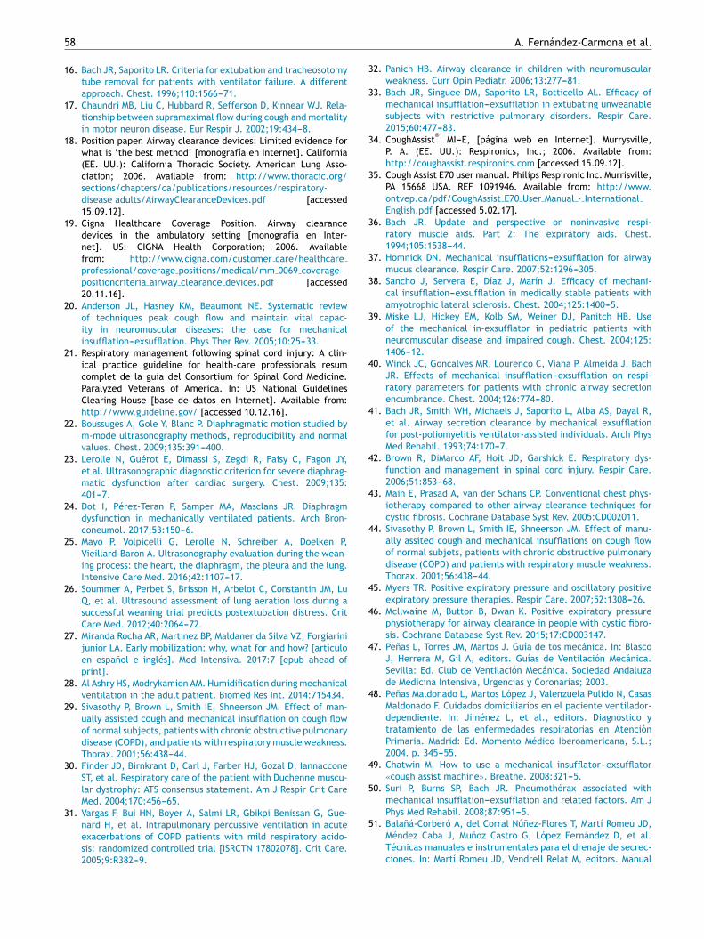
Intrapulmonary percussive ventilation (IPV) is an airwaysecretions clearance technique derived from high-frequencyventilation. It involves the constant administration of a pul-satile gas flow with low successive tidal volumes (TVs) in thepatient airway, with high frequency through an open respira-tory circuit----this affording successive TVs with low pressureand high flow, creating a positive transpulmonary pressuregradient. The insufflated gas volumes are small (between1 ml and 300 ml), and are administered on a continuousbasis, superimposed or not upon spontaneous ventilation ofthe patient (Fig. 1). Expiration is achieved passively.54
The frequency of the percussions in most of the marketeddevices varies between 80 and 650 cycles/min. The circuitused is continuously open, regardless of whether applied ona mask, mouthpiece or tracheostomy cannula. The circuitincludes a Hudson type high-flow pneumatic nebulizer.
Mobilization of the respiratory secretions from the lowertoward the upper airway can cause obstruction of the tra-chea and main bronchi. This technique therefore must becompleted with immediate evacuation of the secretions,using instrumental methods or sequentially applying MC inpatients with ineffective cough. The manufacturers recom-mend a total treatment time of up to 20 min. There is a risk

56 A. Fernández-Carmona et al.
mbar PAW 5
Flow = 0
V = 0.000
60
50
40
30
20
10
0
–10
l/min200
150
100
50
0
–50
–100
–150
–200
1.00
.800
.600
.400
.200
.000
L
0.0 2.0 4.0
Figure 1 Intrapulmonary percussive ventilation (IPV) con-
nected in series in a patient subjected to conventional
mechanical ventilation in pressure control mode.
of causing hyperventilation, however; the time thereforeshould be individualized according to clinical and labora-tory test criteria. On the other hand, dead space can beadded to the circuit with tubing, in order to control thehyperventilation induced by treatment with IPV.6,53,55
Intrapulmonary percussive ventilation is applied bymeans of a series of devices that include:
- Gas source (most devices incorporate an internal compres-sor).
- Phasitron®: a part that modifies delivery of the com-
pressed air-gas to the patient, with the aim of convertingTV of high pressure and low flow into TV of low pres-sure and high flow in a dynamic manner, according to thepulmonary mechanics of the patient.
- Nebulizer: connected in parallel to Phasitron®.
- Interface.
Indications of intrapulmonary percussiveventilation
This technique is indicated for the mobilization of bronchialsecretions in the context of the management of periph-eral obstructions and ventilation disorders in obstructive orrestrictive diseases characterized by hypersecretion, inde-pendently of the degree of patient collaboration, since it issuperimposed upon patient respiration. The technique canbe applied in adults and children, and in both acute and
long-term treatments. This therapeutic modality in turn canbe applied in the presence of an artificial airway or a naturalairway using a face mask or mouthpiece.6,53---55
The conditions in which most experience has been gainedare:
- Cystic fibrosis43,56
- Neuromuscular diseases57---59
- Chronic obstructive pulmonary disease31,60
- Neonates61,62
However, experience with IPV has also been gained inpatients with muscle weakness acquired in the ICU, obeseindividuals, burn victims, etc. In the last few years inwhich extracorporeal oxygenation techniques have becomeincreasingly used as life support systems, treatment com-bined with IPV has been shown to reduce the extracorporealmembrane oxygenation (ECMO) times in patients with acuterespiratory distress syndrome (ARDS).63---67
The outcomes of IPV can be optimized by combining itwith physiotherapy, postural drainage, cough assist, MC andany other type of respiratory physiotherapy technique.
Contraindications53,55:
- Absolute: non-drained pneumothorax.- Relative: Lyell syndrome, severe hemoptysis,
coagulopathy-anticoagulation, epilepsy, peak flow<3 l/s (180 l/min), provided there is no available artificialairway aspiration system or MC device in patients withoutan artificial airway.
Intrapulmonary percussive ventilation applicationtechniques
As a general rule, we use this technique53,55 in patients sub-jected to mechanical ventilation. It can be applied in serieswith the mechanical ventilator in the different ventilationmodes (both controlled and spontaneous). The main param-eters when applying IPV are:
- Operating pressure (OP). This is the pressure generatedby the compressor; its adjustment determines the flowrate feeding the respiratory circuit (Phasitron
®). The value
ranges between 0.8 and 3.6 mbar. Low OP (<2 mbar) favorsvibratory effect, while higher OP implies a greater venti-latory effect.
- Percussion frequency. This is the number of percussions orcycles administered to the patient per minute. It variesbetween 75 (±10) and 400 (±20) cycles per minute or11.2---6.7 Hz. High frequencies of >300 cycles/min improvethe percussive or vibratory effect, while low frequenciesof <200 cycles/min imply a greater recruitment and ven-tilatory effect.
- I:E ratio. This is the ratio between the incrementand decrement times of the administered percussionpressures. The ratio varies between 1:2.5 and 1.5:1, andis modified according to the respiratory pattern of thepatient and the background disease condition.
- Proximal pressure (PP). This is the pressure measured inthe proximal airway, and is determined by the frequency,OP, flow transmitted to the patient, the pulmonary

Mechanical mucociliary clearance techniques 57
resistances, thoracopulmonary compliance and the inspi-ratory and expiratory flows generated by the patient. Thisparameter is displayed on the data screen. When the pres-sure limit of 40 cmH2O is reached, the system interruptsIPV.
The parameters that determine IPV are interdependent:a change in adjustment of one parameter implies the modi-fication of other parameters and therefore also of the effectupon the patient. The selection of parameters is madetaking into account the age, disease and clinical condi-tion of the patient. In this regard, it must be consideredthat an OP between 0.8 mbar and 2 mbar favors the vibra-tory effect----thereby causing increased mobilization of thesecretions----while higher OP settings favor the ventilatoryand recruitment effects of the technique. High frequen-cies (250---400 cycles/min) generate a considerable vibratoryeffect that facilitates the clearance of secretions, whilelower frequencies (75---250 cycles/min) enhance the venti-latory effect of IPV.
It is advisable for the first treatment of the patient to bemade with a low OP setting and high frequency, in order toallow improved adjustment to therapy. In practice, once thebase OP and frequency have been established, it is advisableto vary the percussion frequency, alternating its predomi-nantly vibratory effect with the predominantly ventilatoryeffect.53,55,68,69
Although it is advisable for the patient to be in recumbentsemi-decubitus for this treatment, in some cases IPV canbe applied adopting passive lung drainage techniques thatrequire different patient positions.53
Complications and side effects of intrapulmonarypercussive ventilation
Intrapulmonary percussive ventilation53,55 is a safe tech-nique: the different series published to date do not reportserious complications. Indeed, most studies describe IPVas a well tolerated technique. Some of the describedcomplications are:
- Intercostal pain.- Activation of pharyngeal reflexes (nausea, vomiting, etc.).- Elevation of body temperature (mobilization of bronchial
secretions).- Dry mouth.- Dyspnea.- Headache (hyperventilation, sinus disorders).- Conjunctivitis (defective mask fitting).- Tracheal stoma discomfort (mobilization of the cannula).- Aerophagia.- Dizziness (hyperventilation).- Muscle cramps or paresthesias of the extremities (hyper-
ventilation).- Hypoventilation.- Desaturation (inappropriate adjustment, closing of the
glottis, insufficient oxygen supply, mobilization of anexcessive amount of bronchial secretions).
Conclusions
Both MC and IPV are safe and effective techniques thatrequire specific equipment that is easy to use by trainedstaff.
These techniques could improve the extubation protocolsand avoid reintubations in critical patients with ineffectivecough.
Further controlled studies involving the differentmechanical mucociliary clearance modalities in the criticalcare setting would be advisable.
Conflicts of interest
The authors declare that they have no conflicts of interest.
References
1. Canning BJ, Chang AB, Bolser DC, Smith JA, Mazzone SB, McGar-vey L, CHEST Expert Cough Panel. Anatomy and neurophysiologyof cough: CHEST guideline and expert panel report. Chest.2014;146:1633---48.
2. Servera E, Sancho J, Zafra MJ. Tos y enfermedades neuromuscu-lares. Manejo no invasivo de las secreciones respiratorias. ArchBronconeumol. 2003;39:418---27.
3. Bach JR. Disorders of ventilation. Weakness, stiffness, andmobilitation. Chest. 2000;117:301---3.
4. Fahy JV, Dickey BF. Airway mucus function and dysfunction. NEngl J Med. 2010;363:2233---47.
5. Chatwin M, Ross E, Hart N, Nickol AH, Polkey MI, Simonds AK.Cough augmentation with mechanical insufflations/exsufflationin patients with neuromuscular weakness. Eur Respir J.2003;21:502---8.
6. Strickland SL, Rubin BK, Drescher GS, Haas CF, O’Malley CA,Volsko TA, et al. AARC clinical practice guideline: effectivenessof nonpharmacologic airway clearance therapies in hospitalizedpatients. Respir Care. 2013;58:2187---93.
7. Strickland SL. Year in review 2014: airway clearance. RespirCare. 2015;60:603---5.
8. Servera E, Sancho J, Zafra MJ, Marín J. Secretion managementmust be consigned when reporting success or failure of nonin-vasive ventilation. Chest. 2000;123:1773.
9. Irwin RS, Widdicombe J. Cough, in Murray and Nadel. Textbookof respiration medicine. 3.a ed. Philadelphia: WB Saunders;2000. p. 553---66.
10. Goncalves MR, Honrado T, Winck JC, Palva JA. Effects ofmechanical insufflations---exsufflation in preventing respiratoryfailure after extubation: a randomized controlled trial. CritCare. 2012;16:R48, http://dx.doi.org/10.1186/cc11249.
11. Rose L, Adhikari NK, Poon J, Leasa D, McKim DA, CANuVENTgroup. Cough augmentation techniques in the critically ill: aCanadian national survey. Respir Care. 2016;61:1360---8.
12. Makim D, Rose L. Efficacy of mechanicalinsufflations---exsufflation in extubating unweanable sub-jects with restrictive pulmonary disorders. Respir Care.2015;60:621---2.
13. Bach JR, Saportito LR, Shah HR, Singuee D. Decanulationof patients with severe respritory muscle insufficiency: effi-cacy of mechanical insufflations---exsufflation. J Rehabil Med.2014;46:1037---41.
14. Grassino AE, Moxham J, Aldrich TK, Allen J, Bellemare F,Brochard L, et al. ATS/ERS Statement on respiratory muscletesting. Am J Respir Crit Care Med. 2002;166:518---624.
15. Kang SE, Bach JR. Maximum insufflaction capacity. Chest.2000;118:61---3.
58 A. Fernández-Carmona et al.
16. Bach JR, Saporito LR. Criteria for extubation and tracheosotomytube removal for patients with ventilator failure. A differentapproach. Chest. 1996;110:1566---71.
17. Chaundri MB, Liu C, Hubbard R, Sefferson D, Kinnear WJ. Rela-tionship between supramaximal flow during cough and mortalityin motor neuron disease. Eur Respir J. 2002;19:434---8.
18. Position paper. Airway clearance devices: Limited evidence forwhat is ‘the best method’ [monografía en Internet]. California(EE. UU.): California Thoracic Society. American Lung Asso-ciation; 2006. Available from: http://www.thoracic.org/sections/chapters/ca/publications/resources/respiratory-disease adults/AirwayClearanceDevices.pdf [accessed15.09.12].
19. Cigna Healthcare Coverage Position. Airway clearancedevices in the ambulatory setting [monografía en Inter-net]. US: CIGNA Health Corporation; 2006. Availablefrom: http://www.cigna.com/customer care/healthcareprofessional/coverage positions/medical/mm 0069 coverage-positioncriteria airway clearance devices.pdf [accessed20.11.16].
20. Anderson JL, Hasney KM, Beaumont NE. Systematic reviewof techniques peak cough flow and maintain vital capac-ity in neuromuscular diseases: the case for mechanicalinsufflation---exsufflation. Phys Ther Rev. 2005;10:25---33.
21. Respiratory management following spinal cord injury: A clin-ical practice guideline for health-care professionals resumcomplet de la guia del Consortium for Spinal Cord Medicine.Paralyzed Veterans of America. In: US National GuidelinesClearing House [base de datos en Internet]. Available from:http://www.guideline.gov/ [accessed 10.12.16].
22. Boussuges A, Gole Y, Blanc P. Diaphragmatic motion studied bym-mode ultrasonography methods, reproducibility and normalvalues. Chest. 2009;135:391---400.
23. Lerolle N, Guérot E, Dimassi S, Zegdi R, Faisy C, Fagon JY,et al. Ultrasonographic diagnostic criterion for severe diaphrag-matic dysfunction after cardiac surgery. Chest. 2009;135:401---7.
24. Dot I, Pérez-Teran P, Samper MA, Masclans JR. Diaphragmdysfunction in mechanically ventilated patients. Arch Bron-coneumol. 2017;53:150---6.
25. Mayo P, Volpicelli G, Lerolle N, Schreiber A, Doelken P,Vieillard-Baron A. Ultrasonography evaluation during the wean-ing process: the heart, the diaphragm, the pleura and the lung.Intensive Care Med. 2016;42:1107---17.
26. Soummer A, Perbet S, Brisson H, Arbelot C, Constantin JM, LuQ, et al. Ultrasound assessment of lung aeration loss during asuccessful weaning trial predicts postextubation distress. CritCare Med. 2012;40:2064---72.
27. Miranda Rocha AR, Martinez BP, Maldaner da Silva VZ, Forgiarinijunior LA. Early mobilization: why, what for and how? [artículoen espanol e inglés]. Med Intensiva. 2017:7 [epub ahead ofprint].
28. Al Ashry HS, Modrykamien AM. Humidification during mechanicalventilation in the adult patient. Biomed Res Int. 2014:715434.
29. Sivasothy P, Brown L, Smith IE, Shneerson JM. Effect of man-ually assisted cough and mechanical insufflation on cough flowof normal subjects, patients with chronic obstructive pulmonarydisease (COPD), and patients with respiratory muscle weakness.Thorax. 2001;56:438---44.
30. Finder JD, Birnkrant D, Carl J, Farber HJ, Gozal D, IannacconeST, et al. Respiratory care of the patient with Duchenne muscu-lar dystrophy: ATS consensus statement. Am J Respir Crit CareMed. 2004;170:456---65.
31. Vargas F, Bui HN, Boyer A, Salmi LR, Gbikpi Benissan G, Gue-nard H, et al. Intrapulmonary percussive ventilation in acuteexacerbations of COPD patients with mild respiratory acido-sis: randomized controlled trial [ISRCTN 17802078]. Crit Care.2005;9:R382---9.
32. Panich HB. Airway clearance in children with neuromuscularweakness. Curr Opin Pediatr. 2006;13:277---81.
33. Bach JR, Singuee DM, Saporito LR, Botticello AL. Efficacy ofmechanical insufflation---exsufflation in extubating unweanablesubjects with restrictive pulmonary disorders. Respir Care.2015;60:477---83.
34. CoughAssist®
MI---E, [página web en Internet]. Murrysville,P. A. (EE. UU.): Respironics, Inc.; 2006. Available from:http://coughassist.respironics.com [accessed 15.09.12].
35. Cough Assist E70 user manual. Philips Respironic Inc. Murrisville,PA 15668 USA. REF 1091946. Available from: http://www.ontvep.ca/pdf/CoughAssist E70 User Manual - InternationalEnglish.pdf [accessed 5.02.17].
36. Bach JR. Update and perspective on noninvasive respi-ratory muscle aids. Part 2: The expiratory aids. Chest.1994;105:1538---44.
37. Homnick DN. Mechanical insufflations---exsufflation for airwaymucus clearance. Respir Care. 2007;52:1296---305.
38. Sancho J, Servera E, Díaz J, Marín J. Efficacy of mechani-cal insufflation---exsufflation in medically stable patients withamyotrophic lateral sclerosis. Chest. 2004;125:1400---5.
39. Miske LJ, Hickey EM, Kolb SM, Weiner DJ, Panitch HB. Useof the mechanical in-exsufflator in pediatric patients withneuromuscular disease and impaired cough. Chest. 2004;125:1406---12.
40. Winck JC, Goncalves MR, Lourenco C, Viana P, Almeida J, BachJR. Effects of mechanical insufflation---exsufflation on respi-ratory parameters for patients with chronic airway secretionencumbrance. Chest. 2004;126:774---80.
41. Bach JR, Smith WH, Michaels J, Saporito L, Alba AS, Dayal R,et al. Airway secretion clearance by mechanical exsufflationfor post-poliomyelitis ventilator-assisted individuals. Arch PhysMed Rehabil. 1993;74:170---7.
42. Brown R, DiMarco AF, Hoit JD, Garshick E. Respiratory dys-function and management in spinal cord injury. Respir Care.2006;51:853---68.
43. Main E, Prasad A, van der Schans CP. Conventional chest phys-iotherapy compared to other airway clearance techniques forcystic fibrosis. Cochrane Database Syst Rev. 2005:CD002011.
44. Sivasothy P, Brown L, Smith IE, Shneerson JM. Effect of manu-ally assited cough and mechanical insufflations on cough flowof normal subjets, patients with chronic obstructive pulmonarydisease (COPD) and patients with respiratory muscle weakness.Thorax. 2001;56:438---44.
45. Myers TR. Positive expiratory pressure and oscillatory positiveexpiratory pressure therapies. Respir Care. 2007;52:1308---26.
46. Mcllwaine M, Button B, Dwan K. Positive expiratory pressurephysiotherapy for airway clearance in people with cystic fibro-sis. Cochrane Database Syst Rev. 2015;17:CD003147.
47. Penas L, Torres JM, Martos J. Guía de tos mecánica. In: BlascoJ, Herrera M, Gil A, editors. Guías de Ventilación Mecánica.Sevilla: Ed. Club de Ventilación Mecánica. Sociedad Andaluzade Medicina Intensiva, Urgencias y Coronarias; 2003.
48. Penas Maldonado L, Martos López J, Valenzuela Pulido N, CasasMaldonado F. Cuidados domiciliarios en el paciente ventilador-dependiente. In: Jiménez L, et al., editors. Diagnóstico ytratamiento de las enfermedades respiratorias en AtenciónPrimaria. Madrid: Ed. Momento Médico Iberoamericana, S.L.;2004. p. 345---55.
49. Chatwin M. How to use a mechanical insufflator---exsufflator«cough assist machine». Breathe. 2008:321---5.
50. Suri P, Burns SP, Bach JR. Pneumothórax associated withmechanical insufflation---exsufflation and related factors. Am JPhys Med Rehabil. 2008;87:951---5.
51. Balaná-Corberó A, del Corral Núnez-Flores T, Martí Romeu JD,Méndez Caba J, Munoz Castro G, López Fernández D, et al.Técnicas manuales e instrumentales para el drenaje de secrec-ciones. In: Martí Romeu JD, Vendrell Relat M, editors. Manual

Mechanical mucociliary clearance techniques 59
Separ de procedimientos. Barcelona: Ed. Respira-Fundaciónespanola del pulmón-SEPAR; 2013. p. 65---97.
52. Bach JR. Mechanical insufflation---exsufflation. Comparison ofpeak expiratory flows with manually assisted an unassistedcoughing techniques. Chest. 1993;104:1553---62.
53. Bento J, Goncalves M, Silva N, Pinto T, Marinho A, Winck JC.Indications and compliance of home mechanical insufflations-exsufflation in patients with neuromuscular diseases. ArchBonconeumol. 2010;46:420---5.
54. Percusionair®
. https://percussionaire.com/ [accessed5.02.17].
55. Esquinas A, Gómez ML, Ríos AT, van Loey C. Indicaciones de lossistemas de percusión IMP2
®en cuidados críticos. In: Esquinas
Rodriguez AM, editor. Tratado cuidados respiratorios en críticos.Bases y principios. Murcia: Ed. Antonio M. Esquinas; 2009. p.345---50.
56. Dmello D, Nayak RP, Matuschak GM. High-frequency percussiveventilation for airway clearance in cystic fibrosis: a brief report.Lung. 2010, http://dx.doi.org/10.1007/s00408-010-9252-5.
57. Dawson J. Bronchial higiene therapy. Am J Nurs. 2002;102:1---11.58. Chaisson KM, Walsh S, Simmons Z, Vender RL. A clinical pilot
study: high frecuency chest wall oscillation airway clearance inpatients with amyotrophic lateral sclerosis. Amyotroph LateralScler. 2006;7:107---11.
59. Toussaint M, de Win H, Steens M, Soudon P. Effect of intrapul-monary percussive ventilation on mucus clearance in Duchennemuscular dystrophy patients: a preliminary report. Respir Care.2003;48:940---7.
60. Antonaglia V, Lucangelo U, Zin WA, Peratoner A, de SimoniL, Capitano G, et al. Intrapulmonary percussive ventilationimproves the outcome of patients with acute exacerbation ofchronic obstructive pulmonary disease using a helmet
®. Crit
Care Med. 2006;12:2940---5.61. Rizkalla NA, Dominick CL, Fitzgerald JC, Thomas NJ, Yehya
N. High-frequency percussive ventilation improves oxygenation
and ventilation in pediatric patients with acute respiratory fail-ure. J Crit Care. 2014;29:314e1---7.
62. Campbell PJ, Chilton HW, Garvey PA, Gupta JMJ. Volumetricdiffusive respirator use in neonatal respiratory failure. PaediatrChild Health. 1991;27:31---3.
63. Chung KK, Wolf SE, Renz EM, Allan PF, Aden JK, Merrill GA, et al.High-frequency percussive ventilation and low tidal volume ven-tilation burns: a randomized controlled trial. Crit Care Med.2010;38:1970---7.
64. White CE, Park MS, Renz EM, Kim SH, Ritenour AE, Wolf SE,et al. Burn center treatment of patients with severe anhydrousammonia injury: case reports and literature review. J Burn CareRes. 2007;28:922---8.
65. Tsuruta R, Kasaoka S, Okabayashi K, Maekawa T. Efficacy andsafety of intrapulmonary percussive ventilation superimposedon conventional ventilation in obese patients with compressionatelectasis. J Crit Care. 2006;21:328---32.
66. Michaels AJ, Hill JG, Sperley BP, Young BP, Ogston TL, Wiles CL,et al. Use of HFPV for Adults with ARDS: the protocolized useof high-frequency percussive ventilation for adults with acuterespiratory failure treated with extracorporeal membrane oxy-genation. ASAIO J. 2015. Pulmonary 345---349.
67. Michaels AJ, Hill JG, Long WB, Young BP, Sperley BP, Shanks TR,et al. Adult refractory hypoxemic acute respiratory distress syn-drome treated with extracorporeal membrane oxygenation: therole of a regional referral center. Am J Surg. 2013;205:492---8.
68. Riffard G, Toussaint M. Ventilation à percussions intrapul-monaires: fonctionnement et modalités de réglage. Rev MalRespir. 2012;29:347---54.
69. Toussaint M, Guillet MC, Paternotte S, Soudon P, Haan J.Intrapulmonary effects of setting parameters in portableintrapulmonary percussive ventilation devices. Respir Care.2012;57:735---74.