indirect inactivation of bacteriophage during and after exposure to ionizing radiation
TRANSCRIPT

234 I N A C T I V A T I O N OF B A C T E R I O P H A G E
INDIRECT INACTIVATION OF BACTERIOPHAGE DURING AND AFTER EXPOSURE TO
IONIZING RADIATION
BY TIKVAH ALPER Radiotherapeutic Research Unit, Hammersmith Hospital, Ducane Road, W. 12
Received 3 1st January, 1952
Bacteriophages S13 and T3 have been irradiated with X-rays, y-rays and U.V. light while suspended, in varying concentrations, in buffer solution. The problems studied included (i) variation of inactivation dose with concentration, (ii) shape of survival curves, (iii) inactivating effect of H202, (iv) after effect of radiation.
Phage particles surviving irradiation (with ionizing rays) in dilute solution were found to be much more susceptible thereafter to the inactivating effect of H202. This change in the phage particles is an indirect effect of radiation, but could not be attributed to the action of OH or HO2 radicals.
Where material is capable of biological assay, radiation effects can be studied at very low concentrations, as was pointed out by Dale1 in the classical work in which he discovered the indirect effect of ionizing radiations on various enzymes. Bacterial viruses are commonly assayed by the plaque-counting technique : single particles penetrate the host bacteria, multiply, and produce, in a confluent growth of the bacteria, areas of lysis which are visible to the naked eye. It is possible therefore to estimate the number of viable particles in very dilute suspensions. Studies on the effects of radiation on such suspensions can yield information both on the mechanism of radical action, and on the behaviour during and after ir- radiation of the smallest living organisms, which may in some cases be regarded as single macromolecules.
E X P E R I M E N T A L
(a) BACTERIOPHAGE PREPARATION.-In most of the investigations to be described bacteriophage S13 was used. This is a dysentery phage, active against S. flexneri strain Y6R, but for all assays a sensitive E. coli strain was used. Some work has also been done with the coliphage T3.
Publ
ishe
d on
01
Janu
ary
1952
. Dow
nloa
ded
by N
orth
east
ern
Uni
vers
ity o
n 31
/10/
2014
04:
28:1
8.
View Article Online / Journal Homepage / Table of Contents for this issue
T I K V A H ALPER 235
The diameter of S13 was determined by Elford 2 as being 13-18 mp, and was estimated at 16 mp by Lea,3 who used radiations of different ion densities, to obtain data which he interpreted in the light of the “one-hit” hypothesis. This good agreement between the size determination by Lea’s methods and others lends S13 a particular interest in radiobiology. Recent electron microscope studies by Elford 4 have revealed that S13 is a spherical virus, and have confirmed that the diameter is about 15 mp, The coliphage T3 is estimated as being about 45 mp in diameter, and is a round phage with no tail.5 S13 stock suspensions were prepared by a method which was intended to reduce as far as possible organic material other than phage. The best stock preparation contained about 1.4 x 10⁻³ g/ml total solids, of which about one part in 5 x 10⁴ consisted of viable phage particles, 1010/m1 in number. The lowest concentration of this stock used was a dilution of about 5 x 10⁻⁷, containing about 5000 phage particles per ml. In all the experiments, suspensions of phage were in 10-3 M phosphate buffer, pH 7, made up with Analar chemicals and glass distilled water. All glassware used was chemically clean.
(b) IRRADIATION AND DosmmY.-(i) y-ruys.-In several sets of experiments the sources were 1 g radium, 200 mg radium and 200 mc C060, the y-ray dose rates being respectively 140 r/min, 30 r/min and 6 r/min. With the radium sources, dose rates
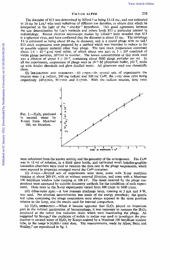
FIG. 1 .-H202 produced in aerated water by X-rays
100, from Maximar no filtration.
were calculated from the known activity and the geometry of the arrangement. The C o 6 0 was in 15 ml of solution, in a thick glass bottle, and calibrated small bakelite-graphite ionization chambers were used to measure the dose rate in the phage suspensions, which were exposed in ampoules arranged round the C060 container.
(ii) X-ruys.-Several sets of experiments were done, some with X-ray machines running at about 200 kV, with or without external filtration, and some with a Maximar 100 beryllium window tube running at 100 kV. The doses received by the phage sus- pensions were measured by suitable dosimetry methods for the conditions of each experi- ment. Dose rates in the X-ray experiments varied from 800 r/mh to 3600 r/min.
(iii) Ultra-violet light.-A low pressure discharge lamp, running at 3 mA and 9 W, was used. No absolute determination was made of the energy absorbed; the quartz test tubes containing the phage suspensions were always exposed in the same position relative to the lamp, and the results used for internal comparison.
(c) H202 ESTIMATION.-when it became apparent that H202 played an important part in the indirect inactivation of bacteriophage, it was necessary to measure the H2Oz produced at the rather low radiation doses which were inactivating the phage. As suggested by Savage,6 the oxidation of iodide to iodine was used to investigate the pro- duction in aerated water of H202 by X-rays emitted by a Maximar 100 beryllium window tube, in the range 0-10,000 r total dose. The measurements, made by Alper, Stein and Wakley,7 are reproduced in fig. 1.
Publ
ishe
d on
01
Janu
ary
1952
. Dow
nloa
ded
by N
orth
east
ern
Uni
vers
ity o
n 31
/10/
2014
04:
28:1
8.
View Article Online

236 INACTIVATION OF BACTERIOPHAGE
I
0 L - FIG. 2.-Observations in
8 irradiations of S13, 1.4 x 10⁻⁹ g/ml, y-rays at
140 r/min.
._
2:
-- 07 3 :
> 7 - L
. _
4, ' Dose in Rontycns X lo3 8
I1 12 (3 14 15 16 17 18 19 10,
RESULTS
SHAPE OF SURVIVAL CURVES.-The primary object of the first experiments (undertaken in collaboration with Dr. D. E. Lea) was to establish the relationship between inactivation dose and solid content of the phage suspension. It was at that time thought that indirect inactivation of phage particles resulted simply from single collisions with active radicals, the sort of mechanism which has since been called monotopic by Gray.* Where only this indirect effect of radiation is taking place, survival curves should be exponential. However, the survival curves for the lowest concentrations of phage, exposed to y-rays at 140r/min, showed a marked departure from the exponential. Fig. 2 presents the results of eight experiments on suspensions of 1.4 X 10⁻⁹ g solids/ml. The survival curves for the whole range of concentrations used, up to 3.3 x 10⁻² g/ml, are presented in fig. 3 and 4. As the concentration was increased the curves apparently approached more nearly to the exponential.
exponential nature of the curves might be explained by the presence of a protective sub- stance which, on combining with active radicals, lost its protective action. Such a protective substance could gradually be " cleaned up " by radiation and the survival curve would become exponential after the cleaning-up process was complete. Thus preliminary irradiation of the suspending buffer solution should dispose of such protec- tion, Ampoules containing 0.38 ml buffer were exposed to large doses of y-rays (30,000 r or more) and the pre-irradiated buffer was used in the last dilution stage of the phage preparation, the standard procedure being to add 0.02 ml of a phage suspension to the
SURVIVAL CURVES, PHAGE I N PRE-IRRADIATED BUFFER.-It Was thought that the non-
irradiated ampoule. The highest phage concentration which could be prepared in this way was, therefore, 5 x 10⁻² of the stock phage preparation. Parallel ampoules were always prepared, one to be used as a control for the other which was to undergo further irradiation. Since the number of active phage particles in the controls decreased with time, the " surviving fraction " in the irradiated ampoules was taken as the ratio between the number present in the irradiated ampoule and the number in the control at the time of each sampling. The survival curves presented in fig. 5 therefore represent the in- activation due only to the action of the y-rays on the phage in pre-irradiated buffer, the action of the irradiated buffer on the phage being automatically allowed for.
Comparison of fig. 5 with fig. 3 and 4 shows that the inactivation doses were con- siderably less when the phage was irradiated in pre-irradiated buffer. The survival curves are exponential for the lowest concentrations, but depart from the exponential at higher concentrations, showing that the cleaning-up hypothesis was not tenable.
TEMPERATURE DEPENDENcE.-It was found that the slope of the survival curves was dependent on temperature, in the y-ray experiments described, the temperature dependence being particularly marked for the suspensions in pre-irradiated buffer (fig. 6 and 7).
INACTIVATION BY H202.-An extensive series of experiments was undertaken to determine the effect of added H202 on S13, in order to assess the part played by the formed H202 in the irradiation experiments. The results were, briefly,
(i) inactivation of S13 by chemical H202 proceeded exponentially ; (ii) inactivation by H202 was dependent on temperature, on H202 concentration,
and on the solid content of the phage suspension (fig. 8, 9 and 10, illustrate these results).
Publ
ishe
d on
01
Janu
ary
1952
. Dow
nloa
ded
by N
orth
east
ern
Uni
vers
ity o
n 31
/10/
2014
04:
28:1
8.
View Article Online

T I K V A H ALPER 237
IRRADIATION IN PRESENCE OF CATALASL-Since H202 inactivated the phage, but re- quired time to do this, it seemed possible that the curvature of the semi-logarithmically plotted survival curves might be due to the gradual building up in the phage suspensions of H202, or H202 together with some other persistent toxic product of irradiation. It was thought that some light might be thrown on the action of irradiation-formed H202 if catalase were present in a suspension undergoing irradiation. In case catalase could act as a competitor for radicals, a control was used containing in the suspension an equal
I / !06 1
iFTtL 2 4 - , IO'STOCK 10' STOCK + 17- PEPTONE
T A G € ----I. [LEA AND SALAMAK]
'L J<.STOCK + 5% PEPTONE
3
',STOCK PHAGE
\
5- \,37"C
A , , I 2 3 4 5 6 7 1 8 9 10
DOSE ROENTGENS x 103
I 20°C 0 25oc
3- TIME HOURS
FIG. 3.-Survival curves, S13 in various dilutions of stock preparation : 'y-rays at 140 rlmin. FIG. 4.-Survival curves, S13 in various dilutions : y-rays at 140 r/min.
FIG. 5.-SurvivaI curves, S13 in various dilutions in pre-irradiated buffer : y-rays, 140 r/min.
FIG. 6.-Temperature dependence of survival curves : y-rays, 30 r/min. FIG. 7.-Temperature dependence of survival curves, phage in pre-irradiated
FIG. 8.-Survival curves, phage exposed to H202 at two temperatures.
amount of catalase which had been inactivated by heating for 15 min at 104" C. A phage suspension containing no catalase was irradiated simultaneously with the other two, at 6 r/min. The survival curves (fig. 11) demonstrated that the presence of active catalase caused inactivation of the phage to proceed exponentially, in contrast with the catalase free suspensions. It seemed probable, therefore, that where the time taken for irradiation was long enough, the gradual build-up of peroxides was responsible for an ever increasing rate of inactivation.
SURVIVAL CURVES AT A HIGHER DOSE RATE.-A series of experiments was then per- formed with X-rays at a much higher dose rate (3600 r/min), so that the total irradiation time was too short to allow of much action by the formed H202. It was found (fig. 12)
buffer : y-rays, 140 r/min.
Publ
ishe
d on
01
Janu
ary
1952
. Dow
nloa
ded
by N
orth
east
ern
Uni
vers
ity o
n 31
/10/
2014
04:
28:1
8.
View Article Online
238 INACTIVATION O F BACTERIOPHAGE
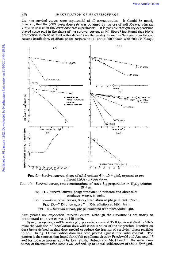
that the survival curves were exponential at all concentrations. It should be noted, however, that the 3600 r/min dose rate was obtained by the use of soft X-rays, whereas y-rays were used in the lower dose rate experiments. It is possible that quality dependence played some part in the shape of the survival curves, as M. Ebert 9 has found that H202 production in clean aerated water depends on the quality as well as the type of radiation. Recent irradiations of dilute phage suspensions at about 3000 r/min with 200 kV X-rays
(9)
I
4
HOUR5
1 2 3 4 5
m e + INACTIVATED CATALASE
P H & E + C A T K * Y \ I 2 3 4 5 . 6 7
OOK ROENTGENS x 103
:.I;..-- (10)
\ 2
3
5 x 10. STOU
2 X Id6 STOCK
FIG. 9.-Survival curves, phage of solid content 6 x 10⁻⁹ g/ml, exposed to two different H202 concentrations.
FIG. 10.-Survival curves, two concentrations of stock S13 preparation in H202 solution 10-4 m.
FIG. 11 .-Survival curves, phage irradiated in presence and absence of catalase : y-rays, 6 r/min.
FIG. 12.-All survival curves, X-ray irradiation of phage at 3600 rlmin. FIG. 13.-" Dilution curve " : X-irradiation at 3600 r/min.
FIG. 14.-Survival curves, phage irradiated with ultra-violet light.
have yielded non-exponential survival curves, although the curvature is not nearly so pronounced as in the curves at 140 r/min.
EFFECT OF DILunoN.-The series of exponential curves at 3600 r/min was used to deter- mine the variation of inactivation dose with concentration of the suspension, inactivation dose being defined as that dose needed to reduce the fraction of surviving phage particles to e-1. In fig. 13 inactivation dose has been plotted against total solid content. The pattern is the same as that found for rabbit papilloma virus by Friedewald and Anderson,*() and for tobacco mosaic virus by Lea, Smith, Holmes and Markham.11 The initial con- stancy of the inactivation dose is well defined, up to a total solid content of about 10-6 g/ml.
Publ
ishe
d on
01
Janu
ary
1952
. Dow
nloa
ded
by N
orth
east
ern
Uni
vers
ity o
n 31
/10/
2014
04:
28:1
8.
View Article Online

TIKVAH ALPER 239
INACTIVATION BY ULTRA-VIOLET LIGHT.-It may be of interest to compare these results with those for inactivation by ultra-violet light. Inactivation was independent of con- centration in the range tested, viz. 1.4 X 10⁻⁹ g/ml to 1.4 x 10⁻⁴ g/ml, and proceeded exponentially at all concentrations (fig. 14). As the irradiation times were short, no delayed effect would have shown up in the survival curves, and such an effect was not looked for at the time these experiments were done. The fact that inactivation dose was independent of concentration was in contrast with the results for ionizing radiations or for added H202.
AFTER-EFFECTS OF RADunoN.-Evidence from the experiments so far described led to the expectation that, after the end of X- or y-irradiation, phage would continue to be inactivated by the products of the irradiation formed in the suspending medium. This was in fact found to be the case, as illustrated in fig. 15 by survival curves after various doses of X-rays. The curves were exponential, as far as they went. In subsequent work, however, greater initial concentrations of phage were used, so that inactivation could
FIG. 15.-Inactivation of phage - after various doses of X-rays
36CO R
4 5 0 0 R
1 6 0 0 0 R
- - - L : L I , , , I
20 42 60 80 i00 !20 lo0 160 ;cO & MIPJUTES AFTER IRRADIATIOhI COb44EKE3
FIG. 16.-Survival curves, irradiated phage and non-irradiated phage in irradiated buffer, and non-irradiated phage in heat-inactivated, irradiated
+ Phaye A 6uJu /rruda/ed
phage suspension.
be followed for longer times and to smaller surviving fractions. It was then found that the curves departed from the exponential, the inactivation rate decreasing with time.
ATTEMPT TO FJND EFFECT OF ORGANIC PERoxnxs.-In all the after-effect experiments the rate of inactivation was considerably greater than that found as a result of adding to phage suspensions the amount of H202 which would be produced by the radiation. Similarly, it was found that phage still surviving at the end of radiation was subsequently inactivated at a much greater rate than phage put into buffer which had been exposed to the same dose of radiation. It was thought that organic material, other than phage, present in the suspensions might react with the active radicals to form toxic organic per- oxides, and that this would explain the difference in the results. In order to test this explanation a suspension of phage was inactivated by heating for about 30 min at 60" C, and then irradiated simultaneously with clean buffer and with a suspension of active phage. At the end of irradiation active phage was introduced into the irradiated buffer and the irradiated killed phage suspensions, and sampling of these suspensions, together with the irradiated suspension of active phage, was continued for 140 min. The survival curves, presented in fig. 16, show that the inactivation rate was the same for active phage intro- duced into the irradiated buffer or the irradiated killed phage suspension, and much less
Publ
ishe
d on
01
Janu
ary
1952
. Dow
nloa
ded
by N
orth
east
ern
Uni
vers
ity o
n 31
/10/
2014
04:
28:1
8.
View Article Online

240 INACTIVATION OF BACTERIOPHAGE
than the inactivation rate of the phage which had been present during irradiation and had survived the immediate effects thereof.
ENHANCED SUSCEPTIBILITY TO ACTION OF IRRADIATED BUFFER SOLUTION AND H202.- Subsequent work has established the fact that the greater part of the delayed inactivation of phage after the end of irradiation is due to a change, occurring as a result of the action of radicals, which makes it much more susceptible thereafter to the action either of added H202 or of the H202 formed by irradiation of the suspending medium. Fig. 17 illus- trates the fraction of surviving phage particles, at various times after the end of irradiation, in the following suspensions of S13 :
(A) A dilute suspension (about 0.8 pg/ml total solids) in 10-3 M phosphate buffer,
(B) 0-1 ml of suspension A introduced after irradiation into 1.9 ml buffer which had exposed to 15,000 r of 200 kV X-rays.
been irradiated simultaneously with A.
FIG. 17.4urvival curves of various suspensions, after 16000 roentgens
of X-rays.
FIG. 18.4urvival curves, phage in 3 x 10⁻⁵ M H202.
(C) 0.1 ml of non-irradiated suspension, equal in concentration to A, introduced
(D) 0.1 ml of suspension A afier irradiation introduced into 1-9 ml of non-irradiated
(E) 0.1 ml of non-irradiated suspension, equal in concentration to A, introduced
The curves illustrate the fact that the inactivation rate was identical for suspensions A and B, and that to produce this rapid rate of inactivation it was necessary (i) that the phage suspension be exposed to radiation, (ii) that it should be in contact thereafter with irradiated buffer solution. The additional curve F is the ratio between curves A (or B) and C, and therefore expresses the number of survivors in the irradiated suspension as a fraction of the number of phage particles not affected by radiation which would survive the H202 produced. Curve F therefore depicts the inactivation due only to the interaction between phage damaged by the radiation and H202. This inactivation apparently pro- ceeded until a constant fraction remained, presumably those particles not affected by the radiation.
into 1.9 ml of buffer which had been irradiated simultaneously with A.
buffer.
into 1.9 ml of non-irradiated buffer. (Control.)
Publ
ishe
d on
01
Janu
ary
1952
. Dow
nloa
ded
by N
orth
east
ern
Uni
vers
ity o
n 31
/10/
2014
04:
28:1
8.
View Article Online

T I K V A H ALPER 241
In further experiments phage was exposed to a I3202 solution of concentration 3 X 10⁻⁵ My this being roughly the concentration which was produced in aerated water by 15,000 r of 100 kV X-rays (fig. 1). As can be seen from fig. 18, the inactivation rate for irradiated phage exposed to this concentration of Hz02 was very much greater than for non-irradiated phage exposed to the same concentration. The results with phage S13 have been reproduced with T3, which has been found to be much more radiation sensitive. The delayed effect on S13, after 15,000 r, could be reproduced with T3 after 2,000r. No systematic comparison has as yet been made of the radiosensitivity of the two phages, but the figures quoted indicate that the doses necessary to produce the same after-effects are roughly inversely proportional to the surface area of the phage particles.
S13 to the action of H202 was observed in suspensions from which dissolved oxygen had been removed, as well as in fully aerated suspensions. In order to demonstrate the effect it was necessary to introduce aliquots after irradiation into irradiated aerated buffer or into H202 solutions. A certain delayed effect was demonstrable with phage irradiated in oxygen free conditions, as with irradiated phage introduced into non-irradiated buffer, but this effect was small when compared with the inactivation resulting from the exposure
AFTER-EFFECT ON OXYGEN-FREE SUSPENSIONS.-The enhanced susceptibility of irradiated
1
M/hotex offer end of /rrodblion
FIG. 19.-Survival curves : irradiated oxygen free suspension after about 10,000 r ; and aliquot intro-
duced into irradiated aerated buffer.
/ O 15 20
FIG. 20.-Survival curves aerated and oxygen-free suspensions of S13 : X-rays,
2800 r/min.
of the irradiated phage to irradiated aerated buffer. Fig. 19 presents the survival curves from an experiment in which the irradiated phage suspension had been rendered oxygen- free by bubbling nitrogen through for about 40 min before irradiation commenced.
the X-irradiation of oxygen-free suspensions of S13, inactivation proceeded at a rate which was certainly no slower than that observed in aerated suspensions. The in- activation rate became greater in the latter only when reaction with H202 had begun to contribute to the inactivation (fig. 20).
SURVIVAL CURVES, AERATED AND OXYGEN-FREE SUSPENSIONS.-It Was found that during
DISCUSSION
As bacterial viruses have been shown to consist to a large extent of DNA,12 it is interesting to speculate on whether enhanced susceptibility to H202 after exposure to active radicals is, in fact, a property of DNA. Butler and Conway 13 found that DNA continued to undergo degradation after the cessation of radiation only when dissolved oxygen was present during irradiation. They also found that, while DNA was affected by H202, the concentration required to produce the same effect as a given dose of radiation was too high to have been formed by the radiation. On the other hand they found that the immediate effects of radiation
Publ
ishe
d on
01
Janu
ary
1952
. Dow
nloa
ded
by N
orth
east
ern
Uni
vers
ity o
n 31
/10/
2014
04:
28:1
8.
View Article Online

242 INACTIVATION OF BACTERIOPHAGE
were not dependent on oxygen concentration. These results might well be ex- plained on the basis of DNA acquiring an enhanced sensitivity to H202 during irradiation.
If the phenomenon which has been described is, in fact, a property of DNA, and therefore of living cells, it is clear that ionic yields based on observations on susceptible in vitro material, and made immediately after cessation of radiation, would appear much lower than if such damage as has here been reported for bacteriophage were given time to express itself. It is perhaps significant that damage to tissue cells, well known to occur, in general, at much lower radiation levels than damage to in vitro material, is commonly assessed by observations made some time after the end of radiation. The results of the experiment illustrated by fig. 17 make it possible to compare very roughly the ionic yield assessed in terms of immediate inactivation with that assessed in terms of damage to phage particles which makes them susceptible to Hz02. Curve F of fig. 17 shows that inactivation due only to reaction between damaged phage and H202 proceeded until, after about 80 min, the survivors remained at about 15 % of the number still active at the end of irradiation, which was 50 % of the number of particles at the beginning of irradiation. Thus the fraction remaining completely un- damaged by the actual irradiation was 7.5 %. If the action of the radicals in damaging the phage was monotopic, the dose required to damage 92.5 % would be about four times the dose required to damage 50 % (since 0.5 = e-0.7, and 0.075 = e-2*6), and an assessment of ionic yield based only on immediate inactiva- tion would be smaller by a factor 4 than an assessment based on the immediate inactivation plus the after-effect. As has been stated, the results obtained with 15,000 r for S13 were roughly duplicated for T3 with a dose of 2,000 r ; which was sufficient to bring about inactivation of 93 % of the particles within 80 min. after irradiation ceased. It would seem that the order of magnitude of irradiation dose required to affect this type of in vitro material therefore does approach the doses which are commonly used in in vitro experiments on radiation effects.
Some of the results described may be of interest in throwinglight on the behaviour of active radicals 14 in irradiated aqueous solutions. At the lowest concentrations of S13, the inactivation dose was constant, so that ionic yield was directly pro- portional to phage concentration, up to 10-6 glml. This accords with the theory set out by Dainton,ls and shows that the recombination of radicals plays the greatest part in their elimination in suspensions which contain fewer than about 1011 particles/ml of diameter whose order of magnitude is 15mp.
The experiments in which the immediate effects of radiation on aerated and oxygen-free phage suspensions were compared demonstrate that, for bacteriophage at least, the presence of dissolved oxygen does not give rise to an indirect effect of greater efficiency, except in so far as the oxygen is necessary for the formation of H202 which plays a part in a secondary reaction with phage particles already damaged by the radicals. An apparently enhanced inactivation in the presence of oxygen would, however, arise from the reaction between affected phage partides and Hz02, which can be formed by X- or y-rays only when oxygen is present. There is no evidence from this work, therefore, of the action of radicals of the H02 type, which, it has been thought, might account for some of the increased effects of radiation on oxygenated material. In this connection it is interesting to note that Butler and Conway 13 in their studies on the X-irradiation of DNA, found no dependence of immediate effects on the oxygen concentration of their solutions.
These investigations have been carried out in several laboratories, namely, the Strangeways Research Laboratory, Cambridge ; Onderstepoort Laboratories, Pretoria, South Africa ; the National Physical Laboratory, Council for Scientific and Industrial Research, Pretoria : and the Radiotherapeutic Research Unit of the Medical Research Council, Hammersmith Hospital. T am very grateful to the authorities at all these institutions for the generous facilities granted me. I
Publ
ishe
d on
01
Janu
ary
1952
. Dow
nloa
ded
by N
orth
east
ern
Uni
vers
ity o
n 31
/10/
2014
04:
28:1
8.
View Article Online

TIKVAH ALPER 243
am indebted to Mr. D. J. Savage and Miss Ilmary Reeler, of the National Physical Laboratory, South African Council for Scientific and Industrial Research, for technical assistance. I should like to acknowledge my gratitude to Dr. W. Hayes, of the Postgraduate Medical School, for his assistance, helpful advice, and also for preparing and giving me a stock of the T3 phage. Dr. M. Ebert has been very kind in assisting me with many of the chemical problems.
I have had the great privilege of Dr. L. H. Gray’s interest in this work from the time it was initiated, and owe to him many of the ideas which have been followed UP-
1 Dale, Biochem. J., 1940, 34, 1367. 2 Elford and Andrews, Brit. J . Expt. Path., 1932, 13, 446. 3 Lea, Actions of Radiations on Living Cells (C.U.P., Cambridge, 1946), chap. 3. 4 Elford, personal communication. 5 Anderson, paper in Symposium, The Nature of the Bacterial Surface, (Blackwell,
6 Savage, Analyst, 1951, 76, 224. 7 Alper, Stein and Wakley, to be published. 8 Gray, Brit. J. Radiol. (in press). 9 Ebert, personal communication ; to be reported at this Discussion.
10 Friedewald and Anderson, J. Expt. Med., 1940,45, 713. 11 Lea, Smith, Holmes and Markham, Parasitology, 1944, 36, 110. 12 Cohen and Anderson, J. Expt. Med., 1946, 84, 511. 13 Butler and Conway, J. Chem. Soc., 1950, 670, 3418. 14 Weiss, Nature, 1944, 153, 748. 15 Dainton, Ann. Reports, 1949, 45, 5.
Oxford, 1949), p. 87.
Publ
ishe
d on
01
Janu
ary
1952
. Dow
nloa
ded
by N
orth
east
ern
Uni
vers
ity o
n 31
/10/
2014
04:
28:1
8.
View Article Online