indications for electrophysiological testing · indications for electrophysiological testing samuel...
TRANSCRIPT
1
Indications for Electrophysiological Testing
Samuel C. Dudley, Jr., M.D., Ph.D.Division of CardiologyDepartment of PhysiologyEmory University/Atlanta VAMC

2
What EP testing can doWhat EP testing can do
l Measure conduction intervals– good for bradyarrhythmias
l Add extrastimuli– good for reentrant tachyarrhythmias
l Ablation– good for focal and reentrant tachycardias

3
Conduction systemConduction system

4
Measurements madeMeasurements made
l Recovery of automaticityl Conduction velocityl Refractorinessl Activation mappingl Pace mapping

5
Mechanisms of arrhythmiaMechanisms of arrhythmial Automaticity
– normal (e.g. sinus tachycardia)– abnormal (e.g. reperfusion arrhythmias)
l Triggered activity– Early afterdepolarizations associated with QT
prolongation (torsades de pointes)– Delayed afterdepolarizations associated with Ca2+
overload (e.g. digoxin)l Reentry
– fixed obstruction (e.g. atrial flutter)– leading circle (e.g. ventricular fibrillation)

6
Reentry Reentry -- initiationinitiation
Josephson. 1993. Clinical Cardiac Electrophysiology 2nd Edition. 183.

7
Reentry Reentry -- response to response to extrastimulusextrastimulus
NothingNothing EntrainmentEntrainment TerminationTerminationJosephson. 1993. Clinical Cardiac Electrophysiology 2nd Edition. 209.

8
Triggered activityTriggered activity
EADs EADs -- Bradycardia DependentBradycardia Dependent DADs DADs -- Tachycardia DependentTachycardia Dependent
Wit and Rosen. 1992. In The Heart and Cardiovascular System, Ed. Fozzard et al. Raven Press.

9
Responses of arrhythmias during PESResponses of arrhythmias during PES
NormalAutomaticity
AbnormalAutomaticity
EADs DADs Reentry
Initiated bydrive train
No No No Yes Yes
Initiated byextrastimuli
No No No Variable Yes
Suppresion byoverdrive
Yes, notermination
No, notermination
Yes Variable Rare, possibleentrainment
Terminationbyextrastimulus
No No Variable Unlikely Yes,termination ina range

10
Problems addressed by EP studiesProblems addressed by EP studies
l Bradyarrhythmias (site of block)– Sinus node function– AV block– IVCD
l Tachyarrhythmias– SVT
• AV reentrant tachycardia• AV nodal reentry
– VT
l Syncopel Evaluate implanted
device programming options
l Evaluate efficacy of therapy
11
Basic rulesBasic rules
l Always try to make an EKG diagnosis first.l Fix ischemia firstl If you cannot bring on the tachycardia, it is
hard to ablate it.– Think twice about starting drugs
l If the rhythm is not stable, it is hard to ablate it.
12
When not to do EPSWhen not to do EPSl Symptoms correlating with
ECG findingsl Asymptomatic patients with
sinus slowing or Wenckebach during sleep only
l Asymptomatic bifascicularblock
l Asymptomatic preexcitation
l Congenital long QT and acquired long QT correlating with symptoms
l Asymptomatic patients without risk factors for SCD
l Patients with cardiac arrest within 48 hrs of ischemia/MI
l Cardiac arrest from other causes

13
Complications (<2%)Complications (<2%)l Hemorrhagel Phlebitisl Thromboembolusl Arrhythmiasl Tamponadel CVA (Left sided procedures)l Pneumothoraxl RF ablation
– valve damage– AV block

14
HRA
RV
CSHIS
RV
Catheters positionsCatheters positions

15
Normal ElectrogramNormal Electrogram
His spike
Josephson. 1993. Clinical Cardiac Electrophysiology 2nd Edition. 98.

16
Sinus node dysfunctionSinus node dysfunction
Prystowsky and Klein. 1994. Cardiac Arrhythmias. 307.
17
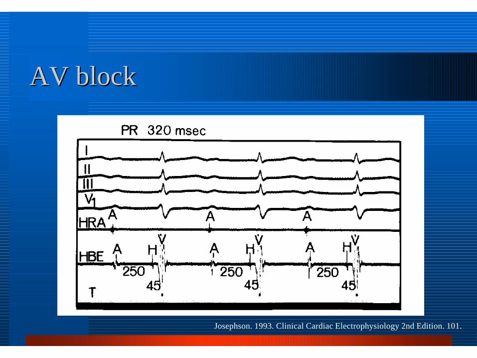
AV blockAV block
Josephson. 1993. Clinical Cardiac Electrophysiology 2nd Edition. 101.

18
AV BlockAV Blockl Type I 2° AVB or 3° AV block
with narrow QRS– AV node– rarely intra His
l Type I 2° AVB or 3° AV block wide QRS– anywhere
l Type II 2° AVB, wide QRS– infra His– intra His– AV node (rare)Josephson. 1993. Clinical Cardiac Electrophysiology 2nd
Edition. 110.

19
HV intervals & HV intervals & bifascicularbifascicular blockblock
l HV > 55 ms high sensitivity but low specificity for progression (2-3%/yr CHB)
l Infra His block during atrial pacing has low sensitivity but high specificity
Josephson. 1993. Clinical Cardiac Electrophysiology 2nd Edition. 108.
20
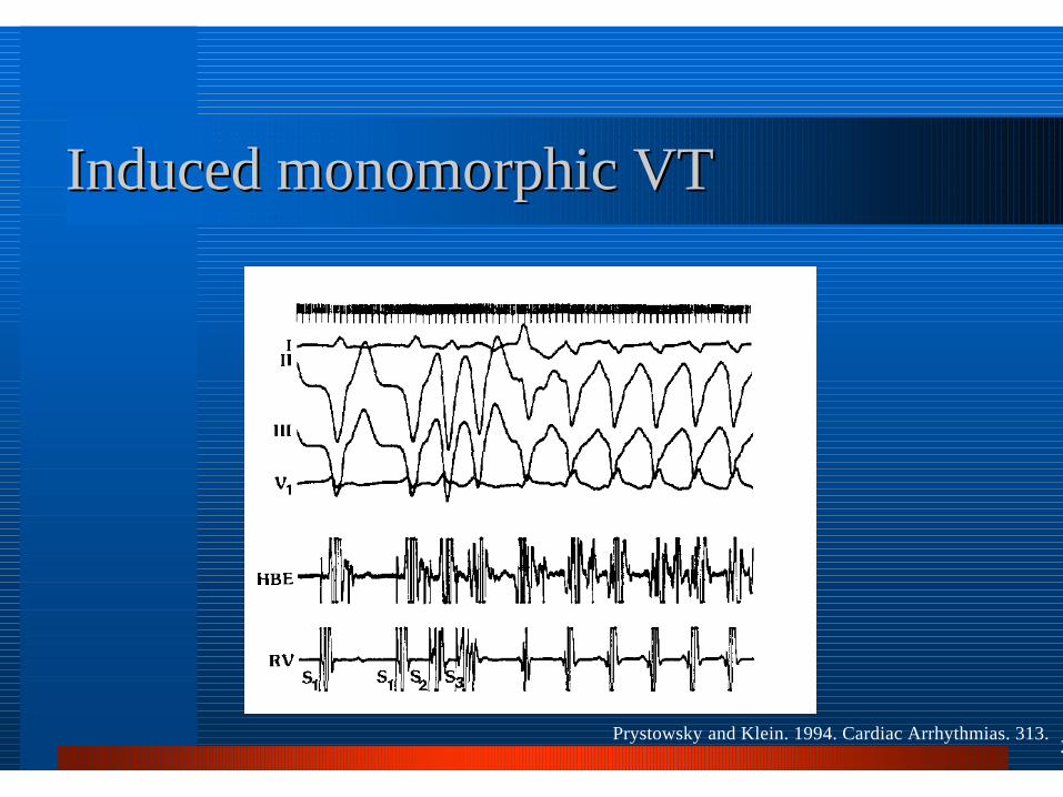
Induced monomorphic VT Induced monomorphic VT
Prystowsky and Klein. 1994. Cardiac Arrhythmias. 313.

21
VT or SVT?VT or SVT?
Josephson. 1993. Clinical Cardiac Electrophysiology 2nd Edition. 422.

22
MUSTT registryMUSTT registry
Bruxton et al. 1999. NEJM: 341, 1882.
23
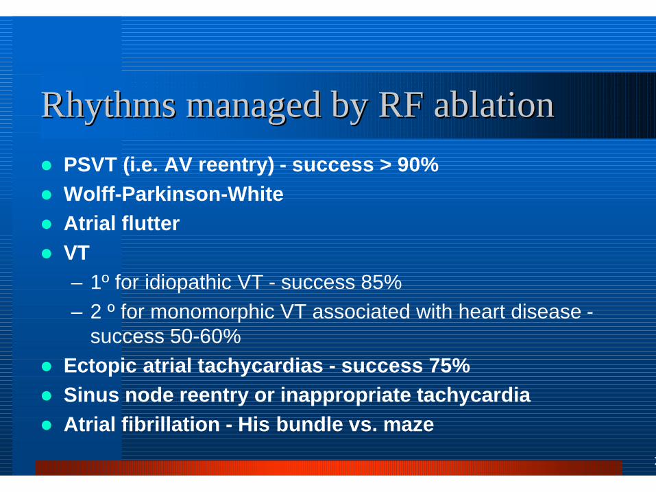
Rhythms managed by RF ablationRhythms managed by RF ablationl PSVT (i.e. AV reentry) - success > 90%l Wolff-Parkinson-Whitel Atrial flutterl VT
– 1º for idiopathic VT - success 85%– 2 º for monomorphic VT associated with heart disease -
success 50-60%l Ectopic atrial tachycardias - success 75%l Sinus node reentry or inappropriate tachycardial Atrial fibrillation - His bundle vs. maze

24
SVT ablationSVT ablation
SVT SVT -- Long RPLong RP
Post ablationPost ablation
Pre ablationPre ablation

25
Mapping WPWMapping WPW
Josephson. 1993. Clinical Cardiac Electrophysiology 2nd Edition. 347.

26
SVT ablationSVT ablation
Josephson. 1993. Clinical Cardiac Electrophysiology 2nd Edition. 743.

27
AV nodal reentryAV nodal reentry

28
Mapping SVTMapping SVT
Josephson. 1993. Clinical Cardiac Electrophysiology 2nd Edition. 188.

29
Ablating SVT Ablating SVT -- Triangle of KochTriangle of Koch
Coronary os
Tricuspid annulus
Compact AV nodeTendon of todaroCrista terminalis
IVC os Slow pathwaySlow pathway
Fast Pathway