indian society of gastroenterology - springer
TRANSCRIPT

ABSTRACTS
Indian Society of Gastroenterology
Received: 2 October 2013 /Accepted: 2 October 2013 /Published online: 14 November 2013# Indian Society of Gastroenterology 2013
Plenary Session
A-1
Cancer stem cells in hepatocellular carcinoma: Animmunohistochemical study with clinicopathologicalcorrelation
Smita Mary Matthai, Banumathi RamakrishnaDepartment of Pathology, Christian Medical College, Vellore632 004, India
Background and Aims : Cancer stem cells (CSC) may beresponsible for tumor recurrence and resistance to che-motherapy in hepatocellular carcinomas (HCC). Thisstudy evaluated the associations between histologicalparameters and liver CSCs (LCSC) in HCC, and comparedCSC in HCC associated with and without hepatitis B virus(HBV) infection.Methods : Seventy-nine tumors (49 surgical resections from46 patients, and 30 from autopsy) were reviewed. Immuno-histochemical staining for the LCSC marker EpCAM, liverprogenitor cell (LPC) marker CK19 and NCAM wereperformed and were correlated with histological features oftumor behavior.Results : Thirty-three tumors (41.8 %) showed positive stain-ing for EpCAM. CK19 and NCAM expression were seen in26 (32.9 %) and 4 (5.1%) tumors respectively. The expressionof EpCAM and CK19 correlated significantly with each other(p<0.001). EpCAM expression was significantly associatedwith features indicating aggressive tumor behavior, includingyounger age of onset, higher serum AFP levels, tumor celldedifferentiation, increased mitotic activity, and vascular in-vasiveness. There was no significant difference in expressionof EpCAM, CK19 and NCAM between HBV positive andnegative HCC.
Conclusion : The LCSC marker EpCAMwas expressed in lessthan half of HCC, was independent of HBVetiology, and wasstrongly associated with clinical and histological features ofaggressive tumor behavior. Positive staining for CK19 suggestsa possible LPC origin of the EpCAM positive HCCs.
A-2
Relationship of cytokines, oxidative stress andgastrointestinal motility with bacterial overgrowth inulcerative colitis patients
SVRana , S Sharma, S K Sinha, K K Prasad, AMalik, J Kaur,R K MoryaDepartment of Super Specialty of Gastroenterology, Postgrad-uate Institute of Medical Education and Research, Chandigarh160 012, India
Background : Ulcerative colitis (UC) is idiopathic, chronic andrelapsing inflammatory bowel disease. Factors which initiateand perpetuate UC are not well understood. It is still unclear ifany relationship exists between cytokines, oxidative stress,gastrointestinal (GI) motility, small intestinal bacterial over-growth (SIBO) in UC patients. Therefore, the present studywas planned to examine the relationship between these factorsamong UC patients.Methods : Total 120 UC patients and 125 age and sex matchedcontrols with no GI symptoms were enrolled. Levels of plas-ma IL-6, IL-8, TNF-α and IL-10 were measured in all subjectsby using ELISA. Lipid-peroxidation (LPO) and reduced glu-tathione (GSH) were measured by standard methods.Orocecal transit time (OCTT) and SIBO were measured bylactulose and glucose hydrogen breath tests respectively.Results : Out of 120 UC patients, 74 were males with mean±SD of age 45.6±17.5 years. Plasma levels of IL-6, IL-8,TNF-α and IL-10 in UC patients were significantly higher
Indian J Gastroenterol (November 2013) 32(Suppl 1):A1–A132DOI 10.1007/s12664-013-0417-z

(p<0.01) as compared to controls. LPO in UC patients wassignificantly increased (p<0.01) while reduced glutathione(GSH) was significantly decreased (p<0.01) as compared tocontrols. OCTT and SIBO were significantly higher in UCpatients as compared to controls. It was also observed thatthere was significant correlation between SIBO with IL-6, IL-8, TNF-α, IL-10, LPO and GSH. Although correlation withGSH was inversely proportional to SIBO.Conclusion : This study indicates that increase in cytokines anddecrease in anti-oxidants in UC patients would have resulted inoxidative stress causing delayed GI motility leading to SIBO.
A-3
Colorectal cancers in Indian patients with ulcerative colitis
Devendra Desai , Abhijit Deshmukh, Philip Abraham, AnandJoshi, Sudeep Shah, Tarun Gupta, Ramesh Deshpande, VarunKhandagale, Siji GeorgeDivisions of Gastroenterolgy, Surgery, and Pathology, P DHinduja Hospital, Veer Savarkar Marg, Mahim, Mumbai 400016, India
Background : Data from India suggest that colorectal cancer(CRC) occurs in 0.94 % to 1.8 % of patients with ulcerativecolitis (UC). We report our data on CRC in UC.Methods : Data on 430 patients (248males [57.7 %]; mean age44 [SD 14.6] years; median duration of disease 6 (Interquartilerange 7) years with UC were available for analysis from theinflammatory bowel disease database maintained since 2005.Thirty-eight (8.8 %) patients had proctitis, 95 (22.1 %) procto-sigmoiditis, 131 (30.5 %) left-sided colitis, 159 (37 %)pancolitis and extent was unknown in 7 patients. Patients withhistologically confirmed CRC within the segment with colitiswere compared with those without CRC, to determine riskfactors for the development of CRC.Results : Twelve patients (2.8 %) developed CRC. The inci-dence density was 3.556/1000 patient-years of disease (PYD)overall–2.995/1000 in the first 10 years, 3.338/1000 PYD at10 to 20 years, and 7.030 /1000 PYD at >20 years. Onunivariate analysis age, gender, duration of disease, extent ofcolitis, smoking, duration of 5-aminosalicylic acid therapy,family history of inflammatory bowel disease, medicationcompliance and disease control were considered for analysis.Extensive colitis (disease proximal to splenic flexure) (p=0.01), longer duration of disease (p=0.00001) and poor con-trol of disease (p=0.007) were associated with development ofCRC. On multivariate analysis, duration of disease (p=0.0099) and extent of colitis (p=0.0273) were significantfactors for development of malignancy.Conclusion : In this study, colorectal cancer occurred in 2.8 %of patients with ulcerative colitis. The risk increased withextent and duration of disease.
A-4
Microsatellite instability and promoter hypermethylationin colorectal cancer in India
V V Ravi Kanth , Sandeep Balsingh, M Sasikala, G V Rao,R Pradeep, Steffie Avanthi, D Nageshwar ReddyAsian Healthcare Foundation and Asian Institute of Gastro-enterology, 6-3-661, Somajiguda, Hyderabad 500 082, India
Background and Objectives : The incidence of colorectal can-cer (CRC) is increasing in India. Genomic instability is thehallmark of cancer and microsatellite instability (MSI) is animportant factor in tumor development. MSI is ahypermutable phenotype caused by the loss of DNAmismatch repair activity. It is important to identify MSI clin-ically as they have a better prognosis and differ with responseto chemotherapy. Limited data is available on the incidence ofMSI in Indian CRCs. The objectives of this study were toidentify the extent of MSI in Indian CRC patients below 50years and determine promoter methylation status of hMLH1and hMSH2 in relation to MSI.Methods : Ninety-one individuals with CRC were recruited(Bethesda guidelines); patient characteristics were noteddown. Colorectal tumors were tested for MSI by NCI recom-mended Bethesda panel using labeled primers and the frag-ments were separated and analyzed on Beckman Sequencer.Promoter methylation status was determined by restrictionenzyme digestion and PCR.Results : MSI-High was detected in 13.2 % (12/91), MSI-lowin 35.2 % (32/91) and the rest were Microsatellite stable(MSS) 51.6 % (47/91). Majority (8/12) of the MSI-H tumorswere associated with rectum (site of the tumor, were moder-ately or poorly differentiated (12/12) and the tumor type wasadenocarcinoma (10/12). Promoter hypermethylation was sig-nificantly associated with instability, both MSI-H and MSI-L.Conclusions : In the present study, MSI, both high and lowwas associated with 48.4 % of CRC patients below fifty yearsof age.
A-5
HCV viremia levels are determinant of serum IFN-αexpression via TRAF6 interaction with MAVS and TRIF
Phani Kumar Gumma , Soumya Jyoti Chowdhury, VijayKumar Karra, Rajesh Ruttala, Premashis KarDepartment of Medicine, Maulana Azad Medical College,Bahadur Shah Zafar Marg, Delhi Gate, New Delhi, Delhi110 002, India
Infection with the hepatitis C virus (HCV) leads to chronichepatitis C (CHC) in 50 % to 80 % of individuals. The
A2 Indian J Gastroenterol (November 2013) 32(Suppl 1):A1–A132

recognition of HCV by immune system, triggers pathwaysleading to type-I interferon production, and induce an antiviralstate. HCV associated with hepatocellular carcinoma (HCC)and B cell lymphoma. HCV infection causes a 5 to 10 foldincrease in mutation frequency in Ig heavy chain, BCL-6, p53,and β-catenin genes of in vitro HCV-infected B cell lines andHCV-associated peripheral blood mononuclear cells, lympho-mas, and HCCs, but not in lymphomas of non-viral origin orHBV-associated HCC. HCV induces a mutator phenotype andmay transform cells by a hit-and-run mechanism in the host.Gene sequences codes for TRAF6-binding motifs in TRIF250–PEEMSW–255 and in MAVS, 153–PGENSE–158 and455–PEENEY–460 were analyzed for mutations by directsequencing, and IFN-β levels from 63 patients of CHC and38 healthy individuals. We made an effort to confirm theseprotein interactions by co-immuno precipitation of TRAF6with MAVS and TRIF isolated from biopsy of 47 chronichepatitis C patients and 12 autopsy liver tissue and Westernblotting. Alignment sequences code for MAVS and TRIFmotifs which interacts with TRAF6 revealed they were highlyconserved. MAVS and TRIF interact with TRAF6 in CHCpatients. Thus there is no association of sequence variationswith these protein interactions in CHC patients. HCV viremia<1.4 × 10⁵ IU/mL was associated with TRAF6 interactionwith MAVS and TRIF proteins which causes increase inIFN-β levels in CHC patients.
A-6
Complete hepatitis B virus genome analysis in patientsof hepatocellular carcinoma and asymptomatic carriersfrom Northern, Sothern and North East India
Manash Pratim Sarma , Giasuddin Ahmed, Subhash Medhi,Premashis KarDepartment of Medicine, Maulana Azad Medical College,University of Delhi, Bahadur Shah Zafar Marg, Delhi Gate,New Delhi, Delhi 110 002, India, and Department of Biotech-nology, Gauhati University, Assam
Materials and Method : Seventy-five HBV related HCC casesand 15 HBV related asymptomatic carriers were included inthe study. HBV DNA was amplified by six sets of walkingprimers. PCR amplicons were sequenced commercially, sub-mitted to http://www.ncbi.nlm.nih.gov, translated into aminoacid and aligned using BioEdit v7.0.9.Result : A total of 60, 15, 23 and 1 mutations were observed inthe PC/C, X, P and S genes respectively. Mutations like 10I→L was significantly associated in HCC cases from NEI [(p=0.01; OR=5. 63) VS SI] and [(p<0.01; OR=16.63) VS NI].Mutations like 41S→T (p< 0.001; OR= 19.01), 92 V→G (p<0.001; OR= 19.01), 96N→T (p< 0.001; OR= 19.01) and164Q→P (p=0.0279; OR= 3.085) were found significantly
associated with HCC cases from NI [VS SI].Widely reported28 W→stop mutation was found in few HCC cases. 132→stop [(p= 0.004486; OR= 5.479 VS SI) and [(p= 0.004486;OR= 5.479) VS NEI] was a novel finding. Mutations like267I→N and 268D→Twere exclusively found in HCC casesfrom NEI while 270S→F was confined to NI. Reported drugmutants like 80L→I, 236N→T, 169I→Tand 181A→V) werealso observed.Conclusion : PC/C region of the HBV genome is most proneto mutation followed by P, X and S genes. The highestvariation in the entire HBV genome was observed in HCCcases from North India while the genome was compar-atively conserved in asymptomatic HBV carriers. Novalmutations in surface (132 stop), polymerase (frameshiftmutation at codon 178), core (10 I→L, 41 S→T, 92V→G, 96 N→T and 164 Q→P) and X (33 P→S) geneneeds molecular and cellular studies to be used as anovel kit for the diagnosis and prognosis of HCC.Association of exclusive mutants in HCC cases fromNEI needs close attention for developing region basedHBV related HCC management in India.
Young Investigator Award Session
B-1
Single stage laparoscopic common bile duct explorationand cholecystectomy versus two stage endoscopic stoneextraction followed by laparoscopic cholecystectomyfor patients with concomitant gallbladder stonesand common bile duct stones
Karthik Rajan , Virinder Kumar Bansal, Mahesh C Misra,Ragini Kilambi, Subodh Kumar, Asuri Krishna, Atin Kumar,Chandrakant S Pandav, Rajeshwari Subramaniam, M KArora, Pramod Kumar GargDepartments of Surgery, Radiodiagnosis, Preventive and So-cial Medicine, Anesthesiology, and Gastroenterology, All In-dia Institute of Medical Sciences, New Delhi 110 029, India
Background : Management of gallbladder stones with com-mon bile duct (CBD) stones is debatable with two-stageendoscopic stone extraction followed by laparoscopic chole-cystectomy being the currently preferred method.Objective : To compare the success and cost-effectiveness ofsingle stage vs. two-stage management of patients with con-comitant gallbladder and CBD stones.Methods : Consecutive patients with concomitant gallbladderand CBD stones were randomized to either single-stage lapa-roscopic CBD exploration and cholecystectomy (Group I) orto two-stage procedure ie. ERCP for CBD stones extractionfollowed by laparoscopic cholecystectomy (Group II).
Indian J Gastroenterol (November 2013) 32(Suppl 1):A1–A132 A3

Success was defined as complete clearance of CBD andcholecystectomy by the intended modality.Results : From February 2009 to October 2012, 168 patientswere randomized–84 to single-stage procedure (Group I) and84 to two-stage procedure (Group II). Both the groups werematched with regard to patients’ demographic and clinicalparameters. The success rates of laparoscopic CBD explora-tion and ERCP for clearance of CBD were similar (91.7 % vs.88.1%). Overall success rate was also similar (88.1 % vs. 79.8%; p=0.20). The mean operative time was significantly higherin Group I (135.7±36.6 vs. 72.4±27.6 minutes; p=<0.001) butthe overall hospital stay was significantly shorter (4.6±2.4 vs.5.3±6.2 days; p=0.03). The cost of treatment was lowerin Group I (INR 21258 vs. 27328; p=0.001). There wasno significant difference in complications between the 2groups.Conclusions : Single-stage and two-stage management foruncomplicated concomitant gallbladder and CBD stoneshad similar success and complications but the single-stage strategy was better in terms of shorter hospital stay andcost effectiveness.
B-2
Whole genome expression profile to identify genes involvedin pathogenesis of celiac disease
Asha Mishra , Rajesh Pandey, Abhisek Agnihotri, AnuragMehta, Mitali Mukerji, Govind K MakhariaDepartment of Gastroenterology and Human Nutrition, AllIndia Institute of Medical Sciences, Ansari Nagar, New Delhi110 029, and Institute of Genomics and Integrative Biology,New Delhi, India
Background : Celiac disease (CeD) is a multifactorial disorderinvolving genetic and environmental factors with multiplegenes expected to be involved. We studied whole genomeexpression profile in duodenal mucosa of patients with CeDusing microarray.Patients and Methods : Mucosal biopsies from duodenumwere obtained and total RNA was extracted using RNeasyKit (Qiagen) from 12 HLA-DQ2 positive CeD patients (vil-lous abnormality Marsh grade 3b and 3c) and 12 controls. 500ng of total RNA was used for whole genome expressionprofiling using Illumina HT-12.v4 Bead Chips. Data wasanalyzed using Illumina Genomic Studio, with ±13 (p50 %of differentially expressed genes in metabolic processes. Thisincluded upregulated genes involved in lipid metabolismREP; and downregulated drug metabolizing genes(CYP4A11, CYP4F11 and CYP3A4), and steroid hormonessynthesis (ABCA7). It also included other important process-es which may regulate metabolism like transport, localization,response to stimulus and homeostatic processes. RNA
expression of CYP-3A4 was confirmed by qPCR and it wasdownregulated by mean factor of 0.189 (0.036-1.083) in CeDcompared with control (p=0.001).Conclusion : The gene expression analysis shows differentialexpression of a number of genes in CeD. An underexpressionof CYP3A4 in CeD has been validated earlier by us. Cyp3A4has a potential for being used as a biomarker of villousatrophy.
B-3
Association of polymorphisms of human leukocyteantigen-DQA1 and DQB1 alleles with outcome of HBVinfection from North India
Vijay Kumar Karra , P K Gumma, R Ruttala, S J Chowdhury,S K Polipalli, Anita Chakravarti, P KarPCR-Hepatitis Laboratory, Departments of Medicine, andMedical Microbiology, Maulana Azad Medical College, Uni-versity of Delhi, Bahadur Shah Zafar Marg, Delhi Gate, Delhi110 002, India
Aim : To assess the contribution of the HLA class II DQA1 andDQB1 gene polymorphisms for susceptibility or resistance toacute hepatitis B, chronic hepatitis B, and development ofHBV related liver cirrhosis and HCC.Materials and Methods : HLA-DQA1 and -DQB1 allelesin 134 acute hepatitis B, 48 chronic hepatitis B, 84 livercirrhosis, 14 HCC patients and 280 healthy controlswere analyzed by using the polymerase chain reaction/sequence specific primer (PCR/SSP) technique fromNew Delhi.Results : Our data suggest that allele frequency ofDQA1*0101/0104 (2.2 % vs. 30.0 %), DQA1*0102/3(42.53 % vs. 57.1 %), DQA1*0501 (23.88 % vs. 57.1 %),DQA1*0601 (17.9 % vs. 35.0 %), DQB1*0302/3 (17.9 % vs.71.7 %), DQB1*0303 (2.2 % vs. 15.2 %), DQA1*0601(4.1 % vs. 35.0 %), DQB1*0302/3 (37.5 % vs. 71.7 %),DQA1*0101/0104 (14.2 % vs. 30.0 %), DQA1*0101/2/4(25.0 % vs. 49.2 %), DQA1*0102/3 (57.1 % vs. 40.4 %),DQA1*0103 (22.6 % vs. 44.2 %), DQA1*0501 (33.3 % vs.57. 1%), DQA1*0601 (3.5 % vs. 35.0 %), DQB1*0302/3(38.0 % vs. 71.7 %), DQA1*0102/3 (21.4 % vs. 57.1 %),DQA1*0103 (7.1 % vs. 44.2 %), DQA1*0501 (14.2 % vs.57.1 %), DQA1*0601 (0.0 % vs. 35.0 %), DQB1*0302/3(71.7 % vs. 21.4 %), were markedly lower in HBV relatedacute, chronic, cirrhosis and HCC than in the control groupthere was a significant correlation between them, whichstrongly suggest a protective effect against HBV infection.Allele frequency of DQA1*0101/2/4 (76.11 % vs. 49.2 %),DQA1*0103 (78.3 % vs. 44.2 %), DQB1*0501 (6.7 % vs. 1.7%), DQB1*0402 (29.1 % vs. 1.0 %), DQB1*0601 (45.8 % vs.24.2 %), DQB1*0402 (35.7 % vs. 1.0 %) were markedly
A4 Indian J Gastroenterol (November 2013) 32(Suppl 1):A1–A132

higher in HBV related acute, chronic and cirrhosis than in thecontrol group there was a significant correlation betweenthem. which strongly suggest a susceptible effect againstHBV infection.Conclusion : DQA1*0101/0104, DQA1*0101/2/4,DQA1*0102/3, DQA1*0103, DQA1*0501, DQA1*0601,DQB1*0302/3, DQB1*0303 are closely related with resis-tance to HBV infection, DQA1*0101/2/4, DQA1*0103,DQB1*0501, DQB1*0402, DQB1*0601 closely related withsusceptibility to HBV infection. These findings suggest thathost HLA class II gene is an important factor determining theoutcome of HBV infection.
B-4
Alteration of tight junction proteins (Zo-1, Claudin 2, 3and 4, Occludin and JAM) in patients with celiac diseaseand Crohn's disease
Pooja Goswami, Prasenjit Das , Anil K Verma, ShyamPrakash, T K Das, T C Nag, Vineet Ahuja, Siddhartha DattaGupta, Govind K MakhariaAll India Institute of Medical Sciences, New Delhi 110 029,India
Background : Tight junctions (TJ) maintain the integrityof intercellular pathway and are regulated by transmem-brane and cytoplasmic proteins. Abnormalities of TJhave been implicated in pathogenesis of both celiac(CeD) and Crohn’s disease (CD). Since the mechanismsof abnormalities in intestinal permeability are differentin CeD and CD; we planned to study if there was a differencein expression of key TJ proteins at baseline and at six monthsafter treatment.Patients andMethods : Endoscopic mucosal biopsies fromtreatment naïve patients with CeD (n=24), activeCD (n=28), 15 control subjects and duodenal biopsiesafter 6 months of treatment were subjected to histo-logical examination (Modified Marsh grade), immune-histochemical analysis of key TJ proteins [transmem-brane proteins (claudin-2, 3, 4, occludin and JAM)and cytoplasmic protein (ZO-1)]. The expression patternsof the TJ proteins were validated using western blotanalysis. Ultrastructural image morphometry was performedto study correlation between TJ protein expression and ultra-structural changes.Results : There was a significant overexpression of claudin-2(pore forming protein), occludin (cell polarity) and underex-pression of ZO-1 (cytoplasmic anchoring protein), claudin-3and 4 (pore sealing proteins) at baseline biopsies of CeD andCD. Ultrastructurally there was loss of pentalaminar structuresand dilatation of TJs at baseline. Normalization of some ofthese key pore modulating and structural TJ proteins were
seen 6 months after treatment. Ultrastructurally there wasreduction in TJ diameter.Conclusions : Overexpression of pore forming and underex-pression of pore sealing and cytoplasmic anchoring proteinslead to loss of TJ integrity. These changes are however notdisease specific.
B-5
Molecular characterization of full length HCV genotype 3areveals additional hypervariable region with potentialepitopic region analyzed by an in-silico approach
Manish Chandra Choudhary, Vidhya Natarajan, Shvetank SSharma, Priyanka Pandey, Garima Mishra, Banishree Saha,Tarandeep Singh, Rachana Tripathi, Munpally ShesheerKumar, Syed N Kazim, Ekta Gupta, Shiv K SarinDepartment Research, Institute of Liver and Biliary Sciences,Vasant Kunj Marg, New Delhi 110 070, India, Department ofMedicine, University of Massachusetts Medical School,Worcester, USA, London School of Hygiene and TropicalMedicine, London University, and Centre for InterdisciplinaryResearch in Basic Sciences, Jamia Milia University, NewDelhi 110 025, India
Background : Hepatitis C virus (HCV) causes persistent infec-tion due to its ability to undergo rapid mutations, thus evadinghost’s immune surveillance mechanism. Much of research tounderstand escape mechanism has been directed againstidentifying potential epitopes with some success in var-ious genotypes, however, there is paucity of data forHCV genotype 3a (GT3a).Objective : To clone, sequence and characterize full-lengthHCV from plasma of patients with GT3a infection.Study design : Molecular characterization of five patientswith HCV GT3a infection was performed by assessingits genomic variability with other genotypes and sub-types, by measuring Shannon entropy, analyzing extentof positive selection and performing physico-chemicalanalysis.Results : Genetic analysis by Shannon entropy measurementrevealed two novel hypervariable regions (HVR) within theenvelope protein E2 of HCV GT3a, named as HVR496and HVR576 with a variable 5-8 amino-acid insertionsite and a putative N-glycosylation site. Phylogeneticanalysis revealed these sequences formed a distinctclade and was of earliest evolving representatives ofthe HCV GT3a. Evolutionary analysis of E2 regionencompassing most of HVR region ie. HVR1,HVR496, and HVR576 showed that positively selectedsites were largely restricted to these HVR regions.Physico-chemical and antigenicity prediction analysiswere in concordance with entropy analysis data confirming
Indian J Gastroenterol (November 2013) 32(Suppl 1):A1–A132 A5
these regions as novel hypervariable regions. Further, B- andT-cell epitope prediction using in-silico tools revealed poten-tial immunogenic regions.Conclusions : The newly identified HVRs may be employedas markers while assessing viral genetic diversity in infectedindividuals to better define the course of treatment and mayact as vaccine targets for HCV GT3a.
B-6
Ultra deep high-throughput sequencing of intestinalmicroflora in celiac disease and first-degree relativesand effect of gluten-free diet on them
Sudarshan A Shetty, Dhiraj P Dhotre, Khushbo Bhatia, AnilK Verma, Asha Mishra, Vineet Ahuja, Govind K Makharia,Yogesh S ShoucheNational Centre for Cell Sciences, All India Institute of Med-ical Sciences, New Delhi 110 029, India
Background : Gut microflora has been implicated in patho-genesis of celiac disease (CeD). We aimed to investigate theduodenal microflora in patients with CeD (both before andafter gluten-free diet, [GFD]), first-degree relatives (FDR) andcontrols.Patients and Methods : The small intestinal microflora induodenal biopsies of patients with CeD both before and sixmonths after GFD, first degree relatives and controls wasanalyzed using ultra deep high-throughput amplicon sequenc-ing of V3 region of 16S rRNA gene using IlluminaHiSeq2000 sequencer.Results : The observed species of bacteria in controls, FDRand CeD patients before and after GFD at an even sequencingdepth were approximately 1470, 1311, 1209 and 1586,respectively. Shannon and Chao1 rarefaction measuresuggested near complete sampling of bacterial speciespresent. Treatment naïve CeD patients had a higherabundance of pathogenic bacteria. GFD led to reductionin colonization of pathogenic bacteria and increase inabundance of beneficial microflora. The distribution pat-tern of microbial flora in first-degree relatives, both atphylum and family level, were different from that inCeD patients. The principal component analysis demon-strated a near similarity in duodenal microflora of first-degree relatives and controls.Conclusion : The present study is the first study thatelucidates the duodenal microflora in controls, First-degree relatives and CeD patients (before and afterGFD). GFD in CeD patients shifted the bacterial com-munity composition towards a healthy gut microfloraand comparable to that of FDR and controls. Therewas difference in duodenal microflora of CeD patientsand first-degree relatives.
Presidential Posters
PP-1
A study of SPINK 1 mutation and clinical correlatesin idiopathic recurrent acute pancreatitis
Shiran Shetty, J Krishnaveni, LVenkatakrishnan,RamalingamDepartment of Gastroenterology, P S G Institute of MedicalSciences, 1674, Peelamedu, Coimbatore 641 004 India
Recurrent acute pancreatitis and chronic pancreatitis are la-beled as idiopathic when no identifiable factors are found. Theidentifications of genetic mutations associated with pancrea-titis have provided opportunities for identifying patients at riskfor idiopathic pancreatitis.Aim : To study of clinical profile and prevalence of SPINK 1mutation in idiopathic recurrent acute and chronic pancreatitis.Design : Prospective observational study of patients with idi-opathic recurrent and chronic pancreatitis in a tertiary carehospital from November 2010 to 31st December 2011.Results : Fifty patients were included out which 17 patientswere idiopathic recurrent acute pancreatitis and 33 werechronic. Out of 17 patients with RAP mean age was22.29±9.7 years, duration of illness was 28.23±10.34 months,82 % were male, 94 % had BMI >18.5 kg/m2 41.17 %had SPINK1 mutation. Out of 33 patients with chronicpancreatitis mean age was 31.75±13.07 year, duration ofillness was 31.33±19.89 months, mean fasting sugar was112.57 mg/dL, 67 %were male, 93.94 % had pain 87.8 % hadductal dilatation on CT, 36.36 % were SPINK 1 positive.Conclusion : SPINK1 mutation patients have more frequentepisodes of pancreatitis and parenchymal calcification on CT.The clinical profile of idiopathic chronic pancreatitis is differ-ent from what has been reported in the past.
PP-2
Amebic liver abscess: Diagnostic and therapeutic appraisal
Jayanta Kumar Ghosh, Sundeep Kumar Goyal, ManasKumar Behera, V B Abhilash, Sunit Kumar Shukla, VinodKumar Dixit, A K JainDepartment of Gastroenterology, Institute of MedicalSciences, Banaras Hindu University, Varanasi 221 005,India
Aims and Objectives : i) Clinical profile and diagnosis ofuncomplicated right lobe amebic liver abscess (ALA) byultrasonography and serologic methods, ii) To define prog-nostic markers such as non-resolution of ALA, morbidity andmortality, and iii) Compare the efficacy of metronidazole
A6 Indian J Gastroenterol (November 2013) 32(Suppl 1):A1–A132
treatment alone vs. aspiration and metronidazole combinedtreatment by radiologic and serologic methods.Materials and Methods : Total 160 consecutive patients withright lobe ALA were divided into 4 groups: ALA patientstreated with 1) metronidazole only [(a) < 5 cm and (b) 5 cm)]and 2) metronidazole and aspiration [(a) <5 cm and (b) 5 cm)].Ultrasonography was done to assess the size of ALA, and E.histolytica Gal/GalNAc lectin detection in serum by TechlabII Elisa kit was done with somemodifications on day 1 and 14.Results : Ninety percent of the patients were male and alco-holic. Diabetes was common in females. Abscess cavity size>5 cm, continued alcohol abuse, uncontrolled diabetes werefactors with significantly increased morbidity and mortality.Techlab II Elisa in serum was a useful method for antigendetection in ALA patients. Patients who underwent aspirationplus metronidazole had shown significantly better outcome interms of clinical symptoms, biochemical markers, radiologicaland serologic improvement.Conclusion : In this prospective, quasi-randomized, double-blinded study the role of aspiration plus metronidazole was re-established. The 2009 Cochrane’s review found no evidenceto support or refute aspiration of the abscess cavity plusmetronidazole vs. metronidazole alone in uncomplicatedamebic liver abscess.
PP-3
Expression profile of Toll like receptor-7 and TH1/TH2dependent cytokines in acute viral hepatitis casesfrom Assam: A pilot study
Priyanka Kashyap , Manab Deka, Sujoy Bose, ManashreeSharma, Raj Kumari Deblakshmi, Sangit Dutta, SubhashMedhiDepartments of Biological Science, Biotechnology, and Med-icine, Gauhati University, Guwahati 14, Assam,
Introduction : Toll-like receptors (TLRs) are innate sensorsthat recognize microbial and endogenous ligands, initiatinghost defense response of which TLR-7 recognizes viral single-stranded RNA.Aim : To study the TLR7 expression in acute viral hepatitiscases along with the expression profile of Th1/Th2 dependantcytokines in patients from Assam.Methodology : Subjects enrolled included acute cases of hep-atitis A virus (HAV) n=10, hepatitis E virus (HEV) n=10along with healthy controls n=10 collected from GauhatiMedical College. Expression of TLR7 was done by RealTime-PCR, using β-actin as internal control. Expression pro-file of Th1/Th2 cytokines was assessed by flow cytometry.Result : Expression of TLR7 was increased in case of HEV(fold increase 4.01±2.77) than HAV (fold increase 3.23±0.91)compared to healthy controls and correlation analysis was
statistically non significant (p =0.464) in the foldchange.Th1/Th2 cytokine expression revealed that IL-6 ex-pression was highest in HAV [31.51 (pg/mL)±70.25] as wellas HEV [15.16±18.64] which was statistically non significant(p=0.4861) among the two acute case groups. In HAV IL-4[0.09±0.23], and in HEV IL-10 [3.95±8.61] was leastexpressed in comparison to healthy controls where only cyto-kines TNF-α [3.2] and IL-6 [0.12] were expressed.Conclusion : Our study may indicate that TLR7 signalingplays an important role in acute viral hepatitis and possibleinteraction between TLR7, pathogenesis and disease progres-sion of acute viral hepatitis. IL-6 upregulation in both acuteHAV and HEV may play an important role in the diseaseprogression of acute viral hepatitis.
PP-4
Predictive value of serum actin-free Gc-globulinfor complications and outcome in acute liver failure
Avishek Bagchi , Suresh Kumar, P C Ray, P KarDepartment of Medicine, Maulana Azad Medical College,University of Delhi, Bahadur Shah Zafar Marg, Delhi Gate,Delhi 110 002, India
Introduction : Serum concentration of the actin free frac-tion of Gc-globulin is postulated to represent a goodprognostic indicator in acute liver failure (ALF) patients.This prospective study was designed to evaluate wheth-er early changes in actin free Gc- globulin levels wereassociated with complications and outcomes and toidentify factors associated with persistent low actin freeGc-globulin levels in ALF.Methods : Thirty-two consecutive ALF patients admitted fromOctober 2011 to December 2012 were followed up until deathor complete recovery. All had serum actin free Gc-globulinestimation at admission and at day three or expiry. Logisticregression analysis was performed to identify independentpredictors of mortality. A receiver operating characteristiccurve analysis was also done.Results : Nonsurvivors had significantly lower median actinfree Gc-globulin levels than survivors. A receiver operatingcharacteristic curve analysis showed that serum actin free Gc-globulin level of 124 mg/L would predict mortality with 92 %sensitivity and 71.4 % specificity. Patients with lower serumactin free Gc-globulin levels at admission and with decreasingtrends in serum actin free Gc-globulin levels were found tohave 100%mortality and also developedmore complications.Logistic regression analysis showed that serum actin free Gc-globulin, total leukocyte count and serum creatinine at admis-sion were independent predictors of mortality. Incorporatingthese variables, a score predicting mortality risk at admissionwas derived.
Indian J Gastroenterol (November 2013) 32(Suppl 1):A1–A132 A7

Conclusion : Serum actin free Gc-globulin level at presenta-tion is predictive of outcome and can be used for risk stratifi-cation. Its persistent low level predicts mortality and is corre-lated with various complications.
PP-5
Multilocus genotyping and subgenotyping ofCryptosporidiumspecies infecting humans from India
Pooja Yadav, Bijay R Mirdha, Govind K Makharia, ShinjiniBhatnagarDepartments of Microbiology, Gastroenterology and HumanNutrition and Pediatrics, All India Institute of Medical Sci-ences, New Delhi 110 029, India
Background : Cryptosporidium hominis and Cryptosporidiumparvum are two commonest cause of human cryptosporid-iosis. Very few studies from the sub-continent have ana-lyzed the clinical and socio-demographic features with thatof species, genotypes and sub-genotypes of the species.Hence, the objective of the study was to examine crypto-sporidiosis and their association with clinico-epidemiologi-cal variables.Patients and Methods : Three consecutive stool samples werecollected from 600 patients [immunocompetent (n=300) andimmunocompromized (n=300)] with diarrhea. Besides, mi-croscopic examination, extracted DNA from stool sampleswere subjected to PCR-RFLP assays at small subunit ribo-somal (SSUrRNA), Cryptosporidium oocyst wall protein(COWP), thrombospondin-related adhesive protein of Cryp-tosporidium (TRAP-C1) and Cryptosporidium glycoprotein(Cpgp40/15) loci.Results : Cryptosporidium species was detected microscopi-cally in 82 (14 %, 82/600) [immunocompetent (n=18) andimmunocompromized (n=64)] patients. Sensitivities of PCRassays using SSUrRNA, COWP, TRAP-C1 and Cpgp40/15genes were 92 % (77/82+2) (2 additional positives by PCR),68 % (54/80), 43 % (34/80) and 95 % (70/74), respectively.Seventy percent of the isolates were C. hominis and rest 30 %were C. parvum [monkey and bovine genotypes]. Sub-genotyping using Cpgp40/15 gene could detect seven differ-ent sub-genotypes [Ia(19 %), Ib(13 %), Id(19 %), Ie(38 %),If(4 %), IIc(2 %), IId(4 %)] in 52 C. hominis isolates, andonly three sub-genotypes [IIa(72 %), IIb(11 %), IId(17 %)]in 18 C. parvum isolates. Infections with Cryptosporidiumwere common in children below 5 years of age than olderchildren (p value <0.001) and most of them were infectedwith Ie, Ia and Id subgenotypes.Conclusion : Zoonotic transmissions have an important role inhuman cryptosporidiosis, and multilocus genotyping tool isbest suited for dissecting anthroponotic and zoonotic trans-missions in a community.
PP-6
Titres of anti-tissue transglutaminase antibody correlatewell with severity of villous abnormality in celiac disease
Prashant Singh, Lalit Kurray, Abhishek Agnihotri, PrasenjitDas, Anil Kumar Verma,Vishnubhatla Sreenivas, SiddharthDattagupta, Govind K MakhariaAll India Institute of Medical Sciences, New Delhi 110 029,India
Background and Aim : ESPGHAN 2012 guidelines for diag-nosing celiac disease (CeD) suggest that biopsy could beavoided in some pat ients wi th high ant i - t i ssuetransglutaminase (tTG) antibody (ab) titre. We reviewed ourCeD database to study if anti-tTG ab titres correlate withseverity of villous abnormalities in Asian population and tofind out a cutoff value of anti-tTG ab fold rise which couldbest predict CeD.Methods : We reviewed a cohort of 366 anti-tTG ab positiveindividuals in whom duodenal biopsies were performed. Anti-tTG ab results were expressed in terms of fold-rise by calcu-lating ratio of observed values with cut-off value. CeD wasdiagnosed in presence of positive serology, villous atrophy(>Marsh grade 2) and unequivocal response to gluten-freediet.Results : The mean anti-tTG fold-rise in groups with Marshgrade-2 was 2.6 (±2.5), grade 3a was 4.0 (±3.9), 3b was 5.7(±5.1) and 3c was 11.8 (±8.0). The positive likelihood ratio fordiagnosing CeD was 15.4 and 27.4 at 12 and 14 fold-rise ofanti-tTG titre, respectively. The positve predictive value ofdiagnosis of CeDwas 100%when anti-tTG antibody titre was14 folds higher over the cutoff value. Fifty-seven (43.9 %)individuals with anti-tTG titre rise <2 folds also had CeD.Conclusions : As severity of villous abnormality increases,titre of anti-tTG also rises. Presence of villous atrophy canbe predicted at very high anti-tTG titre. Contrary to emergingbelief, mucosal biopsies should be performed even if anti-tTGantibody titre is less than 2 times, because many patients withCeD have low titres.
PP-7
RS2230201 polymorphism may dictate complement C3levels and response to treatment in chronic hepatitis patients
Soumya Jyoti Chowdhury, Phani Kumar Gumma, VijayKumar Karra, Premashis KarMaulana AzadMedical College, University of Delhi, BahadurShah Zafar Marg, Delhi Gate, Delhi 110 002, India
Background : The basis of response of chronic hepatitis C(CHC) patients to treatment is still unclear and there may be
A8 Indian J Gastroenterol (November 2013) 32(Suppl 1):A1–A132

many other factors which influence treatment outcome otherthan the existing ones. The serum concentration of C3 closelyreflects the total complement activity and Individuals affected byC3 deficiency suffer from recurrent pyogenic infections. Thisstudy aims to find out if there is any relation between levels ofC3 in serum and its functional SNPs with response to treatment.Methods : The study included 132 CHC patients and 81healthy controls, genotyped for 3 known functional SNP’s,out of which 48 patients obtained conventional IFN+ribavirin.Results : C3 Level of the healthy group was significantlyhigher (88.5±19 mg/dL) when compared to CHC group (56±18 mg/dL; p<0.001). Thirty-three out of 36 responders wereCC genotype carriers whereas 9 out of 12 non-responderswere non-CC genotype of rs2230201. The ‘C’ allele ofrs2230201 was found to be associated with increased serumC3 levels when compared to other genotypes in healthy groupwhereas CT genotype was associated with lowered serum C3in CHC group. A serum C3 value of <53 mg/dL was predic-tive of SVR with sensitivity 63.89 % and specificity 66.67 %.Conclusion : Our study indicates that rs2230201 ‘C’ allele isassociated with increasing of serum C3 levels when comparedto “T” allele which may confer advantage in attaining SVRwhen present in homozygous condition. Our study indicatesthat patients with serum C3 value <53 mg/dL and non-CCgenotypes may not respond to treatment.
PP-8
Prevalence of cirrhotic cardiomyopathy and its correlationwith S.BNP levels
Neha Kapoor, Suresh Kumar, Vimal Mehta, Sanjay Tyagi, PKarDepartment of Medicine, Maulana Azad Medical College,University of Delhi, Bahadur Shah Zafar Marg, Delhi Gate,New Delhi, Delhi 110 002, IndiaIntroduction : Decompensate liver cirrhosis is characterized bya peripheral vasodilation with a low-resistance hyperdynamiccirculation. The sustained increase of cardiac work load asso-ciated with such a condition may result in an inconstant andoften subclinical series of heart abnormalities, constituting anew clinical entity known as cirrhotic cardiomyopathy.Diastoilc dysfunction has been described in cirrhosis.Methods : Tissue doppler echocardiography is a newer sensi-tive technique to detect diastolic dysfunction. We used thisnewer technique and also did conventional 2D and dopplerechocardiography in cirrhotic patients. Additionally, QTc in-terval, BNP, Tropinin T levels were measured. Ultrasoundabdomen, UGI endoscopy, doppler SPA, MELD score andCTP was done.Result : In our study 30 cirrhotic patients were compared with30 controls. The results in 30 cirrhotic patients (mean age 44.7±14.8 yr, M:F:28:2) etiology:alcoholic-12, nonalcoholic-18,
Child score (A-0, B-11, C-19), MELD Score 14.7±6.4. Tissuedoppler echocardiography showed mean early diastolic veloc-ity of 11.6±2.6 vs. 19.8±6 cm/sec (p )
The abstract matter was not received in full at the time ofsending to press, despite reminders to corresponding author.
Esophagus
E-1
Manometric evaluation in patients with reflux symptomsbut normal upper gastrointestinal endoscopy
Mayank Jain , Sandip Ware, Amit Bundiwal, Shohini Sircar,Ajay JainChoithram Hospital and Research Centre, Manik Bagh Road,Indore 452 014, IndiaAim : To study the manometric findings in all patients withreflux symptoms of more than six months duration and anormal upper gastrointestinal endoscopy.Materials and Methods : This retrospective study was done atChoithram Hospital and Research Centre, Indore. Records ofall patients with reflux symptoms of >6months duration and adocumented normal OGD at our centre were analyzed. Thestudy period was from October 2011 to March 2013. Highresolution manomtery was done in left lateral position using16 channel water perfusion system. Basal LES pressures wererecorded for 1 minute. Peristalsis was recorded for ten swal-lows of 5 mL water each. Age and sex distribution, basal andnadir LES pressures and esophageal peristaltic pattern wereanalyzed.Results : A total of forty-five patients formed the study group.The mean age was 46.1 years and male to female ratio was34:11. Sixteen (35.5 %) patients had a normal manometricstudy. The major abnormalities detected included hypotensivelower esophageal sphincter (21), nonspecific dysmotility (11),absent peristalsis in lower 2/3 of esophagus (1) and diffuseesophageal spasm (3).Conclusion : Only a third of patients with normal OGD scopyand long standing reflux symptoms have normal manometricfindings. Hypotensive lower esophageal sphincter andnonspecific dysmotility are themajormanometric abnormalities.
E-2
The characters of lower esophageal sphincter functionand esophageal motility in patients with reflux disease
Mayank JainChoithram Hospital and Research Centre, Manik Bagh Road,Indore 452 014, India
Indian J Gastroenterol (November 2013) 32(Suppl 1):A1–A132 A9

Background : Reflux esophagitis (RE) and nonerosive refluxdisease (NERD) are subtypes of gastroesophageal reflux dis-ease (GERD). Lower esophageal sphincter (LES) functionand esophageal motility play an important role in antirefluxin patients with GERD.Aim : To study the characters of esophageal motility and LESfunction in patients with RE and NERD.Materials and Methods : The study was done at ChoithramHospital and Research Centre. All patients who underwentesophageal manometry for reflux symptoms between April2012 to March 2013 formed the study group. They weredivided into two groups based on the upper gastrointestinalendoscopy reports-reflux esophagitis and NERD. High reso-lution manomtery was done in left lateral position using 16channel water perfusion system. Basal LES pressures wererecorded for 1 minute. Peristalsis was recorded for tenswallows of 5 mL water each. Age and sex distribution,basal and nadir LES pressures and esophageal peristalticpattern were analyzed in the two groups. Unpaired t testwas applied.Results : A total of 46 patients (31-RE and 15-NERD) formedthe study group. The age and sex distribution in the twogroups was similar. The basal LES pressures were significant-ly lower (p=0.003) in the RE group. The peristaltic patternshowed normal peristalsis in eleven and nonspecificdysmotility in 4/15 patients with NERD. In patients withreflux esophagitis, normal peristalsis (18/31), nonspecificdysmotility (7/31), hypertensive peristalsis (2/31), absent peri-stalsis in lower 2/3 of esophagus (3/31) and hypotensiveperistalsis (1/31) were noted.Conclusion : Patients with RE have significantly lower LESpressures and show abnormal peristaltic patterns compared topatients with NERD.
E-3
WITHDRAWN
E-4
Methylene blue chromoendoscopy for early diagnosisof Barrett’s metaplasia, dysplasia and early esophagealadenocarcinoma: A pilot study
S Mukundan , K Muthukumaran, G Ramkumar, R BalamuGanesh, S Jeevan KumarDepartment of Digestive Health and Diseases, Kilpauk Med-ical College and Hospital, Chennai 600 010, India
Introduction : Chronic GERD predisposes to Barrett’s meta-plasia, dysplasia and adenocarcinoma. Early detection of theselesions is important for making treatment decisions and sur-veillance stratergies.
Aim : To evaluate and compare the effectiveness of methyleneblue chromoendoscopy directed biopsies in the early diagno-sis of Barrett’s metaplasia, Dysplasia and early esophagealadenocarcinoma in high risk population compared to routinerandom biopsy.Materials and Methods : This is an ongoing prospective studyin Department of Digestive Health and Diseases, Gov-ernment Peripheral Hospital, Annanagar, Chennai fromApril 2013. Patients with chronic GERD were includedin the study based on certain inclusion criteria. Eachpatient underwent routine whitelight and 0.5 % methy-lene blue chromoendoscopy with biopsy as per protocol.They were assessed based on staining pattern, numberof biopsies required and histology.Results : So far 12 patients (9 male and 3 female) were includ-ed in the study. Six patients had chronic GERD (3 had noc-turnal symptoms), 4 had heartburn and 2 had noncardiacchestpain. Uniform dark blue staining was seen in 5 patientsand patchy staining in 7 patients. None of the patients hadBarett’s metaplasia or dysplasia on routine biopsy. Afterchromoendoscopy directed biopsy, 2 patients had evidenceof classical Barretts on histology, 3 had gastric metaplasia, 1had esophagitis and none had dysplasia.Conclusion : With the available data Methylene bluechromoendoscopy and targeted biopsy in comparison towhitelight endoscopy and random biopsy, improved the iden-tification and diagnosis of Barett’s metaplasia and impactedtreatment strategies.
E-5
Prevalence and associations of gastroesophageal refluxdisease: A community study in South India
Sudipta Dhar Chowdhury , Gemlyn George, KartikRamakrishna, S Pugazendhi, Balamurugan Ramadoss, JohnMechenro, B S RamakrishnaDepartment of Gastrointestinal Sciences, Christian MedicalCollege, Vellore 632 004, India
Background and Aim : There exist scarce community-baseddata on prevalence of gastroesophageal reflux disease(GERD) in India. This study was conducted to determine theprevalence of symptoms of GERD and to identify potentialassociations with the disease.Method : A community-based survey of adults (aged 18-65years) was done through proportionate sampling in urban andrural areas of Vellore District, Tamil Nadu, India. Theparticipants were questioned about symptoms of heartburn and sour or acid reflux in the last 12 months andfrequency and severity of symptoms noted. We alsoevaluated associations of GERD with place of residence(urban or rural), age, gender, diabetes mellitus,
A10 Indian J Gastroenterol (November 2013) 32(Suppl 1):A1–A132

socioeconomic status, body mass index (BMI), centralobesity, blood pressure, tobacco and alcohol use. Oddsratios (OR) with 95 % confidence intervals were derivedfrom logistic regression models.Results : Six thousand six hundred and thirty-nine adults wereinterviewed, of whom 905 (13.6 %) had reflux symptoms.Amongst the subjects with reflux symptoms 24.2 % (219) hadsymptoms every day. 19.1 % (173) were on daily medicationsfor GERD. GERD symptoms were positively associated withurban dwelling (OR=2.2, 95 % CI: 1.9 -2.6 for urban), higherage (OR=1.3, 95 % CI: 1.1 -1.5 for age >40), higher BMI(OR=1.3, 95 % CI: 1.1-1.5 for BMI 25), central obesity (OR=1.5, 95% CI: 1.2-1.8) and alcohol use (OR=1.5, 95% CI: 1.1-2.04). There was no significant association with gender, dia-betes mellitus, blood pressure, socioeconomic status, or to-bacco use.Conclusion : The prevalence of GERD symptoms in thisrepresentative south Indian community was 13.6 %.Urban dwelling, age >40 years, BMI > 25 kg/m2,central obesity and alcohol use were associated withGERD.
E-6
Endoscopic stenting for leaks following surgical esophagealprocedures: A case series
Mohd. Juned Khan , P S Rajan, P Karthikeyan, Amol Shinde,C PalaniveluG EM Hospital and Research Centre, 45, Pankaja Mill Road,Ramanathapuram, Coimbatore 641 045, India
Background/Aims : Leak after esophageal anastomosis orperforation has high morbidity and mortality especially dueto sepsis, mediastinitis and malnutrition. Management of thisis a therapeutic challenge. This study reviews our experiencetreating postoperative esophageal leaks with the esophagealstent.Methods : Between 2007 and 2012, 11 patients with intratho-racic anastomotic leak after esophagectomy (n=8), perforationfollowing hellers cardiomyotomy (n=3) were treated withendoscopic placement of a removable covered SEMS. Theclinical details of these patients were analyzed and recoverypattern studied.Result : Eleven patients had stents placed for leak occlusionafter esophagectomy (n=8), or myotomy (n=3). The meaninterval between surgical intervention and stent placementwas 7 days. Occlusion of the leak occurred in all 11 patientsas documented by gastrograffin study on 2nd day followingstenting. One patient had stent migration partially which wasreplaced with another lengthy stent and required feedingjejunostomy also temporarily. None of the other patients hadStent migration or any stent related complications. Eleven
stents were removed without residual leak at mean durationof 75±33 days. One patient had a stricture after stent removalthat required endoscopic dilatation.Conclusion : The esophageal stent is an effective method foroccluding a postoperative esophageal leak. It effectively elim-inates mediastinal and peritoneal contamination, promotesenteral nutrition and is easily removable. These stents are aneffective alternative to traditional esophageal diversion andsubsequent reconstruction in patients with a persistent esoph-ageal leak.
E-7
Combined thoracoscopic-endoscopic approach for leaksafter esophageal surgery
Mohd. Juned Khan , P S Rajan, P Karthikeyan, Amol Shinde,C PalaniveluG EM Hospital and Research Centre, 45, Pankaja Mill Road,Ramanathapuram, Coimbatore 641 045, India
Background/Aims : Esophageal surgery, especially esopha-geal resections are associated with high morbidity. Intra-thoracic leaks, at esophagogastric anastomosis can lead tomediastinitis and sepsis. Early detection and appropriatemeasures to drain intrathoracic collection and preventingcontinuing leak into mediastinum should be the aim. Rarelyleaks can also occur after Heller’s cardiomyotomy. Here wereport 3 cases where we used combined thorocoscopy andendoscopic approach to successfully manage intrathoracicleak.Methods : Of the three patients, two had undergone minimallyinvasive thoraco-laparoscopic Ivor-Lewis esophagectomy foradenocarcinoma of lower esophagus. Both the leaks weredetected on postoperative day 4 when patient developed fea-tures of mediastinitis and collection. The third patient hadundergone laparoscopic Heller’s cardiomyotomy. The leakwas detected on postoperative day three. In all three patientsthe approach to drain the pleural collection was rightthoracoscopic with patient in prone position. Thorough drain-age and lavage of the mediastinum and right pleural cavitywas done. In one case a small segment of the gastric conduitwhich was discolored at the anastomotic line was ex-cised and re-suturing done. This was followed by place-ment of self expanding removable PTFE coated metallicstent endoscopically.Result : All patients improved dramatically in the postopera-tive period. There was no leak in the postoperative period asdocumented by gastrograffin study. They were able to tolerateorally from post procedure day five. The stents were removed3 months following the procedure.Conclusion : Combined thoraco-endoscopic approach can belife saving.
Indian J Gastroenterol (November 2013) 32(Suppl 1):A1–A132 A11
E-8
Role of high resolution manometry in determining responseto pneumatic dilation in achalasia cardia in a tertiaryreferral center
K Srinivas , M Ramanna, M Umadevi, P Shravan KumarDepartment of Gastroenterology, Gandhi Medical Collegeand Hospital, Hyderabad 500 029, India
Background : Achalasia cardia is the commonest cause ofmotor dysphagia.Aim : To study the role of high resolution manometry (HRM)in determining the effect of pneumatic dilation in achalasia ina tertiary referral centre in south India.Materials and Methods: We have retrospectively reviewed 20cases of achalasia cardia over a 3-year period from 2010 to 2013.Results : Males were more often affected than females in a ratioof 1.5:1. Mean age of presentation is 38 years. Two malepediatric cases are below the age of 10 years. When symptomswere analyzed, dysphagia for solids and liquids was found in90 %, chest pain in 40 %, regurgitation in 30 %. Bariumswallow X-ray studies of the esophagus were diagnostic. Fif-teen patients underwent HRM and the ratio of type1 to 2 to 3 is6:8:1. Long-term follow up of 15 patients over a 3-year periodrevealed excellent response of 95 % at 1 year, 80 % at 3 years.One male child underwent second dilation after 8 months. The2 pediatric cases and the one with type 3 achalasia had recur-rence of symptoms after 2 years and were referred for surgery.Conclusions : The response to pneumatic dilation was moreeffective for type 1 and 2 , but not for type 3 and pediatricachalasia cardia.
E-9
Esophageal variceal stent for uncontrolled post endoscopicvariceal ligation ulcer bleed
B S Ravindra, Naveen Ganjoo, Eranna B Maanthagonda ,Kiran ShindeMedical Gastroenterology, B G S Global Hospital, Kengeri,Bengaluru 560 060, India
Sixty-eight-years-male, a case of hepatitis B cirrhosis (ChildB) with diabetes, presented with acute massive hemetemesis.No history of alcoholism, smoking, hepatic encephalopathy,SBP, HRS. Past history, had undergone endoscopic varicealligation. Mucosal pallor noted. Systemic examination: NAD.Investigations : Hemoglobin-7.6 %, RBS-279, PT INR-1.2,LFT-s.bilirubin-0.7, total proteins-5.6, serum albumin-2,SGOT-55, SGPT-24, alp-43, GGT-37. Ultrasound abdomenshowed liver cirrhosis, spleenomegaly, mild ascites. Asciticfluid examination-no evidence of SBP. Upper gastrointestinal
endoscopy showed esophageal varices with post-endoscopicvariceal ligation ulcer. Management: endoscopic glue injec-tion was attempted but patient continued to bleed, due tounavailability of TIPS, esophageal variceal stent (DANISSTENT). Followed by the procedure patient was hemody-namically stable and bleeding stopped. After 7 days stentwas removed. No bleeding was observed. Later he underwentTIPS and is symptom free since then.
E-10
Predictors of Barrett’s esophagus in patients presenting to atertiary care centre: A case control study
Yamuna R Pillai , H Abhilash, Anil P John, Devang Tank,E H Rooby, N Premaletha, K R VinayakumarDepartment of Medical Gastroenterology, Government Med-ical College, Thiruvananthapuram 695 011, India
Background : Barrett’s esophagus is the most importantsurrogate marker for the development of esophagealadenocarcinoma. This study was aimed to identify the predic-tors of Barrett’s esophagus in patients presenting for uppergastrointestinal (GI) endoscopy for any indication.Methods : A case control study conducted in patients undergo-ing upper GI endoscopy in Medical Gastroenterology Depart-ment from January to June 2013. Patients with Barrett’s (cases)were compared to those without Barrett’s (controls). Baselinecharacteristics including demographic information, body massindex, history of smoking, use of aspirin/ NSAIDs, endoscopicdetails of endoscopically suspected esophageal metaplasia(ESEM) presence of hiatus hernia were noted. Biopsies weretaken from ESEM to assess for intestinal/gastric metaplasia andsubjected to histology, doubtful cases confirmed by PAS andAlcian blue staining. Data analyzed using SPSS version 16.0.Results : Barrett’s was present in 7.8 % of the 590 studysubjects. On univariate analysis BMI>24.5 (p -002), hiatushernia (p -0.034) and nocturnal symptoms (p -0.00) werefound to be predictive of Barrett’s.Conclusions : High BMI, hiatus hernia and nocturnal symp-toms were found to be predictors of Barrett’s esophagus.
E-11
Two years follow up in patients who underwent dilationfor esophageal strictures
C Vaishnavi Priyaa , S Jeevan Kumar, P Ganesh,R Balamurali, K Muthukumaran, G RamkumarDepartment of Digestive Health and Diseases, Kilpauk Med-ical College, Poonamallee High Road, Kilpauk, Chennai 600010, India and Dr M G RMedical University, 69, Anna Salai,Guindy, Chennai 600 032, India
A12 Indian J Gastroenterol (November 2013) 32(Suppl 1):A1–A132

Objectives : To compare the frequency of dilation amongvarious esophageal strictures over 2 years, in patients whounderwent Savary-Gilliard bougie dilation.Materials and Methods : A cross sectional studywas, done in 63patients, with esophageal strictures who underwent dilation ses-sion, between March 2011 and June 2011 at Kilpauk MedicalCollege. The frequency of repeat dilation in them, over a period of2 years follow up, were analyzed based on, etiology of strictures.Results : Among 63 patients dilated, postradiotherapy stric-tures were more (23 cases=36%). Among corrosive strictures,two patients underwent dilation 10 times, and two patientsunderwent dilation five times, three patients dilated twice.Among postradiotherapy strictures 2 patients underwent dila-tion seven times, and 3 patients underwent dilatation 5 times.Conclusion : In our centre, postradiation strictures constitutedmaximum number of cases dilated. Corrosive stricture group,closely followed by postradiation, had maximum numbersessions of dilation. Corrosive stricture needed frequent dila-tions, at shorter intervals than postradiation.
E-12
Achalasia is associated with eNOS4a4a, iNOS22GAand nNOS29 TT genotypes: A case-control study
Rajan Singh , Uday C Ghoshal, Asha Misra, Balraj MittalDepartments of Gastroenterology and Genetics, Sanjay Gan-dhi Postgraduate Institute of Medical Sciences, RaebareliRoad, Lucknow 226 014, India
Background : Achalasia, a common esophageal motor disor-der, is associated with incomplete lower esophageal sphincter(LES) relaxation and esophageal aperistalsis resulting fromdegeneration of ganglion cells in the myenteric plexus. Nitricoxide (NO), produced by NO synthase (NOS), plays an im-portant role in peristalsis and LES relaxation. We aimed tostudy the role of NOS gene isoforms (eNOS, iNOS andnNOS) polymorphisms in patients with achalasia as comparedto healthy subjects (HS).Methods : Consecutive patients with achalasia (diagnosedusing high resolution manometry) and HS were genotypedfor 27 bp eNOS variable number of tandem repeats (VNTR),iNOS22 G/A (rs1060826), nNOS C/T (rs2682826) polymor-phisms by PCR and PCR-restriction fragment length poly-morphism (RFLP), respectively.Results : Among 183 patients (118 [64.5%]male, age 39.5±13-y) with achalasia and 366 HS (254 [69.4 %] male, age 40.8±11-y), eNOS4a4a genotype of 27bp VNTRwas commoner amongpatients compared to HS (20 [10.9 %] vs. 13 [3.6 %], p=0.000,OR 3.72, 95 % CI 1.8-7.7). Patients with achalasia more oftenhad iNOS22GA genotypes than HS (95 [51.9 %] vs. 93[25.4 %], p=0.000, OR 3.0, 95 % CI 2.1-4.4). Frequency ofgenotypes GA+AA was frequent among patents compared to
HS (97[53%] vs. 107 [29.2%], p=0.000, OR 2.7, 95%CI 1.8-3.9). Also, nNOS29TT variant genotype in rs2682826 wascommoner among patients compared to HS (14 [7.7 %] vs. 6[1.6 %], p=0.000, OR 5.91, 95 % CI 2.2-15.8).Conclusion : Achalasia is associated with eNOS4a4a,iNOS22GA and nNOS29TT genotypes, respectively. Thisassociation may explain the importance of polymorphismsof eNOS, iNOS and nNOS genes for development ofachalasia.
E-13
Ingestion of carbonated drink decreases lower esophagealsphincter pressure and increases transient lower esophagealsphincter relaxations in patients with gastroesophagealreflux
Shobna Bhatia, Akash Shukla , Megha Meshram, PrashantDhore, Gaurav SharmaDepartment of Gastroenterology, Seth G S Medical Collegeand K E M Hospital, Mumbai 400 012, India
Introduction : Transient lower esophageal sphincter relaxa-tions (tLESr) and diminished baseline lower esophagealsphincter pressure (LESP) are postulated mechanisms of gas-troesophageal reflux (GER). Ingestion of carbonated drinkincreases tLESr and lowers LESP in healthy subjects; weassessed the effects of carbonated drink on these in GERpatients.Methods : Eleven patients (mean age 33.3 [10.9] y; 10 men)with GER, diagnosed by symptoms and endoscopy,underwent high resolution manometry for 120 minutes eachat baseline, and after ingestion of 200 mL of chilled potablewater, and 200 mL of chilled Coke (Coca-Cola, India) ondifferent days. The sequence of drinks was decided bycomputer-generated random numbers. A physician who wasblinded to sequence of drinks did the analysis (Trace 1.2,Hebbard, Australia). No patient was receiving PPI orprokinetics at the time of study. The institutional ethics com-mittee permission was obtained, and all patients consented tothe study.Results : tLESr were more frequent after carbonated beverage(3 [0-43]) as compared to water (1 [0-19]; p=0.03, OR 9.3[1.7-17.9]). LESP was lower after ingestion of carbonatedbeverage (13.9 [7.5] mmHg) as compared to baseline (24.2[10.9]), or after water ingestion (18.6 [8.3] mmHg, p=0.000).Median tLESr duration was longer after carbonated beverage(8.5 [0-26] sec) as compared to that after water (5.7 [0-11] sec,p=0.000, OR 9.12 [6.3-11.9]). Gastric pressure was similar inthe three parts of the study.Conclusions Ingestion of carbonated beverage is associatedwith lowering of LESP, increase in number and duration oftLESr.
Indian J Gastroenterol (November 2013) 32(Suppl 1):A1–A132 A13
E-14
WITHDRAWN
E-15
WITHDRAWN
E-16
Per-oral endoscopic myotomy (POEM) for achalasia cardia- Results from a single centre
Amol Bapaye , Nachiket Dubale, Rajendra Pujari, SuhasDateDepartment of Digestive Diseases and Endoscopy, DeenanathMangeshkar Hospital and Research Center, Erandawne, Pune411 004, India
Background and Aim : Treatment for achalasia cardia in-cludes endoscopic balloon dilatation or surgical/laparoscopic Heller’s myotomy. Peroral endoscopicmyotomy (POEM) is an upcoming modality. This studyreports preliminary results of POEM in a single centerstudy.Materials and Methods : Patients with confirmed achalasiaon EGD and high-resolution manometry (HRM) under-going POEM included. Preoperative work up: OGD,HRM and barium swallow. Associated comorbiditiesand previous treatments for achalasia noted. Procedureunder general anesthesia. Equipment : Standard endo-scope, ESD cap, hybrid knife, CO2 insufflation.Hemoclips for closure. Procedure time, technical successand immediate complications noted. Clinical successde te rmined by reduc t ion in dysphagia score .Postprocedure reflux noted. Postprocedure OGD andHRM repeated at 4 weeks.Results : N: 8, achalasia type: I – 2, II – 6, III – 0. Meanpre-op LES pressures (mmHg): 40.6 (26-59). Significantcomorbidities: 3/8. Previous therapy: Heller’s–0, Botox–1 and Dilatation–2. Mean symptom duration (months):70.25 (4-180).Technical success – 100 %. Mean procedure duration(minutes): 187.5 (60-330). Intra-op complications: Nil. Tech-nical difficulties: 2/8. Mean follow up LES pressure (mmHg):10.8 (5-16).Clinical success – dysphagia relief – 8/8 (100%). Postop reflux– 2/8 (25 %) – PPI therapy. Follow up (mean) – 6 months.Conclusions : POEM is safe and effective treatment for acha-lasia cardia. It is effective in previously treated patients and issafe in comorbid patients. GERD occurred in 25 % but waseasily managed with PPI. Prospective randomized studieswith larger sample size are warranted.
E-17
Eosinophilic esophagitis: An evaluation of it's importancein a tropical region
P Murali Krishna, Sailesh Atmakuri , L R S GirinathAndhra Medical College, Jagadamba Junction, Visakhapat-nam Part, Andhra Pradesh 530 002, India
Background : Esophagitis is a inflammatory condition charac-terized by eosinophilic infiltration of the esophagus. Eosino-philic esophagitis is emerging cause for dysphagia in westernsociety.Aim : The purpose of this study was to reevaluate theincidence of eosinophilic esophagitis in patients suffer-ing from dysphagia and to assert it's importance as adistinct clinical entity.Methods : Over 100 patients suffering from dysphagia weresought out from the Southern states of India. Biopsy andfailure to respond to PPI were selected. Further screening ofpatients by rejecting patients with histories of NSAID intake,corrosive injuries, malignancy, comorbid conditions or anysystemic disease. The age of the patients ranged between 12and 75 years.Conclusion : Since the incidence of eosinophilic esophagitis islower in the tropics than temperate climates-despite the higherlevel of allergens in the former-we might also consider re-search into the etiology of this entity confirming whether ornot it truly is an immunological condition.
E-18
Study of mucosal healing after high dose PPI in acutecorrosive injury esophagus
N A Rajesh , Kani Sheik Mohammed, Ratnakar Kini, K PremKumar, T Pughazhendi, Mohd. AliMadras Medical College, Chennai 600 003, and Rajiv GandhiGovernment General Hospital (RGGGH), Park Town,Chennai 600 003, India
There is no standard treatment protocol for acute corrosiveinjury of esophagus and most patients are treated with protonpump inhibitors. However clinical studies evaluating efficacyof PPIs in caustic injury are lacking. Ours is a ongoingprospective study evaluating efficacy of high dose protonpump inhibitor in causing mucosal healing in acute corrosiveinjury of esophagus. Study began during June 2013, haveenrolled 15 patients so far. Adult patients presenting to thehospital within 12 hours of consumption of corrosive agentwere included in the study. Upper gastrointestinal endoscopywas done within 24 hrs of presentation and injury gradedaccording to Zargar classification. All patients were treated
A14 Indian J Gastroenterol (November 2013) 32(Suppl 1):A1–A132
high dose proton pump inhibitors from day 1 of admission.Patients with 1, 2a, 2b, 3a were scheduled for repeat endos-copy after 72 hours which was limited to assessment ofesophageal injury. Of the 15 patients studied 6 patients hadgrade 1, 4 patients had grade 2a, 4 patients had grade 2b, 1patient had grade 3a esophageal injury during initial OGD.Second endoscopy by the same observer showed improve-ment in mucosal healing by 1 or 2 grades in 13 out of 15patients. No adverse events related to endoscopy were ob-served. Our study which is still in its preliminary stages havealready shown promising results with respect to usefulness ofproton pump inhibitors in causing mucosal healing in acutecorrosive injury.
E-19
Endoscopic evaluation of dysphagia in North coastalAndhra Pradesh
T Ravishankar, L R S Girinadh, P Murali KrishnaAndhra Medical College, Jagadamba Junction, VisakhapatnamPart, Andhra Pradesh 530 002, India
Background : Dysphagia is one of the most alarming amongall upper gastrointestinal symptoms. The clinical profile ofdysphagia varies with race and geographic regions.Aim : To identify the etiology and clinical profile of dysphagiain patients of North coastal Andhra Pradesh.Methods : All the subjects attending Gastroenterology OPDare evaluated clinically and subjected to UGIE as a primaryindication if the presenting complaint is dysphagia. The studyperiod is between January 2011 and May 2013. Upper gastro-intestinal (GI) endoscopy is done with Olympus GIE 150video gastroscope with white light. The data was analyzed tosee the prevalence of various etiological factors causingdysphagia.Results : Total no of 1,016 subjects came with dysphagia as theprimary symptom. Dysphagia accounted for 8.3% of total (12,202) endoscopies. Male:female ratio is 1.17:1. Most commonbenign cause being post cricoid web (18.5 %). The order offrequency of esophageal malignancy (30.9 %) in upper, middleand lower third is 19.7 %, 5.2 % and 6.0 % respectively. Postcricoid carcinoma accounts for significant percentage (7.8 %)of cases of dysphagia. No finding in endoscopy is found in14.8 % of subjects. Twenty-four hour pH manometry andimpedance may be important in the evaluation of these cases.Conclusions : Dysphagia accounts for 8.3 % of the total upperGI endoscopies. Esophageal malignancy especially upper onethird accounts for majority causes of dysphagia. Significantnumber of patients doesn’t have findings on endoscopy whichcannot explain the cause for dysphagia. Further evaluationwith pH manometry and impedance studies are useful inendoscopically normal dysphagic cases.
E-20
Otorhinolaryngological manifestations of laryngopharyngealreflux disease and gastroesophageal reflux disease
Sundeep Goyal , V K Dixit, A K Jain, R K Jain, J Ghosh,S RoyInstitute of Medical Sciences, Banaras Hindu University,Varanasi 221 005, IndiaBackground : Recent evidences suggest that gastroesophagealreflux (GER) causes laryngeal symptoms and lead to wideacknowledgement of laryngopharyngeal reflux (LPR). Weaimed to study: (1) Various otorhinolaryngeal manifestationof LPR and GER, (2) Association between different riskfactors and laryngoscopic findings in LPR.Method : Laryngologic evaluation with indirect laryngoscopy,otoscopy, anterior and posterior rhinoscopy and gastro-enterologic evaluation with esophagogastroduodenoscopy(UGIE) were performed in 50 patients.Result : Mean age of the patients was 34.8±10.4 years. Fe-males were more affected as compared to males (56 %vs. 44 %). Ten percent patients were obese and 24 %were overweight. The frequency of various symptomswere lump in throat (88 %), cough (68 %), excessivethroat mucus (60 %), dysphagia (18 %), nasal discharge(10 %) and earache in 10 %. History of addiction(tobacco, alcohol) was present in 50 % cases. Heartburnwas present in 28 % cases. On laryngoscopy changesfound were hyperemia (96 %), ventricular obliteration(74 %), vocal fold edema (74 %), posterior commissurehypertrophy (68 %), and vocal cord granuloma (18 %).Erosive esophagitis was present only in 10 % cases.Fourteen percent had hiatus hernia. Mean reflux symp-tom index (RSI) was 20.34±5.8 and mean reflux findingscore (RFS) was 10.5±2.1. Association between addic-tion and high RSI and RFS score was not significantbut spicy food consumption habit had significant asso-ciation with high RSI and RFS value.Conclusion : Thus, LPR has variety of presentations. Highclinical suspicion along with consultation with an otolaryn-gologist, who can evaluate for laryngeal findings, is necessaryto accurately diagnose LPRD.
Stomach (H. pylori)
STH-1
Genotyping of virulent genes of Helicobacter pylorifrom Guwahati, Assam
Shweta Mahant , Rajashree Das, Sangitanjan Dutta,Valentina Gehlot, Asish Kumar Mukhopadhyay, Kunal Das
Indian J Gastroenterol (November 2013) 32(Suppl 1):A1–A132 A15

Amity Institute of Biotechnology, Amity University, Noida201 303, Gauhati Medical College, Bhangagarh Guwahati,781 034, Assam, National Institute of Cholera and EntericDiseases, Kolkata, Max Super Speciality Hospital, Patparganj,New Delhi 110 092, India
Background and Objective : Helicobacter pylori is a gram-negative, spiral, microaerophilic bacterium 1. Its infection isvery common affecting approximately 80% to 90% of Indianpopulation 2. The cagA gene ie. (cytotoxin-associated gene) isconsidered to be a marker for the presence of a pathogenicityisland (cag PAI), and individuals infected with cagA-positiveH. pylori strains have a higher risk of developing peptic ulcersand gastric cancer 4. The vacA gene encodes a vacuolatingtoxin excreted by H. pylori present in all strains and havingtwo variable parts 5 the s-region (signal) and the m region(middle).Materials and Methods: Sixty-five gastric biopsies were col-lected from the patients suffering from various gastroduodenaldiseases in Guwahati, Assam. Out of 65 biopsies H. pyloriwas successfully isolated from 10 gastric biopsies. The geno-mic DNA was isolated by C-TAB method from the isolatedstrains. For the rest of the 55 biopsies from which we failed toisolated H. pylori , genomic DNA was isolated directly fromthe biopsies by using mammalian DNA isolation kit(Himedia). Urease PCR was positive in 30 genomic DNAindicating the presence ofH. pylori DNA.Multiplex PCRwasdone for the strains which were urease positive. All the H.pylori strains were cagA, vacA, positive.Results : The average age of the 30 patients included in thestudy was 38.06 yr. and theM:F ratio was 1:0.5. All the strainswere cagA and vac A s1m1 positive (100 %) as shown inTable 1. Among all the cagA positive strains 28 were cag typeA (93.3 %) and 2 were cag type B (6.66 %), IceA1 (33 %),IceA2 (33%) and (33%)with both IceA1 and IceA2 negative.Discussion : All the H. pylori strains isolated fromGuwahati were cagA and vacA positive. This is similarto the strains isolated from China and Japan 7, 8. Thisis in sharp contrast to the strains isolated from NorthIndia where the incidence of cagA positive strains isless than 50 % (our unpublished Data). The high rate ofcagA positive strains may be related to incidence ofulcer leading to gastric cancer 9.
STH-2
Prevalence of H. pylori infection in patients with portalhypertensive gastropathy
Mona Dhakal, O P DhakalSikkim-Manipal Institute of Medical Sciences (SMIMS) andCentral Referral Hospital (CRH), 5th Mile Tadong, Gangtok,Sikkim 737 102, India
Portal hypertensive gastropathy refers to changes in themucosa of the stomach in patients with portal hyperten-sion; by far the most common cause of this is cirrhosisof the liver. These changes in the mucosa include fria-bility of the mucosa and the presence of ectatic bloodvessels at the surface. On endoscopy evaluation of thestomach, this condition shows a characteristic mosaic orsnake-skin appearance of the mucosa.Aim of Study : To see the prevalence of H. pylori infection inpatient with portal gastropathy.Method : One hundred cases of liver cirrhosis having portalgastropathy were taken and during routine endoscopy proce-dure, antral biopsy was taken and rapid urease test wasperformed.Result : Twenty cases were positive for RUT, 80 cases werenegative for RUT, out of 20 positive cases 18 had duodenalulcer and 2 cases had antral lesions.Conclusion : It was seen that majority patient tested negativeforH. pylori infection in portal gastropathic mucosa, but thosewho had associated peptic ulcer disease, H. pylori positivitywas seen higher in them.
STH-3
Molecular characterization of Helicobacter pylori: A pilotstudy
VAnand , D Babu Vinish, K Muthukumaran, G Ramkumar, RBalamurali, P Ganesh, S Jeevan KumarDepartment of Digestive Health Diseases, Government Pe-ripheral Hospital and Kilpauk Medical College, Anna Nagar,Chennai 600 010, India
Aim : To determine virulence of Helicobacter pylori in ourcenter among cases of PUD and dyspepsia.Materials and Methods : This is an ongoing cross sectionalstudy in Department of Digestive Health and Diseases, Gov-ernment Peripheral Hospital, Annanagar, Chennai from April2013. Patients with active peptic ulcer disease, confirmedhistory of peptic ulcer. Gastric MALT-lymphoma, followingendoscopic resection of early gastric cancer, uninvestigateddyspepsia were included. Functional dyspepsia, GERD, per-sons using NSAIDs, current or recent use of medications suchas antibiotics, bismuth-containing compounds, or acid inhib-itors, especially PPIs were excluded. Helicobacter pyloritesting by rapid urease test and histological examination byantral biopsy done using gastroscopy. All patients weresubjected for molecular characterization by PCR targetingglmm on H. pylori glmm positive specimen were analyzedfor presence of cag and vac genes by PCR.Results : So far 29 patients were included out of which 20werepositive for rapid urease test, H. pylori was identified histo-pathologically in 3 patients, OGD revealed normal study in 9,
A16 Indian J Gastroenterol (November 2013) 32(Suppl 1):A1–A132

clean based duodenal ulcer in 2, Type 1 gastritis (Taiwaneseclassification) in 4, Type 2 in 6, Type 3 in 8, molecularcharacterization by PCR targeting glmm positive for 6 pa-tients, both cag and vac positivity is seen in 5 and only Vacpositivity is seen in 1.Conclusion : H. pylori positive individuals were positive forboth Cag and Vac indicating high degree of virulence ofstrains circulating in this region.
STH-4
Prevalence of cagA in Helicobacter pylori infection and itsrelationship with peptic ulcer disease
Amit Bhasin , R Se lv i , Mayank S Pansher i ya ,S Shanmuganathan, Shanthi VijayaraghavanSri Ramachandra Medical College, Porur, Chennai 600 116,India
Background and Aims : The cagA gene is one of the majorvirulence determinants of Helicobactor pylori and associatedwith increased risk for gastric ulcers or cancer development.The aim of this study was to determine prevalence ofH. pyloricagA and its correlation with associated chronic gastritis andulcer disease.Methods : DNAwas isolated from gastric biopsies of H. pyloripositive cases by phenol chloroform method. Quantity andquality check was done by agar gel elctrophoresis andnanodrop and polymerase chain reaction (PCR). PCR pdtsubjected to DNA sequencing and cagA status was determined.The prevalence of cagA based on ulcer was also compared.Results : H. pylori was detected in 13/40 (32 %) dyspepticpatients. The positivity rate of cagA gene was 38 % (5/13) ofH. pylori positive cases. Four (4/13) had gastric (prepyloric)ulcer. The prevalence of cagA strains in ulcer patients (75 %)was significantly greater than in non-ulcer patients (7 %). Ofthe patients infected with cagA strain, almost equal numbershad nonulcer dyspepsia (2/5) or peptic ulceration (3/5). Chronicinflammation was more severe in cagA+than in cagA- group.However one patient infected with cagA- strains had antralintestinal metaplasia compared with none in cagA+group. Anantral predominant gastritis was present in majority of cagA-patients (5/8). Caga+ patients had corporal/diffuse gastritis.Conclusion : H. pylori strains of cagA is an indication ofseverity of gastricmucosal damage and presence of peptic ulcer.
STH-5
In vitro drug resistance pattern in the treatmentof Helicobacter pylori
Hardik S Shah , SumitraMitra, Mehul Panchal, Anjali Shetty,Devendra C Desai, Philip Abraham
Division of Gastroenterology, Departments of Medicine, Lab-oratory Medicine, and Microbiology, P D Hinduja NationalHospital and Medical Research Centre, Mumbai 400 016,India
Introduction : Antibiotic resistance is a key factor in the failureof eradication therapy for Helicobacter pylori and is a grow-ing problem worldwide.Aim : To determine using Epsilometer test (E-test) the preva-lence of resistance to commonly used antibiotics for treatmentof Helicobacter pylori and to check if this correlates withclinical response.Patients and Methods : A prospective study done in atertiary-care centre from September 2012 to April 2013.All patients who underwent upper GI endoscopy for dys-pepsia and gave written consent to participate in the studywere included. Those on antibiotics or proton-pump inhib-itor therapy in the previous four weeks were excluded.Helicobacter pylori infection was screened for by the rapidurease test (RUT) on biopsies from the gastric antrum andbody. Specimens testing positive were processed for cultureand subsequent antibiotic susceptibility test against amox-icillin (AMX), clarithromycin (CLA), levofloxacin (LEV),tetracycline (TET) and metronidazole (MTZ), using the E-test as per BSAC 2012 guidelines.Results : Of the 61 RUT-positive biopsy specimens, 44 werecultured successfully (72.1 % success). In vitro resistancerates were: metronidazole (100 %), amoxicillin (84.1 %),clarithromycin (81.8 %), tetracycline (11.4 %) andlevofloxacin (6.8 %). Fifteen of these 44 patients (34.1 %)were tested by the urea breath test for Helicobacter pylorieradication 4 weeks after 10 days’ sequential therapy.Successful eradication was achieved in 14 of the 15patients (93.3 %).Conclusion : In vitro testing of Helicobacter pylori for antibi-otic resistance showed a high resistance rate to commonlyused agents; however, most of the followed up patients hadsuccessful eradication with sequential therapy.
STH-6
Assessment of survivin and STAT3 as biomarkers of gastriccancer progression
Kailash Mishra , Arvind Pandey, Satyendra ChandraTripathi, Sutapa Mahata, Shirish Shukla, Kanchan Vishnoi,Sri Prakash Misra, Vatsala Misra, Manisha Dwivedi, Alok CBharti, Kapil Sharma, Ravikant KumarDepartments of Gastroenterology, and Pathology, Moti LalNehru Medical College, Allahabad 221 001, Division ofMolecular Oncology, Institute of Cytology and PreventiveOncology (ICMR), Noida, and Center for Biotechnology,University of Allahabad, Allahabad
Indian J Gastroenterol (November 2013) 32(Suppl 1):A1–A132 A17

Aim : To analyze expression and localization pattern ofsurvivin with STAT3/active pSTAT3(Y705) during progres-sion of gastric cancer and H. pylori infection.Methods : We examined 156 (61 normal, 30 pre-cancer and 65adenocarcinoma) clinicopathologically-confirmed gastric en-doscopic biopsies for H. pylori infection by cagA and ureCPCR. The expression and localization pattern of survivin andSTAT3/pSTAT3 was studied by western blotting andimmunohistochemistry.Results : Survivin was highly expressed in diffuse and intesti-nal type of gastric adenocarcinoma in both cytoplasm andnucleus. The intestinal metaplastic and dysplastic tissuesshowed moderate cytoplasmic expression of survivin withlow nuclear involvement. A high STAT3 and pSTAT3immuno-positivity was invariably detected in nucleus andcytoplasm of majority of adenocarcinoma lesions irrespectiveof their types. Intestinal metaplasia lesions showed moderateexpression of STAT3 diffusely located in cytoplasm while indysplasia lesions, moderate expression equally localized tonucleus and cytoplasm was observed. The ureC significantlycorrelated with nuclear survivin in adenocarcinoma lesions.On the contrary, cagA positivity was correlated with cytoplas-mic survivin and STAT3 in pre-cancer lesions. Survivin pos-itivity was found strongly associatedwith STAT3 and pSTAT3positivity irrespective of subcellular localization of these pro-teins. The AUCs obtained in normal vs. adenocarcinoma was0.949 and 0.900 (p<0.001) for cytoplasmic survivin andSTAT3, respectively. The nuclear survivin, STAT3 andpSTAT3 showed AUC values of 0.822, 0.800 and 0.809(p<0.001) respectively.Conclusion : The differentially localized survivin and STAT3could be the potential biomarkers in pre-cancer and adenocar-cinoma cases of stomach that correlate with progression ofgastric cancer.
STH-7
Carcinogenic H. pylori infection in gastric pre-cancer andcancer lesions and its association with habits of tobaccoand alcohol abuse
Kailash Mishra , Arvind Pandey, Satyendra ChandraTripathi, Sutapa Mahata, Shirish Shukla, Kanchan Vishnoi,Sri Prakash Misra, Vatsala Misra, Manisha Dwivedi, Alok CBharti, Kapil Sharma, Ravikant KumarDepartments of Gastroenterology, and Pathology, Moti LalNehru Medical College, Allahabad 221 001, Division ofMolecular Oncology, Institute of Cytology and PreventiveOncology (ICMR), Noida, and Center for Biotechnology,University of Allahabad, Allahabad
Background : India has comparatively low incidence rateof gastric carcinoma relative to the highly prevalent H.
pylori infection. Data relevant to H. pylori infectionduring gastric carcinogenesis in Indian scenario is cur-rently lacking.Aims : 1) To study the prevalence of carcinogenic H. pyloriinfection in gastric pre-cancer and cancer lesions. 2) To studyassociation of H. pylori infection with habits of tobacco andalcohol abuse.Materials and Methods : We examined prevalence of H.pylori infection in DNA derived from 156 endoscopicgastric biopsies of pre-cancer (intestinal metaplasia, n=15 and dysplasia, n=15), cancer (diffuse adenocarcino-ma, n=44 and intestinal adenocarcinoma, n=21) andsymptomatic but histopathologically normal control(n=61) by a generic ureC PCR or cagA-specific PCRthat specifically identifies carcinogenic H. pyloristrain.Results : H. pylori infection was present in ~61 % symp-tomatic histopathologically normal individuals, howeveronly 34 % of among these groups were harboringcagA+H. pylori strains. Interestingly, similar proportionof H. pylori infection and cagA positivity was observedin gastric cancer group. On the contrary, H. pyloriinfection in general (90 %), or cagA positive H. pyloriinfection (73 %) in particular, was significantly high ingastric pre-cancer lesions. H. pylori infection showedassociation with habit of tobacco chewing, whereas noassociation was seen with tobacco smoking or alcoholabuse in any of the patient groups.Conclusion : There was high incidence of H. pylori in-fection and carcinogenic cagA positive strain in pre-cancerous lesions that may be associated with the habitof tobacco chewing.
STH-8
Comparison of capsulated versus non-capsulated 14C-ureabreath test for the detection of Helicobacter pylori infection:A scintigraphy study
Chander M Pathak , Balwinder Kaur , Deepak K Bhasin,Bhagwant R Mittal, Sarika Sharma, Krishan L Khanduja,Lalit Aggarwal, Surinder S RanaDepartments of Biophysics, Gastroenterology, and NuclearMedicine, Postgraduate Institute of Medical Education andResearch, Chandigarh 160 012, and Department of Surgery,Lady Harding Medical College, New Delhi 110 067, India
14C-urea breath test (14C-UBT) is considered as gold stan-dard for detection of active gastric H. pylori infection.However, till date no comparative study using capsulatedand non-capsulated 14C-UBT protocols has been conductedin same subjects in identical conditions. We have monitoredthe gastric fate of capsule containing 14C-urea with real
A18 Indian J Gastroenterol (November 2013) 32(Suppl 1):A1–A132

time display and compared sensitivities of these protocolsat different time points of breath collection. Non-capsulated14C-UBT was performed using 74 kBq of 14C-urea in 100dyspeptic patients by collecting breath samples at 10, 15and 20 min. Thereafter, within 2 days a gelatin capsulecontaining 14C-urea along with 6.0 MBq of 99mTc-diethylene triamine penta-acetic acid was administered toeach patient for real time display of capsule movement andits fate in gastrointestinal tract by gamma camera. Simulta-neously, breath samples were collected for 14CO2 measure-ment during image aquisition. Employing non-capsulated14C-UBT, 74 out of 100 dyspeptic patients were found to beH. pylori positive. Discordant 14C-UBT results were ob-tained in 4/74 (5.4 %) cases using these two protocols. Byemploying capsulated and non-capsulated 14C-UBT proto-cols, sensitivities of 14C-UBT were found to be 90.5 % vs.98.6 % at 10 and 91.8 % vs. 97.2 % at 15 min respectively;while these were 94.6 vs. 100, 90.7 vs. 98.6 and 83.7 vs.93.2 % considering any one, two or all three positive valuesrespectively. In conclusion, incomplete/non-resolution of14C-urea capsule in stomach during the phase of breathcollections appears to decrease sensitivity of capsulated14C-UBT as compared to non-capsulated protocol for de-tection of H. pylori infection.
STH-9
Gastric cancer in Indian population is associated with Pro(IL-8-251T/A) and anti-inflammatory (IL-10 -819/592 C/T)gene polymorphisms and serum levels of these cytokinesparticularly in H. pylori infected subjects
Sushil Kumar, R D Mittal, Samir Mohindra, Niraj Kumari,Uday C GhoshalDepartments of Gastroenterology, Urology and Pathology,Sanjay Gandhi Postgraduate Institute of Medical Sciences,Lucknow 226 014, India
Background : IL-8-251T/A and IL-10 (-1082G/A and -819/592 C/T) polymorphisms may influence gastritis,gastric atrophy, intestinal metaplasia (IM) and gastriccancer (GC) following H. pylori infection altering theirexpression.Methods : Genotyping of these genes was performed(ASO-PCR) in 180 each patients with GC and function-al dyspepsia (FD) and 250 healthy subjects (HS). SerumIgG-antibody against H. pylori was tested in all subjectsand IL-8 and IL-10 were measured in 60 subjects ineach group ELISA.Results : IL-8 AA and IL-10-819 TT (-592 AA) geno-types were commoner among GC than HS (43/180[23.9%] vs. 35/250[14.0 %]; OR 1.9 [1.09-3.3], p=0.022 and35/180[19.4 %] vs. 30/250[12.0 %]; OR 2.03 [1.12-3.7],
p=0.02) but comparable with FD (35/180[19.4 %], and35/180[19.4 %]). IL-8 AA and Il-10 -819 T allele car-riers were also commoner in H. pylori -infected GC thanHS (28/101[27.7 %] vs. 22/168[13.1 %]; OR 2.8 [1.38-5.71], p=0.004 and 18/101[17.8 %] vs. 21/168[12.5 %];OR 1.7 [1.01-2.96], p=0.046, respectively). IL-10-1082G/A genotype and IL-10 haplotypes (ACC, GCC, ATAand GTA) were comparable in all groups. IL-8 and IL-10 level was higher among patients with GC and FDthan HS (57.64 [6.44-319.46] vs. 54.35 [4.24-318.96]vs. 26.33 [4.67-304.54] pg/mL respectively for IL-8;p<0.001 for GC vs. HS and 15.47 [1.01-270.87] vs.12.28 [0.96-64.88] vs. 3.79 [1.24-56.65] for IL-10; p=0.024). However, IL-10 level was lower among H.pylori -infected than non-infected subjects (3.79 [8.34[1.24-54.43] vs. 12.28 [0.96-64.87], p=0.012).Conclusions : IL-8-251AA and IL-10 -819TT gene polymor-phisms and their serum levels is associated with GC. Thesecytokines may play role in H. pylori-associated gastric carci-nogenesis in India.
STH-10
Association of Interleukin 10 (1082G/A) polymorphismin gastritis, gastric ulcer and gastric carcinoma
Jitendra Kumar Choudhary, Neha Singh, Smita Verma,Manish Kumar Tripathi, Neha Gupta, Arrtrika Ranjan, AshokKumar Jain, Vinod Kumar DixitDepartment of Gastroenterology, Institute of Medical Sci-ences, Banaras Hindu University, Varanasi 221 005, India
Background : Interleukin 10 (IL-10) is an important anti-inflammatory and immunosuppressive cytokine, playsrole in initiation of the B-cell mediated immune re-sponse. It inhibits the pro-inflammatory cytokines byinhibition of T-helper1(Th1) lymphocytes and stimula-tion of B lymphocyte and Th2 lymphocytes. Singlenucleotide polymorphism (SNP) in IL-10 promoter re-gion has been reported to be associated with gastriccancer. Helicobacter pylori infection is considered tobe a principal cause of chronic gastritis, peptic ulcerand gastric adenocarcinoma.Aim : To analyze the association of IL 10 1082 G/Apolymorphism with gastritis, gastric ulcer and gastriccarcinoma.Methods : A prospective study was performed on patients withantral gastritis, peptic ulcer, and gastric carcinoma, diagnosedon endoscopy and histopathology. All the patients weresubjected to antral biopsy for ultra rapid urease test (URUT),DNA isolation and histopathology. Polymerase chain reactionfor H. pylori specific 16SrDNA, and IL 10 -1082 G/C SNPgenotyping was done.
Indian J Gastroenterol (November 2013) 32(Suppl 1):A1–A132 A19

Results : There were total of 102 patients (M/F-73/29) withmean age 47.77±17.77. Fifty-four patients (52.94 %) werepositive for URUT. H. pylori infection was confirmed by16SrDNA amplification. The distribution frequencies ofIL10-1082 A/G in gastric cancer genotype AA (55.55 %)was high followed by genotype GG (18.51 %) and AG(25.92.5 %). In gastritis distribution of genotype GG (37.21%) and AA (34.88 %) was slightly high than AG (27.91 %)and in gastric ulcer genotype AA, GG and AG was (37.5 %),(37.21 %) and (31.25 %) respectively.Conclusion : Genotype AA was highly associated withgastric carcinoma than other genotype, whereas in gas-tritis and gastric ulcer distribution of all genotype werealmost similar.
STH-11
Effect of H. pylori density by histopathology on efficacyof eradication treatment and complications related toH. pylori
Dharmesh K Shah , Samit S Jain, Piyush O Somani, Ashok RMohite, Q Q Contractor, Pravin M RathiDepartment of Gastroenterology, B Y L Nair CharitableHospital and T NMedical College, Mumbai Central, Mumbai400 008, IndiaIntroduction : Helicobacter pylori infection causes chron-ic gastritis and is a major risk factor for duodenal andgastric ulceration, gastric adenocarcinoma, and primarygastric lymphoma. Increased gastric bacterial densitymay lead to increased levels of inflammation and epi-thelial injury.Aims and Objectives : To study the effect of H. pylori densityon 1) Histological changes in stomach. 2) Efficacy of standardtriple drug anti-H. pylori eradication treatment. 3) Complica-tion related to H. pylori .Materials and Methods : Patients with the symptoms of dys-pepsia not responding to proton pump inhibitor or havingalarm symptoms were subjected to upper GI endoscopy andbiopsy. All the patients whose biopsies showedH. pylori wereenrolled in the study. All the biopsy specimens were studied indetail by pathologist and reported according to Sydney clas-sification. All the patients were given standard 14 day tripledrugs therapy for H. pylori eradication. H. pylori eradicationwas confirmed by urea breath test after six weeks of comple-tion of treatment.Results : Out of 250 patients screened 120 patients wereenrolled in the study. Success rate of H. pylori tripledrug therapy was 80 %. Changes of chronic gastritiswere seen in 81.7 %, while rest of the patients hadacute inflammation. Rate of eradication was significant-ly lower among the patients with higher H. pyloridensity (p<0.05). Among all the complications related
to H. pyori ; duodenal ulcers, reflux esophagitis andantral erosions were higher among the patients withhigher H. pylori density (p<0.05).Conclusion : H. pylori density by histopathology corre-lates with the complication related to H. pylori, ie.duodenal ulcer, reflux esophagitis and antral erosions.It also correlates with the success of the standard tripledrug eradication treatment.
STH-12
Interleukin 10 polymorphism at position 819C/T and 592A/C and its association with gastroduodenal disease
Jitendra Kumar Choudhary, Neha Singh, Smita Verma,Manish Kumar Tripathi, Neha Gupta, Ashok Kumar, AshokKumar Jain, Vinod Kumar DixitDepartment of Gastroenterology, Institute of Medical Sci-ences, School of Biotechnology, and Faculty of Science,Banaras Hindu University,Varanasi 221 005, India
Background : Interleukin 10 (IL-10) is a pleiotropic cytokinewith important immune regulatory and immune stimulatoryfunction. It can act as an anti-inflammatory cytokine and canenhance B-cell proliferation, differentiation and immunoglobu-lin production. It has been confirmed that IL 10 single nucleo-tide polymorphism (SNP) are correlated with the expression ofmucosal IL 10 in patients with Helicobacter pylori infection.Aim : To evaluate association of IL-10 819 C/T and 592A/Cpolymorphism in gastroduodenal disease.Methods : A prospective study was performed on patients withantral gastritis, peptic ulcer, and gastric carcinoma, diagnosedon endoscopy and histopathology. All the patients weresubjected to antral biopsy for ultra rapid urease test (URUT),DNA isolation and histopathology. Isolated DNA weresubjected to polymerase chain reaction for H. pylori specific16SrDNA, and IL10 -819 C/T and -592A/C gene characteri-zation. Polymorphism at -819 C/T and -592A/C are in linkagedisequilibrium with each other.Results : There were total of 102 patients (M/F-73/29) withmean age 47.77±17.77. Fifty-four patients (52.94 %) werepositive for URUT. H. pylori infection was confirmed by16SrDNA amplification. The distribution frequencies ofIL10-819 C/T and 592 C/A in gastritis genotype CT/CA(48.84 %) was high followed by genotype CC/CC (30.23 %)and TT/AA (20.93 %) respectively for -819 C/T and 592 C/A.In gastric ulcer distribution of genotype CC/CC (46.88%)washigher than TT/AA (34.37 %), CT/CA (18.75 %). In gastriculcer genotype TT/AA, CT/CA and CC/CC was (44.44 %),(37.04 %) and (18.52 %) respectively.Conclusion : Genotype CT/CA, CC/CC, TT/AA more associ-ated with gastritis, gastric ulcer and gastric carcinoma respec-tively in -819 C/T and 592 C/A.
A20 Indian J Gastroenterol (November 2013) 32(Suppl 1):A1–A132

STH-13
Diagnostic value of upper gastrointestinal endoscopy priorto cholecystectomy
Samit S Jain , Deepa Peswani, Dharmesh K Shah, Piyush OSomani, Qais Q Contractor, Pravin M RathiDepartment of Gastroenterology, BY LNair Hospital and TNMedical College, Mumbai Central, Mumbai 400 008, IndiaIntroduction : Upper abdominal symptoms may wrongly beattributed to gallstones and many patients continue to havesymptoms after cholecystectomy. Undiagnosed peptic ulcer orgastritis may be the cause of persistent symptoms aftercholecystectomy.Aims : 1) To assess the value of preoperative OGD in patientsundergoing cholecystectomy. 2) To assess whether surgerycan be avoided for atypical pain in patients having positivefindings on OGD.Methods : This prospective observational study was conductedin a tertiary hospital over a period of one year, May 2011- April2012. Patients with abdominal pain with sonographically doc-umented gallstones were subjected to OGD and under consid-eration for elective cholecystectomy were studied, managedand followed up over a period of 6 weeks. OGD performed inall patients 3 days before surgery.Results : Of 60 patients, 37 (38.33 %) presented with typicalpain and rest 23 (61.67 %) with atypical pain. OGD wasabnormal in 22 patients (36.67 %), [14 (23.33 %) had antralgastritis, 3 (5.00 %) pangastritis and duodenal ulcer each, 1(1.67 %) each had Barrett’s esophagus and gastric ulcer]. H.pylori were seen in 26 patients on histopathology. Twenty-onepatients (91.3 %) and one patient (2.7%) had positive findingson OGD in atypical and typical pain groups (p<0.005). H.pylori positive gastritis patients were given H. pylori kit andothers were given pantoprazole. Out of 22 patients with pos-itive findings on OGD, 19 patients (86 %) were symptomsfree with the management after 6 weeks and cholecystectomywas avoided in those.Conclusions : OGD prior to cholecystectomy is valuable inpatients with gallstone disease for detection of upper GIpathology responsible for symptoms.
STH-14
Association between pepsinogen (PG-II) 100 bpinsertion/deletion gene polymorphism and precancerousgastric lesion (intestinal metaplasia)
Sushil Kumar , Niraj Kumari, Samir Mohindra, Rama DMittal, Uday C GhoshalDepartments of Gastroenterology, Pathology, and Urology,Sanjay Gandhi Postgraduate Institute of Medical Sciences,Lucknow 226 014, India
Background : Pepsinogen (PG)-II gene polymorphism andPG-I/PG-II ratio is an effective biomarker for terminal differ-entiation of gastric mucosa into intestinal metaplasia (IM) andgastric cancer.Methods : Genotyping of PG-II 100 bp insertion/deletionpolymorphism was performed in age and gender matchedpatients with GC (n=192), functional dyspepsia (FD, n=180) and healthy subjects (HS, n=240). Serum PG-I andPG-II were measured in 145 patients in each group and 60HS using ELISA.Results : Five alleles were amplified by PCR: allele 1 (510 bp),allele 2 (480 bp), allele 3 (450 bp), allele 4 (400 bp) and allele5 (310 bp). Allele 5 was less frequent among patients with GCand FD thanHS (124/384 [32.3 %] vs. 147/360 [40.8 %], 212/480 [44.2 %], respectively; GC vs. FD; OR 0.69 [95 % CI0.51-0.94], p=0.015 and GC vs. HC; OR 0.61 [95 % CI 0.45-0.81], p=0.0003) but allele 2 was more common amongpatients with GC than FD and HS (106/384 [27.6 %] vs. 70/360 [19.4], 87/480 [18.1 %]; OR 1.58 [95 % CI 1.1-2.26], p=0.008; OR 1.72 [95 % CI 1.23-2.41], p=0.007, respectively).PG-I, PG-II and PG-I/PG-II ratio was comparable among allsubjects. Patients with IM more often had allele 5 (65/160[40.6 %] vs. 169/554 [30.5 %]; OR 1.56 [95 % CI1.07-2.28],p=0.001 and higher levels of PG-II (mean±S.D; 24.4±14.4 vs.18.1 μg/l, p=0.046) whereas PG-I/PG-II was lower (5.9±9.3vs. 8.4±11.4, p=0.038) than those without.Conclusions : Pepsinogen (PG-II) 100 bp insertion/deletionpolymorphism is associated with GC. Moreover, allele 5(shorter allele) and low PG-I/PG-II ratio are associated withIM, a precursor of GC.
STH-15
A randomized clinical trial to evaluate the safetyand efficacy of UNIM-701 compound in the treatmentof duodenal ulcer and eradication of Helicobacter pylori
M A Raheem Rafeeq ,MA Shareef, Avinash Bardia, SandeepKumar Vishwakarma, Md. Aejaz Habeeb, Aleem A KhanCentre for Liver Research and Diagnostics, Deccan College ofMedical Sciences, Hyderabad 500 058, and Central ResearchInstitute of Unani Medicine, Erragadda, Hyderabad 500 038,India
The gastric pathogen Helicobacter pylori , has been a majorcause of peptic ulcer disease and is an early risk factor forgastric carcinoma. It is the second most common chronicbacterial infection in humans. Eradication of Helicobacterpylori is an important event in overcoming gastric diseases.Many regimens are currently available but none of them couldachieve 100 % success in eradication. Therefore the presentstudy was designed to evaluate the efficacy of herbal medicine(UNIM-701). A clinical trial was conducted to evaluate the
Indian J Gastroenterol (November 2013) 32(Suppl 1):A1–A132 A21

efficacy of herbal medicine in duodenal ulcer. Eighty-fivecases of duodenal ulcer were assessed for the clinical trial(56 males and 29 females) with endoscopically proven duo-denal ulcer were subjected to trial (UNIM-701 divided in 2doses) for a period of eight weeks. H. pylori infection wasassessed by diagnostic tests, rapid urease, direct smear, andhistological exam. Clinical assessment was done every weekand repeat endoscopy was performed at the end of the therapy.Seventy-five patients completed the trial and 10 patientsdropped out. H. pylori became negative in 56 out of the75 patients who completed trial according to the proto-col. Twenty-eight patients were cured and 39 patientsrelieved symptomatically whereas 8 patients had noresponse to the therapy. The drug was well toleratedand there was no complaint of any side effects. Theseresults demonstrate that our compound UNIM-701 ex-hibits bactericidal activity against H. pylori . This mayhave potential as new and safe agents for treatment ofduodenal gastric diseases.
STH-16
Prevelance of Helicobacter pylori infection in first degreerelatives of carcinoma stomach patients
Babu Vinish , Jayakumar Jaikrishnan, Balamurali,Muthukumaran, Ramkumar, A Murali, P Ganesh, S JeevanKumarDepartment of Digestive and Health Diseases, Kilpauk Med-ical College and Hospital, Chennai 600 010, India
Aim : To assess the prevalence of Helicobacter pylori infec-tion and premalignant histological changes in the stomach offirst degree relatives of gastric carcinoma patients.Materials and Methods : This prospective case controlstudy was carried out in a tertiary center for gastroin-testinal diseases. A total of 100 persons were recruitedwith 50 in the study and control group respectively. Allpatients underwent upper GI endoscopy and multiplebiopsies were taken from the stomach. One tissue spec-imen was used for the rapid urease test, the others werehistopathological analysis.Results : The gender distribution was study 31 males and19 females in the study group and 30 males and 20females in the control group. The rapid urease test waspositive in 39 persons in the study group (p=0.032*)and in 29 controls histology for Helicobacter pylori waspositive in 34 persons in the study group (p=0.067) andin 25 persons in the control group. There was concor-dance with histology and urease test in 34 of 39 (87 %)in the study group and 25 of 29 persons (86 %) in the controlgroup. All patients who demonstrated Helicobacter pylorihistologically were positive for the rapid urease test.
Premalignant changes were seen in 4 persons, all fromstudy group (p= 0.268).Conclusion : Prevalence of Helicobacter pylori is higherin first degree relatives of patients with gastric carcino-ma than that seen in than in the general population.Premalignant changes (atrophic gastritis) were seen onlyin study group and who were positive for Helicobacterpylori .
Stomach (Non─H pylori)
STNH-1
Frequency of HER2 positive gastric cancer and itsclinicopathological correlation: A pilot study from southIndia
P Thirumal , Muthukumaran, G Ramkumar, R Balamurali,P Ganesh, S Jeevan Kumar
Background and Aim : Gastric cancer show varying clini-cal behavior and response to chemotherapy, determinedby its biological aberrations and molecular vulnerabilities.Overexpression of human epidermal growth factor receptor(HER2, also known neu, ERBB2) is associated with poorprognosis, aggressive disease and poor response to chemother-apy. Trastuzumab, a monoclonal antibody against HER2 incombination with chemotherapy is becoming the standard ofcare in HER2 positive gastric cancer. Frequency of HER2positivity has been reported to be 4.4 % to 28 % in differentgeographic zones. We aim to study the frequency of HER2positive gastric and esophagogastric junction (OGjn) cancer insouth India and its correlation with clinical and pathologicalstaging.Methods : Study cohort was gastric and esophagogastricjunction adenocarcinoma patients evaluated in Depart-ment of Digestive Health and Diseases, Kilpauk Medi-cal College, Chennai from July 2013. HER2 overex-pression in cancer was studied using immunohistochem-istry (IHC) with score of 3+considered positive forHER2.Results : So far we had 20 patients. HER2 was positive(3+) in 9 patients (45 %). There is no difference in HER2positivity or negativity in relation to age, gender and riskfactors. All gastric cancer were located in antrum (66 %)or body (34 %) with 3 cases being diffuse type and therest being intestinal type. We didn’t have OGjn cancer sofar. HER2 positivity was stastically significant in clinicalstage IV (78 % vs. 45 %) cancer and in diffuse type ofgastric cancer (all 3 cases).Conclusion : With the available preliminary data, frequency ofHER2 overexpression was relatively more (45 %) in south
A22 Indian J Gastroenterol (November 2013) 32(Suppl 1):A1–A132

India, with significant positive correlation with advancedstage and diffuse type gastric cancer.
STNH -2
Xanthelasmas of UGI tract
Sandeep Patil , B Ravi ShankarYashoda Hospitals, Behind Hari Hara Kala Bhavan, S P Road,Secunderabad 500 003, IndiaGastric xanthelasmas are macroscopically well demarcatedyellow or yellowish-white plaques and microscopically com-posed of agglomerated foamy cells in lamina propria of stom-ach. Foamy cells in xanthelasma are positive for CD 68 anduniformly negative with PAS staining. Out of 6,959 subjectswho underwent UGI endoscopy, 38 (0.5 %) were found tohave UGI xanthelasma. A predominance of men over women(2.2:1) was noted. Age of patients ranged between 21-85 yrs(mean 53 yrs). Single lesion was found in 20 (53 %) patientsand multiple in 18 (47 %). The most common location ofxanthelasmas was the stomach (64 %), followed by the esoph-agus (18 %) and duodenum (18 %). Xanthelasmas wereassociated with other lesions. Eight (21 %) patientshad gastric erosions, 2 (5.3 %) had esophagitis, 2 (5.3 %)had gastric antral vascular ectasia (GAVE), 1 (2.6 %) hadesophageal candidiasis, 1 (2.6 %) had gastric polyp, 1(2.6 %) had duodenal ulcer, while the remainder hadnormal gastric mucosa (60.6 %). Gastric xanthelasmasare most frequently found in the antrum (47 %). Thecause of gastric xanthelasma is unknown, but chronicgastritis may be the most probable etiologic factor.Biopsy is practiced as a preferable method rather than othermethods of follow up.
STNH -3
An observational study on duodenal eosinophiliain functional dyspepsia
Anish Philip , Vinu Joseph, M Mukunda, D Krishnadas,R Sobhana DeviDepartment of Gastroenterology, Government Medical Col-lege, Kottayam 686 008, India
Background : Many uncontrolled studies have shown thatsignificant increase in duodenal eosinophil count occur infunctional dyspepsia.Aim : To study the prevalence of significant duodenal eosino-philia in functional dyspepsia.Methods : The study included functional dyspepsia pa-tients diagnosed by Rome III consensus criteria attend-ing our outpatient clinic over a period of 6 months.Mucosal biopsies were taken from gastric antrum, D1
and D2 by OGDscopy. Gastroduodenal eosinophilcounts and presence of Helicobacter pylori after specialstaining were assessed by pathologist. Eosinophils werequantied by counting their number per 5 high-powerfields (HPF) at antrum, D1 and D2 and presence ofH. pylori was noted.Results : Total number of cases studied was 84. The mean agewas 42.64 years (range 19-78 years). Male patients were 40(47.62 %). Twenty-three patients (27.3 %) had H. pylori inantral biopsy. No subjects had intestinal parasites or celiacdisease in biopsy.Mean eosinophil count was 8.1 per 5 HPF inD1 and 11.8 per 5 HPF in D2. Based on median values ofeosinophil count in the previous study by Talley et al. cut-offvalues for eosinophil count in D1were 22 and in D2was takenas 21. Six (7.1 %) and fourteen (16.6 %) patients hadeosinophil count above the cut-off values in D1 and D2respectively. No significant association between gastricH. pylori infection and eosinophilia in D1 (p=0.54) andD2 (p=0.34).Conclusions : There was no significant increase in duodenaleosinophilia in patients with functional dyspepsia in thisstudy.
STNH -4
Whitish lesions in the stomach and their histopathologicalcorrelation
B Vinoth , Thomas Alexander, George Kurian, Renu G BoyVarghese, Anita Ramdas, Manjiri Dilip Phansalkar, G NidhyaDepartments of Gastroenterology, and Pathology, PondicherryInstitute of Medical Sciences, Ganapathichettikulam,Pondicherry 605 014, India
Aim : To evaluate the incidence and endoscopic characteristicsof small (<10 mm) white lesions of the stomach at white lightendoscopy and to correlate them with histopathologicalfindings.Methods : In this retrospective study, all endoscopic re-cords between May 2009 and June 2013, were analyzedto detect the incidence of such whitish lesions. Theendoscopic characteristics were independently analyzedfrom the saved pictorial data, by two Gastroenterologistswho were blinded to the endoscopy and histopathologyreports. Similarly the histopathology slides werereviewed by three pathologists, who were blinded tothe endoscopy and histopathology reports.Results : The incidence of small, whitish lesions in our studywas 1.68 % (52 out of 3,091 UGI scopies). The antrum wasthe most common site (79 %) followed by the body of thestomach (19 %). On histopathology majority of the lesionsshowed foamy macrophages indicative of xanthelasmas(43 %), followed by foveolar hyperplasia (33 %), intestinal
Indian J Gastroenterol (November 2013) 32(Suppl 1):A1–A132 A23

metaplasia (28 %) and atrophy (24 %). Endoscopically, thepresence of well defined margins correlated with the diagnosisof xanthelasma.Conclusion : Though small whitish lesions of the stomachare rather uncommon they should not be ignored be-cause 41 % of such lesions showed intestinal metaplasiaor gastric atrophy, which are considered as premalignantconditions of the stomach. A prospective study is re-quired to confirm our observations and characterizeadditional endoscopic features.
STNH -5
FRG1a potential angiogenesis regulator and its expressionin gastric cancer
Ankit Tiwaria , S P Singh, Manjusha DixitSchool of Biological Sciences, National Institute of ScienceEducation and Research, Bhubaneswar 751 005, Departmentof Gastroenterology, S C B Medical College, Cuttack753 007, and Kalinga Gastroenterology Foundation,Cuttack, India
Background and Aim : FSHD region gene 1 (FRG1), which islocated at chromosome 4q35 is a candidate gene forFacioscapulohumeral muscular dystrophy (FSHD). Fifty per-cent to seventy percent of the FSHD patients are associatedwith retinal vasculopathy. However, a recent study inXenopuslaevis has proposed FRG1 to be an angiogenesis regulatoraltering the expression of vascular marker dab2. DAB2 is aconserved protein and a candidate tumor suppressor which isdown regulated in various cancers. The aim of our study is tounderstand expression levels of FRG1 and DAB2 in gastriccancer.Materials and Methods : We obtained gastric biopsy from 11gastric cancer patients and 11 controls from Digestive Dis-ease Centre, Cuttack, Odisha. Consent was taken from eachof the participant recruited for the study as per the protocolapproved by the Institutional Human Ethics Committee.RNA was extracted from the collected biopsy and expres-sion of FRG1 and DAB2 were analyzed by quantitative realtime PCR.Results : Expression levels of FRG1 was found to be higher inpatients compared to controls, with an average fold change of1.99, but was not statistically significant (p =0.077). In case ofDAB2 the expression levels were not much altered with anaverage fold change 0.92 but this too was not statisticallysignificant (p=0.86).Conclusion : This ongoing study has demonstrated thatFRG1 is up regulated, but DAB2 expression level doesnot change in gastric cancer, as observed in other can-cers. FRG1 may be a potential oncogene associated withangiogenesis.
STNH -6
A study to assess the risk factors for exacerbationof symptoms of functional dyspepsia
George Sarin Zacharia , Varghese ThomasDepartment of Gastroenterology, Government Medical Col-lege Campus, Kozhikode 673 008, India
Aims : To identify the role of dietary and psychologicalfactors in exacerbation of symptoms in functional dys-pepsia (FD).Methods : One hundred and nineteen consecutive patientswith FD who satisfied the Rome III criteria were enrolledin this cross sectional study. Patients were evaluated torule out organic causes. A detailed history regardingsymptoms, dietary habits and food materials associatedwith worsening of symptoms was obtained. Patients wereasked to fill up a questionnaire containing the Presump-tive Stressful Life Events Scale (PSLES), PerceivedStress Scale (PSS) and Hospital Anxiety and DepressionScale (HADS).Results : Mean age of cohort was 35.5±11.04 years withmale:female ratio of 47:72. Mean total duration of symptomswas 44.2 months. The most predominant symptoms wereepigastric burning sensation (57.1 %), epigastic pain (21 %)and postprandial fullness (12.6 %). Ninety-one (76.4 %) re-ported worsening with spicy food. Worsening was noted withbrown chickpeas (n=29; 24.4 %), dal (n=26; 21.8 %), longbeans (n=23; 19.3 %), green gram (n=16; 13.4 %), cassava(n=17; 14.2 %), and potatoes (n=17; 14.2 %). Oil fried fooditems were associated with worsening of symptoms in 20(16.7 %). Ninety-two (77.3 %) had improvement with reduc-tion of spices in diet. Mean PSLES and PSS scores were258.38±146.21 and 19±7.35 respectively. Severe and moder-ate stress was reported by 63 % and 31.9 % respectively.HADS scores were abnormal for anxiety (n=54; 45.4 %)and depression (n=21; 17.6 %). Mean HADS A and D scoreswere 9.36±4.30 and 7.03±3.88 respectively.Conclusion : Spicy food, pulses and tubers were found to beassociated with worsening of symptoms in patients with FD.High levels of anxiety, depression and stress were identified inpatients with FD.
STNH -7
Cholecystokinin receptor-A gene polymorphism isprotective against development of functional dyspepsia
Rajan Singh , Balraj Mittal, Uday C GhoshalDepartments of Gastroenterology, and Genetics, SanjayGandhi Postgraduate Institute of Medical Sciences, Lucknow226 014, India
A24 Indian J Gastroenterol (November 2013) 32(Suppl 1):A1–A132

Background : Functional dyspepsia (FD) is characterizedby epigastric pain, burning, early satiety and postpran-dial fullness in absence of organic or metabolic causes.Cholecystokinin receptor-A (CCK-AR) is known tomodulate satiety signal and delay gastric emptying,which are associated with FD. CCK-AR (rs1800857,T/C) polymorphism is associated with a defective splic-ing of the primary transcript of CCK-AR mRNA, whichmay result in the lower expression of the CCK-AR.Therefore, we evaluated the role of genetic polymor-phism of CCK-AR gene (rs1800857, T/C) in FD.Methods : Two hundred and thirty-seven consecutive patientswith FD (Rome III) and 250 healthy controls (HC) were geno-typed for CCK-AR gene polymorphism (PCR-RFLP). Patientswith FDwere sub-classified into epigastric pain syndrome (EPS),postprandial distress syndrome (PDS) and EPS-PDS overlap.Results : Patients with FD [173 (73 %) male, age 38±12-y]were comparable with HC [195 (78 %) male, age 37±12-y]with respect to age and gender. 26/237 (11 %) had EPS, 55(23.2 %) PDS and 156 (65.8 %) EPS-PDS overlap. Among237 patients with FD, CC (variant) genotype of CCK-AR(rs1800857) was infrequent among patients than HC [19(8 %) vs. 46 (18.4 %) p=0.001, odds ratio (OR) =0.36,95 % confidence interval (CI) =0.19-0.66]. However, geno-types distribution was comparable among patients with differ-ent subtypes of FD (p=0.44).Conclusion : CC genotype of CCK-AR polymorphism is pro-tective for FD. EPS-PDS overlap was common among pa-tients with FD.
STNH -8
Clinicopathological profile of gastrointestinal stromaltumor
A B Totagi , P Piramanayagam, R Gaadhe, S Deshmukh,V Vyas, P Borasadia, A Dalal, V Sheshadri, N Murugan,H Hariharan, S A Parameswaran, U Dhus, U Srinivas, A TMohan, K R PalaniswamyMedical Gastroenterology, Apollo Hospitals, Greams Road,Chennai 600 006, India
Background : Gastrointestinal stromal tumor (GIST) is themost common sarcoma of the gastrointestinal tract. Datapertaining to clinical profile of GIST in India is scarce.Method : Consecutive patients of GIST were retrospectivelyreviewed over past 2 yrs and the clinicopathological features,treatments, and outcomes were recorded.Results : There were 23 patients (16 males and 7 females),with a median age of 50 years. All patients were symptomaticand abdominal pain (48 %) was the most common symptomfollowed by, anorexia (39 %) and melena (34 %). The differ-ent sites of GISTwere stomach (12), small intestine (7), colon
(2), extragastrointestinal stromal tumor (E-GIST) (2). Thetumor diameter on imaging varied from 1 to 24 cm.Seventeen percent of patients presented with metastasis.Most common histology was spindle cell morphologyfollowed by mixed spindle cell and epithelioid morphol-ogy. Thirty-nine percent patients showed high risk foraggressive behavior (National Institute of Health GISTworkshop 2001 criteria). Liver was the most commonsite of metastasis. Histologically negative margin ofsurgical specimen noted in 96 % of patients. Recurrencewas seen in two patients.Conclusion : Common site for GISTwas stomach followed bysmall intestine. Thirty-nine percent patients showed high riskfor aggressive behavior of tumor.
STNH -9
Endoscopic full thickness resection of a large gastric GISTwith a serious unexpected complication during tumordelivery
Nachiket Dubale , Amol BapayeDepartment of Digestive Diseases and Endoscopy, DeenanathMangeshkar Hospital and Research Center, Erandawne, Pune411 004, India
The abstract matter was not received at the time of sending topress, despite reminders to corresponding author.
STNH -10
Expression of E-cadherin, VEGF, MMP-1, MMP-2and MMP-3 in gastric carcinoma
Arun Sebastin , Puneet, Gopeshwar Narayan, Mohan Kumar,A K Khanna, V K DixitInstitute of Medical Sciences, Banaras Hindu University,Varanasi 221 005, India
Introduction : Gastric carcinoma is one of the commonmalignancy of GI tract. Various etiological factors areimplicated for its pathogenesis including genetic cause.E-cadherin, VEGF and MMPs expression may be help-ful in better understanding of the disease and its poten-tial therapeutic implication.Material and Method : It is a prospective study performed in73 biopsy samples from gastric carcinoma (n=73) which werecollected following surgery/endoscopy. Twenty-seven con-trols were taken for comparison. Expression of E-cadherin,VEGF, MMP-1, MMP-2 and MMP-3 was done through RT-PCR.Results : Most of the patients were between age group of 40-60year (56.2 %). Forty were male and 33 females. Most patients
Indian J Gastroenterol (November 2013) 32(Suppl 1):A1–A132 A25
had BMI<18.5 (57.5 %). On tumor histology, 48 (65.8 %)were intestinal type whereas 25 diffuse. E–cadherin was underexpressed in 38.4 % cases of gastric carcinoma (p=0.01).However, no correlation of E-cadherin was noted with siteof growth, serosal invasion, lymph node or liver metastases.The underexpression of E-cadherin was more pronounced inadvance gastric cancer (stage III+IV) (p=0.13). VEGF expres-sion was upregulated in 7 (9.6 %) while unchanged in 66(90.4 %) patients of gastric cancer (p=0.76). MMP-1 andMMP-9 expression were not statically significant in gas-tric cancer. Although MMP-2 was upregulated in 21(28.8 %) cases. E-cadherin, VEGF and MMPs hasshowed no significant expression in relation to histolog-ical grade, differentiation, lymphatic, vascular invasionand muscle invasion.Conclusion : E-cadherin is underexpressed in 38.4 % cases ofgastric cancer. However, VEGF and MMPs showed no sig-nificant change in expression.
STNH -11
Argonized tumor bleed: A case report
K Raja Yogesh , Ratnakar Kini, Kani Sheik, K Prem Kumar,T Pugazhendhi, Mohamed AliDepartment of Medical Gastroenterology, Madras MedicalCollege, Chennai 600 003, India
Introduction : Malignant gastroduodenal obstruction is con-sidered a preterminal event. SEMS is an effective mode ofpalliative treatment for such patients. Its complications in-clude re-obstruction (10 %), stent migration (10 %), persistentpain (5 %), perforation (4 %), pancreatitis (4 %) and bleeding5 %. Here, we report a case of tumor bleed following SEMSwhich was successfully managed endoscopically using argonplasma coagulation.Case Report : 38/M, a case of in-operable antropyloric growthwho had underwent palliative pyloroduodenal uncoveredSEMS placement 2 months back for GOO, presented withhistory of seven days of melena, abdominal pain and exer-tional dyspnea. Patient did not have any features of stentobstruction at current presentation. On examination patientwas pale and abdomen revealed a hard immobile mass in theepigastrium. Lab investigation was unremarkable except foranemia. The possibilities considered were tumor bleed, SEMSrelated bleed or bleeding secondary to a cause unrelated to thetumor. Endoscopy revealed multiple areas of diffuse bleedfrom the tumor surface seen through the uncovered SEMS.The bleed was managed effectively by endotherapy usingargon plasma coagulation.Discussion : Bleeding is a known complication with SEMS.Most bleed are diffuse and superficial. Endotherapeuticmodalities which cause superficial noncontact coagulation
such as APC, Nd-YAG laser are ideally suited. Casesnot controlled by endoscopy might require angiographicembolization.
STNH -12
Buried bumper syndrome: An uncommon complicationof percutaneous endoscopic gastrostomy
MShujaath Asif , Ratnakar Kini, Kani Sheik, K Prem Kumar,T Pugazhendhi, Mohamed AliDepartment of Medical Gastroenterology,
Introduction : Percutaneous endoscopic gastrostomy (PEG) isconsidered a safe procedure and simple method of providinglong-term enteral nutrition to patients unable to swallow butwith a functioning gastrointestinal tract. One of the majorcomplication encountered is buried bumper syndrome(BBS). BBS is the external migration of the internal bumperfrom the gastric lumen becoming lodged in the gastric wall oranywhere along the gastrostomy tract. The reported incidenceof BBS ranges from 1.6 % to 21.8 %. Here, we illustrate thefollowing case report from our instituition which was man-aged conservatively initially and replacement PEG tube later.Case Report : A 48-year-old female patient diagnosed to havecarcinoma esophagus arrived at our department with severeodynophagia and dysphagia. Endoscopy showed severemucositis and edema. Of note patient had receivedchemotherapy and radiotherapy. She underwent PEG,as patient required further chemotherapy and anticipatedto develop similar problems in future. Two months later,she presented with fever, pain abdomen, swelling andthick, white discharge at PEG site. Abdominal examrevealed localized tenderness, erythema and indurationat PEG site. Labs were unremarkable. Underwent en-doscopy, which showed internal bumper buried in PEGtract with lots of pus flowing from the tract. BBS wasdiagnosed and PEG tube was removed externally. Paincontrolled, local wound care given. Foley’s catheter wasplaced externally in track to maintain patency. Replace-ment PEG tube was placed later.Discussion : BBS is a preventable complication and, in light ofits significant morbidity and healthcare associated costs, de-serves thorough prevention efforts.
STNH-13
Signet ring cell carcinoma of stomach - Unchangedincidence over the years
B Vinoth , Thomas Alexander, George Kurian, Renu G BoyVarghese, Anita Ramdas, Manjiri Dilip PhansalkarDepartments of Gastroenterology, and Pathology,
A26 Indian J Gastroenterol (November 2013) 32(Suppl 1):A1–A132
Background : Signet ring cell carcinoma (SRCC) of stomachis a specific histological type of gastric carcinoma and theydiffer from classical adenocarcinomas in prognosis accordingto few studies. Data from India is very limited with regard toits incidence and hence we did a retrospective study to find outthe incidence and endoscopic characteristics of this type ofcancers.Methods : In this retrospective study records of all carcinomastomach patients diagnosed by endoscopic biopsy betweenMay 2009 and June 2013 were analyzed to find out theincidence and endoscopic characteristics of signet ring cellcarcinoma of stomach.Results : The total number of stomach cancers diagnosedwas 50 out of 3,091 gastroscopies done (1.62 %) and theincidence of signet ring cell carcinoma was 40 % (20 outof 50). The average age of onset is earlier (50 yrs vs. 60yrs) and male to female ratio is equal (1:1 vs. 4:1) inSRCC when compared to adenocarcinomas. Most com-mon site involved in SRCC of stomach is antrum (75%), followed by body (65 %) and fundus (35 %). Twopatients (10 %) had bilateral ovarian tumors suggestiveof Krukenberg’s tumor.Conclusion : The incidence of signet ring cell carcinomaof stomach from our study was 40 % which correlateswith the previous studies. Making a preoperative histo-logical diagnosis of this type is extremely importants ince they diffe r in prognos is f rom class ica ladenocarcinomas.
Small Intestine
SI-1
A rare cause of PUO
Ashutosh Chaturvedi , Lokesh Jain, Kunal Kothari, GaneshNarain SaxenaMahatma Gandhi Medical College and Hospital, Sitapura,Tonk Road, Jaipur 302 022, India
A 29-year-old male presented with complaints of fever,pain abdomen, anorexia and weight loss of 45 daysduration. Physical examination was unremarkable. In-vestigations revealed Hb 12.5 mg/dL, TLC 11900, ESR65, b.sugar 88 mg/dL, Bil.0.9, SGOT 45, SGPT 35,alkaline phosphatase 111, Cr.0.9. X-ray chest and USGabdomen was within normal limits. UGI endoscopyshowed a 1.5 cm sized smooth surfaced submucosallesion in D1. On biopsy of the lesion, pus came out.CECT abdomen showed peri-portal matted lymph nodeswith central necrosis of variable size compressing firstpart of duodenum which shows thickened wall, small
wedge shaped lesion in segment lll of liver, pancreaticcyst and minimal ascites. Duodenal biopsy showeddense infiltration by acute and chronic inflammatorycells in the lamina propria with granuloma formation.Mauntoux test was 18 mm. HIV was negative. He wasstarted on ATT four drug regime. Fever responded in 3days and he regained appetite and gained weight. Onfollow up endoscopy after 2 months the duodenal le-sion disappeared and lymph nodes decreased in size onCECT abdomen. Stomach and duodenal tuberculosiseach constitute around 1 percent of cases of abdomentuberculosis. Most patients (73 %) had symptoms ofduodenal obstruction. In a majority of these cases ob-struction was due to extrinsic compression by tubercu-lous lymph nodes, rather than by intrinsic duodenallesion. Gastroduodenal tuberculosis should be kept inmind as a cause of PUO.
SI-2
Etiology and epidemiological profile of chronic diarrhea at atertiary care center
Lokesh Jain, Prakhar Garg , Aakash Rajender, ShashankChoudhary, Saahil NohriaMahatma Gandhi Medical College and Hospital, Sitapura,Tonk Road, Jaipur 302 022, India
Aim : To study etiology and epidemiological profile of chronicdiarrhea at a tertiary care hospital.Methods : Patients of chronic diarrhea admitted betweenperiod of 15 October 2012 to 30 June 2013 werestudied for epidemiology and clinical profile andanalyzed.Results : Fifty patients of chronic diarrhea were admitted.Mean age was 18±5 years, M:F 1.8:1, mean duration ofpresentation was 87±17 days. Associated symptoms andsigns included, anemia in 36 (72 %) patients, pain abdomenin 22 (44 %) patients, edema feet in 15 (30 %) patients, oilystools in 7 (14 %) patients, carpopedal spasm in 4 ( 8 %)patients, rachiticickettic rosary in 2 (4 %) patients, bleedingper rectum in 14 (28 %) patients, joint pains in 2 (4 %)patients, weight loss in 42 (84 %) patients and tremors in 2(4 %) patients. Fourteen (28 %) patients were diagnosed asceliac sprue, 7 (14 %) patients as tropical sprue, 7 (14 %)patients as chronic calcific pancreatitis, 7 (14 %) patients asulcerative colitis, 6 (12 %) patients as ileocecal TB, 4 (8 %)as AIDS related, 2 (4 %) patients as giardiasis, 2 (4 %)patients as hyperthyroidism and 1 (2 %) patient was diag-nosed as primary amyloidosis.Conclusion : Celiac sprue remains the commonest cause ofchronic diarrhea followed by tropical sprue, chronic pancrea-titis, ulcerative colitis and intestinal tuberculosis.
Indian J Gastroenterol (November 2013) 32(Suppl 1):A1–A132 A27

SI-3
Superior mesenteric artery syndrome: A case report of oneof the rarest gastrointestinal disorders known to medicalscience
Amit R Gupta , Vandana S Dube, S V Kanitkar, S BalwantkarDepartment of General Surgery, B J Medical College andSassoon Hospital, Pune 411 001, India
Superior mesenteric artery (SMA) syndrome is an uncommonbut well recognized clinical entity characterized by compres-sion of the third or transverse portion of the duodenum be-tween the aorta and the superior mesenteric artery. This resultsin chronic intermittent, acute complete or partial duodenalobstruction. Some studies report the incidence of superiormesenteric artery syndrome to be 0.1 % to 0.3 %. Approxi-mately 0.013 % to 0.78 % of barium upper GI studies evalu-ating for superior mesenteric artery syndrome support thediagnosis. As the syndrome involves a lack of essential fat,four of every five afflicted are underweight, often to the pointof sickliness and emaciation. Females are impacted twice asoften as males, with 75% of cases occurring between the agesof 10 and 30. Common comorbid conditions includehyperchlorhydria (noted in 50% of cases), peptic ulcer disease(25 % to 45 %), pancreatitis and scoliosis. SMA syndrome isestimated to have a mortality rate of 1 in 3. Delay in thediagnosis of SMA syndrome can result in fatal catabolysis(advanced malnutrition), dehydration, oliguria, electrolyte ab-normalities, hypokalemia, acute gastric rupture or intestinalperforation (from prolonged mesenteric ischemia),gastrectasia, spontaneous upper gastrointestinal bleeding, hy-povolemic shock, aspiration pneumonia, or sudden car-diovascular collapse (from increased velocity of bloodflow in the SMA due to the reduced mesenteric angle).Here is a case report of how a 17-year-old male, whowas diagnosed as a case of SMA syndrome, was managedconservatively and surgically when conservative managementfailed.
SI-4
A rare case of management of ischemic jejunal stricture dueto superior mesenteric artery and celiac artery stenosis
S Sathiamoorthy, T S Rameshkumar, H Azimudin, M GManoj, Manimaran, Chitra, Murali, Malarvizhi, M S Revathy,A R VenkateswaranDepartment of Medical Gastroenterology, Stanley MedicalCollege, Old Jail Road, Chennai 600 001, India
Background : Small bowel strictures associated with mesen-teric vessel thrombosis are an uncommon entity. Here we
report an interesting case of ischemic jejunal stricture present-ing as subacute intestinal obstruction due to superior mesen-teric and celiac artery stenosis and its subsequentmanagement.Case Report : A 45-year-old man, engineer, nonsmoker,nonalcoholic, with no comorbidies admitted with history ofabdominal pain, vomiting, abdominal distensionintermiteently for 1 month duration with significant weightloss, anorexia, constipation and ball rolling movements. Sim-ilar complaints 3 months ago managed conservatively withNBM, Ryle’s tube and I/V fluids. No history of fever, surgery,ATT. On examination: upper abdominal distension with visi-ble intestinal peristalsis seen. Blood investigations normal.OGD scopy: dilated stomach, D1 and D2 with bile stasis-proximal small bowel obstruction. Barium meal study: mostlikely stricture at proximal jejunal loop at L3 vertebra levelright side. CECT abdomen:superior mesenteric artery (SMA)thrombosis with proximal jejunal stricture. Sixteen slice CTabdominal angiogram: complete thrombosis of proximal andmid SMA, jejunal and ileal arteries which arises from the leftside of the SMA are thrombosed, focal concentric narrowingof proximal celiac trunk with post stenotic dilatation.Prothrombotic and autoimmune work:negative. Cardiologyevaluation normal. Vascular surgery opinion obtained. Patientwas initially managed conservatively for intestinal obstruc-tion. With the help of interventional radiologist celiac angio-plasty done and was started on s/c LMWH, aspirin,clopidogrel, atorvastatin. Patient improved symptomatically.Planned for mesentric revascularization procedure beforeplanning for surgical intervention of involved segment(stricturoplasty/resection).Conclusion : A greater awareness of the association betweenmesenteric thrombosis and intestinal strictures would reducethe delay in diagnosis and subsequent treatment.Careful CT scan interpretation may be of benefit but in thislight, the barium small bowel follow-through and CT angiog-raphy are the investigation of choice.
SI-5
Small intestinal bacterial overgrowth and oro-cecal transittime in irrritable bowel syndrome patients
Laxmikant Desai , Ebby George Simon, Sudipta DharChowdhury, Balakrishnan Siddhartha RamakrishnaChristian Medical College, Vellore 632 004, India
Aim : To study the prevalence of small intestinal bacterialovergrowth (SIBO) and to determine oro-cecal transit time(OCTT) in patients with irritable bowel syndrome (IBS).Materials and Methods : Consecutive patients aged >18 yearswith IBS as per Rome II criteria who attended Gastroenterol-ogy, OPD of Christian Medical College, Vellore; between
A28 Indian J Gastroenterol (November 2013) 32(Suppl 1):A1–A132

August 2012 and July 2013 were included. Patients who hadtaken antibiotics within last 4 weeks, on lactulose within 1week or had any other cause for SIBOwere excluded. Patientsunderwent glucose and lactulose HBT on 2 consecutive daysas per standard protocol using Bedfont Hydrogen BreathAnalyser, UK. GHBT was used to study SIBO and LHBTwas used to calculate OCTT.Results : One hundred and twenty-one patients were en-rolled, of which 102 underwent both tests (17 did notcomplete tests). Mean age was 40.5 (±10.9) yrs and 70(68.63 %) were males. IBS-diarrhea predominant (IBS-D) was diagnosed in 45 (44.1 %), IBS-constipationpredominant (IBS-C) in 13 (12.8 %) and IBS-mixed(IBS-M) in 44 (43.1 %). GHBT was positive in 7 (6.8 %) -3 (6.6 %) in IBS-D, 1 (7.6 %) in IBS-C and 3 (6.8 %) in IBS-M.OCTT could not be calculated in 16 patients, OCTTwas 115.1(±24.1) min in all subjects together, 114 (±26.8) min in IBS-D,125(±31.5) min in IBS-C, 114 (±20.4) min in IBS-M. ANOVAtest (p=0.421) did not show any difference in OCTT betweensubgroups.Conclusions : Prevalence of SIBO in IBS patients was 6.8 %.OCTTwas 115.1 (±24.1) min. There was no statistical differ-ence between OCTT in different subgroups of IBS.
SI-6
Role of capsule endoscopy in obscure occult gastrointestinalbleed
Vikas Pandey, Nilesh Pandav, Sandeep Patil, Meghraj Ingle,Aniruddha Phadke, Prabha SawantDepartment of Gastroenterology, Lokmanya Tilak MunicipalMedical College and Lokmanya Tilak Municipal GeneralHospital, Sion, Mumbai 400 022, India
Aim : To investigate the efficacy of capsule endoscopy indiagnosing causes of obscure occult gastrointestinal (GI)bleed.Method : All patients with obscure occult GI bleed visiting ourGastroenterology Department were subjected to either bariumfollow through or CTenteroclysis to rule out obvious stricture.All patients were subjected to capsule endoscopy (OMOM,China) after a full bowel preparation (Polyetheylene glycol,138 grams). Follow up of patients were done as per OPDconsultation or telephonic conversation. The findings of cap-sule endoscopy were categorized as a) Obvious/definitive b)Equivocal c) Negative.Results : Thirty-four patients (males 24, females 10, medianage) were studied. The total yield of capsule endoscopy withdefinitive lesions was in 22/34 (65 %) of patients. In descend-ing order a) AV malformations (AVM)-8/34 (23.53 %) b)Crohn’s disease 5/34 (14.7 %) c) NSAID enteropathy 3/34(8.8%) d) Small bowel ulcers 2/34 (5.9%) e) Jejunal and ileal
polyps 1/34 (2.94%) f) Intestinal lymphangectasias 2/34 (2.94%) g) Ileal hemangiomas 1/34 (2.94 %). Equivocal findings 6/34 (17.65 %). Negative study was seen in 6/34 (17.65 %).Complications in the form of capsule retention in distal ileumwas noted in 2/34 (5.88 %). Endoscopic insertion of capsulewas performed in 1/34 (2.94 %). Among 6 with equivocalresult 3 had intermittent history of short duration of NSAIDingestion.Conclusion : In our study AVM was the commonest findingswith few having overlapping history of NSAID ingestion.Crohn’s, NSAID enteropathy, small bowel ulcers are othercommon etiologies. Thus capsule endoscopy is an importanttool in diagnosis of obscure occult GI bleed.
SI-7
The prevalence of celiac disease in patients with irritablebowel syndrome
Nilesh Pandav, Vikas Pandey, Kaivan Shah, Mukesh Nasa,Aniruddha Phadke, Prabha SawantDepartment of Gastroenterology, Lokmanya Tilak MunicipalMedical College and Lokmanya Tilak Municipal GeneralHospital, Sion, Mumbai 400 022, India
Aims : Celiac disease (CD) shares many clinical features withirritable bowel syndrome (IBS). Many patients with celiacdisease are mistakenly considered as IBS. This study aimedto investigate the prevalence of CD among those that fulfill theRome III criteria for IBS.Methods : Patients fulfilling the Rome III criteria for IBSwere prospectively enrolled in the study. Anti-tissuetransglutaminase (IgA anti-tTG) serology was checkedto initially recognize possible CD cases. Patients withdetectable antibody levels underwent endoscopic duode-nal biopsy to confirm a diagnosis of celiac disease.Results : Two hundred patients (127 males, mean age35.27 years, range 19-60 years) fulfilling the Rome IIIcriteria for IBS were prospectively enrolled in the study.Out of 200 patients 110 were IBS-C, 81were IBS-D and9 were IBS-M. Four patients tested positive for IgAanti-tTG antibodies, and duodenal biopsies confirmedthe diagnosis in all of them. All four patients hadIBS-D. All four patients were put on gluten-free diet,two patients who are following up are symptomaticallybetter. Thus, in this study population patients who fulfillRome III criteria for IBS, 2 % actually had celiacdisease. No patient with IBS-C had positive IgA anti-tTG as compared to IBS-D (p<0.05).Conclusion : These findings emphasize the need to rule outceliac disease in patients with diarrhea predominant IBS.Larger studies in our population are required to confirm thisassociation.
Indian J Gastroenterol (November 2013) 32(Suppl 1):A1–A132 A29

SI-8
Role of routine small intestinal biopsy in patientswith chronic small bowel diarrhea and its endoscopiccorrelation
Pathik Parikh , Kaivan Shah, Vikas Pandey, Milind Patil,Anjali Amrapurkar, Prabha SawantDepartment of Gastroenterology, Lokmanya Tilak MunicipalMedical College and Lokmanya Tilak Municipal GeneralHospital, Sion, Mumbai 400 022, India
Aim : To evaluate the role of small intestinal biopsy in patientswith chronic diarrhea and to establish endoscopic correlationwith histological findings.Methods : Consecutive patients scheduled for upper endoscopyfor evaluation of chronic (>4 weeks) diarrhea were enrolled.Patients with chronic pancreatitis were excluded. At least 4duodenal biopsies were taken for histological evaluation.Results : A total of 863 biopsies from 215 patients (126 males,89 females, Mean age - 35 years, range- 5 to 75 years) wasevaluated. Endoscopy showed normal mucosa (57.2 %), re-duced folds (16.2 %), nodularity (14.4 %), scalloping (7.4 %),thickened folds (2.7 %) and erythema (1.3 %). Microscopicdamage was observed in 164 of 215 patients (76.2 %).Lymphoplasmacytic infiltration, increased intra epitheliallymphocytes (>25/100 epithelial cells), partial villous atrophy,total villious atrophy and Brunner’s gland hyperplasia werehistologically confirmed in 76 %, 7.4 %, 5.1 %, 2.3 % and0.9 % of 215 patients respectively. Of 123 with normalappearing mucosa, 100 had abnormal histological findings.Negative predictive value of normal looking intestinal mucosawas only 23.9 %. Table 1 shows the correlation of endoscopicand histological findings of small intestinal mucosa. Diagno-sis confirmed were celiac disease in 3.02 %, giardiasis, eosin-ophilic gastroenteritis, Crohn’s disease and neuroendocrinetumor in 0.46 % each. 4.6 % were treated as tropical enterop-athy after exclusion of all other causes.Conclusion : Normal macroscopic mucosa on endoscopy hasvery low negative predictive value and it should not restrict usfrom taking small intestinal biopsies in patients of chronicdiarrhea.
SI-9
Diagnostic and therapeutic impact of double-balloonenteroscopy in patients with suspected small bowel diseaseundiagnosed after both upper and lower endoscopies
J Chandran , A P Chettupuzha, P Augustine, M JChooracken, J V Francis, R J Mukkada, A KoshyDigestive Diseases Centre, Lakeshore Hospital and ResearchCentre, Kochi 682 304, India
Background : Double-balloon enteroscopy (DBE) permits vi-sualization and intervention for lesions in the small bowel.Aim : To determine the diagnostic yield and therapeutic impactof DBE.Materials andMethods : Ninety-two consecutive patients withsuspected small bowel disease, undiagnosed after upper andlower GI endoscopy from August 2011 to July 2013, werestudied.Result : Patients were 46+19 years, (M+SD), male:female,55:37.Indications : Obscure gastrointestinal bleed (OGIB, 44), abdom-inal pain (20), suspicion of Crohn’s on CT (16), diarrhea (8) andpolyp surveillance (4). A new diagnosis was obtained in 63/92(68 %) patients. In patients with chronic small bowel diarrhea,7/8, 87% had Crohn’s not suspected on other tests, changing thetreatment plan. In OGIB (n=44), the cause was identified in 31patients (70 %). In 19/31 (61 %) a new diagnosis was found.Endoscopic therapy was required in 25/92, 27 %. Success ofendoscopic therapy was 100 %; hemostasis in 15/15 (100 %)patients with angioectasia and 2 with anastomotic ulcer bleed;and polypectomy in 8 patients with intestinal polyp. The 43biopsies revealed Crohn’s disease in 26 (60%), lymphomas in 4(9 %), NSAID enteropathy and neuroendocrine tumor in 2 each(4 %); GIST, vasculitis, eosinophilic gastroenteritis and tuber-culosis in 1 each (2 %) and Peutz-Jeghers syndrome in 3 (7 %).Only one complication developed: ulcerative pharyngitis requir-ing hospitalization for 3 days.Conclusion : DBE had 68 % diagnostic yield and 100 %therapeutic success in the 27 % who required endoscopictherapy.
SI-10
Prevalence of microscopic colitis and celiac diseasein patients with irritable bowel syndrome
Anish Philip , Vinu Joseph, M Mukunda, D Krishnadas, RSobhana DeviDepartment of Gastroenterology, Government Medical Col-lege, Medical College Campus Road, Gandhinagar,Aarppookkara, Kottayam 686 008, India
Background : Irritable bowel syndrome (IBS) is a chroniccondition with limited therapeutic options. Celiac disease(CD) and microscopic colitis (MC) can present with IBS-like symptoms and their diagnosis can offer a chance of cure,for patients labeled as IBS.Aim : To investigate the prevalence of celiac disease or micro-scopic colitis among patients with IBS.Methods : We included patients with IBS, satisfying theRome III consensus criteria, attending outpatient clinicduring a 6 month period. MC was investigated byperforming total colonoscopy with multiple biopsies
A30 Indian J Gastroenterol (November 2013) 32(Suppl 1):A1–A132

from mucosa from proximal colon. CD was investigatedby doing an upper GI endo OGDscopy with D2 biopsyand with IgA transglutaminase antibodies in patientswith positive biopsy.Results : Total number of patients with IBS included was 58.Mean age was 40.51 years (range 18-70 years). Male patientswere 40 (68.9 %). The CD was identified in 3 patients(5.17 %). The Marsh types in each of the three cases were3b, 3b, 3c respectively. MC was identified in 15 patients(17.24 %). The histological type was collagenous colitis in 1patient and lymphocytic colitis in 14 patients. Eosinophiliccolitis was identified in colonic mucosal biopsy in 2 patientssatisfying the Rome III IBS criteria. One of the patients withceliac disease also had lymphocytic colitis.Conclusion : Microscopic colitis and celiac disease frequentlymeet the diagnostic criteria for IBS. So, diagnostic work up forboth diseases should be under taken, in every patient withpersistent symptoms of IBS.
SI-11
Prevalence of small intestinal bacterial overgrowthin patients with irritable bowel syndrome using lactulosehydrogen breath test
Anish Philip , Vinu Joseph, M Mukunda, D Krishnadas, RSobhana DeviDepartment of Gastroenterology, Government Medical Col-lege, Medical College Campus Road, Gandhinagar,Aarppookkara, Kottayam 686 008, India
Introduction : Irritable bowel syndrome (IBS) is a commonfunctional disorder and its pathophysiology is poorly under-stood. Various studies have pointed out that there can beconsiderable overlap of symptoms between IBS and smallintestinal bacterial overgrowth (SIBO).Aim : To study prevalence of SIBO in our population of patientswith IBS using lactulose hydrogen breath test (LHBT).Methods : Patients with IBS satisfying Rome III criteria at-tending our outpatient clinic over a period of 6 months and anequal number of controls, without any gastrointestinal symp-toms were selected. Both groups were evaluated for SIBOusing LHBT. After ingestion of 10 g of lactulose, an increasein breath hydrogen concentration, more than 20 ppm abovebasal value, before a second peak of rise in breath hydrogenafter 90 minutes, was diagnostic of SIBO.Results : One hundred and twenty patients with IBS and 120healthy controls were included. Fifty-six patients had diarrhealtype and 64 patients had mixed type IBS. The mean age ofcases (43.1±24.4 years) was similar to controls (45.9±24.6years) (p=0.86). Male patients were predominant in IBSgroup; 87 males (72 %), compared to control group; 48 males(40 %) (p=0.001). IBS patients had a higher proportion of
LHBT positivity with 9 patients (7.8 %) compared to 2 pa-tients (1.6 %) in control group. But there was no statisticalsignificant difference in LHBT positivity between the twogroups (p= 0.06).Conclusion : There is no significant difference in the prevalenceof SIBO in patients with IBS compared to control population.
SI-12
Reduced HD-5 gene expression in inflammatory boweldisease
J Asha Latha , G Prasad, Rupa Banerjee, M Sasikala,D Nageshwar ReddyAsian Healthcare Foundation, and Asian Institute of Gastro-enterology, 6-3-661, Somajiguda, Hyderabad 500 082, India
Introduction : Defensins are antimicrobial peptides secreted bypaneth cells which are the key effectors of innate mucosaldefense. Human Defensin-5(HD-5) is a paneth cell specificdefensin expressed at the base of small intestine. Reducedexpression of HD-5 could compromise mucosal host defensesand predispose to intestinal mucosal inflammation in in-flammatory bowel disease (IBD). A decrease of thepaneth cell±-defensins can alter the intestinal microbiomeallowing mucosal invasion and triggering inflammation.Objective : To evaluate the expression status of HD-5 mRNAin IBD patients in comparison to non-IBD healthy controls.Methods : Biopsies from the terminal ileum were obtained from24 patients with confirmed IBD (WHOcriteria) and 27 non-IBDhealthy controls. RNA was extracted and amplified throughgeneration of cDNA. PCR amplification was done for candidategene (HD-5). Amplicons were resolved through agarose gelelectrophoresis and semi-quantified by densitometry.Result : The gene expression of HD-5 was found to be de-creased significantly (p=0.03) in IBD patients (0.24) as com-pared to non-IBD controls (0.32).Conclusion : There is a reduced expression of HD-5 in theterminal ileum in Crohn's disease (CD). This could account forIBD associated alteration in the microbiome. This could pave theway for a molecular alternative to restore gut barrier function.
SI-13
Etiological profile of post solid organ transplant patientspresenting with diarrhea
Viral Vyas , K R Palaniswamy, A T Mohan, Usha Srinivasan,Sarojini Permeswaran, UbalDhus, MHariharan, NMurugan,P Piramanayagam, V Seshadri, Ravindra Gaadhe, SharadDeshmukh, Anand Totagi, Pravin Borsadia, Ankit DalalDepartment of Gastroenterolog, Apollo Hospital, 21, GreamsLane, Off. Greams Road, Chennai 600 006, India
Indian J Gastroenterol (November 2013) 32(Suppl 1):A1–A132 A31

Introduction : Diarrhea is frequently encountered in post trans-plant period. Severe diarrhea can lead to weight loss, fluctu-ating immunosuppressive drug levels and can threaten graftsurvival. There is limited literature from India regarding etio-logical spectrum of post-solid organ transplant diarrhea.Materials and Methods : Prospective observational study.All post solid organ transplant patients developing diar-rhea (defined as 3 loose or liquid bowel movements perday) irrespective of the time from transplant and theimmunosuppressive protocol were included. Patient withchronic diarrheal disease existing before transplant andwith severe comorbid conditions were excluded. Inves-tigations done were stool examination including for ovaand cyst, stool for clostridium difficile toxin, CMVPCR, gastroduodenoscopy and colonoscopy.Results : Out of 35 (26 male) patients, 31 (88.57 %) hadundergone renal transplant while 4 (11.4 %) had undergoneliver transplant. Small bowel type of diarrhea was seen in 28/35 (80 %), while large bowel type was seen in 7 (20 %).Diarrhea - 2 weeks was seen in 21/35 (60 %), while <2 weeksseen in 14/35 (40 %). For diarrhea >2 weeks, common etiol-ogies were protozoal infection 10 /21 (47.61 %), drug toxicityin 5/21 (23.8 %), CMV infection 5/21 (23.8 %); no etiologywas found in one. In diarrhea <2weeks, 9/14 (64.3%) had selflimiting enteritis; specific cause was found in two (1-CMV, 1-V.cholera). Bilioma was identified in 2 (14.3 %) post livertransplant patients.Conclusion : In patients with <2 weeks diarrhea, self limitingenteritis is most common, while in those with diarrhea ≥2weeks, protozoal infections, drugs and CMV were commonetiologies.
SI-14
WITHDRAWN
SI-15
Chronic diarrhea among immunocompetent patients dueto intestinal cryptosporidiosis
D Banerjee , A K Handa, R Shrivasta, N Hazra, S Sen,K KapilaDepartments of Gastroenterology, and Microbiology, Com-mand Hospital (SC), Armed Forces Medical College, Pune411 040, India
Introduction : Intestinal protozoa are increasingly being stud-ied because of their association with sporadic cases of acuteand chronic diarrhea in immunocompromised as well as im-munocompetent patients. Various community outbreaks dueto contamination of water and/or food with these protozoahave further highlighted their importance in public health.
Methods : Over three years, consecutive patients of chronicdiarrhea were evaluated for protozoan infection. All immuno-compromised patients were excluded. Freshly collected stoolsamples were analyzed for presence of oocysts of Cryptospo-ridium by modified ZN stain and smears were graded asdescribed by Mathan et al.Results : One hundred and thirty-two samples from 110 im-munocompetent patients with chronic diarrhea were analyzed,including 22 post treatment samples from 19 patients afterthree days of Nitazoxanide therapy. Out of the 110 patients, 32were tested positive (prevalence 29 %). Mean age group was35 yrs (Range 20-62 years), 21 of these patients had dualdeficiency anemia.Discussion : Our study revealed a prevalence of 29 %, whichis higher than that reported in various other studies from India.Grading of smears can be used to access response to therapy.Conclusion : In addition to common enteropathogenic organ-isms, Cryptosporidium is indicated as a key causative agent ofdiarrhea in humans. In many cases, it can cause persistentrecalcitrant diarrhea requiring preventive and therapeuticmeasures. An aware clinician must consider Cryptosoridialinfection in the differential diagnosis of chronic diarrhea inimmunocompetent patient also.
SI-16
Prevalence of autoimmune markers in patients of celiacdisease
Jahangeer Basha , Pradeep Siddappa, Vishal Bodh, SanjayKumar Bhadada, Saroj Kant Sinha, Kartar Singh, RakeshKochharDepartments of Gastroenterology, and Endocrinology, Post-graduate Institute of Medical Education and Research, Chan-digarh 160 012, India
Introduction : Recent data suggests that there is an increasedprevalence of autoimmune marker positivity among patientsof celiac disease with increasing age. However data regardingsuch prevalence is lacking from India.Aim : To evaluate the prevalence of various autoimmunemarkers among patients of adult celiac disease (CD).Methods : Thirty consecutive patients (53.3 % females, meanage 22.15 years) of adult CD (age >12 years) presenting toGastroenterology Clinic over a period of 6 months wereincluded. The diagnosis of CD was based on modifiedESPGHAN criteria. All patients underwent complete clinicalassessment and investigated for autoimmune markers includ-ing ANA, AMA, LKM, SMA and PCA, thyroid profile andblood sugar profile.Results : Of 30 patients, 4 (13.33 %) patients (2 males, 2females) were found to have auto antibody positivity, withANA positivity in 2 and SMA positivity in 2 patients. Three
A32 Indian J Gastroenterol (November 2013) 32(Suppl 1):A1–A132

out of 4 patients had atypical clinical presentation and shortstature. On investigation, all 4 patients had anemia, 3 of themhad transaminitis (AST/ALT levels >1.5 times), high tTGtiters (>100 IU/L) and subtotal villous atrophy on histopathol-ogy. One patient had hypothyroidism also none of them hadtype 1 diabetes mellitus. However, in the remaining patients26 patients, 2 had hypothyroidism and 4 had type 1 diabetesmellitus.Conclusion : This preliminary data suggests that there is in-creased incidence of autoantibodies in CD.
SI-17
Anemia in celiac disease is multifactorial in etiology
Jahangeer Basha , Sreekanth Appasani, Pradeep Siddappa,Vishal Bodh, Subhash Chander Varma, Neelam Varma, SarojKant Sinha, Kartar Singh, Rakesh KochharDepartments of Gastroenterology, Internal Medicine, and He-matology, Postgraduate Institute of Medical Education andResearch, Chandigarh 160 012, India
Aims : To prospectively evaluate the etiological profile ofanemia in patients with adult celiac disease (CD).Materials and Methods : One hundred and three consecu-tive patients (54.4 % females, mean age 26.15±11.1years) of adult CD (age >12 years) diagnosed on basisof modified ESPGHAN criteria were evaluated. Allpatients underwent complete clinical assessment andwere investigated for hematological parameters includ-ing complete hemogram, iron profile, vitamin B12 andfolate levels. Anemia was defined as hemoglobin<0.001).Conclusion : Anemia in CD is multifactorial in etiology. Inaddition to iron deficiency; deficiencies of vitamin B12, folateand anemia of chronic disease have significant role in thepathogenesis.
SI-18
A rare case of intraabdominal cocoon
Sandip Vare, Amit Bundiwal, Mayank Jain , Ajay K Jain,Shohini Sircar, Anup Gupta, Chandrashekhar ChamaniaDepartments of Gastroenterology, Radiology, and GeneralSurgery, Choithram Hospital and Research Centre,Manikbagh Road, Indore 452 017, India
Introduction : Intraabdominal cocoon or encapsulating perito-neal sclerosis (EPS) is a rare and life threatening condition ofsmall bowel obstruction, characterized by a complete or par-tial encasement of the small bowel by a fibrocollageniccocoon-like sac.
Case Report : 30-years-old male patient, chronic alcoholicpresented with history of upper abdominal pain and distentionfor 8 months, non-projectile bilious vomiting for last 2months, weight loss of 15 kgs over 2 months and constipationand borborygmi for last one month. He was admitted atprivate hospital 2 weeks prior to his current admission. Hewas treated conservatively on the lines of intestinal obstruc-tion and improved with treatment. Soon after starting orally hehad recurrence of similar symptoms and hence referred to ourcentre. There was 6 cm x 4 cm lump in the right half ofabdomen 3 cm below right costal margin, firm in consistency,rounded, non-mobile, not moving with the respiration, freefrom skin, nontender with normal temperature. Carnet’s signwas negative. On percussion it was tympanic. Complete bloodcounts, serum creatinine and electrolytes were normal. Com-puted tomography study of the abdomen showed multipleperitoneal bands and thickened mesentery enveloping thesmall bowel loops and jejunum and forming multiple sacswith dilated stomach, duodenum and proximal jejunum, smallbowel cocooning. The patient was subjected to laparotomyand adhesiolysis. Histopathological examination of the sacshowed fibrous tissue with patchy lymphocytic infiltrate.Granulation tissue is also seen. No evidence of granuloma ormalignancy was noted.
SI-19
Fructose malabsorption is uncommon among patientswith irritable bowel syndrome in India: A casecontrol study
Atul Sharma , Deepakshi Srivastava, Abhai Verma, AshaMisra, Vivek A Saraswat, Uday C GhoshalDepartment of Gastroenterology, Sanjay Gandhi PostgraduateInstitute of Medical Sciences, Lucknow 226 014, India
Introduction : In previous studies, 38 % to 75 % patients withfunctional bowel disorders have been reported to have fruc-tose malabsorption (FM), though there is no study on thisissue from India. Hence, we studied the frequency of FMamong patients with IBS and healthy controls from India.Methods : Eighty-five patients with IBS (diagnosed usingRome III criteria) and 27 healthy controls were evaluated forFM by hydrogen breath test (FHBT) using 25 g fructosedissolved in 250 mL water. Breath hydrogen was estimatedat baseline and every 15 minutes thereafter for 3 hours. Per-sistent rise in breath hydrogen 20 ppm above basal wasconsidered diagnostic of FM.Result : Patients (median age 37 y [range 21-66], 63/85 [74 %]male) and controls (31 y [23-56], 17/27 [63 %] male) werecomparable in age (p=0.105) and gender (p=0.264). 12/85(14.1 %) patients with IBS had FM compared to 1/27 (3.7 %)controls (OR 4.3, 95 % CI 0.5-34.5; p=0.173).
Indian J Gastroenterol (November 2013) 32(Suppl 1):A1–A132 A33

Conclusion : Fourteen percent of patients with IBS in thisstudy had FM, which was not higher than the frequencyamong healthy controls.
SI-20
Quantitative alterations in the selected fecal bacteriain patients with irritable bowel syndrome and healthycontrols by real time PCR
Ratnakar Shukla , Ujjala Ghoshal, Vikas Agarwal, Tapan NDhole, Uday C GhoshalDepartments of Microbiology, Immunology, and Gastroenter-ology, Sanjay Gandhi Postgraduate Institute of Medical Sci-ences, Lucknow 226 014, India
Background : Alterations in the gut microbiota, such asreduction in population of beneficial bacteria like Lac-tobacillus and Bifidobacterium and increase in patho-gens such as Pseudomonas aeruginosa may play a rolein pathogenesis of irritable bowel syndrome (IBS).Therefore, we aimed to study fecal Lactobacillus,Bifidobacterium and Pseudomonas in patients with IBSand healthy controls (HC) using quantitative real timepolymerase chain reaction (qPCR).Methods : Three consecutive stool samples were collectedfrom 40 patients with IBS (Rome III criteria) and 30 HC.DNAwas extracted from stool samples and quantified. Geno-mic DNA from Lactobacillus acidophilus ATCC 4356,Bifidobacterium breve ATCC 15700 and Pseudomonasaeruginosa ATCC 27853 was used as positive controls. Quan-titative determination of each bacterium was performed byqPCR.Results : Patients with IBS (n=40) were comparable in age andgender (33-y [17-68], 32/40 male [80 %]) with HC (n=30;35-y [21-69], 22/30 male [73 %]). Pseudomonas aeruginosawas more frequently detected in patients than HC [39/40(97.5%) vs. 10/30 (33.3%), p<0.001]. Copy number of Pseu-domonas aeruginosa [Log103.85 (0.00-4.23) vs. Log100.00(0.00-3.86)] was higher in patients than HC (p<0.001). Copynumber of Lactobacillus [Log104.07, IQR (3.50-4.65) vs.Log104.18 (2.76-5.28)] and Bifidobacterium spp. [Log105.64 (4.96-6.24) vs. Log105.03 (3.19-5.83)] was higher inHC than patients with IBS (p<0.001).Conclusion : Patients with IBS had pathogenic bacteria likePseudomonas aeruginosa more often in their feces and inhigher number. Number of beneficial bacteria was lower infeces of IBS patients than HC. Hence, we suggest thatdysbiosis may play a role in IBS.
SI-21
WITHDRAWN
SI-22
Small bowel adenocarcinomas: A single center experience
Siva Sankar Reddy Gangireddy, Shaesta Mehta, Prachi SPatilDepartment of Digestive Diseases and Clinical Nutrition, TataMemorial Hospital, Parel, Mumbai 400 012, India
Introduction and Aims : Small bowel adenocarcinomas arerare tumors which are treated like colorectal cancers and areconsidered to have a poorer prognosis. We planned an audit ofthe small bowel adenocarcinomas seen at the Tata MemorialHospital from 2008-2013.Methods : A retrospective chart review was done. Chartsof 27 consecutive patients with small bowel adenocar-cinomas (non-periampullary) were evaluated to assessthe presentation, treatment, nutritional status, andoutcomes.Results : The mean age was 45 years (range 17-82 years).There were 18 males and 9 females. The commonpresenting symptoms were vomiting in 7 (36 %), ab-dominal pain in 5 (26 %) and both vomiting and painin 7 (36 %). Fourteen (70 %) patients presented withintestinal obstruction. The mean duration of symptomswas 56 days. The tumors originated in the jejunum in19 (70 %), the ileum in 3 (11 %) and the distalduodenum in 2 (7 %) patients. The tumors were welldifferentiated in 2 (7 %), moderately differentiated in 14(51 %), poorly differentiated in 6 (22 %) patients.Metastatic disease was seen in 4 patients (16 %). Themean hemoglobin was 12.3 g/dL and the mean albuminwas 4.0 g/dL. Eleven (57 %) patients were moderatelymalnourished and 7 (36 %) patients were severely mal-nourished. Eighteen patients (66 %) underwent surgery-resection anastomosis in 16 and palliative bypass in 2patients. Eleven of these patients received adjuvantchemotherapy.Conclusions : Small bowel adenocarcinomas are rare tu-mors. Most patients present with obstructive symptomsand moderate to severe malnutrition. More than 80 %patients have non-metastatic disease and can be cured bysurgery.
SI-23
Outcome of incidentally detected asymptomatic terminalileal ulcers
D Viswanath Reddy, Sudipta Dhar Choudhury, Ebby GeorgeSimon, A J JosephDepartment of Gastrointestinal Sciences, Christian MedicalCollege, Vellore 632 004, India
A34 Indian J Gastroenterol (November 2013) 32(Suppl 1):A1–A132

Background : Incidentally detected terminal ileal ulcers oncolonoscopy of is seen occasionally and there is scarce liter-ature on the approach to the management of them.Aim : To know the histology and clinical outcome of patientswho have been incidentally detected to have terminal ilealulcers on colonoscopy.Design : Prospective and retrospective cohort study in a tertia-ry care centre in south India.Methods : Patients with incidentally detected asymptomaticterminal ileal ulcers on colonoscopy from January 2011 toSeptember 2013 were recruited and their clinical profile andulcer histology reviewed. The patients with lesions in otherpart of colon or have a clinical picture or diagnosis of inflam-matory bowel disease or with history of chronic NSAIDSintake have been excluded. These patients were followed upand their clinical picture, repeat colonoscopy and histologywas reassessed.Results : One hundred and thirty-five patients with incidentallydetected terminal ileal ulcers on colonoscopy were included inthe study. These patients had mild to moderate chronic ileitison histology. Of these, 15 patients had repeat colonoscopywhich showed reduction in size of ulcer in 10 patients anddisappearance of the ulcers in 5 patients. The follow uphistology also showed reduction in severity of inflammationin them. Another 15 patients who had a clinical follow upwithout colonoscopy were doing well.Limitation : Small number of patients had repeat colonoscopyand duration of follow up was short (less than 2 years).Conclusions : The incidentally detected terminal ileal ulcersare usually self-resolving and may not need treatment orsurveillance colonoscopy.
SI-24
All patients with celiac disease do not have anemia
Prashant Singh, Shubhangi Arora, Govind K MakhariaDepartment of Gastroenterology and Human Nutrition, AllIndia Institute of Medical Sciences, Ansari Nagar, New Delhi110 029, India
Introduction : Most patients with celiac disease (CeD) haveanemia and diagnosis of CeD is seldom considered inpresence of normal hemoglobin levels. However, overpast few years, we have observed a few CeD patientshaving normal hemoglobin. Therefore, we reviewed ourdatabase of CeD to find the proportion of CeD patientswith and without anemia and looked for any differencesin their clinical, laboratory, serological and histologicalcharacteristics.Patients and Methods : In this retrospective analysis, wereviewed the case records of 338 patients with CeD. Thediagnosis of CeD was made as per European Society of
Pediatric Gastroenterology, Hepatology and Nutrition guide-lines. Anemia was diagnosed using World Health Organiza-tion cutoffs.Results : Fifty (14.8 %) patients with CeD had normalhemoglobin levels at the time of diagnosis. When com-pared with CeD patients without anemia, CeD patientswith anemia had significantly longer median duration ofsymptoms [48 months (18-120) vs. 24 months (8-60);p=.004], higher proportion having abnormal d-xylosetests (75.6 % vs. 53.6 %, p=.01), lower mean albuminlevels (4.19g/dL (±0.86) vs. 4.66g/dL (±0.91), p<0.001),higher median tissue transglutaminase level rise (7.3(2.6-14.7) vs. 3.6 (1.7-10), p=.0161) and higher propor-tion of Marsh 3c lesions (60.8 % vs. 32 %, p=0.0002).Conclusion : CeD patients with anemia have more severedisease than those without anemia. It is therefore importantto diagnose these patients at earlier stage of the disease evenwhen the classical feature such as anemia is not clinicallyevident.
SI-25
WITHDRAWN
SI-26
A case report of metastatic duodenal melanomawith unknown primary
P Shanija , Rathnakar Kini, Kani Sheik, Prem KumarPugazhendhi, Mohamed AliDepartment of Medical Gastroenterology, Madras MedicalCollege Hospital, Chennai 600 003, India
Small intestine is commonest site of metastasis of ma-lignant melanoma. This is a case report of duodenalmelanoma with locoregional and distant metastasis with-out skin lesion.A 42-year-old male patient presented to Medical Gastroenter-ology, OPD with history of melena and weight loss for 3months. Clinical examination finding was only pallor; other-wise normal.Investigations and Hospital Course : Blood investigationshowed anemia. Upper GI endoscopy revealed two nodularlesion with central umbilication with blackish pigmentation inthe second part of duodenum. HPE suggestive of malignantmelanoma of duodenum with IHC marker HMB-45 and focalS-100 positivity. Colonoscopy was normal.On further evaluation there was no skin lesion; external gen-italia –normal. Eye fundus examination showed bilateralpapilledema. CECTchest and abdomen showed bilateral lung,liver, kidney and lytic bone metastasis; nodal deposit in me-diastinum and mesentery. CT brain revealed metastasis.
Indian J Gastroenterol (November 2013) 32(Suppl 1):A1–A132 A35

Treated with IV steroid for the control of raised ICT. As thepatient having skeletal metastasis, palliative radiotherapy giv-en followed by chemotherapy.Conclusion: Malignant melanoma is the most commontumor metastatic to the gastrointestinal tract. Beforemaking the diagnosis of malignant melanoma of thesmall bowel, other primary melanomas have to be ex-cluded and there should be no distant metastasis. Thispatient has no evidence of primary melanoma, there isdistant metastasis, so diagnosed as metastatic duodenalmelanoma with unknown primary.
SI-27
IBS patients produced more H2 with wheat than rice intakecompared to healthy subjects though bloatingwas unrelatedto H2 production
Uday C Ghoshal, Anshika Agarwal, Ankit Shukla ,Geetanjali Singh, Deepakshi Srivastava, Asha MisraDepartment of Gastroenterology, Sanjay Gandhi PostgraduateInstitute of Medical Sciences, Lucknow 226 014, India
Background : Rice may be better tolerated than wheat inpatients with irritable bowel syndrome (IBS). We hy-pothesized that intolerance to wheat among patientswith IBS is related to increased hydrogen productionby fermentation of malabsorbed starch by colonicbacteria.Method : Initially, 9 healthy subjects (HS) underwenthydrogen breath tests (every 15-min for 4 hours) after100 g rice and 100 g wheat on two consecutive days;since there was no difference in hydrogen production,the same tests were repeated for 6 hours on 8 other HS.Subsequently, 6-h tests were performed on 10 patientswith IBS (Rome III criteria), and postprandial bloatingand pain were scored using standard severity scales overthe recording time.Results : Levels of breath hydrogen were comparableafter wheat and rice on 9 HS [26 y (21-28), 5 male]initially tested for 4 h (Fig. 1C). On the other 8 healthysubjects [(25 y (24-29), 4 male], levels of breath hy-drogen were comparable after wheat and rice over 6 hrecording period (Fig. 1A). In contrast, wheat producedmore hydrogen than rice in 10 patients with IBS [age32 y (24-54), 6 male] and the difference started at 315minutes after ingestion (Fig. 1B). Wheat ingestion led tomore bloating than rice and it started 15 min afteringestion (Fig. 1D). However, there was no differencein pain score with wheat and rice ingestion (Fig. 1E).
Conclusion : IBS patients produced more hydrogen inbreath after ingestion of wheat than rice in contrast toHS.
SI-28
Hyperesonophilic syndrome with secondary eosinophilicgastroenteritis: A case report and review
S Arulprakash, T Arun, Tirou Kannane, Vikas DevendraKohli , George M ChandyMIOT Advanced Center for GI and Liver Diseases,MIOT International, MIOT Hospitals, 4/112, MountPoonamalle Road, Manapakkam, Chennai 600 089,India
Hypereosinophilic syndrome (HES) is defined as persistenteosinophilia (>1.5—109/L for more than six consecutivemonths) with evidence of eosinophil-induced organ dam-age, where other causes of hypereosinophilia have beenexcluded. Eosinophilic gastroenteritis has a chronic relaps-ing course, characterized by intense eosinophilic infiltratein one or multiple segments of the intestine (esophagus torectum) with varying depth of infiltration (mucosal, mus-cular and serosal) and unpredictability in presenting symp-toms. HES is one of the causes for secondary eosinophilicgastroenteritis. We report a 51-years-old lady with recurrentepisodes of abdominal pain, eosinophilic ascites and loosestools from 2008 till 2013 associated with peripheraleosiniphilia (TC-21,960 mm3, eosinophils-44.8 %). Afterdetailed serological and radiological work up autoim-mune, allergic, bacterial, viral, tubercular, parasitic andmalignant causes were ruled out. Duodenal biopsyshowed significant eosinophilic infiltrates. Ascitic fluidanalysis was suggestive of high protein eosinophilicascites. Diffuse small bowel wall thickening and asciteswere found on abdominal CT. Diagnostic laproscopyand full thickness ileal biopsy revealed a heavy infiltrateof eosinophils in the submucosa, muscularis propriaand serosa confirming the diagnosis of eosinophilicgastroenteritis. Bone marrow biopsy showed 40 %mature eosinophils, eosinophilic myelocytes andmetamyelocytes. cytogenetics and molecular studies(JAK 2, PDGFR alfa and beta, Fibroblast growth factorand FISH for BCR-ABL) were negative. She was treat-ed with diethylcarbamazine and albendazole for 4weeks. She responded well. If her symptoms recur theplan is to start her on steroids. This represents a rarecase of hyperesonophilic syndrome with secondary eo-sinophilic gastroenteritis.
A36 Indian J Gastroenterol (November 2013) 32(Suppl 1):A1–A132
SI-29
WITHDRAWN
Large Intestine
LI-1
Prevalence of Campylobacters in and around Chandigarhand their antibiotic resistance profile by molecular methods
Chetana Vaishnavi , Meenakshi Singh, B R ThapaDepartment of Gastroenterology, Postgraduate Instituteof Medical Education and Research, Chandigarh 160012, India
Background : Campylobacter is an important cause ofbacterial gastroenteritis with C. jejuni and C. colibeing commonly associated. In the present investiga-tion the prevalence of Campylobacters in and aroundChandigarh and their antibiotic resistance profile wasdone.Methods : Stool samples from 1,145 diarrheal patientsand 102 healthy subjects from hospital and communitywere cultured microaerophilically on Campylobacter me-dia for 48 h. The suspected colonies were identified byGram stain, biochemical and serologic investigations.Molecular confirmation was done using specific primersto unique regions of 16SrRNA, C. jejuni (hipO), C.coli (aspK), C. lari (glyA) and C. upsaliensis (lpxA)genes. Identification of specific genes to look for resistance tonalidixic acid, ciprofloxacin, tetracycline and streptomycinwas also done.Results : Campylobacters were isolated from 30 (2.6 %) diar-rheal samples but none from healthy subjects. The mostfrequent serogroups were Group S:27, Group B:2, GroupZ5:52 and Group V:32 in descending order. Of the 30 positivepatient samples for Campylobacter species, 27 were C. jejuniand 3 were C. coli . DNA amplification revealed C. jejuni in78.9 %, C. coli in 1.8 % and co-infection of C. coli and C.jejuni in 19.3 % of the cultures but no C. lari or C.upsaliensis . Antibiotic resistance was 40.0 % for nalidixicacid, 23.3 % for ciprofloxacin, 50 % for tetracycline and20 % for streptomycin.Conclusion : C. jejuni species is most commonly isolatedamong the Campylobacter diarrheas. High degree of resis-tance was seen for nalidixic acid and tetracycline and moder-ate degree of resistance were seen for ciprofloxacin andstreptomycin.
LI-2
Phenotypic and genotypic investigation of Clostridiumdifficile isolates obtained from patients reporting to atertiary care hospital
Meenakshi Singh, Chetana Vaishnavi, Prashant Kapoor,Rakesh KochharDepartment of Gastroenterology, Postgraduate Institute ofMedical Education and Research, Chandigarh 160 012, India
Background : Clostridium difficile produces two major entero-toxins, Toxins A and B which are responsible for nosocomialdiarrhea in patients who receive antibiotics and other medica-tions. The disease can lead to life threatening manifestationslike pseudomembranous colitis and toxic megacolon. A pheno-typic and genotypic investigation of C. difficile isolates obtain-ed from patients reporting to a tertiary care hospital was done.Methods : Fecal samples from 356 patients suspected to haveC. difficile infection were incubated overnight anaerobicallyin Robertson’s cooked meat media. From this culture wasdone directly and after alcohol-shock on Columbia blood agarand incubated for 48 h. Suspected colonies of C. difficile wereidentified biochemically and also by amplifying species-specific triose phosphate isomerase (TPI) gene. Isolates foundto be positive for C. difficile by both phenotypic and geno-typic methods were further processed for identification oftoxins A and B. Toxigenic status of C. difficile was identifiedboth by ELISA and by PCR.Results : Phenotypically and genotypically 29 (8.1 %) C.difficile isolates were obtained. C. difficile toxins A/B weredetected by ELISA in 12/29 (41.4%) of the total isolates. PCRfor tcdA and/or tcdB genes were identified in 16 (55 %) of theisolates. Two (12.5 %) of the toxigenic isolates were positivefor tcdA alone whereas 5 (31.2 %) of the toxigenic isolateselaborated tcdB gene alone.Conclusion : PCR is a better tool than ELISA to identifytoxigenic C. difficile producing either one toxin or bothtoxins. Toxigenic C. difficile is prevalent along with non-toxigenic ones in patients reporting to hospital.
LI-3
Spectrum of colonic polyps: Our centre experience
S Mukundan , K Muthukumaran, G Ramkumar,R Balamurali, P Ganesan, S Jeevan KumarDepartment of Digestive Health and Diseases, Kilpauk Med-ical College, Poonamallee High Road, Kilpauk, Chennai 600003, India
Indian J Gastroenterol (November 2013) 32(Suppl 1):A1–A132 A37

Introduction : Colonic polyps though rarely symptomaticare important because of their silent potential to becomemalignant. This is a prospective study of all patientswho underwent colonoscopy from June 2011- June 2013in the Department of Digestive Health and Diseases,Anna Nagar, Kilpauk Medical College, Chennai.Aims : The study aims to present the incidence of colonicpolyps detected during colonoscopy and analyze the morphol-ogy and histopathology with possible prognostic implications.Methodology : All the patients who were found to have polypsat the time of colonoscopy during the study period wereincluded.Observations : A total of 97 patients were found to havecolonic polyps out of the total 1,447 colonoscopies. Thenumber of polyps per patient ranged from 1 to numerous.The commonest presentation of patients was abdominal painwith altered bowel habits in elderly population and bleedingper rectum in the younger population. The presentationshowed a higher incidence in the 5th, 6th and 7th decadesand was more common in males. The highest incidence ofpolyps was in the rectum (26%) and sigmoid (19%).Majorityof the polyps were less than 1 cm in diameter. The polyps wereremoved for histopathological examination which showedadenomas (58 %) as the commonest type followed by hyper-plastic polyps (31 %).Conclusion : Colonic polyps have higher incidence in the5th, 6th and 7th decades. The commonest location is thedistal colon with adenoma being the commonest histo-logical type and removal of the polyp is indicated allcases.
LI-4
Clinical and endoscopic correlation of disease activityin ulcerative colitis
VAnand , R Poppy Rejoice, K Muthukumaran, G Ramkumar,R Balamurali, P Ganesh, S Jeevan KumarDepartment of Digestive Health and Diseases, Kilpauk Med-ical College, Poonamallee High Road, Kilpauk, Chennai 600003, India
Aim : To evaluate clinical and endoscopic correlation of dis-ease activity in ulcerative colitis.Materials and Methods : A cohort study of ulcerative colitispatients identified by clinical, laboratory, colonoscopic exam-ination and histological parameters in the age group of 18 to80 years during the period of April 2012-February 2013 isincluded. Baseline clinical severity, laboratory parameters andendoscopic grading were recorded and treated under 3 groupsas follows, group-I: sulfasalazine, group-II sulfasalazine+oralprednisone, group-III azathioprine+prednisone. These
patients were in follow up and assesed for clinical responseand endoscopic follow up at 3 and 6 months.Results : Out of 101 patients 48 were excluded. The 53 pa-tients were included. Age group- 16 to 64 years. Nocturnaldiarrhea, fever had a positive correlation with severe disease.36,14,03 patients were treated under group I, II and III respec-tively. All had clinical remission in this study. Endoscopicallyno mucosal healing was observed in 80.5 % and 70.5 % at 3and 6 months in group I. In group II only 42.9 % had anendoscopic response. In group III endoscopically completeresponse was seen in 33 %.Conclusion : Mucosal healing as assessed by endoscopy is auseful tool for evaluating and guiding response to therapy inIBD, however, endoscopic mucosal healing has no correlationwith disease activity.
LI-5
A study on rifaximin therapy for patients with IBS-diarrhea
A Santhi Selvi , G Ramkumar, G Muthukumaran, RBalamurali, P Ganesh, S Jeevan KumarDepartment of Digestive Health and Diseases, KilpaukMedical College, Poonamallee High Road, Kilpauk, Chennai600 003, India
Aim : To determine whether the nonabsorbable antibioticrifaximin is effective than placebo in reducing symptoms inpatients with IBS-diarrhea.Study : Prospective Place : Department of Digestive Healthand Diseases, KilpaukMedical College, Chennai. Period: July2012-December 2012.Methodology : Forty-nine participants satisfying ROME-IIIcriteria for IBS were included. Twenty participants receivedplacebo and 29 received 400 mg rifaximin TDS for 14 days.Two rifaximin recipients lost follow up. Patients werereviewed on 1st and 2nd week, then 4 weeks and 10 weeksafter completing therapy.Results : Total cases - 47. Males – 21 (44.68 %). Females – 26(55.31 %). Age - 20 to 50 years. Rifaximin group : [13(48.14%) patients reported abd pain relief, 20 (74.07 %)-reducedstool frequency, 19 (70.37 %)- improved stool consistency, 19(70.37 %) - relief of bloating] after 2weeks. Placebo group :[12 (60 %) had abd pain relief, 13 (65 %) -reduced stoolfrequency, 11 (55 %) - improved stool consistency, 11 (55%) -improvement in bloating] after 2 weeks. Ten weeks aftercompleting rifaximin [9 (33.33 %)- had abd pain recurrence,11 (40.74 %) -increased stool frequency, 9 (33.33 %) - loosestools and 8 (29.62 %)- bloating]. Ten weeks after completingplacebo [7(35 %)- abd pain recurrence, 5 (25 %) -increasedstool frequency, 6 (30 %)-loose stools and 9 (45 %)-bloating]with placebo.
A38 Indian J Gastroenterol (November 2013) 32(Suppl 1):A1–A132
Conclusion : Rifaximin provided adequate relief of IBS symp-toms (stool consistency, frequency and bloat) than placebo.But abdominal pain relief was better with placebo thanrifaximin. Recurrence of symptoms was reported afterdiscontinuing rifaximin. Rifaximin was well tolerated, with-out undue adverse effect.
LI-6
Prevalence of TPMT gene polymorphisms in inflammatorybowel disease patients
Sandeep Patil , B Ravi ShankarYashoda Hospitals, Behind Hari Hara, Kala Bhavan, S PRoad, Secunderabad 500 003, India
Background : Thiopurine drugs-Azathioprine and 6-mercaptopurine (6-MP) are well-established in the treatmentof moderate to severe inflammatory bowel disease (IBD).Their efficacy has been shown in placebo-controlled trialsfor maintenance of remission in both Crohn's disease andulcerative colitis and for induction of remission in Crohn'sdisease. TPMT genotyping seems to play a role in the predic-tion of early drug toxicity in patients who have not beenpreviously exposed to AZA or MP and in determining stan-dard starting dose. The present study is carried to determinethe prevalence of TPMT gene polymorphisms in inflammato-ry bowel disease patients.Material and Methods : Twenty-one patients with inflamma-tory bowel disease referred to Yashoda Hospital weresubjected to blood sampling to determine the concentrationof thiopurine methyltransferase (TPMT) enzyme in RBC andTPMT genotyping using PCR technique.Results : Among 21 IBD patients included in the study, 9patients were diagnosed with ulcerative colitis, 9 patients withCrohn's disease and in 7 patients diagnosis was unknown.Mean age of patients included in the study was 45.5 years.Concentration of TPMT ranged between 11.8 to 58.1 U/mL.90.5 % of patients had the TPMT*1/*1 (Homozygous wild-type) genotype, 9.5 % of patients had the TPMT*1/*3C(Heterozygote-type) genotype.Conclusion : Homozygous wild-type (TPMT*1/*1) genotypewas found to have high (90.5 %) prevalence compared toHeterozygote-type (TPMT*1/*3C) genotype (9.5 %).
LI-7
Vesicointestinal fistulae: A short series
Sandeep Patil , B Ravi ShankarYashoda Hospitals, Behind Hari Hara, Kala Bhavan, S PRoad, Secunderabad 500 003, India
Vesicointestinal fistula disease is a rare entity and usuallymanifests as urinary tract infection (UTI) pneumaturia orfaecaluria. The commonest cause in the western world isdiverticulitis; diverticular disease is uncommon in Asiancountries.We present six cases of urinary bladder fistulae withvaried etiology. ie. Crohn’s disease (CD), radiation and ma-lignancy. S. no age gender diagnosis location of fistula treat-ment outcome 1 55 F CD rectovesical infliximab survied 282 M carcinoma rectum rectovesical RT; surgery survied 3 31F CD ileovesical infliximab; surgery survived 4 58 F carcino-ma cervix colo-vesical untreated No follow up 5 45 M carci-noma rectum rectovesical surgery; chemotherapy survived 636 F carcinoma rectovesical conservative no follow up.Conclusions : Most cases of vesicointestinal fistulae are due tomalignancy or Crohn's disease. Main therapeutic option issurgery. Infliximab may be useful in CD presenting withvesicointestinal fistulae. The patients are likely to have acomplicated course.
LI-8
Attenuation of toxicity and increase in chemotherapeuticpotential of 5-Fluorouracil with fish oil as an adjuvantin experimental colon carcinogenesis
Isha Rani , Navneet AgnihotriDepartment of Biochemistry, Panjab University, Sector 14,Chandigarh 160 014, India
Fish oil (FO) rich in n-3 PUFAs (polyunsaturated fattyacids) has been reported to improve the efficacy andattenuate the toxicities of chemotherapeutic drugs.Therefore, the current study is designed to evaluate therole of FO as adjuvant with 5-FU, standard treatmentfor colorectal cancer in an experimental model. DMH/DSS treated Balb/c mice were kept for 20 weeks for thedevelopment of colon cancer and further subdividedbased on the treatment with 5-FU and/or FO. Aftertreatment, the efficacy of the drug was analyzed throughgross and histopathological examination of colon, serumsialic acid levels, CK19 expression and cell cycle inisolated colonocytes. Toxicity profile was evaluated bythe structural and functional alteration in liver, kidney,spleen and hematological parameters. The decrease inbody weight and survival rate in carcinogen treated and5-FU treated group was attenuated by FO. There was amajor decrease in tumor volume and count in colon of5-FU+FO+DMH. A decline ie. serum sialic acid andCK19 expression as tumor biomarkers was observed in5-FU+FO+DMH group. 5-FU mediated S phase arrestin cell cycle was potentiated with FO. On administrationof FO and 5-FU, there was a decrease in the toxicity as
Indian J Gastroenterol (November 2013) 32(Suppl 1):A1–A132 A39
depicted by liver and renal function tests as well ashematological parameters. In addition, reduced inflam-mation, necrosis and fibrosis was observed in liver,kidney and spleen on histopathological examination.The results suggests that administration of 5-FU withFO as adjuvant may attenuate the drug toxicity andincrease its efficacy against colon cancer.
LI-9
Upregulation of apoptotic pathways in chemopreventiveaction of different ratios of f ish oil and corn oilin experimental colon carcinogenesis
Gayatri Sharma, Isha Rani, Archana Bhatnagar, NavneetAgnihotriDepartment of Biochemistry, Panjab University, Sector 14,Chandigarh 160 014, India
Apoptosis plays an important role in prevention ofcolon cancer. Therefore, the present study was designedto understand mechanism of apoptosis in the chemopre-ventive action of Fish oil (FO) with respect to corn oil(CO) in colon cancer. The animals were given FO+CO(1:1) and FO+CO(2.5:1) containing diet and N, N-dimethylhydrazine dihydrochloride (DMH)/EDTA (onceweekly) for a period of 4 weeks. The groups werelabelled as control (EDTA) and DMH treated dependingon the treatment regimen. Animals were sacrificed either48 hrs (initiation phase) or 16 weeks (post initiationphase) later. The extrinsic apoptotic pathway was delin-eated by Fas, FasL estimation and intrinsic by Bax, Bcl-2 and cyto c expression. Caspase-3 activity and mito-chondrial membrane potential (MMP) was also mea-sured. FO and CO in different ratios led to an increasein Fas expression in both phases and an increase inFasL expression in FO+CO(2.5:1)+DMH group as com-pared to DMH treatment. In FO+CO(1:1)+DMH andFO+CO(2.5:1)+DMH groups, Bax was increased inpostinitiation phase only in comparison to DMH treat-ment. An increase in Bcl-2 expression initially followedby a decrease in FO+CO(1:1)+DMH and FO+CO(2.5:1)+DMH groups was observed. Cyto c expres-sion was increased in FO+CO(2.5:1)+ DMH and FO+CO(1:1)+DMH as compared to DMH treatment. FO+CO(2.5:1)+DMH administration increased caspase-3 inboth phases as compared to FO+CO(1:1)+DMH andDMH animals. In both phases, there was an increasein MMP levels in FO+CO(1:1)+DMH and FO+CO(2.5:1)+DMH in comparison to DMH group. Thechemopreventive action of FO may involve activationof both intrinsic and extrinsic apoptotic pathway inexperimental colon carcinogenesis.
LI-10
DNA copy number variations and their signif icancein the diagnosis of ulcerative colitis associatedand sporadic colorectal neoplasia
B M Shivakumar, Lakshmi Rao, Vasudevan Geetha, B V Tantry,HemaKini, RajeshDharamsi, Kapaettu Satyamoorthy, CGaneshPaiDepartments of Gastroenterology and Hepatology, and Pa-thology, Kasturba Medical College, Manipal University,Manipal, and School of Life Sciences, Manipal University,Manipal 576 104, India
Introduction : Colorectal cancer (CRC) incidence and mortal-ity can be reduced by the detection and removal of early-stage,treatable neoplasia. There is currently lack of establishedmarkers to detect early neoplastic changes. The aim of thisstudy was to determine copy number variations (CNVs) ingenes within CNVs as potential markers for the detection ofneoplasia in both ulcerative colitis-associated (UC-CRN) andsporadic colorectal neoplasia (S-CRN).Materials and Methods : We employed array comparativegenome hybridization (aCGH) to identify copy number aber-rations in tissue samples of UC nonprogressor, progressor andS-CRC. Microarray study was followed by validation of apanel of markers by using Taqman based real time PCRmethod along with the microsatellite instability (MSI) statusin a subset of samples. Immunohistochemistry analyses werealso performed in a subset samples.Results : Integrated analysis showed 10 overlapping regionsbetween UC-Progressor and S-CRC, majorly in 8q and 12pregions, comprising of C-MYC and CCND2 respectively. Apanel of 6 genes with or without MSI was successful indetecting neoplasia in S-CRC with overall accuracy of 54 %and 58 % respectively in comparison to 29 % in UC high risksamples. The IHC study showed that p53 and CCND1 weresignificantly overexpressed in an increasing pattern from highrisk pre-neoplastic to neoplastic stages. EGFR and AMACRexpressions were observed only in neoplastic conditions ofUC-HR samples.Conclusion : Study identifies genomic aberrations in the formof CNVs present in UC-CRN and S-CRN and may helpidentify genes driving carcinogenesis and further understand-ing of disease pathogenesis.
LI-11
Spectrum of inflammatory bowel disease in central India
Mohd Talha Noor, Satyarth Choudhary, Bhagwan SinghThakur, Neeraj Jain, Sunil JainDepartment of Gastroenterology, Sri Aurobindo Institute ofMedical Sciences, Indore 543 111, India
A40 Indian J Gastroenterol (November 2013) 32(Suppl 1):A1–A132
Background : Inflammatory bowel disease is an importantcause of morbidity and mortality. In this study we describethe spectrum of inflammatory bowel disease from a tertiarycare centre in central India.Methods : Between June 2011 and June 2013, clinicalprofile of 51 patients with inflammatory bowel diseaseattending the outpatient and emergency departments ofour hospital was analyzed. Evaluation of these patientsincluded history taking, clinical examination, hemogram,liver function tests, stool examination and colonoscopy.Patients of Crohn’s disease were classified according tomontreal classification.Results : Out of 51 patients, 34 were diagnosed as ulcerativecolitis and 17 as Crohn’s disease. Among 34 patients (meanage 40.2±10.4 years) of ulcerative colitis 10 (29.4 %) hadpancolitis, 9 (26.5 %) left sided colitis, 12 (35.3 %)proctosigmoiditis and 3 (8.8 %) had proctitis. Four (11.8 %)had extraintestinal manifestations (peripheral arthropathy 3,ankylosing spondylitis 1, episcleritis 1). All of these patientswere managed medically. Among 17 patients (mean age 37.5±13.8 years) of Crohns disease, 7 (41.2 %) had non-stricturing, non-penetrating disease (B1), 8 (47.1 %) hadstricturing disease (B2) and 2(11.8 %) had penetrating disease(B3). Perianal involvement was present in 3 (17.6 %). Two(11.8 %) had small bowel involvement (L1), 9 (52.9 %) hadcolonic involvement (L2) and 6 (35.3 %) had involvement ofboth small and large bowel (L3). Three (17.6 %) had extrain-testinal manifestations (pyoderma gangrenosum 1, sacroiliitis1, peripheral arthropathy 1). Majority of these patients weremanaged medically, only two required surgery.Conclusion : Both ulcerative colitis and Crohn’s disease arequite common in central India. Most of these patients can bemanaged medically.
LI-12
Risk factors for exacerbation of symptoms of irritable bowelsyndrome
P Sivaprasad , Varghese ThomasDepartment of Gastroenterology, Calicut Medical College,Kozhikode 673 008, India
Objectives : Irritable bowel syndrome (IBS) is a chronic disor-der lasting for many years, but very often patients present withexacerbation of symptoms. We analyzed the role of variouspsychological and dietary factors in the exacerbation of IBS.Methods : One hundred consecutive patients with IBS whosatisfied the Rome III criteria were enrolled in this cross sec-tional study. Patients were evaluated to rule out organic causes.Detailed dietary history and a questionnaire containing thePresumptive Stressful Life events Scale (PSLES) and the Hos-pital Anxiety and Depression Scale (HADS) was answered.
Results : Mean age of the cohort was 30.74 (SD 9.31). Themale:female ratio was 68:32. 62 % patients had IBS-D, 28 %had IBS-M and 10 % had IBS-C. Mean stool frequency ofIBS-D patients was 4.5/day vs. 1.9/week for IBS-C. MeanPSLES score of the patients was 262.21(SD 143.86.) indicat-ing high level of stress. Moderate to high scores for anxietyand depression were documented. Higher scores of PSLES,HADS-A and HADS-D were associated with increased stoolfrequency. However statistical significance was achieved onlyfor HADS-A scores (p=0.05). Mean PSLES and HADS-Dscores were significantly higher for patients with psychiatricillness (p=0.007 and 0.027 respectively). Majority of subjects(96 %) consumed a spicy diet and 67 (69.8 %) subjectsreported subjective symptomatic improvement after reducingthe spices in their diet.Conclusion : High levels of stress, anxiety and depressionwere noted in patients suffering from IBS. Majority of patientsnoted relief of symptoms on reduction of spices in their diet.
LI-13
Colonoscopic band ligation for internal hemorrhoids. Atertiary care experience
L V Lokesh , Umesh Jalihal, B S Satyaprakash, B AvinashDepartment of Gastroenterology, New B E L Road, M SRamaiah Nagar, MSRIT Post, Mathikere, Bangalore 560054, India
Introduction : Rubber band ligation (BL) is the most widelyused technique for treatment of symptomatic internal hemor-rhoids (IH) that are refractory to conservative treatment. Theaim of this study is to assess the efficacy of colonoscopic BLas therapy for symptomatic IH.Methodology : Patients seen at our center with symptomaticIH who underwent BL from January 2006 to December 2011were included in this prospective study. The clinical andcolonoscopic details were entered in uniform structured dataforms.Results : Two hundred and eighteen consecutive patients withsymptomatic IH were enrolled in the study. The presentationwas rectal bleed in 150 (69 %) and prolapse in remaining 68(31 %) patients. All the patients were treated with sameoperator. The severity of the IH was classified by usingGoligher grading system. The mean age was 48.3+15 yearswith age range of 22-85 years. The mean follow up was 3months (range 1 month -36 months). In 209 patients (96 %)there was at least 1 grade reduction in hemorrhoids as well thesymptoms were controlled. Two patients required surgery andanother 7 patients required repeat session of banding. Afterbanding session 32 (15%) patients had perianal pain and 13 (6%) had mild bleeding. Twenty-four patients (11 %) had chron-ic liver disease (Child B-C).
Indian J Gastroenterol (November 2013) 32(Suppl 1):A1–A132 A41

Conclusions : Colonoscopic BL is a safe, effective and outpa-tient therapeutic procedure for symptomatic internal hemor-rhoids. More so the BL is helpful in patients of coagulopathyassociated with chronic liver disease.
LI-14
Cigarette smoke and benzo(±)pyrene-induced changesin colon cancer cells: Role of phospholipase A2
S K Sharma , S K Yadav, G Kaushik, C M Pathak, S V Rana,K L KhandujaDepartments of Biophysics, and Gastroenterology, Postgrad-uate Institute of Medical Education and Research, Chandigarh160 012, IndiaBackground : Cigarette smoke which contains as Benzo(±)py-rene the major carcinogen is responsible for colon cancer.PLA2s have been implicated in diverse pathological condi-tions, including inflammation, tissue repair and cancer. Thedifferent PLA2s, identifïed to date, can be classifïed into threegroups: secreted PLA2 (sPLA2), calcium-independent PLA2(iPLA2) and calcium-dependent cytosolic PLA2 (cPLA2).Aim : To evaluate the role of PLA2 isoforms in cigarette smoke andbenzo(a)pyrene-induced molecular changes in colon cancer cells.Materials and Methods : The effects of different concentrationsof cigarette smoke and benzo(±)pyrene at different time intervalwere assessed on two lineages of colon cancer cells (HCT-15and HT-29). The effect of cigarette smoke and benzo(±)pyreneon cell viability, reactive oxygen species, cell membrane integ-rity, cell apoptosis and proliferation was assessed by MTT,DCFHDA, DHE, DHR123, FDA, AnnexinV-PI, andCYQUANT kit respectively. We screened both cell lines fordifferent PLA2 isoforms expression by using RT-PCR.Results : The cell viability, cell membrane integrity and pro-liferation, deteriorated with time and increase in cigarettesmoke and benzo(±)pyrene concentration which may be dueto increased ROS production and apoptosis. Both the cell linesexpressed all the PLA2 isoforms except IIA, IIE, V, and VII.Expression of IB and IVAwere increased with higher concen-tration of benzo(±)pyrene in HT-29 and HCT-15 cells respec-tively whereas increase in IID were cell specifïc.Conclusion : It seems that at least IID form of PLA2 and ROSis responsible for inflammatory reaction induced by cigarettesmoke and benzo(a)pyrene in the colon cancer cells.
LI-15
Risk factors for relapse of ulcerative colitis: A case controlstudy
Jino Thomas , Varghese ThomasDepartment of Gastroenterology, Calicut Medical College,Kozhikode 673 008, India
Introduction : Ulcerative colitis (UC) characteristically has achronic relapsing course. There are several factors whichprecipitate relapse.Objective : To assess the role of various risk actors in precipitat-ing relapse in UC.Methods : Forty-two consecutive patients with a relapse of UCwere recruited. A detailed clinical and laboratory evaluationincluding stool examination for parasites, stool culture, sigmoid-oscopy and immunohistochemistry for cytomegalovirus on rec-tal biopsy specimens were done. Severity assessment was doneas per Truelove and Witts criteria and Mayo severity index.Perceived stress score (PSS) over previous one month wascalculated for all using a validated questionnaire. Age and sexmatched patients with UC in remission were studied as controls.Results : There were 42 study subjects (39 severe and 3 mod-erate UC; mean age 44.2±15.1 years, males: 29) and 30control patients (mean age 42.1±15.4 years, males 19). Non-compliance to treatment was present in 12 (28.6%; p=0.001),inadequate drug dosage in 7 (16.7 %; p=0.018) stool culturefor pathogenic bacteria in 3 (7.1 %). Entameba trophozoiteswere seen in stool in 2, history of recent NSAID intake in 4,antibiotic intake in 4 and intake of indigenous drugs in 4patients. One patient had CMV infection and 2 had stoppedsmoking recently. Perceived stress score was 17.6 in studygroup and 14.9 in controls (p=0.003).Conclusion : Noncompliance to treatment, inadequate drugdosage and high PSS are associated with significantly in-creased risk for relapse of UC.
LI-16
Customized removable fully covered SEMS for ano-rectalstricture
B S Ravindra, Naveen Ganjoo, Kiran Dhananjay Shinde ,Eranna B MaanthagondaMedical Gastroenterology Department, B G S Global Hospi-tal, #67, Uttarahalli Road, Kengeri, Bangalore 560 060, India
A 31-years-male, a case of rectosigmoid adenocarcinoma, postLAR presented with rectal obstruction secondary to recurrentand refractory rectal stricture at 3-4 cms from anal verge. Thispatient due to obstructive symptoms and vomiting underwentmultiple CRE (controlled radial expansion) balloon dilatations,followed by which patient underwent laparoscopic assistedtransanal excision of anastomotic stricture with diversionileostomy. Obstructive symptoms persisted even afterileostomy closure. Customized fully covered 8 cm stent place-ment was done. The stent was in situ for 7 days after which itwas removed in view of pain and occasional bleeding. Thepatient was comfortable and had no obstructive symptoms. Thestricture site was patent on fexible sigmoidoscopy. Follow updone after 2 months, patient was symptom-free.
A42 Indian J Gastroenterol (November 2013) 32(Suppl 1):A1–A132

LI-17
APC and β-catenin mutations and expression analysisin sporadic colorectal tumors: A mutational “hotspot”for tumorigenesis
Mumtaz Anwar,NehaNanda, RakeshKochhar, Shabeer Ahmad,Alka Bhatia, Rajinder Singh, Kim Vaiphei, Safrun MahmoodDepartments of Gastroenterology, General Surgery, Histopa-thology, and ExperimentalMedicine and Biotechnology, Post-graduate Institute of Medical Education and Research, Chan-digarh 160 012, India
Background : The incidence of colorectal cancer (CRC) isincreasing rapidly in Asian countries during the past fewdecades, but no comprehensive analysis has been done to findout the exact cause of this disease. In the present study weinvestigated the frequencies of mutations and expression anal-ysis of APC and β-catenin in tumor, adjoining and distantnormal mucosa and correlated these alterations with patientsclinicopathological parameters.Methods : PCR-SSCP (Single Strand Conformation Polymor-phism) analysis followed by DNA sequencing was used todetect mutations inMCR of Exon 15 of APC and Exon 3 ofβ-catenin. The concentration of APC and β-catenin mRNA intumor, adjoining and distant normal mucosa specimens (35each) was determined by real-time quantitative RT-PCR. Theratio of APC and β-catenin cDNA copies/β-actin cDNAcopies was used to represent the mRNA expression level indifferent tissues. Immunohistochemistry was used to detectthe protein expression pattern of APC and β-catenin.Results : The frequencies of mutations in MCR of exon 15 ofAPC and exon 3 ofβ-catenin in 35 tumor tissue samples were45.0 % and 20.0 % respectively. Furthermore, the overallmRNA expression of APC gene was down-regulated and thatofβ-catenin genewas up-regulated in tumor tissue samples by16 fold and 22.5 fold respectively as compared to distantnormal mucosa. In addition to this, β-catenin mRNA levelsin tumors with lymph node metastasis positive cases weresignificantly increased as compared to tumors without lymphnode metastasis. The protein expression of APC was de-creased and that of β-catenin was increased in tumor tissuesamples as compared to normal and adjoining mucosa. Therewas no association between the mutations and expressionpattern of APC and β-catenin (p>0.05). Moreover, theseresults highly correlate with the patients clinicopathologicalfactors.Conclusions : In this study, it was found that the alterationsin APC and β-catenin are different than those found inWesterns, which suggests that these genetic alterationsmight have independent influence on CRC developmentand there are multiple alternative genetic pathways toCRC in our patients.
LI-18
Role of QuantiFERON GOLD TB in differentiatinggastrointestinal tuberculosis from Crohn’s disease
K R Pradeep , Harshad Joshi, Nilesh N Toke, Rony Thomas,Charles Panackel, Sunil K MathaiDepartment of Gastroenterology and Hepatology, MedicalTrust Hospital, M G Road, Kochi 682 016, India
Objective : The clinical, endoscopic, and histological fea-tures of Crohn’s disease (CD) and intestinal tuberculosismimic each other so much that it becomes difficult todifferentiate between them. The aim of our study was toassess the sensitivity and specificity of QuantiFERON-GOLD TB (QGTB) test in differentiating the twodiseases.Methods : We prospectively included 44 patients withileo-colonic ulcers and 10 normal subjects as controls.Demographic, clinical, laboratory, endoscopic and histo-logical features were noted. All patients were evaluatedwith a QGTB. Patients were diagnosed as either tuber-culosis or CD based on clinical, endoscopic and histo-logic criteria. Patients were followed up and a repeatcolonoscopy was performed at the end of 3 months;diagnosis was revised if the patient did not demonstratemucosal healing or an improvement when compared tothe previous colonoscopy.Results : Twenty-nine of the 54 patients (54 %) were male, andthe mean age was 39.4 years (Range 10-81 years). A finaldiagnosis of CD was made in 26 patients (52 %), tuberculosisin 18 patients (36 %). QGTB test was positive in 20 patients,of which 16 patients had a final diagnosis of tuberculosis and4 patients had CD. Two patients who had a final diagnosis oftuberculosis had a negative QGTB test. All 10 control subjectshad a negative test. The sensitivity of the test was 88.9 % andspecificity was 85 %. Positive predictive value was 80 %,negative predictive value was 91.7 %.Conclusion : The QGTB test is both sensitive and specific fordifferentiating gastrointestinal tuberculosis from Crohn’sdisease.
LI-19
Alterations of MMP-9 and Caspase-3 expression in colonictumors induced by 1,2-dimethylhydrazine (DMH) in rats
Neha Nanda , Mumtaz Anwar, D K Dhawan, SafrunMahmoodDepartments of Experimental Medicine and Biotechnology,Postgraduate Institute of Medical Education and Research,Chandigarh 160 012, and Department of Biophysics, PanjabUniversity, Sector 14, Chandigarh 160 014, India
Indian J Gastroenterol (November 2013) 32(Suppl 1):A1–A132 A43

Rationale : 1,2-dimethylhydrazine (DMH)-induced colon car-cinogenesis in rats has many similarities with human sporadiccolorectal cancer and is a reliable model to explore molecularmechanism involved in progression of colorectal cancer fromadenoma to carcinoma sequence.Objective : To study the expression of various proteins in-volved in tumorigenesis pathway of DMH induced rat model.Methods : Two groups of chow-diet-fed, male SpragueDawley rats, aged 10 weeks (n=12/group) were fed a normaldiet and injected subcutaneously for two time durations of 10and 20 weeks DMH at a dose of 30 mg/kg body weight/weekor with Ethylene diamine tetra-acetic acid (EDTA) saline.Macroscopic and microscopic analyses were performed forconfirmation of adenoma and carcinoma. Protein expressionof MMP-9 and Caspase-3 were analyzed by immunohisto-chemistry studies.Results : Gross examination of 10 weeks DMH treated colonshowed polypoid lesions. And multiple tumors were formedafter 20 weeks DMH treatment. Histopathological studiesconfirmed the colon carcinogenesis from adenoma carcinomasequence by type of tumor, degree of differentiation andinvasion of tumors. Immunohistochemistry studies showedthe increase expression of MMP-9 and reduced expressionof Caspase-3 in colonic tissues of DMH induced rat model ascompared to controls.Conclusion : The observed data strongly implicates that DMHinduced colon carcinogenesis altered the apoptotic machineryby modulating the expression of various proteins involved inthis pathway.
LI-20
Relat ionship between MGMT gene promoterhypermethylation and tumor profile in sporadic colorectalcancer in Indian population
Rupal Sinha , Showket Hussain, R Suresh Kumar, KapilKumar, Pankaj Pande, Shivendra Singh, Dinesh ChandraDoval, Seemi Farhat Basir, Mausumi BharadwajRajiv Gandhi Cancer Institute and Research Centre, Institu-tional Area, Rohini, New Delhi 110 085, Institute of Cytologyand Preventive Oncology (ICMR). Plot No. 1-7, Sector-39,Noida 201 301, and Jamia Millia Islamia, Jamia Nagar, NewDelhi 110 025, India
Background and Objective : Colorectal cancer (CRC) is aheterogeneous disease with complex etiology including alter-ations of epigenetic information. The study was conducted toanalyze the relationship between epigenetic modification inMGMT gene and tumor profile in sporadic CRC in Indianpopulation.
Materials and Methods : Altogether 124 consecutive surgi-cally resected fresh tissue specimens (62 tumor tissuesand adjacent normal control regions each) of primarysporadic CRC were included. Bisulphite modificationand methylation specific PCR was done for studyingMGMT gene.Results : Methylated MGMT gene was observed in 47 % and13 % cases of CRC in the tumor and adjacent control regions,respectively. Higher number of cases with methylatedMGMTgene were tumor stage 3 (52 %) and moderately differentiatedgrade (66 %). Tumor stage, metastasis and lymphatic invasionwere found to be significantly correlated with the presence ofmethylated MGMT gene (p -values 0.018, 0.044 and 0.048,respectively).Conclusion : Presence of epigenetic modification in MGMTgene suggests its possible role in the causation and develop-ment of sporadic CRC in the Indian population. Prospectivestudies with larger sample size are however required in thisdirection.
LI-21
CMV in ulcerative colitis: Pathogen or an innocentbystander?
Sumit Bhatia , C Ganesh Pai, Ganesh Bhat, Deepak SuvarnaDepartment of Gastroenterology and Hepatology, KasturbaMedical College, Manipal 576 104, India
Background : Cytomegalovirus infection is considered toworsen disease severity and outcomes in patients with ulcer-ative colitis but this is controversial.Methods : Consecutive patients with flare of ulcerative colitisfrom January 2011 - May 2013 had colonoscopy and colonicbiopsies performed for histopathology.Results : Nine patients (7 male, 2 female) with active ulcera-tive colitis were found to have CMV infection on histopathol-ogy. Their median age was 38 years (range: 20-58 yrs). Atpresentation 4 patients had severe, 4 had moderate and 1patient had mild disease. Four patients were on azathioprinemaintenance and 5 patients on mesalamine treatment at thetime of diagnosis. All 9 patients received corticosteroids fortheir flare pending histopathology reports. Azathioprine waswithheld only in 1 patient due to persistent symptoms and inrest of the patients immunosuppressants were continued.Treatment outcomes: Six patients achieved complete remis-sion with standard treatment. Two received Ganciclovir treat-ment in view of persistent symptoms-1 patient achieved com-plete remission, 1 had partial remission. One patient on stan-dard treatment was lost to follow up. Follow up: One patienton antiviral underwent colectomy 3 months later due to
A44 Indian J Gastroenterol (November 2013) 32(Suppl 1):A1–A132
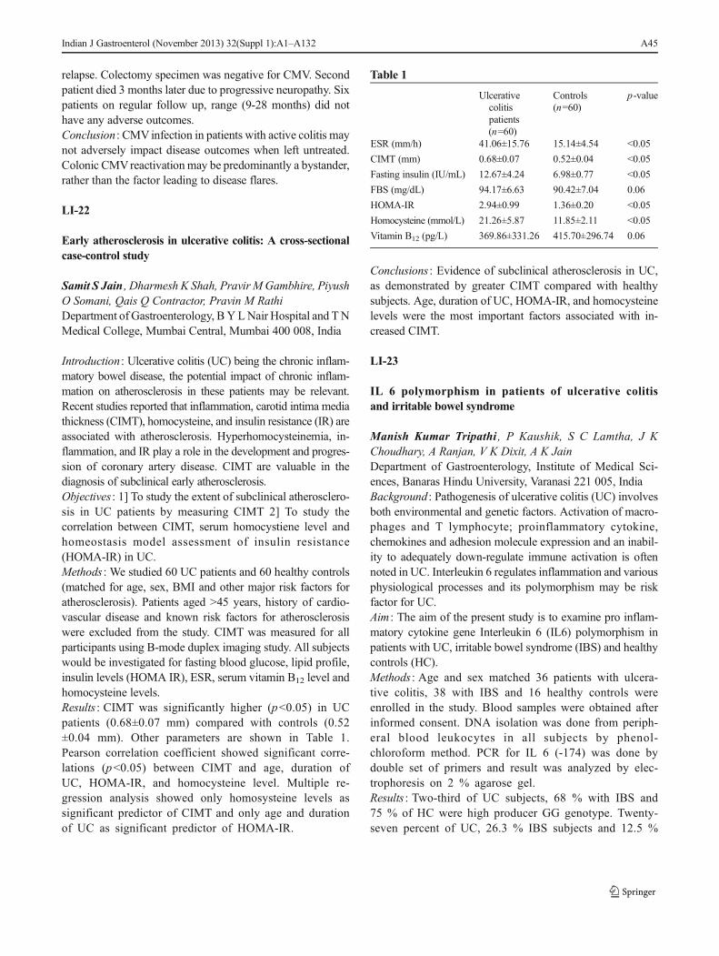
relapse. Colectomy specimen was negative for CMV. Secondpatient died 3 months later due to progressive neuropathy. Sixpatients on regular follow up, range (9-28 months) did nothave any adverse outcomes.Conclusion : CMVinfection in patients with active colitis maynot adversely impact disease outcomes when left untreated.Colonic CMVreactivation may be predominantly a bystander,rather than the factor leading to disease flares.
LI-22
Early atherosclerosis in ulcerative colitis: A cross-sectionalcase-control study
Samit S Jain , Dharmesh K Shah, Pravir MGambhire, PiyushO Somani, Qais Q Contractor, Pravin M RathiDepartment of Gastroenterology, BY LNair Hospital and TNMedical College, Mumbai Central, Mumbai 400 008, India
Introduction : Ulcerative colitis (UC) being the chronic inflam-matory bowel disease, the potential impact of chronic inflam-mation on atherosclerosis in these patients may be relevant.Recent studies reported that inflammation, carotid intima mediathickness (CIMT), homocysteine, and insulin resistance (IR) areassociated with atherosclerosis. Hyperhomocysteinemia, in-flammation, and IR play a role in the development and progres-sion of coronary artery disease. CIMT are valuable in thediagnosis of subclinical early atherosclerosis.Objectives : 1] To study the extent of subclinical atherosclero-sis in UC patients by measuring CIMT 2] To study thecorrelation between CIMT, serum homocystiene level andhomeostasis model assessment of insulin resistance(HOMA-IR) in UC.Methods : We studied 60 UC patients and 60 healthy controls(matched for age, sex, BMI and other major risk factors foratherosclerosis). Patients aged >45 years, history of cardio-vascular disease and known risk factors for atherosclerosiswere excluded from the study. CIMT was measured for allparticipants using B-mode duplex imaging study. All subjectswould be investigated for fasting blood glucose, lipid profile,insulin levels (HOMA IR), ESR, serum vitamin B12 level andhomocysteine levels.Results : CIMT was significantly higher (p<0.05) in UCpatients (0.68±0.07 mm) compared with controls (0.52±0.04 mm). Other parameters are shown in Table 1.Pearson correlation coefficient showed significant corre-lations (p<0.05) between CIMT and age, duration ofUC, HOMA-IR, and homocysteine level. Multiple re-gression analysis showed only homosysteine levels assignificant predictor of CIMT and only age and durationof UC as significant predictor of HOMA-IR.
Table 1
Ulcerativecolitispatients(n=60)
Controls(n=60)
p-value
ESR (mm/h) 41.06±15.76 15.14±4.54 <0.05
CIMT (mm) 0.68±0.07 0.52±0.04 <0.05
Fasting insulin (IU/mL) 12.67±4.24 6.98±0.77 <0.05
FBS (mg/dL) 94.17±6.63 90.42±7.04 0.06
HOMA-IR 2.94±0.99 1.36±0.20 <0.05
Homocysteine (mmol/L) 21.26±5.87 11.85±2.11 <0.05
Vitamin B12 (pg/L) 369.86±331.26 415.70±296.74 0.06
Conclusions : Evidence of subclinical atherosclerosis in UC,as demonstrated by greater CIMT compared with healthysubjects. Age, duration of UC, HOMA-IR, and homocysteinelevels were the most important factors associated with in-creased CIMT.
LI-23
IL 6 polymorphism in patients of ulcerative colitisand irritable bowel syndrome
Manish Kumar Tripathi , P Kaushik, S C Lamtha, J KChoudhary, A Ranjan, V K Dixit, A K JainDepartment of Gastroenterology, Institute of Medical Sci-ences, Banaras Hindu University, Varanasi 221 005, IndiaBackground : Pathogenesis of ulcerative colitis (UC) involvesboth environmental and genetic factors. Activation of macro-phages and T lymphocyte; proinflammatory cytokine,chemokines and adhesion molecule expression and an inabil-ity to adequately down-regulate immune activation is oftennoted in UC. Interleukin 6 regulates inflammation and variousphysiological processes and its polymorphism may be riskfactor for UC.Aim : The aim of the present study is to examine pro inflam-matory cytokine gene Interleukin 6 (IL6) polymorphism inpatients with UC, irritable bowel syndrome (IBS) and healthycontrols (HC).Methods : Age and sex matched 36 patients with ulcera-tive colitis, 38 with IBS and 16 healthy controls wereenrolled in the study. Blood samples were obtained afterinformed consent. DNA isolation was done from periph-eral blood leukocytes in all subjects by phenol-chloroform method. PCR for IL 6 (-174) was done bydouble set of primers and result was analyzed by elec-trophoresis on 2 % agarose gel.Results : Two-third of UC subjects, 68 % with IBS and75 % of HC were high producer GG genotype. Twenty-seven percent of UC, 26.3 % IBS subjects and 12.5 %
Indian J Gastroenterol (November 2013) 32(Suppl 1):A1–A132 A45

of HC genotype were intermediate producer GC. Lowproducers CC genotype was found to be 5.5 % in UC,5.3 % in IBS and 12.5 % in HC.Conclusion : This study could not substantiate association ofulcerative colitis with GG genotype polymorphic form of pro-inflammatory cytokine IL6 in our region.
LI-24
Endoscopic signif icance of bowel wall thickeningon abdominal computed tomography scan (CT scan)of patients with no prior history of gastrointestinal disease-An observational study
K S Sijil , Devang Tank, A B Suthanu, N Premaletha, K R VKumarDepartment of Medical Gastroenterology, Government Med-ical College, Thiruvananthapuram 695 011, India
Background : Computed tomography scan is used as adiagnostic modality in many abdominal situations. Bow-el wall thickening is a common finding in patients withno prior history of gastroenterological disease. The sig-nificance of this nonspecific incidental finding is notclear. Wall thickening in the lower GI tract is a realchallenge as there is concern for neoplasia and inflam-matory bowel disease.Aim : To study the significance of bowel wall thickening in theCT scan by comparing it with colonoscopic findings within 1month of CT scan.Methods : All patients with CT abdomen finding ofthickened bowel wall who underwent colonoscopy wereincluded in this study. Colonoscopic findings were no-ticed and biopsy taken whenever necessary. Patientswith known history of gastrointestinal disease wereexcluded.Results : Sixty-five patients were included in this study.Abdominal pain was the indication for CT in majority(n=48, 73.85 %). Thirty-nine had abnormal colonoscopyfindings with a positive predictive value of 60 %.Twenty-nine patients (74.36 %) with abnormal colonos-copy findings required further intervention. The resultsof colonoscopy were colorectal malignancy in 9 patients(13.85 %), ileocecal tuberculosis in 5 (7.69 %), inflam-matory bowel disease in 8 patients (12.31 %), polyps in7 patients (10.77 %), nonspecific in 8 (12.30 %) anddiverticular disease in 2 (3.07 %).Conclusion : Bowel wall thickening on computed tomography(CT) scan has high predictive value for abnormalcolonoscopic findings. It warrants colonoscopic evaluationin all patients bowel wall thickening in CT scan.
LI-25
Genetic polymorphism in DNA repair genes and theircontribution to ulcerative colitis
Avinash Bardia , Santosh K Tiwari, Sivaram Gunisetty,Sandeep K Vishwakarma, Pratibha Nallari, Md. AejazHabeeb, Aleem A KhanCentre for Liver Research and Diagnostics, Deccan College ofMedical Sciences, Kanchanbagh, Hyderabad 500 058, India
Ulcerative colitis (UC) is an inflammatory destructive diseaseof the large intestine characterized by motility and secretiondisorders. The inter-individual variation is partly due to thegenetic polymorphisms in genes involved in DNA repairgenes mechanisms which may modulate the functions of theencoded proteins. Genetic polymorphisms in XPD, XRCC3and hMSH2 genes have been reported to increase the suscep-tibility of various cancers; however their precise role in UCstill remains elusive. The present study was designed to ex-amine the polymorphisms in XPD, XRCC3, hMSH2 andinvestigate the level of NO andMDA in the risk of UC. Bloodsamples from 384 unrelated subjects (171- UC, 213- healthycontrols) were collected. Genomic DNA was isolated andgenotyped for XPD Lys751Gln, XRCC3 Thr241Met andhMSH2 Gln322Asp using PCR and RFLP. No andMDA levels in serum were also measured. The frequen-cy of genotype Lys/Gln at codon 751 of XPD gene didnot show significant difference in UC than the controls(95 % CI 0.98-2.23, p=0.06). The genotypic frequencyof XRCC3 Thr/Met at 241 codon and hMSH2 Gln/Aspat 322 codon showed statistically significant among UCsubjects than the controls (95%CI 1.15-2.65, p=0.008) (95%CI 1.15-2.62, p=0.009) respectively. Polymorphism inXRCC3/XPD and XRCC3/hMSH2 together considerably in-creased the risk of UC (95 % CI 1.31-3.29, p=0.001)(95 % CI 1.24-3.15, p=0.004). High level of NO andMDA were seen in UC subjects than the controls (p=0.01). Polymorphisms in XRCC3 and hMSH2 significantlyincreased the risk of UC in our population not in XPDpolymorphisms.
LI-26
Influence of genetic polymorphisms in X-ray repaircross-complementing group-1 and apurinic/apyrimidinicendonuclease-1 on apoptosis and ulcerative colitis
Avinash Bardia , Santosh K Tiwari, Sivaram Gunisetty,Sandeep K Vishwakarma, Pratibha Nallari, Aleem A Khan,Md Aejaz Habeeb
A46 Indian J Gastroenterol (November 2013) 32(Suppl 1):A1–A132
Centre for Liver Research and Diagnostics, Deccan College ofMedical Sciences, Kanchanbagh, Hyderabad 500 058, andDepartment of Genetics, Osmania University, Hyderabad500 00, India
Ulcerative colitis is one of the two major manifesta-tions of inflammatory bowel disease. Base excisionrepair (BER) is an important DNA repair mechanismwhich every cell utilizes to repair the damaged DNA.X-ray cross-complementing group 1 (XRCC1) andapurinic/apyrimidinic endonuclease 1 (APE1) are twovital DNA repair proteins that play definitive roles inBER pathway. Genetic polymorphisms in these geneshave been reported to increase the susceptibility ofvarious cancers; however their precise role in apoptosisand etiology of ulcerative colitis still remains elusive.Therefore the present study was designed to examinethe polymorphisms in XRCC1 and APE1 and investi-gate their role on the rate of apoptosis and ulcerativecolitis risk. Blood samples from 384 unrelated subjects(171- UC, 213- healthy controls) were collected. Geno-mic DNA was isolated and genotyped for XRCC1Arg399Gln and APE1 Glu148Asp. Apoptosis andintracellular ROS levels in peripheral blood mononucle-ar cel ls were measured using Annexin-V andH2DCFDA assay respectively. The frequency of geno-type Arg/Gln at codon 399 of XRCC1 and APE1 Asp/Glu at 148 codon gene was statistically significantly inpatients with UC than the controls. High rate of apo-ptosis coupled with elevated ROS levels were seen inUC subjects than the controls (p=0.01). Increased per-centage of necrotic and late apoptotic cells were ob-served among UC subjects with XRCC1 Arg399Glnand those with both XRCC1 Arg399Gln and APE1Asp148Glu. Polymorphisms in XRCC1 Arg399Glnand APE1 Asp148Glu significantly increased theapoptotic rates and risk of ulcerative colitis in ourpopulation.
LI-27
A rare case of non-Hodgkin lymphoma in inflammatorybowel disease patient on immunosuppressive therapy:Case report
S Kavitha , S Rathanarkini, Mohamed Kani sheik, KPremkumar, T Pugazhendhi, Mohamed AliDepartment of Medical Gastroenterology, Madras MedicalCollege, Chennai 600 003, and Government Rajiv GandhiGovernment General Hospital (RGGGH), Park Town, Chen-nai 600 003, India
The association between ulcerative colitis and lympho-ma is rare. We report the case of a 22-year-old femalewho presented a clinical picture and radiological andcolonoscopic findings suggesting the diagnosis of idio-pathic ulcerative colitis. Patient presented after threeyears with increased symptoms. When proceeded withinvestigation found colonic mucosa studded with multi-ple polypoidal lesions from sigmoid colon upto cecum.Histopathological and immunohistochemical studies oftissue specimens obtained at biopsy led to the diagnosisof non-Hodgkin's B-cell rich lymphoma throughout thecolon. Possible relations between ulcerative colitis andgastrointestinal lymphoma are discussed.
LI-28
Clinical characteristics, disease course and outcomeof patients hospitalized with severe ulcerative colitis
Mayank Agrawal , Sanjay Banerjee, G K Dhali, SumitSanyal, Rajib SarkarSchool of Digestive and Liver Disease, S S K M Hospital,Kolkata 700 020, India
Background : Around 30 % of severe ulcerative colitis fail torespond to intravenous steroids, rescue therapy or colectomyis required in these. Result of colectomy become poor asmedical therapy is prolonged so there is need for parametersto predict nonresponse early.Material and Methods : Demographic, clinical and labora-tory parameters were collected on day 1, 3 and 5 afteradmission of 22 consecutive patients (23 episodes) andprospectively recorded. Each episode was treated withintravenous hydrocortisone, fluid administration andelectrolyte management. The data of patients who failedto respond or developed complications requiring emer-gency surgery or died within 5 days of hospitalization,were compared with the data of those who respondedduring this period using student t test for nominalvariable and chi-square test for ordinal variables.Results : Medical treatment failed in 9 of the 23 episodes, all 9underwent colectomy, 1 of the patient died after colectomy.Female sex, higher lichtiger score on day 1, 2, 3, 4, 5 and alesser decline in lichtiger score from baseline over day 2, 3, 4and 5, higher CRP on day 3 and 5 and lesser fall in CRP fromday 1 to day 5 and low serum albumin on day 1, 3 and 5 wasassociated with failure of medical therapy on univariateanalysis.Conclusion : It may be possible to predict the failure of med-ical treatment in exacerbations of UC using a scoring systembased on clinical parameters and simple laboratory tests.
Indian J Gastroenterol (November 2013) 32(Suppl 1):A1–A132 A47
LI-29
Effect of argon plasma coagulation in the treatmentof symptomatic chronic radiation proctitis
Sultania SurenderSchool of Digestive and Liver Diseases, Institute of PostGraduate Medical Education and Research, 244, A J C BoseRoad, Kolkatta 700 020, India
Background : Argon plasma coagulation (APC) has increasing-ly became treatment of choice for bleeding in chronic radiationproctitis (CRP). Worldwide studies showed radiotherapy forcarcinoma prostate as themain cause of CRP. In India carcinomacervix is the predominant cause. This is the first Indian study toevaluate role of argon plasma coagulation in CRP.Objective : To evaluate - 1. The efficacy of APC on rectalbleeding and other symptoms in CRP. 2. Short and long-termcomplication of APC. 3. Correlation between success of ther-apy and colonoscopic severity or rectal bleeding score (RBS).Design : Prospective study Setting: Tertiary care hospital.Methods : APC (max 4) treatment sessions given in CRP patientswith rectal bleeding score -2, at the interval of 1 month till RBSremained -2. Therapeutic success is RBS -1. Follow up done for6 months. Colonoscopic severity grading was also done.Results : Forty-two female patients (mean age 51.88 yrs) withcarcinoma cervix (37) and carcinoma endometrium (5) patientswere studied. Thirty-four (80 %) patients had therapeutic successout of which 50% had complete cessation of bleeding. Therapeu-tic success rate was 100%, 94%, 63% inmild (7), moderate (16),severe (19) colonoscopic severity patients respectively. In RBS 2(13 %), 3 (22 %), 4 (7 %) success rate was 92 %, 87 %, 57 %respectively. Rectal ulcer developed in 7 (17 %) patients. Onepatient had recurrence of symptoms after 3 months of follow up.Conclusion : APC is excellent treatment modality in mild andmoderate CRP but less effective in severe CRP.
LI-30
Preprobiotics vs. rifaximin in the treatment of IBS-D
Pravir Gambhire , Samit Jain, Piyush Somani, Q Contractor,Pravin RathiDepartment of Gastroenterology, T N Medical College and BY L Nair Hospital, Mumbai Central, Mumbai 400 008, IndiaBackground : IBS is a chronic condition with no known cure.Clinically important benefits might be achieved in patientswith preprobiotics particularly in those with diarrhea-predominant symptoms in light of alterations in commensalenteric bacterial species in this subset. There is also accumu-lating evidence pointing towards the benefit of a short courseof rifaximin in the global improvement of patients with IBS-D.
Aims and Objectives : To assess the efficacy of preprobioticsvs. rifaximin for patients with IBS-D.Materials and Methods : It was a comparative parallelrandomized study. Male/female patients between 18 to65 yrs, with a diagnosis of IBS as per Rome III criteria,were included, the patients in the preprobiotic arm weregiven perprobiotics for 14 days while those in therifaximin arm got Tab 400 mg TID for 14 days. Thepatients were interviewed at baseline and weekly for 4weeks; the parameters to scale the symptoms includedlikert scale, subjective global assesement score and irri-table bowel syndrome score.Stastical Analysis : Qualitative data was represented inform of frequency and percentage. Association betweenqualitative variables was be assesed by chi-square test.Before –after comparison of qualitative data within eachgroup was be done using McNemar Test. Quantitaivedata was be represented using mean±SD and medianand IQR (Interquartile range).Results : Significant improvements in global symptoms of IBS(p<0.05) and bloating (p<0.05) throughout the 4-week followup in the rifaximin group, although differences in relief ofdiarrhea, and generalized ill feeling between the two groupswere not significant.Conclusions : Short course of rifaximin was better than thepreprobiotics in the IBS-D subset.
GI MOTILITY
GIM-1
Invalidity of ROME III criteria in diagnosing Indianpatients with irritable bowel syndrome
Gautam RayB R Singh Hospital, Sealdah, Kolkata 700 014, India
Background : Though Rome III criteria is used to diagnoseIBS in West, the same is not validated in India where normalbowel habit is quite different.Methods : A questionnaire in English (and in local vernacular)containing questions of ROME III criteria was administered to100 patients consulting for symptoms of IBS who had normalcolonoscopy (39), sigmoidoscopy (61) and negative stoolOBT. All had normal hemoglobin, ESR, blood sugar andthyroid status.Results : Age 41.62+16.2 years, Male:Female: 60:40, 100 %resident of the area for at least 8 years [88 % lifetime].Symptom duration 8.53+3.2 [2-18] years, constipation 14,diarrhea 63, alternate constipation and diarrhea 0, feeling ofincomplete evacuation 93. Average stool frequency/day 2.2
A48 Indian J Gastroenterol (November 2013) 32(Suppl 1):A1–A132
[1-5] and per week 16.6 [7-27]. Stool type–not well formed80, watery 5, combination 15 and type 4 [22], 5 [60], 6 [18] onBristol Stool Scale. Symptoms were abdominal pain 23/discomfort 54, bloat 21, mucus in stool 94 [all >25 % time],visible distension in none, more frequent/looser stool withpain onset 65/72, relief with stool passage 53, passing stoolafter meals 67, straining at stool in none, pain at least 3 days/month and 3 months/year 75, milk intolerance leading tostoppage of taking milk 96. Thirty-seven felt they had nodiarrhea and 11 felt constipated with incomplete evacuationeven when passing 3 stools/day.Conclusion : Rome III criteria is invalid in Indian patients.Patients’ feeling of incomplete evacuation and mucus in stoolare important as also associated milk intolerance.
GIM-2
An evaluation of the symptom of constipation in Indianpatients
Gautam RayB R Singh Hospital, Sealdah, Kolkata 700 014, India
Background : Normal bowel habit of Indians is quite differentfrom theWest and hence the definition of constipation but thishas not been appropriately studied.Methods : Seventy consecutive patients complaining of sub-jective feeling of constipation (1) either presenting to OPD(consulters) or (2) during evaluation of other diseases (non-consulters) were evaluated by clinical, biochemical andcolonoscopic examination.Results : Mean age 63.9+13.45 (46-84) years, 49 (70 %)were above 60 years of age. Male:female: 55:15, con-sulters 48 (male 45 [81.8 %], females 3 [20 %], p<.05).Forty-two (60 %) had feeling of incomplete evacuationbut passed 1-2 motions/day. Fifteen (21.4 %) had 4-6motions/week and only 13 (18.6 %) had less than 3motions/week (the western definition of constipation).On Bristol Stool Scale, stool forms were 11 type 1(15.7 %), 10 type 2 (14.3 %), 14 type 3 (20 %), 35type 4 (50 %). On colonoscopy, only 3 (4.3 %), allnon-consulters, had colon cancer and all had othersymptoms as well. Others had atonic dilated colon 7(10 %), diverticulosis 5 (7.2 %) or were normal (78.5%). Comordities were diabetes mellitus 30 (42.9 %),hypothyroism 10 (14.3 %), hypertension mandating in-take of calcium channel blockers and beta blocker drugs45 (64.3 %), organic brain disease 14 (20 %).Conclusions : A feeling of incomplete bowel evacuationis interpreted as constipation in India, most have benignneuromuscular etiology due to drug intake or comorbidillness. Most consulters are elderly males. The definition
of constipation needs to be coined by large scalepopulaton studies.
GIM-3
Clinical and investigative assessment of chronicconstipation
Rajiv Baijal , Praveen Kumar , Nimish Shah, SandeepKulkarni, Soham Doshi, Deepak Gupta, DeepakAmarapurkarJagjivan Ram Railway Hospital, Maratha Mandir Marg,Behind Maratha Mandir Cinema, Mumbai Central, Mumbai400 008, India
Introduction : Constipation may be primary or secondary.Pathophysiologic subtypes of primary constipation aredyssynergic defecation (DD), slow (STC) and normaltransit constipation (NTC). Clinical subtypes are func-tional constipation (FC) and constipation predominantIBS (C-IBS).Aims :1) To study the clinical profile, categorize and comparevarious subtypes of primary constipation. 2) To assess successof biofeedback therapy (BFT) in DD.Material and Methods : Consecutive constipation patients(April 2011-December 2012) were evaluated. Patients<18 years and secondary constipation were excluded.‘FC’ and ‘C-IBS’ were classified by Rome-3 module.Patients underwent FBS, calcium, TFT, stool examina-tion, colonoscopy, anorectal manometry (ARM) withballoon expulsion test and colon transit study (CTS).Patients with DD were given BFT.Results : Out of 128 patients 23 %, 58 % and 19 % hadsecondary constipation, FC and C-IBS respectively.Ninety-nine patients had primary constipation. Meanage-53.50 (18–86) years, (77 % males). Forty-six, 15and 40 had NTC, STC, DD respectively. 34/40 hadparadoxical anal contraction and 6/40 had impaired rec-tal propulsion. ‘FC’ and ‘C-IBS’, were clinically andpathophysiologically similar except for abdominal pain.Patients with DD were more likely to have history offinger evacuation, straining, incomplete evacuation, sen-sation of anorectal obstruction than no DD (p<0.001).Sixty-nine percent patients with STC had ≤3 stools/week compared to 37 % with NTC. 30/40 patients withDD underwent BFT but 20 completed ≥4 sessions.Seventy percent with ≥4 sessions had improved CSBMwith increase in anal relaxation time.Conclusion : NTC is most common subtype of primary con-stipation. Symptom profile can predict pathophysiologicsubtype but investigations (ARM and CTS) are needed toconfirm them. DD showed good response to BFT.
Indian J Gastroenterol (November 2013) 32(Suppl 1):A1–A132 A49

Liver – Portal Hypertension
LPH-1
Prevalence of hyponatremia in treatment naiïve patientswith cirrhosis and ascites
Mayank JainChoithram Hospital and Research Centre, Manikbagh Road,Indore 452 014, India
Background : Serum sodium is a simple, readily availableand objective marker of disease severity in cirrhosis.Several studies have shown that hyponatremia is astrong predictor of early mortality. Moreover, the accu-racy of MELD - Na has been shown to be slightlysuperior to that of MELD in candidates for transplanta-tion. The present study was done to determine theprevalence of hyponatremia (serum sodium<130 meq/L) intreatment naïve patients of cirrhosis and ascites during theirfirst hospital admissionMethods : This is a prospective and observational study.It enrolled 125 consecutive treatment naïve patients ofcirrhosis and ascites who were admitted for the firsttime at Jagjivanram Hospital, Mumbai and ChoithramHospital and Research Centre, Indore between May2008 to March 2013. A detailed history regardinguse of diuretics, antibiotics, albumin, intravenousfluids, previous episodes of ascites and therapeuticparacentesis was taken. Diagnosis of cirrhosis andascites was confirmed by clinical, radiological, endo-scopic and laboratory investigations. Serum sodiumlevels were sent within two hours of admission. Thedata collected was analyzed to determine prevalence ofhyponatremia.Results : The mean age of patients was 54.92 years (23-78 years). The majority of patients were males (80 %).The etiological factors for cirrhosis were alcohol (86,68.8 %), HCV (17, 13.6 %), HBV (8, 6.4 %), crypto-genic (9, 7.2 %) and others (5, 4 %). Thirty-four pa-tients had serum sodium levels of <130 meq/L. Majorityof these patients (29, 85.3 %) had serum sodium levelsbetween 120-129 meq/L while two patients had serumsodium levels below 110 meq/L. Three patients hadserum sodium levels between 111 to 119 meq/L. Themean MELD score was 20.94 (range 7-39). Twenty-fourpatients had MELD score of more than 15 while onlyone patient had a score of <9.Conclusions : The prevalence of hyponatremia in treat-ment naïve patients with cirrhosis and ascites duringfirst hospital admission is 20.7 %. It is more commonin patients with MELD score of >15 suggesting thepresence of advanced liver disease.
LPH-2
Utility of serum pro-BNP as a marker of cardiacdysfunction in liver cirrhosis
B Varun Rao , K Panduranga Rao, B Ramesh Kumar,K Venugopal Reddy
Background : Unrecognized cardiac dysfunction is a cause forincreased mortality in the peri trans-plant period (arrhythmiasand overt heart failure) and after TIPS (pulmonary edema).Aim : 1) To find the prevalence of cardiac dysfunction in patientswith cirrhosis. 2) To study utility of serum pro-BNP as a markerof cardiac dysfunction in patients with liver cirrhosis.Methods : Total of 85 patients with cirrhosis were included in thestudy after satisfying the inclusion and exclusion criteria betweenSeptember 2011 to September 2013. All of them underwentevaluation for presence of cardiac dysfunction by 2D-Echo(systolic/diastolic dysfunction) and ECG (QT Prolongation).Serum pro-BNP was measured in all the patients. Patients weredivided into two groups group A - those with cardiac dysfunc-tion, group B - those without cardiac dysfunction. Clinical andbiochemical indices in both the groups were compared.Results : Out of 85 patients Child A-18, Child B-39, Child C-28. A total of 38 (44.7 %) patients had cardiac dysfunction.Prevalance of cardiac dysfunction was more in Child C 21/28(75 %) compared to Child B 15/39 (38.46 %) and Child A 2/18 (11.11 %). Out of 85 patients corrected QT interval wasprolonged in 30/85 (35.29). QT prolongation was more com-mon in Child C (46.42%) compared to Child B (35.89%) andChild A (16.66 %). QT was prolonged in 21/38 (55.26 %)patients with diastolic dysfunction. Serum pro BNP was ele-vated in 33/38 (86.84 %) patients with cardiac dysfunction.Mean serum pro-BNP levels are higher in Child C comparedto Child A, B. The levels also correlate with MELD score.Conclusions : Prevalence of cardiac dysfunction in patientswith liver cirrhosis is 44.7 %. Pro-BNP is elevated in majorityof the patients with cardiac dysfunction (86.84 %). Highermean levels of pro-BNP are seen in Child C as compared toChild A, B. The levels also correlate with MELD score.
LPH-3
A study of portal hypertensive polyps
V Anand , K Muthukumaran, G Ramkumar, R Balamurali,P Ganesh, S Jeevan KumarDepartment of Digestive Health and Diseases, Kilpauk Med-ical College, Chennai 600 010, India
Background and Aim of Study : To estimate the prevalence ofportal hypertensive polyps in patients with portal hypertensiondue to any etiology.
A50 Indian J Gastroenterol (November 2013) 32(Suppl 1):A1–A132

Patients and Methods : This is a prospective observational studyof patients with portal hypertensive polyps. All patients in agegroup of 18 to 80 years who had portal hypertension diagnosedby clinical, laboratory and imaging criteria who went upper GIscopy from the period of May 2012 to April 2013 were includedin the study. Diagnosis of portal hypertensive polyps was con-firmed histologically. Patients with proton pump inhibitor use,gastric varices, H. pylori gastritis, history of inherited polyposissyndromes, patients who refused consent were excluded.Results : A total of 3,621 upper GI endoscopies were done ofwhich 428 patients had portal hypertension. Polyps were notedin 9 patients (2.1 %), out of 428 patients 366 had chornic liverdisease and 62 had extrahepatic portal hypertention. Out ofthem 1 was excluded due to rapid urease test positivity. Histo-pathological examination showed four had extensive vascularproliferation and glandular hyperplasia (portal hypertensivepolyp) two had hyperplastic polyp and other two had neuroen-docrine tumor. Endoscopic appearances of polyps varied con-siderably, with sizes ranging from 5mm to 15mm. Polyps weresessile or pedunculated, singular or multiple, found in theantrum, body of the stomach. There is no specific endoscopicfeatures to identify portal hypertensive polyps.Conclusion : Portal hypertensive polyps are rare. There is nospecific endoscopic features. Biopsy is necessary for diagnosis.Long-term studies are needed to characterize their significance.
LPH-4
Budd-Chiari syndrome in abdominal tuberculosis-causativeor coincidental: A rare case report
S Sathiamoorthy, T S Rameshkumar, H Azimudin, M GManoj, Manimaran, Chitra, Murali, Malarvizhi, M S Revathy,A R VenkateswaranDepartment of Medical Gastroenterology, Government Stan-ley Medical College, Chennai 600 001, India
Background : Tuberculosis (TB) as a cause of Budd-Chiarisyndrome (BCS) has less been documented in literature. Herewe report a rare case of BCS in abdominal TB.Case Report : A 21-year-young female, admitted with history ofabdominal distension, abdominal pain of 3 months duration.Evaluated elsewhere, diagnosed as cryptogenic cirrhosis referedfor liver transplant registration to our center. Patient wasreevaluated. Past history of antituberculous treatment (ATT) forwell documented tuberculous ascites in 2009 for 6 monthsfollowingwhich patient improved symptomatically. Her physicalexamination was normal except prominent back veins, moderateascites and spleenomegaly. Her LFT was normal. Ascites fluidanalysis: low SAAG, high protein. ADA normal. Cytologynegative for malignant cells. USG abdomen with portal veindoppler:coarse, irregular liver with caudate lobe hypertropy.Right, middle and left hepatic vein occluded with comma shaped
collaterals, spleenomegalywith ascites. OGDScopy: grade 3 to 2esophageal varices with PHG. Underwent EVL. CECT abdo-men: irregular liver with caudate lobe hypertrophy, hepatic veinscould not be visualized, spleenomegaly with ascites, ileocecalthickeningwith pulled up contracted cecum. Prothromboticworkup normal. Colonoscopy: passed up to cecum, ileum notintubated. Ulceroproliferative lesion in semilunar fold biopsydone. HPE report-chronic inflammatory pathology.Conclusion : Development of BCS in the background of ab-dominal tuberculosis is less reported.This case is reported dueto rare occurance.
LPH-5
Prevalence of cirrhotic cardiomyopathy: Our centreexperience
P Thirumal , Muthukumaran, G Ramkumar, R Balamural,P Ganesh, S Jeevan KumarDepartment of Digestive Health and Disease, Kilpauk Medi-cal College, Chennai 600 010, India
Aim : To study the frequency of cirrhotic cardiomyopathy in livercirrhosis patients and its correlation with severity of liver disease.Method : This is a case series study conducted in Department ofDigestive Health and Disease, a tertiary care centre in Chennaiover the period from July 2012 to March 2013. First, restingECGwas done in all enrolled cirrhotic patients. QTc values werecalculated and value >0.44 sec were considered as prolonged.Systolic dysfunction was assessed by reduced ejection fraction(value <55 %). Diastolic dysfunction assessed by reduced E/Aratio (value <1 was present in 20 (30 %) cases, prolong QTinterval (>0.44 sec) in 16 (22.8 %) and ejection fraction (EF)<0.55was in 11 (15.7%) patients. Cirrhotic cardiomyopathywaspresent in 25 (35.7 %) cases and frequency correlates directlywith severity of liver disease. There was no significant differencein frequency of CCM among alcoholics and nonalcoholics.Conclusion : We observed that cirrhotic cardiomyopathy waspresent in 25 percent of our cirrhotic patients and significantlymore in Child C. Presence of this clinical entity may havemajor impact on prognosis in these patients.
LPH-6
Acute kidney injury at admission and its responseto terlipressin as a predictor of mortality in patientswith acute-on-chronic liver failure
Ankur Jindal , Manoj Kumar, Shiv Kumar SarinDepartment of Hepatology, Institute of Liver and BiliarySciences, Vasant Kunj Marg, New Delhi 110 070, IndiaBackground : Unlike decompensated cirrhotics, the preva-lence, associated complications, mortality and treatment
Indian J Gastroenterol (November 2013) 32(Suppl 1):A1–A132 A51

outcomes for acute kidney injury (AKI) in acute-on-chronicliver failure (ACLF) are less well defined.Methods : Consecutive acute-on-chronic liver failure patients withAKI [serum creatinine >1.5 mg/dL] at admission were comparedwith controls for short-term (7 day) and long-term (1, 3 month)mortality and development of HE, SBP and acute variceal bleed(AVB). Patients were further compared based on severity of AKI(mild AKI; S.cr 1.5-3 mg/dL; severe AKI; S.cr > 3 mg/dL).Results : Of 267 ACLF patients, 55 (22.8 %) had AKI at admis-sion. Patients with AKI at admission had higher short and long-term mortality (7 day, 1 and 3 month) and more often developedHE [54.1 % vs. 30.6 %; p=0.001] and SBP [9.1 % vs. 5.9 %; p=0.022]. However, severity of AKI neither predicted survival nordevelopment of complications. Presence of AKI (Odd’s ratio,OR: 3.121) and serum bilirubin (OR: 9.136) were independentbaseline predictors of survival. Terlipressin was used in 28 of 55patients with AKI who were volume non-responders; 11/28(35.28 %) showed response (S. Cr <1.5 mg/dL) after a medianof 4 days and had lower mortality compared to non-responders(10 % vs. 44.3 %, p=0.05). There was no difference interlipressin response rates in mild vs. severe AKI.Conclusion : Almost one fourth of the ACLF patients haveAKI at admission and presence of AKI, but not its severity,predicts the development of complications and high mortality.Terlipressin is effective in reversing AKI in ~35 % of patientswithin a week and results in improved survival.
LPH-7
Noninvasive assessment of portal hypertension: Aretrospective study in a tertiary care hospital
Sanket Shah , Amandeep, M M Prabhu, NareshMedicineDepartment, KasturbaHospital,Manipal 576 104, India
It is currently recommended that all patients with livercirrhosis undergo upper gastrointestinal endoscopy (UGIE)to identify those who have large esophageal varices thatcarry a high risk of bleeding and may benefit from pro-phylactic measures. This approach leads to unnecessaryUGIE in those without large esophageal varices. This pro-spective study was conducted for 130 patients in a tertiarycare hospital to evaluate noninvasive predictors of largevarices (LV). Specificity and sensitivities were assessed forthe each measure.
LPH-8
Clinical and etiological profile of patients with decompensatedchronic liver disease at a tertiary care centre
Jatin Patel , Prasad Bhate, Dhaval Gupta, Meghraj Ingle,Aniruddha Phadke, Prabha Sawant
Department of Gastroenterology, Lokmanya Tilak MunicipalMedical College and Lokmanya Tilak Municipal GeneralHospital, Sion, Mumbai 400 022, India
Aim : To study the clinical and etiological profile of patientswith decompensated chronic liver disease.Methods : Patients with decompensated liver disease admitted inour hospital between May 2011 and May 2013 were included instudy. After detailed history and examination all patientsunderwent routine blood investigations (hemogram, liver functiontests, renal function tests, prothrombin time), ascitic fluid investi-gations, autoimmune markers, viral markers, s.ceruloplasmin,ultrasonography with doppler and esophagogastroduodenoscopy.Results : Age group of the patients included is 4 to 80 yearswith mean age of 44.89 years. There were 382 male (80.76%)and 91 female (19.23 %) patients with M:F ratio of 4.2:1.Most common symptom at presentation is abdominal disten-sion in 71 % patients followed by pedal edema in 47.99 %,abdominal pain in 41.86 %, jaundice in 39.75 %, malena in37.84 %, hematemesis in 31.5 %, encephalopathy in 21.99 %and decreased urine output in 6.55 % patients. Most commonetiology of decompensated liver disease was alcoholic liverdisease in 56.23 % patients followed by hepatitis B in 19.66 %,cryptogenic in 12 %, autoimmune hepatitis in 3.8 %, hepatitis Cin 2.75%,Budd-Chiari syndrome in 2.54% and others in 2.96%patients. Twenty-two percent patients belonged to CTP class Band 77.8 % patients in CTP class C.Conclusion : Alcoholic liver disease followed by hepatitis Bwere the leading causes of decompensated liver disease andascites was the most common symptom and majority patientsbelonged to CTP-C.
LPH-9
Incidence of portal vein thrombosis in decompensatedchronic liver disease
Prasad Bhate , Jatin Patel, Dhaval Gupta, Meghraj Ingle,Aniruddha Phadke, Prabha SawantDepartment of Gastroenterology, Lokmanya Tilak MunicipalMedical College and Lokmanya Tilak Municipal GeneralHospital, Sion, Mumbai 400 022, India
Aim : To study the incidence of portal vein thrombosis indecompensated chronic liver disease (CLD).Methods : Patients of decompensated CLD admitted in ourhospital between July 2012 to June 2013 were included instudy. Patients with hepatocellular carcinoma were excluded.All patients are investigated by routine blood investigationslike hemogram, blood urea, s.creatinine, s.bil, AST, ALT andUSG abdomen with portosplenic Doppler.Results : Out of 193 patients included in study 27 (13.99 %) (22males) patients showed evidence of portal vein thrombosis on
A52 Indian J Gastroenterol (November 2013) 32(Suppl 1):A1–A132
Doppler. Mean age of patients with portal vein thrombosiswas 48 years. Most common symptom was abdominal dis-tension in 18 (66.67 %) abdominal pain in 16 (59.26 %),malena in 12 (44.45 %), pedal edema in 12 (44.45 %),jaundice in 7 (25.93 %) and hematemesis in 7 (25.93 %)patients. Liver was palpable in 11 (40.74 %) patients whilepalpable spleen was present in 14 (51.85 %) patients. Mostcommon etiology of chronic liver disease associated withportal vein thrombosis was alcoholic liver disease in 13(48.15 %) patients, hepatitis B in 6 (22.23 %), cryptogenicliver disease in 5 (18.52 %), nonalcoholic steatohepatitis in 1(3.7 %) and hepatitis C in 1 (3.7 %) patient. Ascites waspresent in 23 (85.18 %), upper GI bleed in 6 (22.23 %),spontaneous bacterial peritonitis in 4 (14.81 %), hepaticencephalopathy in 3 (11.11 %) and hepatorenal syndrome in2 (7.4 %) patients. Three patients were in CTP-B and 24(88.89 %) were in CTP-C.Conclusion : Portal vein thrombosis was found in almost 14% ofall our decompensated CLD patients. It could represent an im-portant cause of decompensation especially inCTPCpatients andshould be actively sought for in patients with decompensation.
LPH-10
Incidence of post EVL ulcer bleed after endoscopic varicealbanding and its correlation with CTP score
B Sajeeth Manikanda Prabu , K Muthukumaran,G Ramkumar, R Balamurali, P Ganesh, S Jeevan KumarDepartment of Digestive Health and Disease, Kilpauk Medi-cal College, Chennai 600 010, India
Aim : To study the incidence of ulcer bleeding after endoscopicvariceal ligation and its correlation with Child-Pugh score.Materials and Methods : One hundred and ten cirrhotic patientswith esophageal varices who required endoscopic variceal liga-tion to prevent bleeding or rebleeding in our centrewere includedin the study. The incidence of post EVL ulcer bleeding werenoted and correlated with their Child-Pugh scoring.Results : The incidence of rebleeding after EVL was 7.85 % (14patients). Of 110 patients the etiological cause for cirrhosis wasethanol in 82 patients (74 %), hepatitis B related in 15 patients(13.63 %), EHPVO related in 8 patients (7.27 %) and others(4.54 %). Of 14 patients who had rebleeding 11 (78.57 %) hadrebleeding secondary to varcies and 3 (21.42 %) had post EVLulcer bleed. One patient with EVL ulcer bleed died inspite ofactive resuscitation. Among 14 patients who had rebleeding(either hematemesis or malena), 10 (71.4 9 %) patients belongedto Child-Pugh class C and 5 (28.57 %) patients to Class B. All 3patients with post EVL ulcer bleed belonged to Child-Class C.Conclusion : This study demonstrated that the prevalence ofulcer bleed after EVL is uncommon and ulcer bleed is morelikely to occur in patients who are in Child-Class C.
LPH-11
Incidence of spontaneous bacterial peritonitis in decom-pensated cirrhotic patients and its correlation with clinicaland laboratory parameters
B Sajeeth Manikanda Prabu , K Muthukumaran,G Ramkumar, R Balamurali, P Ganesh, S Jeevan KumarDepartment of Gastroenterology, Kilpauk Medical College,Chennai 600 010, India
Aim : 1. To study the incidence of spontaneous bacterialperitonitis (SBP) in DCLD patients. 2. To correlate withclinical, lab parameters and scoring system.Materials andMethods : One hundred decompensated cirrhot-ic patients with suspected SBP, admitted to our hospital over aperiod of one year were included in our study after excludingother causes. They were assessed based on history, examina-tion, investigations including ascitic fluid analysis and follow-ed up for one month.Results : Among 100 patients, 29 patients had SBP.Among 29 patients, 7 had culture proven SBP and 22had culture negative neutrocytic ascities (CNNA). Thecommon presentation were worsening of abdominal dis-tension - 29 patients (100 %), Hepatic encephalopathy -13 patients (44.82 %), fever and abdominal pain 11patients (37.93 %) and UGI bleed 6 patients (20.69 %). AllSBP patients had ascitic fluid albumin 25, 18 (62.06 %) and8 (27.58 %) patients with score between 16 -24 and <15respectively. Five (62.50 %) patients died during follow up,of which 4 had classic SBP and 1 with CNNA.Among those 5patients, 2 developed HRS, 1 had UGI bleed and 1 had hepaticencephalopathy.Conclusion : 1. Cirrhotic patient with SBP can haveatypical presenting symptoms. Hence high degree ofsuspicion is required to rule out SBP and manage ap-propriately. 2. Cirrhotic patients with low ascitic albu-min and with high CTP and MELD score are moreprone to develop SBP. 3. Mortality increases with asso-ciated complications.
LPH-12
AKI in patients with acute on chronic liver failure isdifferent from patients with cirrhosis
Rakhi Maiwall , S Kumar, S Lata, Bhaskar Thakur,C Vashishtha, M K Sharma, H Garg, S Taneja, S K SarinDepartments of Hepatology, and Nephrology, Institute ofLiver and Biliary Sciences, D 1, Vasant Kunj, New Delhi110 070, and Departments of Hematology, and Biostatistics,All India Institute of Medical Sciences, Ansari Nagar, NewDelhi 110 029, India
Indian J Gastroenterol (November 2013) 32(Suppl 1):A1–A132 A53

Background and Aims : The current definitions for AKI(AKIN criteria) and HRS have been derived from patientswith cirrhosis with paucity of data on AKI in acute on chronicliver failure (ACLF). We investigated the prevalence, naturalhistory and outcome of AKI and Type 1 HRS in patients withACLF (n=534) and compared it with a cohort of hospitalizedpatients with cirrhosis (n=2,083).Methods : Serial creatinine up to day 30 was recorded for allpatients.Results : AKI was more common in ACLF (50.5 % vs.32.3 %; p<0.0001) and associated with increased risk ofmortality (59.9 % vs. 41.3 %; p<0.0001) as compared toCLD. Presence of ACLF (vs. CLD) was associated with asix-fold increased risk of AKI (p<0.0001, OR 6.4 95 % CI4.9-8.3) on multivariate analysis. There was no significantdifference seen in the AKIN stage at baseline (Stage 1 : 61 %vs. 69%; Stage 2: 29.5% vs. 23.5%; Stage 3 : 9.9% vs. 8.5%),decrease in serum creatinine at 48 hours (67.3 % vs. 65.3 %),however, progression to AKIN stage 3 (23.5 % vs. 12.6 %) wassignificantlymore common inACLF (p<0.0001). Prevalence ofType 1 HRS (28.7% vs. 29.2%) was similar, however responseto terlipressin (15.5%vs. 50.1%), mortality (67.9%vs. 45.9%;p=0.001) and progression to ATN and need of RRT (53.8 % vs.20.4 %; p<0.0001) was higher for patients with ACLF and type1 HRS.Conclusion : The prevalence, natural history and mortality ofAKI in ACLF is distinctly different from patients with CLD.Patients with ACLF and type 1 HRS have the highest risk ofmortality.
LPH-13
Clinical and etiological profile of acute febrile illnessin cirrhosis
P Laksmana Chandra , P Shravan Kumar, M Umadevi,M RamannaDepartment of Gastroenterology, Gandhi Hospital, Secunder-abad 500 025, India
Aim : Acute febrile illness is common in cirrhotic patients. Theaim of this study was to identify the clinical profile andetiological factors presenting as acute febrile illness in cirrhot-ic patients.Methods : All cirrhotic patients admitted as inpatients betweenJanuary 2013 and December 2013 with a history of fever of lessthan seven days duration. Detailed history, clinical examinationand investigations- complete blood picture, complete urine anal-ysis, liver function test, ascitic fluid analysis, serum ammonia,smear for MP, blood culture sensitivity, chest X-ray, ultrasoundabdomen were done. The data collected were analyzed.Results : Out of 78 patients male-to-female ratio was males 66(85 %) females 12 (15 %) ratio male/female 5.5:1. The mean
age at presentation was 44 years (24-64 years). The otherclinical features associated with fever include abdominal pain48 (62 %), jaundice 15 (19 %), swelling of lower limb12 (15 %), vomiting 12 (15 %) altered sensorium 8 (10 %),cough 8 (10 %), burning micturition 6 (8 %). The etiologicalfactors were: spontaneous bacterial peritonitis 40 (51 %),lower limb cellulitis 12 (15 %), lower respiratory tract infec-tion 8 (10 %), viral fever 8 (10 %), urinary tract infection6 (8 %), acute cholecystitis 2 (2.5 %), and malaria 2 (2.5 %).Hepatic encephalopathy was associated in 8 cases of sponta-neous bacterial peritonitis.Conclusion : Spontaneous bacterial peritonitis is the mostcommon cause of acute febrile illness in cirrhosis thoughmultiple other causes have been identified in this study.
LPH-14
The association between the serum sodium leveland the severity of complications in liver cirrhosis
S Sukumaran , A R Akilandeswari, K Muthukumaran,G Ramkumar, R Balamurali, P Ganesh, S Jeevan KumarDepartment of Digestive Health and Diseases, Kilpauk Med-ical College, Chennai 600 010, India
Introduction : Cirrhotic patients with ascites have low survivalrate of 23% and 7% in 5 and 10 years respectively. In patientswith hyponatremia and ascites the risk of pretransplant deathwithin 180 days may exceed 40 %. Hyponatremia is indepen-dent predictor of mortality in cirrhotic patients and associatedwith increased rate of various complications.Aim : To find the prevalence of hyponatremia in cirrhosis andstudy the association between the serum sodium level andseverity of complications in liver cirrhosis.Materials and Methods : Study included 50 consecutive pa-tients with liver cirrhosis admitted in our institution from July2012 to February 2013. Patients with complications due tocirrhosis such as ascites, hepatic encephalopathy, hepatic hy-drothorax, spontaneous bacterial peritonitis were included.Patients with hepatocellular carcinoma, diuretics intake cur-rently or within one month and intrinsic renal disease, heartdisease were excluded. Enrolled patients were subjected toclinical and laboratory investigations.Results : Hyponatremia (<130 meq/L) was found in 27 pa-tients (54 %). Patients with hyponatremia had severe ascites(grade 2 and 3 in 6 and 8 patients respectively p -0.005) andincreased frequency of hepatic encephalopathy (12 patients44 %) than other complications which did not increase signif-icantly. Hyponatremia is more common in patients with CTPscore B (18 patients- 36 %) and C (26 patients-52 %).Conclusion : The prevalence of hyponatremia in cirrhotics isfound to be in more than half of the patients and they are moreprone for severe ascites and hepatic encephalopathy than other
A54 Indian J Gastroenterol (November 2013) 32(Suppl 1):A1–A132
complications. Hyponatremia is found more commonly inthose patients with CTP score B and C.
LPH-15
Predictors of mortality in alcoholic hepatitis and to comparethe MELD score with the Maddrey`s DF (mDF) score asa predictor of short-term mortality
K Srinivas , M Ramanna, M Umadevi, P Shravan KumarDepartment of Gastroenterology, Gandhi Medical College,Hyderabad 500 025, India
Background : Alcoholic hepatitis (AH) is associated with badprognosis. So it is important to know the predictors of mor-tality and to manage the patient accordingly.Aim : To assess prognostic factors for patients with alcoholichepatitis and to compare MELD score with mDF score as apredictor of short-term mortality.Methods : Fifty patients with diagnosis of AH were included.Demographic, laboratory data, complications and 30 day mor-tality were registered. A comparison was performed betweensurvivors and non-survivors.Results: Mean alcohol consumption is about 120.96±59.35gm/day. mDF (96.81 vs. 56.75, p<0.0001) and MELD score(33.27 vs. 21.82, p<0.0001 ) was higher in patients who diedthan those who survived respectively. The performancecharacterisitcs of MELD score (AUC–0.89) is similar to thatof mDF score (AUC-0.85). No differences between groupswere recorded regarding albumin, age and Glasgow scale.Increased levels of creatinine (2.18 vs. 1.13, p<0.0001),bilurubin (20.27 vs. 12.60, p<0.0001) and INR (2.32 vs.1.78, p<0.0025) are related with higher risk of death.Conclusion : Increased levels of creatinine, bilurubin, INR,mDF and MELD score are the significant risk factors associ-ated with mortality. Both MELD and mDF scores are equallygood predictors of short-term mortality in patients with alco-holic hepatitis.
LPH-16
Factors predisposing to bleeding from esophageal ulcersoccurring after endoscopic variceal band ligation
Roji Ray, Varghese ThomasDepartment of Gastroenterology, Government Medical Col-lege, Kozhikode 673 008, India
Background : Bleeding in the post endotherapy period frompost endoscopic variceal band ligation (EVL) ulcers has beendescribed and sometimes it can be life threatening.Aims : To assess the risk factors and outcome of bleeding fromesophageal ulcers occurring after EVL.
Methods : Consecutive patients with cirrhosis who had under-gone EVL for esophageal varices were studied. Clinical,hematological and biochemical parameters as well as Childand MELD scores were compared between those with andwithout post EVL ulcer bleed which occurred within fourweeks of performing EVL.Results : One hundred and fifty-one patients were enrolled in thestudy. One hundred and twenty-nine (85.4 %) were males andthe most common etiology of cirrhosis was alcohol (n=104;68.9%). Twelve (7.95%) patients developed post EVL bleed ofwhich 10 (83.3 %) had bleed from post EVL ulcers. The meanduration to ulcer bleed was 14.1 days (SD 2.64) after EVL.Mean platelet count and INR were 0.85 lakhs/mm3 (SD 0.14)and 2.07 (SD 1.17) in those with ulcer bleed compared to 1.37lakhs/mm3 (SD 0.31) and 1.25 (SD 0.14) respectively in thosewithout bleed (p=<0.001). The mean MELD score was 16.5(SD 7.35) in those who bled compared to 9.5 (SD 1.63) in thosewithout ulcer bleed (p=<0.001). Post EVL ulcer bleed wasassociated with hemodynamic disturbance in four patients.One patient expired due to post EVL bleed.Conclusion : 6.6 % of cirrhotic patients who had undergoneEVL developed bleeding from post EVL esophageal ulcers.Twenty-five percent of these patients had hemodynamic in-stability. A low platelet count, prolonged INR, and higherMELD scores were associated with the risk of bleeding frompost EVL esophageal ulcers.
LPH-17
Incidence and prognosis of functional renal failurein cirrhotic patients
M Tarakeshwari , K Muthukumaran, G Ram Kumar,R Balamurali, P Ganesh, S Jeevan KumarDepartment of Digestive Health and Diseases, GovernmentPeripheral Hospital, Kilpauk Medical College, Chennai 600010, India
Aim : To assess the incidence of functional renal failure incirrhosis and investigate the prognostic factors.Materials and Methods : An observational study of cirrhoticpatients admitted in our hospital between July 2012-June2013 was carried out. Patients diagnosed with cirrhosis andpresenting with elevated renal parameters were included in thestudy. Cirrhotic patients with underlying structural renal dis-ease were excluded from the study. Patients with functionalrenal failure were categorized in three subgroups as pre renalfailure (group 1), renal failure with infection (group 2) andhepatorenal syndrome (group 3). Clinical characters of thesepatients were analyzed for precipitating factors. They weretreated according to the standard protocol, evaluated for clin-ical, biochemical and radiological parameters and followed upfor survival rates and prognostic factors during hospital stay.
Indian J Gastroenterol (November 2013) 32(Suppl 1):A1–A132 A55

Results : Out of the 210 patients 86 (40.95 %) developedfunctional renal failure. Highest incidence was in the 5thdecade (79 %). The most frequent was prerenal failure 63(73.25 %) followed by renal failue with infection 15(17.44 %), and hepatorenal syndrome 8 (9.30 %). Uppergastrointestinal bleed and acute gastroenteritis with an inci-dence of 39.6 % and 31% respectively were the most frequentprecipitants of functional renal failure. The incidence of func-tional renal failure in Child class A, B, C were 56%, 36% and8 % respectively. Among cirrhotics with Child class C theincidence of hepatorenal syndrome was 61.5 %.Conclusion : Approximately 41 % of patients with cirrhosisdeveloped functional renal failure. Higher CTP score wasassociated with increased risk of occurrence of hepatorenalsyndrome. Most of the patients with functional renal failurerecovered with treatment except those with hepatorenalsyndrome.
LPH-18
Outcome of extrahepatic portal venous obstruction with orwithout endoscopic and/or surgical treatment
Nisar A Shah , S A Zargar, O J Shah, I Rabani, G Javed, G NYatu, B A Khan, A H Shah, A Sadaf, G M Gulzar, S SJaswinder, M A KhanDepartments of Gastroenterology, Surgical Gastroenterology,and Radiodiagnosis, Sher-i-Kashmir Institute of Medical Sci-ences, Soura, Srinagar, Kashmir 190 011, India
Background : Extrahepatic portal venous obstruction is an im-portant cause of portal hypertension in children and adultsworld over, and a major cause in Asian and African population.Objective : To look into the clinical presentation, lab parameters,etiological factors, radiological features, morbidity andmortalityin patients of EHPVO on different treatment modalities.Design : Retrospective cum prospectiveMethods : Extensive demographic lab data, radiological data,anthropometric assessment, endoscopic and interventional da-ta, surgical data from consecutive patients diagnosed andregistered since 1982 and those registered prospectively fromAugust 2007 to January 2010 with the Departments of Gas-troenterology and Surgical Gastroenterology was revived.Data on the diagnostic work up, etiological profile, endoscop-ic follow up, complications and their management was alsonoted. Consecutive patients on pharmacotherapy, EST/EVLalone and patients who had undergone devascularization orshunt were assessed for their outcome.Results : A total of 240 patients, 125 males and 145 childrenwith mean follow up of 9.3±7.7 years were registered. Mainpresenting symptoms were upper GI bleed, symptomaticsplenomegaly, growth retardation, hypersplenism, jaundice,cholangitis and non specific pain abdomen. Thirty-three
percent of children had relevant past history especially umbil-ical and abdominal sepsis. 7.5 % of children had congenitalanomalies. Transabdominal ultrasound was initial importantinformative investigation. CECT, ERCP, MRCP were alsofound informative supplementary investigations. Eighty-twopercent of patients had esophageal varicies, 16 % had gastricvaricies and 7.5 % had PHGP. Thirty-four percent had growthretardation. Recurrent GI bleeds and biliopathy were impor-tant complications followed by hypersplenism, growth retar-dation and decompensated liver disease. Previously describedclassification of biliopathy was found in-adequate and un-informative, a new classification designed to address the se-verity of biliopathy was used. Surgery was found better treat-ment modality, especially shunt procedure; however an opti-mal treatment modality needs to be discovered.Limitations : A retrospective cum prospective study, completeetiological profile not available.Conclusion : EHPVO is an important cause of portal hyper-tension in both children and adults and an important cause ofmorbidity. GI bleed and symptomatic splenomegaly are mostcommon presenting symptoms. Esophago gastric varices areimportant cause of morbidity. Biliopathy, growth retardationand hypersplenism need early surgical intervention. Surgery isa better treatment modality.
LPH-19
Insulin resistance in euglycemic chronic liver disease
Amitava Goswami , Narendra Bhargava, Sunil Dadhich,Ganaraj KulamarvaDr. Sampurnanand Medical College, Shastri Nagar, Residen-cy Road, Jodhpur 342 001, India
Introduction : Chronic liver disease is associated with insulinresistence and IR progressively increases with the advance-ment of liver disease. So identification of IR can identify therisk for development of hepatogenous DM. Hence a prospec-tive observational study was conducted.Aim : 1. IR in CLD of varied etiology 2. IR assessment indifferent stages of CLD 3. Pancreatic beta cell insulin secre-tions in relation with stages of CLD.Inclusion Criteria : 1. Euglycemic cirrhotic patients, diagnosisof cirrhosis were based on histopathological evidence or clin-ical grounds, impaired liver function tests and ultrasonograph-ic features consistent with cirrhosis. 2. HCC exclusion criteria:1. Known Type 2 DM or FBS >126 mg/dL.Methods : Insulin resistance was assessed by the homeostasismodel assessmentmethod for the evaluation of insulin resistance.QUICKI an alternative surrogate marker of IR HOMA-beta aparameter for insulin secretion ability of pancreatic beta-cells.Results : IR in euglycemic CLD was seen in 68.5 %. IR invaried etiology was seen in the order HCV (100%), AIH (100
A56 Indian J Gastroenterol (November 2013) 32(Suppl 1):A1–A132
%), HCC (80 %), ALD (72 %) and HBV (32 %). HOMA -IRvalue was significantly raised in CTP score >9 (p -value0.0007) and MELD score >15 (p -value 0.02). HOMA betawas raised in CTP score >9 (p -value 0.006) and MELD score>15 (p -value 0.00001).Conclusion : Insulin resistance is a common manifestation inchronic liver disease of varied etiology and with advancementof liver disease, the pancreatic beta-cell insulin secretion in-creases which results in development of hepatogenousdiabetes.
LPH-20
WITHDRAWN
LPH-21
Variceal patterns in male and female portal hypertensives
C Vaishnavi Priyaa , S Jeevan Kumar, P Ganesh,R Balamurali, K Muthukumaran, G RamkumarDepartment of Digestive Health and Diseases, GovernmentKilpauk Medical College, Chennai 600 010, and Dr M G RMedical University, 69, Anna Salai, Guindy, Chennai 600 032,IndiaObjectives : To compare the pattern of varices between maleand female portal hypertensives of various etiologies.Methods and Materials : A prospective analysis of varicealpatterns was made in Kilpauk Medical College, in 190patients who underwent OGD from July 2012 to July2013 in both male and female portal hypertensives ofvarious etiologies. Among, total 190 patients analyzed,M/F=130/60. Types of varices, with grading werecompared.Results : Among, 190 patients, including 130 males and 60females, females hadmore fundal varices, 8/60 (13%), where-as in males fundal varices were 6/130 (5 %). GOV 1 (gastro-esophageal varices) was seen 2 females (2/60=3 %) andGOVII was seen in 1 female (1/60=1.6 %) females. Twomales had GOV II (2/130=1.5 %). IGV 1 (isolated gastricvarices) was seen in four female cases (4/60=6.6 %) and 5male cases (5/130=4 %). Esophageal varices grade IV wasseen in 6 (10 %) cases of females, and 8 male patients (6 %).Grade III esophageal varies were seen in 22 cases of females(37 %) and 39 males (30 %), Grade II esophageal varies wasfound in 74 males (57 % ) and 28 females (47 %), Grade Iesophageal varices were found in 65 males (50 %) and 19females (32 %).Conclusion : In our centre, female portal hypertensives hadmore prevalence of fundal varices. More GOV, and esopha-geal varices Grade IV, and Grade III were seen in females thanmales. Grade II and grade I esophageal varices, were foundmore in males.
LPH-22
The prevalence of cirrhotic cardiomyopathy in livercirrhosis and its correlation with pro-BNP level, QTc,and severity of cirrhosis
Kailash Mishra , Sri Prakash Misra, Manisha Dwivedi, KapilSharma, Ravikant KumarDepartment of Gastroenterology, Moti Lal Nehru MedicalCollege, University of Allahabad, Allahabad 211 001, India
Aim : To determine the frequency of cirrhotic cardiomyopathyand its correlation with the spectrum of disease severity, Pro-BNP levels and QTc.Methodology : This study was conducted in Department ofGastroenterology, M L N Medical College, Allahabad andincluded consecutive cases of cirrhosis of liver of either sexabove 14 years of age. Resting ECG was done in all thepatients. QTc values were calculated. QTc interval of >0.44sec was considered as prolonged. Systolic dysfunction wasassessed by ejection fraction (< 1 in 29 (82.86 %) cases, meanQTc was 0.47±0.07 second, ejection fraction (EF) 44 sec,ejection fraction <55 % and E/A ratio <1.Conclusion : Cirrhotic cardiomyopathy is highly prevalent incirrhosis and positively correlated with degree of liverdysfunction.
LPH-23
Occurence of failure of empirical treatment of spontanousbacterial peritonits and role of other antibiotic in failure
Devang Chandrakant Tank , Yamuna Pillai, K S Sijil, SSreejaya, N Premalatha, K R VinayakumarDepartment of Medical Gastroenterology, Government Med-ical College, Thiruvananthapuram 695 011, India
Background : Spontaneous bacterial peritonitis (SBP) is acommon infection in patients with cirrhosis. In the era ofincreasing use of antibiotics the ascitic fluid culture is negativehence difficult to treat patients with failure of empiricalregime.Aim : To know occurrence of failure of cefotaxime treatment inpatients with SBP and explore role of other antibiotics incefotaxime failure.Material and Method : It was retrospective observationalstudy conducted between July 2012 to July 2013 at Depart-ment of Gastroenterology, Government Medical College,Thiruvananthapuram. Data collected in cirrhotic patients withSBPwith special regard to previous use of norfloxacillin. Dataalso collected for hospital acquired SBP. Total 285 patientswith SBP were enrolled in the study. All patients with diag-nosis of SBP were given standard treatment. More than 25 %
Indian J Gastroenterol (November 2013) 32(Suppl 1):A1–A132 A57
reduction in the ANC level after 48 hours of antibiotic wasconsidered as response and in nonresponding patients antibi-otics were changed appropriately.Results : Total 285 patients treated empirically withcefotaxime of which 177 (62.1 %) responded. Remaining108 patients, 95 patients responded to piperacillin+tazobactum. One hundred and forty-five patients wereon norfloxacillin prophylaxis for SBP of which 81 (57.9%) did not respond to cefotaxime (p<0.05). Patientsresponding to piperacillin+tazobactum, 75 were onnorfloxacillin prophylaxis. Total 56 patients were havinghospital acquired SBP.Conclusion : Cefotaxime failure occurred in 37.9 %. Pa-tients who are on norfloxacillin are more likely to failon cefotaxime. Piperacillin+tazobactum is useful incefotaxime failure cases and patients who developsSBP on norfloxacillin.
LPH-24
Renal resistive index and renal function before andafter terlipressin therapy in patients with hepatorenalsyndrome
Asif Iqbal , Jayanta Kumar Ghosh, Amitnandan DharDwivedi, Vinod Kumar Dixit, Kamlakar TripathiDepartments of Internal Medicine, Gastroenterology, andRadiodiagnosis, Institute of Medical Sciences, Banaras HinduUniversity, Varanasi 221 005, India
Aim : To evaluate effects of terlipressin on renal hemodynam-ics, with the help of resistive index (RI) of renal arteries inpatients of hepatorenal syndrome (HRS).Materials and Methods : Total 40 patients (20 as study groupand 20 as control group) of HRS admitted in Internal Medicineward from the period of January 2012 to June 2013 wereevaluated prospectively. Terlipressin 1mg 6 hourly intravenous-ly was administered to patients in the study group for 48 hoursand radiological evaluation was done by recalculating the RI inthe interlobar arteries and evaluating effect of terlipressin onresistive index.Results : No significant change in cortical thickness of kidneyin both groups was observed elaborating the fact that thisparameter is indicative of chronic disease process and notaltered by short-term terlipressin therapy (ie. 48 hours). Therewas significant decrease in RI of interlobar renal arteries instudy group after terlipressin therapy, indicating the fact thatterlipressin causes significant reduction in renal vasoconstric-tion as reflected by decrease in resistive indices of interlobarrenal arteries.Conclusion : Terlipressin significantly decreased the elevatedRI in patients with HRS and its role in HRSwas re-establishedby this study.
LPH-25
Factors affecting the outcome of Type 1 HRS in a tertiarycare centre in South Kerala
E H Rooby , P V Mashhood, George Peter, Sreejaya,N Premalatha, K R VinayakumarDepartment of Medical Gastroenterology, Government Med-ical College, Thiruvananthapuram 695 011, India
Background : Hepatorenal syndrome type1 (HRS1) is a rap-idly progressive but potentially reversible form of renal failureoccurring in cirrhosis.Objective : To find the outcome of terlipressin plus albumintherapy (TAT) in HRS1 and predictors of outcome, survival at1 and 6 months.Material and Methods : Consecutive patients with HRS1 fromJune 2011 to January 2013 included in this observational study.Inclusion Criteria : Adults with HRS1 defined by IAC. Ex-clusion Criteria : Obstructive or parenchymal renal dysfunc-tion, C V disease.Method : Patients treated with terlipressin 1 mg IV 6 hourlyand IValbumin 1 gm/kg on day1 followed by 20-40 gm/day.Dose of terlipressin increased to a maximum of 2 mg/4–6 h ifthere is no reduction in s.creatinine of minimum 25 % ofbaseline at day 3. TAT maintained until creatinine <1.5 ordevelops complication/death. Primary outcome: reversal ofHRS or death. Secondary: recurrence, side effects, transplantfree survival at 30 and 180 days.Results : Of 32 patients 29 taken TAT. All were males. Mean age51.9. Main precipitating factors: SBP, alcoholic hepatitis andcellulitis. 58.2 % recovered. Twelve not responded (7 died, 5withdrawn). 78.9% at 1month and 15.7% at 6months survived.Four lost followup.HRS recurred in 4. Baseline serum creatininewas predictive of response to treatment (2.9+/-0.4 vs. 4.0+/-1.2),p-value 0.015. MELD score showed a significant difference for6 months survival 20.5+/-3.7 vs. 30.4+/- 6.9 (p 0.015).Conclusion : Baseline s.creatinine significantly correlates withHRS treatment response. MELD score showed significantcorrelation with 6 months survival.
LPH-26
Metformin reduces hepatic resistance and portal pressurein CCl4 and BDL cirrhotic rats
Dinesh Tripathi,Eva Erice, Jordi Gracia-Sancho, Hector Garcia-Calder, Juan Carlos Garcia-Pagn, Jaime Bosch, Shiv K SarinDepartment of Hepatology, Institute of Liver and BiliarySciences, Vasant Kunj Marg, New Delhi 110 070, India andBarcelona Hepatic Hemodynamic Laboratory, Liver Unit,Hospital Clinic-IDIBAPS and CIBERehd, University of Bar-celona, Spain
A58 Indian J Gastroenterol (November 2013) 32(Suppl 1):A1–A132
Background : Increased hepatic vascular resistance is primaryfactor in development of portal hypertension. We evaluatedthe effects of metformin on hepatic and systemic hemody-namics and underlying mechanisms in cirrhotic rats and itspossible interaction with propranolol.Methods : CCl4-cirrhotic rats received metformin (300 mg/kg)or its vehicle (n=12/group) once daily for 1 week, beforemeasuring hemodynamic parameters (MAP, portal pressure-PP, portal blood flow-PBF, hepatic vascular resistance-HVR),and molecular/cellular potential mechanisms. PP and MAPresponse to acute propranolol (5 mg/kg IV) was assessed (n=10/group). Effects of metformin±propranolol on PP and MAPwere further evaluated in BDL-cirrhotic rats (n=8/group).Results : Metformin-treated CCl4 cirrhotic rats had lower PP(10.2±0.8 vs. 13.9±0.8 mmHg;-27 %; p
The abstract matter was not received in full at the time ofsending to press, despite reminders to corresponding author.
LPH-27
Development of a new prognostic model to predict mortalityin hospitalized patients with cirrhosis
Rakhi Maiwall , S Kumar, S K SarinDepartment of Hepatology, Institute of Liver and BiliarySciences, Vasant Kunj Marg, New Delhi 110 070, IndiaDepartment of Hematology, All India Institute of MedicalSciences, Ansari Nagar, New Delhi 110 029, IndiaBackground and Aim : The current MELD score which isvalidated for prediction of 3-month mortality in cirrhotics isdevoid of most of the factors which significantly impactmortality in these patients. Also, it has not been validated topredict mortality at 1 and 6 months. Hence, we aimed todevelop a prognostic model in which we evaluated the roleof different prognostic variables for prediction of short termmortality at 1, 3 and 6 months for inpatients with cirrhosis.Methods: In a prospective cohort study of 2,083 inpatientswith cirrhosis, a multivariate prognostic model was built usingCox Proportion Hazard Regression and was validated andcalibrated using internal bootstrapping method by Frank EHarrell.Results: Patients with CLD (1,631 males, with mean age 49.4(±15.6) years were followed for 12 (0-1269) days of which338 (16.2 %) died. Age, presence of sepsis, HE, MELD,serum sodium (Na) and potassium (K) were significant onmultivariate analysis and considered for model building. Asignificant increase in mortality was seen with increasingMELD (HR 3.87) and age (HR 1.29), decreasing sodium(HR 0.87), both increasing and decreasing potassium (HR1.63), presence of sepsis (HR 3.05) and HE (HR 3.12). Themodel performed better than MELD score in prediction ofliver related death.
Conclusion : The proposed model predicts liver related deathat 1, 3 and 6 months with an accuracy better than the MELDscore by combining novel prognostic factors in cirrhotics.
LPH-28
WITHDRAWN
LPH-29
Combination therapy using granulocyte colony stimulatingfactor and recombinant erythropoietin (rEPO) improvestransplant free survival in patients of decompensated livercirrhosis
Chandan Kumar Kedarisetty, Ankit Bhardwaj, ChhaganBihari, Archana Rastogi, Paul David, Nirupama Trehanapati,Shiv Kumar SarinDepartments of Hepatology, Research, and Pathology, Insti-tute of Liver and Biliary Sciences, D-1, Vasant Kunj, NewDelhi 110 070, India
Background: Decompensated cirrhosis is the end of spectrum ofchronic liver disease with amedian survival of <2 years. The onlyeffective treatment is liver transplantation.We had recently shownthat use of granulocyte colony stimulating factor (GCSF) en-hanced survival in acute-on-chronic liver failure patients (Gastro-enterology 2012). EPO has been shown to improve liver regen-eration in post-hepatectomy and fulminant liver failure ratmodels.Aim: The present study was to study whether the use ofGCSF and Darbopeitin alpha (DPO) could lead to im-proved survival and liver regeneration in decompensatedcirrhotics.Patients andMethods : Consecutive patients with decompensatedcirrhosis were enrolled after approval by IRB, to receive5 μg/kg GCSF on day 1, 2, 3, 4, 5 and then every third day(total 12) and a weekly dose 40 IU DPO for 1 month (GroupA, n=29) or matched placebo (Group B, n=26). Standard ofcare continued in both groups. Transjugular liver biopsy(TJLB) and hepatic venous pressure gradient (HVPG) weredone at baseline and in a proportion of patients at 1 month.Follow up was done on days 2, 4, 7, 14, 28 and then at month3, 6, 9, and 12.Results : The two groups were well matched in baseline char-acteristics. The most common etiology was alcohol, followedby cryptogenic and viral hepatitis. There was significant im-provement in CTP scores from 10.9±1.2 at baseline to 9.67±1.8 at 1 month, 9.1±1.7 at 3 months and 8.8±1.4 at 6 monthsfollow up (p<0.005) in Group A. Significant reduction seen inalbumin requirement in Group A from baseline to one month(p - 0.045). There was reduction in need for large volumeparacentesis in Group A. Baseline alpha fetoprotein levelswere similar in both groups. At 1 month, it was 6.6±3.6 ng/
Indian J Gastroenterol (November 2013) 32(Suppl 1):A1–A132 A59

mL inGroup A and 4.7±2.7 in Group B (p - 0.018). No changein HVPG seen from baseline to 1 month (p -0.26). Transplantfree survival at 12 months was 68.9 % in Group A and 46.2 %in Group B (p -0.049) (Fig. 1) and at 6 months was 82.7 % inGroupA and 50% inGroupB (p -0.009).Actuarial probabilityof survival at 6 months and 12 months was significant inGroup A and not in Group B. Immunohistochemistry in repeatTJLB of 1 patient inGroupA showed an increase in proportionof CD 34+cells, CD 133+cells, Ki-67 and proliferating cellnuclear antigen (PCNA) suggestive of liver regeneration(Fig. 2). No adverse events were noted.Conclusions : This is the first RCT (NCT01384565) showingclinical benefit and a transplant free survival by using a com-bination of G-CSF and EPO in patients with decompensatedcirrhosis.
LPH-30
Reduction in liver stiffness at one month is a predictorof survival in acute-on-chronic liver failure
Chandan Kumar Kedarisetty, Ankit Bhardwaj, HitendraGarg, Shiv Kumar SarinDepartments of Hepatology, and Research, Institute of Liver andBiliary Sciences, D-1, Vasant Kunj, New Delhi 110 070, India
Background and Aims : Acute-on-chronic liver failure (ACLF)is a distinct syndrome with high mortality. As there is anelement of acute hepatic injury and inflammation, there is apotential of reversibility and a need for identifying non-invasiveprognostic markers. We hypothesized and investigated whetherregression of acute hepatic insult and inflammation is reflectedin the reduction in liver stiffness and improved survival.Patients and Methods : Fifty-three consecutive patients ofACLF were enrolled into the prospective observational study.All patients had baseline liver stiffness measurement (LSM)measured by Fibroscan (Echosens, France) and hepatic venouspressure gradient (HVPG) measurement and received the stan-dard of care. The primary end-point was survival at one month.Results : Themean age was 45.5±11.3 years. Themost commonetiology for acute insult was alcohol (35 %), acute hepatitis E(35 %) followed by reactivation of hepatitis B (13 %) while theetiology for underlying chronic liver disease was alcohol (33%),cryptogenic (27%) and hepatitis C. The baseline CTP score was10.6±1.1 and MELD score was 20.8±7.1. The baseline LSMwas 55.9±19.5 kPa. Mean reduction in LSM from baseline toone month (LSM) was 23.2±13.1 KPa (p-0.000), CTP scorewas 1.79±1.36 (p-0.111);MELD score was 8.47±7.4 (p-0.004).Mean HVPG at baseline was 15.04±5.21 mmHg and at onemonth was 16.89±5.76 (p-0.126). Mean baseline total bilirubinwas 19.9±9.4 mg/dL and mean baseline INR was 1.9±0.58.Bbilirubin at one month was 9.08±7.4 (p-0.000) and INR was0.598±0.44 (p-0.000). At onemonth, therewere 48/55 survivors
(87.3 %) and 7/55 nonsurvivors (12.7 %). There was a signifi-cant correlation between LSM and survival at one month. Thethreshold of fall in LSM from baseline to 1 month to predictsurvival was 13.25 kPawith a sensitivity of 89% and specificityof 71 % (AUROC 0.79; 95 % CI: 0.584-1.005; p -0.016)(Fig. 1). No correlation was seen between survival and bilirubin(p-0.259),ΔINR (p-0.403), CTP (p-0.505), MELD (p- 0.899).Conclusions : There is a significant reduction in the liverstiffness on improvement of the inflammation due to regres-sion of acute hepatic insult in ACLF. Reduction of 13.25 kPais the appropriate cut-off for the reduction in liver stiffnessfrom baseline to one month to predict survival.
LPH-31
A prospective, double-blind, randomized placebo-controlledtrial of carvedilol for early primary prophylaxisof esophageal varices in cirrhosis
Ankit Bhardwaj, Chitranshu Vashishtha, K N Chandan, RakhiMaiwall, Manoj Kumar Sharma, Shiv Kumar SarinDepartments of Hepatology and Research, Institute of Liver andBiliary Sciences, D-1, Vasant Kunj, New Delhi 110 070, India
Background and Aims : Gastroesophageal variceal (GEV)hemorrhage is a major complication of portal hypertensionresulting from cirrhosis of the liver. The risk of bleeding fromsmall (≤5 mm) varices is 7 % at 2 years. Median rate ofprogression to larger varices is 5% to 23% per year. Carvedilol,a non-cardio selective vasodilating beta-blocker, has beenclaimed to be as effective in preventing variceal bleeding asEVL.We compared the efficacy and safety of carvedilol againstplacebo in the prevention of growth of small to large varices.Methods : One hundred and seventy-five consecutive patientswith cirrhosis and portal hypertension with small esophagealvarices (<5 mm in size and no red color signs) were prospec-tively enrolled fromNovember 2010 to December 2012. Eight-eight patients received carvedilol (Group A) and 87 patientsreceived matched placebo (Group B). Upper gastrointestinalendoscopy was done at baseline, 6 months and 12 months.Fibroscan and HVPG were done at baseline and at 1 year offollow up. The primary endpoint of the study was prevention ofdevelopment of large (>5 mm) varices. The secondary end-points were adverse events and mortality in both groups.Results : Sixty-three patients in Group A and 69 patients inGroup B completed 1 year follow up. The mean CTP andMELD in the carvedilol arm was 7.31±2.1 and 13.65±5.4 whilein the placebo arm was 7.48±2.2 and 14.2±5.28 respectively.The mean carvedilol dose was 11.923±2.05 mg/day and targetheart rate achieved in Group A was 58±3 beats per minute.Baseline HVPG in Group A and B were 15.3±3.9 and 16.06±5.7 respectively. After 1 year of treatment, there was no statis-tical difference in reduction of HVPG in the two groups
A60 Indian J Gastroenterol (November 2013) 32(Suppl 1):A1–A132

(p-0.224). There was no correlation between change in HVPGand fibro scan in carvedilol and placebo group after the period of1 year (p=0.415 vs. p=0.799). Twelve (19 %) patients in GroupA and 14 (20.1 %) in Group B developed large esophagealvarices (p=ns). The actuarial probability of prevention of devel-opment of large esophageal varices at 30 month was also notdifferent between the groups (p-0.218). The kappa index forassessment of variceal size was <5 %. There were no differencein adverse events reported (7.9% inGroupAvs. 1.4% inGroupB, p=0.109). No patient bled from varices in either group.Conclusion : This is the first RCT (NCT01196507) showingthat while carvedilol was well tolerated, it was not effective inpreventing growth of small to large esophageal varices. Thedrug was also not effective in achieving significant reductionin HVPG in this group of patients at 12 months.
LPH-32
Exercise induced maximal heart rate and risk of varicealbleeding in patients with cirrhosis: A prospectiveobservational study
Anil Jangir, S K Mahiuddin Ahammed, Kausik Das, G KDhali, Abhijit ChowdhuryDepartments of Medical Gastroenterology and Hepatology,School of Digestive and Liver Diseases, Institute of Post Grad-uate Medical Education and Research, Kolkata 700 020, India
Background : Variceal hemorrhage, most direct consequenceof portal hypertension, cause substantial morbidity and mor-tality. Exercise induced changes in hemodynamic parametersare expected to influence the portal pressure and hence the riskof variceal bleeding.Aims and Objectives : To study, prospectively, the relationshipbetween exercise induced maximal heart rate and risk ofvariceal bleeding in patients with cirrhosis.Materials and Methods : Consecutive patients of cirrhosiswith portal HTN were prospectively enrolled attending ourOPD. Group A comprised of non-bleeder patients with eitherlarge esophageal varices (CTP class A or B) or with smallesophageal varices (CTP class B only). Group B comprised ofpatients with variceal bleed within 4 weeks of attending OPD.Group A patients were put on propranolol with heart ratemonitoring and Group B patients were given propranololalong with EBL to cause obliteration of varices. All patientsunderwent TMT (treadmill test) to look for maximal HRachieved at 9.3 METS which is equivalent to moderate exer-cise. Endpoint was variceal bleed in groupA and recurrence ofvarices in Group B.Results : Out of 40 patients, 35 belonged to group A and 5 togroup B. Mean (±SD) age was 43.82 (±9.78) years in group Aand 48 (±7.87) years in group B. Etiology of cirrhosis was – 12patients each due to HBVand ethanol related, 10 cryptogenic, 5
HCV related and one NASH related. Twenty-three out of 40(57.5 %) patients required 160 mg daily dose of propranolol toachieve desired HR. Mean (±SD) HR at rest was 62.95±8.04per minute (min HR 46, max HR 84). Mean (±SD) HR at 9.3METS was 103.8±13.07 per minute (min HR 81, max HR141). There were only 2 endpoint events in group A. Demo-graphic, clinical and exercise characteristics of these two pa-tients were similar to rest of the study population. There was noendpoint event in group B. Only 3/40 patients reported adverseevents to propranolol and none required withdrawal.Conclusion : In early cirrhosis with portal HTN, propranolol isan effective agent for both primary and secondary prophylaxiswith minimal adverse effects. Greater number of patients arerequired to correlate moderate exercise induced maximal HRto risk of variceal bleeding.
Liver – Viral Hepatitis
LVH-1
Design of immune therapy for treating chronic hepatitis Bon the basis of accumulated evidences in the benchesand bedsides over last three decades
Mamun Al-Mahtab , Fazle Akbar, Helal Uddin, SalimurRahman, Julio Cesar Aguilar Rubido, Gerardo EnriqueGuillen Nieto, Yoichi HiasaDepartment of Hepatology, Bangabandhu SheikhMujibMed-ical University, Dhaka, Bangladesh, Department of MedicalSciences, Toshiba General Hospital, Tokyo, Japan, ClinicalResearch Organization, Dhaka, Bangladesh, and Center forGenetic Engineering and Bio
Introduction : Distorted immunity in CHB and in animalmodels of chronic HBV infection and inadequate im-mune restoration by antiviral drugs has opened thescope of immunetherapy for CHB with immu-ne-modulators . However, ongoing regimens ofimmunetherapy were not effective during last 3 decades.It appears that non-HBV antigen-specific agents (cyto-kines, interleukins, growth factors etc.) will not be ef-fective for CHB. Clinical trials show that these areeither ineffective or adequate amounts could not be useddue to safety concerns. HBsAg-based prophylactic vac-cines for therapeutic purposes also could not stand thetest of time and was unable to show therapeutic effectin combination with antiviral drugs or with cell-based orDNA-based therapy.Methods : Studies in HBV transgenic mice revealed thatHBcAg should be an integral part of any therapeutic vaccine,because HBcAg-specific cellular immunity down regulatesHBV replication and protects liver. A phase I/II clinical trial
Indian J Gastroenterol (November 2013) 32(Suppl 1):A1–A132 A61

was accomplished in 18 CHB patients and the long-termsafety profile and efficacy led to a phase III clinical trial withthis vaccine in 151 patients, in which 75 patients received thevaccine and 76 patients received pegylated-interferon onceweekly for 48 weeks.Results : HBsAg/HBcAg-vaccine is safer therapeutic ap-proach and efficacy is comparable to or better thanpegylated-interferon. Opportunities remain to attain resultsby altering dose, duration and route of administration. Com-bination of HBsAg/HBcAg-vaccine with antiviral or otherimmune modulators remain an additional option.Conclusion : Success of therapeutic vaccine in CHB wouldunveil new fields of immune therapeutic approaches againstcancer, autoimmunity, allergies and transplantation.
LVH-2
Impact of entecavir versus lamivudine on hepatic covalentlyclosed-circular DNA and total hepatic HBV DNAin nucleoside-naive HBeAg positive chronic hepatitis Bpatients
Scott Bowden, Stephen Locarnini, Ting-Tsung Chang, You-Chen Chao, Kwang-hyub Han, Robert G Gish, Robert deMan, Cyril Llamoso, Hong TangVictorian Infectious Diseases Reference Laboratory, NorthMelbourne, Victoria; National Cheng Kung University Med-ical College, Tainan, Taiwan; Tri-Service General Hospital,Taipei, Taiwan; Severance Hospital, Seoul, Korea, Republicof; and University of
Background : Hepatic cccDNA and chromosomal HBVintegration, together with liver inflammation resultingfrom the immunological reaction to the infection, arebelieved to contribute to HCC development. Limiteddata are available on the effect of nucleos(t)ide ana-logues on hepatic cccDNA and total HBV DNA levels.These results describe the effect of entecavir (ETV) onhepatic cccDNA and total HBV DNA levels comparedwith lamivudine (LVD) in biopsies from patients en-rolled in the phase III study ETV-022.Methods : Patients with evaluable hepatic cccDNA and totalhepatic HBV DNA pairs (ie. both baseline and week 48measurements from biopsies) were included. Differences(ETV vs. LVD) in mean log10 changes in hepatic cccDNAand total hepatic HBV DNA were estimated using linearregression adjusted for baseline levels. Total hepatic HBVDNA was extracted from frozen liver samples usingthe Epicenter Masterpure kit. Hepatic cccDNA and totalhepatic HBV DNA were quantified by real-time PCR(Roche Light Cycler), and copy numbers per humangenome equivalent (HGEq) were determined by normal-izing samples to the cellular beta-globin gene (limit of
detection for both hepatic cccDNA and total hepaticHBV DNA: 0.002 copies/HGEq).Results : Overall, 305 patients had evaluable pairs (ETV: 159;LVD: 146). Baseline demographics and disease characteristicswere comparable between the two arms. Compared with LVD,ETV demonstrated significantly greater reductions of hepaticcccDNA and total hepatic DNA levels at week 48 frombaseline.Conclusion : At Week 48, treatment with ETV is superior toLVD in reducing hepatic cccDNA and total hepatic HBVDNA from baseline.
LVH-3
Over-expression of microRNA-30a inhibits HBx-inducedautophagosome formation in hepatic cells
Satendra Kumar, Sweta Khanal, Aashirwad Shahi, ParulGupta, Preeti Damania, Senthil Kumar VenugopalSouth Asian University, Akbar Bhawan, Chanakyapuri, NewDelhi 110 021, India
Background : One of the ways that HBV survives and repli-cates in the host cells is by inducing autophagy. Previousreports have shown that miRNA-30a inhibits autophagosomeformation in cancer cells. Hence, we hypothesized that over-expression of miRNA-30a could inhibit HBV-inducedautohphagosome formation in HCC.Methods : microRNA-30a was over-expressed in Hep G2cells and Hep G2.2.1.5 cells. After 72 hours, total RNAwas isolated and real time PCR was performed. Westernblots were performed for beclin-1, caspase-3 and β-actin. Hep G2 cells were transfected with pSG5-HBxplasmid and its effect on miRNA-30a and beclin-1 wasdetermined.Results : Over-expression of miRNA-30a resulted in a 20-foldincrease (n=3; p<0.001) in the intracellular levels of miRNA-30a. The expression of beclin-1 was 4-fold higher in HepG2.2.1.5 cells compared to Hep G2 cells. miRNA-30a over-expression in Hep G2 and Hep G2.2.1.5 cells resulted in asignificant decrease in the expression of beclin-1 proteinlevels (8-fold and 4-fold respectively; n=3; p<0.05). HepG2 cells were transfected with pSG5-HBx plasmid or emptyvector. It was found there was a significant increase in beclin-1expression (6-fold increase in HBx transfected cells). Therewas no effect of HBx on miRNA-30a. Over-expression ofmiRNA-30a significantly increased cleaved caspase-3 proteinlevels, suggesting that over-expression of miRNA-30a in-duces apoptosis.Conclusion : These data demonstrate that HBx inducesbeclin-1 expression and over-expression of miRNA-30acould successfully inhibit the autophagy induced byHBx.
A62 Indian J Gastroenterol (November 2013) 32(Suppl 1):A1–A132
LVH-4
Etiological pattern of acute viral hepatitis in Sikkim (NorthEastern State)
O P Dhakal , Mona DhakalSikkim-Manipal Institute of Medical Sciences (SMIMS) andCentral Referral Hospital (CRH), 5th Mile Tadong, Gangtok,Sikkim 737 102, India
Introduction : Hepatitis is an inflammation of the liver,commonly caused by a viral infection, alcohol, drugs andother systemic causes. The five main hepatitis viruses areA, B, C, D and E. These are of greatest concern because ofthe burden of illness and potential for outbreaks and epi-demic spread. Types B and C leads to chronic disease andare the most common cause of liver cirrhosis and cancer.Hepatitis A and E are typically caused by ingestion ofcontaminated food or water. Hepatitis B, C and D usuallyoccur as a result of blood or fluid contact. Acute infectionmay occur with limited or no symptoms, or may includesymptoms such as jaundice, dark urine, extreme fatigue,nausea, vomiting and abdominal pain.Aim of Study : To see the etiological pattern of acute viralhepatitis.Method : One hundred cases of acute hepatitis over aperiod of two years were taken for study and collectedblood sample was tested for HBsAg, anti-HCV, IgMHAV and IgM HEV.Result : Out of 100 cases 54 cases were males and 46 werefemales. Two percent of total were positive for HBsAg, onepercent were positive for anti-HCV, 80 % were positive forIgMHAVand 6%were positive for IgMHEVand 11% testedfor none.Conclusion : Majority of patients tested positive for hep-atitis A infection and few tested for hepatitis E infec-tion. Those who tested positive for hepatitis E infectionwere all females.
LVH-5
Maternal anthropometric and biochemical factorin hepatitis E virus infection during pregnancy
Sheetal Sharma , Ashok Kumar, P Kar, Sarita AggarwalDepartments of Obstetrics and Gynecology, Department ofMedicine, Department of Biochemistry
Background : Hepatitis E virus (HEV) infection during is fatalduring pregnancy. Pregnant women in developing countriessuffer from malnutrition which as a host factors is known tocause reduced immunocompetence leading to greater risk ofmultiple viral infections.
Objective : To evaluate the maternal nutritional status in viralhepatitis during pregnancy and correlate it with severity ofhepatitis E in pregnancy.Method : The study includes 45 HEV infected pregnant cases,54 non-HEV pregnant cases and 45 control cases were recruitedin the study during February 2012 to May 2013. Nutritionalfactors were evaluated by measuring the anthropometric param-eters and biochemical factors. Liver function test was done.Results : BMI, mid-arm circumference and tricep skin foldthickness were found to be significantly lower in ALF preg-nant patients compared between AVH pregnant patients in theHEV group and control group (p<0.01). Significant decreasein level of total protein, serum albumin and globulin in AVHand ALF cases of HEV group was observed when comparedto control cases (p<0.01). All the anthropometric parametersshowed negative correlation with PT and TB and positivecorrelation with SGOT. Although only PT showed a signifi-cant correlation with BMI and TFT (r=0.297, p=0.05 and r=0.304, p=0.04 respectively).Conclusion : Nutritional factors were low in ALF patients inHEV group which might be able to explain poor prognosis ofdisease during pregnancy.
LVH-6
Profile of hepatitis B screening in a tertiary hospitalin Salem
T Rajkumar Solomon , Caroline Selvi K, A AravindDepartment of Medical Gastroenterology, GovernmentMohan Kumaramangalam Medical College Hospital, Salem636 001, India
Aim : To present the profile of hepatitis B screening pro-gramme at the Department of Medical Gastroenterology at atertiary referral centre.Methods : This is a prospective study. One thousand fivehundred and eleven persons were screened for HBsAg be-tween July 2012 and June 2013. Subsequently liver functiontests, HBeAg, HBV DNA quantification and abdominal ultra-sound were done for HBsAg positive persons.Results : Of these 1,511 persons, eighty one patients testedpositive (5.36 %). Males outnumbered females (62 males vs.19 females) M:F ratio was 3.26. Those who tested positive forHBsAg were from ages 12 to 70 years. The HBsAg positivepersons were evaluated with liver function tests, abdominalultrasound, HBeAg and HBV DNA quantification by PCRmethod. Family screening was done for persons positive forHBsAg. Hepatitis B vaccine was advised for those negativefor HBsAg. Nine patients who were HBeAg positive had viralDNA more than 20000 IU/mL and 21 patients who wereHBeAg negative had HBV DNA more than 2000 IU/mL.The rest (61 persons) were HBeAg negative, HBV DNA
Indian J Gastroenterol (November 2013) 32(Suppl 1):A1–A132 A63
negative with normal transaminases. ALT and AST weresignificantly elevated in those with significant viral load inboth HBeAg positive and HBeAg negative subjects. Thesepatients were inducted for anti-viral therapy and they were(and are) reviewed and monitored with liver function testsevery 3 months and viral load every 6 months. One family offour is on anti-viral therapy.Conclusion : Hepatitis B screening helps to identify HBVinfection in this community and to assess those that requireactive treatment.
LVH-7
Hepatitis B virus and hepatitis C virus infectionamong chronic alcoholics in tertiary care hospital
A K Sharma , A K Pannu, V Singh, U Debi, K SinghDepartments of Super Speciality of Gastroenterology andHepatology, Postgraduate Institute of Medical Education andResearch, Chandigarh 160 012, India
Background : Alcohol drinking and viral hepatitis are majorcause of liver disease worldwide. The prevalence of hepatitisB virus (HBV) and hepatitis C virus (HCV) among alcoholicswith morbidity attending for medical care is not clearly knownfrom this region.Objective : To know the prevalence of HBV and HCVinfection among chronic alcoholics attending tertiarycare hospital.Methods : One hundred and three consecutive alcoholic pa-tients with various ailments attending Department of Gastro-enterology and Hepatology, Nehru Hospital, PGIMER wereincluded in the study. Questionnaire based information wascollected on alcohol consumption and disease profile. Routineblood investigations and USG were done. All were also testedfor HBsAg and anti-HCV by ELISA.Results : Among chronic alcoholics 102 were males (99 %)and 01 female (1 %). Mean age of alcoholics was 44.11 yrs±10.69. The mean daily intake of alcohol was 108.35 g /day±66.88. The mean duration of alcohol drinking was 17.27yrs.±8.79. Overall 77.6 % (80/103) chronic alcoholics hadliver disease based on clinical, biochemical and USG. Twentypercent (16/80) alcoholics had fatty liver disease while 6.2 %(5/80) had only hepatitis. 72.5 % (58/80) patients were havingliver cirrhosis. Among these alcoholics 15 % (12/80) hadacute on chronic liver disease while 57.5 % (46/80) haddecompensated cirrhosis. 3.9 % (4/103) alcoholics were reac-tive for HBsAg while 9.7 % (10/103) were reactive for anti-HCV by ELISA.Conclusion : Prevalence of HCV infection among chronicalcoholics was 9.7 %. HBV infection was found in 3.9 %chronic alcoholic patients attending tertiary hospital from thisregion.
LVH-8
Hepatitis B seroprevalence among pregnant women and itsvertical transmission rate in Department of Digestive Healthand Diseases, KMC, Chennai
M Tarakeshwari , K Muthukumaran, G Ramkumar,R Balamurali, P Ganesh, SJeevan KumarDepartment of Digestive Health and Diseases, GovernmentPeripheral Hospital, Kilpauk Medical College, Chennai 600010, India
Background : A teaching hospital based population study is astrong indicator of true HBV infection rate in the communityas large number of patients from different backgrounds attendthe hospital.Aim : To determine the prevalence of HBV infection amongpregnant women at Kilpauk Medical College Hospital.Methods : A total of 5,463 pregnant females with 38 weeks ofgestation constituted the target of the present study. Bloodsamples were drawn from all subjects and sera was tested forhepatitis B surface antigen by ELISA technique. Hepatitis Bpositive mothers were further tested for hepatitis B e antigen,liver function tests and HBV DNA titre. Infants of hepatitis Bpositive mothers were tested for surface antigen at nine months.Results : Overall 53 (0.97 %) pregnant women were identifiedas positive for hepatitis B surface antigen. Women in the agegroup of 21-25 had the highest prevalence at 60.37 %. Of theseropositive mothers hepatitis B e antigen was positive in 13patients with 2 having elevated DNA titres. Of the 40 patientswho were hepatitis e antigen negative one had elevated DNAtitre. All three patients with elevated DNA titre had elevatedliver enzymes and were started on antivirals. One infant born ofinfected mothers tested positive for hepatitis B surface antigen.Conclusion : The seroprevalence of hepatitis B among preg-nant women in our study was found to be 0.97 % which wasless than the national average of 2 % to 7 %. Most of thepatients were asymptomatic. The vertical transmission ratewas 6.25 %.
LVH-9
Maternal and fetal outcome in HEVand non-HEV infectedpregnant patients with acute viral hepatitis
Jasmine Parihar, Aparna Agrawal, Abha Singh, Manoj JaisUniversity of Delhi
In this prospective observational study of 63 pregnant patientswith acute viral hepatitis during pregnancy, 13 had hepatitis Evirus (HEV) infection (group A) and 50 had non-HEV infec-tion (group B). The mean age of study population was 24.4±2.8 years, majority being primigravida (38.9 %) and in 3rd
A64 Indian J Gastroenterol (November 2013) 32(Suppl 1):A1–A132

trimester of pregnancy (60.32 %). The difference in the de-mographic profile, clinical presentation, hematological andbiochemical parameters between 2 groups at presentationwas statistically insignificant. Group A and B had comparablepeak serum levels of biliuribin (7.6±4.7mg/dL vs. 9.3±4.9mg/dL; p=0.4), AST (1640±798IU/L vs. 1645±924IU/L; p=0.98)and ALT (1918±1085IU/L vs. 1585±899IU/L; p=2.6). Cere-bral edema was more common in group A than B (23.1 % vs.2.1 %; p=0.006). Fulminant hepatic failure developed in 23%of group A patients and 12.5 % of group B patients (p>0.05).The frequency of none of the other complications was signif-icantly different between 2 groups. 46.2 % of HEV infectedpatients had preterm labor as compared to 18.7% of non-HEVpatients (p=0.044). Maternal mortality was similar in twogroups (7.7 % vs. 4.2 %; p=0.581). Obstetric outcome wasalso comparable. Eleven HEV infected and 40 non-HEVinfected patients delivered live babies. Among these 36.4 %of group A and 75 % of group B patients had term appropriatefor gestational age (AGA) babies (p=0.043) whereas 54.5 %of group A patients had preterm AGA babies (vs. 17.5 % ingroup B; p=0.014). Neonatal deaths were more commonbabies of HEV infected mothers (18.2 % vs. 2.5 %; p=0.05).
LVH-10
Selective suppression of NF-kBp65 in hepatitis virus-infected pregnant women manifesting severe liver damageand high mortality
Bhupesh K Prusty, Suresh Hedau, Ajay Singh, PremashisKar, Bhudev C DasDivision of Molecular Oncology, Institute of Cytology andPreventive Oncology, Noida, India, Department of Medicine,Maulana Azad Medical College, Bahadur Shah Zafar Marg,Delhi Gate, Delhi 110 002, India
Fulminant hepatitis in Asian pregnant women is generallycaused by hepatitis E virus infection, and extremely highmortality is most common in them. Decreased cell-mediated immunity is considered a major cause of deathin these cases, but what exactly influences decreased im-munity and high mortality specifically during pregnancy isnot known. We used electrophoretic mobility shift assays,immunoblotting, and immunohistochemical analysis tostudy the expression and DNA binding activity of NF-kBp50 and NF-kB p65 in pregnant fulminant hepatic failure(FHF) patients and compared them with their nonpregnantcounterparts. In both PBMC and postmortem liver biopsyspecimens the DNA-binding activity of NF-kB was veryhigh in samples from pregnant FHF patients compared withthose from nonpregnant women as well as pregnant womenwith acute viral hepatitis (AVH) without FHF. Further dis-section of the NF-kB complex in supershift assays
demonstrated complete absence of p65 in the NF-kB com-plex, which is formed by homodimerization of the p50component in pregnant FHF patients. Western blottingand immunohistochemical analysis of the expression ofp50 and p65 proteins both showed higher levels of p50expression and a complete absence or a minimal expressionof p65, indicating its nonparticipation in NF-kB dependenttransactivation in pregnant FHF patients. We suggest thatthe exclusion of p65 from the NF-kB transactivation com-plex seems to be a crucial step that may cause deregulatedimmunity and severe liver damage, leading to the death ofthe patient. Our findings provide a molecular basis, fordeveloping novel therapeutic approaches.
LVH-11
IL-8 serum levels and CXCR-2 gene polymorphismsin HBVand HCV related chronic hepatitis and cirrhosis
Shubham Vatsya , Richa Dewan, P KarDepartments of Medicine and Microbiology, Maulana AzadMedical College, University of Delhi, Bahadur Shah ZafarMarg, Delhi Gate, Delhi 110 002, India
Background and Aims : Interleukin-8 is a potent chemo attrac-tant for neutrophils and contributes to acute liver inflamma-tion. Much less is known about IL-8 in chronic liver diseases(CLD). We investigated the regulation of IL-8, its receptorsCXCR1 and CXCR2 in CLD patients.Methodology : A total of 50 cases of chronic hepatitis, 50cases of liver cirrhosis and 50 healthy age sex matchedindividuals as control were included in the study. SerumIL-8 and CXCL5 levels were estimated by using respec-tive ELISA kit. The isolated total genomic DNA, SNPsin CXCR2 (rs2230054 [C/T]) and CXCL5 promoter(rs352046 [C/G]), were detected by PCR techniqueusing type specific primers.Results : Three genotypes of the same SNP rs2230054 ofreceptor CXCR-2 namely CC, TC and TT were ana-lyzed to see the difference in serum levels ofInterleukin-8 between the HBV and HCV group andamongst the three genotypes of this SNP. The differencein the serum levels of Interleukin-8 between the HBVand HCV group was statistically significant as far as theTC genotype was concerned p -value=0.001.Conclusions : Serum Interleukin-8 levels were highest inthe chronic hepatitis group followed by the cirrhoticgroup and lowest in the control group. In HBV infec-tions the SNP rs2230054 TC genotype of receptorCXCR-2 influenced Interleukin-8 serum levels and sig-nificantly high levels were found in these cases, whichmean association of rs2230054 TC genotype with HBVenhances progression of the disease.
Indian J Gastroenterol (November 2013) 32(Suppl 1):A1–A132 A65

LVH-12
WITHDRAWN
LVH-13
Hepatitis B virus precore and basal core promoter mutationin HBeAg negative inactive carriers
Neha Gupta , Jitendra Kumar Choudhary, Manish KumarTripathi, Smita Verma, Arttrika Ranjan, Vinod Kumar Dixit,Ashok Kumar JainDepartment of Gastroenterology, Institute of MedicalSciences, Banaras Hindu University, Varanasi 221 005,India
Background : Chronic hepatitis B virus (HBV) infectionhas high prevalence in our country accounting foralmost 50 million subjects. Mostly chronic HBVinfected subjects are asymptomatic. Many of thesehave inactive infection with HBeAntigen (HBeAg) neg-ative and others have replicative infection with HBeAgpositivity. However, these asymptomatic individualsneed evaluation with particular reference to presenceof mutations in precore (PC) and basal core promoter(BCP) region.Aim : To study the relevance of HBVmutants in asymptomaticHBV infected subjects.Patients and Methods : In this study 37 (34 HBeAg neg-ative) asymptomatic subjects with normal liver functionwere enrolled. The determination and confirmation ofprecore and basal core promoter sequence and mutationof HBV was done by using PCR followed by RFLP anddirect sequencing.Results : Majority 34 (92 %) subjects were HBeAg neg-ative and 3 (8 %) were HBeAg positive. HBeAg posi-tive subjects might had immunotolerant infection with-out any clinical and biochemical abnormality. Meanviral load in immunotolerant 7.08±1.16 and in HBeAgnegative subjects 1.64±1.21 was significant (p<0.05).The prevalence of mutation was PC 3 (8.8 %), BCP 8(23.5 %) and PC+BCP 12 (35.3 %) in HBeAg negativesubjects ie. inactive chronic carriers.Comparison of biochemical parameters in mutant and wildtype in HBeAg negative subjects
Variable Mutant (n=23) Wild type (n=11) p-value
ALT (IU/L) 31.00±9.83 26.64±5.71 0.114
Serum albumin (g/dL) 4.65±0.48 4.64±0.58 0.937
Prothrombin time (INR) 1.11±0.15 1.22±0.19 0.112
DNA (log10 copies/mL) 1.81±1.03 1.27±0.86 0.146
Conclusion: BCP/PC mutation was noted in two third ofHBeAg negative asymptomatic chronic infection (inactivecarriers). Clinical and biochemical profile was similar irre-spective of presence or absence of mutant strains.
LVH-14
WITHDRAWN
LVH-15
Community prevalence of HBV in an urban industrialpopulation in south India
Chintan Kansagra , B Sukanya, Nayana Joshi, Sandip Shah,Nikhil Shirole, Kunal Vyawahare, Ajit KumarDepartment of Medical Gastroenterology, Nizam’s Institue ofMedical Sciences, Punjagutta, Hyderabad 500 082, India
Background : India comes in the intermediate zone for theprevalence for HBV. There exist only few studies, whichreflect the status in the general population as most studiescome from the blood banks.Aim : To study the prevalence of hepatitis-B infection in SouthIndian industrial population.Methods : The study population comprised of 4,021 healthyindustrial workers and their families who were screened forhepatitis-B surface antigen (HBsAg) spot test. Seroprevalencerate was calculated and stratified by age and sex. Appropriatestatistics was applied.Results : Out of 4,021 workers, 66 (1.64 %) were HBsAgpositive. Higher prevalence of HBsAg was found amongstmales 1.67 % (58/3453) than females 1.4 % (8/568) (p=NS).The age specific prevalence increased from 0.81 % (1/123) inworkers aged 11-20 years to a maximum of 2.45 % (21/855) inworkers aged 41-50 years and decreased in older age groups.The peaks were detected in male participants aged 41-50 yearsand in females aged 31-40 (2.20 %) years. HBeAg positivitywas low (5/66) (7.56 %). Only one out of 66 patients requiredtreatment based on HBV DNA, ALT and other criteria.Conclusion : Prevalence of HBsAg was very low in our pop-ulation. Majority were in non-replicative phase of HBV infec-tion. Males and females had equal prevalence.
LVH-16
Role of serum Mannan-Binding lectin levels in chronichepatitis C patients undergoing interferon plus ribavirintreatment
Rajesh Ruttala , Phani Kumar Gumma, Vijay Kumar Karra,Soumya Jyoti Chowdhury, Sunil Kumar Polipalli, Premashis Kar
A66 Indian J Gastroenterol (November 2013) 32(Suppl 1):A1–A132

Department of Medicine, Maulana Azad Medical College,University of Delhi, Bahadur Shah Zafar Marg, Delhi Gate,Delhi 110 002, India.
Background : Chronic hepatitis C virus (HCV) infectionis a major worldwide public health problem affecting anestimated 180 million people worldwide and leads tochronic liver disease. Mannan-Binding lectin (MBL) is aliver-derived pluripotent serum lectin that plays a role inthe innate immune system of the host. It is an acute-phase protein that is involved in the activation of theclassical complement pathway. MBL may play a defen-sive role in HCV infection.Objectives : To investigate the relationship between MBLconcentration and HCV infection in North Indian patientssuffering from chronic hepatitis C.Materials and Methods : Serum samples obtained from 60North Indian population visiting Lok Nayak Hospital, OPDwho were diagnosed with hepatitis C and underwent conven-tional treatment with interferon+ribavirin. Sixty normal con-trols were assayed for MBL using commercial ELISA kit.MBL concentrations were correlated to disease characteristicsand treatment response.Results : Mean age±S.D of the patient group was 37.43±9.65.The mean+S.D. of MBL levels was significantly different inhealthy controls and CHC patients (1325±492 ng/mL vs. 619±130 ng/mL [p<0.001, t=10.65]). The MBL levels of re-sponders (n=39, 1870±288 ng/mL) was significantly higherthan non-responders (n=21, 1020±160 ng/mL) (p<0.0001, t=11.26).Conclusions : MBL levels were significantly higher in CHCpatients when compared to healthy controls. The significantdifference between the responder and non-responder in treat-ment group indicates MBL as an important factor influencingthe outcome of CHC to treatment.
LVH-17
WITHDRAWN
LVH-18
IL-28B genotype testing in chronic hepatitis C patientsand healthy populations in India
Baibaswata Nayak , Chandreswar Prasad, Jyotish KumarJha, Shyam Praksah, Shalimar, Subrat Kumar AcharyaDepartment of Gastroenterology, All India Institute of Medi-cal Sciences, Ansari Nagar, New Delhi 110 029, India
The IL28B gene encodes protein that belongs to type IIIIFN family known as IFN 3. The expression of IL-28B gene
can activate IFN-stimulated regulatory factor 3 and induceexpression of other genes containing IFN-stimulated re-sponse elements through JAK/STAT pathway and exertantiviral response. Single nucleotide polymorphisms(SNPs) of IL-28b gene can affect its expression and itsantiviral effect. Recent genome wide association study(GWAS) in chronic hepatitis C (CHC) patients indicate thatIL28B gene SNPs can be used as a predictive marker for thesuccess of PEG-IFN/RBV therapy. The IL28B gene SNPgenotype testing will be useful for determining IFN respon-siveness and the likelihood of achieving rapid virologicalresponse (RVR) and sustained virological response (SVR)in CHC patients. However, association of SVR to PEG-IFN/ribavirin therapy is shown for IL28b SNPs rs12979860and rs8099917. We have developed test for IL28b genotypefor SNPs rs12979860 and rs8099917 for mass genotypingin Indian populations. The genomic DNA from wholeblood of CHC patient and healthy volunteer was isolated.For genotyping, three tests were standardized that includesTaqman probe based genotyping, Genome Lab SNP startprimer extension kit for multiplexing and SYBR greendetection based real time ARMs-PCR test and confirmedby sequencing. We have evaluated sixty CHC patients andsixty healthy volunteer for IL28b genotyping. Frequencyand distribution of favorable phenotype (CC or C allele forrs12979860 and TT or T allele for rs8099917) in healthyand CHC patients has been evaluated and prospectivelyfollowed for virological response in CHC patients.
LVH-19
Prevalent of hepatitis B virus genotype D in a tertiary carehospital in Guwahati, Assam
Namrata Kumari , Snigdha Saikia, Parag Nath, MoumitaBose, Subhash Medhi, Bhavadev Goswami, Premashis Kar,Manab DekaDepartment of Biological Science, Gauhati University,Guwahati, Assam, Department of Gastroenterology, GauhatiMedical College, Guwahati, Assam, and Department of Med-icine, Maulana Azad Medical College, Bahadur Shah ZafarMarg, Delhi Gate, Delhi 110 002, India
Background and Aim : The geographical distribution patternof HBV genotypes have a distinct molecular epidemiology inthe eastern part compared to other parts of India. This study isan effort to examine the prevalence of HBV genotypes diag-nosed in HBV infection with respect to various spectrums ofliver diseases in Assam.Method : A total of 450 liver diseases cases attending thegeneral OPD of Gauhati Medical College were screenedfor HBsAg positivity by ELISA. HBsAg positive cases
Indian J Gastroenterol (November 2013) 32(Suppl 1):A1–A132 A67

were further examined for HBV DNA using primersspecific for the core and precore region. HBV genotypeswere determined by multiplex PCR (Krischberg et al.January 2004).Result : Serological screening showed 109 samples to beHBsAg positive (24.2 %) which included acute viralhepatitis 43/109 (39.4 %), chronic hepatitis 62/109(56.88 %), cirrhosis 3/109 (2.75 %), HCC 1/109 (0.91%), with a mean age of 39.3±13 and Male: female::5:1.Out of the 109 HBsAg positive cases, HBV DNA werepositive in 20 cases (18.3 %). Of which 17 were chron-ic and 3 were acute viral hepatitis. Genotype-D werefound in all HBV positive cases.Conclusion : HBV genotype D were most prevalent inGuwahati. HBV genotyping became a routine exercise inclinical medicine and molecular epidemiology.
LVH-20
Development of in-house method for quantification of HCVRNA by real time PCR
Shyam Prakash , Priyatma, Shalimar, S K AcharyaDepartment of Gastroenterology and Human Nutrition, AllIndia Institute of Medical Sciences, Ansari Nagar, New Delhi110 029, India
Quantitation of HCV RNA is important for monitoring ofdisease progression and assessment of viral response toantiviral therapy. Various in-house assays have been de-veloped, but many of these have limitations of relativelylow sensitivity and limited dynamic range. To develop afluorogenic probe based detection of viral RNA by realtime PCR. Two hundred and seventeen serum sampleswere collected from the liver clinic of the Department ofGastroenterology, AIIMS, New Delhi. Each of them wasscreened for anti-HCV, HBsAg, HBeAg and subsequentlysubgroups were made for HCV RNA quantitation. Therange of age was 13-75 years (M/F ratio 1.8:1). Twentyhealthy subjects were also included and screened for anti-HCV, HBsAg and HBeAg in the study. In the etiology ofhepatitis clinical, biochemical and ultrasonographical in-vestigations were done in patients. The real time PCRshowed good intra-inter reproducibility over a widedynamic range (1 x 100 to 1 x 1000000 copies/mL)and correlated well with those from commercial assay(r=0.99, (p ,0.001). For assay calibration, the AcromatrixHCV international standard was used and procured fromthe Applied Biosciences. Statistical analysis was doneand p -values <0.05 were deemed statistically significant.This method is reliable, accurate and reproducible. HCVRNA quantification by real time PCR can be used tomonitor the efficacy of HCV therapy and for studying
the relationship between HCV viral load and stage ofthe disease.
LVH-21
Screening the influence of folate receptor alpha expressionon hepatitis E related pregnancy outcome
Purabi Deka Bose , Diptika Tiwari, Chandana Ray, M Bose,Tarun Basumatary, Manab Deka, Sujoy BoseDepartment of Biotechnology, Pandu College, Guwahati,Assam, Departments of Biotechnology, and BiologicalScience, Gauhati University, Guwahati, Assam, Depart-ment of Gynecology, Gauhati Medical College,Guwahati, Assam, and Department of Life Science andBioinformatics, Assam University, Diphu Campus,Diphu, Assam, India
Folate transport from the mother to the fetus across the pla-centa involving folate transporters is critical for proper fetalgrowth and development. Possible associations between pla-cental abruption, altered folate receptor expression requiresadditional attention with respect to HEV-infected-pregnancy-related complications like preterm birth, and both fetal andmaternal mortality.Aim : Study the role of differential expression of folatereceptor-alpha (FR-α) in deciding the fate of pregnancy out-come or severity of Hep-E related pregnancy cases.Methods : HEV-infected-pregnancy cases (AVH=16,FHF=09), preterm delivery cases (n=50) and healthypregnant women from the same community (n=65) wereincluded in the present study and followed till deliveryor outcome. HEV genotyping was performed by PCR-sequencing analysis. Differential mRNA profile of FR-αexpression was studied by RT-PCR using β-actin asinternal control. Statistical analysis was performed bySPSSv13 software.Results : Preterm delivery was observed in Hep-E relatedHFH cases (100 %) and high number of AVH cases (4/10=40 %). Fetal death (p=0.009) and maternal death(0.012) was significantly higher in FHF cases comparedto AVH. FR-α expression was found to be down-regulated multiple folds in preterm delivery, AVH andFHF cases; the lowest expression observed in FHFcases showing more than 20 fold down-regulation ofFR-α compared to controls. FR-α expression was sig-nificantly lower in FHF cases compared to AVH (p=0.05) and preterm delivery cases (p<0.001); and signif-icantly lower in AVH compared to healthy pretermdelivery cases (p<0.001).Conclusion : Down-regulation of FRα expression espe-cially in Hep-E related FHF cases can lead to reducedfolate transfer to the fetus which may critically effect
A68 Indian J Gastroenterol (November 2013) 32(Suppl 1):A1–A132
pregnancy complications and outcome like fetal andmaternal mortality.
LVH-22
Clinical significance of serum HBV DNA viral load levelsin patients of chronic hepatitis B
Shilpa Arora , Rajesh Ruttala, Premashis KarDepartment of Medicine, Maulana Azad Medical College,Bahadur Shah Zafar Marg, Delhi Gate, New Delhi, Delhi110 002, India
Background/Aims : Serum levels of hepatitis B virus (HBV)DNA are direct measures of viral replication in hepatocytes.We investigated the correlations between HBV viral load andthe clinical implications in chronic hepatitis B (CHB) infectedpatients.Methods : Three hundred CHB [103 inactive HBsAg carriers,55 asymptomatic replicative carriers, 111 CHB patients and31 patients with liver cirrhosis] were enrolled. Study groupincluded subjects with detectable serum HBsAg, HBeAg oranti-HBc in serum at the time of screening and for at least sixmonths before study entry. Quantitation of HBV DNA wasperformed by real-time PCR.Results : One hundred and eight (36 %) were HBeAg positiveand 192 were (64 %) HBeAg negative. HBV DNA levels ofthe replicative carriers, chronic hepatitis, and cirrhosis wereexpressed as geometric mean±SD, 1.65×10³±0.97 copies/mL, 3.83 × 10⁶±1.28 copies/mL and 5.93 × 10⁵±1.34copies/mL, respectively. The meanALT levels in patients withhigh viral content (66.3±1.4 mg/dL) were significantly higherthan in patients with lower viral content (54.7±0.89 mg/dL;p<0.001). HBV DNA levels in HBeAg positive group weresignificantly higher than those in HBeAg negative group(p <0.0001). The correlation coefficient between HBV DNAlevel and HBeAg was only 0.351.Conclusions : There is significant correlation between HBVDNA levels and HBeAg status as well as HBV DNA levelsand ALT levels.
LVH-23
Association of serum HBsAg levels with liver histologyin HBeAg positive chronic hepatitis B patients
Sundeep Goyal , A K Jain, V K Dixit, S Shukla, J Ghosh,M Behra, V B AbhilashInstitute of Medical Sciences, Banaras Hindu University,Varanasi 221 005, India
Background : There is preliminary data suggesting lower hep-atitis B surface antigen (HBsAgl) serum levels is associated
with more severe liver fibrosis in HBeAg-positive patients.We studied whether single point HBsAg quantification maycontribute to diagnosis of moderate to advance liver fibrosis inuntreated hepatitis B e antigen-positive Indian patients.Methods : HBsAgsl were measured at baseline in 374 carriersfollowed up prospectively (median, 12; range, 12-24 months).HBV phases were defined after 1-year monthly monitoring ofHBV-DNA and transaminases. Liver histology was scoredusing the METAVIR system.Results : HBeAg (+) patients showed significantly higherHBsAgl compared with HBeAg (-) patients: median,29785.77 (range, 240–9,00,987) vs. median, 5475 (range,0.07–65,105) IU/mL; p<.001. Among HBeAg (-) patientsHBsAgsl were significantly lower in 107 inactive carriers(IC) than 166 active carriers (AC): median, 1,018 (range,0.07–7725) vs. median, 12,673 (range, 135–65,105) IU/mL;p<.001. Serum HBsAg and HBV DNA levels in HBeAg (+)patients showed moderate correlation (r=0.32, p<0.01), as didserum HBsAg levels and fibrosis severity (r=-0.61, p<0.01).HBeAg (+) patients with fibrosis score ≥2 exhibited signifi-cantly lower serum HBsAg levels compared with patientswith minimal or no fibrosis. In HBeAg (+) patients only factorindependently associated with fibrosis ≥2 on multivariateanalysis was the level of serum HBsAg.Conclusion : Lower baseline levels of HBsAg might reflectthe status of advanced liver fibrosis in HBeAg positive chron-ic hepatitis B patients.
LVH-24
Expression of Th1/Th2 cytokines in hepatitis B and Crelated chronic liver disease from Assam
Kangkana Kataki , Subhash Medhi, Sujoy Bose, PriyankaKashyap, Namrata Kumari, B B Borthakur, Anupam Sarma,Amal Chandra Kataki, Manab DekaDepartment of Biological Science, Gauhati University,Guwahati 781 014, Assam, and Dr B Borooah Cancer Insti-tute, Gopinath Nagar, Guwahati 781 016, Assam, and Depart-ment of Biotechnology, Gauhati University, Guwahati 781014, Assam, India
Background and Aim : Hepatitis B and C are the main causes ofchronic liver disease and HCC worldwide. This study wastargeted to correlate HBV and HCV associated chronic liverdisease with Th1/Th2 cytokines (IL-2, IL-4, IL-6, IL-10,TNF-α± and INF-γ).Method : A total of 37 chronic liver diseases cases wereincluded in the study which include HBV related chronichepatitis N=12, cirrhosis N=2, hepatocellular carcinoma(HCC) N=10. HCV related chronic hepatitis N=9, cirrho-sis N=3 and HCC N=1 and healthy control N=5. Cyto-kine profiles of the samples were analyzed using BD
Indian J Gastroenterol (November 2013) 32(Suppl 1):A1–A132 A69

cytometric bead array (CBA) human Th1/Th2 cytokine KitII.Result : In all the samples IL6 showed highest expression.Irrespective of the etiology among HCC and chronic casesexpression IL-6 is statistically significant (p -value 0.0250).The highest concentration of IL-6 was shown in HBV medi-ated HCC samples with an average of 1732.89 pg/μL and thelowest concentration of IL-6 was shown in HCV mediatedcirrhosis samples with an average of 10.96 pg/μL.Conclusion : From the above study conducted it can be con-cluded that there is a significant increase in the expression ofIL-6 in HBV and HCV mediated chronic liver disease com-pared to normal control where there is no expression and maybe a prognostic marker. The small sample size is a limitingfactor in this study which need further evaluation.
LVH-25
Molecular epidemiology of hepatitis C virus from India
Syed Rahamathulla , Raju Nagarapu, A Anusha, AejazHabeeb Mohammed, V V Ramachandra Rao, MadhaviChandra, Aleem Ahmed Khan, Vishnupriya Satti, KhajaMohammed NanneCentre for Liver Research and Diagnostics, Deccan College ofMedical Sciences, Hyderabad 500 058, India
Hepatitis C virus (HCV) infection is becoming a major menaceand is evolving as a public health problem globally. Studies onthe prevalence of HCV in different countries suggest that morethan 300million people worldwide got infected, out of which atleast 20 million people reside in India. The population size inthe present investigation is 3,168 which include 194 (6.12 %)individuals positive for HCV infection as detected by RT-PCRmethod, but the third generation ELISA detected only 126(3.97 %) positive individuals. Among the 586 patients whoattended the gastroenterology outpatient unit, 5 (0.85 %), 6(1.02 %) individuals were positive for HCV by ELISA andRT-PCR methods respectively. Out of 1,698 voluntary blooddonors screened, 23 (1.35 %) were positive in ELISA, while 28(1.64 %) were positive by RT-PCR method. In the risk groupcategory total number of patients screened were 877, out ofwhich 98 (11.17%) and 160 (18.24%)were positive by ELISAand RT-PCR methods respectively. Genotypic distribution of194 HCV patients in various categories of population. Among6 patients attended gastroenterology camps, genotype 1a wasfound in 1 patient, genotype 1b in 1, genotype 3a in 1, genotype3b in 1 patient, mixed genotypes in 2 patients. Among 28 blooddonors genotype 1a was found in 2, genotype 1b in 13, geno-type 3a in 2, genotype 3b in 10, and one new genotype5a was reported. Genotyping in risk groups revealedthat genotype 1b with 45.36 % is predominant followedby genotype 3b with 24.74 %.
LVH-26
Development of in vitro model for hepatitis c virus infectionin human hepatocyte progenitors
Aleem Ahmed Khan , Raju Nagarapu, Sandeep KumarViswakarma, Rahamathulla Syed, Avinash Bardia, AnushaAggi, Ganga Bhavani, Aejaz Habeeb MohammedCentre for Liver Research and Diagnostics, Deccan College ofMedical Sciences, Hyderabad 500 058, India
Hepatitis C virus (HCV) is a major health concern causingchronic liver disease in humans. Understanding the pathogen-esis of HCV infection in vivo presents several hurdles for thedevelopment of more appropriate treatment modalities. Invitro culture model of HCV infection in primary hepatocytesmay provide a significant tool to study the mechanism ofpathogenesis of HCV in acute and chronic liver diseases. Inpresent study EpCAMpositive human hepatic progenitor cellswere cultured in vitro and transfected with different genotypesof HCV serum and RNA samples. In vitro proliferation andsurvival of cells was determined using MTTassay, changes incell morphology was identified using phase contrast micros-copy. The transfection efficiency of HCV infection was deter-mined by identification of viral RNA amplification using RT-PCR. The study provides a better understanding about thepathogenesis, viral replication, and in vitro susceptibility ofHCV. Characterization of HCV/serum infected in vitro modelwill give a clear image for the study of HCV infection. It alsodemonstrates the response of hepatocytes with different geno-types and HCV infected serum samples.
LVH-27
Reduction in CD4+ T cells and altered monocytes/macrophages is associated with fatal acute liver failurehepatitis E virus infected pregnant patients
Rashi Sehgal , Paul David, Ashish Vyas, Preeti Rawal, SyedHissar, Gouri Nath Samudrala, Sharda Patra, Shubha SagarTrivedi, Shiv Kumar Sarin, Nirupma TrehanpatiDepartment of Research, Institute of Liver and Biliary Sci-ences, Vasant Kunj Marg, New Delhi 110 070, Department ofObstetrics and Gynecology, Lady Harding Medical College,and School of Life Sciences, Jawaharlal Nehru University,New Delhi 110 067, India
Aim : To investigate the innate and adaptive immune responsesin pregnant women with or without fatal HEV infection.Methods : We investigated the difference in innate and adap-tive immune cells in HEV infected fatal acute liver failure inpregnant patients (Group A, n=5), pregnant females withacute viral hepatitis (Group. B, n=35), HEV infected non-
A70 Indian J Gastroenterol (November 2013) 32(Suppl 1):A1–A132

pregnant females (Group C, n=10), healthy pregnant (GroupD, n=20). Using six color flow cytometry, expression ofTLR3, TLR7 and TLR9 on peripheral monocytes/macrophages, B cells was done. Memory cells in CD4+/CD8+ T cells, CCR1, CCR5, CCR6, CCR9, CXCR3 andCXCR5 were analyzed. Frequency of CD4+/CD8+cytokine-secreting cells were compared in all groups after stimulationof PBMCs with HEV ORF2, ORF3 and PMA/Ionomycin.Results : Significantly increased CD14monocytes andCD11b+CD163 macrophages (28 % and 32 % p=0.001, 0.002) andTLR3, TLR7 expression (p=0.01, p=0.002,) observed inGroup A, B than C, D. Frequency of monocytes were signifi-cantly increased in patients with low viral load than high viralload. In Group A, reduced frequencies of CD4 Tcells (p=0.05)and expression of CXCR5 on CD4 T cells (p=0.04) wereobserved than Group B. PBMCs stimulated with HEV ORF2and HEV ORF3 showed decreased IL-10, IL-2 and IL-6 andincreased TNF alpha, IFN- in Group A than Group B.Conclusion : In fatal HEV related ALF in pregnant females,significantly reduced frequency of T cells, reduced expressionof CXCR5 on CD4 T cells and consecutive increased mono-cytes macrophages suggests a major role of monocytes inregulating CD4 T cells.
LVH-28
Interferon induced reactivation of tuberculosis: An ignoredreality!
N Kumar, S. Kumar, Manoj Kumar, Vikram Bhatia, RakhiMaiwal, Chitranshu Vashista, Hitendra Garg, S K SarinDepartments of Hepatology and Pulmonology, Institute ofLiver and Biliary Sciences, Vasant Kunj Marg, New Delhi110 070, India
Background and Aims : Interferon treatment inducesimmunomodulation. There have been some cases of patientdeveloping reactivation of tuberculosis as a consequence ofinterferon therapy but overall data is limited. The study wasdone to study the profile of patients developing tuberculosisduring/after interferon treatment for chronic HCV or HBVinfection.Methods : A prospective analysis of data involving IFN treat-ment in HCVor HBV infection (2009-2013) was done.Results : Two hundred and thirty-three patients were adminis-tered IFN for chronic HCV infection and 35 patients forchronic HBV infection. Twelve patients developed tuberculo-sis as a consequence of interferon treatment for HCVor HBVrelated infection. Mean age was around 50 years (42-67)(Male:Female 6:6). Four patients developed tuberculosis dur-ing interferon therapy, 3 developed tuberculosis within 6months after stopping interferon therapy and 5 patients devel-oped tuberculosis more than 1 year after completion of
interferon therapy. Three patients had diabetes and 1 alsohad HIV infection. Four patients had liver decompensationas presentation of development of tuberculosis. Two patientshad prior tuberculosis. Focus was pulmonary in 7 patients.After treatment 11 were declared cured and 1 patient died.Conclusion : Interferon treatment for chronic HCV or HBVinfection increases chances of reactivation of tuberculosisinfection possibly through immunomodulation. Presentationof tuberculosis could be as liver decompensation and can bedelayed also. High percentage of tuberculosis occurrence siteafter interferon treatment was pulmonary. Routine Monteuxtesting and exclusion of TB infection is recommended beforestarting IFN therapy in chronic liver disease patients.
LVH-29
HCV genotype 3a core polymorphism in Indian patients isnot associated with hepatic steatosis
Manish Chandra Choudhary, Banishree Saha, VidhyaNatarajan, Priyanka Pandey, Ekta Gupta, Syed N Kazim,Shvetank Sharma, Shiv K SarinDepartments of Research and Virology, Institute of Liver andBiliary Sciences, Vasant Kunj Marg, New Delhi 110 070,India and Department of Medicine, University of Massachu-setts Medical School, Worcester, USA, Cent
Background : Hepatic steatosis is a common histological fea-ture of hepatitis C infection (HCV). HCV genotype (GT) 3acore protein is associatedwithmarked accumulation of neutrallipids contained in large lipid droplets compared to GT1b coreprotein. However, there is still no consensus on association ofHCV core sequence polymorphism and steatosis in HCVGT3a patients. We aimed at investigating specific polymor-phism in HCV GT3a core protein and its association withincreased lipid accumulation in these patients.Method : The study group included patients who were treat-ment naive, nonalcoholic, HCV genotype 3a infected withfibrosis score F=0-2 on liver biopsy. Patients were groupedinto nonsteatotic (n=17) and steatotic (n=14); moderate tosevere steatosis, grade 2; 33 % to 67 % cells with fat, mildsteatosis, grade 1; 5 % to 32 % cells with fat and no steatosis,grade 0; 0 % to 5 % cells with fat. HCV core gene was PCRamplified, cloned and sequenced. Translated HCV core se-quences were aligned using ClustalW program in Bioedit,v7.2.0 to identify candidate polymorphisms associated withincreased intracellular lipid levels. Phylogenetic and mo-lecular evolutionary analyzes were conducted withMEGA5 software by maximum likelihood method. Forin vitro assessment of lipid accumulation, full lengthHCV core gene were subcloned in pEGFP-C3 vectorand transfected in Huh7 cells and lipid accumulationwas assessed by Nile Red staining.
Indian J Gastroenterol (November 2013) 32(Suppl 1):A1–A132 A71

Result : We observed 2 polymorphisms at amino acid position106; domain1; S to N/T (p=0.45) and 162; domain 2; I to V(p=0.47), however, no significant association was found be-tween particular amino-acid residues or motifs in the fullgenome core region (domains 1, 2 and 3) and steatosis.Residues specific to genotype 3a strain, such as F164 werefound in all patients irrespective of steatosis severity, as op-posed to previous report implicating it with steatosis in vitro.Similarly, phenylalanine-valine (FV) at positions 182 and 186(as opposed to FI) were not found in our study, albeit leucine-isoleucine (LI) was found more frequently in both the studygroups. Phylogenetic analysis of the core sequences fromsteatosis and non-steatosis patients revealed that sequenceswere found to be equally distributed throughout the phyloge-netic tree. Further, we did not observe a difference in intracel-lular lipid accumulation in GT3a core transfected Huh7 cellscompared to vector transfected cells.Conclusion : Thus, we conclude that polymorphisms observedin GT3a core sequence do not associate with steatosis in Indianpatients, suggesting the involvement of other HCV proteins orindividual host factors in mediating hepatic steatosis.
LVH-30
Etiolology and prognostic factors in patients of acute liverfailure
M K Behera , A K Jain, V K Dixit, V B Abhilash, S Goel,J K Ghosh, M P TripathyDepartment of Gastroenterology, Institute of Medical Sci-ences, Banaras Hindu University, Varanasi 221 005, India
Introduction : Acute hepatic failure is characterized by therapid deterioration of liver function resulting in altered mentalstatus and coagulopathy in patients with previous normal liveror at least well compensated liver disease. The etiology offulminant hepatic failure refers to a wide variety of causes, ofwhich viral hepatitis or toxin-induced are most common.Material and Method: This was a retrospective study donebetween January 2008 to March 2013. A total of 61 patientswere included in the study based on retrospective collection ofdata of acute liver failure patients. Multiple parameters werecompared between survivors and nonsurvivors in a univariateanalysis. All significant factors on univariate analysis wereanalyzed further into stepwise logistic regression analysis toidentify independent variables of prognosis.Results : Out of 61 patients, there was 35 female and 26 malepatients and the sex ratio was 1.34:1. Acute hepatitis B wascausative agent in 22 (36.06%) patients, and acute hepatitis E,alcoholic hepatitis, acute hepatitis A, antitubercular drugs,acute Budd-Chairi syndrome acoounts for 6 (9.83 %), 5(8.19), 3 (4.91), 2 (3.27 %) patients. There was overall mor-tality rate of 81.9 % (50 patients) and survival rate was 19.1 %
(11 patients). On multivariate analysis, non-hepatitis A etiol-ogy, prothrombin time >30 s, grade of coma >2 predicted pooroutcome.Conclusions: Most common cause of acute liver failure in ourinstution was acute hepatitis B and early predictors of mortal-ity were non-hepatitis A etiology, prothrombin time>30 s,Grade of coma >2.
LVH-31
Prevalence and trends of hepatitis C virus seropositivityamong blood donors in Punjab: A 5 year analysis
Sonia Gupta, Omesh Goyal , Rajesh, Amarjeet KaurDayanand Medical College and Hospital, D M C Road,Tagore Nagar, Ludhiana 141 001, India
Background : Hepatitis C virus (HCV) is a major cause ofpost-transfusion hepatitis. Seropositivity of HCV amongblood donors varies from 0.34 % to 5.1 % in different partsof India. We aimed to study the prevalence of anti-HCVantibodies among healthy blood donors in Punjab.Material and Methods : This retrospective analysis includedblood donor screening data from January 2007 to December2011 (5 years). Screening for anti-HCV antibodies was doneusing 3rd generation ELISA kits.Results : Of the total 1,50,752 blood units screened, 1.62 %(n=2453) were found to be reactive for anti-HCV antibodies.The seropositivity of HCV varied from 1.14 % to 1.89 % inthe last 5 years, with an overall decline in year 2011 (1.44 %).Out of the total blood donors positive for anti-HCVantibody,89.9%were replacement donors while 10.1%were voluntaryblood donors.Conclusion : Mean seropositivity rate of HCVin blood donors inPunjab during 5 years (2007-2011) was 1.62 %, with a gradualdecline in the last 2 years. Of the total donors positive, 89.9 %were replacement donors while 10.1% were voluntary donors.
Liver – Others
LO-1
Role of dietary supplement for management of end-stageHCC
Mamun Al-Mahtab , Fazle Akbar, Helal Uddin, LovelyBegum, Nasrin Zahan, Salimur RahmanDepartment of Hepatology, Bangabandhu SheikhMujibMed-ical University, Dhaka, Bangladesh, Department of MedicalSciences, Toshiba General Hospital, Tokyo, Japan, ClinicalResearch Organization, Dhaka, Bangladesh, and CatalysisSouth Asia Operations, Dh
A72 Indian J Gastroenterol (November 2013) 32(Suppl 1):A1–A132

Introduction : Management of end-stage HCC represents chal-lenge in developing countries. Most patients here come withend-stage HCC. This study assesses utility of dietary supple-ment for management of end-stage HCC.Methods : Thirty-six patients with end-stage HCC were en-rolled. Twenty received Oncoxin (50 mL syrup plus 3 cap-sules, Catalysis, Madrid, Spain), a dietary supplement, for 3months containing glycine 2000 mg, glucosamine 2000 mg,malic acid 1200 mg, arginine 640 mg, cystine 204 mg, zinc,green-tea extract and folic acid. Another 16 did not consentand were managed conservatively (control group). Generalwell-being of both groups was similar (30 % to 40 %according to Karnofsky Performance Scale Index). All werefollowed until death.Results : 2/16 (12.5 %) in control group survived >2 months.9/20 (45 %) receiving oncoxin survived >2 months (p6months. Karnofsky Performance Scale Index decreased in allin control group (50 % from at 1 month compared to that atstudy commencement of 100 %), but increased in patientsreceiving oncoxin (150 % at 1 month compared to 100 % atstudy commencement).Conclusion : This study inspires optimism about safety andefficacy of a food supplement for management of end-stageHCC, some of whom may become subject to moleculartargeted therapy, like sorafenib.
LO-2
Interesting case of jaundice
T S Ramesh Kumar, A R Venkateswaran, RevathyMedical Gastroenterology Department, Stanley Medical Col-lege Hospital, Chennai 600 001, India
Hepatotoxicity following acute poisoning with rodenticideshas been infrequently reported in literature. To emphasize thefact that this form of clinical presentation is not unusual. Weare reporting one case of rodenticide poisoning masqueradingas severe hepatic dysfunction and patient recovered fromfulminant hepatic failure.
LO-3
Wilson's disease associated with pancreatitis
T S Ramesh Kumar, Sathiamoorthy Preethi, Hariprasad,Azimudeen, Manoj, Chitra, Manimaran, Murali, A RVenkateswaranMedical Gastroenterology Department, Stanley Medical Col-lege Hospital, Chennai 600 001, India
A 14-year-old girl presented with a 1-month history of abdom-inal pain and distention. A diagnosis of Wilson's disease was
established, and zinc acetate therapywas initiated. An associatedpancreatitis was diagnosed on presentation, based on elevatedserum amylase and an enlarged pancreas ultrasonically. Subse-quently, regular follow up disclosed no abdominal pain, withrepeatedly normal serum amylase level and a normal pancreason ultrasonography. Since abdominal pain is a common symp-tom in Wilson's disease on presentation, this possibility shouldbe considered in untreated patients. It is concluded that pancre-atitis may be associated with Wilson's disease, possibly becauseof copper deposition in the pancreas, and is probably responsiveto copper chelation therapy.
LO-4
Denovo autoimmunehepatitis following liver transplantation:A case report
T S Ramesh Kumar, A R Venkateswaran, RevathyMedical Gastroenterology Department, Stanley MedicalCollege Hospital, Chennai 600 001, India
De novo autoimmune hepatitis (AIH), a rare disorder firstdescribed in 1998, appears in patients with liver transplantsdue to autoimmune and nonautoimmune etiologies. De novoAIH occurs in 2.5 % to 3.4 % of allografts; children seem tohave a predilection for this syndrome. We have present hereina case of a liver allograft recipient who developed chronichepatitis associated with autoimmune features outlining theclinical course, liver histology, and response to treatment.
LO-5
Association between minimal hepatic encephalopathyand H. pylori infection in cirrhotics
S Mukundan , R Vinoth Kumar, K Muthukumaran,G Ramkumar, R Balamurali, P Ganesh, S Jeevan KumarDepartment of Digestive Health and Diseases, Kilpauk Med-ical College, Chennai 600 010, India
Introduction : Helicobacter pylori bacteria convert urea toammonia, which has been implicated in causation of hepaticencephalopathy in patients with liver cirrhosis. The role of H.pylori infection in causation of minimal hepatic encephalop-athy (MHE) has not been well studied. This is a prospectivestudy on the relationship of H. pylori infection with MHE in50 patients with liver cirrhosis from August 2012 to January2013 in the Department of Digestive Health and Diseases,Kilpauk Medical College, Chennai.Aim : To study the prevalence of H. pylori in asymptomaticcirrhotics with MHE.Materials and Methodology : A total of 50 patients with livercirrhosis underwent psychometric testing forMHE after ruling
Indian J Gastroenterol (November 2013) 32(Suppl 1):A1–A132 A73
out overt HE. Rapid urease test was done to diagnose H.pylori infection.Results : A total of 50 patients were included in the study.Thirty-five (70 %) had MHE based on psychometric testing.H. pylori was positive in 30 (60 %) of patients. Amongpatients with MHE, H. pylori was seen in 26 (74 %) and inpatients without MHE it was seen only in 4 (26 %).Conclusion : This study concludes there is a high incidence ofH. pylori infection in patients with minimal hepatic encepha-lopathy, thereby suggesting a probable cause effect relationship.Further large scale studies are required to assess H. pyloriprevalence and outcome of treatment in patients with MHE.
LO-6
Predictors of mortality in severe alcoholic hepatitis treatedwith pentoxifylline
P Maya , P Augustine, M J Chooracken, P G Mathew,J V Francis, A P Chettupuzha, R J Mukkada, A KoshyDigestive Diseases Centre, Lakeshore Hospital and ResearchCentre, Kochi 682 304, India
Background : A stoppage rule for severe alcoholic hepatitis(SAH) not responding to pentoxifylline does not exist, al-though Lille model is used to stop steroids.Aim : To identify factors which predict non-response topentoxifylline in SAH.Methods : Retrospective comparison of survivor (n=36) andnonsurvivor (n=21) SAH patients on pentoxifylline (2008-2012). SAHwas defined as patients with alcohol consumptionin last 3 weeks, AST>ALTwith AST >2ULN, andMaddrey’sdiscriminant function (MDF) 32. Patients who receivedpentoxifylline for <7 days, aged 60 y, received corticosteroidsor hepatotoxic drug, had viral hepatitis or hepatocellular car-cinoma were excluded. Parameters on day of startingpentoxifylline and day 7 (D7) were used. Primary outcome:30 day mortality.Results : Baseline parameters survivors vs. non-survivors:Age, s.bilirubin, AST, ALT, s.albumin and presence of asciteswere similar. INR (1.9±0.6 vs. 2.9±0.9, p<0.001), s.creatinine(1.5±0.9 vs. 0.9±0.3, p=0.01), encephalopathy (7/36 vs. 13/21, p=0.002), MDF (113±41 vs. 66±32, p<0.001) andMELD(27±7 vs. 32±7, p=0.03) were significantly different. D7parameters, s.bilirubin (15±7 vs. 22±10, p=0.01), INR (2.1±1.2 vs. 2.8±0.7, p=0.006), s.creatinine (0.8±0.2 vs. 1.5±0.9,p=0.004), encephalopathy (0/36 vs. 16/21, p<0.001), ascites(9/36 vs. 18/21, p<0.001), MDF (64±34 vs. 116±39,p<0.001) and MELD (26±7 vs. 33±7, p=0.001) were signif-icantly different. In multivariate analysis, D7ascites andD7MELD predicted mortality, ROC 0.92. Survival (cut-off0.54)=8.182 - 0.192 (D7MELD) - 3.584 (D7 ascites), sensi-tivity: 89 %, specificity: 85 %.
Conclusion : In pentoxifylline treated SAH, D7ascites andD7MELD predicted non-response and mortality. These pa-tients may be considered for liver transplantation.
LO-7
Incidence of duodenal ulcers in patients with cirrhosisof liver presenting with upper gastrointestinal bleeding
A Santhi Selvi , G Ramkumar, G Muthukumaran,R Balamurali, P Ganesh, S Jeevan KumarDepartment of Digestive Health and Diseases, Kilpauk Med-ical College, Chennai 600 010, India
Aim : To determine the incidence of duodenal ulcers in patientswith liver cirrhosis presenting with upper gastrointestinalbleeding.Study : Prospective Place : Department of Digestive Healthand Diseases, Government Peripheral Hospital, Annanagar,KMC, Chennai.Period : January 2012 to December 2012.Methodology : Total 132 patients who were established casesof liver cirrhosis, presenting with upper gastrointestinal bleedwere included in the study. All patients were subjected toendoscopic examination after hemodynamic stabilization.Results : Twenty-four (18.18 %) patients had duodenal ulcers onOGD. Age ranged from 30-55 years. Among 24 patients, 22(91.66 %) were males, 2 (8.33 %) were females. Almost allduodenal ulcers were Forrest-III. Three (12.5 %) patients hadstigmata of recent bleed. Twenty-one (87.5 %) patients hadesophageal varices along with DU. Sixteen (66.66 %) patientshad grade I-II varices, 3 (12.5 %) had grade III varices. Four(16.66 %) patients had esophageal varices with red signs. Three(12.5 %) patients had isolated fundic varices. Eleven (45.83 %)patients had associatedmild portal hypertensive gastropathy and 4(16.66 %) had severe PHG. Five (20.83 %) patients had associ-ated prepyloric ulcer and 2 (8.33 %) patients had antral ulcer.Conclusion : This study showed that duodenal ulcer was pres-ent in 18.8 % patients of liver cirrhosis. Its application willhelp in identifying the cause of bleeding in cirrhotic patientswhile performing upper gastrointestinal endoscopy and willhelp in appropriate management of such patients.
LO-8
Lipid profile in patients with nonalcoholic fatty liver diseasediagnosed on ultrasound basis in Sikkimese populations(North-Eastern State)
O P Dhakal , Mona DhakalSikkim-Manipal Institute of Medical Sciences (SMIMS) andCentral Referral Hospital (CRH), 5th Mile Tadong, Gangtok,Sikkim 737 102, India
A74 Indian J Gastroenterol (November 2013) 32(Suppl 1):A1–A132

Introduction : Nonalcoholic fatty liver disease (NAFLD) in-cludes a spectrum of hepatic pathology that resembles alcoholinduced liver disease but develops in individuals who are notalcohol abuser.Aim of Study : To analyze lipid profile in patients withnonalcoholic fatty liver disease.Method : Blood sample was collected from 100 cases of fattyliver diagnosed on ultrasound and serum lipid profile wasanalyzed in fasting state.Result : Out of 100 patient 56were males and 44were females.Nineteen percent fatty liver was seen in age group 21 to 40years, 54 % in age group 41 % to 60 % and 27% in age groupmore than 60 years. Total cholesterol less than 200 mg/dL wasseen in 22 %, 201 to 300 mg/dL in 64 % and more than300mg/dLwas seen in 14%. Triglycerides less than 150mg/dLwas seen 30 %, 151 to 300 mg/dL in 65 % and more than300 mg/dL in 5 %. HDL and LDL was normal in all patient.Conclusion : Majority of persons were males. Fatty liver wasseen more in middle aged people and more in males. Totalcholesterol and triglycerides was seen significantly elevated inmore than 60 %.
LO-9
Comparison of acoustic radiation force impulse imagingwith transient elastography (Fibro Scan®) in thenoninvasive assessment of cirrhosis
Sandeep Patil , B Ravi ShankarYashoda Hospitals, BehindHariHara, Kala Bhavan, S P Road,Secunderabad 500 003, India
Background : In certain situations the diagnosis of cirrhosiswould not be obvious by routinely available methods. A liverbiopsy may be needed for diagnosis. Three noninvasive alter-nate methods are MRI, TE and radiation force impulse imaging(ARFI). The present study was carried out to assess the sensi-tivity of ARFI and TE in confirmed cases of cirrhosis (F4).Material and Methods : A total of 72 confirmed cirrhotic pa-tients were included in the study. Both ARFI imaging and TEwere performed in 25 patients, 27 underwent only TE and rest ofthe 20 underwent only ARFI. A median of 15 successful mea-surements were obtained for each patient using ARFI and TE.Results : Age of patients included was 26-80 yrs, 17 (23.6 %)were male and 55 (76.4 %) female. Etiology of cirrhosis wasHBV (15), NAFLD (03), HCV (08), alcohol (30), others (16).We found a mean LSM value of 40.77 kPa (range of 8.1-75kPa) with TE and mean SWV measurement of 2.38 m/sec(range of 1.26-4.40 m/s) with ARFI. A sensitivity of 94 %was observed in determining the F4 stage (cirrhosis) using TEconsidering a cuto-ff of >12 kPa; and a sensitivity was foundto be 69 % using ARFI , considering a cut-off of 2.38 m/sec;80 % with 2.25 m/sec; 93 % with 2.00 m/sec.
Conclusion : ARFI score of >2.25 m/sec and a fibroscore of>12 kPa is suggestive of cirrhosis. TE scores over ARFI incorrectly identifying cirrhosis. However, in view of widerange of measurement values, follow up assessment couldbe appropriate with TE, rather than with ARFI.
LO-10
Alcoholic hepatitis: MELD versus ABIC scoringin predicting 28 day mortality
S Pratibha , P Shravan Kumar, M Umadevi, M Ramanna,I BhraniDepartment of Gastroenterology, 5th floor, Gandhi Hospital,Secunderabad 500 025, India
Aim : To compare the MELD with ABIC score in predictingthe 28 day mortality in patients with alcoholic hepatitis.Materials and Methods : In this prospective study. All patientswith features of alcoholic hepatitis were included. The patienthistory and clinical examination were recorded. Laboratorydata was taken the scores and the 28 day mortality calculated.Results : A total of 50 males were included. Seventy-eightpercent of patients were in the age group of 30-49 years. Onehundred percent presented with jaundice followed by anorexia(74 %) and pain abdomen (56 %). On examination icterus wasseen in 100 %, ascites (84 %), encephalopathy (66 %) andhepatomegaly (58 %). Average s. bilirubin was 15.98 mg %.AST was elevated in all and average was 179 U/L. AST/ALTratio was more than 2 in 48 (96 %) patients. Twenty-three (46%) patients had high serum creatinine. TheΔPTwas >3 secondsin 45 (90%) patients. Forty (80%) had an INR > 1.5. Forty-fourpercent expired. MELD score was >21 in thirty-four (68 %)patients. All expired patients had with ABIC score >8.81.Conclusions : Many scoring systems have been proposed toassess the severity of the disease. On comparing MELD scorewith ABIC score to predict the 28 day mortality. AIBC scorewas a better predictor of 28 day mortality. Also in our studywe noticed the AST/ALT ratio was above in 96 % of patientswhich is a surrogate marker of underlying cirrhosis.
LO-11
WITHDRAWN
LO-12
An interesting case of vanishing bile duct syndrome causedby native medicine
H Azimudin , Manoj, Sathiamoorthy, Ramesh Kumar,Venketeswaran, Revathy, Chitra, Manimaran, Murali,Malarvizhi
Indian J Gastroenterol (November 2013) 32(Suppl 1):A1–A132 A75
Departmernt of Medical Gastroenterology, Government Stan-ley Hospital and Medical College, Chennai 600 001, India
Background : Vanishing bile duct syndrome (VBS) is a termloosely applied to a group of disorders associated with pro-gressive destruction of mainly intrahepatic bile ducts andultimately cholestasis. Among various causes postulated likeimmunological, congenital, genetic, fibrocystic, Hodgkin’slymphoma are xenobiotics like antibiotics (amox-clavulanicacid), tri-cyclic anti-depressants and native drugsCase Report : We hereby report a case of an unmarried youngfemale with no significant past medical or family history whopresented to us with a 8 months duration of progressivejaundice, severe fatigue, itching, weight loss around 20 kgsallegedly following an episode of exanthemous illness (?chicken pox) for which she took native treatment for 4months. Her LFT pattern showed predominantly conjugatedtype of hyperbilirubenemia with elevated alkaline phospha-tase. An extensive work up including viral markers, exhaus-tive autoimmune markers and Wilson’s work up (serum ceru-loplasmin and screening for KF ring) were negative. A trial ofsteroids was tried and failed to yield any response. A liverbiopsy was done which showed majority of portal tracts >50 % having absence of intralobular bile ducts and remainingshowing markedly degenerating bile ducts with loss of polar-ity, cytoplasm vacuolization and nuclear pyknosis. Also seenwere chronic cholestasis, portal and pericellular fibrosis sug-gestive of VBS for which various differential diagnosis weregiven like idiopathic adult onset ductopenia, AMA negativePBC, small duct PSC, late onset nonspecific paucity ofintrahepatic bile-ducts, MDR-3 deficiency and drug inducedliver injury.Conclusion : We present a case of vanishing bile duct syn-drome in a young female presenting as intrahepatic cholestasisprobably caused due to ingestion of native medications.
LO-13
Role of miRNA-21, miRNA-22 and miRNA-451in modulating gluconeogenesis, and the role of highglucose and free fatty acids in regulating the expressionof these miRNAs in hepatic cells
Parul Gupta , Sweta Khanal, Aashirwad Shahi, SatendraKumar, Preeti Damania, Senthil Kumar VenugopalSouth Asian University, Akbar Bhawan, Chanakyapuri, NewDelhi 110 021, India
Background : Nonalcoholic fatty liver disease (NAFLD) isassociated with obesity and insulin resistance, fat depositionin liver, and altered gluconeogenesis pathway. miRNAs aresmall non-coding RNAs that regulate cell functions. We hy-pothesized that dysregulation of miRNAs might modulate
gluconeogenesis, thereby allowing NAFLD progression to-wards NASH.Methods : miRNA-21, miRNA-22 and miRNA-451 were over-expressed in Huh7 cells and their effect on modulating gluco-neogenesis was evaluated. After 72 hours, total RNA enrichedwith miRNAs, was isolated and real time PCR for miRNAswas performed. Western blots were performed for SIRT-1,PGC-1α, PEPCK and β-actin. Effect of high glucose (HG, 25mM), palmitic acid (PA, 200 μM), oleic acid (OA, 150 μM) orcombination with HG was evaluated on the expression of thesemiRNAs and gluconeogenesis by Western blots.Results : Over-expression of these miRNAs led to increasedintracellular levels of each miRNA in hepatic cells. miRNA-21 increased SIRT-1 levels to 4-fold in Huh7 cells, whilemiRNA-22 and miRNA-451 inhibited SIRT-1 to 3-fold and2-fold respectively. Only miRNA-451 inhibited PGC-1α sig-nificantly (n=3; p<0.01). Expression of PEPCK was in-creased by miRNA-21 (10-fold; n=3; p<0.01), and inhibitedby miRNA-22 and miRNA-451 (5-fold and 15-fold respec-tively; n=3; p<0.01). OA and OA plus HG had increasedexpression of miRNA-21. PA increased SIRT-1 expression to4-fold compared to control cells.Conclusion : These data demonstrate that over-expression ofmiRNA-21 increased gluconeogenesis while miRNA-22 andmiRNA-451 inhibited gluconeogenesis. Some of the free fattyacids alone or in combination with HG modulated the expres-sion of these miRNAs and regulate gluconeogenesis in hepaticcells.
LO-14
Utility of liver biopsy in clinical practice
Kaivan Shah , Dhaval Gupta, Nilesh Pandav, Meghraj Ingle,Aniruddha Phadke, Prabha SawantDepartment of Gastroenterology, Lokmanya Tilak MunicipalMedical College and Lokmanya Tilak Municipal GeneralHospital, Sion, Mumbai 400 022, India
Aim : To study utility of liver biopsy in clinical practice inpatients with persistent abnormal liver function test.Methods and Material : Patients from age 18 to 60 withpersistently abnormal liver function test in form of alteredALT, AST and/or ALP underwent percutaneous liver biopsy.Patients with history of significant alcohol intake, focal liverlesions on imaging and positive viral markers (HIV,HbsAg, anti-HCV antibody) were excluded from study.Patients with autoimmune markers positivity were in-cluded in study. All liver biopsies were done after priorwritten consent with prior sonography guided markingof biopsy site.Results : Out of 72 patients 40 were male. 51.3 % werediagnosed as NASH, 13.8 % found to be autoimmune
A76 Indian J Gastroenterol (November 2013) 32(Suppl 1):A1–A132

hepatitis, 5.5 % had autoimmune hepatitis with overlap syn-drome, 5.5 % had drug induced liver injury, 5.5 % hadcryptogenic cirrhosis, 2.7 % had features of biliary injury,2.7 % had no specific changes, 1.3 % had features ofextramedullary hemopoiesis, 1.3 % had small duct primarysclerosing cholangitis and 8.3 % had normal liver biopsies. In55.5 % patients diagnosis were confirmed in form of NASH,autoimmune hepatitis and DILI. New diagnosis were found in33.3 % patients for autoimmune hepatitis, autoimmune hepa-titis with overlap syndrome, small duct PSC, cryptogeniccirrhosis and biliary injury, 8.3 % biopsies were found normaland 2.7 % were nonspecific for any etiology.Conclusion : Liver biopsy is thereby useful forconfirming suspected diagnosis as well as in evaluatingdiagnostic dilemma cases in patients with persistentlyabnormal liver function test.
LO-15
Nonalcoholic fatty liver disease and hypothyroidism
Pathik Parikh , Sandeep Patil, Prasad Bhate, Mukesh Nasa,Aniruddha Phadke, Prabha SawantDepartment of Gastroenterology, Lokmanya Tilak MunicipalMedical College and Lokmanya Tilak Municipal GeneralHospital, Sion, Mumbai 400 022, India
Aim : To confirm the correlation between hypothyroidism andnonalcoholic fatty liver disease (NAFLD).Methods : Patients visiting Gastroenterology OPD be-tween September 2011 and September 2012 at our ter-tiary care center were stratified into NAFLD and con-trols on the basis of ultrasound examination. All pa-tients above 18 years were included. All patients withalcohol intake greater than 20 g/d, HBsAg or anti-HCVpositivity and history of liver disease were excluded.Full thyroid profile was carried out in all patients andthey were classified as having subclinical (TSH>5.5 IU/mL but 10 mIU/L).Results : Four hundred (250 NAFLD and 150 controls) pa-tients were studied. The mean age and female: male ratio ofNAFLD patients was 44.3±3.2 and 1.8:1 and of controls was41.6±3.89 and 1.94:1 respectively (p>0.05). Hypothyroidismwas seen in 16.8 % and 1.3 % of NAFLD and controlsrespectively (p<0.001). Twenty-six percent of these NAFLDpatients had subclinical hypothyroidism. None of the controlshad overt hypothyroidism. ALT, ASTand BMI in hypothyroidNAFLD was 53 IU/L, 44 IU/L and 29.17±0.56 kg/m2 and inhypothyroid controls was 21 IU/L, 18 IU/L and 25.14±0.14kg/m2 respectively (p<0.05). Multivariate regression analysisshowed that NAFLD was statistically significantly associatedwith hypothyroidism (odds ratio (OR) 14.94, 95% confidenceinterval (CI), 3.5 to 62.6)
Conclusion : The prevalence of hypothyroidism in NAFLD is16.8 %. Hypothyroidism is closely associated with NAFLDindependently of known metabolic risk factors, confirming asignificant clinical relationship between these two diseases.
LO-16
Clinical profile of patients with alcoholic hepatitis
I Bhrani , P Shravan Kumar, M Umadevi, M Ramanna,S PratibhaDepartment of Gastroenterology, 5th Floor, Gandhi Hospital,Secunderabad 500 025, India
Aim : To study the clinical profile of patients with alcoholichepatitis.Materials and Methods : This is a prospective study of 50patients. All data was collected from the patients and themortality at 28 days was recorded. The MELD score and the28 day mortality risk were calculated.Results : Total patients were 50. All were males. Forty-four percent were in the age group of 30-39 yearsfollowed by 34 % in the age group of 40-49 years.Majority of the patients (40 %) were consuming alcoholof 81-120 g/day, 34 % consumed between 121-200 g/day. Mean alcohol consumption by the patients in ourstudy was 120.96±59.35 g/day. Most common symptomwas jaund ice (100 %) fo l lowed by anorex ia(74 %), pain abdomen (56 %), nausea or vomiting (52%). Most common sign is jaundice (100 %). Among the50 patients who were followed up for 28 days, 56 % ofthem survived. The mean admission serum bilirubin,serum creatinine, difference of PT, INR and scores weresignificantly higher in the death group compared tosurvival group.Conclusion : All the patients in this study group were maleswith the most common age group of presentation being be-tween 30-39 years. Most of the patients consumed a significantamount of alcohol ie. more than 80 g/day. The mortality at 28dayswas high and predictors of mortality being serum bilirubin,serum creatinine, ΔPT, INR, high mDF and high MELD.
LO-17
Awareness and opinion of nonalcoholic fatty liver diseasepatients about obesity and its consequences
Shivaram Prasad Singh, Bijay Misra , Debasis Misra, GirishKumar Pati, Ayaskanta Singh, Sanjib Kumar Kar, ManasKumar Panigrahi, Chudamani Meher, Omprakash AgrawalDepartment of Gastroenterology, S C B Medical College,Cuttack 753 007, and Department of Radiology, Beam Diag-nostics, Cuttack, India
Indian J Gastroenterol (November 2013) 32(Suppl 1):A1–A132 A77

Background and Aim : The prevalence of nonalcoholic fattyliver disease (NAFLD) is rising globally. The aim of this studyis to know the opinion of NAFLD patients regarding NAFLDand obesity and its consequences.Material and Methods : This prospective study was conductedin 326 incidentally detected NAFLD patients using a face-to-face questionnaire.Results : Of 326 patients, 68 (20.86 %) had normal weight(BMI23). 23.93 % didn’t have knowledge about harmfuleffects of obesity. About half didn’t know that consumptionof fast food and pre-packed meals promoted obesity. Two-thirds (64.42 %) had not been advised by their doctors toreduce weight. Besides, 38.76 % (100/258) of the obesepresumed that they had normal body weight in comparisonto lean NAFLD (46/68) [38.76 % vs. 67.65 %; p<0.0002].61.24 % of patients who considered themselves obese attrib-uted it to inactivity, overeating, familial and combined causesin 55.06 %, 28.48 %, 3.8 % and 12.03 % cases respectively.One-third of obese NAFLD [50/158(31.65 %)] didn’t haveguilt of being overweight and 9.49 % (15/158) didn’t want tolose weight.Conclusion : Despite high prevalence of NAFLD, awarenessamong NAFLD patients regarding obesity is grossly inade-quate. A third of NAFLD patients are ignorant about harmfuleffects of obesity. Two fifths of obese NAFLD felt that theywere non-obese. Two thirds of NAFLD patients were neithereducated by their physicians about effects of obesity noradvised to shed weight. There is a gross lack of awarenessregarding NAFLD and obesity in patients and physicians.
LO-18
Utility of liver stiffness measurement using Fibroscanin differentiating acute severe viral hepatitis and acuteon chronic liver failure at admission
Rinkesh Kumar Bansal , Praveen Sharma, Vinit Shah, VarunGupta, Abdul Rauf, Pankaj Tyagi, Ashish Kumar, VikasSingla, Naresh Bansal, Anil AroraGastroenterology and Hepatology, Sir Ganga Ram Hospital,Old Rajinder Nagar, Delhi 110 060, India
Introduction : Liver stiffness (LS) measurement usingFibroScan is a reproducible and accurate technique for assess-ment of fibrosis. Often it is difficult to differentiate severeacute viral hepatitis (AVH) from patients with acute on chron-ic liver failure (ACLF) at admission. Aim is to determineutility of LS measurement in differentiating patients fromsevere AVH and ACLF at admission.Aims and Methods : Ninety patients with severe AVH (serumbilirubin >5 and INR >1.5) and ACLF as per APASL guide-lines were recruited prospectively. LS and biochemical testsperformed at admission.
Results : The mean age 37.7±14.9 years and (M:F:78:12). Theetiology of acute hepatitis (n=45) included (HAV, n=12, HEV,n=18, drug induced, n=3, HBV, n=3 and unknown, n=9).Similarly etiology of ACLF (n=45) were HBV with severereactivation, n=10, alcoholic with alcoholic hepatitis, n=24,acute viral hepatitis on cryptogenic cirrhosis, n=11. There wasno significant difference at baseline between ACLF and AVHin platelet count (202±88 vs. 212±78 thousand/cumm), serumbilirubin (15±8 vs. 13±8.5 mg/dL) and INR (1.8±0.4 vs. 1.8±0.3). However there was significant difference in ACLF andAVH in median AST (123, 33-1049 vs. 230, 54-3721 IU/L,p=0.01) and ALT (118, 24-751 vs. 246, 66-6349 IU/L, p=0.001). Mean LS (53.3±21.5 vs. 16.1±9 kPa, p=0.001) weresignificant more in ACLF compared to AVH. Multivariateanalysis showed only LS at admission could differentiatesevere AVH versus ACLF (p=0.0001). Taking a cut-off forLS as 28.2kPa sensitivity and specificity for diagnosingACLF was 84 % and 85 % respectively.Conclusion : Baseline liver stiffness measurement byfibroscan can differentiate severe acute viral hepatitis fromacute on chronic liver failure at admission.
LO-19
Predictors of mortality in patients with acute on chronicliver failure: Do we need sub classification?
Vinit Sanjay Shah , Praveen Sharma, Rinkesh K Bansal,Pankaj Tyagi, Ashish Kumar, Naresh Bansal, Vikas Singla,Anil Arora, Abdul Rauf, Varun GuptaDepartment of Gastroenterology and Hepatology, Sir GangaramHospital, Old Rajender Nagar, New Delhi 110 060, India
Background : We describe clinical, biochemical profile ofacute on chronic liver failure (ACLF), effect of acute insultand organ failures on mortality.Methods : Patients classified as ACLF-1 when no organ failureexcept liver, ACLF-2 (one organ failure along with liver),ACLF-3 ( two organ failures along with liver) and ACLF-4with (3 organ failures along with liver).Results : Three hundred and eighteen patients (mean age 44.4±11.5 years; M/F 292:26), median serum bilirubin 15.4 (5-64 mg %), CTP score (11±1.9), MELD score (26.7±7.9) andhospital stay 8 (1-82 days) were prospectively enrolled. Acuteinsults were alcoholic hepatitis (n=185, 58 %), HBVreactivation (n=32, 10 %) and acute hepatitis E (n=23, 7%). Etiology of chronic liver disease were alcohol (n=204,64 %), HBV (n=35, 11 %), cryptogenic (n=67, 21 %) andothers (12, 4 %). Ascites present in 284 (89 %), hepaticencephalopathy (HE) 152 (48 %), septicemia 41 (13 %), chestinfection 62 (20 %), spontaneous bacterial peritonitis 30 (9%), acute kidney injury (AKI) in 143 (45 %) at the time ofadmission. ACLF-1 were 155 (49 %); ACLF-2, 87 (27 %);
A78 Indian J Gastroenterol (November 2013) 32(Suppl 1):A1–A132

ACLF-3, 39 (12%) and ACLF-4, 37 (12%). Mortality was 50(32%) inACLF-1, 47 (54%) in ACLF-2, 28 (71%) in ACLF-3 and 36 (97 %) in ACLF-4. On multivariate analysis totalleukocyte count (TLC) and loss of 2 organ failures either atpresentation or during hospital stay were predictors of mortal-ity. Combination of AKI and HE were associated with highestmortality.Conclusions : Loss of 2 organ functions either at presentationor during hospital stay and high TLC at baseline are indepen-dent predictors of mortality in ACLF.
LO-20
Translation and validation of Hindi version of quality of lifequestionnaire for liver cirrhosis patients
Manisha Kakaji , Gourdas Choudhuri, Sushil Gupta, U CGhoshal, Rachna Mishra, Uttam SinghDepartments of Endocrinology and Gastroenterology, SanjayGandhi Postgraduate Institute of Medical Sciences, Lucknow226 014, India
The chronic liver disease questionnaire (CLDQ) is diseasespecific questionnaire, developed to evaluate the impact ofliver cirrhosis (LC) on QoL. But till date it has not beenvalidated in Hindi. This study attempted to translate originalCLDQ into Hindi, validated it for assessment of QoL in LCpatients.Methods : Translation permission was taken from author, TheCLDQ was translated from the original version to the Hindiversion. Total 124 subjects were recruited, 62 LC patients and62 normal subjects. The Hindi version of CLDQ was admin-istered. The diagnosis of LC was based on abdominal ultra-sound, and esophageal varices. Grading of LC was assessedaccording to Child-Pugh scoring into class A, B and C. All ofthe participants were subjected to history taking, disease du-ration, clinical examination, abdominal ultrasonography. Thedata was analyzed using the SPSS 16.0.Results : Forty-three (69.4 %) patients, forty-six (74.2 %) weremen in both the groups, the mean age of the patients wasalmost similar (p>0.05) among cases (47.56±9.25) and con-trols (57.16±9.88). Twenty-one (33.8 %), thirty-two (51.6 %)and nine (14.5 %) were categorizes into CTP score A, B, C.The reliability of Hindi CLDQ was determined from theCronbach’s alpha was overall scores 0.61. However, amongthe cases, the Cronbach’s alpha for individual domains was0.90, the lowest being 0.89 and highest 0.91. The test-retestcorrelation coefficient for average CLDQ among the caseswas 0.78 (p<0.0001). The test-retest correlation coefficientamong the cases was above 0.62Conclusion : Our study confirmed the validity of Hindi CLDQversion in assessing the QoL among patients with livercirrhosis.
LO-21
Poor quality of life and predictors of health relatedquality of life (HRQoL) in cirrhosis at a tertiary carehospital India
Manisha Kakaji , Gourdas Choudhuri, Sushil Gupta, U CGhoshal, Rachna Mishra, Uttam SinghDepartments of Endocrinology and Gastroenterology, SanjayGandhi Postgraduate Institute of Medical Sciences, Lucknow226 014, India
Background : Cirrhosis produces variety of symptomswhich eventually lead to a negative impact on healthrelated quality of life (HRQoL). The general aim of thisstudy was to evaluate the magnitude of poor HRQoLand to assess factors related with HRQoL in patientswith cirrhosis in India.Findings : This was a cross sectional study conducted inGastroenterology Outpatient Clinics of Sanjay Gandhi Post-graduate Institute of Medical Sciences, Lucknow on adultpatients with cirrhosis. In this study chronic liver diseasequestionnaire (CLDQ) was used to assess HRQoL of thesepatients and CLDQ score was used as an outcome measure todetermine factors related with HRQoL. Two hundredand twenty-two participants were recruited in the study166 (74.75 %) were males. Mean age of participantswas 45.99 ±8.71. The most common cause for cirrhosiswas cryptogenic 98 (44.15 %). Mean model for end-stage liver disease (MELD) score was 13.36±4.07 andmore than half of patients 144 (64.86 %) had advancedcirrhosis in Child-Turcot-Pugh (CTP) B or C stage.Mean CLDQ score was 3.65±0.58, 3.06±0.68, 2.04±0.36 respectively in Child class A, B, C. Amongstall of the domains, fatigue and activity domain priorhistory of decompensation were significant factors asso-ciated with HRQoL in patients with liver cirrhosis.Conclusion : Frequency of poor HRQoL determined byCLDQ score is high in patients with liver cirrhosis. Hemoglo-bin, serum albumen, prior history of decompensation (likeencephalopathy and upper gastrointestinal bleed), are associ-ated with health related quality of life.
LO-22
Epidemiology, disease characterization and managementof alcoholic liver disease in India
Deepak Kumar Agarwal, Lucknow; G N Ramesh, Cochin;K Narayanaswamy, Chennai; Dharmesh Kapoor, Hyderabad;Deepak N Amarapurkar, Mumbai; Ajit Sood, Ludhiana;G Badari Narayanan, Tirunelveli; Anil Arora, Delhi; SMukewar, Nagpur; SanjayKumar, Bhopal; VGMohan Prasad
Indian J Gastroenterol (November 2013) 32(Suppl 1):A1–A132 A79
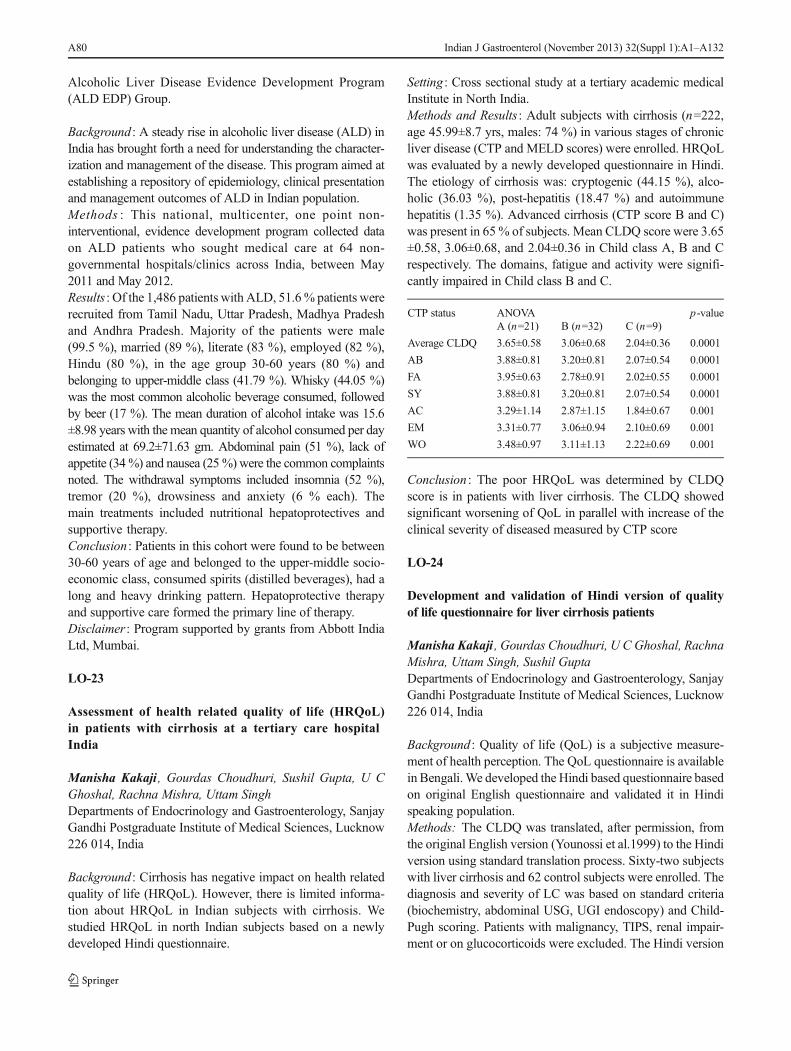
Alcoholic Liver Disease Evidence Development Program(ALD EDP) Group.
Background : A steady rise in alcoholic liver disease (ALD) inIndia has brought forth a need for understanding the character-ization and management of the disease. This program aimed atestablishing a repository of epidemiology, clinical presentationand management outcomes of ALD in Indian population.Methods : This national, multicenter, one point non-interventional, evidence development program collected dataon ALD patients who sought medical care at 64 non-governmental hospitals/clinics across India, between May2011 and May 2012.Results : Of the 1,486 patients with ALD, 51.6% patients wererecruited from Tamil Nadu, Uttar Pradesh, Madhya Pradeshand Andhra Pradesh. Majority of the patients were male(99.5 %), married (89 %), literate (83 %), employed (82 %),Hindu (80 %), in the age group 30-60 years (80 %) andbelonging to upper-middle class (41.79 %). Whisky (44.05 %)was the most common alcoholic beverage consumed, followedby beer (17 %). The mean duration of alcohol intake was 15.6±8.98 years with the mean quantity of alcohol consumed per dayestimated at 69.2±71.63 gm. Abdominal pain (51 %), lack ofappetite (34%) and nausea (25%) were the common complaintsnoted. The withdrawal symptoms included insomnia (52 %),tremor (20 %), drowsiness and anxiety (6 % each). Themain treatments included nutritional hepatoprotectives andsupportive therapy.Conclusion : Patients in this cohort were found to be between30-60 years of age and belonged to the upper-middle socio-economic class, consumed spirits (distilled beverages), had along and heavy drinking pattern. Hepatoprotective therapyand supportive care formed the primary line of therapy.Disclaimer : Program supported by grants from Abbott IndiaLtd, Mumbai.
LO-23
Assessment of health related quality of life (HRQoL)in patients with cirrhosis at a tertiary care hospitalIndia
Manisha Kakaji , Gourdas Choudhuri, Sushil Gupta, U CGhoshal, Rachna Mishra, Uttam SinghDepartments of Endocrinology and Gastroenterology, SanjayGandhi Postgraduate Institute of Medical Sciences, Lucknow226 014, India
Background : Cirrhosis has negative impact on health relatedquality of life (HRQoL). However, there is limited informa-tion about HRQoL in Indian subjects with cirrhosis. Westudied HRQoL in north Indian subjects based on a newlydeveloped Hindi questionnaire.
Setting : Cross sectional study at a tertiary academic medicalInstitute in North India.Methods and Results : Adult subjects with cirrhosis (n=222,age 45.99±8.7 yrs, males: 74 %) in various stages of chronicliver disease (CTP and MELD scores) were enrolled. HRQoLwas evaluated by a newly developed questionnaire in Hindi.The etiology of cirrhosis was: cryptogenic (44.15 %), alco-holic (36.03 %), post-hepatitis (18.47 %) and autoimmunehepatitis (1.35 %). Advanced cirrhosis (CTP score B and C)was present in 65 % of subjects. Mean CLDQ score were 3.65±0.58, 3.06±0.68, and 2.04±0.36 in Child class A, B and Crespectively. The domains, fatigue and activity were signifi-cantly impaired in Child class B and C.
CTP status ANOVA p-valueA (n=21) B (n=32) C (n=9)
Average CLDQ 3.65±0.58 3.06±0.68 2.04±0.36 0.0001
AB 3.88±0.81 3.20±0.81 2.07±0.54 0.0001
FA 3.95±0.63 2.78±0.91 2.02±0.55 0.0001
SY 3.88±0.81 3.20±0.81 2.07±0.54 0.0001
AC 3.29±1.14 2.87±1.15 1.84±0.67 0.001
EM 3.31±0.77 3.06±0.94 2.10±0.69 0.001
WO 3.48±0.97 3.11±1.13 2.22±0.69 0.001
Conclusion : The poor HRQoL was determined by CLDQscore is in patients with liver cirrhosis. The CLDQ showedsignificant worsening of QoL in parallel with increase of theclinical severity of diseased measured by CTP score
LO-24
Development and validation of Hindi version of qualityof life questionnaire for liver cirrhosis patients
Manisha Kakaji , Gourdas Choudhuri, U C Ghoshal, RachnaMishra, Uttam Singh, Sushil GuptaDepartments of Endocrinology and Gastroenterology, SanjayGandhi Postgraduate Institute of Medical Sciences, Lucknow226 014, India
Background : Quality of life (QoL) is a subjective measure-ment of health perception. The QoL questionnaire is availablein Bengali.We developed the Hindi based questionnaire basedon original English questionnaire and validated it in Hindispeaking population.Methods: The CLDQ was translated, after permission, fromthe original English version (Younossi et al.1999) to the Hindiversion using standard translation process. Sixty-two subjectswith liver cirrhosis and 62 control subjects were enrolled. Thediagnosis and severity of LC was based on standard criteria(biochemistry, abdominal USG, UGI endoscopy) and Child-Pugh scoring. Patients with malignancy, TIPS, renal impair-ment or on glucocorticoids were excluded. The Hindi version
A80 Indian J Gastroenterol (November 2013) 32(Suppl 1):A1–A132

of CLDQ was administered to patients and controls. The dataused was analyzed using the SPSS 16.0.Results : Forty-three (69.4 %) patients, forty-six (74.2 %) weremen in both the groups, the mean age of the patients was almostsimilar (p>0.05) among cases (47.56±9.25) and controls (57.16±9.88). Among the patients twenty-one (33%), thirty-two (51.6%) and nine (14.5 %) were categorizes into CTP score A, B, Crespectively. The reliability of Hindi CLDQ was determinedfrom the Cronbach’s alpha was overall scores 0.61. However,among the cases, the Cronbach’s alpha for individual domainswas nearly 0.90, the lowest being 0.89 and highest 0.91. Thetest-retest correlation coefficient for average CLDQ among thecases was 0.78 (p<0.0001) which was above 0.62.Conclusion : Our study confirmed the validity of Hindi CLDQversion in assessing the QoL among patients with LC.
LO-25
Serum apoptosis markers [CK 18 {M30 andAND M 65}]in acute liver failure
Chetanya Malik , Anita Chakravarti, Suresh Kumar,Premashis KarDepartments of Medicine and Microbiology, Maulana AzadMedical College, University of Delhi, Bahadur Shah ZafarMarg, Delhi Gate, New Delhi, Delhi 110 002, India
Introduction : Several clinical studies of acute liver failure(ALF) patients have demonstrated significantly increasedlevels of circulating apoptosis markers. Interest has recentlyfocused upon the measurement of cell death markers such ascaspase-cleaved and total cytokeratin [CK 18 {M30 and M65}] as a means of identifying the extent and relative propor-tions of apoptotic and necrotic cell death in ALF patients. Veryfew studies have been performed on patients with ALF. Thisstudy was aimed to assess whether the circulating apoptoticmarkers [CK 18 {M30 and M 65}] are altered in ALF and tocorrelate it with the prognosis.Methods : Thirty cases of ALF (sample collected at day ofadmission and day 3), 30 cases of acute viral hepatitis and 30healthy controls were included in the study. The serum levelsof M30 and M65 were measured using commercially avail-able ELISA kits.Results and Conclusion : The levels of M30 in ALF caseson the day of admission were 497.04±115.93 mIU/mL and451.24±369 mIU/mL at day 3. The levels of M65 in ALFcases on the day of admission were 138.02±120.62 mIU/mLand 109.40±77.23 mIU/mL at day 3. The ALF patients withM30 and M65 levels had grave prognosis as assessed byKings’ College criteria. Serum levels of apoptotic bio-markers [CK 18 {M30 and M 65}] were significantly(p>0.05) elevated in ALF cases compared to AVH andcontrol groups.
LO-26
Serum cholinesterase: A useful marker for liver syntheticfunction
Kunal Vyawahare , Ajit Kumar, Nayana Joshi, B Sukanya,Chintan Kansagra, Sandip Shah, Nikhil ShiroleDepartment of Gastroenterology, Nizam’s Institue of MedicalSciences, Punjagutta, Hyderabad 500 082, India
Background : Serum cholinesterase or pseudocholinesterase is anenzyme synthesized by liver and its levels decrease with de-creased synthetic function of liver as in cirrhosis. It is also foundto decrease further with the degree of liver decompensation.Aims : To compare the levels of enzyme in cirrhotic andnoncirrhotic patients and to determine whether fall in thelevels correlate with the degree of decompensation.Methods and Results : This is a cross sectional analytical studyconducted at the Department of Gastroenterology at our hospi-tal. Thirty-four patients were studied. Sixteen were cirrhoticsand 18 were noncirrhotics. The median levels of serum cholin-esterase in cirrhotic group was 135 U/L (Range:5-520) whereasit was 645 U/L (Range: 230-1300) in the noncirrhotic group (p-value <0.0001). Cirrhotic patients were further classified on thebasis of Child-Pugh`s criteria in to classes A, B and C whichincluded 2, 8 and 6 patients respectively. The median levels ofserum cholinesterase in these three classes were 515 U/L, 135U/L and 123 U/L respectively (p-value: 0.016).Conclusion : Thus serum cholinesterase is an important liversynthetic function test that can be used to identify cirrhoticpatients and its levels decrease further with the degree ofdecompensation.
LO-27
Renal resistive index and its utility in cirrhotic patients
Harshad Vinay Joshi , Pradeep Kakkadasam Ramaswami,Nilesh Toke, Sunil K Mathai, Charles Panackl, Rony ThomasMedical Trust Hospital, M G Road, Kochi 682 016, India
Background : Renal failure in patients with end-stage liverdisease is associated with significant morbidity and mortality.In patients with cirrhosis activated renin angiotensis system(RAS) is responsible for maintaining effective systemic vol-ume with its attendant complication in form of renalhypoperfusion due to renal arterial vasoconstriction. Renalhemodynamic changes are evident even before changes inserum creatinine occur. Renal resistive index (RI) is a directmeasure of renal vasoconstriction and can be a good tool inidentifying patients who have high renal vasoconstrictionhence are at risk of HRS. The purpose of this study is toevaluate the usefulness of RI in cirrhotic patients. So far not
Indian J Gastroenterol (November 2013) 32(Suppl 1):A1–A132 A81

many studies have assessed correlation between Child’s class,MELD scores and RI.Objectives of Study : A prospective study to compare resistiveindex in cirrhotic patients with controls, and to assess relationof RI with Child–Pugh’s class and MELD score.Materials and Methods : Adult in-patients with cirrhosisfrom May 2011 to April 2013 in our tertiary care hospitalwere included in the study after exclusion usingpredetermined facts. These patients were grouped into 3according to Child’s class as A, B and C. Forth groupconsisted of controls. Each group consisted of 25 patients.Renal resistive index (RI) was calculated in each patientusing duplex ultrasound with color doppler by two radiol-ogists. A value of less than 0.7 was taken as normal basedon several past studies in literature.Results : Amongst the study population of 100 patients, 80 weremales (80 %) while 20 were female. Overall amongst all Child’sclass alcohol topped the list of etiology, followed by cryptogen-ic, HBV, HCV and NASH in descending order. Majority ofpatients were in 40-60 years age group (~ 42%). Mean RI valuein patients with Child’s A cirrhosis was 0.69 (+/-0.015), inChild’s B cirrhosis was 0.71 (+/-0.08) and in Child’s C cirrhosiswas 0.89 (+/-0.026) whereas mean RI amongst controls was0.622 (+/-0.053). Compared to controls the RI was higheramongst all Child’s classes. There was no significant differencewithin individual Child’s classes. RI showed statistically signif-icant correlation with MELD. With higher MELD score the RIvalue increased with a correlation coefficient of 0.78.This correlation was even stronger for value of MELD>25. RI was found to increase in linear with age andBUN. Inverse correlation was seen between serum albu-min, sodium and RI. These correlations although werestatistically not very significant. RI did not vary indifferent etiologies of cirrhosis.Conclusion : Our study showed that resistive index signifi-cantly increases with a higher MELD score. Higher RI showpositive correlation with cirrhosis compared to controls. RI isindependent of etiology of cirrhosis. Higher RI is associatedwith higher TBIL, BUN, INR and lower albumin and sodiumlevels, although statistically less significant. RI could be agood tool to detect patients who are likely to develop HRSearly in clinical course even before worsening of creatinine.Long-term follow up studies are needed to study howmany ofpatients with high RI develop overt renal failure on follow up,which could help in establishing predictive value of RIamongst cirrhotics.
LO-28
Hepatocellular carcinoma: An unusual case presentingwith right heart failure
Arvind Namdeo , Pankaj Manoria, Rajesh Mehra
Shivalaya Gastroenterology and Liver Clinic, 6 Deepak Hous-ing Society DoodhDairy Bus Stop, Chuna Bhatti, Bhopal, India
Hepatocellular carcinoma (HCC) is most common primaryliver tumor. Most commonly presents as asymptomatic onscreening, or with deterioration of symptoms of cirrhosis likejaundice and ascites, weight loss, weakness, anorexia, palpa-ble liver. The case of a 59-year-old man presented with pro-gressive breathlessness on exertion for 3 months with markedswelling in lower limbs and abdominal distension for 1month.On examination patient had raised JVP, palpable liver withmarked pedel edema. Echo showed a large mass in rightatrium, lab showed HBsAg positive, USG showed large massin liver infiltrating in IVC and right atrium. AFP was >30000.CECT showed typical HCC with increased arterial uptake insegment VII and VIII with invasion in hepatic part of proximalIVC and complete extension into right atrium. To conclude itsan unusual case of large HCC presented with symptoms ofright sided heart failure with infiltration in IVC and heart.
LO-29
Histopathological features of liver in patients with chronichypertransaminaesaemia of unknown etiology
Rajesh Bathini , Praveen Kumar, Praveen Mathew,Somasekhar, Md DawoodVydehi Institute ofMedical Sciences,Whitefield, Nallurahalli,Bangalore 560 066, India
Objective : Asymptomatic persistent transaminaesemia is acommon finding and may alter treatment plan if correct diag-nosis is made.Aim : The aim of the study is to determine the spectrum ofhistopathological features of liver and probable etiology inthese patients.Methods : From August 2012 to June 2013, 14 patients withpersistent hypertransaminaesemia of more than 6 months withno clear diagnosis on clinical, sonological and serologicalevaluation were included. Liver biopsies were done and fibro-sis was staged by Ishak method.Results : Total 14 patients, 8 (58 %) were male. Mean age was33 yrs. Histopathological features of liver biopsies showedbland fibrosis in 3 (21.5 %), fibrosis with chronic hepatitis in 1(7.5 %), steatohepatitis in 5 (35.5 %), cirrhosis in 1 (7.5 %),Dubin-Johnson syndrome in 2 (14 %) and nonspecificchanges in 2 (14 %). In patients with bland fibrosis, onepatient had noncirrhotic portal fibrosis and another patientwas found to have occult hepatitis B. In patient with fibrosiswith chronic hepatitis, patient had interface hepatitis withfibrosis who later turned out to be ASMA positive. Insteatohepatitis patients, 2 had 3/6 fibrosis. In cirrhotic patient,no cause was found.
A82 Indian J Gastroenterol (November 2013) 32(Suppl 1):A1–A132

Conclusion : Liver biopsy is a useful investigation in patientswith persistent hypertransaminaesemia based onwhich furtherevaluation could pick up underlying etiology. In this studycommon histopathological findings were steatohepatitis,bland fibrosis.
LO-30
Transaminase levels are better than platelet count aspredictors of adverse outcome in dengue virus infection
C S Thyagaraj , M NarendranathanDepartment of Gastroenterology, Kerala Institute of MedicalSciences, Vinod Nagar Road, Post Anayara, Anamukham,Thiruvananthapuram 695 029, India
Background : Hepatic involvement has been recognized as acomplication of dengue fever. Traditionally platelet countshave been used to assess the severity of dengue fever. Oneof the objectives of this study was to develop a score to predictadverse outcome in these cases.Methods : Admissions with serologically proved dengue infec-tion were included in the study. The lab data of the cases wereanalyzed to identify the predictors of mortality. Mortality wasthe independent variable. The independent variables studied areage, platelet count, AST, ALT and alkaline phosphatase.Results : Ninety-one cases of dengue were included in thestudy. Eleven patients from this group died. Ninety-two per-cent had platelet count below 100,000. Platelet count was notsignificantly different in the two groups (53.7 x 10³ in thesurvivors compared to 42.9 x 10³ in the fatal cases; p=0.922).Transaminase levels were significantly higher (p<0.001) inthe mortality group (ALT: 2211 vs. 182; AST: 1579 vs. 131).From the data a score was developed (ALT/100)+age) - Add60 to this score in case of infants. A score above 70 couldpredict mortality with a sensitivity of 100 % and a specificityof 94.8 %.Conclusions : The study was done in a select group ofsevere cases of dengue virus infection admitted to thehospital. In this group of cases transaminase levels werebetter predictors of mortality than platelet count. Thescore incorporating age and ALT could predict mortalitywith an accuracy of 95.5 %. This score has to bevalidated in a prospective study.
LO-31
Kidney biopsy changes in patients with ethanol relatedcirrhosis and severe alcoholic hepatitis
S K Ravikiran , C K Adarsh, G K Prashanth, MallikarjunPatil, Keyur A Seth, H Aradya Venu, T R Vijay Kumar,Harshad Devarbhavi
Department of Gastroenterology, St Johns Medical College,Bangalore 560 034, India
Background : In cirrhotics, the renal pathological changesare not easily available as renal biopsy has high rate ofcomplications and often renal diagnosis is based onclinical scenario, urine examination, and renal imaging.Our objective is to analyze the spectrum of renal lesionsin patients who die of ethanol related cirrhosis andalcoholic hepatitis.Methods : Ultrasonography guided, percutaneous renal andliver biopsies were performed in 25 consecutive patients im-mediately after death (within 10 minutes) due to ethanolrelated liver disease. Urine analysis and serum biochemicaltests were available during hospitalization at the same sitting.Results : Patients had advanced liver disease as reflected in aMELD score of 30 and a Maddrey’s discriminant score of 87.Fifteen of 25 patients (60 %) had serum creatinine > 1.5 mg/dL. Five (20 %) patients had urinary tract infection, 8 (32 %)had spontaneous bacterial peritonitis and 4 (16 %) had comor-bidities. Renal biopsies were normal in 8 (28 %) patients.Most patients (48 %) had ATN. Bile casts were seen in 2(8 %) patients. Patients who had ATN also had higher creat-inine and it correlated positively. Nonspecific mesangial ma-trix expansion was seen in 3 and proliferation in 2 patients.Conclusions : Contrary to the popular belief that cirrhoticpatients with renal dysfunction have hepatorenal syn-drome (HRS) where renal histology is normal, we founda significant number of ATN. AKI due to ATN ispreventable with appropriate attention to hemodynamics,infections and diuretics. This study lays emphasis onruling out reversible causes of AKI before making alabel of HRS.
LO-32
Clinical spectrum and the role of non-surgical interventionin Budd-Chiari syndrome: Experience at a tertiary carecenter
Manoj Kumar, Manu Tandan, Rajesh Gupta, R Jagadeesh,Piyal Nag, D Nageshwar ReddyDepartments of Gastroenterology and Intervention Radiology,Asian Institute of Gastroenterology, 6-3-661, Somajiguda,Hyderabad 500 082, India
Aims and Objectives : To evaluate the clinical spectrum, pat-tern of vascular obstruction and role of nonsurgical treatmentin Budd-Chiari syndrome (BCS).Methods : Seventy cases of primary BCS were prospec-tively evaluated for clinical spectrum and pattern ofvascular obstruction. Patients with refractory ascitesand/or deteriorating liver function, depending on
Indian J Gastroenterol (November 2013) 32(Suppl 1):A1–A132 A83

obstruction of inferior-vena-cava (IVC) and/or hepaticvein (HV), were triaged for radiological intervention(RI). Patients with diuretic-responsive ascites or stableliver function were managed conservatively. Follow upwas done at the end of the 1st, 3rd, 6th and 12thmonth. Repeat RI were preformed as and when requiredduring follow up.Results : HV obstruction was most common (55.7 %)followed by IVC obstruction (31.4 %). An underlyingcoagulation disorder was defined in 54 % cases withpolycythemia-vera being the commonest (11.4 %) and36 % patients showed multiple coagulation disorders. RIwere performed in 50 patients, IVC stenting being thecommonest (38 %) followed by HV stenting (32 %).TIPS was performed in 11 patients. Technical successachieved in all patients. Re-occlusion of HV and IVCstent was observed in 12.5 % and 19 % respectively.Among TIPS patients primary patency rate at 1 yearwas 90.9 % and mortality was 9 %. Of the 20 patientsmanaged conservatively, four required RI due to pro-gressive ascites during follow up. Overall mortality was7.6 %.Conclusions : Hypercoagulability is important etiological fac-tor and the work up should not be stopped after detectingsingle cause. Pattern of vascular obstruction is changing.Medical therapy is indicated for asymptomatic patients. RI ispreferred in patients with refractory ascites or deterioratingliver functions.
LO-33
BCLC staging of patients with HCC presenting for firsttime in a tertiary care centre
Anil P John , Yamuna R Pillai, Siljo Jose, K R V KumarMedical Gastroenterology Department, Trivandrum MedicalCollege, Thiruvananthapuram 695 011, IndiaBackground : The clinical profile of HCC patients in detailwill help us to optimize the surveillance of at risk patients andhence the timely therapeutic intervention of affected patients.Aim : BCLC staging of patients with HCC presenting for firsttime in a tertiary care centre.Methods : Retrospective analysis of clinical data of patientsdiagnosed with HCC between 2008 and 2012 in MedicalGastroenterology Department, Government Medical College,Thiruvananthapuram, was done.Results : One hundred and fourteen patients, male n=90(78.9 %), female n=24 (21.1 %) were included into theanalysis. The mean age at diagnosis was 62 years, rangeof 30 to 88. HBV related chronic liver disease was 32%, followed by alcoholic liver disease (25 %), NASH(19 %), HCV (6 %), alcohol with HBV (15 %) andalcohol with HCV (3 %). Sixty percent of patients had
AFP >200. Forty percent of patients with liver cirrhosishad no impaired liver function (Child A) at diagnosis ofHCC, 18 % were in Child B and 42 % in Child C. Vastmajority of patients presenting in advanced and end-stageof the disease (BCLC B 7.2 %, BCLC C 47.8 %, BCLC D45 %).Conclusions : HBV related liver cirrhosis is the mostimportant risk factor for HCC in our tertiary centre.Majority of patients presents with advance stage orend-stage disease at diagnosis.
LO-34
Autoimmune liver disease: Impact on etiology of chronicliver disease and its disease spectrum: Experiencefrom North-Western India
Paras Shah , Vivek Agarwal, Sandeep Nijhawan, AmitMathur, Gaurav GuptaDepartment of Gastroenterology, Sawai Man Singh MedicalCollege, J L N Marg, Jaipur 302 004, India
Background and Aim : Autoimmune liver disease (AILD) isconsidered rare cause of chronic liver disease (CLD) in Indiawith few studies till date. We aim to study the impact of AILDas etiology in our patients of CLD and to evaluate the diseasespectrum of AILD patients.Methods : We prospectively analyzed 936 consecutivepatients of CLD admitted to Gastroenterology Depart-ment, S M S Medical College, Jaipur, between July2011 till June 2013, for presence of AILD and analyzedtheir clinical, biochemical and histological characteris-tics. Patients in whom etiological work up could not becompleted were excluded.Results : Out of 936 CLD patients (612 males-65.38 %and 324 females-34.62 %), AILD was found in 58 (6.19%). Females outnumbered males (49 females, 09 males,M:F=1:5.4). Prevalence of AILD was 1.47 % in malesand 15.12 % in females. Mean age of presentation was 32±14 years. In females AILD was major cause of non-ethanol, non-B, non-C related CLD, with adjusted preva-lence of 45.37 %. Presenting complains in majority ofAILD patients was jaundice (72.41 %) and ascites (53.44%), while GI bleed and HE being uncommon. Out ofAILD-AIH was seen in 47 (35-definite, 12-probable),PBC in 6, PBC/AIH overlap in 2, PSC in 3. Associatedautoimmune disorders were seen in 27.58 %.Conclusion : Autoimmune liver disease is not uncommoncause of CLD in Northwestern India with prevalencebeing 6.19 %. Majority of our AILD patients were fe-males, presented late with advanced liver disease andassociated other autoimmune disease were not commonlyseen.
A84 Indian J Gastroenterol (November 2013) 32(Suppl 1):A1–A132

LO-35
Evaluation of blood sample for the diagnosis of amebic liverabscess
Ujjala Ghoshal , Virendra Jaiswal, Sanjay S Baijal, Tapan NDhole, Uday C GhoshalDepartments of Microbiology, Radiodiagnostic and Gastroen-terology, Sanjay Gandhi Postgraduate Institute of MedicalSciences, Raebareli Road, Lucknow 226 014, India
Background : Diagnosis of amebic liver abscess (ALA) ischallenging, as sensitivity of microscopy is very low (0 % to15 %). Serum IgG antibody may not distinguish past fromcurrent infection, especially in endemic regions and is positivein a proportion of healthy population. Although, liver aspirateis the most suitable for diagnosis but this facility is not widelyavailable.Objective : We evaluated utility of blood sample for diagnosisof ALA using PCR.Method : Twenty-eight patients with ALA and 10 patients withpyogenic liver abscess (PLA) were included. Diagnosis of liverabscess was based, I. Clinical symptoms (fever, pain in the righthypochondrium, lower chest, shoulder. II. Enlarged and/ortender liver without jaundice. III. Space occupying lesion inthe liver diagnosed by ultra sound IV. For ALA- presence ofamebic DNA in liver aspirate and for PLA-aspirate cultureshowing growth of a pyogenic organism. Liver aspirate andwhole blood were collected from each patient. DNA wasextracted from liver aspirate and blood samples. E. histolyticaDNAwas amplified using species specific primer.Results : 25/28 patients with liver abscess had amebic DNA intheir blood sample. 22/25 (88 %) of these patients ALApatients had already received prior amoebic treatment. Sensi-tivity, specificity of PCR for the diagnosis of ALA in bloodsamples were 89 % and 100 % respectively. PCR wasremained negative in blood samples of all PLA patients.Conclusion : Detection of E. histolytica DNA in blood samplecould be used for diagnosis of ALA patients where aspirationof pus is not feasible.
LO-36
Study of non-alcoholic fatty liver disease in higher-socioeconomic population of western India
S Patwari, Nikhil Lala, Bhavesh PanchalSterling Hospital, Sterling Hospital Road, Memnagar,Ahmedabad 380 052, India
Aim and Background : The prevalence of non-alcoholic fattyliver disease (NAFLD) has doubled during last 20 years,whereas the prevalence of other chronic liver diseases has
remained stable or even decreased. progression of NAFLDto nonalcoholic steatohepatitis (NASH) increases risks ofliver cirrhosis, liver failure, and hepatocellular carcinoma(HCC). Cirrhosis due to NASH is an increasingly frequentreason for liver transplantation. Available data of NAFLDprevalence in India is scanty.Objectives and Method : Four hundred and thirty-seven ob-jectives were screened by basic ultrasonography of abdo-men to diagnose fatty liver. All the objectives werescreened by age, gender, body mass index (BMI), w/h ratio(for central obesity), medical examination, primary lfts,history of chronic diseases (dislipidemia, diabetes, hypo-thyroidism, etc.) and alcohol consumption. Two differentviews have been taken to study. First NAFLD andsuspected NASH (altered lfts with fatty liver) objectiveswere screened with different factors like age, gender, BMI,w/h ratio etc. Prevalence of these factors among NAFLDpool were noted. Secondly, different groups of populationlike overweight, diabetics etc were screened by proportionof NAFLD/suspected NASH to study NAFLD propor-tions among chronic diseases.Result : Among 437 (266 males, 171 females) objectives, 165(37.7 %) had NAFLD. Thirty-one of 165 (18.7 %) had alteredlfts (suspected NASH). It accounts 7.3 % of total objectives.Out of 165 NAFLD, 126 (76.4 %) were males and 39 (23.6%) were females. Among NAFLD, 149 (90.3 %)dislipidemic, 145 (87.8 %) overweight (BMI >25), 67 (41%)obese (BMI >30), 125 (75.5 %) centrally obese (w/h >0.85),52 (31.5 %) diabetics, 16 (10 %) hypothyroids, were found.One hundred and thirty-one (79.3 %) of NAFLD objectiveswere both overweight (BMI >25) and dislipidemic. 87.9 %(131/149) of dislipidemic NAFLD and 88.4 % (46/52) ofdiabetic NAFLD have BMI > 25. 84.3 % of dislipidemicand 76.9 % diabetic NAFLD have w/h ratio >0.85. AvergeBMI and w/h ratio of NAFLD group are 30.01 kg/m2 and0.93 respectively. NAFLD proportions in males and femalesare 47.3 % (126 of 266) and 22.8 % (39 of 171) respectively.NAFLD proportion in 51 to 60 yrs age group is 40.3% (23 of57). NAFLD among dislipidemic 47.3 % (164 of 346), over-weight 43.8 % (145 of 331), obese 51.9 % (66 of 127),centrally obese 46 % (123 of 267), diabetics 55.3 % (52 of94) were found. 51.6 % (16 of 31) of suspected NASH grouphave been found to have globulin level greater than 3.5 g/dL.Conclusion . Proportion of NAFLD is more in higher socio-economic population in western region of India. NAFLD ismore prevalent in males than females. With increase in age,NAFLD proportion increases (highest among 51-60 yrs).There is significant drop in NAFLD proportion on removalof any form of obesity. Average NAFLD individual is central-ly obese/obese. Diabetics and dislipidemics have higherNAFLD proportions, if their BMI is >25. Majority ofNAFLD is in combination of dislipidemia and BMI >25.Higher globulins levels in suspected NASH objectives were
Indian J Gastroenterol (November 2013) 32(Suppl 1):A1–A132 A85

found, which requires further study of immunologicalphenomena.
LO-37
Radiofrequency ablation of hepatocellular carcinoma:Recurrence, satellite lesions and needle seed tracking
Antony Raphel Joy, R N Harikumar, S Ismail, M Sreekanth,K P Sreekumar, P K NazarAmrita Institute of Medical Sciences and Research Centre,AIMS Ponekkara PO, Kochi 682 041, India
Background : Radiofrequency ablation (RFA) has emerged inclinical practice, as a simple and effective technique for treat-ment of hepatocellular carcinoma (HCC). It is not associatedwith major side effects and can be performed with ease, safetyand reasonable cost. RFA as a modality has been used withcurative intent, palliative intent and as a bridge to livertransplantation.Aim : The aim of this study is to assess the response rate ofRFA in the form of recurrence, satellite lesions and needletrack seeding with a follow up of up to 1 year.Methods : A retrospective descriptive study of 108 patientswith 115 HCC (mean diameter 3.23, 1.4 SD) were submittedto RFA between May 2004 and December 2011. In all casesRFAwas performed with percutaneous approach under ultra-sound guidance using Covidien's Cool Tip RF ablation needle,size depending on the lesion size. Treatment efficacy wasestimated with MDCT abdomen and AFP levels.Results : Complete ablation after treatment was 66 %, 56.3 %and 28.5 % in HCC smaller than 3 cm, between 3 and 5 cmand larger than 5 cm respectively. Eighteen (26.4 %) lesionsout of 68 developed local recurrence after complete ablationduring a mean follow up of 3 months. Twenty-two (20.3 %)patients out of the total developed new satellite lesions duringa follow up of 1 year. There were no needle tracking seenduring a follow up period of 1 year after RFA. There were notreatment related deaths.Conclusion : RFA is an effective treatment for HCC smallerthan 5cmwith complete ablation in more than 60% of lesions.Patients with elevated AFP had a worse prognosis with de-velopment of recurrence and satellite lesions.
LO-38
Association of dendritic cell SIGN polymorphismin hepatocellular carcinoma patients from India
Dipu Bharali , Manash P Sarma, Premashis KarDepartment of Medicine, Maulana Azad Medical College,Bahadur Shah Zafar Marg, Delhi Gate, New Delhi, Delhi110 002, India
Objective : To investigate the association of mutation indendritic cell-specific intercellular adhesion molecule-3-grabbing non-integrin (DC SIGN) promoter region inhepatocellular carcinoma (HCC) patients and healthycontrol without any history of liver diseases and alsoto analyze the mutation as a risk factor for HCC.Methods : A total of 40 cases of HCC and 40 healthy controlswithout any liver abnormalities were included in the study.Total 5 mL peripheral blood samples were collected andgenomic DNA was isolated followed by PCR amplificationand direct sequencing (Macrogen, Korea) along with compar-ing those sequences with the published database.Results : The characteristic mutation within DC SIGN promot-er region in HBV infected HCC individuals was observed.There was 2 hot spot mutation located in positions -139, -142which were observed in HCC patients. Only 1 spot mutationlocated in position -139 was observed in healthy controls. The-142 which was absent in the healthy control was observed in17 HCC (42.5 %). The -139T was far more frequent in thehealthy control (98 %) than in the HCC patients (35 %).Conclusion : In the DC SIGN promoter region, -142 may beassociated as a genetic factor for developing HBV associatedHCC but -139 may be associated with protection against.
LO-39
Assessment of HRQOL in patients with cirrhosis usingCLDQ (chronic liver disease questionnaire): A pilot studyfrom north costal Andhra Pradesh
P SChakravarthy, Vidya Viswanath, L R SGirinath, PMuraliKrishnaAndhra Medical College, Jagadamba Junction, Visakhapat-nam Part, Andhra Pradesh 530 002, India
Aim : To validate a liver specific questionnaire CLDQ incorrelation to CTP scoring in patients with cirrhosis in theregion of north costal Andhra Pradesh.Methods : Subjects with chronic liver disease who are willingto participative and CTP scoring=5+(except those with com-plications of cirrhosis such as malignancy, SBP, renal failure,pulmonary failure and unwilling patients) were enquiredCLDQ in local language (Telugu). Each patient’s responseswere assessed according to 1 to 7 scale in CLDQ in 6 healthrelated domains prescribed in CLDQ scoring system. Eachpatient’s CTP score was calculated. Data was analyzed usingchi-square testing and p -value <0.05 was taken as significant.Results : A total of 32 patients were included in the study. InCTPA group (n=3) of patients, the score was moderate (4-5)in all domains. In CTP-B group (n=16), score was severe(1-3) in abdominal domain and moderate in other domains. InCTP C group (n=13), score was severe in abdominal andfatigue domain and moderate in other domains.
A86 Indian J Gastroenterol (November 2013) 32(Suppl 1):A1–A132

Conclusion : The administered CLDQ thus showed theabdomen domain and fatigue domain as areas with mostdistressing symptoms, followed by systemic domain.The severity of the CLDQ scoring is matching withseverity of the disease according to standard CTP scor-ing for liver disease.
LO-40
Spontaneous intracranial hemorrhage in patientsof cirrhosis
A K Choudhury, Anand Lovkesh, S K Taneja, C Vasistha, S KSarinDepartment of Hepatology, Institute of Liver and BiliarySciences. D-1 Vasant Kunj, New Delhi 110 070, India
Introduction : Spontaneous intracranial hemorrhage (SICH) isthough rare it often mimics hepatic encephalopathy. There is apaucity of data on SICH in cirrhotic patients presenting withencephalopathy.Aims and Objectives : To determine the incidence, clinicalprofile and outcome of SICH in cirrhotic patients with HE.Materials and Methods : A retrospective analysis of 3,170cirrhotic patients out of which 1,243 admitted to the intensiveliver coma unit of a tertiary referral hospital over a period of 4years from 2009 to 2013 taken.Results : Hepatic encephalopathy is seen in 682 patients andabnormal neuroimaging seen in 26 patients (12.75 %). Whichinclude hemorrhage in 20 (9.8 %) and infarct in 6 (2.9 %). Ofthese 20 patients with SICH, 16 were males and 4 femaleswith a median age of 55.5 (IQR 50-61.5). The etiology of liverdisease was alcohol in 9, hepatitis C in 3, hepatitis B andNASH in 2 each and cryptogenic in 4 patients. Thecommonest presentation was progressive deep coma in 7,seizures in 6, persistent HE in 3, focal neurological deficit in2 and headache in 1 patient. Of these 20 patients of SICH 9patients were in Grade IVHE, 3 in Grade III and 8 in Grade II.The median platelet count was 70 x 103/mm3 (IQR 57-96)and INRwas 1.7 (IQR 1.5- 2.3). The median bilirubin was 4.6mg/dL (IQR 1.9-9.8), serum creatinine was 1.07 mg/dL (IQR0.7-2.1) and serum sodium was 142.6 (IQR 135.5-150.8).According to CTP score 14 patients had Child C grade, 4had Child B and 2 had Child A grade. The median MELDscore was 24 (IQR 16-27). The median MELD Na was 26(IQR 22-29). The commonest site of bleed was frontoparietalin 6, followed by subdural and basal ganglion in 3 patientseach. None of the patient underwent surgical intervention dueto coagulopathy and severe liver disease. The overall mortal-ity was 80 %, with 4 patients (20 %) showing spontaneousrecovery. There was no significant difference in age, sex,clinical presentation; biochemical parameters, MELD, MELDNa, INR and platelet count in the survivors as compared to
those who died. Mannitol was used in 10 patients but was notfound to significantly alter outcome.Conclusion : Spontaneous intracranial hemorrhage is rare incirrhotic patients but caries very high mortality. It is morecommon in male alcoholics with severe liver disease.
LO-41
A new prognostic score for severe alcoholic hepatitis
Neeraj Nagaich , Sandeep Nijhawan, Radha Sharma,Subhash Nepalia, Amit Mathur, Gaurav GuptaDepartment of Gastroenterology, Sawai Man Singh MedicalCollege, J L N Marg, Jaipur 302 004, India
Background : Alcoholic hepatitis (AH) is a cause of consider-able mortality and morbidity. Short-term prognosis of alco-holic hepatitis is worse than that of decompensated cirrhosisas agreed at the Baveno IV consensus conference. The strat-ification of patients with severe alcoholic hepatitis (SAH)have potential clinical implications. The modified Maddrey’sdiscriminant factor (mDF) is most frequently used score. Thisstudy was aimed to determine role and validity of a new scorefor predicting short and long-term prognosis in patients withSAH.Methods : In 151 patients admitted with SAH (DF -2). A scorecombining individual score assigned for each of these 5 var-iables [age, bilirubin level, Delta PT (prothrombin time) (nor-mal PT value - PT of patient), creatinine level and grade ofencephalopathy] was compared to DF in determining survivalat 30 days, 90 days and 6 months.Results : The predictive value of this SCORE was better thanDF. By stepwise multivariate analysis, this SCORE was iden-tified as independently associated with 6-month mortality.This new score value greater than 7 was associated withincreased risk of mortality (18 % vs. 63%) and showed higherpredictability as compared to DF, particularly so for 6 monthmortality, AUROC 0.84 (0.71-0.96) vs. 0.64 ( 0.44-0.84).Conclusion : This new SCORE includes parameters easilyobtained at admission, which allows a rapid identification ofthe prognosis. In this study it compared well with the DF andrather performed better. It may help in early risk stratificationand management of patients with SAH.
LO-42
A study of spectrum of hepatic dysfunction in denguefever
Vineet Behera , Velu Nair, N Naithani, Pankaj Puri, V RMujeeb, Juhi Varshney, B SainiDepartments of Gastroenterology and Internal Medicine,Armed Forces Medical College, Pune 411 040, India
Indian J Gastroenterol (November 2013) 32(Suppl 1):A1–A132 A87

Objective : To study the clinical and laboratory spectrum ofhepatic dysfunction in dengue and study the factors predis-posing to hepatic dysfunction.Materials and Methods : We designed a two year cross sec-tional study in a tertiary care hospital. The study populationcomprised of patients of dengue fever (as per revised WHOcriteria). The clinical presentation especially gastrointestinalfeatures were recorded. Hemogram, routine biochemistry, bili-rubin, LFT, dengue serology and ultrasonography of abdomenwas done for all. Other causes of hepatitis like viral markers,NASH, alcohol intake markers, drugs were excluded.Results : 1.204 patients of dengue were included in the study.The gastrointestinal symptoms seen were nausea/vomiting in52 % cases, anorexia in 42 %, pain abdomen in 17.6 %,jaundice in 12.2 % cases. 2. USG showed hepatomegaly in23.5%, splenomegaly in 15.1%, ascites in 24% and acalculouscholecystitis in 20 % cases. LFT revealed hyperbilirubinemia(>1.5 mg/dL) in 13.2 %, elevated AST and ALT (>50 IU/L) in54.9 % and 47.05 % respectively (15.7 % >3 ULN enzymes),hypoalbuminemia (<3.5 g/dL) in 33.3 % cases (30.8 % of these<50,000/cu mm, hemorrhagic manifestations and features ofcapillary leak were statistically significant.Discussion : Dengue has clinical hepatic and gastrointestinalmanifestations as given, hypoalbuminemia, transaminitis (<3times ULN) and rarely jaundice with acalculous cholecystitisand ascites on USG. It is more frequent and severe in patientswith thrombocytopenia, hemorrhagic manifestations and cap-illary leak.Conclusion : Hepatic manifestations are seen in patients ofdengue and are a marker of severe disease.
LO-43
Clinical profile and factors associated with liver enzymeabnormalities among HIV-infected persons
Ashok Mohite , Pravir Gambhire, Dharmesh Shah, PiyushSomani, Qais Contractor, Pravin RathiDepartment of Gastroenterology, T N Medical College and BY L Nair Hospital, Mumbai Central, Mumbai 400 008, India
Background : As HIV infected patients are experiencing lon-ger life expectancies, liver disease has been recognized as amajor issue but data is more limited and is mostly fromwestern countries.Aims and Objectives : To study the clinical spectrum of liverenzyme abnormalities in HIV infected patients. To study thepotential cause(s) and identify factors associated with liver testelevations among HIV-infected patients.Materials and Methods : Cross sectional, prospective study intertiary care hospital. Thirty-three HIV infected patients withderanged liver enzymes are followed and evaluated for possi-ble causes and outcome of deranged LFT.
Results : Mean age of patients was 39.3±9.46 with 9 (27 %)females and 24 (73 %) males. The most common diagnosiswas alcoholic liver disease (21 %) and chronic hepatitis Binfection (21 %) followed by ATT induced hepatitis (18 %).Three (9 %) patients had multiple causes and one had hepatictuberculosis. Liver biopsy was done in 4 patients. Elevenpatients (33 %) had cirrhosis at the time of presentation.Despite laboratory tests and ultrasound examination, abnor-mal liver test results was unexplained in 10 (30 %) patients ofwhich 7 patients was on HAART. During mean follow up of 6months two patients died, one worsened and others improved.Conclusions : Liver test abnormalities are common amongHIV patients during the HAART era. The most commondiagnosis was alcoholic liver disease and chronic hepatitis Binfection. Despite laboratory and radiologic investigationsinto the cause of liver dysfunction, 30 % were unexplained,but might be related to unrecognized fatty liver disease in-duced by HAART.
LO-44
Hepatocellular carcinoma in the era of newer treatments: Asingle center experience
Prachi S Patil, Shaesta Mehta, Mahesh MahadikDepartment of Digestive Diseases and Clinical Nutrition, TataMemorial Hospital, Parel, Mumbai 400 012, India
Introduction and Aims : Hepatocellular carcinomas (HCC)are associated with poor prognosis. Newer treatmentmodalities like Sorafenib and Trans Arterial Radioembolisation(TARE) have been introduced recently. We present anaudit of HCC patients seen at the Tata Memorial Hos-pital from 2012-2013.Methods : Review of a prospective database of 183 consecu-tive patients with HCC to assess the etiology, stage, treatmentand outcomes.Results : The mean age was 57 years (range 18-80 years).Most patients were male (85.2 %). The mean alpha-fetoprotein (AFP) levels were 82,308 ng/mL (range 2.5-3376570 ng/mL). AFP levels more than 400 ng/mL were seenin 93 patients (52 %) while levels between 100-400 ng/mLwere seen in 17 (9.4 %). The commonest etiological factorswere HBV infection in 77 patients (42 %), HCV infection in18 (9.8 %) and alcohol consumption in 72 (39.6 %). TheBCLC stage was A in 5 %, B in 32 %, C in 30 % and D in33 % patients. Most patients had an ECOG-PS of 2 (69 %) or3 (24 %). Chronic liver disease was present in 131(72 %). One hundred and twelve patients (62 %) wereChild-B and 69 (38 %) Child-C. Sixty-eight patients(38 %) presented with decompensation. Eighteen patients(9.8 %) underwent surgical resection, 8 (4.4 %) underwentRFA and 54 (29.5 %) underwent TACE. Newer treatment
A88 Indian J Gastroenterol (November 2013) 32(Suppl 1):A1–A132

modalities like TARE and Sorafenib were used in 19 (10.4 %)and 42 (23 %) patients respectively. Twenty-nine (15.8 %)were planned for best supportive care only.Conclusions : Most HCC present in an advanced stage andwith a suboptimal performance status. Newer treatmentmodalities are used in almost one third of all HCC patients.
LO-45
A rare case of amebic liver abscess presenting as nephroticsyndrome
Vineet Behera , Pankaj Puri, S Srivastava, N Naithani,A Sagar, R Yadav, P KumarDepartments of Gastroenterology and Internal Medicine,Armed Forces Medical College, Pune 411 040, India
Background : Amebic liver abscess presenting as nephroticsyndrome is very rare. Case Report: A 38-years-old-manpresented with fever with pain right upper abdomen of 7 daysand swelling of feet and had tender hepatomegaly. His inves-tigations showed leukocytosis, creatinine 1.7 mg/dL and con-jugated hyperbilirubinemia. USG and CT scan confirmed an9.5 cm X 10.8 X 10 cm abscess in right lobe of liverimpending rupture with evidence of IVC compression ondoppler. His amebic serology was positive. He had 3+protein-uria with 24 hour urine protein of 3600mg, serum albumin 2.4mg/dL and hypertriglyceridemia confirming a nephrotic syn-drome. A percutaneous drainage of the abscess was done andwas given IV metronidazole and cefotaxime. His symptomsimproved over 48 hours and had gradual resolution of abscesssize, IVC compression, proteinuria and other parameters.Discussion : Amebic liver abscess can cause thrombosisof the portal vein or IVC or can compress these veinscausing bilateral pedal edema and ascites. But signifi-cant IVC compression causing renal vein congestionand leading to a picture like nephrotic syndrome isextremely rare. Huddle reported a case of ALA compli-cated with nephrotic syndrome due to external IVCcompression which recovered after drainage of the ab-scess. Liver abscess in close proximity to the IVC orhepatic veins, >5 cm in size or with pedal edema oranasarca should undergo CT or doppler USG to dem-onstrate compression or abnormal flow. It requires ag-gressive medical therapy, anticoagulation and urgentdrainage of the abscess.
LO-46
A rare case of storage cirrhosis –Niemann-Pick disease
V SaravananMadras Medical College, Chennai 600 003, India
Niemann-Pick disease is a rare lysosomal storage disorderwhich manifest mainly in childhood. Here we present a caseof Niemann-Pick disease who had presented during his adult-hood. A 23-year-old male presented to our pod with painabdomen, jaundice. On examination, he had short 4th meta-tarsal hand, hepatomegaly. He had thrombocytopenia, alteredlipid profile and liver function tests. His liver biopsyshowed gralumatous hepatitis with noncaseating granu-loma with lymphocytic infiltrates. His bone marrowshowed foamy cells. Diagnosis was made with de-creased sphingomyelinase activity in blood. Since hedid not have neurological involvement, he was diag-nosed to have Niemann-Pick disease, type2. He waskept under follow up. Niemann-Pick disease as such arare entity can very occasionally present in adulthoodand can pose difficulty in diagnosis.
LO-47
Chemokine ligand CXCL-8 mediated activation andneutrophil influx aggravates liver injury and correlateswith clinical severity indices in acute-on-chronic liverfailure
Arshi Khanam , Peggy Reise, Nirupma Trehanpati, ChandanKumar, Carlos Albert Guzman, Shiv Kumar SarinDepartments of Research and Hepatology, Institute of Liverand Biliary Sciences, Vasant Kunj Marg, New Delhi 110 070,and Department of Vaccinology and Applied Microbiology,Helmohltz Centre for Infection Research, Braunschweig,Germany
Background : Acute-on-chronic liver failure (ACLF) is a seri-ous and often fatal condition, having very high incidence ofinfection and mortality. Impaired neutrophil and Th17cellfunction has been observed in ALF and served as a biomarkerinvolved in organ dysfunction and sepsis. We investi-gated role of neutrophils and Th17cells associated withdisease progression in ACLF.Methods : Eighteen alcohol induced, 17HBV-related ACLFand 18 healthy controls (HC) were enrolled. TJLB was col-lected from patients. Peripheral blood was taken from patientsand HC. Percentage of neutrophil, CD16 (activation marker),CXCR-1and 2 was observed. Absolute neutrophil, lympho-cyte and TLC was compared in patients with and withoutsepsis. IL-6,IL-23,CCL-20 and GM-CSF level were detected.Circulating and intrahepatic Th17cells, IL-23 receptor andtranscription factor ROR γT was investigated. CytokinesIL-17, IL-22, CXCL-8, and IFN γ were measured.Results : Significant increase in percentage of neutrophils,CD16, CXCR-1/2 was observed in alcohol (p>0.008, 0.00,0.00, 0.003) and HBV-induced ACLF (p>0.00, 0.00, 0.00,0.00) than HC. No differences were seen in alcohol vs. HBV-
Indian J Gastroenterol (November 2013) 32(Suppl 1):A1–A132 A89

induced ACLF. Absolute neutrophil and lymphocyte countwas higher in patients with sepsis than without. Inflammatorycytokines (IL-6 p>0.00, GM-CSF-0.00, IL-23-0.01) werehigh in both groups and positively correlated with INR(p>0.008), MELD (p>0.03) and CTP score. Th17cells(p>0.003), IL-23 receptor (p>0.04) and ROR γT was higherin peripheral blood of ACLF than HC and same was evenhigher in liver tissuewith respect to peripheral blood in ACLF.Increased Th17cells positively correlated with ALT (p>0.00),s bilirubin (p>0.00) and INR value (p>0.00). Circulating IL-17 (p>0.002), IL-22 (p>0.05), CXCL-8 (p>0.03) and IFN γproduced by Th17cells was highly elevated in ACLF than HCand even higher in liver than peripheral blood in ACLF. HighCXCL-8 showed strong correlation with increased neutrophilcounts (p>0.00).Conclusion : Increased intrahepatic Th17cells produceCXCL-8 which recruits neutrophils to liver in CXCR-1/2dependent manner. Hyper activated neutrophil produces abun-dant inflammatory cytokines; thus encourages hepatic inflam-mation and further disease progression.
LO-48
Acquired hemophagocytic lymphohistiocytosis mimickingacute hepatic insult presenting as ACLF: Experiencefrom a dedicated Liver Unit
Ankur Jindal , Awinash Kumar, Ashok Kumar, NaveenKumar, Manoj Kumar Sharma, Rakhi Maiwall, HitendraGarg, S K SarinDepartment of Hepatology, Institute of Liver and BiliarySciences, New Delhi 110 070, India
Background : Hemophagocytic lymphohistiocytosis (HLH) isfrequently fatal (overall mortality >50 %) and oftenunderdiagnosed. It involves a final common pathway ofhypercytokinemia. A timely diagnosis is imperative to facili-tate immunosuppressive therapy and decrease mortality. HLHpresenting as severe acute viral hepatitis or acute-on-chronicliver failure is extremely rare. We present our experience withHLH in a dedicated liver ICU presenting mainly for severeacute hepatic insult.Methods : Retrospective analysis of admitted patients withsystemic inflammatory response (SIRS) fulfilling diagnosticHLH criteria (≥5/8).Results : Seventeen patients [Male:Female -14:3; Adults:Child- 14:3; median age- 26 years (range-1 month to 66 years]were diagnosed with acquired HLH at our hospital from year2010 to 2012. Twelve (70.6 %) patients presented clinically asACLF (7) and AVH (5) at admission. Viral associated HLH (8patients; 47.1 %) was most common (HAV-3, HEV-2, EBV-1,Dengue-1, Parvovirus-1). Although etiology remainedundiagnosed in 7 (41.2 %) patients, few patients had
lymphoma (1) and visceral leishmaniasis (1). While fever[16 patients (94.2 %); median duration- 30 days (4-90 days)]and jaundice [14 patients; mean bilirubin-20.34±8.6 mg/dL]were most common symptoms at presentation, clinical signssuch as presence of enlarged liver (76.4 %), spleen (76.4 %)and ascites (58.8 %) were most frequent. Five patients alsohad AKI (serum creatinine >1.5 mg/dL) at admission. Impor-tant biochemical parameters included hyperferritinemia [allpatients; mean-16064.7±7652 ng/dL], hypertriglyceridemia(10 patients; mean-354.7±145.1 mg/dL), raised LDH (mean-2952.66±726) and low fibrinogen (12 patients; mean-146.1±32.4) levels. Bone marrow aspiration was done in 12 patientsof which 11 shows presence of hemophagocytosed histio-cytes. Ten patients (58.8 %) had in-hospital mortality andthe main cause was eventual sepsis and multiorgan failure.Twelve patients (70.6 %) received specific immunosuppres-sive therapy (steroids-4, IV IG-4, plasmaphersis-4,cyclosporine-1) but it made no difference in clinical outcome(In hospital mortality- 66.67 % vs. 62.25 % respectively;p>0.05).Conclusions : HLH may masquerade as acute hepatic insult, itis important to suspect and recognize this entity early enoughin patients with ACLF, as prognosis is poor despite immuno-suppressive therapy.
LO-49
Altered hepatic microenvironment governs the natureof hepatic regenerative response to acute hepatic insult
Smriti Shubham , Jaswinder S Maras, Saira Hussain,Dhananjay Kumar, Chhagan Bihari, Hitendra K Garg, RakhiMaiwall, Viniyendra Pamecha, Archana Rastogi, AnupamKumar, Shiv K SarinDepartments of Research, Hepatology, Pathology, and HPBSurgery, Institute of Liver and Biliary Sciences, New Delhi110 070, India
Background : Acute hepatic insult triggers hepatic regenera-tion in both normal and chronic liver. Hepatic microenviron-ment plays an important role in both hepatocyte and hepaticprogenitor cell (HPC) mediated hepatic regeneration.Aim : To understand the effect of altered hepatic microenvi-ronment on hepatic regeneration in response to acute hepaticinsult.Patients and Methods : Hepatic vein plasma and liver sectionsof acute-on-chronic liver failure (ACLF, n=31; Gr. 1), acuteliver failure (ALF, n=10; Gr. 2) decompensated liver disease(DLD, n=10; Gr. 3), were collected and subjected to immu-nohistochemical staining for Ki67 (hepatocyte self replica-tion) and cytokeratin7 (HPC and intermediate hepatocytes)for characterization of nature of hepatic regeneration. Hepaticvein plasma of the same patients was subjected to cytokine
A90 Indian J Gastroenterol (November 2013) 32(Suppl 1):A1–A132
bead array (Bio-Rad) and was correlated with hepatocyte selfreplication, HPC activation and maturation.Results : Immunohistochemical analysis documented a signif-icant decrease in Ki67+hepatocytes in Gr.1 as compared to Gr.2 (p=0.003). Further the number of CK7+HPC and its matu-rational lineages was found to be significantly increased in Gr.1 as compared to Gr. 2 (p=0.027). Cytokine array analysisshowed that GCSF, GMCSF, TNFα, IL-2Rα, BNGF, HGF,IL-3, SCGFB, BFGF were all significantly down regulated inGr.1 as compared to Gr. 2 (p<0.05). On correlation we ob-served that BFGF, GCSF, GMCSF correlated directly to he-patocytes self replication [Ki67 (p<0.05, r2>0.3)]. WhileTNF-α, GCSF and GMCSF documented an inverse correla-tion with CK7+HPC and its maturational lineages (p<0.05,r2>-0.3). Multinomial logistic regression analysis document-ed BFGF independently correlates with hepatocytes self rep-lication with a LR=6.3 and HR=26.4 CI (1.4-495). WhileGCSF and TNF-α are inversely associated with CK7+HPCactivation and its maturational lineages [HR=7.4 (0.9-58), 6.4(0.9-43)].Conclusion : Overall the result suggest that acute insult in ahealthy liver triggers hepatocytes self replication while acuteinsult in chronic liver triggers HPC mediated hepatic regener-ation. Loss of BFGF in ACLF may be responsible for com-promised hepatocytes self replication and loss of GCSF andTNF-α may be associated with increased HPC activation andits maturation.
LO-50
Relative adrenal insufficiency in chronic liver disease issimilar to sepsis
Faiz Ahmed , Sharad Malhotra, Ankush Bansal, R KHimthaniBatra Hospital and Medical Research Centre, 1,TughlakabadInstitutional Area, Near Saket Metro Station, MehrauliBadarpur Road, New Delhi 110 062, India
Introduction : Patients with severe sepsis and septic shockcommonly have abnormalities in adrenal function and this isassociated with decreased response to catecholamine therapy,hemodynamic instability and a high mortality. Similar find-ings have been observed in patients with chronic liver disease.This phenomenon has been termed relative adrenal insuffi-ciency (RAI). We therefore decided to screen for relativeadrenal insufficiency in severely stressed chronic liver diseasepatients and compare them with patients of sepsis.Methods : We enrolled 20 patients with chronic liver diseaseand 20 patients with severe sepsis. Patients who had receivedsteroids in last 6 weeks were excluded. Cortisol levels weremeasured by a radioimmune assay technology (Sourcerer RIAcounter, Oakfield Health Care). The peak cortisol level was
defined as the highest cortisol level obtained after synacthenadministration at 60 minutes. The criteria for adrenal insuffi-ciency were defined as follows: baseline value less than15 mcg/dL (414 nmoL/litre), or cortisol response less than9mcg/dL (less than 248 nmoL/litre) with a baseline valuebetween 15 and 34 mcg/dL.Results : Adrenal insufficiency is common in patients withboth chronic liver disease and severe sepsis. In our study wefound the incidence of adrenal insufficiency to be 60 % and55 % in chronic liver disease and sepsis respectively, In ourstudy mean arterial pressure, serum bilirubin, vasopressindependency, renal dysfunction and bacteremia were indepen-dent factors predicting adrenal insufficiency in critically illpatients with chronic liver disease and severe sepsis.Conclusion : Clinically relevant Adrenal insufficiency is seenin chronic liver disease similar to sepsis and may requiresimilar management.
LO-51
Prospective study of spontaneous bacterial peritonitis in atertiary care center
A Bharath Kumar, Jeyamani Ramachandran, Sajith, AshishGoel, V BalajiDepartments of Hepatology and Microbiology, ChristianMedical College, Vellore 632 004, India
Aim and Methods : We assessed the microbiolgical profile,response to antibiotics and factors affecting outcome prospec-tively in cirrhotic patients with SBP from July 2012 to July. SBPwas categorized based on mode of acquisition into-communityacquired (CA), health care related (HCR) and nosocomial (NC).Protocol was to treat CA and HCRwith piperacillin tazobactum;NC and SBP with shock and multiorgan dysfunction withmeropenem. Antibiotic was escalated in nonresponders, definedas Ë‚ 25 % reduction in ascitic neutrophil count after 48 hours.Results : Among 150 episodes (in 130 patients, 86%male) 45%,42% and 13%were CA, HCR andNC. Predominant etiology ofcirrhosis was ethanol (46 %). Ascitic fluid cultures were positivein 39%.Commonest organismwasE. coli in CA andHCR.BothE. coli and gram positive organisms were the culprits in NC.Antibiotic escalationwas required in 32%, 28%and 26% inCA,HCR and NC. Organ failure (acute kidney injury) at admissionoccurred in 57 %, 67 % and 80 % in CA, HCR and NC.Piperacillin resistance was 37 %, 40 % and 75 % in CA, HCRand NC. One case each in CA and HCR had carbapenemaseresistance. In hospital mortalitywas 26% inCA andHCR and 34% in NC. Recurrent infections were seen in 15 patients (11 %).Conclusions : A significant proportion of CA showed lack ofresponse to piperacillin. Presence of organ dysfunction atadmission, need to escalate antibiotic, NC type and resistanceto piperacillin were associated with poor outcome.
Indian J Gastroenterol (November 2013) 32(Suppl 1):A1–A132 A91

LO-52
Clinical profile of drug induced liver injury: A prospectivesingle centre study in Eastern India
Abhijit Chowdhury, G K Dhali, Kaushik Das, Amit MishraDepartment of Gastroenterology and Hepatology, S S K MHospital, Kolkata 700 020, Institute of Post Graduate MedicalEducation and Research, Kolkata 700 020, India
Background : Drug induced liver injury (DILI) is an increas-ingly recognized cause of morbidity and mortality in patientsreceiving prescription drugs and complementary medications.Spectrum of DILI ranges from asymptomatic derangement inliver function tests to chronic liver disease and outcomes varyfrom early resolution to death.Aims and Objectives : Prospective assessment of causes, clin-ical features, pattern, severity, strength of causality and out-comes of DILI in Eastern India.Material and Methods : All consecutive patients admitted asinpatients in SDLD, SSKM Hospital or attending HepatologyOPD from February 2012 to July 2013, fulfilling predefinedinclusion and exclusion criteria were included in this study.Predefined clinical and laboratory assessment was done for allpatients. Strength of causality was assessed using RUCAMcriteria.Results : Of 39 included patients, 21 were female. Anti-tubercular drugs were causative agent in 19 (49 %), oralcontraceptive pills in 10 (26 %), methotrexate in 4 (10 %),Ayurvedic drugs in 4 (10%) and antiepileptic drugs in 2 (5%).Twelve (31 %) patients had hepatocellular pattern, 23 (59 %)had mixed pattern and 4 (10 %) had cholestatic pattern ofinjury. Twenty patients had possible association and 18 pa-tients had probable association according to RUCAM criteria.Jaundice (62 %) was most common symptom at presentation.Twenty-two (56.4 %) patients had >50 % improvement inALT within 30 days of stopping the drug while 1 patientexpired due to possible DILI event.Conclusions : Antitubercular drugs are most common cause ofDILI in our population followed by oral contraceptive pills.Ayurvedic medications may under-recognised cause of DILI.Most DILI events have early resolution after withdrawal ofcausative agent.
LO-53
Effect of rifaximin, probiotics and L-ornithine L-aspartateon minimal hepatic rncephalopathy: A case control study
Sanjay Pant, Kapil Sharma , S P Misra, Manisha Dwivedi,Sushil Narang, Kailash Mishra, Ravikant Kumar
Department of Gastroenterology and Hepatology, Moti LalNehru Medical College, Allahabad 211 001, India
Background : Minimal hepatic encephalopathy (MHE) im-plies subtle impairment of cognitive functions in absence offeatures of overt encephalopathy.Aims : To determine the prevalence of MHE in patients withliver cirrhosis and find out effect of rifaximin, probiotics andLOLA individually in reversal of MHE by comparing it withno treatment group.Methods : A total of 317 cirrhotics were screened for the studyof which 111 were excluded due to various reasons.Remaining 206 patients were screened for MHE usingneuropsychometric tests (NPTs) and/or critical flicker fre-quency (CFF). Of these 124 patients with MHEwere random-ized to receive LOLA (drug 1), rifaximin (drug 2), probiotics(drug 3), for two months and were compared to patients whowere not given any treatment for MHE.Results : Out of the 206 cirrhotics, 124 (60.19 %) had MHE.Amongst these 124 MHE patients, 87 (70.16 %) patients withMHE had CFF <39Hz, 112 (90.32%) patients withMHE had 2or more abnormal psychometric tests and 75 (60.48 %) patientshad abnormality on both the CFF values and more than twoNPTs. Intention to treat analysis showed patients who improvedafter giving treatment were 21/31, 22/31, 16/32 and 9 /30 forLOLA, rifaximin, probiotic and no treatment groups respec-tively. CFF scores were statistically significant (p-valueThe abstract matter was not received in full at the time ofsending to press, despite reminders to corresponding author.
LO-54
Etiologic and clinical spectrum of chronic liver disease in atertiary referral hospital in India
S M Shasthry, M Kumar, H Garg, R Maiwal, C Vashisht,V Bhatia, R Kumar, Y K Joshi, S K SarinInstitute of Liver and Biliary Sciences, Sector D1, VasantKunj New Delhi 110 070, India
Aims : To evaluate the etiological and clinical spectrum of chron-ic liver disease [CLD], in a tertiary care center in Northern India.Methods : We evaluated 3,170 chronic liver disease patientsadmitted at ILBS from June 2009 to April 2013. The diagnosisof CLD was based on clinical, radiological or endoscopicfeatures.Results : Alcoholic liver disease was the cause in 1,507 [47.5%] CLD patients followed by viral etiologies 805 [25.4 %];13.4 % of HCV related CLD and 12 % HBV related CLD.Cirrhosis was due to NASH+NAFLD in 425 [13.3 %], cryp-togenic in 380 [12 %] and autoimmune in 113 [3.6 %] of the
A92 Indian J Gastroenterol (November 2013) 32(Suppl 1):A1–A132

total CLD patients. The mean age at presentation was 51.76+/-12.45 yrs with males being predominant 2,627 [82.7 %]. Themean CTP, MELD and MELDNa scores at presentation were9.06+/-2.42 and 17.92+/-8.23, and 21.17+/-8.12. Two third ofpatients [1916; 66.6 %] had ascites and 731 [23.1 %] hadhepatic encephalopathy and 336 [10.6%] had associatedHCCat presentation. ALD patients had significantly lower age(mean 48.34+/-10.7 yrs) compared to other etiologies(p<0.001); with higher CTP (10.1+/-2.1), MELD (21.1+/-8.2) MELDNa (24.5+/-7.6), liver stiffness (49+/-23 kPa) andHVPG (17.75+/-4.73 mmHg) in comparison with HBV, HCV,NAFLD, AIH and ALD+ liver diseases (p<0.001 with eachgroup). Higher mortality at first presentation was seen in ALD(9.6 %) compared with HBV (6.6 %), HCV (3.8 %) andNAFLD (3.8 %) (p<0.05 with each groups)Conclusion : ALD constitutes nearly half of chronic liverdiseases requiring hospital admissions in tertiary care centerin India with younger age at presentation, more severe diseaseand higher mortality compared to other causes of chronic liverdiseases.
LO-55
Autoimmune hepatitis/primary biliary cirrhosis overlap
Karthikeyan , Premkumar, Ratnakar Kini, KanisheikMohammad, T Pugazhendhi, Mohammed AliDepartment of Medical Gastroenterology, Madras MedicalCollege, Chennai 600 003, India
Overlap syndrome is used to describe a form of autoimmunehepatitis (AIH) which present with characteristics of AIH andprimary biliary cirrhosis (PBC) or primary sclerosingcholangitis (PSC). Patients with overlap syndromes presentwith both hepatic and cholestatic serum liver tests and havehistological features of AIH and PBC or PSC. AIH-PBC is themost common form of overlap syndrome, affecting almost 10% of adults with AIH or PBC. Transitions from PBC to AIH-PBC overlap syndrome have also been reported. Overlapsyndromes show a progressive course without treatment, andtherapy is empirical. Anticholestatic therapy withursodeoxycholic acid is usually combined with immunosup-pressive therapy with corticosteroids and/or azathioprine inboth AIH-PBC and overlap syndromes. In end-stage disease,liver transplantation is the treatment of choice. Here we reportone such case of autoimmune hepatitis-primary biliary cirrho-sis overlap. Our patient was a 45 yrs old female with featuresof decompensated liver disease, portal hypertension and cho-lestasis. She had grossly elevated IgG, positive AMA, ANAand strongly positive AMA- M2. Liver biopsy showed fea-tures of both PBC and AIH.
LO-56
Etiology and prognosis of acute liver failure in a tertiarycentre
Siljo Jose , E H RoobyMedical Gastroentrology, S S B Medical College,Trivandrum 695 011, IndiaBackground : Acute liver failure (ALF) is a rare condition inwhich rapid deterioration of liver function results in alteredmentation and coagulopathy in previously normal individuals.Objective : To study the etiology and prognosis of acute liverfailure patients admitted in Medical Gastroenterology Depart-ment, Trivandrum Medical College from August 2010 to July2011.Methods : There were 24 patients, 8 males and 16 females. Themost common etiology was Hep B, drug induced, pancreatitsfollowed by others. Six patients survived withouttansplantation, 18 patients expired. None of the patientsunderwent transplantation. Liver biopsy was done in onepatient with SLE and ALF, etiology found to be HSV.Conclusion : ALF has a mortality of 75 % and prognosis ispoor. The etiology was wide range, the most common beingHep B, drug induced, and pancreatits.
LO-57
Altered hepatic microenvironment governs the natureof hepatic regenerative response to acute hepatic insult
Smriti Shubham , Jaswinder S Maras, Saira Hussain,Dhananjay Kumar, Chhagan Bihari, Hitendra K Garg, RakhiMaiwall, Viniyendra Pamecha, Archana Rastogi, AnupamKumar, Shiv K SarinDepartments of Research, Hepatology, Pathology, and HPBSurgery, Institute of Liver and Biliary Sciences, Vasant KunjMarg, New Delhi 110 070, India
Background : Acute hepatic insult triggers hepatic regenera-tion in both normal and chronic liver. Hepatic microenviron-ment plays an important role in both hepatocyte and hepaticprogenitor cell (HPC) mediated hepatic regeneration.Aim : To understand the effect of hepatic microenvironment onnature of hepatic regeneration in response to acute hepatic insult.Patients and Methods : Hepatic vein plasma and liver sectionsof acute-on-chronic liver failure (ACLF, n=31; Gr. 1), acuteliver failure (ALF, n=10; Gr. 2) decompensated liver disease(DLD, n=10; Gr. 3), were collected and subjected to immu-nohistochemical staining for Ki67 (hepatocyte self replica-tion) and cytokeratin 7 (HPC and intermediate hepatocytes)for characterization of the nature of hepatic regeneration.
Indian J Gastroenterol (November 2013) 32(Suppl 1):A1–A132 A93

Hepatic vein plasma samples of the same patients wassubjected to cytokine bead array (bioplex-47 cytokinesarray-Bio-Rad) and was correlated with hepatocyte self repli-cation, hepatic progenitor cell activation and maturation.Results : Immune-histochemical analysis documented a signif-icant decrease in Ki67+hepatocytes in Gr.1 as compared to Gr.2 (p=0.003). Further the number of CK7+HPC and its matu-rational lineages was found to be significantly increased inGr.1 as compared to Gr. 2 (p=0.027). Cytokine bead arrayanalysis showed that GCSF, GMCSF, TNF-alpha, IL-2R Al-pha, BNGF, HGF, IL-3, SCGFB, BFGF were all significantlydown regulated in Gr.1 as compared to Gr.2 (p<0.05). Oncorrelation we observed that BFGF, GCSF, GMCSF correlat-ed directly to hepatocytes self replication [Ki67(p<0.05,r2>0.3)]. While TNF-alpha, GCSF and GMCSF documentedan inverse correlation with CK7+HPC and its maturationallineages (p<0.05,r2>-0.3). Multinomial logistic regressionanalysis documented BFGF independently correlates withhepatocytes self replication with a LR=6.3 and HR= 26.4 CI(1.4-495). While GCSF and TNF-alpha are inversely associ-ated with CK7+HPC activation and its maturational lineages[HR=7.4 (0.9-58), 6.4 (0.9-43)].Conclusion : Overall the result suggest that acute insult in ahealthy liver triggers hepatocytes self replication while acuteinsult in chronic liver triggers HPC mediated hepatic regener-ation. Loss of BFGF in ACLF may be responsible for com-promised hepatocytes self replication and loss of GCSF andTNF-alpha may be associated with increased HPC activationand its maturation.
LO-58
Overlap syndrome: Autoimmune hepatitis with AMAnegative primary biliary cirrhosis
T S Ramesh Kumar, A R Venkateswaran, RevathyDepartment of Medical Gastroenterology, GovernmentStanley Medical Hospital, Chennai 600 001, India
“Overlap syndrome” is used to describe variant forms ofautoimmune hepatitis (AIH) which present with characteris-tics of AIH and primary biliary cirrhosis (PBC) or primarysclerosing cholangitis (PSC). Patients with overlap syndromespresent with both hepatitic and cholestatic serum liver testsand have histological features of AIH and PBC or PSC. AIH-PBC is the most common form of overlap syndrome, affectingalmost 10 % of adults with AIH or PBC. Transitions fromPBC to AIH-PBC overlap syndrome have also been reported.Overlap syndromes show a progressive course without treat-ment, and therapy is empiric. Ursodeoxycholic acid is usuallycombined with immunosuppressive therapy but end-stagedisease requires liver transplantation. We report a case ofAIH/PBC overlap with relevant discussion of literature.
LO-59
The comparison of bone mineral density between patientswith chronic liver disease and the control group
Manisha Kakaji , Gourdas Choudhuri, Sushil GuptaDepartments of Endocrinology, and Gastroenterology, SanjayGandhi Postgraduate Institute of Medical Sciences, Lucknow226 014, IndiaBackground and Aim : Hepatic osteodystrophy is a complica-tion of longstanding liver disease results in osteopenia andosteoporosis. It can significantly affect morbidity and qualityof life of these patients. The aim of this study was to comparethe rate of bone mineral density (BMD) between patients withchronic liver disease (CLD) and healthy control group.Materials and Methods : One hundred and sixty-four pa-tients with CLD and 222 individuals without CLD (controlgroup) were enrolled in this study. The CLD group includedalcoholic liver disease, idiopathic, C or B virus related, andautoimmune hepatitis patients. The CLD patients were di-vided to Child class - A, B and C by severity. Bone mineraldensity (BMD) was measured by dual energy X-ray absorp-tiometry (DEXA).Results : Adult patients (46.38±8.96 M: F: 3:1) was enrolled.CTP score of A, B, and C was present in 31.10 %, 56.70 %and 12.20 % respectively. BMD were measures at three sites,patients with child A has 0.832±0.133, 0.848±0.135, 0.609±0.101, B 0.822±0.120, 0.843±0.160, 0.604±0.122 and C has0.797±0.112, 0.773±0.274, 0.593±0.125 and normal were0.921±0.105, 0.896±0.127, 0.627±0.122 respectively.Seventy-nine (48.18 % has osteoporosis 78 (47.56 %) hadosteopenia and 7 (4.26 %) were normal according to T score.Significant correlations were found between BMD and CTPscore. Although statistically not significant, the mean bonemass (mean Z. score) was lower and the frequency of osteo-porosis was higher in CLD compared to those of the controlgroup (p>0.05). The mean bone mass was decreased fromChild-A to Child-C (p>0.05).Conclusion : CLD could reduce BMD. The risk increases withseverity of cirrhosis.
Gallbladder and Biliary Diseases
GB-1
Risk factors associated with gallbladder cancer in coastalOdisha
Ankit Tiwari, S P Singh, Niranjan Rout, Manjusha DixitSchool of Biological Sciences, National Institute of ScienceEducation and Research, Bhubaneswar, Odisha 751 005,Department of Gastroenterology, S C B Medical College,
A94 Indian J Gastroenterol (November 2013) 32(Suppl 1):A1–A132

Cuttack, Odisha 753 007, AHRCC, Cuttack, Odisha, andKalinga Gastroenterology Foundation, Cuttack, Odisha, India
Background and Aim : Gallbladder carcinoma (GBC), themost common biliary tract cancer has highest incidence rateand is the commonest cancer in North Indian women (21.5/100,000). The distribution suggests a high-incidence region,popularly known as GBC belt, comprising Uttar Pradesh,Bihar, Orissa, West Bengal, and Assam. Epidemiologic stud-ies suggest that GBC is multifactorial in nature. This studyaims to identify risk factors associated with GBC in coastalOdisha.Patients and Methods : We recruited 45 GBC patients and524 controls from Beam Diagnostics Center, Cuttack,Odisha. A questionnaire was developed for this studyto record various environmental risk factors includingdietary habits, reproductive history, previous infections,family history for cancers, occupation, age, BMI andgender. The dietary evaluation was carried out by thedietary recall method. Data was analyzed using SPSS 17software. Data analysis was done for known risk factorsincluding use of impure water, mustard oil, and familyhistory of cancer.Results : Frequency of GBC was found to be muchhigher in females (2.27:1). Odds ratio for gender femaleto male was 2.198 (95 % CI 1.17-4.13). Consumptionof impure water and mustard oil were strongly associ-ated (p<0.000) with increased risk (Odds ratio 11.32, 95% CI 4.70-27.27 and 3.25, 95 % CI 1.69-6.27) of GBC.Family history of cancer was also found as highlysignificant (p<0.000, Odds ratio 16.45, 95 % CI 7.49-36.16) risk factor for GBC.Conclusion : This ongoing study shows that female gender,impure water, mustard oil and family history of cancer are riskfactors for gallbladder cancer.
GB-2
Clinical and etiological profile of obstructive jaundicein tertiary referral hospital
P Laksmana Chandra , P Shravan Kumar, M Umadevi,M RamannaGandhi Medical College Hospital, Secunderabad 500 025,India
Background/Aim : Obstructive jaundice is one of the com-mon refferal cases to gastroenterology. In this study, weaimed at identifying clinical and etiological profile ofobstructive jaundice in patients referred for ERCPprocedure.Patients and Methods : Three hundred and twenty-three pa-tients with obstructive jaundice referred for ERCP over a
period of 4 years from 2009-2013 were studied retrospective-ly. Demographic, clinical, biochemical and ultrasonography,CT, MRCP, ERCP findings were analyzed and biopsy detailswere noted.Results : Of all the 323 patients, 128 males (40 %); median age42 years; range 17-67 years and females 195 (60 %); medianage 38 range 19-64 years. The clinical profile of patientsinclude jaundice 323 (100 %), abdominal pain 170 (55 %),vomiting 94 (29 %), anorexia 90 (29 %), loss of weight 80 (26%), pruritus 50 (15 %), pale stools 58 (18%). The spectrum ofdiseases diagnosed include choledocholithiasis 139 (43 %),periampullary carcinoma 47 (15 %), benign biliary stricture44 (14 %), hilar cholangiocarcinoma 31 (10 %), distal chol-angiocarcinoma 20 (6), carcinoma head of pancreas 16 (5 %),postoperative bile leak 15 (5 %), choledochal cyst 4 (1 %),carcinoma gallbladder 3 (0.9%), hydatid cyst 2 (0.6%), portalbiliopathy 2 (0.6%), PSC 2 (0.6%). Gender wise analysis 128males (40 %), 195 females (60%). Common etiology in malescholedocholithiasis 53 (17 %), periampullary carcinoma 38(12 %), hilar cholangiocarcinoma 20 (6 %) and females-choledocholithiasis 86 (27 %), benign biliary stricture 28 (9%).Conclusion : Choledocholithiasis found to be the commonestcause of obstructive jaundice followed by periampullary car-cinoma in this study.
GB-3
Double-guidewire-assisted technique for difficult biliarycannulation: A case series
Amit Bhasin , Anoop Alva, Shanthi Vijayaraghavan,S ShanmuganathanDepartment of Medical Gastroenterology, Sri RamachandraMedical College, Porur, Chennai 600 116, India
Background : A common problem with endoscopic retrogradecholangiopancreatography (ERCP) is difficulty reaching a deepbiliary cannulation. An alternative method for difficult biliarycannulation is the double-guidewire technique (DGT). Ourstudy aimed to assess the feasibility and safety of the double-guidewire-assisted biliary cannulation using this technique.Methods : A total of 10 patients who were undergoing ERCPwith intent to cannulate the CBD between January to June 2013were included in the study. DGTwas applied after three repeatedpancreatic guidewire entry. The application of the DGT for deepbiliary cannulation and the complications of ERCP procedure(pancreatitis, perforation, bleeding) using the DGT were deter-mined. The overall success rate for biliary cannulation in thesecases also was determined. Main outcome measurements CBDcannulation rate and ERCP-related complications.Results : A total of seven female and three male patients withage range of 29-72 years were included in the study. Eight out
Indian J Gastroenterol (November 2013) 32(Suppl 1):A1–A132 A95
of ten patients had underlying CBD stone. One patient was postcholecystectomy CBD leak and one had carcinoma head ofpancreas. The DGT was applied in the above 10 cases afterthree attempts. CBD cannulation was achieved in 100 % (10/10). None of the patients (0 %) developed any post proceduralcomplications.Conclusions : The double-guidewire technique is a feasibleand safe method for biliary cannulation in all patients withfailed standard cannulation technique with low rate ofcomplications.
GB-4
Diagnostic and therapeutic role of ERCP in patientswith post laparoscopic cholecystectomy bile leak
S Pratibha , P Shravan Kumar, M Umadevi, M Ramanna,I BharaniDepartment of Gastroenterology, 5th Floor, Gandhi Hospital,Secunderabad 500 025, India
Aim : To study ERCP findings in post laparoscopic cholecys-tectomy bile leak.Materials and Methods : This is a retrospective analysis of 17patients. The details were recorded from the medical records.All patients who had a bile leak post cholecystectomy andunderwent an ERCP were included.Results : Total of 17 patients was analyzed. The age range was25-65 years. There were 12 males and 5 females. Cannulationwas successful in 13 patients. Cholangiogram revealed leak in8 patients, 3 had leak at CBD level, 4 at cystic duct insertionlevel. A complete cut-off was seen in 4 patients. Two patientshad stones in the CBD. All 17 patients underwentsphincterotomy and stenting was successful in 9 patients.Stone extraction was successful in one patient. Four patientsin whom cannulation was unsuccessful were referred for CBDexploration. All the stented patients had shown a resolution ofleak at 1 week follow up.Conclusions : ERCP and stenting is an effective modality oftreatment for bile leak. The common sites of leak were CBDand at cystic duct insertion. However stenting was successfulin approximately half of the patients.
GB-5
Outcome of emergency endoscopic biliary stentingwithout imaging guidance in patients with obstructivejaundice
S Sukumaran , K Muthukumaran, G Ramkumar,R Balamurali, P Ganesh, S Jeevan KumarDepartment of Digestive Health and Disease, Kilpauk Medi-cal College, Chennai 600 010, India
Introduction : Endoscopic biliary stenting (EBS) is an effec-tive method to achieve biliary drainage in patients with ob-structive jaundice due to malignancy or benign conditions. Inroutine practice, ERCP and biliary stenting are performedwithfluoroscopy. However, in emergency conditions, patients withan impaired general status who cannot be transported to theradiology units, emergency EBS stenting without imagingguidance may be applied.Aim: To study outcome of emergency EBS without imaging guid-ance performed in patients with obstructive jaundice in our centre.Materials and Methods : In this retrospective study, technicalsuccess and outcome of emergency EBS without imaging guid-ance that were performed in our centre in 45 patients withobstructive jaundice were analyzed. Patients were evaluated withimaging studies like MRCP and CECT abdomen. Patient underconscious sedation; SVS done and selective cannulation of CBD(bile aspiration in cannula)+/-sphincterotomy and stent placementwere done. X-ray abdomen done for confirming stent position.Results : In 45 patients, successful stenting was achieved in 39patients (89 %) of which sphincterotomy done in 24 patients(61 %). In two patients stenting was done aftercholedochotomy and through choledochoduodenal fistula inone patient. Substantial reduction in bilirubin levels wasachieved in all patients with successful stenting. There wereno major immediate and late complications.Conclusion : Technical success and outcome of EBS is verymuch similar to conventional ERCP. EBS is safe and effica-cious method of biliary drainage in emergency conditions andpatients with an impaired general status who cannot betransported to radiology units.
GB-6
Single incision laparoscopic cholecystectomy and peroperative ERCP as single stage treatment
P S Rajan , Mohd. Juned Khan, P Karthikeyan, C PalaniveluG EM Hospital and Research Centre, 45, Pankaja Mill Road,Ramanathapuram, Coimbatore 641 045, India
Background/Aims : Even though laparoscopic cholecystectomyand ERCP have revolutionized themanagement of common bileduct stones, the use of these modalities as a single stage proce-dure remains controversial. More over, with increasing applica-tion of single incision minimally invasive procedure, we carriedout our study to determine the feasibility of single incisionlaparoscopic cholecystectomy and ERCP as a single procedure.Methods : We involved all patients planned for management ofboth gallbladder and CBD stones excluding those with acutecholecystitis, empyema of gallbladder from August 2010. Wesubmitted them for single incision laparoscopic cholecystec-tomy and per operative ERCP. Age group from 6 to 63 years.Total number of patients 55.
A96 Indian J Gastroenterol (November 2013) 32(Suppl 1):A1–A132

Result : CBD access and stone clearance was achieved in 100% of patients. Out of 55, 11 patients required needle knifesphincterotomy because of failure to cannulate ampulla. Threepatients required lithotripter to crush the larger size stones.With regard to gallbladder, cholecystectomy was performedwith single incision multiport technique. Nine patients re-quired converstion to standard laparoscopy in view of denseadhesions around Calot’s triangle and the need for suturinginfundibuluam where the cystic duct dissection and clippingnot possible.Conclusion : Single incision laparoscopic cholecystectomyand ERCP provides effective therapy for CBD and gallbladderstones with least morbidity and may be beneficial to selectedpatients as both are done under single anesthesia.
GB-7
Laparoscopic assisted endoscopic retrograde cholangiographyfollowing Roux-En-Y reconstruction
P S Rajan , Mohd. Juned Khan, P Karthikeyan, Amol Shinde,C PalaniveluG EM Hospital and Research Centre, 45, Pankaja Mill Road,Ramanathapuram, Coimbatore 641 045, India
Background/Aims : Laparoscopy assisted endoscopic retro-grade cholangiography (LAERC) allows the diagnosis andtreatment of biliopancreatic conditions in patients with alteredanatomy following any kind of Roux-En-Y reconstruction.However experience with this technique is limited and itsresults are still emerging.Methods : To report on the experience with LAERC in con-secutive patients from a tertiary centre with high-volume ofbariatric surgeries and to evaluate success rates of ERC withthe laparoscopy-assisted approach.Result : Five patients underwent LAERC, three for choled-ocholithiasis after RYGB, one for recurrent stricture afterhepaticojejunostomy for choledochal cyst and one for cho-ledocholithiasis following subtotal gastrectomy. Endoscopicaccess was obtained through the gastric remnant (in threeRYGB patients) or biliopancreatic limb (in other twopatients). Biliary cannulation was successfully achieved inall the patients. Biliary sphincterotomy was performed inthe three patients with intact sphincters. Average timetaken to achieve laparoscopic assisted endoscopic cannula-tion was 40 min (range 20 min-55 min) and average timetaken to complete the entire procedure was 140 minutes(range 110 min-160 min). All the four choledocholithiasispatients were treated by balloon sweep, and widesphincterotomy. The patient with recurrent cholangitis fol-lowing hepaticojejunostomy stricture for choledochal cystmanaged with stenting. The median post-procedure hospi-tal stay was 4 days.
Conclusion : LAERC is safe and successful for the treatmentof biliopancreartic condition in patients with Roux-en-Yanatomy.
GB-8
Single stage laparoscopic peritoneal lavage and ERCPin biliary peritonitis following cholecystectomy
P S Rajan , Mohd. Juned Khan, P Karthikeyan,Sathyamurthy, Amol Shinde, C PalaniveluG E M Hospital and Research Centre, 45-A, Pankaja MillRoad, Ramanathapuram, Coimbatore 641 005, India
Background : Post cholecystectomywith biliary peritonitis is achallenging clinical scenario where there is role of both sur-gery and endoscopy. Our objective of this study is to see thefeasibility and advantages in aiding fast recovery by combin-ing both laparoscopic peritoneal lavage and ERCP withstenting at single stage.Methods : From September 2008 to January 2013, 43 patientswith biliary injury presented with peritonitis in our Institute(30 M and 13 F). After preoperative work up, they werescheduled for combined laparoscopic peritoneal lavage andERCP. Three patients were converted to open surgery in viewof dense adhesions.Technique : After general anesthesia, thorough peritoneallavage done. Multiple intraabdominal drains placed in allcompartments. Before completion, ERCP was performed insame supine position. Cannulation was successful in 32patients and in 11 patients following pre-cut needle knifesphincterotomy, guide-wire could be taken into CBD andplastic stent deployed in 33 patients. In 10 patients, therewas complete transaction of CBD seen and hence stentingcould not be performed.Result : Out of 43 cases, 10 patients had complete CBDtransaction and were kept on with drains for 3 months beforetaken up for definitive hepaticojejunostomy procedure. In 33cases, 24 patients had leak from cystic duct or infundibulamcut-end. Nine patients had lateral wall injury to CBD. All thepatients settled well with endoscopic stenting followingsphincterotomy. Two patients had mild postoperative pancre-atitis treated conservatively.Conclusion : Combined application of laparoscopy and ERCPaids in faster recovery in patients with biliary injury.
GB-9
Prognostic signif icance of maspin mRNA expressionin patients with resected gallbladder cancer
Kavita Baghel , Hasan Raza Kazmi, Saloni Raj, AbhijitChandra, Rajeshwar Nath Srivastava
Indian J Gastroenterol (November 2013) 32(Suppl 1):A1–A132 A97

Departments of Surgical Gastroenterology, and OrthopedicSurgery, King George's Medical University, Lucknow 226003, India
Introduction : In the present study, we investigated expressionof maspin mRNA in normal, gallstones and gallbladder cancer(GBC) tissues and its prognostic significance for patients withGBC.Materials and Methods : The study consists of 81 patientsincluding 25 each of normal and gallstones and 31 of GBC(stage II: n=16, stage III: n=15). RNA was isolated fromtissues followed by cDNA synthesis. Maspin mRNA expres-sion was evaluated by reverse transcriptase PCR and real timePCR.Results : Significant higher (p=0.043) expression ofmaspin mRNA was observed in GBC by reverse tran-scriptase PCR. Further, 36 % and 65 % increase (P 1.5(median survival; 11 months) as compared with thosepatients in which R.Q. <1.5 (median survival; 17.5months) (HR=3.02, 95 % CI=2.304-11.53). Significantdifference (p=0.02) in survival was observed for stageII patients however no significant difference (p=0.117)was observed for stage III disease. The HR for stage IIand III disease was 8.9 (95 % CI=1.395-56.8) and 2.17(9 5% CI=0.7907-8.477) respectively.Conclusion : Increased maspin mRNA expression from gall-stones to GBC indicates presence of gallstones may lead tomalignant transformation and poor survival. Maspin mRNAmay be used as a prognostic marker for resected stage II GBC.
GB-10
Epidemiological factors in gallbladder carcinomain northern India
Smita Verma , Jitendra Kumar Choudhary, Neha Singh, NehaGupta, Manish Kumar Tripathi, Sapna, Vinod Kumar Dixit,Ashok Kumar JainDepartment of Gastroenterology, Institute of Medical Sci-ences, Banaras Hindu University, Varanasi 221 005, India
Background : Gallbladder cancer (GBC) is one of the mostcommon gastrointestinal malignancies in northern India. It is afatal and incurable disease and has reputation for being ag-gressive and incurable. The poor prognosis of gallbladdercarcinoma is attributed to late diagnosis due to absence ofspecific clinical findings in early stage. However, the dataregarding GBC are limited.Aim : To evaluate role of epidemiological factors in gallblad-der carcinoma in north-eastern region of India.Method : Subjects presented with GBC were included inthis prospective study. Study has been carried out in 45patients, attending the Gastroenterological Unit of S S
Hospital, BHU. A thorough evaluation was carried outon the basis of clinical history and physical examina-tion. Diagnosis was made on the ultrasonography, com-puted tomography and/or MRI findings.Result : Prospective study of 45 GBC subjects in our hospitalover 16 months analyzing how the epidemiological factors areinfluencing the disease. Mean age of subjects was 55.8±12.10(M/F-18/27). Majority were in lower socioeconomic status 44(97.8 %), with non vegetarians 31 (68.9 %). Male smokerswere significantly high (p=0.000). BMI value is non signifi-cant in male and female (p=0.361). Gallstones were present in23 (51.1 %, p=0.000). Pain abdomen was present in 28 (62.2%), followed by lump, nausea and vomiting and jaundice in69.84 %, 86.7 %, 88.9 % patients respectively.Conclusions : This study shows that GBC is more prone tofemale that belong from low socioeconomic status. Gallstone,nonvegetarian diet and smokers were significantly associatedwith GBC.
GB-11
Vitamin D deficiency in patients with gallstones: Prevalenceand effect of supplementation of parenteral vitamin Don gallbladder ejection fraction
G Petluri, U Dutta , A Bhattacharya, S K Bhadada, K SinghDepartments of Gastroenterology, Nuclear Medicine, and En-docrinology, Postgraduate Institute of Medical Education andResearch, Chandigarh 160 012, India
Background : Gallbladder stasis is a cofactor in the pathogene-sis of gallstones (GS). Vitamin-D deficiency (VDD) if presentin patients with GS, is likely to further promote GB stasis.Patients with GS are prone to VDD due poor fat absorption.Aims : To study the vitamin-D profile in patients with GS anddetermine the effect of vitamin-D supplementation (VDS) onGBEF.Methods : Prospective study at a tertiary center in North-India(2009 - 2011). Those with symptomatic GS were enrolled ascases (n=91). Sera were analyzed for 25-OH vitamin-D levelsusing chemiluminiscence assay. They were classified asvitamin-D deficient (30 ng/mL). GBEF was estimated usingcholescintigraphy after a standard fatty meal and classified asnonfunctional (0 %), static (75 %). Those with suboptimalGBEF underwent repeat evaluation for GBEF, 2 weeks afterVDS (single IM dose of 6,00,000 IU).Results : Mean vitamin-D levels among cases [n=91;mean age of 48 (13) years; 78 % F; 86 % multipleGS] was 14 (7.5) ng/mL. Of them, 75 (82.4 %) weredeficient, 12 (13.2 %) insufficient and 4 (4.4 %) hadsufficient vitamin-D levels. Of them, 14.4 % hadnonfunctional, 16.4 % had static, 47.2 % hadsuboptimal and 22 % had optimal GB function. After
A98 Indian J Gastroenterol (November 2013) 32(Suppl 1):A1–A132
VDS in 19 of those with suboptimal GBEF, the meanGBEF increased from 52 (16.3) to 62.7 (13.8) [p -val-ue=0.001]. Median increase in GBEF was by 17 %(IQR 9.6,44.4). In all the five patients with static GB,GBEF was restored to normal range after VDS.Conclusions : Vitamin-D deficiency is highly prevalent amongpatients with GS. Vitamin-D supplementation resulted in sig-nificant improvement in GBEF and normalized GB functionin those with static GB.
GB-12
Comparison of EUS-CD, EUS-HG and EUS-AG astechniques of EUS guided biliary drainage (EUS-BD)
Nachiket Dubale , Advay Aher, Amol BapayeDepartment of Digestive Diseases and Endoscopy, DeenanathMangeshkar Hospital and Research Center, Erandawne, Pune411 004, India
Background : ERCP fails in 5% to 10% due to various causes.EUS guided biliary drainage (EUSBD) is an alternative.EUSBD is possible by EUS-ERCP rendezvous (EUSRV); orby transmural choledochoduodenostomy (EUSCD) orhepaticogastrostomy (EUSHG); or antegrade transpapillarystenting (EUSAG).Aim and Methods : To compare technical aspects, success rates,clinical outcomes and complications of EUSCD, EUSHG andEUSAG. Retrospective study. Included -EUSCD, EUSHG orEUSAG. Excluded - EUSRV. All EUSBD conducted by singleendoscopist. Statistical analysis simple t-test and chi-squaretest. P-value <0.05 considered significant.Results : Thirty-one EUSBD procedures in 7 years (2005-12); EUSCD-13 (42 %), EUSH -9 (29 %), EUSAG-9(29 %). Three groups comparable for baseline charac-teristics. EUSAG technically successful in 90 % vs.77.7 % (EUSHG) and 84 % (EUSCD) (p>0.05, NS).Clinical success similar in 3 groups. Failures convertedto alternative EUSBD (1 each-EUSCD and EUSAG) orpercutaneous drainage (EUSHG). Failed drainage-1(EUSHG). Procedure time shortest-EUSCD, longest-EUSHG. Aggressive track dilatation (diathermy/balloon)most frequently required-EUSHG, never-EUSAG.Complications-5/14 - EUSCD (4 minor leaks), 2/9 -EUSHG (1 major biliary peritonitis and death) and 1/10-EUSAG (late stent occlusion).Conclusions : The 3 EUSBD techniques are comparablefor technical success and clinical efficacy. EUSCD hadshortest procedure time. Aggressive track dilatation wasnot required in EUSAG. EUSHG was technically moredifficult. Further randomized prospective studies com-paring these 3 techniques are needed to confirm thesefindings.
GB-13
Delineation of the critical molecular switches controllingthe development of gallbladder diseases and its progressionto carcinoma
Rajkumari Deblakshmi , S Bose, M Bose, A Saikia,D Thanjam, B Thanjam, Bir Kumar Sharma, Manab DekaDepartments of Biotechnology, and Biological Sciences,Gauhati University, Guwahati, Assam, Central Hospital, NFRailway, Guwahati 781 014, Assam, Babina Diagnostic Cen-tre, Lamphelpat, Manipur, India RIMS, Lamph
Deregulation in the expression of proto-oncogenes, tumorsuppressor genes and cell cycle regulators are critical forcarcinogenesis.Aim : To study the role of deregulation of proto-oncogene β-catenin, downstream cell cycle regulatorygene CyclinD1, and tumour suppressor gene p53 expres-sion in the development and progression of gallbladderdisease and carcinoma.Methods : Surgically ressected tissuees of clinically and histo-pathologically proven cases of gallbladder disease patients{Cholelithiasis(CL, n=25), cholecystitis (CS, n=20) and CaGB(n=20) along with autopsy based controls (n=10)} were col-lected from Central Hospital, NF Railway, Guwahati; andRIMS, Manipur. β-catenin, CyclinD1 and p53 mRNA expres-sion was studied by RT-PCR, using β-actin as internal control.Differential protein expression was studied by immunohisto-chemistry and/or western blotting. Statistical analysis wasperformed by SPSS software.Results : β-catenin mRNA expression was up-regulatedch r ono l og i c a l l y i n t h e ga l l b l a dde r d i s e a s e s(control<CL<CS<CaGB). CyclinD1 expression was sig-nificantly increased in CaGB cases (11.954±3.11 folds)and CL (5.03±3.61 folds, p=0.036) but not in CS com-pared to controls; and correlated significantly with in-creased β-catenin (p=0.015) expression. p53 expressionwas found to be significantly down-regulated in CL (p=0.002) and CaGB (p=0.004) compared to controls. β-catenin levels negatively co-related with p53 expressionsignifican-tly (p=0.049). The protein expression datawas in accordance with the mRNA expression profile.Protein expression study by western blot also shows up-regulated expression of β-catenin and CyclinD1 inCaGB and CL cases compared to controls while down-regulation of p53 was observed in the same cases. IHCbased analysis showed increased membranous and cyto-plasmic expression β-catenin in CaGB cases comp-aredto controls and CL.Conclusion : Deregulation of β-catenin, CyclinD1 and p53 ispivotal for the development of gallbladder disease andcarcinogenesis.
Indian J Gastroenterol (November 2013) 32(Suppl 1):A1–A132 A99

GB-14
Direct peroral cholangioscopy using standard pediatricgastroscope: A pilot study
A S Puri , N Gupta, S Sachdeva, A SanghiDepartment of Gastroenterology, G B Pant Hospital, J LNehru Marg, New Delhi 110 002, India
Background : Direct peroral cholangioscopy (POCS) is anemerging technique with several diagnostic and therapeuticindications.Aims : To study the feasibility and safety of direct peroralcholangioscopy using standard pediatric gastroscope.Study Design : Retrospective analysis of prospectively collect-ed data.Material and Methods : Patients referred for ERCP for variousindications and found to have CBD dilated (>1 cm) till lowerend were included in the study. Initial procedure of biliarycannulation and papillotomy was done using standard adulttherapeutic duodenoscope. Then POCS was performed usingstandard pediatric gastroscope in the same sitting. Success andcomplication rate were analyzed.Results : Nine patients (median age 37 yrs, seven females) wereenrolled in the study. Indication for ERCP was choledocholi-thiasis in 8 and lower end CBD stricture in one patient. Suc-cessful intubation of CBD was possible in 8 of the 9 patientsgiving a technical success rate of 89%.Median procedural timewas 4 minutes (35 sec to 12 minutes). One patient developedretroperitoneal perforation which was managed conservativelywith hospital stay of 3 days. No other complications wereobserved. One patient underwent biopsy of choledochal muco-sa which changed the diagnosis and management plan.Conclusion : Direct peroral cholangioscopy using standardpediatric gastroscope is feasible without significant complica-tion rate with several diagnostic and therapeutic implications.
GB-15
Spectrumof pancreaticobiliary pathologies in a tertiary carecentre in northern India
V B Abhilash , Lovekesh, M K Behera, V K Dixit, A K JainDepartments of Gastroenterology and Radiology, Institute ofMedical Sciences, Banaras Hindu University, Varanasi 221005, India
Objectives : The objective was to study the spectrum ofcongenital, obstructive and neoplastic pancreaticobiliaryconditions found in patients of northern India usingMRCP. The incidence of anomalous pancreaticobiliaryjunction (APBJ) in hepatobiliary disease was alsoidentified.
Methods : The study was conducted in Sir Sunderlal Hospital,BHU, Varanasi during the period from August 2011 to June2013. Fifty patients who presented with symptoms andinvestigations suggestive of pancreaticobiliary diseasewere subjected to MRCP. MRCP images were analyzedfor the morphology of ductal system, ductal calculi,pancreaticobiliary junction, length of common channeland accessory duct.Results : The majority of patients were females (33).Choledochal cysts were more common in females (62.5 %).Eighty-seven percent of the choledochal cysts were of type I.Three cases of choledochal cysts were complicated by cho-ledocholithiasis. 87.5 % of choledochal cysts were associatedwith APBJ. Most common cause of biliary obstruction wascholedocholithiasis (43 %) followed by benign stricture(33 %) and malignant stricture (14 %). All cases of chronicpancreatitis showed a dilated pancreatic duct all through thepancreas.Conclusion : APBJ was found in 15 cases and was associatedmainly with choledochal cyst. Ten cases (67 %) of APBJ wereright angled type and 5 (33 %) were of acute angled type.Post-surgical biliary conditions identified were biliary stric-tures in 62.5 % followed by biliary stones (25 %) and biliaryleakage (12.5 %). MRCP is currently the imaging modalitythat best demonstrates the biliary and pancreatic duct withsimultaneous visualization of the ducts proximal and distal tothe site of pathology.
GB-16
Role of altered modulation β-catenin and hTERTin the development and progression of gall bladderdiseases and carcinoma
Sujoy Bose, Rajkumari Deblakshmi, Anjan Saikia, ManabDeka, Dhabali Thanjam, Bipin ThanjamDepartment of Biotechnology, Gauhati University, Guwahati,Assam, Department of Biological Sciences, Gauhati University,Guwahati, Assam, Central Hospital, NF Railway, Guwahati,Assam, Babina Diagnostic Centre, Lamphelpat, Manipur, India
Background : Activation and reactivation of proto-onco-gene β-catenin and telomerase respectively are criticalin the development of cancers of various cellular etiol-ogies. Limited data is available on the role of these keysignal transducers in the development of gallbladderdiseases and progression to gallbladder carcinoma(CaGB).Aim : To study the modulation of β-catenin and hTERT (hu-man telomerase RT component) in the development and pro-gression of gallbladder diseases and carcinoma.Methods : Surgically ressected tissues of clinically andhistopatho-logically proven cases of gallbladder disease
A100 Indian J Gastroenterol (November 2013) 32(Suppl 1):A1–A132
patients {cholelithiasis (CL, n=25), cholecystitis (CS, n=20)and CaGB (n=20) along with autopsy based controls (n=10)}were collected from Central Hospital, NF Railway, Guwahati;and RIMS, Manipur. β-catenin , and hTERT mRNA expres-sion by real time-PCR, using β-actin as internal control.Mutation analysis of β-catenin exon3 region was performedby PCR-sequencing based analysis. β-catenin protein expres-sion was studied on 5μm formalin fixed paraffin embeddedsections by immunohistochemistry and/or western blotting,while hTERT expression was studied by western blot analy-sis. Statistical analysis was performed by SPSSv13.0software.Results : β-catenin mRNA expression was up-regulated chro-nologically in the gallbladder diseases (control <cholelithiasis<cholecystitis <CaGB). Mutation analysis for the exon3 re-gion of β -catenin showed presence of mutation in 2/20 (10%)of CaGB cases only. The up-regulation of β -catenin co-relatedstatistically significantly with upstream HGF mRNA expres-sion and down-stream cyclin D1 expression at mRNA andprotein level. hTERTexpression was up-regulated in gallblad-der diseases, highest being in CL (1.999±0.0282), followed byCS (1.479±0.5656) and CaGB (1.0065±0.009) cases com-pared to controls. The protein based expression analysis byIHC showed increased expression of membranous and cyto-plasmic β-catenin in CaGB cases compared to controls andCL. Protein expression study by western blot also shows up-regulated expression of β-catenin and hTERT in CaGB andCL cases compared to controls.Conclusion : Deregulation of Wnt/β -catenin and telomerasesignalling promotes gallbladder disease development and pro-gression to carcinogenesis. Reactivation of hTERT is an earlyevent in gallbladder disease development and may be usedas a selective biomarker to access disease predisposition.β -catenin may be a used as a therapeutic target to controldisease development and progression.
GB-17
Snare ampullectomy for ampullary tumors - Single centreretrospective study
Nachiket Dubale , Amol BalpayeDepartment of Digestive Diseases and Endoscopy, DeenanathMangeshkar Hospital, Erandawne, Pune 411 004, India
Background : Tumors of duodenal papillae may be malignantor premalignant. Endoscopic snare papillectomy (ESP) maybe a minimally invasive solution to treat these lesions.Methods : Included-patients with ampullary tumors treatedwith ESP during 6-years (February 2007 to January 2013).All underwent pre-ESP imaging confirming suitability for theprocedure. ESP was performed using a diathermy snare byfulcrum technique followed by biliary and pancreatic stenting.
Patients with adenocarcinoma were counseled for surgery orfollow up. Follow up done at 1, 3, 6, 12, 18, and 24 monthsand yearly thereafter.Results : N-36 mean age 63 (33-83), 23-males. Mean tumordiameter 18 mm (7-37). Complications-2 minor bleeds, onebiliary stenosis, one fatal pancreatitis. Histopathology: adeno-carcinoma 20 (56%), adenoma 15 (41%), NET-1.Mean followup 13.6months (1-58). Four (11%) lost to follow up, adenoma -no recurrence-10 (67 %), recurrence-3 (treated by APC), NET3-month no recurrence. Adenocarcinoma-8 (40 %) surgery,seven (58 %) - no recurrence at mean 26-month (14- 58),recurrence -2, fatal pancreatitis-1, lost to follow up-2.Conclusions : ESP for ampullary tumors is effective and safe.It can be curative for most ampullary adenomas. ESP forlocalized adenocarcinoma may be potentially curative in >50% patients.
GB-18
Endoscopic management of post-cholecystectomy benignbiliary strictures: The SGPGI experience
Vivek A Saraswat, Praveer Rai, Samir Mohindra, Sachin SShetty, Sreejith VenugopalDepartment of Gastroenterology, Sanjay Gandhi PostgraduateInstitute of Medical Sciences, Lucknow 226 014, India
Background : Endoscopic management of post-cholecys-tectomy benign biliary strictures (BBS-PC) is first linetherapy in many centers. Experience at SGPGIMS fromJanuary 2006 to June 2013 with this modality isreviewed.Methods : BBS-PC was defined as biliary stricture after cho-lecystectomy, negative for malignancy, and was classifiedaccording to Bismuth. Technical success was the ability todilate and stent the stricture. Stents were exchanged every 3-months and increasing numbers of stents were placed till thediameter in the stricture was same as downstream to it andwithout significant recoil 48 hours after removal of all stents.Results : Total of 147 procedures were performed in 38 pa-tients (mean age 41.9±12 years, 65%women). Strictures werehilar in one-thirds [Bismuth type 1 -5, type 2 -20, type 3 -5,type 4 - 8]. Median treatment duration was 10 months (range1 - 38 months) and median of 4 procedures were performedper patient (range 2 -10). The maximum number of stentsplaced per patient were 4.57 (range 3-6). Technical successwas achieved in 95 % (36/38). Therapy is completed in 19patients, who are asymptomatic on median follow up 16.8months (range 1 month - 6 years) after stent removal, and isongoing in 9 patients. Treatment failed in 2 patients, 1 dieddue to cardiac cause and 7 defaulted after 1-3 sessions.Conclusion : An aggressive protocol is effective for BBS-PC,even for type 3 and 4 strictures. While early results are
Indian J Gastroenterol (November 2013) 32(Suppl 1):A1–A132 A101

encouraging, longer follow up in more number of patients isneeded.
GB-19
Endoscopic management of post-operative bile leaks-Atertiary care experience
S Vadivel KumaranMadras Medical College, Chennai 600 003, India
Background : Significant bile leak as an uncommon com-plication after biliary tract surgery may constitute aserious and difficult management problem. Surgicalmanagement of biliary fistulae is associated with highmorbidity and mortality. Biliary endoscopic procedureshave become the treatment of choice for management ofbiliary fistulae.Methods : Twenty-five patients presented with bile leaks aftercholecystectomy (open cholecystectomy in 1 patient, laparo-scopic cholecystectomy in 24). The presence of bile leaks wasconfirmed by appearance of bile in percutaneous drainage ofabdominal collections and ERCP. Of the 25 patients, 8 patientshad complete transection of the common bile duct as demon-strated by ERCP and were subjected to bilioenteric anastomo-sis. In the remaining patients after cholangiography and local-ization of the site of bile leaks, therapeutic procedures likesphincterotomy, biliary stenting were performed. Stents of 7Fsize were placed according to the standard techniques. Thestents were removed after an interval of 6-8 weeks.Results : Sites of bile leaks were cystic duct - 8, common bileduct - 6, right hepatic duct-1, T-tube-1. Stenting was success-ful in 12 patients. Bile leaks stopped in all patients at a medianinterval of 3 days (range 3-16 days) after endoscopicinterventions.Conclusions : Post-cholecystectomy bile leaks occur mostcommonly in the cystic duct. Endoscopic therapy is safe andeffective in the management of bile leaks and after surgery.
GB-20
A rare case report of umbilical metastasis from gallbladdercarcinoma
C Vijai Shankar, Mohammed Ali, T Pugazhendhi, K PremKumar, Kani Shaikh Muhammed, P Ratnakar KiniDepartment of Medical Gastroenterology, Madras MedicalCollege, Chennai 600 003, India
Metastatic umbilical deposits from abdominal and pelvic ma-lignancy is termed Sister Mary Joseph’s nodule. Metastasis ofgallbladder carcinoma to the umbilicus is extremely rare, evenrarer is for this metastasis to be the initial presentation of
gallbladder carcinoma. A 74-year-old male presented with hardumbilical swelling with foul smelling discharge for 1 monthduration and jaundice for 1 week. Physical examination re-vealed a hard globular mass in right hypochondrial region.Investigations : Work up revealed raised bilirubin and alkalinephosphatase. Fine needle aspiration of umbilical swelling wassuggestive of metastatic adenocarcinoma. CT abdomen re-vealed a heterogeneous enhancing mass lesion in distal bodyand neck of gallbladder suggestive of gallbladder carcinomawith liver infiltration and transverse colon involvement, mildintrahepatic biliary radicles dilatation and periportal nodes.Treatment and Course : Patient was referred for palliative che-motherapy after relief of obstructive jaundice by endoscopicretrograde cholangiographic stenting of right biliary system.Conclusion : Umbilical metastasis from gallbladder primary isvery rarely reported though common after seedling in umbil-ical port site following laparoscopic cholecystectomy and itspresence indicates worse prognosis.
GB-21
Reverse transcription polymerase chain reaction is sensitivemethod for lymph node micrometastasis in gallbladdercancer, cholangiocarcinoma and pancreas cancer
Pranjal Deka, Ritu Khosla , Nirupma Trehanpati, ArchanaRastogi, Shiv K Sarin, Tushar K ChattopadhyayHepato-Pancreato-Biliary, Institute of Liver and Biliary Sci-ences, D-1 Vasant Kunj, New Delhi 1100 70, India
Background: Gallbladder, bile-duct and pancreas cancer are signifi-cant clinical problems. Cancer recurrence occurs in regional lymphnodes (LNs) in substantial number of patients. Routine histopatho-logical examination of resected LNs is unable to diagnosemicrometastasis and hence immunohistochemical analysis was initi-ated. Recently, RT-PCR is being considered more sensitive method.Aim : To study the prevalence of micrometastasis in biliary andpancreatic cancer using RT-PCR and ImmunohistochemistryPatients andMethods : Total 71 LNs (68 from cancers and threefrom non-cancer) were dissected along the hepatoduodenalligament, common hepatic-artery, celiac-axis, peripancreatic,suprapyloric and infrapyloric region from cholangio, gallbladderand pancreatic carcinoma. Histological examination was madeby H&E staining and immunohistochemistry was performed onserial sections of paraffin-embedded tissues using CAM5. Twoand K-Ras monoclonal antibodies. Total RNAwas isolated and1μg was used to prepare cDNA. Quantitative RT-PCR wasperformed in triplicate using syber green and specific primersfor K-Ras,MAGE3, CK20 and CEA. 18S RNAwas used as thecontrol for normalization. Relative expression of each gene wasanalyzed by calculating the LogRQ of Ct value.Results : Out of 68 LNs, 23 were positive for malignancy onhistopathological examination. Of these positive LNs, 17
A102 Indian J Gastroenterol (November 2013) 32(Suppl 1):A1–A132

showed more than two fold up-regulation of K-Ras, MAGE3,CK20 or CEA gene expression. Most of the nodes werepositive for dual expression of K-Ras, MAGE3 or CK20 byRT-PCR. Eighteen HE negative LNs also showed upregulatedexpression of either one of these genes. However, these nodesdid not reveal CAM5.2 and K-Ras staining by IHC.Conclusion : RT-PCR appears to be a more sensitive techniquefor detecting micro-metastasis than IHC. Long-term follow upfor recurrence ofmicro-metastasis in these patients will furtherstrengthen the importance of RT-PCR in prognostic work up.
GB-22
Incidence of gallbladder cancer in rural and semi-urbanpopulation of South Indian population
T S Ramesh Kumar, A R Venkateswaran, RevathyDepartment of Medical Gastroenterology, Stanley MedicalCollege, Chennai 600 001, IndiaThere is marked increase in the incidence of gallbladdercancer (GBC) in Indian subcontinent in recent times. Nohospital based data or registry is available for this importantcancer from south of India. The present study aims to make afirst insight into the pattern of GBC in southern region of Indiaduring the year (2011-2013). A retrospective study was car-ried out at the Stanley Hospital, Chennai, South India, toidentify the pattern of GBC in different districts of south India.The data obtained were analyzed for statistical significance bycalculating the average value of the parameters followed bysample t -test using Graph Pad Prism 5. Most of the patientswere from rural background with poor economic status(55.07 %) with low body mass index (BMI - 18.5) and wereeither uneducated or educated up to primary level only. Thecrude incidence rate of GBC was 15.5/1,00,000 in femalesand 5.9/1,00,000 in males. The female/male ratio was 2.3 forgallbladder diseases.
Pancreas
P-1
A rare cause of recurrent acute pancreatitis: Case report
Lokesh Jain , Praveen Gupta, Dilip Dubey, PraveenMangluniaSaket Hospital and Research Center, Sector 10, Meera Marg,Mansarovar, Jaipur 302 020, India
A 45-year-old male presented to us with history of epigastricpain and vomiting of three days duration. Investigations re-vealedHb 12.5mg/dL, TLC 11900, b.sugar 88mg/dL, bil.0.9,SGOT 45, SGPT 35, alkaline phosphatase 111, amylase 445,
lipase 599, Cr. 0.9. USG abdomen and total lipid profile waswithin normal limits. He had similar attacks of pancreatitisthree times in last one year. Each episode was mild in severityand lasted for three to five days. CECT abdomen showedbulky pancreas with fat stranding. MRCP showed normalCBD andMPD. ERCPwas planned and biliary and pancreaticsphincterotomy done. He was discharged in a stable conditionbut admitted again after six weeks with attack of acute pan-creatitis. Serum Ca and PTHwere sent as a part of work up forrecurrent acute pancreatitis. Serum Ca was 12.7 mg/dL andPTH was 325 pg/mL. USG scan of neck showed a welldefined solid hypoechoic heterogenous mass measuring 1.8x1.1 x 1.0 cm with cystic areas and vascularity within it notedbehind the upper pole of right lobe of thyroid. Tc SestaMIBIparathyroid scan was suggestive of parathyroid adenoma inright upper lobe. He underwent right upper lobeparathyroidadenoidectomy. After 24 hrs of surgery, serumCa was 9.2 mg /dL and PTH was 75 pg/mL. He is symptomfree at six months of follow up. PHPT has been associatedwith different types of pancreatitis, such as acute, sub-acute, or chronic calcifying pancreatitis. It is importantto estimate serum calcium after an episode ofunexplained pancreatitis. This will minimize the delaybefore the diagnosis of PHPT is made.
P-2
Risk factors for development of secondary diabetesin chronic pancreatitis and impact of ductal decompression
Rupjyoti Talukdar, G Venkat Rao, D Nageshwar ReddyAsian Institute of Gastroenterology, 6-3-661, Somajiguda,Hyderabad 500 082, India
Introduction : Pancreatic calcification has been shown tobe a risk factor for diabetes (DM) chronic pancreatitis(CP). It was unclear whether ductal calculi or parenchy-mal calcification attributes to the risk. We prospectivelyre-explored the disease-related risk factors for DM inCP and studied the impact of ductal decompression onDM.Patient andMethods : Six hundred and forty-five patients withCP were followed for a mean (range) duration of 3.5 (1.3-10.1) yrs. Data on clinical/radiological characteristics of CP,diabetic status, and ductal decompression were recorded. CPrelated data from evaluation performed elsewhere were alsorecorded. Univariate analysis was performed to evaluate theassociation of etiology, clinical characteristics (steatorrhea/acute exacerbation), morphology (ductal diameter/ductalcalculi/parenchymal calcification/ductal/biliary stricture) withDM. Variables with significant difference were put to multi-variate analysis. Impact of ductal decompression on diabeteswas evaluated using logistic regression.
Indian J Gastroenterol (November 2013) 32(Suppl 1):A1–A132 A103

Results : One hundred and thirty-eight patients did not havecomplete data and were excluded and 507 analyzed. Mean age(IQR) of CP onset was 26 (18-32) yrs; 385 (76.1 %) patientswere males. One hundred and ninety (38 %) patients had DM,of which 107 (56.3 %) developed DM after onset of CP. Mean(95 % CI) duration between onset of CP and DM diagnosiswas 2.2 (0.9-3.5) yrs. Univariate analysis identified followingsignificantly associated parameters (OR[95 % CI]; p): steat-orrhea (2.1[1.03-4.16];0.04), ductal calculi (6.4[1.8-22.3];0.0001) and biliary stricture (5.7[1.71-18.71];0.0034).Multivariate analysis identified ductal calculi (p=0.005)as the single independent risk-factor for DM. There wasno association of ductal decompression on developmentof DM (OR 0.88; p=0.54).Conclusions : Presence of ductal calculi is the single importantrisk factor for the development of DM in patients withCP. Impact of ductal decompression on DM warrantsfurther study.
P-3
Validation of the revised Atlanta definitions of severityof acute pancreatitis: Have all loose ends being tied?
Rupjyoti Talukdar, Abhik Bhattacharrya, Bhavana Rao,Mithun Sharma, G Venkat Rao, D Nageshwar ReddyAsian Institute of Gastroenterology, 6-3-661, Somajiguda,Hyderabad 500 082, India
Introduction : Revision of the Atlanta criteria for acutepancreatitis (AP) was long awaited. Though the revised At-lanta criteria has been recently proposed, this has not beenprospectively validated. We validate the new definitions in aprospective cohort.Patient and Methods : One hundred and sixty-three consecu-tively admitted patients with AP were followed for 6 monthsafter discharge. AP was categorized as mild (MAP) (no localcomplication [LC] and organ failure [OF]), moderate (MSAP)(transient OF or local/systemic complication but no persistentOF) and severe (SAP) AP (persistent OF). LC included acuteperipancreatic fluid collections, pseudocyst, acute necroticcollection and walled-off necrosis. Baseline characteristics(age/gender/hematocrit/BUN/SIRS/BISAP) and outcomes(total hospital stay/need for ICU care/ICU days/infected pan-creatic necrosis [IPN] in-hospital death) were compared.Results : Eighty-seven (53.4 %) patients had MAP, 58 (35.6%) MSAP and 18 (11.04 %) SAP. Among the baseline char-acteristics, BISAP was significantly higher in MSAP com-pared to MAP [1.2(1.9-2.4) vs. 1.6 (1.5-2.01); p=0.002]; andBUN was significantly higher in SAP compared to MSAP[64.9(50.7-79.1) vs. 24.9 (20.7-29.1); p<0.0001]. All out-comes except mortality were significantly higher in MSAPcompared to MAP. Need for ICU care (83.3 % vs. 43.1 %; p=
0.01), total ICU days (7.9(4.8-10.9) vs. 3.5 (0.5-2.5); p=0.04)and mortality (38.9 % vs. 1.7 %; p=0.0002) was significantlymore in SAP compared toMSAP; while total hospital stay andfrequency of IPN were similar in both. Four (4.6 %) patientswith MAP required ICU care due to pneumonia and sepsis,and had similar hospital stay as MSAP. Patients with MSAPwho had primary IPN (n=10) had similar outcomes as patientswith SAP.Conclusions : This study validates the utility of the RevisedAtlanta definitions of AP. MSAP patients with primaryinfected necrosis needs further elaborate evaluation.
P-4
An elusive cause of acute pancreatitis from intraluminalhardware
Yousef RedaState University of New York at Upstate, 620 JeffersonAvenue, Suite 17 Winnipeg, Manitoba Canada R2V0P2
Introduction : Percutaneous endoscopic gastrostojejunostomytubes (PEG-J) are devices to bypass oral obstruction,gastroparesis, and any hinderance of food or medication tobe orally taken. They still carry the risk of aspiration, and hostof problems at the insertion site.Case : A 20-year-old male with cerebral palsy required long-term tracheostomy and PEG-J tube feeding developed a cellu-litis at the site of PEG-J tube insertion, requiring replacement.The patient returned to have PEG/J tube malfunction withsevere abdominal pain. CT showed that the J-Limb of thePEG-J-tube extended through the duodenum and into theproximal jejunum. There was a small bowel intussusceptionwithin the jejunum surrounding the J-limb and acutepancreatitis.A PEG/J tube contrast study which confirmed themalpositioned PEG-J-tube. The patient underwent surgicalrepositioning followed by repeat contrast studies for confir-mation. Postoperatively patient’s symptoms had resolved withtolerable feeding.Discussion: Acute pancreatits as a result of duodenalpapillary obstruction from a migrated PEG-J is a rarebut serious complication. PEG/J-tubes, as opposed toPEG-tubes, offer patients the benefit of possible smalldecreased aspiration risk, however puts the patient atincreased risk of iatrogenic pancreatitis. Four cases inliterature involved a Foley catheter and two were from aloosened external bumper. Mechanism of migration isdue to gut peristalisis in the setting of loosened externalbumper. This patient developed a small bowel intussus-ception in addition to pancreatitis which is life threat-ening if prompt recognition to remove the tube is notcompleted.
A104 Indian J Gastroenterol (November 2013) 32(Suppl 1):A1–A132
P-5
Clinical profile of early onset and late onset idiopathicchronic pancreatitis in South India
G Rajesh , A B Veena, M Saumya, V BalakrishnanDepartment of Gastroenterology, Amrita Institute of MedicalSciences, AIMS Ponekkara PO, Kochi 682 041, India
Background and Aim : Idiopathic chronic pancreatitis (ICP) isthe most common form of chronic pancreatitis reported inIndia. There is paucity of literature on the prevalence andprofiles of early onset- and late onset forms of ICP in India.Patients and Methods : We compared the profile of early- andlate onset ICP in a patient population attending a tertiary carehospital in South India. We also tried to correlate clinicalfeatures with amount of alcohol abuse in male patients withearly onset CP.Results : Pain appears to be the characteristic feature as morethan 90 % of both early onset- and late onset ICP had pain asthe most significant symptom. Onset of pain was at 14.97±7.7years in early onset and at 38.12±9.9 in late onset ICP(p<0.001). Diabetes was seen in 41.4 % in early onset- ascompared to 69.1 % in late onset ICP (p<0.001). Pancreaticexocrine insufficiency was seen in 34.4 % in early onset- ascompared to 53.2 % in late onset ICP (p<0.001). Risk ofdiabetes in patients with early onset chronic pancreatitis isincreased among those who use alcohol and is correlated withthe quantity of alcohol used. This risk is further increased bysmoking along with alcohol consumption.Conclusions : There are differences between early onset- andlate onset ICP in south Indian patients. Smoking and con-sumption of small amounts of alcohol influences the clinicalpresentation and course of disease in ICP.
P-6
Factors associated with acute exacerbations in patientswith chronic pancreatitis
Rupjyoti Talukdar, G Venkat Rao, D Nageshwar ReddyAsian Institute of Gastroenterology, 6-3-661, Somajiguda,Hyderabad 500 082, India
Introduction : Chronic pancreatitis (CP) is associated withsignificant comorbidity. Acute on CP adds to the comorbidities.In this study we evaluate factors associated with acute on CP.Patient andMethods : Six hundred and forty-five patients withCP were followed for a mean (range) of 3.5 (1.3-10.1) yrs.Data on clinical/radiological characteristics of CP and acuteexacerbations were recorded. Acute exacerbation was definedas (increased) abdominal pain along with an elevation ofserum amylase/lipase to greater than three times the upper
limit of normal, with or without imaging features of AP.Univariate analysis was performed to evaluate association ofetiology, gender, disease duration, steatorrhea, ductal diame-ter, ductal calculi, parenchymal calcification, ductal and bili-ary stricture with acute exacerbations. Variables with signifi-cant association were put to multivariate analysis.Results : One hundred and thirty-eight patients did not havecomplete data; therefore 507 were analyzed. Mean age (IQR)of CP onset 26 (18-32) yrs; 385 (76.1 %) patients were males.One hundred and forty-one (27.8 %) patients had acute exac-erbations after the diagnosis of CP.Mean (95%CI) number ofacute exacerbations was 1.6 (1.39-1.75). Univariate analysisshowed following significantly associated (OR[95 % CI]; p)factors with a higher frequency of acute exacerbation: malegender (1.77[1.07-2.90];0.03); intake of alcohol+smoking(2.59[1.65-4.08];
The abstract matter was not received in full at the time ofsending to press, despite reminders to corresponding author.
P-7
An interesting case of acute pancreatitis
H Azimudin , Manoj, Sathiamoorthy, Ramesh Kumar,Venketeswaran, Revathy, Chitra, Manimaran, Murali, MalarvizhiDepartment of Medical Gastroenterology, Government StanleyHospital and Medical College, Chennai 600 001, India
Background : Ascariasis being highly motile migrates viaampulla Vater into biliary tree or pancreatic duct andcause a variety of manifestations ranging from biliarycolic, ascending cholangitis, acalculous cholecystitis oracute pancreatitis.Case Report : We hereby report a case of a 34-year-old malenonalcoholic nonsmoker who presented to us with 3 daysduration pain abdomen radiating to back suggestive of acutepancreatitis which was further supported by lab investiga-tions which showed elevated serum amylase (963 u/L) andlipase (557 u/L). USG abdomen showed GB wall to bethickened 0.4 mm, edematous filled with sludge featuressuggestive of acalculous cholecystitis and 2 obstructing cal-culi in a dilated distal CBD measuring 7 mm. Upper GIendoscopy showed ascariasis worms which were removedvia forceps. SVS was done which showed a diverticulumabove ampulla. As cannulation of bile duct via ampullafailed it was attempted via the diverticulum successfullywhich showed a linear filling defect in mid CBD at cysticduct insertion probably due to worms. The patient wassubsequently put on albendazole managed conservativelyand was discharged on follow up.Conclusion : Ascariasis as a cause of acute pancreatitismust be always be kept in one’s mind as a differential
Indian J Gastroenterol (November 2013) 32(Suppl 1):A1–A132 A105

diagnosis especially in a developing country like ours.Also CBD stone disease was concurrently found in ourcase. A choledochodeodenal fistula type II was inciden-tally detected via which a novel ERCP was performed.This case is being presented to highlight ascariasiscausing pancreatitis and also incidental interesting find-ing of a choledochodeodenal fistula and novel ERCPattempted.
P-8
Protection of chronic pancreatitis in animal model of ratswith wheatgrass
Surendra Sharma , Satya Vati Rana, Deepak Kumar Bhasin,Surinder Rana, Ritambhra Nada, Samir MalhotraDepartments of Super Speciality Gastroenterology, Histopa-thology, and Pharmacology, Postgraduate Institute of MedicalEducation and Research, Chandigarh 160 012, India
Background :Wheatgrass (Triticum aestivum), has been foundeffective in preventing various diseases in human as well asanimals. However it’s role in chronic pancreatitis has not yetbeen studied.Methods : Twenty-four Wistar rats of either sex weredivided into 4 groups. Group 1 (control): Rats weregiven IP injections of normal saline on day 1,4,7,10,13,16 and 19 as well as intragastrically water daily 2days before starting. Group 2: L-arginine hydrochloride(250 mg/100 g bw/day) IP in 2 repeated doses of 1 hrinterval on day 1, then single dose on day 4,7,10,13,16and 19. Group 3: Rats in this group received orallywheatgrass dissolved in water at a dose of 0.8 gm/kgb.wt daily 2 days before starting of L-arginine. Group4: Animals in this group received wheatgrass alone at ahighest dose of 1.2 gm/kg b.wt. Levels of serum amy-lase, lipase and pancreatic glutathione (GSH) and lipidperoxidation (LPO) were studied on day 21. H&E andMasson’s trichrome stain were used for histopathology.Results : Levels of serum amylase and lipase were sig-nificantly higher in arginine group. Supplementation ofwheatgrass reduce activity of amylase, lipase. Also, itwas found that level of LPO in pancreatic tissue weresignificantly higher whereas GSH levels were signifi-cantly lower in arginine group and wheatgrass supple-mentation significantly increased GSH level and de-crease LPO levels. Histologically, inflammation, fibrosis,fat infiltration and edema scores were significantlyhigher in arginine group as compared to wheatgrassgiven along with arginine.Conclusion : Wheatgrass supplementation significantly re-duced severity of chronic pancreatitis in experimental modelof rats.
P-9
ERCP as a diagnostic and therapeutic modality in casesof chronic pancreatitis with pancreatic ascites
I Bhrani , P Shravan Kumar, M Umadevi, M Ramanna,S PratibhaDepartment of Gastroenterology, Gandhi Hospital, Secunderabad500 025, India
Background/Aim : Chronic pancreatitis is one of commonestreferral to Gastroenterology. In this study, we identifiedpatients with chronic pancreatitis who presented with ascitesas a complication and who were evaluated and treated withERCP.One hundred and four patients with chronic pancreatitisover a period of 7 years from August 2007- July 2013were studied retrospectively. Demographic characteristics,clinical presentations, ERCP findings and the proceduresdone and response were analyzed. Out of those 11 (10 %)patients presented with ascites all of which showed in-creased ascitic fluid amylase (5000-45000 U/L). Out ofthe 11 patients 1 (10 %) was female and 10 (90 %) weremales. One (10 %) patient had jaundice along with ascitesand 3 (27 %) patients had pseudocyst. On ERCP leak wasseen in 9 (82 %) patients (head/body-4 patients (36 %);body/tail-2 patients (18 %); head-2 (18 %) patients; tail- 1(10 %) patient). Stenting was possible in 7 (63.6 %)patients. All patients showed resolution of ascites in fol-low up of 15 days. And there were no recurrences in a 3month follow up.Conclusion : ERCP is an effective modality of diagnosis andtreatment in cases of chronic pancreatitis with ascites.
P-10
Pleural effusion as a presenting symptom in chronicpancreatitis: Role of therapeutic ERCP
I Bhrani , P Shravan Kumar, M Umadevi, M Ramanna,S PratibhaDepartment of Gastroenterology, Gandhi Hospital, Secunderabad500 025, India
Background/Aim : Chronic pancreatitis is a common re-ferral to Gastroenterology. In this study, we identifiedpatients with chronic pancreatitis who presented withpleural effusion as a complication and underwent thera-peutic ERCPMaterials and Methods : One hundred and four patients fromAugust 2007- July 2013 were studied retrospectively. Demo-graphic characteristics, clinical presentations, ERCP findingsand the procedures done were analyzed.
A106 Indian J Gastroenterol (November 2013) 32(Suppl 1):A1–A132

Results : Thirty-six (34.6 %) patients presented withshortness of breath at admission, 14 (39 %) had mod-erate to severe pleural effusion (8-left 4-bilateral; 2-right) pancreatic effusion was confirmed with pleuralfluid amylase (400 to 2400 units/L). Four (28 %) werefemales and 10 (72 %) were males. Of the 14 patientswho underwent ERCP 8 (57 %) patients showed a leakin PD. Four (28 %) had leak in body and tail region, 3(22 %) at the junction of head and body, 1 (7 %) in theregion of head. Four (28.5 %) patients showed ductaldilation with proximal stricture with effacement of sidebranches; two (14.2 %) patients PD could not be can-nu l a t e d . F i v e ( 35 . 2 %) pa t i e n t s und e rwen tsphincterotomy alone, while stenting was possible in 9(64.2 %) patients. All patients (100 %) showed resolu-tion of effusion in a span of 7 to 10 days. There wasno recurrence of effusion in a three month follow up inany of the patients (100 %).Conclusion: ERCP is an effective modality of diagnosis andtreatment in cases of pancreatic pleural effusions.
P-11
A case series of pancreatic pseudocysts
Vishnu Abishek , Balaji, Babu vinish, Jeevan Kumar,P GaneshKilpauk Medical College, Chennai 600 010, India
Aim : To study the presentation of pancreatitis with complica-tion and treatment experience at a tertiary care centre.Methods and Materials : A prospective analysis of a series ofcases of acute pancreatitis with local complication was donebetween January and March 2013, at Kilpauk Medical Col-lege Hospital. Specific cases of pancreatic pseudocyst wereisolated. The course of the illness and treatment outcomestudied in this cohort.Results : A total of 9 cases of pancreatitis developedcomplication of pseudocyst. Male/female (n=8/1). Thepresentation was acute severe pancreatitis in 4 cases,recurrent pancreatitis in 2 cases, acute on chronic pan-creatitis 3 cases. Etiology was post traumatic (n=1),idiopathic chronic calcific pancreatitis (n=1), alcoholinduced (n=7). Pancreatic ascites was present in 3cases. Average cyst fluid amylase was 5,600 IU/mL.The average size of the pseudocyst is 6.5 cm. Amongthe 3 types of pseudocyst, 3 cases were type 1, 6 caseswere type 2. Pancreatic duct calculus was seen in 3patients. Mortality was 22 % (2/9) due to sepsis andboth had ascites. One patient of acute on chronic pan-creatitis underwent percutaneous drainage of cyst, 1case underwent laparoscopic cystogastrostomy, 2 caseshad spontaneous resolution of cyst in 8 weeks.
Conclusion : Pseudocyst was more common with acute onchronic pancreatitis and type 2 communicating pseudocystwas more common in this series. Intervention was requiredin multiloculated cyst, giant cyst (>10 cm) and cyst withabnormal radiological morphology. Sepsis was seen morecommonly with pancreatic ascites.
P-12
Taurolithocholic acid induced acinar injury: A modelof human acute biliary pancreatitis
Aparna Jakkampudi , Ramaiah Jangala, C Ramji,M Sasikala, G Venkat Rao, D Nageshwar Reddy, RupjyotiTalukdarAsian Healthcare Foundation, and Asian Institute ofGastroenterology, 6-3-661, Somajiguda, Hyderabad 500082, India
Introduction : Acute pancreatitis (AP) is a potentially life-threatening illness. AP begins as a local inflammation ofthe pancreas resulting from acinar cell injury, whicheventually mounts a systemic inflammatory responsesyndrome. Even though several mechanisms of AP havebeen suggested by experimental studies in murinemodels, the precise mechanisms are not clear inhumans.Aim : The aim of the present work was to elucidate the re-sponse of human pancreatic acini to bile acids, a knowncausative factor for AP.Methods : Human pancreatic tissue was collected fromthe patients who underwent pancreatic surgery for indi-cations other than malignancy/chronic pancreatitis.Harvested pancreata were cut into small blocks, whichwere then sliced into thin sections of 140 μm. Trypsinactivity was measured at 37oC fluorimetrically usingKawabata’s method while cathepsin B activity wasassayed by McDonald and Ellis method after exposingthe tissue to 300 and 500 μM taurolithocolic acid(TCLS) for different time points. H&E stain wasperformed on paraffin–embedded tissue blocks to eval-uate histological changes of tissue injury.Result : Functionality of the pancreatic tissue slices wasestablished by the secretion of amylase into the mediumin response to secretagogues. There were 15 and 20folds increase in the trypsin and cathepsin B activitiesrespectively when exposed to 500μM for 2 hrs. H&Estaining showed patchy degeneration of acinar tissue.The acinar cell cytoplasm in these areas appeared paleand edematous; and there were several pyknotic nucleiindicating necrosis.Conclusion : TLCS can cause necrosis of acinar tissue ofhuman pancreas.
Indian J Gastroenterol (November 2013) 32(Suppl 1):A1–A132 A107
P-13
Acinar cell regeneration in chronic pancreatitis:Preliminary studies
Madhavi Meduri, Aparna Jakkampudi , Ramaiah Jangala, PPavan Kumar, M Sasikala, D Nageshwar Reddy, G VenkatRao, Rupjyoti TalukdarAsian Healthcare Foundation, and Asian Institute of Gastro-enterology, 6-3-661, Somajiguda, Hyderabad 500 082, India
Introduction : Chronic pancreatitis (CP) is characterized byirreversible pancreatic damage resulting in exocrine/endocrine dysfunction. Currently no curative treatment existsfor CP. It is not known if there is acinar regeneration in CP,unlike in acute pancreatitis. Demonstration of acinar regener-ation in CP could lead to development of curative treatment.We evaluated if there is acinar cells regeneration in CP in thispreliminary study.Methods : Experiments were performed on pancreatic tissuesobtained from specimens of CP patients undergoing pan-creatic resection/drainage for pain. Healthy pancreatic tis-sue from resected specimens of patients undergoing sur-gery for periampullary adenomas/pancreatic cystic lesions(nonmalignant) constituted controls. Immunofluorescence(IF) was performed to assess beta-catenin (a transcriptionfactor required for maturation and maintenance of acinarcells; and acinar regeneration after acute pancreatitis). Ki-67, (marker of cell proliferation) was also evaluated. Inorder to localize the beta-catenin positive cells, concomi-tant immunostaining for insulin was performed.Results : Studies were conducted on 4 CP and 3 controlsamples. Mean (+/-SD) duration after diagnosis of CP was18+/-3.5 mths. All patients were male; mean (+/-SD) age was22.3 (3.4) yrs. Compared to 2 (+/-0.6) % in control tissue, CPtissue showed 17.8 (+/-4.9) % beta-catenin positive cells.Beta-catenin was observed in cells outside the islets (onmerged images), implying them to be acinar cells. There wasno difference in Ki-67 positive cell fraction (2.3 % vs. 2.8 %)between control and CP, implying absence of proliferation.Conclusion : Our preliminary results suggest that there isacinar cell regeneration in CP, which occur vianonproliferative mechanisms. Further studies are re-quired to confirm the findings and elucidate the regen-erative mechanisms.
P-14
IFN-γ expression is regulated by miR-29c in chronicpancreatitis
A Maheshwari , K Murali Manohar, P Pavan Kumar, K UmaMahesh, M Sasikala, G V Rao, D Nageshwar Reddy
Asian Healthcare Foundation, and Asian Institute of Gastro-enterology, 6-3-661, Somajiguda, Hyderabad 500 082, India
Background and Aim : IFN-γwas shown to be the predominantintra pancreatic cytokine responsible among all other inflamma-tory cytokines and it was shown to correlate with glycemicstatus in chronic pancreatitis. It was reported that miRNA-29controls immune responses by targeting interferon-Î3. The 3-UTR of IFN-γmRNAbears themiR-29c binding sites and thus,IFN-γ is a direct target of miR-29c. In this study, we evaluatedthe miR-29c and correlated with pancreatic Interferon gammalevels in chronic pancreatitis patients.Materials and Methods : Pancreatic tissues were obtainedfrom CP patients undergoing partial pancreatectomy (studygroup, n =6) and patients undergoing Whipple’spancreaticoduodenectomy (control group, n=10). MicroRNAprofiling in CP and controls was done using GenechipmiRNA microarray (Affymetrix, California, USA). Dif-ferentially expressed miRNAs of CP were identifiedusing the significance analysis of microarrays (SAM)version 4.0 with a threshold difference in expressionset to 2. Pancreatic tissue IFN-γ levels were estimatedby flow cytometer.Results : In comparison to controls, miRNA profiles showeddifferential expression of 16 miRNA in CP. SAM analysisindicated miR-29c to be down regulated with a fold change of2.0. Expression levels of IFN-γ in pancreatic tissues werefound to be 4 times higher in CP patients when comparedto controls.Conclusion : This result demonstrates that miR-29c regulatesIFN-γ expression and contributes to inflammatory milieu inchronic pancreatitis.
P-15
WITHDRAWN
P-16
Involvement of Notch 3 and Notch 4 genes in pancreaticductal adenocarcinoma
Jayanta Borkakoti , Ranjana Gondal, Anil Aggarwal, SyedAkhtar Hussain, Premashis KarDepartment of Medicine, Maulana Azad Medical College,Bahadur Shah Zafar Marg, Delhi Gate, Delhi 110 002,Department of Pathology and Gastrointestinal Surgery, G BPant Hospital, J L Nehru Marg, New Delhi 110 002, andDepartment of Biotechnology, Jamia Millia Islamia, JamiaNagar, New Delhi, Delhi 110 025
Background and Objectives: Pancreatic ductal adenocarcinoma(PDAC) is associated with poor prognosis due to late
A108 Indian J Gastroenterol (November 2013) 32(Suppl 1):A1–A132

presentation and aggressive metastatic potential. The study wastargeted to analyze the expression level of Notch-3 and Notch-4prote ins in pancrea t ic carc inoma t issues us ingimmunohistochemistry.Methods : A total of 47 cases of pancreatic ductal adenocarci-noma and 28 cases of chronic pancreatitis were collectedduring the years 2011-2013. Standard immunohistochemistrystaining protocol ABC method was used to analyze the ex-pression levels of Notch-3 and Notch-4 proteins in paraffin-embedded sections of pancreatic carcinoma tissue.Results : The mean age of the cases were 53.88±11.84 years withage range of 26-69 years. Out of total 47 tissues, 16 (34.04 %)showed well-differentiation, 18 (38.29 %) showed moderatedifferentiation and the remaining 13 (27.65 %) showed poordifferentiation. Among well differentiated tumor cases, 3(18.75 %) showed moderate notch-3 expression, whereas 13(81.25%) showed no expression for notch -3 in comparison to8 (50 %) cases which showed high expression of notch-4, 2showed (12.5 %) moderate notch-4 expression, 4 (25 %) casespresented with mild notch-4 expression and 2 (12.5 %)showed no expression for notch-4. Among moderately differen-tiated cases, three (16.67%) showedmild expression of notch-3,whereas 15 (83.33 %) showed no expression for notch-3. Thecases of pancreatic carcinoma showed predominantly positivestaining in the malignant glands for Notch-4 while the Notch-3stained slides did not show significant positivity.Conclusion : The study suggests that the notch pathways upregulation can be related to the human pancreatic carcinoma.Notch 4 is a key regulator in human PDAC.
P-17
Neutrophil gelatinase associated lipocalin: A novel markerof acute lung injury in patients with acute pancreatitis
Pradeep Kumar Siddappa , Jahangeer Basha, ManishManrai, Sreekanth Appasani, N Kalra, S K Sinha, K Singh,R KochharDepartments of Gastroenterology, and Radiology, PostgraduateInstitute of Medical Education and Research, Chandigarh160 012, India
Aim : To study the role of neutrophil gelatinase associatedlipocalin (NGAL) in serum and urine in patients of acutepancreatitis with acute lung injury (ALI).Methods : Fifty consecutive patients with acute pancreatitis(AP) within 3 days of symptom onset and 31 age and sexmatched healthy controls were studied. Patients were testedfor urinary and serum NGAL levels (ELISA), (within 24hours of admission) and once in controls.Results : Pancreatitis patients were aged 13-85 yrs (males60 %), with 31 age and sex matched controls. ALI was seenin 28 patients (56 %), with 19 (67.85 %) having persistent and
9 (32.15 %) having transient ALI. The mean serum and urineNGAL levels (587.66±251.5 ng/mL and 252.84±165.89ng/mL) were significantly higher in patients with AP than incontrols (15.10±6.29 and 4.26±4.57 ng/mL respectively).After excluding patients with ALI on day 1{13 patients(46.42 %)}, patients who developed ALI had significantlyhigher serum and urine NGAL levels compared to thosewho did not (p=0.014 and 0.006 respectively). Both serumand urine NGAL (cut-off 690.19 ng/mL, AUC-0.72 and258.38 ng/mL, AUC 0.74) predicted ALI with good sensitivityand specificity (73 % to 73 % vs. 67 % to 71 % respectively).Similarly urine NGAL was able to predict persistent ALI at acutoff of 258.38 ng/mLwith a sensitivity and specificity of 72%and 70 % respectively (AUC-0.713, sig. 0.032).Conclusion : ALI is the most common organ failure seen inAP with >50 % presenting at onset. Persistent ALI has highmortality (32 %). Serum and urine NGAL levels are higher inpatients of ALI compared to controls. Urine NGAL (day 1) isa novel biomarker which predicts ALI with good accuracy.
P-18
Natural history of acute kidney injury in patients with acutepancreatitis
Pradeep K Siddappa , Jahangeer Basha, Manish Manrai,Sreekanth Appasani, N Kalra, S K Sinha, K Singh, R KochharDepartments of Gastroenterology, and Radiology, Postgradu-ate Institute of Medical Education and Research, Chandigarh160 012, India
Aim : To study the natural history of acute kidney injury (AKI)in patients of acute pancreatitis (AP).Methods : Fifty consecutive patients with AP within 3days of symptom onset were followed, fate of AKIpatients recorded. AKI was defined according to Atlantaclassification.Results : Patients were aged 13-85 yrs (males 60 %) and werefollowed till discharge or death. AKI developed in 21 (42 %)of which 11 (52.38 %) had persistent and 10 (47.62 %) hadtransient AKI. Of the 11 persistent AKI, AKI resolved in 5(45.45 %) and 6 (54.54 %) patients died. Four (36.6 %) ofthese required dialysis (3HD+1PD) of whom 3 died. None ofthe patients with transient AKI required dialysis, but 4 died inthis group (mostly to other organ failure and sepsis). Ten (47.6%) patients had AKI on day 1, 7 (70 %) persistent and 3 (30%) transient AKI. Of the remaining 5 with persistent AKI, 3had onset on day 2 and 1 each on day 6 and 9. Of theremaining 7 with transient AKI, 5 had onset on day 2 and 1had onset on day 14. Fever requiring antibiotics was seen in allpatients who developed AKI beyond first 2 days. Thirteen hadboth ALI and AKI at the same time, with 6 of these havinghypotension, none survived.
Indian J Gastroenterol (November 2013) 32(Suppl 1):A1–A132 A109

Conclusion : AKI is a common complication ofAP, half of thempresenting at onset. Persistent AKI have high mortality (>50%).Half of patients with AKI also have associated ALI and thosewith triple organ failure have 100 % mortality. Patients whodevelop AKI later than first 2 days have associated sepsis.
P-19
Does aggressive fluid resuscitation alter the clinical courseof acute pancreatitis?
P Vasudevan, G R Verma, Ashish Bhalla, Naveen Kalra,Jahangeer Basha, Manish Manrai , Sreekanth Appasani,Puneet Chhabra, Pradeep Siddappa, Saroj Kant Sinha,Kartar Singh, Rakesh KochharDepartments of Gastroenterology, Surgery, Internal Medicine,and Radiology, Postgraduate Institute of Medical Educationand Research, Chandigarh 160 012, IndiaAim : To evaluate whether aggressive fluid resuscitation in thefirst 24 hours alter the clinical course of acute pancreatitis.Methods : Fifty consecutive patients (mean age-41.64 yrs, 33males) of acute pancreatitis >18 yrs of age presenting within 5days of onset of pain between January 2012 and June 2013were subjected to complete demographic profile, clinical andlaboratory evaluation. Intravenous fluid was infused as 20mL/kg bolus till a base line CVP of 8 cm of normal salineand at a rate of 3-5 mL/kg/hr to maintain a CVP of 8-12 cm ofNS and urine output of 0.5 mL/kg/hr. Patients were dividedinto aggressive group (n=19, 38 %) and nonaggressive group(n=31, 62 %) based on fluids received either >33 % or 2weeks (58.06 % vs. 47.36 %, p=0.461). However, in patientswho received >10 liters of fluid at the end of day 5, had morefrequent presence of pancreatic necrosis, infective complica-tion, organ failure, need for mechanical ventilation, dialysis,increased hospital, ICU stay with increased mortality.Conclusion : Aggressive fluid resuscitation in the 0-24hours is associated with decreased frequency of pancre-atic necrosis, decreased need for mechanical ventilationand overall decreased need for ICU stay and subsequentmortality.
P-20
Does the type of fluid used in resuscitation matterin the clinical course of acute pancreatitis?
P Vasudevan, G R Verma, Ashish Bhalla, Naveen Kalra,Jahangeer Basha, Sreekanth Appasani, Puneet Chhabra,Manish Manrai , Saroj Kant Sinha, Kartar Singh, RakeshKochharDepartments of Gastroenterology, Surgery, Internal Medicineand Radiology, Postgraduate Institute of Medical Educationand Research, Chandigarh 160 012, India
Aim : To evaluate whether the type of fluid, Ringer lactate ornormal saline used in resuscitation matter in the clinical courseof acute pancreatitis.Methods : Fifty consecutive patients (mean age: 41.64, n=33 males) of acute pancreatitis >18 yrs of age presentingwithin 5 days of onset of pain between January 2012 andJune 2013 were subjected to complete demographic pro-file, clinical and laboratory evaluation. Hemodynamic pa-rameters were assessed along with a central venous pres-sure and an hourly urine output intravenous fluid wasinfused as 20 mL/kg bolus till a base line CVP of 8 cmof normal saline and at a rate of 3-5 mL/kg/hr to maintaina CVP of 8-12 cm of NS and urine output of 0.5 mL/kg/hr.Patients were randomized into 2 groups to receive either0.9 % normal saline or ringer lactate for resuscitation andwere compared for outcome parameters.Results : Alcohol was the most common etiology (n=30,60 %) followed by gallstone disease (n=19, 38 %). Patientsin the ringer lactate subgroup had lower percentage of pan-creatic necrosis (20 % vs. 32 %, p=0.33), less incidence oforgan failure (68 % vs. 80 %, p=0.33), less duration ofhospital stay >2 weeks (44 % vs. 64 %, p=0.15), less needfor ICU stay (32 % vs. 52 %, p=0.152), less development ofinfective complications (28% vs. 40%, p=0.37) and less needfor intervention (32 % vs. 24 %, p=0.529) though the differ-ence did not reach statistical significance.Conclusion : Patients who received ringer lactate showed atrend towards less incidence of morbidity than those who wereresuscitated with normal saline.
P-21
Natural history of gastrointestinal f istulae in acutepancreatitis
JahangeerBasha, VikasGupta,GRVerma, ThakurDeen Yadav,Naveen Kalra, Puneet Chhabra, Sreekanth Appasani, ManishManrai, Saroj Kant Sinha, Kartar Singh, Rakesh KochharDepartments of Gastroenterology, Surgery and Radiology,Postgraduate Institute of Medical Education and Research,Chandigarh 160 012, India
Objective : To evaluate the prevalence, clinical course andoutcome of gastrointestinal (GI) fistulization in acute pancre-atitis (AP).Methods : All patients of AP with GI fistulae detected both inretrospective (2006-2009) and prospective (2010-2013) anal-ysis were studied for demographic characteristics, clinicalpresentation, etiology of pancreatitis, interval between onsetof pancreatitis and stulization, diagnostic studies, treatment(including surgery), and outcome.Results : GI fistulization occurred in 38 patients (meanage 39.03±12.2 years; 35 males). The interval between
A110 Indian J Gastroenterol (November 2013) 32(Suppl 1):A1–A132
onset of AP and detection of fistulae was 5.84±5.2weeks. The commonest feature was persistent fever(68.4 %), followed by GI bleed (50 %) and pain abdo-men (34.2 %). Fistulae developed spontaneously 73.7 %the rest had history of surgical or percutaneous inter-vention. The commonest site of fistulae was duodenum(44.7 %), followed by colon (39.5 %), stomach (7.9 %),small bowel (2.6 %) and both colon+duodenum (5.3 %).CECT raised suspicion of fistulae by presence of airwithin the necrosis in 50 % and by inflammatory in-volvement of the bowel in 16 %. Of 21 patients, whounderwent endoscopy, 17 (44.7 %) had direct visualiza-tion of fistulae. Majority of patients underwent treatmentincluding surgery (55.3 %) followed by conservativemanagement (21.1 %), endoscopic (15.8 %) and percu-taneous intervention (7.9 %). Overall mortality was 31.6%. Patients who had GI bleed had significantly higherchance of colonic fistulae (p=0.05) and worse outcome(p=0.036). The need for surgical intervention and mor-tality were significantly higher in colonic than upper GIfistulae (p=0.008 and 0.003).Conclusion : Development of GI fistulae in patients with AP isan ominous and uncommon complication requiring high in-dex of suspicion. GI bleed as a presenting feature and coloniclocation are adverse prognostic factors requiring surgical in-tervention and higher mortality.
P-22
Non relief of pain by endoscopic therapy in chronic calcificpancreatitis is a predictor of poor response to surgicaltreatment
Chitta Ranjan Panda , Haribhakti Seba Das, Bijay Misra,Debasish Misra, Shivaram Prasad SinghDepartment of Gastroenterology, S C B Medical College,Cuttack 753 007, India
Background : Duct decompression by pancreatic sphinctero-tomy and stent placement in chronic calcific pancreatitis withdilated MPD is a good short-term method of relief of pain, asan alternative to surgical treatment.Aim : To determine whether surgical procedure helps inthose patients where endoscopic procedure failed torelieve pain.Methods : Fifteen patients of chronic calcific pancreatitis whopresented with intractable pain not responding to medical lineof treatment and underwent pancreatic endotherapy, wereincluded in the study from January 2010 to March 2013.Patients who did not respond to endoscopic therapyunderwent surgery.Results : Twelve patients were male and 3 were female. Meanpatient age was 35.5years (18 yrs - 44 yrs). Six patients had
alcoholic pancreatitis and 9 had idiopathic chronic pancreati-tis. All patients had dilated main pancreatic ducts with calculiand/or stricture. Pancreatic sphincterotomy with stenting donein all patients, with dilatation of stricture whenever indicated.Stent exchanges were carried out on demand. Over a meanfollow up period of 18 months (8-24 months) significant painrelief was achieved in 12 out of 15 patients. Three patientswho did not respond at all to pancreatic stenting underwentsurgical lateral pancreatojejonostomy procedure. Pain re-curred in all the three cases within three months followingsurgery.Conclusion : Non relief of pain after pancreatic endotherapy inchronic calcific pancreatitis is predictor of poor response tosurgery in those cases.
P-23
Complications of chronic pancreatitis: A 3 year experience
Bijay Misra, Haribhakti Seba Das , C R Panda, S P Singh,DebasishMisra, Sanjib Kumar Kar, Manas Kumar PanigrahiDepartment of Gastroenterology, S C B Medical College,Cuttack 753 007, IndiaBackground and Aim : Chronic pancreatitis and its complica-tions has significant morbidity and mortality. The aim of thisstudy was to analyze the complications seen in patients withchronic pancreatitis.Material and Methods : All patients of chronic pancreatitisseen during last 3 years were included in the study with specialreference to its complications. They underwent routine bio-chemical examination, USG, X-ray chest and abdomen, CTscan, upper GI endoscopy, Doppler study, MRCP, ERCP andangiography as and when required for diagnosis of the diseaseand its different complications.Results : Total 104 patients of chronic pancreatitis withM: F ratio of 78:26, age range of 9-75 years and meanage of 39.16+13.12 were studied for complication. Eti-ology was alcoholic in 51, idiopathic in 50, sickle celldisease in one, familial in 2 cases. Complications seenwere pseudocyst in 24, ascites in 19, splenic arteryaneurysm in 2, pancreatico pleural fistula in 2, portalvein thrombosis in 3 and duodenal obstruction in 3,paniculitis with skin nodules were seen in one case.Benign head mass was present in 4 patients, malignancyin 3 and IPMN in 2 cases. Splenic vein thrombosisleading to fundic varices was observed in two cases.Diabetes was seen in 15 cases. Exocrine deficiencyleading to steatorrhea was noted in 4 patients. Acuteexacerbation with pancreatic necrosis was seen in 4cases.Conclusion : Pseudocyst and pancreatic ascites are thecommonest complication. Alcoholic and idiopathic pancreatiticdo not show much difference in terms of complication.
Indian J Gastroenterol (November 2013) 32(Suppl 1):A1–A132 A111
P-24
Small intestinal bacterial overgrowth in patientswith chronic pancreatitis
Kundan Kumar, Uday C Ghoshal, Deepakshi Srivastava,Asha Misra, Samir MohindraDepartment of Gastroenterology, Sanjay Gandhi PostgraduateInstitute of Medical Sciences, Raibareily Road, Lucknow226014, India
Introduction : Patients with chronic pancreatitis can developsmall intestinal bacterial overgrowth (SIBO) due to steator-rhea leading to gut stasis, reduced gut defence (failed conver-sion of prodefensin to defensin) and loss of pancreatic juice.Previous reports describe 30 % to 40 % frequency of SIBO inchronic alcoholic pancreatitis with scanty data on frequency ofSIBO in patients with chronic idiopathic pancreatitis. Wetherefore studied the frequency of SIBO in patients withalcoholic and idiopathic chronic pancreatitis.Material and Methods : Sixty-eight patients with chronic pan-creatitis and 74 age and sex matched healthy controls wereevaluated for SIBO using glucose hydrogen breath test(GHBT). Persistent rise in breath hydrogen 12 ppm abovebasal (at least two readings) was considered diagnostic ofSIBO.Result : SIBO was found in 10/68 patients (14.7 %) ascompared to 1/74 healthy controls (1.3 %) (p=0.003). 3/22 (13.6 %) patients with alcoholic pancreatitis hadbacterial overgrowth compared to 7/46 (15.2 %) patientswith idiopathic chronic pancreatitis (p=0.86). Age, sex,body mass index (BMI), presence of steatorrhea, pres-ence of pain, pancreatic calcifications and use of pan-creatic enzyme supplements had no relationship with thepresence of SIBO.Conclusion : SIBO was found to be significantly morecommon in patients with chronic pancreatitis of any etiol-ogy compared to healthy controls but frequency of SIBOwas not significantly different between alcoholic and idio-pathic etiologies. Presence of SIBO did not correlate withsteatorrhea or other parameters of exocrine and endocrineinsufficiency. Appropriate diagnosis and treatment for SIBOcan alleviate symptoms and improve quality of life in thesepatients.
P-25
Indications for ERCP in patients with chronic pancreatitis
K Jayachandran , S Pratibha, P Shravan Kumar, MUmadevi,M Ramanna, I BharaniDepartment of Gastroenterology, 5th Floor, Gandhi Hospital,Secunderabad 500 025, India
Background/Aim : Chronic pancreatitis is one of commonestdiagnosis in gastroenterology. In this study, we identifiedpatients with chronic pancreatitis who underwent ERCP forvarious indications.Materials and Methods : One hundred and one patients withdefinite chronic pancreatitis on imaging from August 2007-July 2013 were studied retrospectively. Patients with persis-tent pain, features of cholestatic jaundice, pleural effusion,ascites and pseudocyst with complications who underwentERCP were included.Results : Forty-one (40 %) patients presented with persistentpain 36 (35.6 %) presented with jaundice, 5 (4.9 %) hadpleural effusion, 10 (9.9 %) had ascites, 9 (8.9 %) had com-plicated pseudocyst. ERCP showed dilated PD in 37 (36.6 %),stricture PD in 3 (2.9 %) each, leak in 12 (11.8 %) normal in10 (9.9 %), not cannulated in 36 (35.6 %). CBDwas dilated in15 (14.8 %) patients of whom 3 had stones also. Normal in 25(24.7 %), CBD stricture was seen in 31 (30.6 %), not cannu-lated in 30 (29.7 %). One hundred percent patients withascites, pleural effusion responded to stenting. 5/9 (55 %) ofcomplicated pseudocyst with a leak improved with stenting.LFTs after 1 week of stenting jaundiced patient showed im-provement. 10/40 (25 %) with persistent pain showed im-provement in pain score by 1 week.Conclusion : Among the various patients who underwentERCP in chronic pancreatitis with most common indicationwas persistent pain. In our study the response was seen best inpatients with ascites, pleural effusion, jaundice and compli-cated pseudocyst with leak.
P-26
Does the site of fluid collection alter the clinical courseof acute pancreatitis?
Manish Manrai , Thakur Deen Yadav, Niranjan Khandelwal,Jahangeer Basha, Sreekanth Appasani, Pradeep SiddappaNaveen Kalra, Saroj Kant Sinha, Kartar Singh, RakeshKochharDepartments of Gastroenterology, Surgery and Radiology,Postgraduate Institute of Medical Educationand Research,Chandigarh 160 012, India
Aim : To evaluate whether the site of fluid collections alters theclinical course of acute pancreatitis.Methods : One hundred and eighty-nine consecutive pa-tients (mean age-38.85 years, 70 % males) of acute pan-creatitis >12 yrs of age between July 2011 and December2012 were subjected to complete demographic profile,clinical and laboratory evaluation. Details of acute fluidcollections based on CECT findings were noted and pa-tients were categorized according to site of collection intopancreatic alone, peripancreatic alone, distant alone,
A112 Indian J Gastroenterol (November 2013) 32(Suppl 1):A1–A132

peripancreatic with distant and pancreatic with peripancreatic/distant collections. Outcome parameters compared be-tween different groups were organ failure, need for organsupport, need for intervention, hospital and ICU stay andmortalty.Results : Overall, 151 (79.9 %) patients had fluid collec-tions and 38 (20.1 %) had no collections. Location ofcollections were pancreas only in 5 (3.31 %), peri-pancreatic only in 52 (34.43 %), distant only in 5 (3.31%), peripancreatic+distant in 52 (34.43 %), and pancreaticwith peripancreatic/distant in 38 (25.1 %). Patients withperipancreatic with distant collections had more incidenceof organ failure (3 organ failures with Marshall score >2vs. 2 organ failures; 56 % vs. 31 %), more morbidity withincreased ventilator and dialysis requirements, increasedinfections (if pancreatic site is also included; 37 % vs. 8%), increased need for intervention (both percutaneous aswell as surgery), prolonged hospital stay (22 days vs. 17days), and increased mortality (47 % vs. 34 %).Conclusion : Presence of collections in peripancreatic areaalong with distant collections increased the morbidityand mortality along with need for intervention suggest-ing a different protocol for management of collectionsas per the location.
P-27
Pancreatic stellate cells contribute to intra isletinflammatory milieu in chronic pancreatitis
Ratnakar Reddy, R Talukdar, M Sasikala, P Pavan Kumar, AMaheshwari, G V Rao, R Pradeep, D Nageshwar ReddyAsian Healthcare Foundation, and Asian Institute of Gastro-enterology, 6-3-661, Somajiguda, Hyderabad 500 082, India
Introduction: Pancreatic stellate cell (PSCs) activation leading tofibrosis, increased pancreatic IFN-γ causing early β-cell dys-function and apoptosis manifesting into clinical diabetes inchronic pancreatitis (CP) are reported. It was demonstrated thatearly β-cell dysfunction in CP results from increased IFNγsecreting T helper (Th) cell mediated inflammation. This studyevaluates the association of PSCs, Th cells andβcell dysfunction.Methods : Pancreatic tissues obtained from surgical specimensof CP patients without (n=16) and with (n=15) diabetesmellitus (DM) and controls (n=16) were evaluated for mor-phology and fibrosis by hematoxylin and eosin (H&E), andMasson’s trichrome (MT) staining. Presence of α-smoothmuscle actin (SMA) and glial fibrillary acidic protein(GFAP) for identifying activated PSCs within the islets wasassessed by immunohistochemistry (IHC) and immu-fluorescence (IF). Quantification of α-SMA was performedon flow cytometry. Transforming growth factor (TGF)β ex-pression within isolated islets was evaluated by RT-PCR.
Results : Duration of symptoms was 1-2 yrs in non-diabetics and3-10 yrs in diabetic CP patients. Fibrosis was dense in the periislet area as opposed to minimal intra-islet fibrosis. Intra-islet α-SMA positive cells in CP patients without and with DM indicat-ed presence of activated PSCs. FACS analysis of non β cell isletsuspension showed 0.1 % and 1.1 % α-SMA positive cells innon-diabetic and diabetic CP respectively. Presence of intra-isletα-SMA and GFAP double positive cells confirmed intra-isletactivated PSCs. TGF-β mRNAwas evident within the islets.Conclusion : TGFβ secreted by intra-islet activated PSCs mayaid in differentiation of naïve and Th17 cells to IFN-γ secret-ing Th1 cells, thereby contributing to inflammatorymilieu andβcell dysfunction in CP.
P-28
Natural history of fluid collections in acute pancreatitisclassified as per Atlanta 2012 revision
Manish Manrai , Thakur Deen Yadav, Niranjan Khandelwal,Jahangeer Basha, Sreekanth Appasani, Pradeep Siddappa,NaveenKalra, Saroj Kant Sinha, Kartar Singh, RakeshKochharDepartments of Gastroenterology, Surgery, and Radiology,Postgraduate Institute of Medical Educationand Research,Chandigarh 160 012, IndiaAim : To study the course and outcome of pancreatic-extrapancreatic acute fluid collections in patients of acute pancre-atitis (AP).Methods : One hundred and eighty-nine consecutive patients(mean age-38.85 years, 70%males) of AP between July 2011and December 2012 were subjected to complete clinical andlaboratory evaluation. Details of acute fluid collections ie.acute peripancreatic fluid collections (APFCs) and acute ne-crotic collections (ANCs) based on CECT findings werenoted. Patients were followed up for short-term (up to 3months) and long-term (>3 months) for sequelae.Results : Alcohol was the major cause (n=80 (42.3 %). Nec-rotizing pancreatitis seen in 153 (80.9 %) with ANC in 143,interstitial edematous pancreatitis in 36 with APFC in 8 andno fluid collections in 38 (20.1%). Collections were located inpancreas in 5 (3.31 %), peripancreatic in 52 (34.43 %), distantin 5 (3.31 %), peripancreatic+distant in 52 (34.43 %), andpancreatic+peripancreatic/distant in 38 (25.1%). One hundredand forty-two (75.13 %) patients were followed up for 3months and 64 beyond 3 months. Of 105 ANCs, 21 (20 %)resolved and 83 (79%) developed walled-off-necrosis (WON)and 1hadWON+pseudocyst. All APFCs resolved except one,evolved into pseudocyst. Infections were seen in 56.7 % ofANCs and none of APFCs. Fify-four patients required percu-taneous drainage (30-resolved, 16-resolving WON and 8-persistent WON, expired). Endoscopic drainage was done in16 patients (2-resolved, 11-resolving WON and 2-persistentWON. Twenty-four patients required surgery and 12 expired.
Indian J Gastroenterol (November 2013) 32(Suppl 1):A1–A132 A113

Conclusion : Fluid collections developed in 80 % of AP andmost were peripancreatic or distant or combination. ANCsoccurred in 93.4 % of patients with necrotizing pancreatitis, ofwhom, 20 % resolved and rest developed WON. APFCsdeveloped in 22.2 % of interstitial pancreatitis and all of themresolved except 1, developed a pseudocyst.
P-29
CFTR mutations are not associated with chronicpancreatitis in Indian subjects
Urmila Steffie Avanthi , V V Ravi Kanth, Rupjyoti Talukdar,M Sasikala, D Nageshwar ReddyAsian Healthcare Foundation, and Asian Institute of Gastro-enterology, 6-3-661, Somajiguda, Hyderabad 500 082, India
Introduction : Chronic pancreatitis (CP) is characterized byirreversible destruction of pancreatic tissue resulting in exo-crine and endocrine dysfunction. The prevalence of CP is highin India (114-200/100,000 population). N43S in the Spink1gene is significantly associated with Indian CP, howeverSPINK1 polymorphism is only a disease modifier and addi-tional polymorphisms interacting/modifying the Spink1 genehave to be identified. CFTR is an important gene in thiscontext. The objective of this study was to genotype individ-uals with and without CP for 4 mutations in CFTR gene,N34S in Spink1 gene and N29I; R122H in the PRSS1 gene.Methods : One hundred and twenty-five individuals with CPand 50 healthy controls were recruited. ARMS-multiplex PCRwas employed to genotype individuals for 4 CFTR mutationsnamely ï, F508, G551D, G542X and 621+1G>T. Primersflanking the polymorphisms in SPINK1 and PRSS1 geneswere designed and genotyped by direct DNA sequencing.Results : Only 1 individual from CP and none from controlgroup had ï, F508 mutation in the CFTR gene. None of theother mutations of the CFTR or PRSS1 gene were seen eitherin the CP or control group. Of the 125 from the CP group,58.54 % were wild type, 29.27 % were heterozygous and12.20 % were homozygous for the SPINK1 polymorphism.SPINK1 polymorphism in the control group was seen to be2.76 % from our earlier study.Conclusions : Mutations in the CFTR gene were not signifi-cantly associatedwith CP in the present study in Indian subjects.
P-30
Pancreatic calculus causing biliary obstructionat the ampulla - Endoscopic therapy
Anurag J Shetty, C Ganesh Pai, Ganesh Bhat, Sumit BhatiaDepartment of Gastroenterology and Hepatology, KasturbaMedical College, Manipal 576 104, India
Introduction : Biliary obstruction in chronic calcific pancrea-titis (CCP) is often caused by inflammatory or fibrotic stric-tures of the bile duct, carcinoma of head of pancreas or lesscommonly by compression from pseudocysts. Pancreatic cal-culi causing ampullary obstruction and leading to obstructivejaundice is extremely rare.Methods : A hospital based retrospective study was done. Themedical records of all patients with CCP or biliary obstructionwho underwent ERCP over 3 years between 2010-2013 inKasturba Medical College, Manipal were analyzed.Results : Three cases of CCP with impacted pancreatic calculiat the ampulla identified. All 3 patients presented with biliaryobstruction and 2 of these patients had features of cholangitis.All 3 patients were incidentally detected to have chronic cal-cific pancreatitis when evaluated for biliary obstruction.Imaging- CT/ERCP showed biliary dilatation down to theampulla with a pancreatic calculus at the ampulla. There wereno intrapancreatic biliary stricture/neoplasm/pseudocyst caus-ing biliary obstruction. All the patients were managed success-fully with endoscopic papillotomy and extraction of pancreaticcalculi from the ampulla. All patients had complete resolutionof biliary obstruction with a single endoscopic procedure.Conclusion : Pancreatic calculus causing ampullary obstruc-tion though very rare should be considered as a possibility inpatients with chronic calcific pancreatitis presenting with bil-iary obstruction. Endoscopic therapy is effective in extractionof pancreatic calculi impacted in ampulla and resolution ofbiliary obstruction.
P-31
Effects of vitamin D deficiency in patients with acutepancreatitis
Deepak K Bhasin, Chalapathi Rao, Surinder S Rana, RaviKumar Sharma , Vishal Sharma, Naresh Sachdeva, AnilBhansaliDepartments of Gastroenterology, and Endocrinology, Post-graduate Institute of Medical Education and Research, Chan-digarh 160 012, India
Background : The association between hypovitaminosis D andacute pancreatitis (AP) has not been studied.Aim : Study incidence of vitamin D deficiency in patients withAP and assess the correlation, if any, between hypovitaminosisD and severity, complications and its outcome.Methods : Ninety-four patients (56M; age 38±13.34 years)with AP who were admitted between January 2011 and Oc-tober 2012 were retrospectively analyzed. The clinical, labo-ratory, radiological parameters, need for intervention and finaloutcome were retrieved.Results : The most common etiology of AP was alcohol intake(n=39; 41.4 %) followed by gallstone disease (n=32; 34 %).
A114 Indian J Gastroenterol (November 2013) 32(Suppl 1):A1–A132

Eighty (86.2 %) patients had severe disease (Atlanta criteria)and 76 (80.9 %) patients had necrotizing pancreatitis. Persistentorgan failure was noted in 48 (51 %) patients of which respi-ratory failure was the most common (n=39; 41.5 %). MeanCTSI was 6.65±2.70 and 7 (7.4 %) patients succumbed to theirillness. The mean vitamin D levels in the patients studied were21.4±23.7 ng/mL. The mean corrected calcium and phosphatelevels were 7.7±72 mg/dL and 3.0±1.49 mg/dL. Seventy-three(77.7 %) patients were found to be vitamin D deficient (vitaminD levels <30 ng/mL). Persistent organ failure was seen morecommonly in patients with vitamin D deficiency compared topatients with normal levels (56.2 % vs. 33.3 %; p=0.04. How-ever, compared to deficient group, those with normal levels hadsimilar incidence of pancreatic necrosis (p=0.52), acute fluidcollections (p=0.38), severity (p=0.94), sepsis (p=0.17), inter-vention {percutaneous catheter drainage (p=0.42)/surgery (p=0.80)} and mortality (p=0.68).Conclusions : Patients of AP with vitamin D deficiency haveincreased incidence of persistent organ failure.
P-32
EUS study of fluid collections in acute necrotizingpancreatitis: Are they pseudocysts, walled off pancreaticnecrosis or need a new name?
Surinder S Rana , Deepak K Bhasin, Yalaka Rami Reddy,Chalapathi Rao, Ravi K Sharma, K Jeyashree, Rajesh GuptaDepartment of Gastroenterology, School of Public Health andSurgery, Postgraduate Institute of Medical Education andResearch, Sector 12, Chandigarh 160 012, India
Background : There have been attempts to develop universallyapplicable nomenclature for pancreatic fluid collections(PFC’s) in acute pancreatitis. But PFC’s following acute nec-rotizing pancreatitis (ANP) has not been studied by sensitiveimaging techniques like EUS.Aims : Prospectively study PFC’s occurring following ANP byserial endoscopic ultrasound (EUS).Patients : Over last 14 months, 47 patients (34 males; meanage 36.7±11.6 years) of ANPwith PFCwere followed upwithEUS at 6 weeks, 3 months and 6 months respectively.Results : On EUS at 6 weeks, 41/47 (87 %) patients had fluidcollection with solid debris. Follow up EUS at 3 and 6 monthsrevealed progressively decreasing solid content (52 % and 44% patients had solid content respectively). During this period,11 (23.4 %) patients needed intervention for symptomaticPFC and two of these patients had no solid debris. Additionof retrospective data of endoscopic drainage of WOPN doneover last 3 years identified 43 patients and 40 % solid debriswas present in 6, 33 and 4 patients respectively. Patients with<10 % necrotic debris needed only single session of endo-scopic drainage whereas patients with 10 % to 40 % solid
debris needed 2 or more sessions. Patients with >40 % soliddebris either needed direct endoscopic debridement or surgicalnecrosectomy.Conclusions : All PFC following ANP may not have solidnecrotic content and over period of time necrotic content tendsto liquefy. This may have therapeutic implications.
P-33
Comparative study of combined EUS and ERCP with CTscan for evaluation of patients with suspected periampullarycarcinoma and obstructive jaundice
Mohd. Juned Khan , C Palanivelu, P S Rajan, P Kartikeyan,P Senthilnathan, B TarannumG EM Hospital and Research Centre, 45, Pankaja Mill Road,Ramanathapuram, Coimbatore 641 045, India
Objective : (1) To determine feasibility and outcome of EUSand therapeutic ERCP into single session and compare withCT scan findings. (2) To determine inoperability of patientswho were operable on CT scan.Material and Methods : >Total of 30 patients between agegroup of 40-75 years, with suspected malignant obstructinglesion in periampullary region. Combined EUS and ERCPwas done by our team in single sitting. By using linear arrayendoscope with or without radial echoendoscope (Olympus)an EUS evaluation of ampulla, pancrease, bileduct and duo-denum was done.Results : Out of 30 patients, CT scan of 22 showed mass withdilated IHBR and CBD and remaining 8 patients (26.66)showed only dilated IHBR and CBD on CT scan. EUS wasdone in these 22 cases. Out of these 22 EUS; 14 (63.6%) casesare operable, while EUS of 8 (36.4%) showed that patients areinoperable due to involvement of vessels and lymphnodes. CTscan of 8 patients showed only dilated IHBR and CBD. EUSstudy of these 8 patients showed that 3 (37.5 %) cases hasearly small mass in head of pancreas. Two patients were foundto have stricture, one proved to be malignant and one provedto be benign on biopsy. Last one patient have small CBD stoneand sludge in terminal CBD.Conclusion : 1. Combined EUS and ERCP done in samesitting is cost effective and time saving. 2. EUS is superiormodality to CT scan abdomen in redeciding the operability ofperiampularry malignancy
P-34
Prevailing factors and prognosis in post ERCP pancreatitis
I Naresh Md, P Muralikrishna, K Jayaram , L R S GirinadhDepartment of Gastroenterology, Andhra Medical College,King George Hospital, Vishakhapatnam, India
Indian J Gastroenterol (November 2013) 32(Suppl 1):A1–A132 A115

Introduction : Acute pancreatitis is themost common and seriouscomplication of ERCP procedure. Serum amylase elevation iscommon after ERCP, seen up to 75 % of the patients. Fortunate-ly, acute pancreatitis requiring hospitalization is less common.Aim of the Study : 1.To estimate the frequency, severity andoutcome of post ERCP pancreatitis. 2. To identify the riskfactors associated with post ERCP pancreatitis. 3. To predictthe onset of post ERCP pancreatitis.Materials and Methods : Patients undergoing ERCP in Endos-copy Unit, Department of Gastroenterology, King George Hos-pital, Vishakhapatnam, during the period from January 2011 toFebruary 2013 were included in the study. Serum amylaseestimation was done with in 4 hours after ERCP. In patientswith elevated serum amylase at 4 hours post ERCP, repeatserum amylase estimation was done at 24 hours after ERCP.Results : 1. Overall incidence of post ERCP pancreatitis is about8.7 % (16 of 198). Asymptomatic hyperamylesemia is about42.4 %. 2. On univariate analysis patient related factors like age24.9 kg/m2 are found to be significant. 3. Procedure relatedfactors like involvement of trainee/junior consultant and dif-ficult cannulation are found to be associated with post-ERCPpancreatitis.Conclusions : 1.The incidence of post-ERCP pancreatitis isabout 8.7 %. Asymptomatic rise in serum amylase is about42.2 % in our study. 2. Post-ERCP pancreatitis runs a benigncourse and has good outcome. 3. The level of serum amylasemeasured at 4 hours after ERCP is the most reliable predictorof post-ERCP pancreatitis.
P-35
Hemorrhage complicating the course of severe acutepancreatitis
RV Pradeep Krishna , Vikas Gupta, Yadav, D Thakur, Bhalla,Asheesh; J D Wig, Rakesh KochharPostgraduate Institute of Medical Education and Research,Chandigarh 160 012, India
Introduction : The course of severe acute pancreatitis (SAP)can be complicated by hemorrhage, which is associated withpoor outcome.Objectives : To evaluate the factors associated with hemor-rhagic complications in SAP and their effect on outcome.Results : Twenty-four (13.1 %) patients had hemorrhagic com-plications; 12 intraabdominal and 12 intraluminal. Thirteenhad a major and 11 had minor bleed. Sixteen patients bleedbefore and 8 after intervention (radiological 3, surgical 5). Themean duration of pancreatitis prior to bleed was 27+27.2 days.Predictors of bleed on univariate analysis were male sex (p=0.014), organ failure (p=0.008), venous thrombosis (p=0.033),infective necrosis (0.001) and systemic sepsis (0.037). Onmultivariate analysis infected necrosis (p=0.015) was a
significant factor. Radiological drainage was associated withdecreased risk of bleeding (45.8 % vs. 54.4 %; p=0.000). Needfor surgery (50 % vs. 12.6 %, p=0.003), intensive care stay(7.4+7.9 vs. 5.4+5.2 days; p=0.001) and mortality (41.7 % vs.10.7 %; p=0.000) were significantly higher in bleeders. 7/13 ofmajor bleeders had pseudoaneurysms - 4 were embolized, 4needed surgery including 1 embolization failure. 7/12intraabdominal bleeders required surgical intervention, 3 hadsuccessful embolization and 2 had expectant management. Ofthe 12 with luminal bleed, 8 had gastroduodenal ulcers and 4had evidence of hollow viscus erosion, all of which requiredsurgery. CT severity index (p=0.046) and surgical intervention(p= 0.041), were significantly associated with intraabdominalbleed. Organ failure (p=0.043), presence of pseudoaneurysm(53.8 % vs. 9.1 %; p=0.041) and surgical intervention (69.2 %vs. 27.3 %; p=0.020) were associated with major bleed. Nosignificant factor could be identified for post-intervention bleed.Conclusions : Hemorrhage in SAP indicates severe disease.Infection causes local events which predispose to hemorrhage.Luminal bleed may be indicative of erosion into the adjacentviscera. Pseudoaneurysms were associated with major bleed-ing. Bleeding occurring after intervention have similar courseas those without.
P-36
Endoscopic drainage of pancreatic pseudocysts at a tertiarycare center in Northern India
Sandeep Kumar, Samir Mohindra, Praveer Rai, Vivek ASaraswatDepartment of Gastroenterology, Sanjay Gandhi PostgraduateInstitute of Medical Sciences, Lucknow 226 014, India
Background : Endoscopic drainage is an effective non-surgicaltreatment for pancreatic pseudocysts. Experience with thismodality was reviewed at a tertiary care centre.Methods : Records of consecutive patients from 2000 to 2013undergoing endoscopic drainage of pancreatic pseudocystswere analyzed. Cyst were drained after dilating the cyst punc-ture track with a controlled radial expansion balloon (CRE,Boston Scientific Corp; 12-18 mm) and placing plastic doublepigtail stents with or without a nasocystic drain. Stents anddrain were removed after resolution of pseudocyst onimaging.Results : Seventy-seven patients [age 37 years (15-73);81 % men] underwent cystogastrostomy (81 %),cystoduodenostomy (14 %) or transpapillary drainage(5 %). An endoscopic nasocystic drain (ENCD) wasplaced in 39 patients (50 %). Pseudocysts developedmost often in acute pancreatitis (86 %), were located inthe lesser sac (93 %) and were large [median volume677 mL (80-2706 mL)]. Median duration of symptoms
A116 Indian J Gastroenterol (November 2013) 32(Suppl 1):A1–A132

was 15.7 wks (4-68 wks). Drainage was successful in98 %. There was 1 death (1.3 %) due to sepsis. Com-plications occurred in 19 others (25 %) [pancreaticabscess (12), bleeding (5), perforation (2)]. 68 patients(88 %) were followed for a median period of 26months (1-120 months). Endoscopic drainage was de-finitive treatment in 60 (88 %) patients while 4 (5.8 %)had recurrence of pseudocyst. A second endoscopicprocedure was done in 13 patients (19 %; sepsis 12,recurrent PPC 1), surgery was needed in three (3.8 %;bleed 1, recurrence 2) and angiography with emboliza-tion in 1 patient (1.3 %).Conclusions : Endoscopic drainage is effective and safe forpancreatic pseudocysts in the setting of acute pancreatitis.
P-37
Pancreas divisum in Lucknow: Clinical presentationand long-term results of endoscopic therapy
Vivek A Saraswat, Praveer Rai, Samir Mohindra, SachinMunjal , Manoharlal SharmaDepartment of Gastroenterology, Sanjay Gandhi PostgraduateInstitute of Medical Sciences, Lucknow 226 014, IndiaAim : To evaluate clinical presentation and long-term results ofendoscopic therapy in patients with pancreas divisum.Patients and Methods : Database of patients undergoingendotherapy for pancreas divisum from January 2002 to July2013 was analyzed for clinical presentation and results oftherapy. Technical success was completion of minor papillaendotherapy while clinical success was resolution of symp-toms with unhindered pancreatic drainage after endotherapy.Patients with CP were treated with clearance of PD calculi,dilatation and stenting of strictures in the dorsal duct andrepeated stent exchanges till resolution of strictures.Results : Forty-three procedures were done in 21 patients withpancreas divisum [mean age 24 years (8 to 54), 12 male]presenting with recurrent pain for 47 months (6 to 274).Recurrent acute pancreatitis (RAP) was present in 5 while16 had chronic pancreatitis (CP; 10 calcific, 6 non-calcific).Minor papilla sphincterotomy with PD stent (4) provided painrelief in RAP for clinical success of 100 %. Technical successwas 90.5 %; minor papilla canulation failed in 2 patients withCP, who underwent surgery. Dorsal duct CP was treated withdilatation and stenting (8) or by dilatation alone (6). Pain wasrelieved completely in 10, persisted in 1, while 3 are still ontreatment. Mean duration of follow up after endotherapy was17.6 months. Clinical success was achieved in 77 % (10/13)completing therapy. Complications occurred in 5 patients(20 %; mild pancreatitis 4, stent migration 1).Conclusion : Endoscopic therapy was successful in patientswith pancreas divisum, providing long-lasting relief in bothCP and RAP.
P-38
Cysto enteric fistula of pancreatic pseudocyst
S John , Kani Sheik, Rathnakar Kini, Premkumar,Pugazhendhi, Mohammed AliDepartment of Medical Gastroenterology, Madras MedicalCollege, Chennai 600 003, India
Cysto enteric fistula of pancreatic pseudocyst has been rarelyreported. It can be attributed as one of the natural course ofpseudocyst resolution as well as a complication.This is a case report of 18-year-old male who presented withabdominal pain of pancreatic type and abdominal distension.Physical examination revealed a vague mass palpable in theepigastrium and ascites.Investigation and Course : He had raised serum amylaseand lipase. He had high SAAG ascitic fluid and asciticfluid amylase of 3946 IU/L. CECT abdomen revealed apancreatic pseudocyst 18*12 cm in lesser saccompressing the stomach. Patient developed large vol-ume diarrhea following which he was relieved of theabdominal pain and distension and amylase level wasraised (3456 IU/L) in the loose stool sample. Repeatimaging showed significant reduction in the size ofpseudocyst to 5*4.5 cm even though imaging couldnot demonstrate the site of fistula.Conclusion : Cysto enteric fistula of pancreatic pseudocystis a rare mode of resolution of pseudocyst and rarelyassociated with complication due to intrinsic healingcapacity of bowel wall.
P-39
Exocrine insufficiency in chronic pancreatitis
Namrata Singh , Vikas Sachdev, Indu Arora, Ashish DattUpadhyay, Anoop Saraya.Departments of Gastroenterology and Human Nutrition, andBiostatistics, All India Institute of Medical Sciences, NewDelhi 110 029, India
Aim : To see the association between pancreatic exocrineinsufficiency and clinical parameters in patients with chronicpancreatitis (CP).Methods : Fecal elastase, fecal fat and fecal chymotrypsinwere measured in patients with CP. Association of these threeparameters with pain, steatorhea, diabetes, disease duration,BMI and USG findings (Atrophy, ductal dilatation and calci-fication) were examined.Results : Out of 153 patients (mean age 33.26±12 years, males118), 140 presented with pain. Diabetes and steatorhea waspresent in 21 % and 32.03 %, respectively. Fecal elastase
Indian J Gastroenterol (November 2013) 32(Suppl 1):A1–A132 A117

(85(1-520) μg/gm) was normal (>200 μg/gm) in 36 (23.52%), fecal fat (4.6 (0.24-48.87) gm/24 hours) was normal (7units/gm) in 53 (34.64 %) patients. Diabetes was moreprevalent in CP patients with high fecal fat (p=0.001) andlow chymotrypsin (p=0.001) levels. Duration of diseasehad no effect on the three studied parameters. Threemethods did not correlate with ductal dilatation, pancreaticatrophy and calcification. Chymotrypsin levels had a pos-itive correlation with elastase (r=0.6238, p<0.001) andBMI (r=0.2047, p=0.0200). Fecal fat had a weak negativecorrelation with elastase (r=-0.2538, p=0.0032) and chy-motrypsin (r=-0.2058, p=0.0175).Conclusion : Fecal chymotrypsin levels correlated with fecalfat excretion and BMI. Fecal chymotrypsin estimation may behelpful in early detection of malabsorption in chronicpancreatitis.
P-40
Tumor necrosis factor alpha gene polymorphismsand hospital outcomes in patients with acute pancreatitis
V A Jiffy Rasak , Ebby George Simon, S Pugazhendhi, B SRamakrishnaDepartment of Gastroenterolgy, Wellcome Research Labora-tory, Christian Medical College, Vellore 632 004, India
Background : Acute pancreatitis is an inflammation of thepancreas which can be precipitated by multiple factors.The severity of illness can range from mild to lifethreatening. We hypothesized that the polymorphismsin TNF alpha can affect the severity of illness once apatient develops pancreatitis.Aims : 1. To study the clinical profile of patients presentingwith acute pancreatitis. 2. To study the effect of TNF alphagene polymorphisms on the outcome of pancreatitis and se-verity of illness.Methods : All patients presenting with acute pancreatitisto CMC were recruited. Their clinical and laboratoryprofiles were studied and the in-hospital outcomes andseverity of illness were assessed. Venous blood wascollected from all patients for DNA analysis. The poly-morphisms being studied were TNF 308 G/A, TNF 857C/T, TNF 863C.Results : One hundred and ten patients have been recruited forthe study till date - 93 males and 17 females. Eighty-eightpatients presented with the first episode whereas 22 hadrecurrent attacks. Alcohol was the commonest etiology.Fifty-one patients had mild attack and 59 of them had a severeattack. Eleven patients required ICU admission and there wereseven deaths. The most common complication requiring ICUadmission was ARDS/ALI. The genetic analysis of thesepatients is under way.
Conclusion : More than 50 % of the patients in our study hadsevere acute pancreatitis. We expect to find a positive corre-lation to the severity of illness and the TNF polymorphisms.
P-41
Evaluation of hydrogen sulfide as a prognostic markerin patients with acute pancreatitis
Iqbal N Qureshi , A J Joseph, Kavitha Reuben, K ABalasubramanian, Anup RamachandranDepartment of Gastrointestinal Sciences, Christian MedicalCollege, Vellore 632 004, India
Background : Hydrogen sulphide (H2S) is a vasoactive mole-cule with varied function. The study was aimed to evaluate therole of H2S in acute pancreatitis.Materials and Methods : All patients of acute pancreatitisadmitted between June 2012 and May 2013 were in-cluded in the study. Severity of acute pancreatitis wasdefined as per Atlanta classification. Plasma H2S wasmeasured (spectrophotometry) at admission, at 48 hoursand at discharge. H2S was also measured in 13 healthyvolunteers.Results : Thirty-eight patients (male:30; age: 39.5, 13-72years; median, range) were included. Alcohol (18) and biliary(9) were common etiology of pancreatitis. Eleven (29 %) hadsevere pancreatitis; 10 had acute lung injury and 2 had acutekidney injury. Admission plasma H2S levels in patients withacute pancreatitis (n=34) was similar to those of healthyvolunteers (16.4, 7-81 μmol/L vs. 16.4, 11-26 μmol/L; medi-an, range; p -value: 0.8). Admission plasma H2S levels inpatients with mild pancreatitis (n=24) was similar to patientswith severe pancreatitis (15.7, 7-81 μmol/L vs. 36, 10-54μmol/L; p -value: 0.5). Plasma H2S levels at 48 hours afteradmission (n=34; 15, 3.6-76μmol/L vs. 22, 10-59μmol/L; p -value: 0.6) and at discharge (n=23) was also similar in patientswith mild and severe pancreatitis. Admission plasma H2Slevels did not predict acute lung injury in the study patients.Plasma H2S levels at admission in the single patient whoexpired of multi-organ failure was 10 μmol/L.Conclusion : Plasma H2S levels at admission or at 48hours after do not predict severity in patients with acutepancreatitis.
P-42
Utility of the “Harmless Acute Pancreatitis Score”in predicting a non-severe course of acute pancreatitis: Apilot study in an Indian cohort
Rupjyoti Talukdar, Mithun Sharma, Amal Dev Goswami,Arunima Goswami, Anup Baro
A118 Indian J Gastroenterol (November 2013) 32(Suppl 1):A1–A132
Asian Institute of Gastroenterology, 6-3-661, Somajiguda,Hyderabad 500 082, and NEMCARE Hospital, Guwahati,Assam, India
Introduction : Harmless Acute Pancreatitis Score (HAPS) is anewly developed system that predicts a mild course of AP.HAPS includes absence of rebound abdominal tenderness,normal hematocrit and serum creatinine <0.0001). Signifi-cantly higher proportions (%, p ) of non-HAPS patients need-ed ICU care (84.6 %; 0.001), had local complications (81.3 %;0.0004), POF (87.5 %; 0.007) and infections (68.8 %; 0.021).The ROC AUC of HAPS in predicting mild disease was 84.8(95 % CI 74.8-93.2), with sensitivity, specificity, positive andnegative predictive values of 76.3 (95 % CI 63.4-86.4), 85.7(95 %CI 73.6-96.8), 93.8 (95 %CI 82.8-98.6) and 56.3 (95 %CI 37.7-73.6) respectively.Conclusion : HAPS can be useful in severity assessment of APin Indian patients.
Pediatric Gastroenterology
PG-1
An interesting case of trichobezoar
M G Manoj , Azimudin, Ramesh Kumar, Sathiamoorthy,Venketeswaran, Revathy, Chitra, Manimaran, Murali,MalarvizhiDepartment of Medical Gastroenterology, Government StanleyHospital and Medical College, Chennai 600 001, IndiaBackground : Trichobezoars (hair ball) are usually located inthe stomach, but may extend through the pylorus into theduodenum and small bowel (Rapunzel syndrome). It is a rarecondition almost exclusively seen in young females usuallywith psychiatric disturbances. Human hair is resistant to di-gestion as well as peristalsis due to its smooth surface leadingit to accumulate between the mucosal folds of the stomach.The impaction of hair together with mucus and food resultingin the formation of a trichobezoar.Case Report : We hereby report a case of 7-year-old femalechild admitted in Pediatric Surgery referred to us for evalua-tion of abdominal mass, decreased food intake and abdominalpain on and off. Patient was taken up for upper gastrointestinalendoscopy which showed balls of hair mixed with food instomach and duodenum assuming the shape of the stomachand extending in to the duodenum. Patient was taken up forsurgery by the pediatric surgeons and the trichobezoar wasremoved with no postoperative complications and subse-quently the child was discharged.Conclusion : Trichobezoar is to be entertained as one of thedifferential diagnosis in young females and children present-ing with nonspecific abdominal complaints and should be
subsequently investigated with aids such as upper GI endos-copywhich helps in planningmanagement as in our case. Thiscase is presented to highlight the diagnosis of trichobezoar inany cases of vague pain abdomen especially in younger agegroup and whose diagnosis requires a high index of alertnessand suspicion by clinician.
PG-2
Whipple’s disease: A case report
Amit Sitapara , Arvind Vikani, Neema SitaparaLaxmi ChildrenHospital, 2nd floor, Opal Plaza, AksharMarg,Amin Marg Corner, Rajkot 360 004, India
The present article reports the clinical case of a 12-year-old male child presented with complain of diarrhea, bloodin stool, weight loss, anorexia and abdominal pain. Gen-eral appearance of patient was cachexic. The abdomenwas soft, flat and there was palpable tender mass inhypogastrium on per abdomen examination. Per rectalexamination showed mucosal thickening with hardextraluminal fixed mass. Blood report showed decreasedhemoglobin level, increased WBC count and ESR. Periph-eral smear examination of blood showed neutrophilic leu-kocytosis and hypochromic microcytic anemia. USG ab-domen revealed thickening of sigmoid and descendingcolon with surrounding thick mysentry with enlargednodes. CT scan of abdomen with pelvis showed diffusecircumferential mildly enhancing wall thickening involvingthe sigmoid colon, rectum, hepatic flexure and proximaldescending colon with surrounding fat infiltration andmultiple enlarged lymphnodes in the mesentry andischiorectal fossa. Colonoscopy showed multiple thicknodular lesions with ulcer more in rectosigmoid region.Histopathological examination of specimen of colon re-vealed benign colonic mucosa, lumina is crowded by largemacrophages, glands are mildly distorted, severe infiltra-tion of lymphocytes and neutrophil with occasional giantcells seen. Patient was treated by combination of trimeth-oprim with sulphamethoxazole for one year and followedup every three months. On follow up, patient’s generalcondition was good, decrease frequency of stools, increaseappetite, weight gained and colonoscopy showed all le-sions were healed.
PG-3
Prevalence of hepatopulmonary syndrome in childrenwith extrahepatic portal venous obstruction and cirrhosis
Vibhor Vinayak Borkar, U Poddar, A Kapoor, S K Yachha,A Srivastava
Indian J Gastroenterol (November 2013) 32(Suppl 1):A1–A132 A119

Sanjay Gandhi Postgraduate Institute of Medical Sciences,Lucknow 226 014, India
Background/Aim : Hepatopulmonary syndrome (HPS) hasnot so far been reported to occur among children withextrahepatic portal venous obstruction (EHPVO). We haveprospectively studied the prevalence of HPS in children withportal hypertension.Methods : Over a period of 18 months, children with portalhypertension due to cirrhosis and EHPVO between ages 1-18years were studied and prevalence of HPS was compared ineach group. HPS was diagnosed by the presenceintrapulmonary shunt using transthorasic contrast echocardi-ography and hypoxia/orthodeoxia (in ABG).Result : Of 121 children (35 cirrhotics and 86 EHPVO)studied, HPS was significantly more in children with cirrho-sis than with EHPVO (37 %) vs. 14 %, p=0.007]. Both thegroups were comparable as per gender distribution (Male:74 % vs. 84 %) and age (10.8 ±3.7 vs. 10.9±3.9 years).Cirrhotics with HPS had increased frequency of clinicalfeatures (clubbing, dyspnea, cyanosis) and long standingdisease duration (44±38 vs. 20±22 months, p=0.01) ascompared to cirrhotics without HPS. Proportion of advancedliver disease (Child B and C) was similar in both the groups(70 % vs. 72 %). Among the EHPVO, clubbing was ob-served in children with HPS only (33 % vs. 3 %, p=0.003).There were no difference between children with HPS andwithout in terms of their gender, age, spleen size andhypersplenism.Conclusions : This study shows occurrence of HPS amongchildren with EHPVO and it occurs less often as compared tocirrhotic children. Long standing duration of disease withcirrhotics is more common with HPS.
PG-4
Primary intestinal lymphangiectasia: A rare causeof chronic diarrhea in children
K S Prasanth , S Anil Kumar, A B Suthanu, Tony Joseph,N Premaletha, A Santhosh Kumar, K R Vinaya KumarDepartments of Gastroenterology and Pediatrics, GovernmentMedical College, Thiruvananthapuram 695 011, India
A 10-year-girl presented with recurrent small bowel diarrheawith features of malabsorption of fats and protein and growthretardation from the age of 3 years. This illness was notassociated with fever, vomiting, abdominal pain, rash, arthral-gia or urinary symptoms. She was second born to non-consanguineous parents, with a birth weight of 3.5 kg andwas diagnosed to have congenital lymphedema right upperlimb in the new born period. On examination she had gumhypertrophy, teeth overcrowding and prominent maxilla;
lymphedema of right upper limb and bilateral pitting pedaledema. She had height 124 cm and weight 21 kg (both <3rdcentile) and BMI-13.65 kg/m2. She had hepatomegaly andshifting dullness. Other systems were normal. Hemoglobin 13g/dL, WBC count - 8000 cells/cu.mm, differential countP83L14E3, ESR-10 mm/1st hour, platelet count 3.1 lakh/cu.mm.; 24 hour proteinuria-nil; stool fat 10/hpf;LFT:bilirubin-1/0.4, OT/PT-22/18 IU/L, ALP-160, totalprotein/serum albumin-3/1.4 g/dL, INR-1.02, blood urea-18mg/dL, creatinine-0.5 mg/dL; fasting lipid profile: totalcholesterol-75 mg/dL, triglyceride-60 mg/dL; HDL-14 mg/dL and LDL-47mg/dL. Lymphoscintigraphy showed obstruc-tive pattern of right upper limb lymphatic flow. USG abdomenshowed ascites and edematous bowel loops. BMFT showeddiffuse thickening of mucosal folds involving jejunum andileum. The characteristic snow flake appearance of the duo-denum was seen on upper gastrointestinal endoscopy. Duode-nal biopsy showed dilated lymphatics which clinched thediagnosis of primary intestinal lymphangiectasia. She wasgiven medium chain triglyceride based diet and albumintransfusions.
PG-5
Endoscopic therapy of chronic pancreatitis in children: A15-year experience
V A Saraswat, R Malik , P Rai, S K YachhaDepartments of Gastroenterology and Pediatric Gastroenter-ology, Sanjay Gandhi Postgraduate Institute of Medical Sci-ences, Lucknow 226 014, India
Objective : To review indications and results of endoscopicmanagement in children with chronic pancreatitis (CP).Methods : Children (<18 years) with CP undergoing ERCP atthe Sanjay Gandhi Postgraduate Institute of Medical Sciences,Lucknow between January 1997 and May 2013 were studied.Patients were followed up in an outpatient clinic. Procedureoutcomes and complications were evaluated.Results : Twenty-three children (13 boys) underwent 63 pro-cedures. Age at symptom onset was 9.4+4.4 years and at firstERCP 12.4+3.8 years. Indications were pain (n=21), pancre-atic ascites with pain (n=1) and biliary stricture with pain (1).Eleven patients (48 %) had pancreatic calculi, 13 (52 %) hadmain pancreatic duct (PD) stricture while 3 (13 %) had stonewith stricture. Pancreas divisum was seen in 6 patients(34.7 %). Therapeutic procedures included papillotomy ofmajor papilla in 19 (82.6 %), of minor papilla in 3 (13 %),endoscopic stone extraction in 5 and stricture dilatation in 12.PD stents were placed in 13 patients (4 in dorsal duct-3 hadpancreas divisum) with median 2 procedures/patient (range1-3) and median 1 stent per procedure (range 1-3). Threepatients underwent ESWL. Five cases had were referred
A120 Indian J Gastroenterol (November 2013) 32(Suppl 1):A1–A132
surgical referral after initial ERCP (unsuitability for endoscop-ic therapy 2, technical failure 3). In remaining 18, medianfollow up was 24 months (1-144m). 77.77 % (n=14) hadimprovement of symptoms. Complication rate was 14 %(pancreatitis 5, anesthetic 4).Conclusions : In the largest series of ERCP for children withCP from India we demonstrate that endotherapy offers signif-icant long-term pain relief.
PG-6
A case of Caroli syndrome with mental retardationand orofacial syndrome
M G Manoj , Azimudin, Ramesh Kumar, Sathiamoorthy,Venketeswaran, Revathy, Chitra, Manimaran, Murali,MalarvizhiDepartment of Medical Gastroenterology, Government StanleyHospital and Medical College, Chennai 600 001, India
Background : Caroli’s disease and Caroli’s syndrome are rarecongenital disorders of the intrahepatic bile ducts. Caroli’ssyndrome is associated with autosomal recessive polycystickidney disease. There is description of a patient with bothOFD1 (oral-facial-digital syndrome) and Caroli's diseasealong with cystic renal disease occurring in 1:250,000 birthsit is rare. Most cases appear to be sporadic, but others areinherited in an X-linked dominant manner.Case Report : We hereby report a case of a 18-year-old femaleborn out of a second degree consanguineous marriage withhistory of delayed milestones, and past history of recurrentabdominal pain; fever and jaundice requiring multiple admis-sions since one and half years of age. She presented to us withdecompensation in the form of abdominal distension andswelling of legs. Further work up revealed her to be in Childclass B. USG abdomen and MRI was done which showed thecharacteristic bile duct dilatations with portal bundle of ves-sels passing through which was seen as a “central dot sign”.Her previous biopsy report was reviewed which was alsoconsistent with a picture of hepatic fibrosis dilated bile ductsand cholestasis leading to a final diagnosis of Carolis disease.She was subsequently counselled and put on a cadavericdeceased liver donor transplant wait list.Conclusion : When one encounters a case of cholestatsiswith recurrent cholangitis presenting in a younger agegroup as our patient among the various differential diagno-sis one should keep in mind congenital disorders likeCarolis disease. Literature shows that these patients mayend up in decompensation ultimately needing liver trans-plantation as in our case. This case is presented for its rarity,the characteristic central dot sign which was well seen onimaging and to highlight on congenital diseases likeCaroli’s.
Nutrition
N-1
Miraculous role of wheatgrass in prevention of colorectalcancer in rats
J K Kamboj , S V Rana, K K Prasad, K Vaiphei, D K DhawanDepartments of Gastroenterology, and Histopathology, Post-graduate Institute of Medical Education and Research,Chandigarh 160 012, and Department of Biophysics, PanjabUniversity, Sector 14, Chandigarh 160 014, India
Total sialic acid (TSA) level is sensitive marker for earlydetection of colorectal cancer (CRC). Wheatgrass, rich inchlorophyll and antioxidants may play important role CRCprevention.Aim : To study wheatgrass effects on experimentally inducedCRC in rats.Methodology : Thirty-six Sprague Dawley rats were dividedinto 6 groups. Group1 (control): Rats received 1mM EDTAsaline subcutaneously (SC)/week. Group 2: 1, 2 dimethylhy-drazine (DMH) was administered SC at 30 mg/Kg b.wt doseonce/week for 16 weeks. Group 3-5: Wheatgrass was givendaily at doses ie. 80 mg, 100 mg and 120 mg/100 g b.wtrespectively along with weekly DMH injection. Group 6:Alone wheatgrass given orally at dose of 120 mg/100 gb.wt. TSA levels were measured in serum at 8 and 16 weeksand histological studies were also conducted at end of study.Results : At 8 and 16 weeks, level of TSA in serumincreased significantly to 49.2±4.9 and 65.2±8.9 mg/dLrespectively in group 2 as compared to group 1 (35.6±5.4 mg/dL and 33.6±5.4 mg/dL). Wheatgrass treatedgroups showed significant decrease in serum TSA at 8and 16 weeks although there was not much differenceamong group 4 and 5. Histologically, DMH groupshowed well differentiated adenocarcinoma, severe dys-plasia and excessive mitosis. Improvement was seenwith wheatgrass supplementation but there was notmuch difference in histology of groups 4 and 5.Conclusion : Wheatgrass supplementation at dose of 100 mg/100 g b.wt showed similar protection as 120 mg/100 g b.wtboth histologically and in TSA levels. Study also suggests thatTSA can be boon in diagnosis of CRC as it is a cheaper andeasier analysis than CEA.
N-2
Percutaneous endoscopic gastrostomy (PEG): A singlecentre experience
Harshad Vinay Joshi , Pradeep Kakkadasam Ramaswami,Nilesh Toke, Sunil Mathai, Charles Panackel, Rony Thomas
Indian J Gastroenterol (November 2013) 32(Suppl 1):A1–A132 A121

Department of Medical Gastroenterology, Medical TrustHospital, M G Road, Kochi 682 016, Kerala, India
Percutaneous endoscopic gastrostomy (PEG) feeding is animportant route for enteral feeding in patients deemedunfit for oral feeding due to various neurological andnon-neurological causes. All patients who underwentPEG insertion from January 2011 to January 2013 wereincluded in our study. Out of 105 patients 80 % weremale with mean age of 56 years. Intracranial bleed due toroad traffic topped the indication followed by stroke anddegenerative neurologic diseases. Complication rate wasmere 7 % with commonest being peg site infection. Bur-ied bumper syndrome and bleeding were other complica-tions although none necessitated removal of PEG tube. Atmean of 100 days about 25 % patients had their tuberemoved due to clinical improvement. Twenty percent ofour patients died due to non-PEG related complication. Inconclusion, PEG feeding is an essential and safe feedingroute in a set of patients with a minimal risk of compli-cations and negligible mortality.
N-3
Nutritional and clinical abnormalities in cirrhotic patients:A comparison with different etiology of liver disease
Neha Singh , Manish Kumar Tripathi, Smita Verma, JitendraKumar Choudhary, Ashok Kumar Jain, Vinod Kumar DixitDepartment of Gastroenterology, Institute of Medical Sci-ences, Banaras Hindu University, Varanasi 221 005, India
Background : Malnutrition is frequently occurs in patientswith chronic liver disease.Multiple factors which are commonto the underlying disease directly contribute to malnutrition,ie. anorexia, nausea, deficient food intake and absorption andcatabolic state.Aim : To analyze and compare the nutritional and clinicalabnormalities in patients with different etiology of liverdisease.Method and Materials : A total of 294 consecutive patients(M/F-245/49) with Mean age were 39.86±11.51 years. An-thropometric evaluation was performed by the usual parame-ters: BMI, triceps skin fold (TSF) and mid arm circumference(MAC). The disease was diagnosed on the basis of bloodprofile and radiological imaging parameters. Food intakewas retrospectively evaluated using 24-hour dietary recallmethod.Results : Subject with virus related liver disease (VLD) (51%), alcoholic liver disease (ALD) (23.8 %) and cryptogenic(25.2 %) were included in this study. BMI (p=0.004), TSF (p=0.000) and MAC (p=0.000) were significantly correlated inthese patients. There were significant differences in serum
N-4
Regulation of SirT1-a NAD dependent deacetylase asa molecular switch for liver disease development and itsutility as novel therapeutic target
Manashree Sharma , Nidhi Singh, Pinku Rajbongshi,Anupam Sarma, Manab Deka, Sujoy BoseDepartment of Biotechnology, and Biological Science,Gauhati University Guwahati 781 014, and Department ofPathology, Dr B Borooah Cancer Institute, Gopinath Nagar,Guwahati 781 016, Assam, India
SirT1 plays a critical role in the regulation of cell cycle,metabolism and stress response. We aimed to study the roleof SirT1 in liver disease development and investigate if SirT1may be used as therapeutic target.Methods : Phase I: Experimental mice models (n=10) ofliver fibrosis were developed using controlled doses ofCCl4, and differential mRNA expression of SirT1 wasstudied by RT-PCR method. Phase II: Defined concen-tration of resveratrol (a potent Sirt1-activator) was sup-plemented along with requisite doses of CCl4 in micemodels (n=5 each), to screen the therapeutic targetvalue of the gene in controlling fibrosis development.Biochemical and histopathological examination wasperformed to confirm and access the liver fibrosisdevelopment/control. Phase III: Screening for the bio-availability of naturally derived resveratrol wasperformed by HPLC using selected fruits from N EIndia.
A122 Indian J Gastroenterol (November 2013) 32(Suppl 1):A1–A132
albumin, hemoglobin, protein, calorie intake and CTP andMELD scores (Table 1).
Table 1 Comparison of nutritional and clinical status in patients withdifferent etiology
Parameters VLD ALD Cryptogenic p-value
Serum albumin (g/dL) 3.09±0.25 2.90±.666 2.86±.824
Serum protein (g/dL) 6.82±1.9 6.87±1.08 6.28±1.71 0.040
Calorie intake (kcal/day)
1503±353 1126±214 1130±228
Protein intake (gm/day)
37.8±8.57 30.7±4.81 32±5.90 0.004
HB (g/dL) 17.00±5.2 17.00±5.2 17.00±5.2
CTP score 7.85±1.7 8.73±1.84 8.41±1.88 0.000
MELD score 17.37±6.7 20.63±6.8 16.25±6.09
Conclusion : Differences in malnutrition between various eti-ologies of cirrhosis were explored in this study. The frequencyof malnutrition in alcohol related cirrhosis was significantlyhigher.
Results : Biochemical and histopathological results con-firmed development of liver fibrosis after 4 weeks ofregulated CCl4 administration. Sirt1 mRNA expressionwas significantly downregulated (0.199 folds) in fibrosismodel compared to controls. In resveratrol treated mice,biochemical and histopathological examination showedminimal liver tissue damage compared to fibrosis model;and importantly these cases showed an increase in SirT1expression, thereby confirming its therapeutic target value.HPLC based screening of resveratrol showed the presenceof the compound in Embellica officialis, Elaeagnuspyriformis-(heiyai), Averrhoa carambola-(kordoi) etc. inadequate quantity.Conclusion : Down regulation of Sirt1 is critical for liverdiseases and may be effectively targeted for limiting liverdisease development. Fruits containing high amount of res-veratrol has potential neutralceutical values and can be used asalternative therapy against liver disease.
N-5
Approach to parenteral nutrition
Ankush Bansal , Sharad Malhotra, Faiz Ahmed, R KHimthaniBatra Hospital, 1, M B Road, New Delhi 110 064, India
Parenteral nutrition supplies all nutrients without use of intes-tinal tract and is indicated in patients who cannot tolerate oreat enteral feeds. It is indicated in severe malabsorption ofnutrients as in cases of massive small bowel resection and incases of complete bowel obstruction, massive gastrointestinalbleeding and fistulas. It can be given by central venous accesswhen the expected length of therapy is long and peripheralvenous access when the therapy is to be short. Total parentralnutrition (TPN) is a combination of amino acids, glucose andlipid solutions which can be given for a indefinite period.Subclavian vein the best access used and internal jugular veinis the next best. Main drawback is mechanical, catheter relat-ed, infectious and metabolic complications. Parenteral nutri-tion is delivered either through multiple bottles or all in onesystem. A typical parenteral nutrition solution is six timesmore concentrated than blood and consists of approximately30 to 50 gram of protein and 1000 to 1200 calories/L. Inclinical practice a mixture of glucose and long chain fattyacids is given in a ratio of 60 % to 70 % glucose to 30 % to40 % of fat as non-protein calories. This system has manyadvantages as it keeps the osmolarity low and fluid balance ismaintained. Some standard instructions are-infuse onlythrough a new catheter, check vital signs 2 hourly, check X-ray chest, total asepsis, intake-output chart and daily dressing.Nutritional support may need expert adjustments in specificconditions in critically ill patients.
Miscellaneous
M-1
Etiology of nonvariceal upper GI bleed as diagnosedon endoscopy
S Pratibha , P Shravan Kumar, M Umadevi, M Ramanna,I BhraniDepartment of Gastroenterology, Gandhi Hospital, Secunderabad500 025, India
Aim : To determine a source of upper GI bleed on firstendoscopy.Materials and Methods : A retrospective analysis of 169 casesfrom June 2012 to June 2013 with clinically suspectednonvariceal upper GI bleed. Data was collected from medicalrecords.Inclusion Criteria: All patients with either hematemesis ormelena and referred for endoscopy.Exclusion Criteria : History of recent corrosive ingestion.Results : A total of 169 cases were analyzed. The number ofmales were 133 (78 %) and females were 36 (22 %). Adultswere 157 (93 %) and children 12 (07 %). The clinical presen-tation was hematemesis in 80 (90 %) and melena in 9 (10 %)patients. Definite source was identified in 89 (53 %) and notclear in 80 (47 %). Gastritis, duodenal ulcer and gastric ulceraccounted for 17 (20 %), 15 (17 %) and 15 (17 %) respec-tively. Less common causes were esophagitis and Mallory-Weiss tear accounted for 10 (11 %) and 9 (10 %) respectively.Unsuspected varices and duodenitis in 7 % each. Carcinomaand esophageal ulcers accounted for 4 (4 %) each. Leastcommon causes were Cameroon ulcer PHG andcholedodudenal fistula which accounted for 1 % each.Conclusions : In patients with upper GI bleed adult malespatients predominated the females and children. A sourcewas identified in just above half of the cases. The mostcommon cause was gastritis followed by gastric and duodenalulcers. Esophagitis and Mallory-Weiss tear accounted for asignificant proportion of bleeds. The miscellaneous causesaccounted for one fourth of the cases.
M-2
Design, development and feasibility of a novel fecalincontinence device consure 120 SMS
Govind Makharia , Padma Vasantha, Balram Bhargava,Peush Sahni, Sandeep Singh, Sujoy Pal, Rohit Bhatia, AmitK Sharma, Nishith ChasmawalaDepartments of Gastroenterology and Human Nutrition,Neurology, Cardiology, and Gastrointestinal Surgery, AllIndia Institute of Medical Sciences, New Delhi 110 029, India
Indian J Gastroenterol (November 2013) 32(Suppl 1):A1–A132 A123
The abstract matter was not received in part or full at the timeof sending to press, despite reminders to corresponding author.
M-3
WITHDRAWN
M-4
Rockall scoring system in acute upper gastrointestinalbleeding
Khus Raj Dewan , B S Patowary, S BhattaraiDepartment of Gastroenterology, College of MedicalSciences, Teaching Hospital, Bharatpur 10, Chitwan, Nepal
Introduction : Acute upper GI bleeding is a common medicalemergency with a hospital mortality of approximately 10 %.Higher mortality rate is associated with rebleeding. Rockallscoring system identifies patients at higher risk of rebleed andmortality.Methods : This is a prospective, descriptive hospital based studyconducted in Gastroenterology Unit of College of Medical Sci-ences andTeachingHospital, Bharatpur, Nepal from January 2012to January 2013. It included 120 patients at random presentingwith manifestations of acute UGI bleed. Rockall scoring systemwas used to predict the prognosis and rebleeding by dividing thepatients into two groups ie. Group I with Rockall score 6.Results : Males were predominant (75 %). Age ranged from 14to 88 years, mean being 48.76+17.19. At presentation shockwas detected in 21.7 %, severe anemia and high blood ureawere found in 34.2 % and 38.3 % respectively. UGI endoscopyrevealed esophageal varices (47.5%), peptic ulcer disease (33.3%), erosive mucosal disease (11.6 %), Mallory-Weiss tear (4.1%) and malignancy (3.3 %). Median hospital stay was 7.28+3.18 days. Comorbidities were present in 43.3 %. Eighty-sixpatients (71.7 %) had Rockall score <5 and 34 (28.3%) had >6.Five patients (4.2 %) expired. Risk factors for death beingmassive rebleeeding, comorbidities and Rockall score >7.Conclusion : Mortality is associated with massive bleeding,comorbidities and Rockall score >7. Rockall scoring system isan effective system for predicting outcome in patients withupper GI bleeding.
M-5
Extrahepatic replication of hepatitis E virus in humanplacenta:Its clinical implications
Purabi Deka, Bhudev Chandra Das, Rajib Kishore Hazam ,Ashok Kumar, Premashis KarDepartments of Medicine, and Obstetrics and Gynecology,Maulana AzadMedical College, University of Delhi, Bahadur
Shah Zafar Marg, Delhi Gate, New Delhi, Delhi 110 002, andDr B R Ambedkar Center for Biomedical Research, Universityof Delhi, Delhi 110 007, IndiaBackground and Aims : The incidence and severity of hepa-titis E virus (HEV) infection in pregnant women is high indeveloping countries. Transplacental transmission of HEV inthe third trimester of pregnancy has been found to be associ-ated with a high fetal mortality. In humans, extrahepatic site(s)for HEV replication has not been reported to date, thereforethis study was carried out to investigate if HEV replicationoccurs in the placenta of the infected mothers.Methods : The study group included 68 acute viral hepatitis(AVH) and 22 acute liver failure (ALF) pregnant patients.Viral RNA was extracted from blood and placenta. HEVreplication in placenta was confirmed by a replicative negativestrand-specific rt-PCR. Viral load was estimated by real timePCR. Immunohistochemical studies using HEVantibody andelectron microscopy (Tecnai G2, TEM) were also carried outfor in situ detection of HEV in representative placental tissuesections.Results : Replicative HEV RNA was detectable only in theplacenta and not in the blood of the study group. Positivestaining of placental tissue sections stained with hepatitis Evirus antibody against the viral structural protein ORF3 wasidentified in all the representative cases. Hepatitis E virusparticles were also observed by electron microscopy in pla-cental tissue samples. HEV replication in placenta correlatedwith fetal and maternal mortality in ALF.Conclusion : The extrahepatic replication of hepatitis E virusin human placenta is demonstrated for the first time by threemethods. HEV replication in placenta is associated with theseverity of disease in HEV infected pregnant women.
M-6
Endoscopic correlation of radiologic f indingsin the gastrointestinal tract
Krishnadas Devadas, Anish Philip , Mukunda Madhavan,Vinu PA Joseph, R SobhanadeviDepartment of Gastroenterology Government Medical Col-lege, Kottayam 686 008, IndiaBackground and Aims : Patients often come with radiologicalreports of bowel wall thickening on ultrasounds (USS) and CTscans done for even nonbowel related complaints. This resultsin invasive procedures like upper and lower GI endoscopies.We tried to look at the correlation between radiologic inves-tigations and endoscopic findings and to see if presence ofsymptoms and signs could predict positive endoscopy. Wealso tried to see whether there was operator influence academ-ic institutions vs. others.Materials and Methods : Consecutive patients coming to theMedical Gastroenterology Department with radiologic (CT or
A124 Indian J Gastroenterol (November 2013) 32(Suppl 1):A1–A132

USS) findings of bowel wall thickening underwent corre-sponding endoscopic examination.Results : Fifty consecutive patients were included in the study.Thirty-seven patients (74 %) had findings on endoscopy.Patients with positive clinical findings had 91 % chance ofpositive endoscopy findings (p 0.047), but there was nocorrelation with symptoms. All subjects with a palpable masshad a positive endoscopy as against 63 % with no masspalpable. There was no difference between the sexes, acrossthe different age groups, upper GI and lower GI involvementor with USS and CT findings. Reports from academic centreswere more likely to have a positive endoscopic correlation(p=0.047, OR 5.8 (CI 1.13 to 29.9). Clinical findings in apatient with positive radiology from academic centre predictpositive endoscopy in 100 %.Conclusions : All patients with positive radiological findingswill have to undergo invasive testing. Subjects with positiveclinical findings and radiological testing in an academic insti-tution have a very chance of having a positive endoscopy.
M-7
Associative role of MTHFR gene mutation and alteredhomocysteine levels in hepatitis E virus related pretermbirth and pregnancy outcome
Diptika Tiwari , P D Bose, C Ray, A Saikia, M Bose, RizwanaSultana, Manash Pratim Kalita, Devender Mittal, BidishaHazarika, S BoseDepartment of Biological Science, and Biotechnology,Gauhati University, Guwahati 781 014, Department of Bio-technology, Pandu College, Maligaon, Guwahati, 781 012,Guwahati, Assam, and Guwahati Medical College Hospital,Bhangagarh, Guwahati, 781 034, Assam, India
Possible associations between placental abruption, alteredfolate/homocysteine metabolism, and polymorphisms of fo-late pathway genes requires attention with respect to HEVinfected pregnancy related complications like preterm birth,and both fetal and maternal mortality globally.Aim : Study the association of MTHFRC→T genotypes andaltered homocysteine level in preterm birth with special em-phasis on HEV-related-preterm-cases.Methods :MTHFR677C→T genotyping was performed usingDNA extracted from whole blood of 171 preterm pregnancycases {preterm with no pathological etiology (n=155); HEVassociated preterm-[AVH, n=10; FHF, n=6)}, and 65 termpregnancy cases enrolled from GMCH and Central Hospital,NF Rly, Guwahati; followed by PCR-RFLP based analysis.Plasma homocysteine levels were determined commercially.Statistical analysis was performed using SPSSv13 software.Results : MTHFR mutant genotype was predominant in bothpreterm and HEV-related-preterm groups. Presence of mutant
MTHFR genotype resulted in increased risk of preterm {OR=1.725(0.800-3.719), p=0.172} and HEV-related-preterm-birth {OR=9.167 (2.717-30.922), p<0.001} compared to termpregnancy; and HEV-related-preterm-birth {OR=5.315(1.810-15.611), p=0.001} compared to healthy pretermcases. It also resulted in increased risk of fetal death in HEV-related FHF preterm cases compared to AVH cases. Theaverage homocysteine levels was found to increase gradientlyin the following order: Term-pregnancy (5.7±0.89) <AVH(7.44±2.35) <FHF (9.90±3.02) <preterm cases (12.29±4.36).Homocysteine levels were significantly increased in AVH (p=0.023), FHF (p<0.001) and preterm delivery cases (p=0.002)compared to term pregnancy cases; and in FHF (p=0.015) andpreterm cases (p=0.003) compared to AVH cases. MTHFRmutation was associated with significantly higher homocyste-ine levels.Conclusion : MTHFRC→T mutation and resulting higherhomocysteine levels plays a critical role in preterm birth(both HEV and non-viral); and may be utilized as prognosticmarker to evaluate preterm risk and inturn proper patientmanagement.
M-8
Is 5 minutes of high level disinfection of GI endoscopesadequate to prevent transmission of hepatitis B or hepatitisC infection: Results from a tertiary care centre
Paras Shah , Amit Soni, Amit Mathur, Sandeep Nijhawan,Gaurav GuptaDepartment of Gastroenterology, S M S Medical College,Jaipur 302 004, India
Background and Aim : For prevention of transmission of in-fection via GI endoscopy, the recommended soak time in highlevel disinfectant varies from 10 to 20 minutes as per variousguidelines. Reducing the soak time to either 10 min or 5 min,can facilitate centers to reduce waiting time and ultimatelycost. Current study aimed to determine whether reducing soaktime to either 10 min or 5 min is adequate to prevent trans-mission of hepatitis-B or hepatitis-C infection.Methods : Study was conducted at Gastroenterology Depart-ment, S M SMedical College, Jaipur, between July to Decem-ber 2012. Study population consisted 240 patients (60 chronichepatitis B, 60 chronic hepatitis C and 120 patients who werenegative for B and C which comprised study group). Studygroup patients were negative for HBsAg, HBV-DNA, anti-HCVand HCV-RNA and were divided into four arms each of30 patients, viz. B5, B10, C5 and C10. B5 and B10 armconstituted patients who underwent GI endoscopy after en-doscopy of CHB patients with in between soak time of 5 minand 10 min respectively. Same was for C5 and C10 arm whounderwent endoscopy after that of CHC patients.
Indian J Gastroenterol (November 2013) 32(Suppl 1):A1–A132 A125

Results : Study group subjects were followed at month 3 and 6wherein their HBsAg, HBV-DNA, anti-HCVand HCV-RNAwere tested. None of these subjects became positive for anyabove markers during follow up.Conclusion : This suggests that soak time of 10 minutesand 5 minutes is adequate for prevention of transmissionof hepatitis B or hepatitis C infection via endoscopy,provided all other steps of reprocessing are followedwell.
M-9
Needle knife papillotomy is effective and safe methodin failed CBD cannulation during ERCP
S Srivastava, A Jindal , P K Mishra, S Puri, S Sachdeva,N Gupta, B C Sharma, A S PuriDepartments of Gastroenterology, GI Surgery, and Radiol-ogy, G B Pant Hospital, J L Nehru Marg, New Delhi 110002, India
Background and Study Aims : Getting directly into commonbile duct (CBD) is the most important step for success-ful therapeutic endoscopy. Needle-knife papillotomy(NKP) is the technique used in ERCP to facilitateaccess to CBD or PD when standard cannulation tech-niques have failed. Our aim was to examine the safetyand efficacy of NKP to obtain selective cannulation ofthe CBD.Patients and Methods : Retrospective analysis of prospec-tively kept data from January 2007 to June 2012. Weassessed the safety and efficacy of NKP in 1,082 con-secutive patients in whom attempts at standard commonbile duct cannulation were unsuccessful. The data wasobtained from complication register and ERCP reportbooks.Results : From January 2007 to July 2012, 1,082 (9.7 %)NKP were performed out of a total of 11,062 ERCPprocedures. Of these 768 (70.9 %) were females and470 (40.7 %) were in age group of 41-60 years. Theindication for NKP was failed CBD cannulation in allexcept in patients with periampullary divericula (n=36;3.3 %) and ectopic papilla (n=43; 3.9 %). Post NKP,CBD could be cannulated immediately in 765(71 %) patients, 187 patients required second ERCPmaking total cannulation rate following NKP of 91 %(n=982). Post-NKP complications occurred in 7.9 %(n=86) as compared to 4.1 % (n=454) post-ERCP(without NKP).Conclusions : NKP is effective in facilitating cannulation inpatients with failed standard ERCP. The overall success ratewas 91 % and it was associated with non-significant increasein complication rate.
M-10
Impact of completion of primary biliary procedureon management of post endoscopic retrogradecholangiopancreatographic perforation
S Srivastava, A Jindal , L Jain, P K Mishra, S Puri,S Sachdeva, N Gupta, B C Sharma, A S PuriDepartments of Gastroenterology, GI Surgery, and Radiology,G B Pant Hospital, J L NehruMarg, NewDelhi 110 002, India
Background : Perforation is one of the most dreaded compli-cations of therapeutic endoscopic retrograde cholangiopan-creatography (ERCP). Retroduodenal perforation was report-ed in 0.5 to 2.1 percent of sphincterotomies in a number oflarge series. We planned to study incidence and epidemiologyof post-ERCP perforation and risk factors for post-ERCPperforation.Methods : From January 2007 to April 2012, 11,500 ERCPswere performed. Data was collected from complication regis-ter and ERCP report books maintained in department by 2investigators independently. Case sheets of 171 (1.4 %) pa-tients diagnosed as post-ERCP perforation with radiologic,endoscopic or surgical evidence were retrospectivelyreviewed. We analyzed epidemiology of perforation and riskfactors associated with poor outcomes.Results : 60.8 % (n=7000) of all patients undergoing ERCPswere females. One hundred thirty-nine (81.3 %) were femalesamongst patients who had post-ERCP perforation. TherapeuticERCP was performed in all. Mean hospital stay was 14±6days. Majority 129 (75.4 %) were related to use of needleknife precut. Female gender (1.9 % vs. 0.7 %, p<0.001), age>40 years (1.7 % vs. 1.1%, p<0.01), benign disease (1.7 % vs.1.1 % p<0.01) were independent risk factors for post-ERCPperforation. Most of the perforations (n=135, 79 %) weredetected on table. Majority of patients were managed conser-vatively (n=164, 96 %). Although 159 patients recoveredwithout poor outcomes, there was mortality in 13 patients (7%). Of the 13 deaths, 8 patients died of ongoing cholangitiswhile only 5 died as a complication arising out of perforation.Completion of biliary procedure for primary disease was asso-ciated with low risk of mortality (2 % vs. 15.4 %, p<0.001).Conclusions : Post-ERCP perforation is uncommon, majorityof patients can be managed conservatively. Mortality is rareand completion of biliary procedure for primary diseaselowers the mortality significantly.
M-11
Corrosive injury-Tertiary care centre experience
A SangameswaranMadras Medical College, Chennai 600 003, India
A126 Indian J Gastroenterol (November 2013) 32(Suppl 1):A1–A132
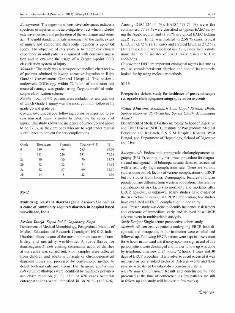
Background : The ingestion of corrosive substances induces aspectrum of injuries to the aero-digestive tract which includesextensive necrosis and perforation of the esophagus and stom-ach. The gold standard for safe assessment of the depth, extentof injury, and appropriate therapeutic regimen is upper GIscopy. The objective of this study is to report our clinicalexperience in adult patients diagnosed with corrosive inges-tion and to evaluate the usage of a Zargar 6-point OGDclassification system of injury.Methods : The study was a retrospective medical chart reviewof patients admitted following corrosive ingestion in RajivGandhi Government General Hospital. The patientsunderwent OGDscopy within 72 hours of admission andmucosal damage was graded using Zargar's modified endo-scopic classification scheme.Results : Total of 445 patients were included for analysis, outof which Grade 1 injury was the most common followed bygrade 2b and grade 3a.Conclusion : Endoscopy following corrosive ingestion to as-sess mucosal injury is useful to determine the severity ofinjury. This study shows the incidence of Grade 2b and aboveto be 37 %, as they are ones who are to kept under regularsurveillance to prevent further complications.
Grade Esophagus Stomach Total (n=445) %
0 189 96 285 64.04
1 111 220 331 74.38
2a 40 30 70 15.73
2b 42 32 74 16.62
3a 32 37 69 15.50
3b 14 8 22 4.94
M-12
Multidrug resistant diarrheagenic Escherichia coli asa cause of community acquired diarrhea in hospital basedsurveillance, India
Neelam Taneja , Sapna Pahil, Gagandeep SinghDepartment of Medical Microbiology, Postgraduate Institute ofMedical Education and Research, Chandigarh 160 012, IndiaDiarrheal illness is one of the most important causes of mor-bidity and mortality worldwide. A surveillance fordiarrheagenic E. coli causing community acquired diarrheaat our centre was carried out. Stool samples were collectedfrom children and adults with acute or chronic/persistentdiarrheal illness and processed by conventional method todetect bacterial enteropathogens. Diarrheagenic Escherichiacoli (DEC) pathotypes were identified by multiplex polymer-ase chain reaction (PCR). Out of 426 cases bacterialenteropathogens were identified in 38.26 % (163/426).
Among DEC (24.41 %), EAEC (19.71 %) were thecommonest, 77.38 % were classified as typical EAEC carry-ing the AggR regulon and 11.90 % as atypical EAEC lackingaggR regulon. EPEC was isolated in 2.58 % cases, typicalEPEC in 72.72 % (8/11) cases and atypical EPEC in 27.27 %(3/11) cases. ETECwere isolated in 2.11% cases. In this studymore than 75 % isolates of EAEC were resistant to fiveantibiotics.Conclusion : DEC are important etiological agents in acute aswell as chronic/persistent diarrhea and should be routinelylooked for by using molecular methods.
M-13
Prospective dohort study for incidence of post-endoscopicretrograde cholangiopancreatography adverse events
Vishal Khurana , Kshaunish Das, Gopal Krishna Dhali,Sanjay Banerjee, Rajib Sarkar, Souvik Ghosh, MahinuddinAhmedDepartment ofMedical Gastroenterology, School of Digestiveand Liver Disease (SDLD), Institute of Postgraduate MedicalEducation and Research, S S K M Hospital, Kolkata, WestBengal, and Department of Hepatology, School of Digestiveand Live
Background : Endoscopic retrograde cholangiopancreato-graphy (ERCP), commonly performed procedure for diagno-sis and management of biliaropancreatic diseases, associatedwith a relatively high complication rate. There are variousstudies done on risk factors of various complications of ERCPbut no studies from India. Demographic features of Indianpopulation are different from western population. The relativecontribution of risk factors to morbidity and mortality afterERCP, however, is unknown. Many studies have evaluatedthe risk factors of individual ERCP complication; few studieshave evaluated all ERCP complication in one study.Aim : Present study was done to identify incidence, risk factorsand outcome of immediate, early and delayed post-ERCPadverse event in multivariable analysis.Study Design : Single centre prospective cohort study,Method : All consecutive patients undergoing ERCP, both di-agnostic and therapeutic, in our institution were enrolled andfollowed up. Following ERCP, patient were kept in observationfor -6 hours in our ward and if no symptom or sign at end of thisperiod patient were discharged and further follow up was doneby telephonic interview at 24 hours, 72 hours, 1 week and 30days of ERCP procedure. If any adverse event occurred it wasmanaged as per standard protocol. Adverse events and theirseverity were dened by established consensus criteria.Results and Conclusions : Result and conclusion will bepresented at the time of conference (as few patients are stillin follow up and study will be over in few weeks).
Indian J Gastroenterol (November 2013) 32(Suppl 1):A1–A132 A127

M-14
Scientif ic yogic meditation ancient techniquefor de-stressing stress
M R Kotwal , C Z Rinchen, Hsin-Li Sony Liu, R N, M S NDepartment of Home and Health, Shunyata, Tibet Road,Sikkim, India, Nursing Department, College of Nursing,Central Taiwan University of Science and Technology,Taichung, Taiwan
Introduction : We live in an era of constant information andalmost infinite possibilities. Multitasking leaves us stressed.Daily meditation physically transforms the cerebral cortex.The most unexpected and comforting recent research confirmthat the human brain retains an astonishing degree of plasticityand capacity for learning throughout life. Our mental perfor-mance, despite a few glitches with short-term memory, doesnot peak until mid life, when the white matter in the loftiestparts of the brain is thickest.Aim and Methods : To evaluate efficacy of a self-learning de-stressing technique Swasthya Sukh Satyam Shivam Sundramin randomly assigned 60 students a meditation group from 114students practiced meditation for 12 weeks and rest formedcontrol group. Scientific technique is from ancient times.Five-step relaxation meditation practiced for 20 minutes 5days weekly was taught to meditation group, while controlgroup did not practice.Results : A questionnaire analyzed confirmed validity, reliabil-ity and reproducibility. Three factors (Psyche, physical andlife adjustment, happiness and moodiness) in the group thatmeditated two times a week for 12 weeks optimally showedstatistically significant (p< 0.0001) results.Conclusion : Preliminary evaluation of an innovative re-laxation meditation practiced twice a week for 12 weeksshowed statistically significant results. The techniquecould be used in many medical situations which generateundue psychological stress and anxiety, to enhance wellbeing. The technique could promote health and preventstress disorders. DVD movie presentation of the tech-nique would be made. The technique is fully explainedand is most simple exercise.
M-15
Acid-fast bacilli culture positivity and drug resistancein primary abdominal tuberculosis
Hrishikesh Samant , Devendra Desai, Philip Abraham,Anand Joshi, Tarun Gupta, Sandeep Davavala, RishikeshKaleria, Camilla RodriguesP D Hinduja National Hospital, Veer Savarkar Marg, Mahim,Mumbai 400 016, India
Introduction: Culture positivity for Mycobacterium tubercu-losis complex (MTB) in abdominal tuberculosis (TB) variesfrom 25 % to 36 %. Data on the prevalence of drug resistancein primary abdominal TB is scant.Aim : To study the AFB culture positivity rate in primaryabdominal TB using Bactec MGITand the prevalence of drugresistance in these patients.Patients and Methods : Records of patients with abdominalTB (diagnosed on clinical features, endoscopy, histology,microbiology) seen during the period 2008-13 were retrievedfrom the Gastroenterology and Microbiology Departments.Patients with extraabdominal TB (5 pulmonary, 2 nodal, 1adnexal) and HIV (1) were excluded from analysis.Results : Of 61 patients, 31 (50.8 %) had AFB culture positive.In the 30 culture-negative patients, histology showed non-caseating granulomas in 25 patients. AFB smear was positivein 4 patients. Drug sensitivity pattern was analyzed in 18patients; resistance was detected in 8 (44.4 %), including 3(16.6 %) who were multi-drug resistant.Conclusion : The rate of AFB culture positivity in primaryabdominal TB was 50.8 % using Bactec MGIT. Drug resis-tance was seen in 44.4 % of those tested, of whom 16.6 %were multi-drug resistant.
M-16
Caustic ingestion in adults: The role of endoscopicclassification in predicting the outcome at a tertiary centre
K Mansoor AliGandhi Medical College and Hospital, Hyderabad, India
Background : The ingestion of caustic substances induces anextensive spectrum of injuries to the Aero digestive tractwhich include extensive necrosis and perforation of the esoph-agus and stomach. The gold standard of safely assessingdepth, extent of injury, and appropriate therapeutic regimenis esophagogastroduodenoscopy (EGD). The objective of thisstudy was to report our clinical experience and to evaluate therole of a 6-point EGD classification system of injury inpredicting outcomes in adult patients diagnosed with causticagent ingestion.Methods : The study was a retrospective medical chart reviewfrom 110 patients admitted to Gandhi Medical College, Se-cunderabad between June 2011-2013 for treatment of causticingestion. The patients underwent EGD within 24 hours ofadmission and mucosal damage was graded using Zagar'smodified endoscopic classification scheme. After treatment,patients were followed in the outpatient clinic for a minimumof 6 months.Results : A total of 110 patients were included for analysis.Grade 2b injury was the most common caustic injury (n=59,53.6 %), followed by grade 3b injuries (n=20, 18.2 %).
A128 Indian J Gastroenterol (November 2013) 32(Suppl 1):A1–A132

Stricture (both esophageal and antral) was the most commoncomplication (n=21, 19.0%), followed by aspiration pneumo-nia (n=10, 9 %) and respiratory failure (n=2, 1.%). Comparedto grade 3a mucosal injury, grade 3b mucosal injuries were atgreater risk of prolonged hospital stay >2 wks and gastroin-testinal and systemic complications.Conclusion : In patients with caustic ingestion, EGDshould be performed within 12 to 24 hours and catego-rized according to a 6-point scale. Patients with grade2b burns and beyond identified on endoscopy have highrates of morbidity. The 6-point scale is useful forpredicting immediate and long-term complications andguiding appropriate therapy.
M-17
Abdominal tuberculosis: Paradigm shift in diagnosticapproach
Pankaj Tyagi , A Arora, V Shingla, P Sharam, A Kumar,A BansalDepartments of Gastroenterology and Hepatology, Sir GangaRam Hospital, Old Rajender Nagar, New Delhi, Delhi 110060, India
Background : The abdomen is involved in 11 % of patientswith extra-pulmonary tuberculosis. It is difficult to get thetissue in abdominal tuberculosis and WHO recommends tohave tissue diagnosis before starting ATT. Endoscopic ultra-sound (EUS) is being used more commonly in most of theendoscopic centres.Methods : Prospectively 134 patients of abdominal tubercu-losis were enrolled in tertiary care hospital in Delhi, andthere clinical, diagnostic investigation and treatment wasrecorded.Results : Out of 134 patients (Mean age 43±15 years)CECT abdomen in 130 patients showed lymphnode in 68%, intestinal wall thicking was seen in 65 patients,mesentric thinking in 35 % of patients. CECT chest wasavailable in sixty-eight patients which showed presence ofmediastinal lymphadenopathy in 48 patients (70 %). Tissuediagnosis was obtained by EUS in 63 patients, surgicallyin 15 patients and endoscopic biopsy in 17 patients. EUSFNAC was done in 63 patients of which mediastinal EUSFNAC was done in 41 and abdominal EUS FNAC wasdone in 22. Histology present in 95 patients in whomgranuloma present in 100 %, AFB was positive in 40 %.Conclusion : Histological evidence can be achieved in 70% ofthe patients of abdominal tuberculosis before starting ATT,EUS helped in getting histology in 66% and mediastinal EUSFNAC was use full in 65 % of total EUS thus EUS should beincluded in the diagnostic algorithm of the abdominaltuberculosis.
M-18
Eff icacy of Xpert MTB/RIF assay for diagnosisof peritoneal tuberculosis in patients with exudative ascites
Chinmay Bera1 , Joy Sarojini Michael, Sridhar Gibikote,Ashish Goel, C E EapenDepartments of Hepatology, Microbiology and Radiology,Christian Medical College, Vellore 632 004, India
Background : Xpert MTB/Rif is a multiplex hemi-nested real-time PCR based assay to detect presence of M. tuberculosiswithin two hours of sample collection. The present studyaimed at assessing efficacy of Xpert MTB/Rif assay for diag-nosis of peritoneal tuberculosis.Methods : Patients presenting to Liver Clinic with exudativeascites and negative Ziehl Nelsen (ZN) staining for acid fastbacilli (AFB) and unyielding cytology for malignant cells,were included after informed consent. Ultrasound guidedomental biopsy sample were obtained in all. Besides histopa-thology and TB culture, Xpert was performed in all tissuesamples. A composite reference standard for diagnosis wasused as no single test is considered as gold standard in thissetting.Results : During January 2012 to July 2013, 18 patients (16males; age: 42.5, 17-65 years; median, range, 3 with underly-ing cirrhosis) were recruited. All had high protein and lowgradient ascites with negative result on ZN staining and cy-tology. Fourteen of the 18 study patients had peritoneal TB asdiagnosed by composite reference standard- culture: 3; gran-ulomatous inflammation of histology: 13 and response to 2months of antituberculous therapy: 3 (unyielding culture/histology). Four patients were diagnosed as peritoneal carci-nomatosis (adenocarcinoma: 2; mesothelioma: 1; metastaticcarcinoma: 1). Xpert was positive in 3 of 14 patients withperitoneal TB and in none of the 4 patients with peritonealcarcinomatosis. The sensitivity, specificity, negative predic-tive value and positive predictive value for Xpert were 21 %,100 %, 27 % and 100 % respectively.Conclusions : Xpert MTB/Rif was not a sensitive test to diag-nose peritoneal tuberculosis, however its positive predictivevalue was 100 %.
M-19
WITHDRAWN
M-20
Step-by-step approach to NOTES - Our learning model
Mohd. Juned Khan , P S Rajan, P Karthikeyan, Amol Shinde,C Palanivelu
Indian J Gastroenterol (November 2013) 32(Suppl 1):A1–A132 A129

G EM Hospital and Research Centre, 45, Pankaja Mill Road,Ramanathapuram, Coimbatore 641 045, India
Background/Aims : NOTES is a surgical procedure to beperformed by endoscopic accessories via natural orifices likemouth, anal canal and vagina in females. With any transitionfrom standard laparoscopy to newer approach, the results ofthe newer procedure should be with lesser morbidity. Hencewe adopted step by step approach.Methods : We progressed from porcine models to trans-umblical approach in human subjects followed bytransvaginal to transgastric approach.Result : Initially we performed transgastric endoscopiccholecystectomy in porcine models. Next we progressedto perform surgery with endoscopic accessories withendoscopy placed in the umbilicus, performed largehuman series of appendectomy and cholecystectomy.After gaining experience of performing transumbilicalendoscopic appendectomy and cholecystectomy, we ad-vanced to vaginal route and performed large series ofappendectomy and cholecystectomy. During this pro-cess, if difficulty was encountered it was converted tostandard laparoscopy without any added morbidity. Fi-nally we proceeded to transgastric approach, at comple-tion requires standard laparoscopic gastrotomy closureuntil we get a good tool to close the gastrostomy byendoscope.Transumbilical Access : Number of cases=20 (12 appendecto-mies and 8 cholecystectomies). Age group=27-52 years. Du-ration of hospital stay=2-4 days. Operative time=30-110minutes.Transvaginal Access : Number of cases=80 (55 appendecto-mies and 25 cholecystectomies). Age group=32-58 years.Duration of hospital stay=2-4 days. Operative time=35-110minutes.Transgstric Access : Number of cases=5 (3 appendecto-mies and 2 cholecystectomies). Age group 27-44 years.Duration of hospital stay=4-6 days. Operative time=90-135 minutes.Conclusion : NOTES being a complex procedure, this methodof step-by-step learning approach in performing this endo-scopic surgical procedures is safe without any morbidity as-sociated with new techniques.
M-21
Efficacy, safety and tolerance of split dose sulphatesolution versus split dose PEG versus single dose PEGfor colonoscopy preparation: A prospective randomizedtrial
George Sarin Zacharia , A R Rajneesh, K Sandesh,K Sunilkumar, T M Ramachandran, Varghese Thomas
Department of Gastroenterology, Government MedicalCollege, Calicut 673 008, India
Aims : To assess the efficacy, safety and tolerance of threetypes of bowel preparation regimens for colonoscopy.Methods : Prospective single blind randomized trial conductedin a tertiary care setting. Patients were randomized to receiveany of the three bowel preparation regimens; (A) split dosesulphate solution or (B) split dose PEG solution or (C) sameday single dose PEG solution. Efficacy of bowel cleansingwas assessed by Boston Bowel Preparation Score (BBPS).Any adverse effects were noted.Results : A total of 172 patientswere considered for the study (A-56, B-61, C-55). Baseline variables were comparable across thegroups. The mean BBPS scores amongst groups A, B and Cwere 8.02, 7.47 and 8.24 respectively (p=0.041). Bonferronianalysis revealed no significant difference between group A vs.groups B and C, where as the difference was significant betweengroups B and C; C with higher BBPS scores. Severe tasteintolerance was reported in 41.1 % (n=23) of patients in groupA compared to 19.7 % (n=12) and 7.3 % (n=4) amongst groupB and C. Volume intolerance was seen in 14.6 % (n=6), 23 %(n=14), 38.2 % (n=21) in groups A, B and C respectively. Sleepdisturbance was observed in groups A (14.3 %) and B (27.9 %).Conclusions : All preparations provided satisfactory bowelcleansing. Same day single dose PEG provided superior bowelcleansing compared to split PEG regimen. Sulphate preparationwas associated with more taste intolerance while single dosePEG was associated with more volume intolerance. Sleep dis-turbances were a problem for overnight bowel preparations.
M-22
Spectrum of gastrointestinal foreign bodies
G Balaji , Muthukumar, G R M Kumar, R Balamurali,P Ganesh, S jeevankumarsDepartment of Gastroenterology, Kilpauk Medical Collegeand Hospital, Chennai 600 010, India
Introduction : Gastrointestinal foreign bodies (GIFBs) arecomprised of food bolus impactions and intentionally andunintentionally ingested or inserted foreign objects. Mostresolve without serious clinical sequelae. Regardless of im-precise morbidity and mortality rates, serious complicationsand deaths occur as a consequence of foreign body ingestions.It is important to understand which patients are at risk forGIFBs, know how to diagnose and treat GIFBs, and deal withtheir complications.Aim : To study the spectrum of GI foreign bodies presented toour hospital with respect to the risk factors and its complications.Materials and Methods : Total of 48 patients were enrolled inthis study.
A130 Indian J Gastroenterol (November 2013) 32(Suppl 1):A1–A132

Study Period : March 2009 to April 2013, retrospective study.Results : Of 48 patients 29 were pediatric group (60.4 %)and 19 were adults (39.6 %). Among pediatric groupcoin was the commonest foreign body (86.2 %) follow-ed by safetypin (7.5 %) and stick (7.5 %). Amongadults 9 patients had impacted food bolus (47.3 %), 6had fishbone (31.5 %) and 4 were others. No risk factorwas found in pediatric group where as in adults 10 hadstricture. Of 48 patients 46 were treated endocopicallyand 2 underwent surgical intervention. One patient diedin this study with mortality rate of 2 %.Conclusion : Gastrointestinal foreign bodies are seen inboth children and adults with increase prevalence inchildren. Endoscopic retrieval of foreign bodies is suc-cessful in most of cases. Surgical intervention was re-quired in complicated cases where endoscopic therapyfailed. Availability of endoscopic and surgical modalitiesof treatment in the same centre significantly decreasesmorbidity.
M-23
Diagnostic utility of EUS guided FNAC of mediastinallymphadenopathy
K R Pradeep , V J Harshad, N T Nilesh, Rony Thomas,Charles Panackel, Sunil K MathaiDepartments of Gastroenterology and Hepatology, MedicalTrust Hospital, M G Road, Kochi 682 016, India
Objective : To analyze the diagnostic utility of EUS guidedFNAC of mediastinal lymphadenopathy.Methods : Retrospective chart review of all EUS guidedFNAC of mediastinal lymphadenopathy from January 2010to July 2013.Results : A total of 86 EUS guided FNAC procedureswere done on 85 patients. Fifty-six patients (65 %) weremale, mean age was 55 years. Cough (76 %) and fever(64 %) were the most common presenting symptoms.Seventy-two patients (84.7 %) received a definitve di-agnosis. Twenty-six (30 %) patients were diagnosedwith granulomatous lymphadenitis, malignancy was di-agnosed in 24 patients (29 %), of which 5 had lympho-ma and 19 had metastases from different primaries (1pancreatic, 1 HCC, 1 renal, 2 squamous cell carcinoma,1 breast, 13 metastatic NSCLC). Interestingly 26 % ofpatients had reactive changes and EUS FNA was incon-clusive in 13 cases (15 %). None of our patients devel-oped any complications post the procedure.Conclusion : EUS guided FNAC of mediastinal lymph-adenopathy is safe and useful. It helps avoiding moreinvasive mediastinal sampling procedures and complexsurgeries.
M-24
A study of cryptosporidial diarrhea inmegaloblastic anemia
Vineet Behera ; Pankaj Puri, S Srivastava, Velu Nair,N Naithani, K D KamalDepartments of Gastroenterology, and Internal Medicine,Armed Forces Medical College, Pune 411 040, India
Objective : To study the incidence of cryptosporidiosis inmegaloblastic anemia (MA) and compare with other anemiasand general population.Materials and Methods : The study population comprised ofpatient of and controls were patients of other anemias andasymptomatic individuals. A detailed clinicopathological re-cord and etiological work up was done for MA. The stoolsamples of all patients was tested for cryptosporidiosis (lightmicroscopy for oocysts and modified ZN staining) two times.Results : 1. Study population – MA (n=48); controls - 50 casesother anemias, 100 healthy controls. 2. The percentage number ofpatients with diarrheal symptoms and other stool examinationabnormalities were 31 % and 12.5 % in MA, 18.7 % and 6.2 %in other anemia and 16%and 6% in healthy controls respectively(p< 0.05). 3. Cryptosporidiosis was seen in 39% ofMA, 16% ofanemia and 14 % of controls (p< 0.05). The presence of crypto-sporidiosis in MAwith (n=15) and without diarrheal symptoms(n=33) was compared and was statistically significant. Crypto-sporidiosis was seen in 41.6 % of MAwith known etiology (n=36) and about 30 % of MAwith unknown etiology (n=12).Discussion : Cryptosporidiosis is seen in 40% ofMAwhich isstatistically significant when compared to other groups; there-fore we can conclude that it is an etiological agent for MA. Itwas commoner in MAwith diarrheal symptoms and can be coassociated with other etiologies in MA.Conclusion : Cryptosporidiosis is an etiological factor forMAandshould be evaluated in all cases of MAwith or without diarrhea.
M-25
Profile of upper gastrointestinal bleeding in a tertiary carehospital in Odisha
Haribhakti Seba Das , M K Panigrahi, C R Panda, S K Kar,B Misra, S P SinghDepartment of Gastroenterology, S C B Medical College,Cuttack 753 007, IndiaBackground : Upper GI bleeding is a potentially life threaten-ing condition.Objective : To study the profile of patient’s presentingwith upper GI bleeding in a tertiary care hospital incoastal Orissa.Materials and Methods : The study population consists of4,665 patients attending the Gastroenterology Outpatient
Indian J Gastroenterol (November 2013) 32(Suppl 1):A1–A132 A131

Department of S C B Medical College and Hospital,Cuttack from June 2007 to July 2011 for upper GIbleeding. Patients underwent upper GI endoscopy,USG abdomen and Doppler USG, CT or MR angiogra-phy whenever required to diagnose the etiology ofbleeding.Results : Out of 4,665 patients 54 % were hospitalized.Mean age group was 28±14.27 years and 76.05 % weremale. Patients presented with melena alone in 79.48 %,hematemesis only in 27.78 % and 44.99 % had both.Mean blood transfusions received was 2.06 units inhospitalized patients. Peptic ulcer was commonest cause(53 %) of upper GI bleed which includes duodenal ulcer
(37 %), gastric ulcer (16 %). Variceal bleeding wasfound in 28 % of patients. Erosive gastritis was foundin 13 % of bleeders. Carcinoma of stomach was foundin 4 %. Other rare causes like Dieulafoy’s lesion,Mallory-Weiss tear, duodenal carcinoid, tubercular ulcerand stromal tumor (GIST) accounted for 2 % of totalcases. EHPVO accounted for 50 % of variceal bleeding.Surgery was done in 4.65 % of duodenal ulcer bleeding.Mortality rate of UGI bleeding was 7.5 %.Conclusion : Peptic ulcer is the commonest cause of UGIbleeding referred to a tertiary care hospital in Odisha. Varicealbleeding and erosive gastritis are among the next commoncause of upper GI bleeding.
A132 Indian J Gastroenterol (November 2013) 32(Suppl 1):A1–A132