increased susceptibility of c. elegans to candida albicans infection ... · specific rnai knockdown...
TRANSCRIPT
Increased Susceptibility of C. elegans to Candida albicans Infection after Tissue-
specific RNAi Knockdown of an ROS-generating NADPH Oxidase
A Major Qualifying Project Report
Submitted to the Faculty of the
WORCESTER POLYTECHNIC INSTITUTE by Kate Pellegriti
Approved by:
Samuel Politz, Ph.D.
Biology and Biotechnology
WPI Project Advisor
Abstract Tissue-specific RNA interference was performed on the model organism Caenorhabditis
elegans to cause the knockdown of bli-3, which is hypothesized to play an important role in
immune defense through the production of reactive oxygen species. Survival assays on C.
elegans infected by the pathogenic yeast Candida albicans revealed highly significant effects on
the survival of the worms when the gene was knocked down in the hypodermal and intestinal
tissues. There was no significant effect on the survival of worms with bli-3 knockdown in body
wall muscle.
Contents Abstract ......................................................................................................................................................... 1
Acknowledgements ....................................................................................................................................... 4
Introduction .................................................................................................................................................. 5
Materials and Methods ............................................................................................................................... 17
Maintenance of Bacteria ............................................................................................................................. 17
Recipe for LB, Ampicillin and Tetracycline Media ............................................................................... 17
Preparation of Feeding Plates ............................................................................................................. 18
Recipe for Feeding Plates .................................................................................................................... 18
Overnight Bacteria Culture ................................................................................................................. 18
Maintenance of Candida albicans............................................................................................................... 18
Recipe for YPD Agar ............................................................................................................................ 18
Overnight Yeast Culture ...................................................................................................................... 19
Survival Assays ............................................................................................................................................ 19
Results ......................................................................................................................................................... 20
Discussion.................................................................................................................................................... 26
References .................................................................................................................................................. 28
Table of Figures Introduction
Figure 1: Essential Roles of Duox Enzymes ...................................................................................... 8
Figure 2: Hypothetical signaling pathways for regulation of Duox (bli-3) in C. elegans ............... 11
Figure 3: Processes of RNAi ........................................................................................................... 13
Figure 4: Areas of Tissue-specific Knockdown of bli-3 ................................................................... 15
Results
Figure 1: Survival Curves of Hypodermal and Whole-Body Knockdown of bli-3 without Dilutions
........................................................................................................................................................ 22
Figure 2: Survival Curves of N2 Worms After Feeding with OP50 transformed with L4440 and
OP50 transformed with F56 C11.1 ................................................................................................. 23
Figure 3: Survival Curves of NR222 Worms After Feeding with OP50 Transformed with L4440 and
OP50 Transformed with F56 C11.1…………………………………………………………………………………………….24
Figure 4: Survival Curves of WP303 Worms After Feeding with OP50 Transformed with L4440
and OP50 Transformed with F56 C11.1……………………………………………………………………………………25
Figure 5: Survival Curves of NR350 Worms After Feeding with OP50 Transformed with L4440 and
OP50 Transformed with F56 C11.1……………………………………………………………………………………………26

Acknowledgements
I would like to thank Professor Politz for being an endless source of knowledge, guidance
and support throughout my research and academic career. I would also like to thank Jessica
Rastad for her revealing and in-depth work with RNAi of bli-3.
Introduction
It is well known that fungal infections can be very difficult to treat, as fungal cells are
eukaryotic, making them difficult to target without also harming human cells (Jain, et al., 2009).
However, effector components of our innate immune response do target these types of infections,
such as a family of enzymes important in fighting numerous types of infections, the NADPH
oxidases (Nox). Homologues of these enzymes have been found in fungi and plants, as well as
animals (Bedard and Krause, 2007). For example, two Nox homologues exist in the fruit fly
model organism Drosophila melanogaster, dNox and dDuox (Ritsick et al., 2007). The dNox
homologue was found to play a role in the regulation of calcium flux in the ovaries, therefore
affecting the fly’s egg-laying abilities, while dDuox helps to maintain balance with the bacterial
fauna of the gut (Ritsick et al., 2007) (Ha et al., 2009). A Nox enzyme also is found in the model
organism Caenorhabditis elegans (C. elegans), a species of roundworm (Edens et al., 2001). We
will describe here the use of this species to study the role of one of these enzymes in innate
immune responses in vivo through RNA interference (RNAi) experiments.
Members of the Nox family all participate in electron transport in order to produce
reactive oxygen species (ROS). The Nox family currently characterized consists of five Nox
enzymes and two Duox enzymes (Bedard and Krause, 2007). Although these enzymes differ
slightly from each other, they generally consist of a NADPH-binding domain at the carboxyl
terminus, a FAD-binding region, six transmembrane domains and four heme-binding histidines
(Bedard and Krause, 2007). The Duox enzymes, however, also contain a peroxidase domain and
EF hand domains that allow Duox activity to be regulated by calcium ions (Edens, et al., 2001;
Leto and Geizst, 2004).
Although many of the Nox enzymes are involved in immune responses, they all have
slightly different functions and can be found in different parts of the body. For example, Nox3
and Nox5 do not have a role in immune responses, as Nox3 is essential for gravity perception
and Nox5 is found in lymphoid cells and important for calcium-dependent signaling (Leto and
Geizst, 2004). Nox1 is thought to play a role in cell division, but is still activated by immune
cytokines as are Nox4 and Nox2 (Bedard and Krause, 2007). Nox2, also called gp91phox
, is often
considered the classic example of this family of enzymes, as it is the most familiar.
Nox2 is expressed in phagocytes, where it plays a role in innate immune defense. Along
with its gp91 catalytic subunit, it also consists of subunits p22, p47, p67 and Rac, all of which
contribute to its functionality (Rada, et al, 2008). Prior to stimulation of fMLP or C5a receptors
by antigens, gp91 and gp22 subunits are located in granular membranes, while p22, p47 and p67
are in the cytosol of the phagocyte (Murphy, 2012). However, stimulation of the aforementioned
receptors leads to Rac activation, causing the subunits in the cytosol to meet with gp91 and gp22,
which activates Nox2 (Murphy, 2012). Similarly to all Nox enzymes, Nox2 helps to transfer
electrons across cellular membranes from NADPH to oxygen, which serves as the electron
acceptor (Rada, et al, 2008). During times of infection, activity of Nox2 enzymes results in
phagocytic consumption of up to fifty times more oxygen, during what is known as the
respiratory burst (Babior, 1984).
The reaction catalyzed by Nox2 and other NOX family members is: O2 + NADPH O2-
+ NADP+ + H
+ (Babior, 1984). Essentially, a pair of electrons is removed from NADPH to be
added to the oxygen, which reduces it, forming a radical known as superoxide (Babior, 1984).
Superoxide is somewhat toxic to pathogens, but more importantly, it serves as a precursor to
other ROS, such as hypochlorite or hydrogen peroxide (Babior, 1984). Superoxide can accept
hydrogen ions in the following reaction: 2O2- + 2H
+ H2O2 + O2, which is catalyzed by the
enzyme superoxide dismutase (Babior, 1984). The hydrogen peroxide produced by this reaction
is a powerful ROS. The movement of electrons in order to form the superoxide produces an
electric potential which is essential to immune defense, as it allows the movement of other ions
in reactions of the ROS with the pathogen’s DNA, lipids and proteins, which inhibits the
pathogen’s growth and ultimately leads to its destruction (Rada, et al, 2008). ROS are also able
to affect the pH of the phagosomes. Although phagosomes generally maintain an acidic
environment in order to eliminate pathogens, the ROS in the neutrophils initially causes an
increase in pH by decreasing the amount of H+ ions in the inside of the phagosome (Lee et al.,
2003). Not only does the production of H2O2 require the consumption of H+
ions, but the
presence of ROS also reduces the amount of H+ ions entering the phagosome through ATPases,
as well as increase the amount of H+ ions leaving the phagosomes due to overall leakiness (Lee
et al., 2003). Subsequent effects on the pH inside of phagosomes are not well understood (Lee et
al., 2003).
Duox1 and Duox2, which are most important to this research, can also be essential to an
organism’s survival against pathogens. The Duox enzymes were originally discovered in the
thyroid glands when research was being conducted on thyroid hormone synthesis and were
called the dual oxidases because they have both a NADPH oxidase domain, as well as an
extracellular peroxidase domain, which is thought to, perhaps, bind hydrogen peroxide (Chavez,
et al., 2009) (Edens, et al., 2001). These enzymes also have EF hand domains, which are not
found in other Nox enzymes, that allow for activation by directly binding calcium (Leto and
Geizst, 2004). In humans, Duox2 was found in the salivary glands, respiratory tract epithelium,
and thyroid tissue, while Duox1 was found in many tissues, such as the lung, thyroid, placenta,
testis, prostate, pancreas and heart (Lambeth, 2004). Because these enzymes are found in many
important epithelial tissues, which are often the first tissues to come into contact with such
bacterial or yeast pathogens, it is thought that these enzymes also play a role in immune defense.
Because Duox2 is essential to the organification of iodide, which is needed for thyroid
hormones, mutations in Duox2 lead to hypothyroidism (Leto and Geizst, 2004). The Duox
enzymes are also found to play an important role in saliva and mucosal secretions in the mouth
and airways by creating hydrogen peroxide, which reacts with thiocyanate to form
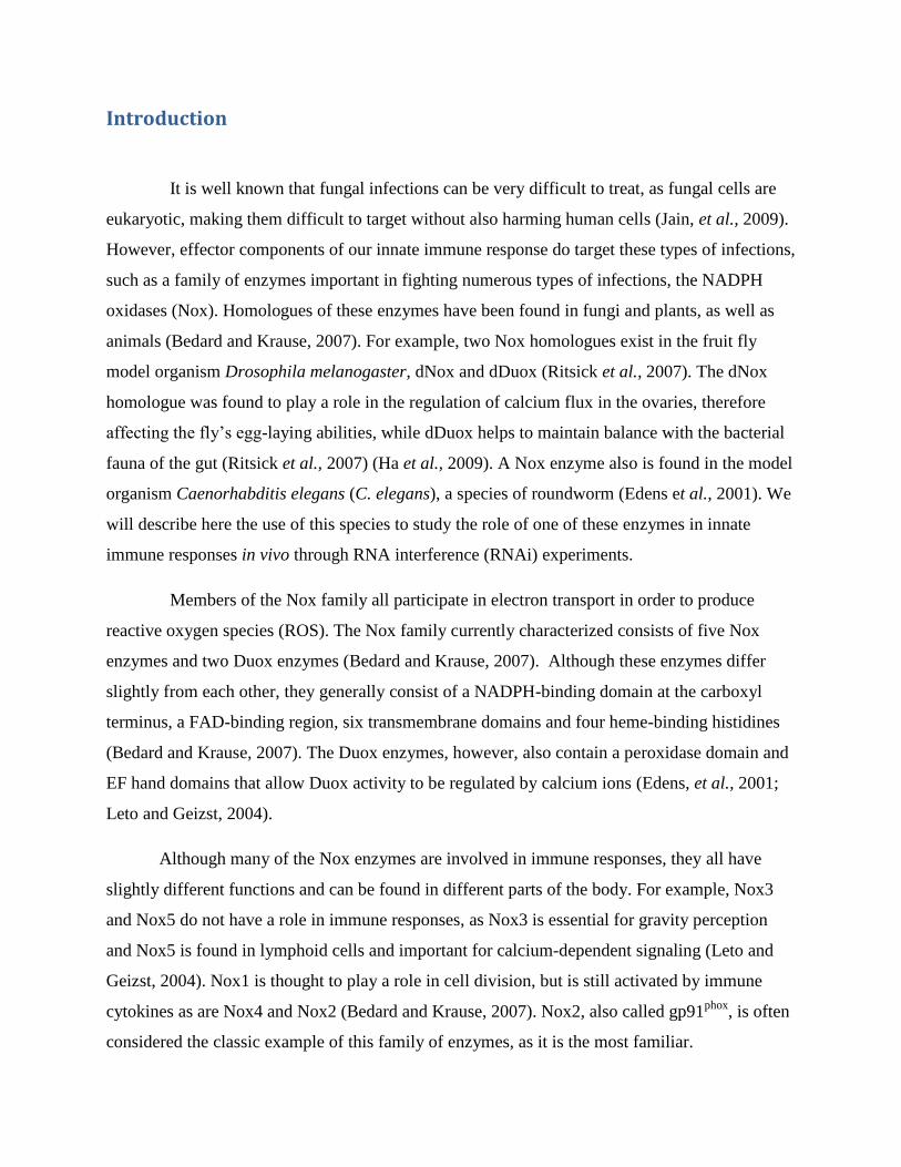
hypothiocyanite, which fights microbial infections (Leto and Geizst, 2004). Figure 1 below
contains an image of the role of Duox2 in the thyroid (top) and in the secretions of epithelial
tissues (bottom).
Figure 1: Essential Roles of Duox Enzymes (Adapted from Leto and Geizst, 2004). The top
diagram shows the role of Duox (shown in red) in the thyroid where it is responsible for forming hormones, while
the bottom diagram demonstrates the role of Duox in epithelial tissues, in which it is responsible for the secretion of
microbicidal secretions that fight off pathogens.
O2-
I-+ H2O2 I° Iodotyrosine Hormone (T4, T3)
O2-
SCN-, I- + H2O2 HOSCN, HOI, I2, etc.
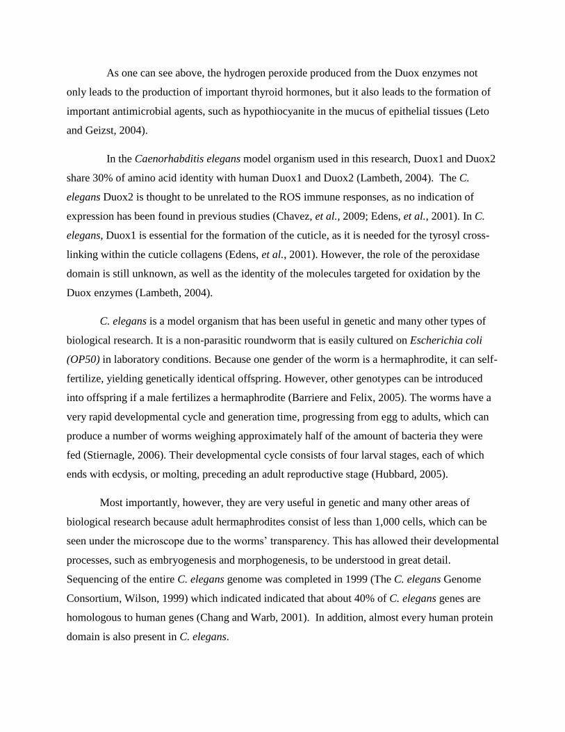
As one can see above, the hydrogen peroxide produced from the Duox enzymes not
only leads to the production of important thyroid hormones, but it also leads to the formation of
important antimicrobial agents, such as hypothiocyanite in the mucus of epithelial tissues (Leto
and Geizst, 2004).
In the Caenorhabditis elegans model organism used in this research, Duox1 and Duox2
share 30% of amino acid identity with human Duox1 and Duox2 (Lambeth, 2004). The C.
elegans Duox2 is thought to be unrelated to the ROS immune responses, as no indication of
expression has been found in previous studies (Chavez, et al., 2009; Edens, et al., 2001). In C.
elegans, Duox1 is essential for the formation of the cuticle, as it is needed for the tyrosyl cross-
linking within the cuticle collagens (Edens, et al., 2001). However, the role of the peroxidase
domain is still unknown, as well as the identity of the molecules targeted for oxidation by the
Duox enzymes (Lambeth, 2004).
C. elegans is a model organism that has been useful in genetic and many other types of
biological research. It is a non-parasitic roundworm that is easily cultured on Escherichia coli
(OP50) in laboratory conditions. Because one gender of the worm is a hermaphrodite, it can self-
fertilize, yielding genetically identical offspring. However, other genotypes can be introduced
into offspring if a male fertilizes a hermaphrodite (Barriere and Felix, 2005). The worms have a
very rapid developmental cycle and generation time, progressing from egg to adults, which can
produce a number of worms weighing approximately half of the amount of bacteria they were
fed (Stiernagle, 2006). Their developmental cycle consists of four larval stages, each of which
ends with ecdysis, or molting, preceding an adult reproductive stage (Hubbard, 2005).
Most importantly, however, they are very useful in genetic and many other areas of
biological research because adult hermaphrodites consist of less than 1,000 cells, which can be
seen under the microscope due to the worms’ transparency. This has allowed their developmental
processes, such as embryogenesis and morphogenesis, to be understood in great detail.
Sequencing of the entire C. elegans genome was completed in 1999 (The C. elegans Genome
Consortium, Wilson, 1999) which indicated indicated that about 40% of C. elegans genes are
homologous to human genes (Chang and Warb, 2001). In addition, almost every human protein
domain is also present in C. elegans.
Not only genes, but whole pathways are conserved between mammals and the C. elegans
model. For example, when a mammal has a pathogenic infection, MAP (mitogen-activated)
kinase pathways are activated, which alerts the body that there is an infection and leads to
various immune responses (Murphy et al., 2012). Three MAP kinase pathways also exist in C.
elegans (Kim et al., 2002). One of these pathways, the p38 pathway, which is also present in
mammals, is shown in the right side of Figure 2.
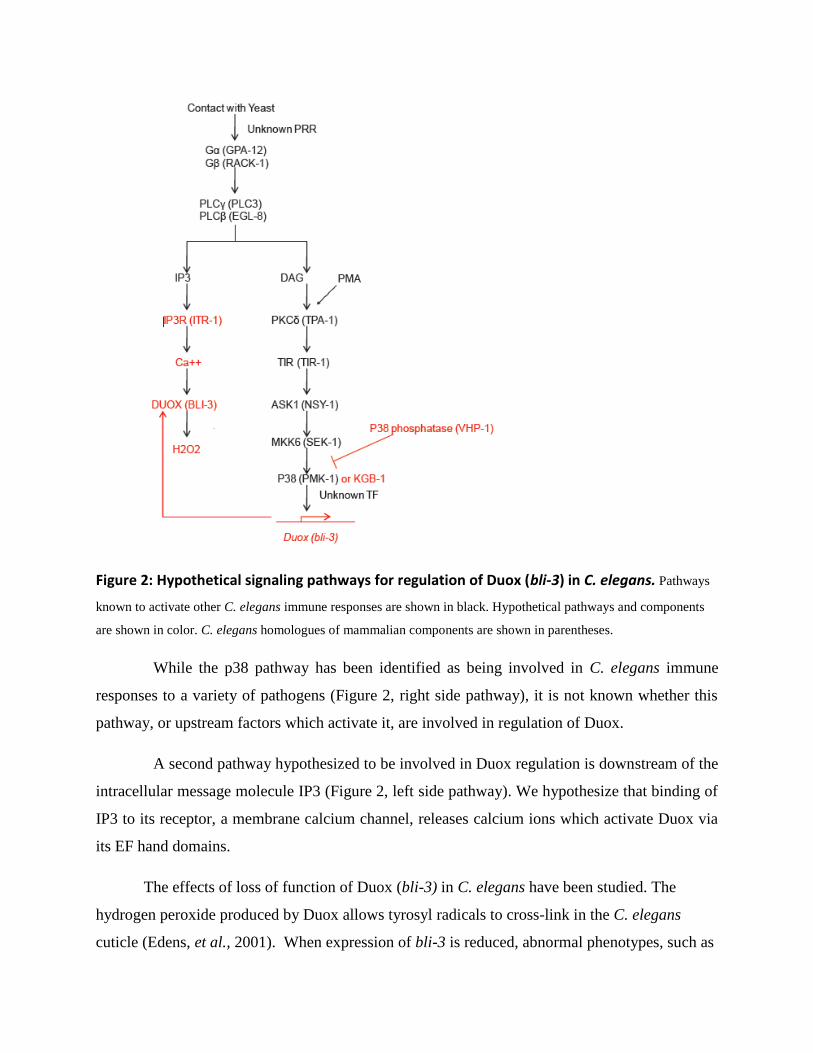
Figure 2: Hypothetical signaling pathways for regulation of Duox (bli-3) in C. elegans. Pathways
known to activate other C. elegans immune responses are shown in black. Hypothetical pathways and components
are shown in color. C. elegans homologues of mammalian components are shown in parentheses.
While the p38 pathway has been identified as being involved in C. elegans immune
responses to a variety of pathogens (Figure 2, right side pathway), it is not known whether this
pathway, or upstream factors which activate it, are involved in regulation of Duox.
A second pathway hypothesized to be involved in Duox regulation is downstream of the
intracellular message molecule IP3 (Figure 2, left side pathway). We hypothesize that binding of
IP3 to its receptor, a membrane calcium channel, releases calcium ions which activate Duox via
its EF hand domains.
The effects of loss of function of Duox (bli-3) in C. elegans have been studied. The
hydrogen peroxide produced by Duox allows tyrosyl radicals to cross-link in the C. elegans
cuticle (Edens, et al., 2001). When expression of bli-3 is reduced, abnormal phenotypes, such as
cuticle blisters appear (Edens, et al., 2001). However, studies performed by Chavez et al. (2009)
and Jain et al.(2009) supported the idea that bli-3 also plays an important role in immune
defense. Research by Jain et al. showed that bli-3 mutants led to reduced ROS production.
Additionally, research by Chavez et al. suggested that when expression of bli-3 was reduced,
either by mutation or RNAi, so was the production of ROS in response to pathogenic bacteria,
which also reduced survival of the worms (Chavez, et al., 2009). We seek to extend these results
by studying the effects of bli-3 loss of function in specific tissues on survival during infection by
the pathogenic yeast Candida albicans.
In vivo RNAi experiments are essential to studying the role of Nox and Duox enzymes
because cells containing these enzymes often cannot be preserved in culture (Leto and Geizst,
2004). RNAi involves introducing dsRNA (double-stranded RNA) into the organism under study
that is capable of gene-specific inhibition of translation of the mRNA of interest (Novina and
Sharp, 2004).
In C. elegans, feeding is often used to introduce dsRNA into the worms, which consists
of literally feeding the worms bacteria that have been transformed with a plasmid that
synthesizes the desired dsRNA. Figure 3 below contains a diagram of how the dsRNA acts
interacts with various proteins in order to knock down the target mRNA.
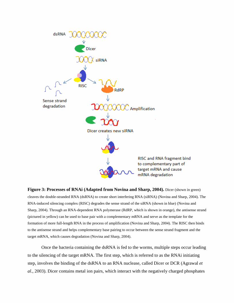
Figure 3: Processes of RNAi (Adapted from Novina and Sharp, 2004). Dicer (shown in green)
cleaves the double-stranded RNA (dsRNA) to create short interfering RNA (siRNA) (Novina and Sharp, 2004). The
RNA-induced silencing complex (RISC) degrades the sense strand of the siRNA (shown in blue) (Novina and
Sharp, 2004). Through an RNA-dependent RNA polymerase (RdRP, which is shown in orange), the antisense strand
(pictured in yellow) can be used to base pair with a complementary mRNA and serve as the template for the
formation of more full-length RNA in the process of amplification (Novina and Sharp, 2004). The RISC then binds
to the antisense strand and helps complementary base pairing to occur between the sense strand fragment and the
target mRNA, which causes degradation (Novina and Sharp, 2004).
Once the bacteria containing the dsRNA is fed to the worms, multiple steps occur leading
to the silencing of the target mRNA. The first step, which is referred to as the RNAi initiating
step, involves the binding of the dsRNA to an RNA nuclease, called Dicer or DCR (Agrawal et
al., 2003). Dicer contains metal ion pairs, which interact with the negatively charged phosphates
of the dsRNA in order to orient the dsRNA into the active site of Dicer (MacRae, 2006). Thus,
the homologous sequence of the dsRNA can correspond to the catalytic sites, allowing for
cleavage to produce short interfering RNA (siRNA) (MacRae, 2006).
In what is sometimes referred to as the effector step, the siRNA then binds to the RNA
induced silencing complex (RISC), a complex that includes the Argonaute2 protein. It is
predicted that the RISC uses ATP in order to unwind the strands of the siRNA (Agrawal et al.,
2003). The RISC binds to the antisense strand, which complementarily binds to the target
mRNA, targeting the desired mRNA for endonucleolytic cleavage (Agrawal et al., 2003).
It is thought that the Argonaute protein might help direct the siRNA to the RISC during the
RNAi process, as interactions between Dicer and Argonaute were found to occur (Hannon,
2002). More importantly, however, Argonaute2 contains endonuclease activity due to its PIWI
domain, which has structural similarities to an RNase domain (Song et al., 2004; Diederichs and
Haber, 2007). Argonaute2, therefore, can be seen as the catalytic subunit of RISC, as it is
responsible for the cleaving activity of RISC and such activity cannot occur without it
(Diederichs and Haber, 2007).
As pictured above, the dsRNA can also be amplified (Agrawal et al., 2003). It is thought
that enzymes known as RNA-dependent RNA polymerases are important in the amplification
process, which are encoded by the ego1 gene in C. elegans. For example, an Rdrp can bind to an
antisense strand of siRNA in order to act as a template to synthesize another dsRNA, thus
allowing the spread of the mRNA silencing throughout the organism when these newly formed
dsRNA are also degraded (Novina and Sharp, 2004).
However, in this research, amplification and spreading were prevented through the use of
tissue-specific promoters. Previous research indicated that rde-1, which encodes an Argonaute
protein, was essential to the process of RNAi catalyzed by dsRNA (Grishok et al., 2000). Qadota
et al. (2007) took advantage of this requirement for rde-1 to create a method for achieving tissue-
specific RNAi in C. elegans. This method was ideal for our purposes.
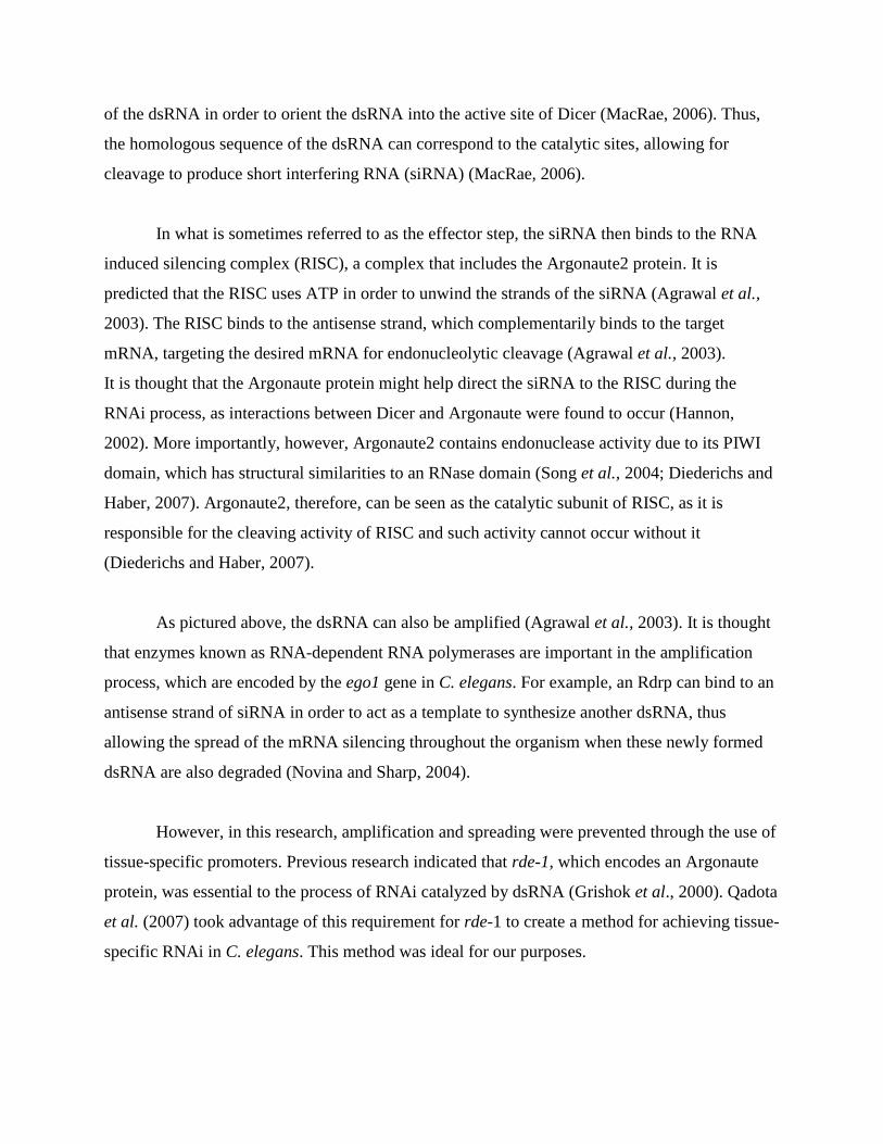
In our experiments, we used a C. elegans strain carrying a mutation in rde-1 which makes
the worms unable to carry out RNAi. By using worm strains carrying an integrated rde-1 gene
under control of promoters that are specifically expressed in the hypodermis, intestine and
muscle, wild-type expression of rde-1 and the production of a wild-type Argonaute protein could
be achieved in those respective tissues. Under these conditions, RNAi occurred only in the
tissues where rde-1 was expressed (Qadota et al., 2007). RNAi could not spread to other tissues,
where RNAi remained defective.
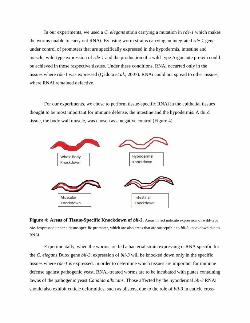
For our experiments, we chose to perform tissue-specific RNAi in the epithelial tissues
thought to be most important for immune defense, the intestine and the hypodermis. A third
tissue, the body wall muscle, was chosen as a negative control (Figure 4).
Figure 4: Areas of Tissue-Specific Knockdown of bli-3. Areas in red indicate expression of wild-type
rde-1expressed under a tissue-specific promoter, which are also areas that are susceptible to bli-3 knockdown due to
RNAi.
Experimentally, when the worms are fed a bacterial strain expressing dsRNA specific for
the C. elegans Duox gene bli-3, expression of bli-3 will be knocked down only in the specific
tissues where rde-1 is expressed. In order to determine which tissues are important for immune
defense against pathogenic yeast, RNAi-treated worms are to be incubated with plates containing
lawns of the pathogenic yeast Candida albicans. Those affected by the hypodermal bli-3 RNAi
should also exhibit cuticle deformities, such as blisters, due to the role of bli-3 in cuticle cross-

linking. However, it will also be interesting to see the effects of bli-3 knockdown in the intestine
because it is predicted that bli-3 and Duox could be important in this organ, as it is the area
infected by the yeast Saccharomyces cerevisiae once it is ingested by the worm.
Materials and Methods
In this project, a C. elegans rde-1 mutant background was chosen to express a transgenic
wild-type rde-1 under control of a hypodermis-specific promoter (strain NR222), a body-wall
muscle-specific promoter (NR350 strain) or an intestine-specific promoter (WP303 strain)
(Qadota et al., 2007). Expression under these specific promoters allowed RNAi knockdown of
the bli-3 gene to occur in those specific tissues. Worms were fed E. coli transformed with a
plasmid containing a bli-3 insert (F56 C11.1), which allowed the formation of bli-3 dsRNA and,
therefore, RNAi of the desired gene or E. coli containing the vector plasmid without the bli-3
insert (L4440), which should not lead to bli-3 RNAi knockdown (Qadota et al., 2007). The E.
coli containing the L4440 plasmid served as a control in these experiments. Effects of the bli-3
RNAi on immune defense were then explored by conducting survival assays on Candida
albicans. The NR222 strain of C. elegans was obtained from the Caenorhabditis Genetics Center,
NR350 was obtained from H. Qadota, and WP303 was obtained from Kevin Strange. All
preparations and methods for these assays are described in this Methodology Section.
Maintenance of Bacteria The transformed E. coli strains used in this project were maintained on plates containing
LB media, supplemented with ampicillin and tetracycline. These plates were prepared according
to the recipe below.
Recipe for LB, Ampicillin and Tetracycline Media
To prepare these plates LB media was first made by adding 10g peptone, 10gNaCl, 5g
yeast extract and 15g agar with 975ml deionized H2O in a 2 liter flask (Maniatis, 1982). Once
thoroughly mixed, this medium was then autoclaved. After the autoclave cycle was complete and
the media was allowed to cool, the antibiotics, 0.05g of solid ampicillin and 0.8mL of 12.5mg/ml
tetracycline were added to a final concentration of 50ug/ml ampicillin and 10ug/ml tetracycline.
Lastly, 30mL of this media was added to each 100mm petri dish utilizing sterilized tubing and a
peristaltic pump. These plates were allowed to sit at room temperature overnight before
eventually being stored in the freezer.
The transformed E. coli strains containing the F56 C11.1 and L4440 plasmids were
streaked, utilizing sterile technique, and grown on these plates overnight at 37°C.
Preparation of Feeding Plates
The F56 C11.1 and L4440 transformed strains grown on the LB, ampicillin and
tetracycline plates were then used to produce a culture for the feeding plates to which the C.
elegans strains would be transferred to induce the RNAi effects.
Recipe for Feeding Plates
To prepare these plates, NGM media was first made by mixing 1.5g NaCl, 8.5g agar,
1.25g peptone and 487.5mL of H2O was in a 1L Erlenmeyer flask. Once thoroughly mixed, this
media was then autoclaved. After the autoclave cycle was complete, the media was allowed to
cool slightly before the rest of the NGM contents were added, which include: 0.5mL 1M CaCl2,
0.5mL 5mg/mL cholesterol, 0.5mL 1M MgSO4, and 12.5mL 1M KPO4. After it was allowed to
cool, 0.5mL of a 25mg/mL liquid solution of carbenicillin was added, as well as 0.5mL of 1M
IPTG. Sterilized tubing and a peristaltic pump were used to add 10mL of this media to each
60mm petri dish. These plates were allowed to sit at room temperature for one week.
Overnight Bacteria Culture
An overnight bacteria culture was made to be used to spot the feeding plates by utilizing
a sterile inoculating loop to transfer one colony from either the F56 C11.1 and L4440 strains
grown on the LB, ampicillin and tetracycline plates to a sterile tube containing 2mL of LB broth
plus 50ug/mL ampicillin. These tubes were then placed in a 37°C incubator and allowed to grow
overnight.
This overnight bacteria culture was then used to spot the feeding plates by transferring
150ul of the overnight culture to each feeding plate and allowing dsRNA expression to be
induced by IPTG at room temperature overnight. For some survival assays, a 1:10 dilution of the
overnight culture into 50ug/ml ampicillin in LB was used to spot the feeding plates.
Maintenance of Candida albicans Wild type Candida albicans (strain SC5314) was streaked and grown on YPD agar plates
at 37°C for 48 hours. These plates were stored at 2°C and used later as test plates for survival
assays. C. albicans was obtained from Reeta Prusty Rao.
Recipe for YPD Agar
One liter of YPD agar was prepared by adding 10g of yeast extract, 20g of peptone, 20g
of dextrose, and 15g of agar in about a liter of H2O. Before autoclaving, the pH of the medium
was adjusted to 6.5, and the volume was brought up to 1000mL. Approximately 30mL of this
medium was added to 100mm petri dishes.
Overnight Yeast Culture
An overnight culture to be used for spotting for the yeast plates was made by transferring
a single SC5314 colony from the YPD plate to a sterile tube with a sterilized inoculating loop
containing 2mL of YPD liquid medium. The culture was placed in a shaker at 37°C overnight.
This overnight yeast culture was then mixed with YPD liquid medium and 50ug/ml
streptomycin in a mixture of 1 part overnight culture, 1 part YPD liquid and 2 parts streptomycin
solution. In assays that were performed on a diluted SC5314 culture, the overnight yeast culture
was first diluted in a 1:30 ratio with YPD liquid broth before being mixed with the YPD liquid
and streptomycin in the 1: 1: 2 ratio. Next, 20ul of the mixture was added to 60mm plates
containing NGM. The spotted plates were then incubated overnight at 37°C.
Survival Assays Each C. elegans strain was first maintained on NGM plates containing OP50 E. coli.
About 5-10 L4 worms were transferred to feeding plates. Worms were kept on the feeding plates
until enough progeny were produced to begin the survival assay or a minimum of three days. If
necessary, progeny were transferred to new feeding plates to avoid starving before the survival
assays were begun.
Four C. elegans strains, N2 (wild type), WP303 (rde-1 under intestinal promoter), NR350
(rde-1 under muscular promoter) and NR222 (rde-1 under hypodermal promoter) were observed
on C. albicans plates after feeding on F56 C11.1 or L4440-expressing E. coli. For each
condition, there were three yeast plates containing 30 worms each. The number of worms on
each plate was counted as close to every 12 hours as possible, at which time the number of live
worms, dead worms, total worms on the plate and censored worms were recorded. Censored
worms were the number of worms that were not counted dead or alive since the previous
counting, as they were not present on the plate. The cause of death for these worms was
undetermined and they were, therefore, not taken into account for data analysis. Dead worms
were removed from the plate and flamed. All data were transferred into Graphpad Prism
Software plotting and further analysis using the log-rank test of significance.
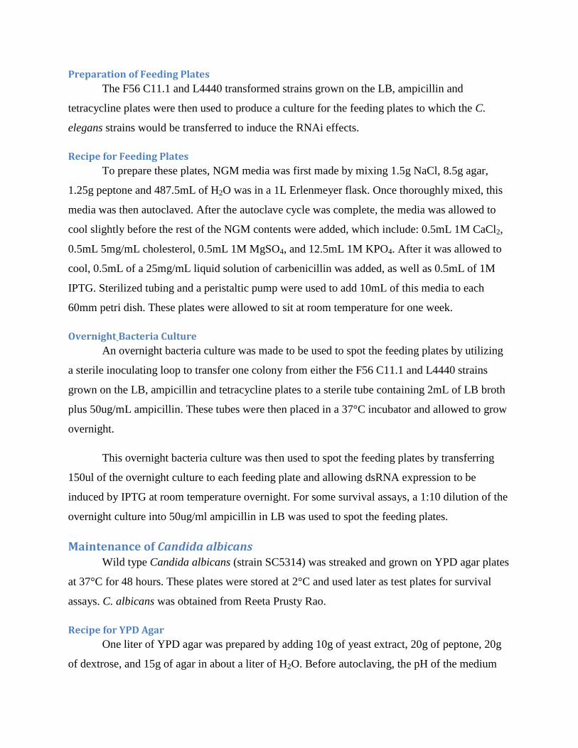
Results
To test the hypothesis that the CeDuox protein is required for defense against the
pathogenic yeast C. albicans, worms were grown under conditions that induced RNAi
knockdown of bli-3, then transferred to plates containing C. albicans. Results were measured by
monitoring survival of worms over time.
In early versions of the survival assays, overnight bacterial cultures were plated undiluted
on the “feeding plates” to induce the RNAi effect. In addition, the C. albicans culture that was
plated for the survival assays was not diluted. Figure 1 shows the survival curves from these
assays. No significant effects of tissue-specific RNAi were observed for the survival assays
shown below performed under undiluted conditions. All worms died rapidly under these
conditions, with 100% of the worms in the hypodermal knockdown dead after less than 100
hours.
Figure 1: Survival Curves of Hypodermal and Whole-Body Knockdown of bli-3 without Dilutions.
These survival assays were performed without any dilutions to bacterial culture for the feeding plates or the yeast
culture used for the survival assays. All other procedures were performed as described in the Materials and Methods
Section. Survival curves for hypodermal knockdown of bli-3 are shown (top), as well as survival curves for whole-
body knockdown of bli-3 (bottom). The log-rank p-values obtained were 0.3390 and 0.1479 for the hypodermal and
whole-body knockdowns, respectively, supporting that significant RNAi effects were not observed.
Survival assays were also conducted in which a 1:10 dilution of the F56 C11.1 and
control L4440 bacterial cultures was performed before they were plated on feeding plates. In
addition, a 1:30 dilution of the yeast culture was performed before it was plated on the survival
assay plates. These adjustments were made in hopes that RNAi effects would be more easily
observed without the severe pathogenicity of the undiluted C. albicans. The survival curves of
each strain are displayed in Figures 3-6 below.
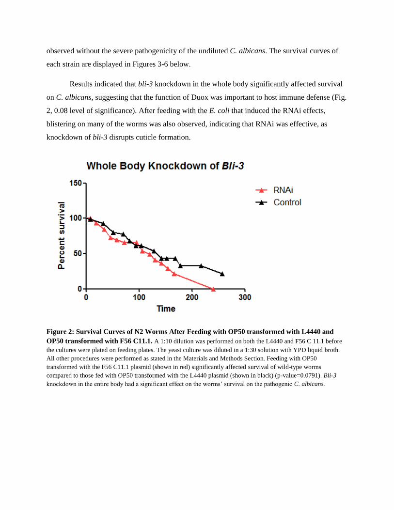
Results indicated that bli-3 knockdown in the whole body significantly affected survival
on C. albicans, suggesting that the function of Duox was important to host immune defense (Fig.
2, 0.08 level of significance). After feeding with the E. coli that induced the RNAi effects,
blistering on many of the worms was also observed, indicating that RNAi was effective, as
knockdown of bli-3 disrupts cuticle formation.
Figure 2: Survival Curves of N2 Worms After Feeding with OP50 transformed with L4440 and
OP50 transformed with F56 C11.1. A 1:10 dilution was performed on both the L4440 and F56 C 11.1 before
the cultures were plated on feeding plates. The yeast culture was diluted in a 1:30 solution with YPD liquid broth.
All other procedures were performed as stated in the Materials and Methods Section. Feeding with OP50
transformed with the F56 C11.1 plasmid (shown in red) significantly affected survival of wild-type worms
compared to those fed with OP50 transformed with the L4440 plasmid (shown in black) (p-value=0.0791). Bli-3
knockdown in the entire body had a significant effect on the worms’ survival on the pathogenic C. albicans.
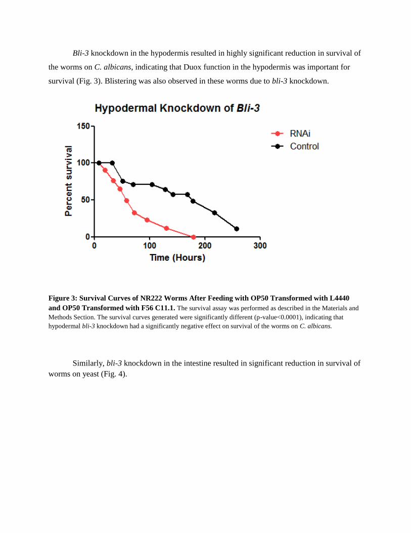
Bli-3 knockdown in the hypodermis resulted in highly significant reduction in survival of
the worms on C. albicans, indicating that Duox function in the hypodermis was important for
survival (Fig. 3). Blistering was also observed in these worms due to bli-3 knockdown.
Figure 3: Survival Curves of NR222 Worms After Feeding with OP50 Transformed with L4440
and OP50 Transformed with F56 C11.1. The survival assay was performed as described in the Materials and
Methods Section. The survival curves generated were significantly different (p-value<0.0001), indicating that
hypodermal bli-3 knockdown had a significantly negative effect on survival of the worms on C. albicans.
Similarly, bli-3 knockdown in the intestine resulted in significant reduction in survival of
worms on yeast (Fig. 4).
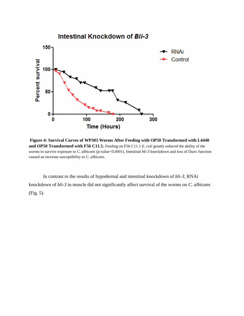
Figure 4: Survival Curves of WP303 Worms After Feeding with OP50 Transformed with L4440
and OP50 Transformed with F56 C11.1. Feeding on F56 C11.1 E. coli greatly reduced the ability of the
worms to survive exposure to C. albicans (p-value<0.0001). Intestinal bli-3 knockdown and loss of Duox function
caused an increase susceptibility to C. albicans.
In contrast to the results of hypodermal and intestinal knockdown of bli-3, RNAi
knockdown of bli-3 in muscle did not significantly affect survival of the worms on C. albicans
(Fig. 5).
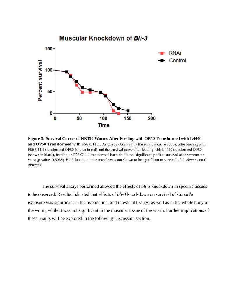
Figure 5: Survival Curves of NR350 Worms After Feeding with OP50 Transformed with L4440
and OP50 Transformed with F56 C11.1. As can be observed by the survival curve above, after feeding with
F56 C11.1 transformed OP50 (shown in red) and the survival curve after feeding with L4440 transformed OP50
(shown in black), feeding on F56 C11.1 transformed bacteria did not significantly affect survival of the worms on
yeast (p-value=0.5038). Bli-3 function in the muscle was not shown to be significant to survival of C. elegans on C.
albicans.
The survival assays performed allowed the effects of bli-3 knockdown in specific tissues
to be observed. Results indicated that effects of bli-3 knockdown on survival of Candida
exposure was significant in the hypodermal and intestinal tissues, as well as in the whole body of
the worm, while it was not significant in the muscular tissue of the worm. Further implications of
these results will be explored in the following Discussion section.
Discussion The results obtained from the RNAi and survival assays performed support the
hypothesis that the Duox activity of bli-3 is important to host immune defense, as bli-3
knockdown in the whole body of the worm significantly decreased survival on the pathogenic
yeast, C. albicans. The finding that bli-3 knockdown in the hypodermal and intestinal tissues
significantly reduced survival, but did not reduce survival in the muscle, further support the
hypothesis that bli-3 and the function of Duox is important to host immunity, as epithelial
tissues, such as the hypodermis and intestine are often the first to come into contact with
pathogens. The intestine is also known to be the main site of infection during infection with the
pathogenic yeast Saccharomyces cerevisiae (Jain et al., 2009).
The results reported here are consistent with those described for the C. elegans response
to the bacterial pathogen Enterococcus faecalis. In that study, RNAi knockdown of bli-3 in the
hypodermis and intestine reduced survival of worms infected with E. faecalis (Chavez et al.,
2009).
However, further research in order to support these findings is suggested by the
importance of bli-3 in cross-linking during cuticle formation (Edens et al., 2001). In these assays,
knockdown of bli-3 not only affected Duox production, but worms with bli-3 knockdown in the
whole body and in the hypodermis also had blistered cuticles. We also observed blistered
cuticles in worms subject to tissue-specific knockdown of bli-3 in the hypodermis. Thus, the
increased susceptibility to C. albicans could have been due to effects on cuticle structure, rather
than being directly caused by a requirement for Duox in host defense. In order to further
investigate this issue, one could perform RNAi and survival assays as previously described with
other blister genes, such as bli-1 or bli-2, which are known to be involved in cuticle formation
and cause blistering, but are not known to be involved with immune defenses (Wormbook,
2007). Comparison of bli-1 or bli-2 knockdown with bli-3 knockdown would allow one to
observe the importance of cuticle formation for survival in contrast to decreased production of
reactive oxygen species.
In addition, the highly significant effects of intestinal knockdown on survival suggest that
Duox is present in the intestinal tissues, although this has not been determined directly. Edens et
al (2001) did not detect bli-3 expression in the intestine, using a Bli-3-specific antibody.
However, expression in the intestine may be induced by infection, as we hypothesized in the
Introduction (Fig. 2). Further investigations, such the use of a reporter gene, would help to
determine whether or not Duox is actually expressed in the intestine. For example, green
fluorescence protein (GFP) could be inserted into the C. elegans genome at the end of the bli-3
gene or downstream of the bli-3 promoter, which would then fluoresce in tissues which express
the bli-3/GFP fusion.
In conclusion, the assays performed allowed for direct comparisons to be made between
worms with full bli-3 function and without bli-3 function in the respective tissues. It was found
that the knockdown of bli-3 and, therefore, the knockdown of reactive oxygen species production
had an increased negative effect on host survivability in the epithelial tissues of the hypodermis
and intestine compared with knockdown in the body wall muscle. These findings suggest that
CeDuox does play a role in defense against the pathogenic yeast C. albicans. If explored further,
the role of CeDuox could perhaps provide some insight to the role of Duox in our bodies and its
possible importance in fighting off harmful and resistant fungal infections.
References
Agrawal, Neema, P.V.N. Dasaradhi, Asif Mohmmed, Pawan Malhotra, Raj K. Bhatnagar, and
Sunil K. Mukherjee. "RNA Interference: Biology, Mechanism, and Applications." Microbiology
and Molecular Biology Reviews 67 (2003): 657-85
Babior, Bernard. "NADPH Oxidase: An Update." Journal of the American Society of
Hematology (1999).
Barrière, A. and Félix, M.-A. Natural variation and population genetics of Caenorhabditis
elegans (December 26, 2005), WormBook, ed. The C. elegans Research Community,
WormBook, doi/10.1895/wormbook.1.43.1, http://www.wormbook.org.
Bedard, Karen, and Karl-Heinz Krause. "The NOX Family of ROS-Generating NADPH
Oxidases: Physiology and Pathophysiology." Physiological Reviews. American Physiological
Society, Jan. 2007. Web.
Chang, Chieh, and Zena Warb. "E Many Faces of Metalloproteases: Cell Growth, Invasion,
Angiogenesis and Metastasis." Trends in Cell Biology 11.11 (2001).
Chavez, Violeta, Akiko Mohri-Shiomi, and Danielle Garsin. "Ce-Duox/Bli-3 Generates Reactive
Oxygen Species as a Protective Innate Immune Mechanism in Caenorhabditis Elegans."
Infection and Immunity. American Society for Microbiology, June 2009. Web.
Diederichs, Sven, and Daniel A. Haber. "Dual Role for Argonautes in MicroRNA Processing and
Posttranscriptional Regulation of MicroRNA Expression." Cell 131.6 (2007): 1097-108.
Edens, W. A., L. Sharling, G. Cheng, R. Shapira, J. M. Kinkade, T. Lee, H. A. Edens, X. Tang,
C. Sullards, D. B. Flaherty, G. M. Benian, and J. D. Lambeth. 2001. Tyrosine cross-linking of
extracellular matrix is catalyzed by Duox, a multidomain oxidase/peroxidase with homology to
the phagocyte oxidase subunit gp91phox. J. Cell Biol. 154:879-891
Geiszt, M., and T. Leto. "The Nox Family of NAD(P)H Oxidases: Host Defense and Beyond."
Journal of Biological Chemistry 279.50 (2004): 51715-1718
Grishok A., Tabara, H., and Mello, C.C. 2000. Genetic requirements for inheritance of RNAi in
C. elegans. Science 287: 2494-2497.
Hannon, Gregory J. "RNA Interference." Nature 418 (2002): 244-51.
Hubbard, E.J.A., and Greenstein, D. Introduction to the germ line (September 1, 2005),
WormBook, ed. The C. elegans Research Community, WormBook,
doi/10.1895/wormbook.1.18.1, http://www.wormbook.org
Jain, Charu, Meijiang Yun, Samuel M. Politz, and Reeta Prusty Rao. "A Pathogenesis Assay
Using Saccharomyces Cerevisiae and Caenorhabditis Elegans Reveals Novel Roles for Yeast
AP-1, Yap1, and Host Dual Oxidase BLI-3 in Fungal Pathogenesis." Eukaryotic Cell. American
Society for Microbiology, May 2009. Web.
Kim, Dennis H., Rhonda Feinbaum, Genevieve Alloing, Fred E. Emerson, Danielle A. Garsin,
Hideki Inoue, Miho Tanaka-Hino, Naoki Hisomoto, Kunihiro Matsumoto, Man-Wah Tan, and
Frederick M. Ausubel. "A Conserved P38 MAP Kinase Pathway in Caenorhabditis Elegans
Innate Immunity." Science 297 (2002): 623-26.
Lambeth, J. David. "NOX Enzymes and the Biology of Reactive Oxygen." Nature Reviews
Immunology 4.3 (2004): 181-89
Lee, Warren L., Rene E. Harrison, and Sergio Grinstein. "Phagocytosis by Neutrophils."
Microbes and Infection 5.14 (2003): 1299-306
MacRae, Ian J., Kaihong Zhou, Fei Li, Adrian Repic, Angela N. Brooks, W. Zacheus Cande,
Paul D. Adams, and Jennifer A. Duodna. "Structural Basis for Double-Stranded RNA Processing
by Dicer." Science 311 (2006): 195-98
Maniatis, T., Fritsch, E. F., & Sambrook, J. (1982). Molecular Cloning: A Laboratory Manual.
New York: Cold Spring Harbor Laboratory.
Murphy, Kenneth, Paul Travers, Mark Walport, and Charles Janeway. Janeway's
Immunobiology. New York: Garland Science, 2012
Novina, Carl D., and Phillip A. Sharp. "The RNAi Revolution." Nature 430.8 (2004): 161-64.
Qadota, Hiroshi, Makiko Inoue, Takao Hikit, Mathias Koppen, Jeffrey D. Hardin, Mutsuki
Amano, Donald G. Moerman, and Kozo Kaibuchi. "Establishment of a Tissue-specific RNAi
System in C. Elegans." Gene 400.1-2 (2007): 166-73.
Ritsick, Darren R., William A. Edens, Victoria Finnerty, and J. David Lambeth. "Nox
Regularion of Smooth." Free Radical Biology and Medicine 43.1 (2007): 31-38
Stiernagle, T. Maintenance of C. elegans (February 11, 2006), WormBook, ed. The C. elegans
Research Community, WormBook, doi/10.1895/wormbook.1.101.1, http://www.wormbook.org.
Rada, Balázs, Csilla Hably, András Meczner, Csaba Timár, Gergely Lakatos, Péter Enyedi, and
Erzsébet Ligeti. "Role of Nox2 in Elimination of Microorganisms." Seminars in
Immunopathology 30.3 (2008): 237-53
Wilson, Richard K., and The C. Elegans Genome Consortium. "How the Worm Was Won: The
C. Elegans Genome Sequencing Project." Trends in Genetics 15.2 (1999): 51-58.