increased post-traumatic survival of neurons in il-6-knockout mice on a background of eae...
TRANSCRIPT

Ž .Journal of Neuroimmunology 119 2001 1–9www.elsevier.comrlocaterjneuroin
Increased post-traumatic survival of neurons in IL-6-knockout mice on abackground of EAE susceptibility
Jasmin Fisher a,1, Tal Mizrahi a,1, Hadas Schori a, Eti Yoles a, Hanna Levkovitch-Verbin a,Shalom Haggiag b, Michel Revel b, Michal Schwartz a,)
a Department of Neurobiology, The Weizmann Institute of Science, 76100 RehoÕot, Israelb Department of Molecular Genetics, The Weizmann Institute of Science, RehoÕot, Israel
Received 30 July 2000; received in revised form 28 March 2001; accepted 17 May 2001
Abstract
Axonal injury initiates a process of neuronal degeneration, with resulting death of neuronal cell bodies. We show here that inC57BLr6J mice, previously shown to have a limited ability to manifest a post-traumatic protective immunity, the rate of neuronalsurvival is increased if IL-6 is deficient during the first 24 hours after optic nerve injury. Immunocytochemical staining preformed 7 daysafter the injury revealed an increased number of activated microglia in the IL-6-deficient mice compared to the wild-type mice. Inaddition, IL-6-deficient mice showed an increased resistance to glutamate toxicity. These findings suggest that the presence of IL-6 duringthe early post-traumatic phase, at least in mice that are susceptible to autoimmune disease development, has a negative effect on neuronalsurvival. This further substantiates the contention that whether immune-derived factors are beneficial or harmful for nerve recovery afterinjury depends on the phenotype of the immune cells and the timing and nature of their dialog with the damaged neural tissue. q 2001Elsevier Science B.V. All rights reserved.
Keywords: Neuroprotection; IL-6 deficiency; Axonal injury; Optic nerve; Glutamate toxicity; Inflammation
1. Introduction
Axonal injury initiates a process of neuronal degenera-Žtion that spreads both laterally and longitudinally Faden,
1993; Faden and Salzman, 1992; McIntosh, 1993; Yoles.and Schwartz, 1998 . The fibers that are directly damaged
by the injury degenerate and their cell bodies eventuallyŽ .die Villegas-Perez et al., 1993 . In addition, there is
degeneration of fibers that escaped the primary lesion,caused by the activity of self-destructive mediators that
Žemerge from the directly damaged fibers Bazan et al.,.1995; Liu et al., 1994; Lynch and Dawson, 1994 . Degen-
eration resulting from the primary injury has been at-tributed to the deficiency of growth factors, normallysupplied by the cellular targets of the fibers, as well as tothe toxicity resulting from unfavorable buffering condi-tions caused by an imbalance of ions, excitatory amino
Žacids, and other metabolic factors Bazan et al., 1995; Liu.et al., 1994; Lynch and Dawson, 1994 .
) Corresponding author. Tel.: q972-8934-2467; fax: q972-8934-4131.Ž .E-mail address: [email protected] M. Schwartz .
1 JF and TM contributed equally to the work.
Recent studies from our laboratory showed that both thelateral and the longitudinal spread of damage after axonalinjury can be reduced by either passive transfer of autoim-
Žmune T cells Hauben et al., 2000a,b; Moalem et al., 1999,.2000a,b or active immunization with myelin-associatedŽ .peptides Fisher et al., 2001; Hauben et al., 2000b . This
autoimmune neuroprotection is probably mediated by cy-tokines and trophic factors secreted by antigen-dependent
Žactivation of the T cells at the lesion site Moalem et al.,.2000a . With regard to the role of individual cytokines in
the process of recovery from injury, the evidence is con-flicting. Cytokines are viewed as key mediators in thepathogenesis of inflammatory lesions of the CNS, butanalysis of their function is complicated by observations of
Žboth helpful and harmful effects Bethea et al., 1999;.Merrill and Benveniste, 1996 .
IL-6, a multifunctional cytokine involved in the hostresponse to infection, is produced by many different celltypes including lymphocytes, monocytes, and parenchymalcells of various organs. In the CNS, astrocytes and mi-croglia secrete IL-6 when infected with viruses or stimu-
Ž .lated by cytokines Frei et al., 1989; Lee et al., 1993 .Both T cell-dependent and T cell-independent pathways of
0165-5728r01r$ - see front matter q 2001 Elsevier Science B.V. All rights reserved.Ž .PII: S0165-5728 01 00342-3
( )J. Fisher et al.rJournal of Neuroimmunology 119 2001 1–92
IL-6 production have been described in mice infected withŽ .different viruses Frei et al., 1989 . The intrathecal IL-6
Ž .response is thought to serve a dual function: i amplifica-Ž .tion of the immune-mediated clearance of virus, and ii
neuronal repair. The latter assumption is based on thefinding that nerve growth factor is produced by astrocytes
Ž .stimulated by IL-6 Frei et al., 1989 . Furthermore, IL-6Žwas found to enhance survival of oligodendrocytes Barres
.et al., 1993 , although in order to be as effective as ciliaryŽ . Ž .neurotrophic factor CNTF Kahn and De Vellis, 1994 in
this regard, it must be combined with its soluble receptorŽ .sIL-6R Mendel et al., 1998 . The latter is a natural agonistŽ .of IL-6 Taga et al., 1989 , and overexpression of IL-6
with sIL-6R was shown to increase nerve regeneration inŽ .transgenic mice Hirota et al., 1996 . However, IL-6 may
also cause neurotoxicity, as evidenced by the neuronaldegeneration that develops in transgenic mice that overex-
Ž .press IL-6 in the CNS parenchyma Campbell et al., 1993 .Recent evidence indicates that IL-6 modulates experi-
Ž .mental autoimmune encephalomyelitis EAE , an inflam-matory demyelinating disease induced by immunization
Ž . Ž .with myelin basic protein MBP , proteolipid protein PLP ,Ž .or myelin oligodendrocyte glycoprotein MOG . Whereas
the encephalitogenic peptide pMOG 35–55 induces EAEin H-2b mice in which the IL-6 gene is intact, IL-6-defi-
Ž .cient mice are resistant to EAE Mendel et al., 1998 .Studies in our laboratory have demonstrated that spon-
taneous recovery from optic nerve injury is critically influ-enced by the animals genetic background. The survivalrate of RGCs after optic nerve injury in EAE-susceptiblemouse strains such as C57BLr6J mice was significantly
Ž .lower than in EAE-resistant strains Kipnis et al., 2001 . Inthe present study we show that the survival rate in anEAE-susceptible mouse strain is increased in the absenceof IL-6. Furthermore, the critical post-injury period duringwhich IL-6 deficiency can improve the survival rate is thefirst 24 h, as the outcome is not affected by the presence orabsence of IL-6 after that time. We further show that thehigher survival rate following axonal injury in the IL-6-de-ficient mice does not correlate with a reduced inflamma-tory response. On the contrary, 7 days after optic nerve
Žinjury, a higher level of Mac-1 immunoreactivity an indi-.cator of microglial activation was observed in IL-6-defi-
cient mice.
2. Materials and methods
2.1. Animals
Ž b. ŽMale and female C57BLr6=129Sv H-2 mice con-. yry Ž .trols and homozygous IL-6 mice 8–12 weeks old
were obtained from the Weizmann Institute of Science,Israel. The mice were housed in a light- and temperature-controlled room and matched for age in each experiment.
2.2. Antigens
ŽRat-derived MOG peptides 1–22 GQFRVIGPGHPI-.RALVGDEAEL were synthesized in the laboratory of
Prof. M. Fridkin at the Department of Chemistry of theWeizmann Institute of Science, using the Fmoc technique
Žwith an automatic multiple peptide synthesizer AMS422,. Ž .Abimed, Langenfeld, Germany . Ovalbumin OVA , frac-
Ž .tion V, was purchased from Sigma Rehovot, Israel .
2.3. ActiÕe immunization
Mice were immunized subcutaneously at one site in theflank with 200 ml of emulsion consisting of MOG 1–22 or
Ž .OVA 300 mg per mouse , emulsified in complete Freund’sŽ .adjuvant CFA supplemented with 500 mg of Mycobac-
terium tuberculosis.
2.4. T cell lines
A T cell line was generated from draining lymph nodecells obtained from H-2b mice immunized with the MOG1–22 antigen described above. Draining lymph nodes fromimmunized mice were removed 11 days after immuniza-tion and pooled in ice-cold PBS. A single-cell suspensionwas prepared and fractionated on plastic tissue-culture
Ž 6 .dishes, and the non-adherent cells 3=10 rml and irradi-Ž . Ž 6 .ated 2000 rad syngeneic normal spleen cells 5=10 rml
Žwere suspended in proliferation medium Ben-Nun and. Ž .Lando, 1983 in 60-mm petri dishes 6 ml per dish . For
selection of lymphoblasts responding to MOG, the culturedŽ .cells were supplemented with MOG 25 mgrml . After
incubation for 72 h, the cultures were collected and washed,Ž 5 .and the cells 4=10 rml were resuspended in propaga-Ž .tion medium Ben-Nun and Lando, 1983 and reseeded in
petri dishes. The cultures were maintained in propagationmedium with changes of medium or splitting of culture for
Ž 5 .8–10 days, and the lymphoblasts 2=10 rml were thenŽ .restimulated with MOG 25 mgrml in the presence of
Ž 6 .irradiated syngeneic normal spleen cells 10=10 rml .The cell line was maintained in cycles of alternate stimula-tion with the MOG peptide and propagation.
2.5. Glutamate toxicity to mouse RGCs
The right eyes of anesthetized mice were puncturedwith a 27-gauge needle in the upper part of the sclera, anda 10-ml Hamilton syringe with a 30-gauge needle wasinserted as far as the vitreal body. Mice were injected with
Ž .a total volume of 1 ml 200 nmol of L-glutamate dissolvedin saline.
2.6. Labeling of RGCs and assessment of their surÕiÕal
For baseline labeling of RGCs, a stereotactic dye wasapplied prior to the crush injury. Eleven days after the first
( )J. Fisher et al.rJournal of Neuroimmunology 119 2001 1–9 3
active immunization, mice were deeply anesthetized byŽintraperitoneal injection of xylazine 14 mgrkg; Vitamed,
. ŽBat Yam, Israel and ketamine 60 mgrkg; Fort Dodge.Laboratories, Fort Dodge, IA and placed in a small stereo-
tactic instrument. The skull was exposed and kept dry andclean using 3% hydrogen peroxide. The bregma was iden-tified and marked. A hole was drilled above the superior
Žcolliculus of each hemisphere 0.292 mm behind and 0.05.mm lateral to the midline . Using a stereotactic measuring
device and a Hamilton injector, the mice were injectedŽwith FluoroGold 3% in saline, Fluorochrome, Denver,
.CO; 1 ml at one site in the superior colliculus of eachhemisphere, at a depth of 0.16 mm from the bony surfaceof the brain. After completion of the injection, the woundwas sutured. Retrograde uptake of the dye provides amarker of the living cells. The same procedure of labelingwas applied in mice exposed to glutamate toxicity, 4 daysafter glutamate injections.
Three days after dye application, the right optic nerveof each mouse was subjected to a crush injury severeenough to cause primary damage to all the axons. Aftersuch an injury, the number of labeled cell bodies at a giventime provides an indication of the rate of secondary degen-eration and cell body death.
Two weeks after the crush injury and one week afterglutamate toxicity, the mice were killed. Eyes showingsigns of ischemia or slight infection were discarded, andonly eyes that looked healthy were used. Each retina wasdetached from the eye, prepared as a flattened wholemount in 4% paraformaldehyde solution, and examined forlabeled RGCs by fluorescence microscopy. Labeled RGCsfrom five to six fields of identical size, located at approxi-mately the same distance from the optic disk, were countedunder the fluorescence microscope and averaged. Calcula-tion of the number of labeled RGCs per square millimeterin the retina of the injured eye provided a quantitativemeasure of the total degeneration.
2.7. Crush injury of mouse optic nerÕe
ŽThree days after stereotactic dye application 14 days.after the first active immunization , the mice were deeply
Žanesthetized by intraperitoneal injection of xylazine 14. Ž .mgrkg and ketamine 60 mgrkg . Using a binocular-op-
erating microscope, the conjunctiva of the right eye wasincised and the optic nerve was exposed. With the aid ofcross-action forceps, the optic nerve was subjected to asevere crush injury 1–2 mm from the eyeball. The unin-jured contralateral nerve was left undisturbed.
2.8. Immunocytochemistry
ŽLongitudinal cryosections of the excised nerves 10 mm.thick were placed on gelatin-coated glass slides and frozen
until preparation for fluorescence staining. Sections werefixed in ethanol for 10 min at room temperature and then
Ž .in acetone for 10 min for MOMA-2 staining , washedtwice in double-distilled water, and incubated for 3 min inPBS containing 0.05% polyoxyethylene–sorbitan mono-
Ž .laurate Tween-20 . Sections were then incubated for 1 hat room temperature with rat antibody to mouse macro-
Ž .phages MOMA-2, Serotec, Oxford, England , mouseŽmonoclonal antibody to mouse neurofilaments N52,
.Sigma diluted 1:200, rat antibody to activated microgliaŽ .Mac-1, Pharmingen diluted 1:50 or rat antibody to B7.2Ž .CD86, Pharmingen diluted 1:20 in PBS containing 3%fetal calf serum and 2% bovine serum albumin. The sec-tions were washed three times with PBS containing 0.05%Tween-20 and then incubated with Cy3-conjugated goat
Ž .anti-rat IgG Jackson ImmunoResearch, West Grove, PA ,Ž .goat anti-mouse IgG Jackson ImmunoResearch or FITC-
Ž .conjugated goat anti-rat IgG Jackson ImmunoResearchfor 1 h at room temperature. They were then washed withPBS containing Tween-20 and treated with glycerol con-
Ž .taining 1,4-diazobicyclo- 2,2,2 octane to inhibit quench-ing of the fluorescence. Sections were viewed with aconfocal microscope. Staining in the absence of the firstantibodies was used as a negative control.
2.9. Statistical analysis
The number of RGCs per square millimeter was calcu-lated for each experiment. Statistical analysis was per-formed by one-way ANOVA.
3. Results
3.1. Increased resistance to secondary degeneration inIL-6-deficient mice
We recently showed that vaccination with the non-en-cephalitogenic myelin-associated peptide MOG 1–22 re-
Ž .duces degeneration of retinal ganglion cells RGCs afterŽ b. Žoptic nerve injury in C3H.SW H-2 mice Fisher et al.,
. Ž .2001 , which are susceptible to EAE Mendel et al., 1995 .C57BLr6J mice, which are also susceptible to EAE, areknown to have a limited ability to manifest a T-cellmediated protective autoimmunity after CNS injury. In thepresent study, we first examined whether MOG vaccina-tion would be beneficial in C57BLr6J mice, which be-cause of a genetically engineered deficiency in IL-6, areresistant to EAE. Two weeks prior to the crush injury,
Ž yry.wild-type and IL-6 knockout IL-6 mice were immu-Ž .nized with MOG 1–22 or with OVA a non-self antigen
as a control. Three days before the injury, their RGCs wereretrogradely labeled by stereotactic application of Fluoro-Gold. Two weeks after the injury, the retinas were excisedand the labeled RGCs were counted. As shown in Fig. 1a,the number of surviving RGCs in the wild-type mice
Ž .actively immunized with MOG 1–22 1101"101, ns4was significantly greater than in those immunized with
( )J. Fisher et al.rJournal of Neuroimmunology 119 2001 1–94
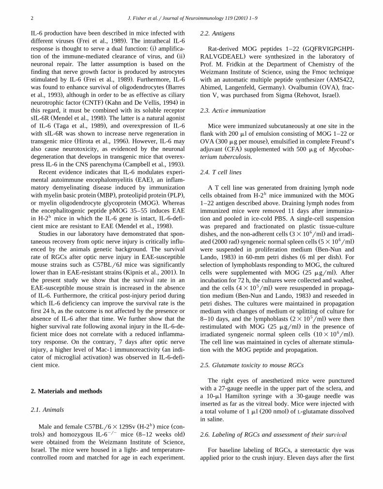
Fig. 1. Post-traumatic survival of RGCs is significantly higher in IL-6-deficient mice than in wild-type mice. The histograms represent the mean numbersŽ . Ž yry. Ž .of labeled RGCs per square millimeter"SEM. a IL-6-deficient IL-6 and control wild-type WT mice were injected with pMOG 1–22 or OVA, 14
days prior to optic nerve injury. The neurotracer dye, FluoroGold, was applied stereotactically 3 days prior to injury. Two weeks after the injury, theŽ .retinas were excised and flat-mounted. Labeled RGCs from five randomly selected fields in each retina all located 1 mm from the optic disk were
Ž .counted by fluorescence microscopy. The mean number of labeled RGCs in the retinas of WT mice pretreated with pMOG 1–22 1101"101, ns4 wasŽ . Ž .significantly higher p-0.01, one-way ANOVA than in mice pretreated with OVA 610"94, ns4 . The mean number of labeled RGCs in the retinas
yry Ž . Ž .of IL-6 mice pretreated with pMOG 1–22 1204"113, ns6 did not differ significantly p)0.05, one-way ANOVA from that in mice pretreatedŽ . Ž . yrywith OVA 857"89, ns8 . b IL-6 and WT mice were injected with T cells specific to MOG 1–22 or PBS immediately after the injury. Dye
Ž .application, preparation, counting of RGCs, and calculation of RGC survival were as described for a . The mean number of labeled RGCs in the retinas ofŽ . Ž . ŽWT mice injected with T 941"54, ns18 was significantly higher p-0.01, one-way ANOVA than in mice injected with PBS 726"36,MO G 1 – 22
. yry Ž .ns16 . The mean number of labeled RGCs in the retinas of IL-6 mice injected with T 1518"40, ns18 did not differ significantlyMO G 1 – 22Ž . Ž .p)0.05, one-way ANOVA from that in mice injected with PBS 1387"47, ns12 . Note the extremely significant difference in RGC survival rates
yry Ž .between the PBS-injected IL-6 mice and the PBS-injected WT mice p-0.001, one-way ANOVA .
Ž .OVA 610"94, ns4; p-0.05, one-way ANOVA . Incontrast, the difference between MOG-immunized and
yry ŽOVA-immunized IL-6 mice 1204"113, ns6 and. Ž857q89, ns8, respectively was not significant p)
.0.05, one-way ANOVA . The number of viable RGCs 2weeks after the crush injury in wild-type and IL-6yry
mice injected immediately after the injury with T cellsŽ .specific to MOG 1–22 T is shown in Fig. 1b.MOG 1 – 22
Significantly more RGCs were viable in the wild-typeŽ .mice injected with T 941q54, ns18 than inMOG 1 – 22
Žthose injected with PBS 726"36, ns16; p-0.01,.one-way ANOVA . The corresponding difference in the
yry ŽIL-6 mice 1518"40, ns18 and 1387"47, ns12,. Žrespectively was not significant p)0.05, one-way
. Ž .ANOVA . Thus, as expected Fisher et al., 2001 , bothactive and passive immunization with a MOG epitope ledto a significantly increased survival of RGCs in the wild-type mice. Interestingly, the RGC survival rate was signifi-cantly higher in the IL-6yry mice injected with PBSŽ . Žns12 than in the PBS-injected wild-type mice ns16;
.p-0.001 one-way ANOVA , but in contrast to the wild-type, MOG immunization, active as well as passive, hadno effect in the IL-6yry mice.
3.2. ReÕersal of neuroprotection in IL-6-deficient mice bya single IL-6 injection
To confirm that the higher survival rate in the IL-6-defi-cient mice is directly related to the absence of IL-6, weattempted to reverse the presumed effect of IL-6 defi-
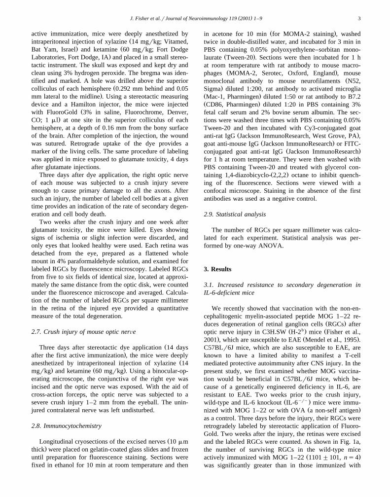
ciency by injecting both IL-6yry and wild-type miceintraperitoneally with IL-6 immediately after the injuryŽ . ŽFig. 2 . Injection of IL-6 compared to PBS injection as a
Fig. 2. Systemic injection of IL-6 immediately after injury reverses theneuroprotective effect of IL-6 deficiency. The histograms record the meannumbers of labeled RGCs per square millimeter"SEM. IL-6yry andWT mice were injected with IL-6 or PBS immediately after the injury.Dye application, preparation, counting of RGCs, and calculation of RGCsurvival were as described for Fig. 1. The mean number of labeled RGCs
Ž .in the retinas of WT mice injected with IL-6 576"68, ns6 did notŽ .differ p)0.05, one-way ANOVA from that in mice injected with PBS
Ž .606"78, ns10 . In contrast, the mean number of labeled RGCs in theyry Ž .retinas of IL-6 mice injected with IL-6 547"67, ns4 was signifi-Ž .cantly lower p-0.001, one-way ANOVA than that in mice injected
Ž .with PBS 1201"88, ns12 .
( )J. Fisher et al.rJournal of Neuroimmunology 119 2001 1–9 5
. yrycontrol, ns10 reduced the RGC survival rate in IL-6Ž . 2 Žmice ns12 from 1201 to 547 per mm p-0.001,
.one-way ANOVA . No significant effect of IL-6 injectionwas observed in the wild-type mice.
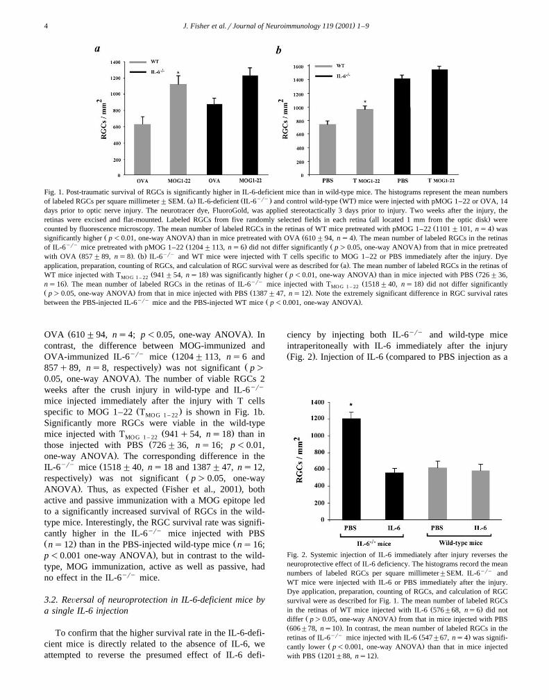
The effect of the injected IL-6 in the IL-6yry miceraised questions about the time period after injury whenthe absence or presence of IL-6 is critical for neuronalsurvival. In other words, it was important to find out atwhat stage the absence of IL-6 is critical for survival. Toaddress this question, IL-6yry mice were injected withIL-6, 1 or 3 days after the injury, and the number ofsurviving RGCs 2 weeks after the injury was comparedwith that in mice injected with IL-6 immediately after the
Ž .injury Fig. 3 . The number of surviving RGCs in IL-6-de-ficient mice injected with IL-6 1 day after the injury was
2 Ž . 21072 per mm ns7 , compared to 547 per mm in themice injected immediately after the injury, indicating thatinjection of IL-6 as early as 1 day after the injury wasalready too late to reverse the endogenous protectivemechanism resulting from the IL-6 deficiency. A similartendency was seen in the mice injected 3 days after injuryŽ .ns5 .
3.3. Increased resistance to glutamate toxicity in IL-6-defi-cient mice
To determine whether the higher survival rate of RGCsafter injury in the IL-6-deficient mice is restricted tomechanical injuries or is common to any CNS insult, wecompared the outcome of a direct biochemical insult causedby glutamate toxicity in IL-6-deficient and wild-type mice.
Fig. 3. Delayed injection of IL-6 does not reverse the neuroprotectiveeffect of IL-6 deficiency. The histograms represent the mean numbers oflabeled RGCs per square millimeter"SEM. IL-6yry mice were injected
Ž . Ž .with IL-6 immediately after the injury ns10 and again 1 day ns7Ž .and 3 days later ns5 . Dye application, preparation and counting of
RGCs, and calculation of RGC survival were as described for Fig. 1.Note, there is almost no difference in RGC survival between mice
Ž .injected with IL-6 3 days after the injury 1214"56 and mice injectedŽ .with PBS immediately after the injury 1201"88 .
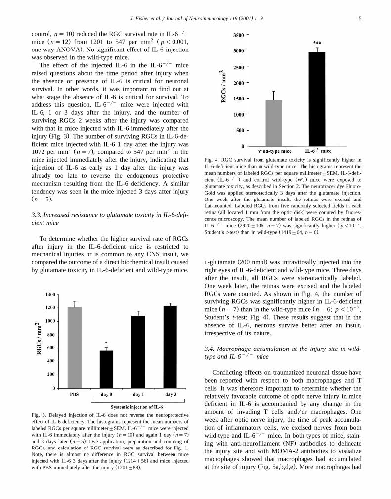
Fig. 4. RGC survival from glutamate toxicity is significantly higher inIL-6-deficient mice than in wild-type mice. The histograms represent themean numbers of labeled RGCs per square millimeter"SEM. IL-6-defi-
Ž yry . Ž .cient IL-6 and control wild-type WT mice were exposed toglutamate toxicity, as described in Section 2. The neurotracer dye Fluoro-Gold was applied stereotactically 3 days after the glutamate injection.One week after the glutamate insult, the retinas were excised andflat-mounted. Labeled RGCs from five randomly selected fields in each
Ž .retina all located 1 mm from the optic disk were counted by fluores-cence microscopy. The mean number of labeled RGCs in the retinas of
yry Ž . Ž y7IL-6 mice 2920"106, ns7 was significantly higher p-10 ,. Ž .Student’s t-test than in wild-type 1419"64, ns6 .
Ž .L-glutamate 200 nmol was intravitreally injected into theright eyes of IL-6-deficient and wild-type mice. Three daysafter the insult, all RGCs were stereotactically labeled.One week later, the retinas were excised and the labeledRGCs were counted. As shown in Fig. 4, the number ofsurviving RGCs was significantly higher in IL-6-deficient
Ž . Ž y7mice ns7 than in the wild-type mice ns6; p-10 ,.Student’s t-test; Fig. 4 . These results suggest that in the
absence of IL-6, neurons survive better after an insult,irrespective of its nature.
3.4. Macrophage accumulation at the injury site in wild-type and IL-6yry mice
Conflicting effects on traumatized neuronal tissue havebeen reported with respect to both macrophages and Tcells. It was therefore important to determine whether therelatively favorable outcome of optic nerve injury in micedeficient in IL-6 is accompanied by any change in theamount of invading T cells andror macrophages. Oneweek after optic nerve injury, the time of peak accumula-tion of inflammatory cells, we excised nerves from bothwild-type and IL-6yry mice. In both types of mice, stain-
Ž .ing with anti-neurofilament NF antibodies to delineatethe injury site and with MOMA-2 antibodies to visualizemacrophages showed that macrophages had accumulated
Ž .at the site of injury Fig. 5a,b,d,e . More macrophages had

( )J. Fisher et al.rJournal of Neuroimmunology 119 2001 1–96
yry Ž .Fig. 5. Macrophage accumulation at the injury site in wild-type and IL-6 mice. Seven days after the injury, the optic nerves of wild-type a, b, c andyry Ž . Ž .IL-6 d, e, f mice were excised and labeled immunocytochemically. Serial optic nerve sections immunolabeled for neurofilaments a, d delineate the
Ž . Ž .site of injury designated by the arrows . Immunolabeling for MOMA-2 b, c, e, f indicates the presence of macrophages at the injury site.
yry Žaccumulated in the IL-6 mice than in wild-type Fig..5b,e . No difference was observed in the numbers of
Ž .accumulated T cells data not shown in the injured opticnerves of the two groups of mice.
The observation that more macrophages accumulated inthe injured optic nerve of mice deficient in IL-6 promptedus to examine whether the difference in the activation state
of macrophages and microglia in IL-6-deficient and wild-type mice. This was done by analyzing macrophagermi-
Ž .croglial expression of Mac-1 C3 complement receptorŽand B7.2, as indicators of microglial activation Jensen et
al., 2000; Koshinaga et al., 2000; Fiske and Brunjes,.2000 . One week after the injury, Mac-1 immunoreactivity
was detected at the injury site of both IL-6-deficient and
Ž .Fig. 6. Intense Mac-1 expression at the injured optic nerves of IL-6-deficient mice. Seven days after the injury, the optic nerves of wild-type a, b andyry Ž . Ž .IL-6 d, e mice were excised and labeled immunocytochemically. Optic nerve sections immunolabeled for Mac-1 a, c indicate the presence of
Ž .activated microglial cells. Immunolabeling for B7.2 b, d indicates the expression of B7.2 molecules at the injured optic nerve.

( )J. Fisher et al.rJournal of Neuroimmunology 119 2001 1–9 7
wild-type mice, but was much more pronounced in theŽ .IL-6-deficient mice Fig. 6a,c . No differences in B7.2
immunoreactivity were observed between injured opticŽ .nerves of the IL-6-deficient and wild-type mice Fig. 6b,d .
4. Discussion
The results of this study suggest that mice deficient inIL-6 are better endowed with a physiological ability tocope with stressful conditions of CNS injury than wild-typemice.
The physiological functions of IL-6 in the CNS areknown to be complex. Numerous studies have indicatedthat IL-6 exerts multiple effects, both beneficial and de-
Žstructive, on CNS cells Van Wagoner and Benveniste,.1999 . In this study, we demonstrated an increased neu-
ronal survival rate in IL-6-deficient mice, assessed 2 weeksafter optic nerve injury or 1 week after intravitreal injec-tion of toxic amounts of glutamate. Interestingly, in arecent study, IL-6-deficient mice showed a decrease in thenumber of activated brain macrophages associated with
Ž .brain focal cryo-injury Penkowa et al., 1999 , suggestinga role for IL-6 in the control CNS inflammation. More-over, dysregulation and overexpression of IL-6 are knownto contribute to the neuropathology and pathophysiology
Ž .associated with many diseases Gadient and Otten, 1997 .Experiments using various transgenic mouse models pointto the destructive potential of dyregulated IL-6 in the CNS.Use of one of these models led to the suggestion thatoverproduction of IL-6 in the CNS may ultimately result inincreased central production of inflammatory cytokines,thus supporting a proinflammatory and detrimental role of
Ž .IL-6 when dysregulated in the CNS Di Santo et al., 1996 .The absence of IL-6 during the first 24 h after the injury
was shown here to be beneficial for neuroprotection. Onepossible explanation is that IL-6 deficiency within theappropriate time window might affect the nature of activa-tion acquired by the microglia, thereby possibly allowingthe creation of protective conditions at a critical stage forpost-traumatic neuronal survival. This interpretation maybe consistent with reports of impaired neuronal regenera-
Ž .tion observed in leukemia inhibitory factor LIF knock-outŽ .mice Sugiura et al., 2000 . It might also be in line with a
recent work showing that IL-10 promotes functional recov-Ž .ery after spinal cord injury Bethea et al., 1999 , provided
that it is supplied immediately after injury; when adminis-tered late, it has adverse effects. The apparently higherlevel of microglialrmacrophage activation seen after in-jury in the IL-6-deficient mice than in the wild-type mice,as observed in the present study, argues in favor of changesin the phenotype of the inflammatory cells or the kineticsof the inflammatory reaction rather than in the amount ofinflammation. It is also possible that the reaction is influ-enced by the existence of a mechanism, unrelated to theimmune activity of the IL-6-deficient mice, which attenu-
ates post-traumatic auto-destructive processes or increasesthe organism’s post-traumatic resistance to them.
The fact that the IL-6-deficient mice are although en-dowed, with a better endogenous protection against stress-ful conditions than the wild type mice do not developEAE, may suggest that they mount beneficial autoimmu-nity rather than destructive autoimmunity as a result of theinjury. Such interpretation is in line with our contention ofthe inverse relationship between susceptibility to developan autoimmune disease and ability to mount beneficial
Ž .autoimmunity Yoles et al., 2001; Kipnis et al., 2001 .The results of the present study, strongly suggest that if
neuroprotective treatment is undertaken using a singleŽ .therapeutic factor monotherapy , its timing must be care-
fully monitored, otherwise it can be more destructive thanhelpful. Moreover, ongoing treatment with one factorwould appear to be risky, as the post-traumatic needs forrecovery change with time. A more promising approachmay be a comprehensive cell therapy, like the one poten-
Žtially provided by autoimmune T cells Moalem et al.,.1999, 2000a,b; Hauben et al., 2000a,b , with continuous
release of various factors at the site of injury. It seemsreasonable to assume that the timing and dynamics ofrelease of such factors would be in accordance with the
Žneeds of the tissue Schwartz and Cohen, 2000; Schwartz.et al., 1999 .
Recently, we showed that active immunization withnon-encephalitogenic myelin-associated peptides, such asMOG 1–22, increases the RGC survival rate 2 weeks after
Ž b. Žoptic nerve crush injury in C3H.SW H-2 mice Fisher et.al., 2001 . The endogenous mechanism of neuroprotection
awakened by injury in our IL-6-deficient mice, which alsopossess the H-2b haplotype, could be only slightly boostedby passive or active immunization with MOG 1–22 pep-tide, whereas the same types of immunization were signifi-cantly effective in the wild-type mice. The insignificanteffect of the MOG immunization in the IL-6-deficientmice might be attributable to their higher basal level ofRGC survival compared to that of the wild-type mice. It ispossible that in the absence of IL-6, whose activity nor-
Žmally evokes a Th-1 type response Mosmann and Coff-.man, 1989 , the spontaneous response to the injury is
differently regulated. Such regulation appears to be criticalŽfor beneficial autoimmunity to be manifested Schwartz
.and Kipnis, 2001 . Alternatively, in the absence of IL-6 thedamaged neural tissue might differently express cytokines,chemokines or other immune-related compounds and theirreceptors that potentially contribute to the local immuneresponse. One such molecule is B7.2, which was recentlyfound by our group to be a better candidate for a type ofcross talk with T cells that produces a beneficial effectŽ .Butovsky et al., 2001 . No differences were found here inB7.2 expression, 1 week after injury, between wild-typeand IL-6-deficient mice. This should be further examinedat earlier time points after the injury. The increased rate ofRGC survival, which was observed here in IL-6 deficient

( )J. Fisher et al.rJournal of Neuroimmunology 119 2001 1–98
mice with a background of EAE susceptibility, might beunique to susceptible strains rather than generally applica-ble. It may be related to the fact that autoimmunity needsto be kept under tight control, a requirement accomplishedhere by the effect of the IL-6 deficiency. The increasedRGC survival rate in these mice was indeed found to becorrelated with the increased resistance to EAE. Furtherstudies should be carried out to determine whether IL-6deficiency in genetically resistant EAE mice will lead to asimilar outcome in terms of post-traumatic RGC survival.
It remains to be determined whether the observed effectof the IL-6 is related to T cells and in particular whetherdestructive and beneficial autoimmunity are mediated byidentical, though differently regulated T cells or by distinc-tive T cells. In either case, the results of the present studycould be interpreted as showing that transient deficiency inIL-6 is beneficial for survival of the cell bodies of trauma-tized CNS axons. In addition, the increased neuronal resis-tance of the IL-6-deficient mice to both mechanical andbiochemical insults, as found in the current study, mightrepresent a more general phenomenon of resistance to awide range of injurious conditions. We believe that theresults of this study may have profound implications forneurodegenerative disorders in general, and those associ-
Žated with optic nerve degeneration Schwartz et al., 1996,.1999; Schwartz and Kipnis, 2001 .
Acknowledgements
We thank S. Smith for editorial assistance and A.Shapira for animal assistance. M. Schwartz holds theMaurice and Ilse Katz Professorial Chair in Neuroim-munology. The work was supported in part by the Glau-coma Research Foundation awarded to M.S.
References
Barres, B.A., Schmid, R., Sendnter, M., Raff, M.C., 1993. Multipleextracellular signals are required for long-term oligodendrocyte sur-vival. Development 118, 283–295.
Bazan, N.G., Rodriguez de Turco, E.B., Allan, G., 1995. Mediators ofinjury in neurotrauma: intracellular signal transduction and geneexpression. J. Neurotrauma 12, 791–814.
Ben-Nun, A., Lando, Z., 1983. Detection of autoimmune cells proliferat-ing to myelin basic protein and selection of T cell lines that mediate
Ž .experimental autoimmune encephalomyelitis EAE in mice. J. Im-munol. 130, 1205–1209.
Bethea, J.R., Nagashima, H., Acosta, M.C., Briceno, C., Gomez, F.,Marcillo, A.E., Loor, K., Green, J., Dietrich, W.D., 1999. Systemi-cally administered interleukin-10 reduces tumor necrosis factor-alphaproduction and significantly improves functional recovery followingtraumatic spinal cord injury in rats. J. Neurotrauma 16, 851–863.
Butovsky, O., Hauben, E., Schwartz, M., 2001. Morphological aspects ofspinal cord autoimmune neuroprotection: colocalization of T cells
Ž .with B7.2 CD86 and prevention of cyst formation. FASEB J. 15,1065–1067.
Campbell, I.L., Abraham, C.R., Masliah, E., Kemper, P., Inglis, J.D.,
Oldstone, M.B., Mucke, L., 1993. Neurologic disease induced intransgenic mice by cerebral overexpression of interleukin 6. Proc.Natl. Acad. Sci. U. S. A. 90, 10061–10065.
Di Santo, E., Alonzi, T., Fattori, E., Poli, V., Ciliberto, G., Sironi, M.,Gnocchi, P., Ricciardi-Castagnoli, P., Ghezzi, P., 1996. Overexpres-sion of interleukin-6 in the central nervous system of transgenic miceincreases central but not systemic proinflammatory cytokine produc-tion. Brain Res. 740, 239–244.
Faden, A.I., 1993. Experimental neurobiology of central nervous systemtrauma. Crit. Rev. Neurobiol. 7, 175–186.
Faden, A.I., Salzman, S., 1992. Pharmacological strategies in CNStrauma. Trends Pharmacol. Sci. 13, 29–35.
Fisher, J., Levkovitch-Verbin, H., Schori, H., Yoles, E., Butovsky, O.,Kaye, J.F., Ben-Nun, A., Schwartz, M., 2001. Vaccination for neuro-protection in the mouse optic nerve: iplications for optic neuropathies.J. Neurosci. 21, 136–142.
Fiske, B.K., Brunjes, P.C., 2000. Microglial activation in the developingrat olfactory bulb. Neuroscience 96, 807–815.
Frei, K., Malipiero, U.V., Leist, T.P., Zinkernagel, R.M., Schwab, M.E.,Fontana, A., 1989. On the cellular source and function of interleukin6 produced in the central nervous system in viral diseases. Eur. J.Immunol. 19, 689–694.
Ž .Gadient, R.A., Otten, U.H., 1997. Interleukin-6 IL-6 —a molecule withboth beneficial and destructive potentials. Prog. Neurobiol. 52, 379–390.
Hauben, E., Butovsky, O., Nevo, U., Yoles, E., Moalem, G., Agranov, E.,Mor, F., Leibowitz-Amit, R., Pevsner, E., Akselrod, S., Neeman, M.,Cohen, I.R., Schwartz, M., 2000a. Passive or active immunizationwith myelin basic protein promotes recovery from spinal cord contu-sion. J. Neurosci. 20, 6421–6430.
Hauben, E., Nevo, U., Yoles, E., Moalem, G., Agranov, E., Mor, F.,Akselrod, S., Neeman, M., Cohen, I.R., Schwartz, M., 2000b. Au-toimmune T cells as potential neuroprotective therapy for spinal cordinjury. Lancet 355, 286–287.
Hirota, H., Kiyama, H., Kishimoto, T., Taga, T., 1996. Accelerated nerveŽ .regeneration in mice by upregulated expression of interleukin IL 6
and IL-6 receptor after trauma. J. Exp. Med. 183, 2627–2634.Jensen, M.B., Hegelund, I.V., Lomholt, N.D., Finsen, B., Owens, T.,
2000. IFNg enhances micriglial reactions to hippocampal axonaldegeneration. J. Neurosci. 20, 3612–3621.
Kahn, M.A., De Vellis, J., 1994. Regulation of an oligodendrocyteprogenitor cell line by the interleukin-6 family of cytokines. Glia 12,87–98.
Kipnis, J., Yoles, E., Schori, H., Hauben, E., Shaked, I., Schwartz, M.,2001. Neuronal survival after CNS insult is determined by a geneti-cally encoded autoimmune response. J. Neurosci 21, 4564–4571.
Koshinaga, M., Katayama, Y., Fukushima, M., Oshima, H., Suma, T.,Takahata, T., 2000. Rapid and widespread microglial activation in-duced by traumatic brain injury in rat brain slices. J. Neurotrauma 17,185–192.
Lee, S.C., Liu, W., Dickson, D.W., Brosnan, C.F., Berman, J.W., 1993.Cytokine production by human fetal microglia and astrocytes. Differ-ential induction by lipopolysaccharide and IL-1 beta. J. Immunol.150, 2659–2667.
Liu, D., Yang, R., Yan, X., McAdoo, D.J., 1994. Hydroxyl radicalsgenerated in vivo kill neurons in the rat spinal cord: electrophysiologi-cal, histological, and neurochemical results. J. Neurochem. 62, 37–44.
Lynch, D.R., Dawson, T.M., 1994. Secondary mechanisms in neuronalŽ .trauma see comments . Curr. Opin. Neurol. 7, 510–516.
McIntosh, T.K., 1993. Novel pharmacologic therapies in the treatment ofexperimental traumatic brain injury: a review. J. Neurotrauma 10,215–261.
Mendel, I., Kerlero de Rosbo, N., Ben-Nun, A., 1995. A myelin oligo-dendrocyte glycoprotein peptide induces typical chronic experimentalautoimmune encephalomyelitis in H-2b mice: fine specificity and Tcell receptor V beta expression of encephalitogenic T cells. Eur. J.Immunol. 25, 1951–1959.
( )J. Fisher et al.rJournal of Neuroimmunology 119 2001 1–9 9
Mendel, I., Katz, A., Kozak, N., Ben-Nun, A., Revel, M., 1998. Inter-leukin-6 functions in autoimmune encephalomyelitis: a study ingene-targeted mice. Eur. J. Immunol. 28, 1727–1737.
Merrill, J.E., Benveniste, E.N., 1996. Cytokines in inflammatory brainlesions: helpful and harmful. Trends Neurosci. 19, 331–338.
Moalem, G., Leibowitz-Amit, R., Yoles, E., Mor, F., Cohen, I.R.,Schwartz, M., 1999. Autoimmune T cells protect neurons from sec-ondary degeneration after central nervous system axotomy. Nat. Med.5, 49–55.
Moalem, G., Gdalyahu, A., Shani, Y., Otten, U., Lazarovici, P., Chen,I.R., Schwartz, M., 2000a. Production of neurotrophins by activated Tcells: implications for neuroprotective autoimmunity. J. Autoimmun.15, 331–345.
Moalem, G., Yoles, E., Leibowitz-Amit, R., Muller-Gilor, S., Mor, F.,Cohen, I.R., Schwartz, M., 2000b. Autoimmune T cells retard the lossof function in injured rat optic nerves. J. Neuroimmunol. 106, 189–197.
Mosmann, T.R., Coffman, R.L., 1989. TH1 and TH2 cells: differentpatterns of lymphokine secretion lead to different functional proper-ties. Annu. Rev. Immunol. 7, 145–173.
Penkowa, M., Moos, T., Carrasco, J., Hadberg, H., Molinero, A., Blueth-mann, H., Hidalgo, J., 1999. Strongly compromised inflammatoryresponse to brain injury in interleukin-6-deficient mice. Glia 25,343–357.
Schwartz, M., Cohen, I.R., 2000. Autoimmunity can benefit self-mainte-nance. Immunol. Today 21, 265–268.
Schwartz, M., Kipnis, J., 2001. Protective autoimmunity: regulation andprospects for vaccination after brain and spinal cord injuries. TrendsMed. 6, 252–258.
Schwartz, M., Belkin, M., Yoles, E., Solomon, A., 1996. Potentialtreatment modalities for glaucomatous neuropathy: neuroprotectionand neuroregeneration. J. Glaucoma 5, 427–432.
Schwartz, M., Moalem, G., Leibowitz-Amit, R., Cohen, I.R., 1999.Innate and adaptive immune responses can be beneficial for CNSrepair. Trends Neurosci. 22, 295–299.
Sugiura, S., Lahav, R., Han, J., Kou, S.Y., Banner, L.R., de Pablo, F.,Patterson, P.H., 2000. Leukaemia inhibitory factor is required fornormal inflammatory responses to injury in the peripheral and centralnervous systems in vivo and is chemotactic for macrophages in vitro.Eur. J. Neurosci. 12, 457–466.
Taga, T., Hibi, M., Hirata, Y., Yamasaki, K., Yasukawa, K., Matsuda, T.,Hirano, T., Kishimoto, T., 1989. Interleukin-6 triggers the associationof its receptor with a possible signal transducer, gp130. Cell 58,573–581.
Van Wagoner, N.J., Benveniste, E.N., 1999. Interleukin-6 expression andregulation in astrocytes. J. Neuroimmunol. 100, 124–139.
Villegas-Perez, M.P., Vidal-Sanz, M., Rasminsky, M., Bray, G.M.,Aguayo, A.J., 1993. Rapid and protracted phases of retinal ganglioncell loss follow axotomy in the optic nerve of adult rats. J. Neurobiol.24, 23–36.
Yoles, E., Schwartz, M., 1998. Degeneration of spared axons followingpartial white matter lesion: implications for optic nerve neuropathies.Exp. Neurol. 153, 1–7.
Yoles, E., Hauben, E., Palgi, O., Agranov, E., Kochroo, V., Cohen, I.R.,Weiner, H., Schwartz, M., 2001. Protective autoimmunity is a physio-logical response to CNS trauma. J. Neurosci. 21, 3740–3748.