increased breast cancer cell toxicity by palladination … · increased breast cancer cell toxicity...
TRANSCRIPT
ORIGINAL ARTICLE
Increased breast cancer cell toxicity by palladinationof the polyamine analogue N1,N11-bis(ethyl)norspermine
Tania M. Silva • Sonia M. Fiuza • Maria P. M. Marques •
Lo Persson • Stina Oredsson
Received: 1 August 2013 / Accepted: 3 November 2013 / Published online: 21 December 2013
� The Author(s) 2013. This article is published with open access at Springerlink.com
Abstract Breast cancer is one of the most common
malignant tumor forms among women and many women
succumb to their disease. Thus, new anticancer agents that
can efficiently improve patient survival are of the utmost
importance. In this study, the effects of the polyamine
analogues N1,N11-bis(ethyl)norspermine (BENSpm) and
N1-cyclo-propylmethyl-N11-ethylnorspermine (CPENSpm)
and the synthesized dinuclear complexes Pd2BENSpm (Pd-
BENSpm), Pt2CPENSpm (Pt-CPENSpm) and Pd2Spm (Pd-
Spm) were investigated in normal-like breast epithelial
MCF-10A cells and the breast cancer cell lines JIMT-1 and
L56BR-C1. The overall data show that palladination of
BENSpm resulted in enhanced cytotoxicity, in contrast to
platination of CPENSpm that reduced cytotoxicity, which
might be explained by differences in the cellular uptake of
Pd-BENSpm and Pt-CPENSpm. BENSpm and Pd-BENS-
pm treatment reduced the CD44?CD24- putative cancer
stem cell population, evaluated by flow cytometry.
Furthermore, Pd-BENSpm was the most efficient com-
pound regarding induction of DNA damage and decrease in
colony formation in soft agar. Pt-CPENSpm and Pd-Spm,
on the other hand, were shown to be the least toxic com-
pounds of all tested. Pd-Spm efficiently reduced the cel-
lular glutathione levels, which probably was a consequence
of its metabolic inactivation by conjugation to this
endogenous thiol. The normal-like cells were found to be
less sensitive to the agents than the breast cancer cells. Our
findings show that Pd-BENSpm exhibits promising anti-
cancer effects which render it suitable for further optimi-
zation to develop a new metal-based chemotherapeutic
drug for breast cancer treatment.
Keywords Breast cancer � Polyamine analogues �Palladium (Pd)(II) complexes � Platinum (Pt)(II)
complexes � DNA damage � Cancer stem cells
Abbreviations
BENSpm N1,N11-bis(ethyl)norspermine
CFE Colony forming efficiency
CPENSpm N1-Cyclo-propylmethyl-N11-
ethylnorspermine
CSC Cancer stem cell
DMSO Dimethyl sulphoxide
FCM Flow cytometry
FCS Fetal calf serum
FITC Fluorescein isothiocyanate
GSH Glutathione
ICP-MS Inductively-coupled plasma mass
spectrometry
MTT 3-(4,5-Dimethyl-thiazolyl-2)-2,5
diphenyltetrazolium bromide
NSpd Norspermidine
PBS Phosphate-buffered saline
Electronic supplementary material The online version of thisarticle (doi:10.1007/s00726-013-1621-y) contains supplementarymaterial, which is available to authorized users.
T. M. Silva (&) � S. M. Fiuza � M. P. M. Marques
Research Unit ‘‘Molecular Physical-Chemistry’’, University
of Coimbra, Coimbra, Portugal
e-mail: [email protected]
T. M. Silva � S. Oredsson
Department of Biology, University of Lund, Lund, Sweden
T. M. Silva � L. Persson
Department of Experimental Medical Science, University
of Lund, Lund, Sweden
M. P. M. Marques
Department of Life Sciences, Faculty of Science and
Technology, University of Coimbra, Coimbra, Portugal
123
Amino Acids (2014) 46:339–352
DOI 10.1007/s00726-013-1621-y
Pd Palladium
Pd-BENSpm Pd2BENSpm
Pd-Spm Pd2Spm
PE Phycoerythrin
PI Propidium iodide
polyHEMA poly(2-hydroxyethyl methacrylate)
Pt Platinum
Pt-CPENSpm Pt2CPENSpm
SCGE Single cell gel electrophoresis
SSAT Spermidine/spermine N1-acetyltransferase
Introduction
The antiproliferative features of cisplatin, [cis-diammin-
edichloroplatinum (II)], (cis-Pt(NH3)2Cl2), were discov-
ered in 1965 by Barnett Rosenberg and resulted in the
successful use of this compound, as the first metal com-
plex, in the treatment of a wide range of solid tumors
(Rosenberg et al. 1965). Platinum (Pt)-based agents are
widely used as chemotherapeutic compounds in today’s
oncological practice, based on their ability to enter the cell
nucleus and covalently bind to DNA, yielding stable
adducts (Brabec and Kasparkova 2005; Esteban-Fernandez
et al. 2010). The formation of several cross-links with
DNA leads to a distortion of the DNA molecule. Thus,
essential biological processes, such as replication and
transcription of DNA are inhibited, which affects protein
synthesis and, consequently, cell proliferation (Brabec and
Kasparkova 2005; Esteban-Fernandez et al. 2010). Prob-
lems with off-target effects such as neurotoxicity and
nephrotoxicity, and the development of acquired resis-
tance, are the main limiting factors of cisplatin treatment
(Brabec and Kasparkova 2005; Esteban-Fernandez et al.
2010). In the few last years, palladium (Pd)(II) complexes
have shown significant antitumor activity against different
cancer cell lines, along with fewer off-target effects, as
compared to cisplatin (Ulukaya et al. 2011).
Another group of compounds extensively used in
experimental cancer research are polyamine analogues
(Casero and Woster 2009). The natural polyamines
putrescine, spermidine and spermine are a group of
ubiquitous positively charged substances with low
molecular weight (Wallace et al. 2003; Palmer and Wal-
lace 2010). In all eukaryotic cells, the polyamines are
involved in a large number of fundamental biological
processes, such as in the regulation of cell proliferation,
differentiation and death (Wallace et al. 2003; Palmer and
Wallace 2010; Traquete et al. 2013). Although all of the
exact roles of polyamines in these processes are not
known, the capacity to interact with DNA and affect DNA
conformation are thought to play a role in their normal
cellular function (Iacomino and Picariello 1823; Pasini
et al. 2013). Polyamine pools are in general higher in
malignant tissue than in normal tissue (Pegg 1988; Tra-
quete et al. 2013), which has suggested the polyamine
metabolic pathway as a target for anticancer therapy (Pegg
1988; Seiler 2005; Casero and Woster 2009; Palmer and
Wallace 2010). Polyamine pool depletion results in inhi-
bition of cell proliferation and, sometimes, in induction of
cell death. One means to achieve polyamine depletion is to
treat with polyamine analogues, which stimulate poly-
amine catabolism and inhibit polyamine biosynthesis,
while they cannot functionally substitute for the depleted
biogenic polyamines (Davidson et al. 1999; Oredsson
et al. 2007; Casero and Woster 2009). Among the most
promising studied polyamine-based antitumor compounds
are the N-ethyl-substituted polyamines, such as N1,N11-
bis(ethyl)norspermine (BENSpm), a symmetrically
substituted spermine analogue and N1-cyclo-propylmethyl-
N11-ethylnorspermine (CPENSpm), an unsymmetrically
substituted spermine analogue. They have been found to
down-regulate polyamine biosynthesis, up-regulate catab-
olism and compete for polyamine uptake, resulting in
dramatic polyamine depletion, inhibition of cell prolifer-
ation and, sometimes, induction of apoptosis (Davidson
et al. 1999; Casero and Woster 2009).
A way to increase the efficiency of a chemotherapeutic
agent, e.g. by increasing interaction with DNA, may be to
combine two different compounds for which DNA is the
biological target, for instance Pt(II) and Pd(II) complexes
with polyamine ligands. In fact, linear alkylpolyamines are
suitable ligands for both Pt(II) and Pd(II) ions, and yield
polynuclear chelates capable of disrupting the native DNA
conformation through the formation of non-conventional
(long-range, interstrand) interactions with the N7 atom of
the purine bases (Hegmans et al. 2008; Ulukaya et al.
2011). Several promising results with these complexes
have been obtained over the last years (Lebwohl and
Canetta 1998; Marques et al. 2002; Fiuza et al. 2006; Fiuza
et al. 2011; Miklasova et al. 2012; Silva et al. 2012).
We have shown that several breast cancer cell lines are
highly sensitive to treatment with the Pd(II) chelate of the
polyamine analogue norspermidine (NSpd) and that this
chelate was more toxic than its Pt(II) counterpart (Silva
et al. 2013). Also, a palladinated spermine was found to be
cytotoxic against breast cancer cell lines (Fiuza et al.
2011).
In the present study, we investigate the cytotoxic effects
of several Pd(II) and Pt(II) polyamine complexes against
two human breast cancer cell lines (JIMT-1 and L56Br-C1)
and one immortalized normal-like breast epithelial cell line
(MCF-10A): two newly synthesized Pd(II) and Pt(II) che-
lates Pd2BENSpm (Pd-BENSpm) and Pt2CPENSpm (Pt-
CPENSpm) (Silva et al. 2012)—and the complex Pd2Spm
(Pd-Spm). Altogether, the results show that palladination of
340 T. M. Silva et al.
123

BENSpm resulted in an increased cytoxicity relative to the
other tested compounds.
Materials and methods
Chemicals
Cell culture medium components were purchased from
Biochrom, Berlin, Germany. Tissue culture plastics were
acquired from Nunc, Roskilde, Denmark. Phosphate-buf-
fered saline (PBS: 8 g/L NaCl, 0.2 g/L KCl, 1.15 g/L
Na2HPO4, 0.2 g/L KH2PO4, pH 7.3) was purchased from
Oxoid Ltd., Basingstoke, Hampshire, UK. Nonidet P-40 was
purchased from VWR, Lund, Sweden. Insulin, hydrocorti-
sone, propidium iodide (PI), Accutase, 3-(4,5-dimethyl-
thiazolyl-2)-2,5 diphenyltetrazolium bromide (MTT) and
poly(2-hydroxyethyl methacrylate) (polyHEMA) were
obtained from Sigma, Stockholm, Sweden. Epithelial growth
factor was purchased from Invitrogen AB, Stockholm,
Sweden. Dimethyl sulphoxide (DMSO) was acquired from
Merck KGaA, Darmstadt, Germany. 14[C]Acetyl-coenzyme
A was purchased from New England Nuclear, DuPont,
Scandinavia AB, Stockholm, Sweden. The monoclonal
antibodies CD44-fluorescein isothiocyanate (FITC) and
CD24-phycoerythrin (PE) together with the FITC- and PE-
conjugated mouse IgG1 isotype controls were obtained from
Becton–Dickinson, Stockholm, Sweden. Nusieve� GTG
low-melting-point agarose, agarose gel supporting medium
and Gel Bond� membranes were obtained from FMC Bio-
Products, Rockland, ME, USA. The GSH-GloTM Glutathione
(GSH) kit was purchased from Promega Biotech AB, Nacka,
Sweden. The Pd-Spm complex was synthesized by Dr. Sonia
Fiuza (Fiuza et al. 2011). BENSpm and CPENSpm were
synthesized and kindly provided by Dr. Patrick Woster,
Department of Drug Discovery and Biomedical Sciences,
Medical University of South Carolina, USA (Casero and
Woster 2009). Pd-BENSpm and Pt-CPENSpm complexes
were synthesized as previously described (Silva et al. 2012).
The complexes are fully characterized by elemental analysis,
as well as through vibrational spectroscopy (Raman and
FTIR). The purity of the analyzed compounds is therefore
assured (Silva et al. 2012).
Drug stock solutions
Stock solutions (2 mM) of BENSpm and CPENSpm were
made in PBS, sterile-filtered and stored at 4 �C. Pd-
BENSpm and Pd-Spm were dissolved in 4 % DMSO in
PBS to give stock solutions of 1 mM that were sterile-
filtered and stored at -20 �C. Pt-CPENSpm was dissolved
in 4 % DMSO in PBS to give a stock solution of 2 mM,
sterile-filtered and stored at -20 �C. Further dilutions were
made in complete cell culture medium to give the final
concentrations.
Cell lines and cell culturing
The L56Br-C1 cell line was established at the Department
of Oncology, Clinical Sciences, Lund University, Sweden
(Johannsson et al. 2003). The JIMT-1 cell line was pur-
chased from the German Collection of Microorganisms and
Cell Cultures (Braunschweig, Germany) and the MCF-10A
cell line was obtained from the American Tissue Type
Culture Collection (Manassas, VA, USA). The cell lines
were cultured as previously described (Silva et al. 2013).
For all experiments, the cells were seeded and allowed to
attach and grow for 24 h, before addition of compound at a
10 lM concentration. A concentration range between 0.1
and 100 lM was used in the MTT assay. The control
received DMSO at the same final concentration as that in
the treated cultures, i.e., 0.1–0.2 %.
Dose response assay
The MTT assay was performed as previously described
(Holst and Oredsson 2005). Briefly, cells were seeded in
96-well microplates with a seeding density of 3,000 (MCF-
10A), 5,000 (JIMT-1) or 8,000 (L56Br-C1) cells in 180 ll
of medium. At 24, 48 and 72 h of drug treatment, 20 ll of
MTT solution (5 mg/ml MTT in PBS) was added to the
cells, which were incubated for 1 h at 37 �C. After removal
of the MTT containing medium, the cells containing
insoluble formazan crystals were dissolved by addition of
100 ll of 100 % DMSO per well. Absorbance was moni-
tored at 540 nm in a Labsystems iEMS Reader MF (Lab-
systems Oy, Helsinki, Finland) using the DeltaSoft II
v.4.14 software (Biometallics Inc., Princeton, NJ, USA).
Cell proliferation
Cells were seeded in Petri dishes (5 cm diameter) at a
density of 0.3 9 106 cells/Petri dish (MCF-10A cells) or
0.6 9 106 cells/Petri dish (JIMT-1 and L56Br-C1 cells) in
5 ml of medium. The cells were allowed to attach and grow
for 24 h and were further harvested by trypsinization, with
the cell number being determined by counting in a hemo-
cytometer after 24, 48 and 72 h of treatment.
Effect of long-time exposure on cell proliferation
This experiment was design to investigate the effect of the
tested compounds on cancer cell proliferation over a longer
period and was performed as previously described (Silva
et al. 2013). Cells (0.3 9 106 MCF-10A cells, 0.7 9 106
JIMT-1 cells and 0.7 9 106 L56Br-C1 cells) were seeded
Increased breast cancer cell toxicity 341
123

into 5 ml of medium in 25 cm2 cell culture flasks and the
compounds were added to the final concentration of 10 lM
24 h later. At each passage, cells were harvested by tryp-
sinization, counted in a hemocytometer, and reseeded at the
same density as above. The cells received the same treat-
ment during each passage. Each week of treatment is
defined as a treatment cycle and the cells were subjected to
five treatment cycles. The total recovery time, i.e., incu-
bation without compound, was 96 h. The data are pre-
sented as the total amount of cells that theoretically would
have accumulated if all cells had been reseeded with a
known cell density after each treatment cycle. Thus, using
the cell number obtained at each passage of a culture
seeded with a known cell density, and applying the formula
log N = log N0 ? nlog2, it was possible to calculate the
number of cells that would have been obtained if all cells
were reseeded at a lower density at each passage.
Intracellular Pd(II) and Pt(II) accumulation
For the study of the intracellular accumulation of
Pd-BENSpm, Pd-Spm or Pt-CPENSpm, cells (1 9 106
MCF-10A cells and 2 9 106 JIMT-1 and L56Br-C1 cells)
were seeded into 12 ml of medium in Petri dishes (9 cm
diameter). After 72 h of treatment with a 10 lM concen-
tration of the tested compounds, cells were washed with
ice-cold PBS, harvested by trypsinization, counted, pel-
leted by centrifugation and stored at -80 �C until analysis.
Prior to analysis, the pellets were digested in 65 % HNO3
for 2 h at 65 �C, diluted to a 5 % acid solution and
centrifuged at 600g for 14 min. The Pd(II) and Pt(II)
accumulation was then analyzed by inductively-coupled plasma
mass spectrometry (ICP-MS) (Thermo X7, Thermo Elemental,
Winsford, UK). The data of metal content were used to calculate
the intracellular concentration of the compounds.
Analysis of polyamine content by high performance
liquid chromatography
L56Br-C1 cells were seeded as described for the prolifer-
ation assay and, after 24 h of treatment, cells were har-
vested, counted, pelleted and stored at -20 �C until
analysis. Chromatographic separation and quantitative
determination of the polyamines in cell extracts in 0.2 M
perchloric acid were carried out using high performance
liquid chromatography (Hewlett Packard 1100) with
h-phthaldialdehyde as the reagent (Seiler and Knodgen 1985).
Spermidine/spermine N1-acetyltransferase activity
assay
L56Br-C1 cells were seeded as described for the prolifer-
ation assay and, after 24 h of treatment, cells were
harvested, counted, pelleted and stored at -80 �C until
analysis. The samples were sonicated in 50 mM Tris–HCl
(pH 7.5) containing 0.25 M sucrose. The spermidine/
spermine N1-acetyltransferase (SSAT) activity was deter-
mined in the sonicates by measuring the synthesis of14[C]acetylspermidine after incubation of the cell extracts
with 14[C]acetyl-coenzyme A and spermidine, as previ-
ously described (Matsui et al. 1981).
Cell cycle phase distribution and cell death analysis
by flow cytometry
MCF-10A, JIMT-1 and L56Br-C1 cells were seeded as
described for the proliferation assay and were further pre-
pared for flow cytometric analysis, as previously described
(Silva et al. 2013). The cells were analyzed using an Accuri
C6 flow cytometer (BD Biosciences, San Jose, CA, USA).
For the computerized evaluation of the cell cycle phase
distribution and the sub-G1 region, the MultiCycle� soft-
ware program (Phoenix Flow Systems, CA, USA) was
used. The sub-G1 region was evaluated as percentage sig-
nals in sub-G1 in relation to all the signals in the sub-G1,
G1, S and G2 histograms. The distribution of the cells in
G1, S and G2 phases was evaluated in % of all cells in G1, S
and G2.
Identification of the CD44?CD24- putative cancer
stem cell population by flow cytometry
JIMT-1 cells (0.6 9 106 cells) were seeded into 5 ml of
medium in Petri dishes (5 cm diameter) and, after a 72 h
treatment period, cells were harvested by Accutase to pre-
vent the disruption of the cell surface proteins. Afterward,
the live cells were washed three times with 5 ml ice-cold
PBS containing 1 % FCS and incubated while shaking for
15 min on ice with the monoclonal antibodies CD44-FITC
and CD24-PE. Next, cells were again washed three times
with ice-cold PBS containing 1 % FCS and identified based
on their expression of the cell surface markers CD44 and
CD24 using an Accuri C6 flow cytometer (BD Biosciences,
San Jose, CA, USA). The data were analyzed using the
CFlow software (BD Biosciences, San Jose, CA, USA).
Colony formation in soft agar
JIMT-1 and L56Br-C1 cells were seeded and treated as
described for the proliferation assay, before reseeding in
soft agar at low density. After 72 h of treatment, cells were
trypsinized, counted and resuspended in agarose containing
medium (0.3 % agarose) at cloning density, i.e., one cell
per microliter, and then reseeded (0.5 ml) in 48-well plates
coated with polyHEMA (Cirenajwis et al. 2010). The cells
were incubated at 37 �C in a humidified incubator with
342 T. M. Silva et al.
123
5 % CO2 in air for 14 days and colonies were counted in an
inverted phase contrast microscope.
Single cell gel electrophoresis assay
The single cell gel electrophoresis (SCGE) assay was
performed as previously described (Freiburghaus et al.
2012). Briefly, MCF-10A cells (0.3 9 106 cells) and JIMT-1
and L56Br-C1 cells (0.7 9 106 cells) were seeded into
5 ml of medium in Petri dishes (5 cm in diameter). After
72 h of treatment with a 10 lM concentration of the tested
compounds, cells were harvested and special attention was
given to the trypsinization time because trypsin can itself
induce DNA damage. Consequently, the time of trypsin-
ization was exactly 13 min for MCF-10A cells, 10 min for
JIMT-1 cells and 6 min for L56Br-C1 cells.
The comets were investigated and photographed with an
epifluorescence microscope (Olympus AX70 equipped
with a Nikon DSRI1 camera). Seven to 22 random images
were captured for each sample. In each image, all nucleoids
were counted by visual inspection. Then, nucleoids with
tails were visually counted in each image. A nucleoid with
tail was defined as a nucleoid with a visible tail indepen-
dent of tail size. The percentage of nucleoids with a tail in
relation to all nucleoids was calculated. A mean was cal-
culated for the counted nucleoids in all images of each
sample. At least 30 nucleoids were scored for each sample.
Afterwards, the mean of the samples for each treatment
was calculated.
GSH-GloTM Glutathione assay
The GSH assay was performed according to the instructions
of the manufacturer (GSH-GloTM Glutathione Assay
Technical Bulletin TB369). Briefly, cells were trypsinized,
counted in a hemocytometer, pelleted and resuspended in
cell culture medium. Aliquots of 180 ll cell suspension
containing 3,000 (MCF-10A), 6,000 (JIMT-1) or 8,000
(L56Br-C1) cells were seeded in the wells of white, opaque
flat-bottomed 96-well plates. At 48 h of drug treatment
(10 lM), the medium was removed from the plates very
carefully. Next, luciferin-NT and glutathione S-transferase
were added to GSH-GloTM reaction buffer to make GSH-
GloTM reagent, which was further added to the wells of the
plates. Before a 30-min incubation period at room temper-
ature, the plates were swirled gently and briefly. Then, the
luciferin detection reagent was added to the wells of the
plates, which were again gently and briefly swirled, fol-
lowed by incubation for 15 min at room temperature.
Finally, luminescence was monitored in a Labsystems
iEMS Reader MF (Labsystems Oy, Helsinki, Finland) using
the DeltaSoft II v.4.14 software (Biometallics Inc.,
Princeton, NJ, USA).
Statistical analysis
For the statistical significance evaluation, a one-way
ANOVA followed by the Newman–Keuls Multiple Com-
parison test was used. Differences were considered statis-
tically significant at p \ 0.05.
Results
MTT reduction
The reduction of MTT is assumed to be a measure of cell
number (Holst and Oredsson 2005) and was initially used
to access the toxicity of BENSpm, Pd-BENSpm,
CPENSpm or Pt-CPENSpm against the three cell lines
investigated, using concentrations that ranged from 0.1 to
100 lM and at 24, 48 and 72 h of treatment (Online
Resource 1). The inhibitory concentration 50 (IC50)
obtained after 72 h of treatment is shown in Table 1. The
normal-like MCF-10A cells were least sensitive while the
L56Br-C1 cell line was most sensitive. Palladination of
BENSPM slightly increased cytotoxicity while platination
of CPENSpm greatly decreased cytotoxicity. Based on
these data and on previously published data on BENSpm
and CPENSpm (McCloskey et al. 2000; Holst and
Oredsson 2005; Oredsson et al. 2007; Uimari et al. 2009),
we decided to use a 10 lM concentration for further
studies.
Effect of one treatment cycle on cell proliferation
We first investigated the effect of the compounds on cell
proliferation during one treatment cycle (Fig. 1a–c). As
also shown in the MTT assay, Pt-CPENSpm was the least
cytotoxic compound. Actually, Pt-CPENSpm did not
affect proliferation of MCF-10A (Fig. 1a) and L56Br-C1
(Fig. 1c) cells, but slightly inhibited proliferation of
Table 1 Absolute IC50 values (lM) of MCF-10A, JIMT-1 and L56-
Br-C1 cells treated for 72 h with BENSpm, Pd-BENSpm, CPENSpm
or Pt-CPENSpm
Cell line MCF-10A JIMT-1 L56Br-C1
BENSpm 52.8 8.7 0.4
Pd-BENSpm 34.2 7.3 0.4
CPENSpm 12.2 9.9 0.2
Pt-CPENSpm [100 lM [100 lM [100 lM
Twenty-four hours after seeding of cells in 96-well plates, the com-
pounds were added in a concentration range of 0.1–100 lM and the
cells were treated for 72 h before evaluation using an MTT assay. The
data were used to construct dose response curves for the evaluation of
absolute IC50 (n = 12)
Increased breast cancer cell toxicity 343
123

JIMT-1 (Fig. 1b) cells. Pd-Spm treatment did not affect
proliferation of MCF-10A cells, while BENSpm, Pd-
BENSpm or CPENSpm treatment slightly reduced the cell
numbers at 48 and 72 h of treatment (Fig. 1a). In L56Br-
C1 cells, treatment with BENSpm, Pd-BENSpm or
CPENSpm resulted in a decrease in cell number after 48
and 72 h of treatment compared to the cell number at
24 h of treatment, implicating cell death (Fig. 1c). Pd-
Spm also resulted in a slight decrease in cell number in
L56Br-C1 cells. In JIMT-1 (Fig. 1b) cells, the inhibition
of cell proliferation was similar after treatment with
BENSpm, Pd-BENSpm, CPENSpm or Pd-Spm. In con-
clusion, the cell proliferation assay shows that the least
sensitive cell line was MCF-10A and the most sensitive
one was L56Br-C1.
Effect of repeated treatment cycles on cell proliferation
In addition to investigating the effect after one 72 h
treatment cycle, we also evaluated the effect of repeated
treatment cycles with 10 lM BENSpm, Pd-BENSpm, Pd-
Spm, CPENSpm or Pt-CPENSpm on cell proliferation of
MCF-10A, JIMT-1 and L56Br-C1 cells (Fig. 1d–f). The
cells were cultivated in cycles with drug treatment for 72 h
followed by a treatment free period of 96 h in between, for
a total number of five cycles. Proliferation of MCF-10A
normal-like breast cells was not affected by repeated
treatment cycles with either Pt-CPENSpm or Pd-Spm
(Fig. 1d), while repeated treatments with these compounds
had a somewhat inhibitory effect on the growth of JIMT-1
cells (Fig. 1e). In L56Br-C1 cells, repeated treatments with
Fig. 1 Effect of BENSpm, Pd-BENSpm, Pd-Spm, CPENSpm or Pt-
CPENSpm treatment on cell proliferation. a–c One treatment cycle.
Twenty-four hours after seeding of cells (0 h time of treatment in the
figure), the compounds were added to a final concentration of 10 lM.
Cells were harvested by trypsinization and counted in a hemocytom-
eter. The results are presented as mean values (n = 6). SEM bars are
covered by the symbols. d–f Five treatment cycles. Cells were seeded
and the compounds were added to the final concentration of 10 lM,
24 h after seeding. After 72 h of treatment, the drug-containing
medium was aspirated and drug free culture medium was added. After
an additional 72 h of incubation, cells were harvested by
trypsinization and counted in a hemocytometer. These 7 days were
defined as one treatment cycle. The cells were reseeded at the same
density as at the previous passage and treated with the same drug for
the next treatment cycle. All together this was repeated for five
treatment cycles. The total recovery time between repeated treatments
was 96 h. The results are presented as mean values (n = 3). SEM bars
are covered by the symbols. Please note that the y-axis has different
scales for the different cell lines because of different rates of cell
proliferation. ***p \ 0.001 compared to control for the curves below
the symbol. Control, BENSpm, Pd-BENSpm, Pd-Spm,
CPENSpm, Pt-CPENSpm
344 T. M. Silva et al.
123

Pd-Spm resulted in a reduction of cell number after four
treatment cycles (Fig. 1f). Repeated treatment with
BENSpm, Pd-BENSpm or CPENSpm resulted in a
decrease in the cell number, compared to the number of
cells seeded at time 0, in all cell lines (Fig. 1d–f).
Intracellular Pd(II) and Pt(II) accumulation
by inductively-coupled plasma mass spectrometry
ICP-MS was used to quantify the Pd(II) or Pt(II) content in
cells treated with 10 lM Pd-BENSpm, Pd-Spm or Pt-
CPENSpm for 72 h and the data were used to calculate the
intracellular concentrations of the drugs (Fig. 2). The data
demonstrate that the intracellular concentration of Pd-
BENSpm was approximately 20, 40 and 360 pmol/106
cells in MCF-10A (Fig. 2a), JIMT-1 (Fig. 2b) and L56Br-
C1 (Fig. 2c) cells, respectively. The Pd-Spm and Pt-
CPENSpm concentrations were between 2 and 18 pmol/
106 cells in the three cell lines, with the highest concen-
trations in L56Br-C1 cells and the lowest concentrations in
MCF-10A cells.
Analysis of polyamine levels
Since L56Br-C1 was found to be the most sensitive cell line,
we decided to measure the polyamine levels in these cells
after 24 h of treatment with 10 lM of BENSpm, Pd-BENS-
pm, Pd-Spm, CPENSpm or Pt-CPENSpm (Fig. 3). As
expected, the putrescine (Fig. 3a), spermidine (Fig. 3b) and
spermine (Fig. 3c) contents decreased significantly upon
treatment with BENSpm, Pd-BENSpm or CPENSpm, com-
pared to control. Pt-CPENSpm treatment resulted in a minor
increase in polyamine levels compared to control, while Pd-
Spm treatment only had a negligible effect (Fig. 3a–c).
Spermidine/spermine N1-acetyltransferase activity
Since BENSpm or Pd-BENSpm treatment efficiently
depleted the polyamines in L56Br-C1 cells, we decided to
measure the activity of the polyamine catabolic enzyme
SSAT. The SSAT activity was very low in untreated
L56Br-C1 cells, but increased significantly after treatment
with BENSpm, Pd-BENSpm or CPENSpm (Table 2). The
increase in SSAT activity was twice as high in cells treated
with BENSpm or Pd-BENSpm than in CPENSpm-treated
cells. SSAT activity was not detected in Pd-Spm- or Pt-
CPENSpm-treated cells.
Cell cycle phase distribution and cell death
Since the various compounds used in the present study
were shown to affect cell proliferation differently, we
analyzed the effect of BENSpm, Pd-BENSpm, Pd-Spm,
CPENSpm or Pt-CPENSpm on cell cycle phase distribu-
tion as well as on cell death using flow cytometry (FCM).
Cells were sampled for analysis of the cell cycle phase
distribution at 24, 48 and 72 h of treatment, but only data
for 48 h of treatment are shown in Table 3, since they show
the major conclusions.
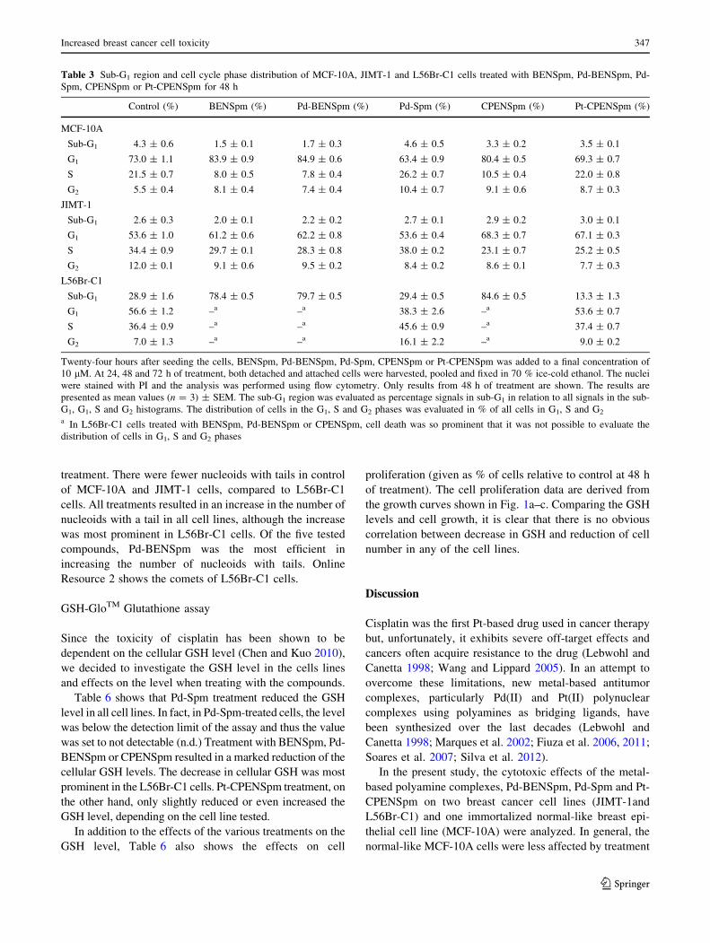
The data show that the only cell line in which the per-
centage of cells in the sub-G1 region, which reflects cell
death, increased substantially was the L56Br-C1 cell line
(Table 3). In L56Br-C1 cells, cell death was so prominent
that is was not possible to evaluate the DNA histograms at
48 and 72 h of treatment. L56Br-C1 cells have a high
degree of spontaneous apoptotic cell death, which is
obvious in Table 3 where the sub-G1 region in control cells
is around 20 % (Hegardt et al. 2002). No cell death was
observed in MCF-10A and JIMT-1 cells.
Fig. 2 Intracellular concentration of Pd-BENSpm, Pd-Spm and Pt-
CPENSpm in MCF-10A, JIMT-1 and L56Br-C1 cells. After 72 h of
treatment with a 10 lM concentration of the compounds, cells were
harvested, pooled and digested in HNO3. The supernatant was used
for analysis of Pd(II) and Pt(II) by ICP-MS and the data used to
calculate the intracellular Pd-BENSpm, Pd-Spm and Pt-CPENSpm
concentrations in MCF-10A (a), JIMT-1 (b) and L56Br-C1 (c) cells.
The results are presented as mean values (n = 3) and bars
represent ± SEM. ***p \ 0.001 compared to Pd-Spm or Pt-
CPENSpm treatment. Pd-BENSpm, Pd-Spm, Pt-CPENSpm
Increased breast cancer cell toxicity 345
123

In MCF-10A cells, the number of cells in the G1 phase
increased from around 50 to 80 % between 24 and 48 h of
treatment with BENSpm, Pd-BENSpm or CPENSpm
(Table 3). During the same time period, the percentage of
cells in the S phase decreased from 40 to 10 %. Less evi-
dent changes were found in cell cycle phase distribution of
JIMT-1 cells after treatment (Table 3).
Identification of the CD44?CD24- putative cancer
stem cell population by flow cytometry
To investigate the effect on the putative breast cancer
stem cell (CSC) population, here defined as CD44?CD24-
(Al-Hajj et al. 2003; Cirenajwis et al. 2010), JIMT-1 cells
were treated for 72 h with 10 lM BENSpm, Pd-BENSpm,
Pd-Spm, CPENSpm or Pt-CPENSpm and the cells were
analyzed by FCM after labeling with the CD44-FITC and
CD24-PE antibodies. As shown in Fig. 4, treatment with
either BENSpm or Pd-BENSpm significantly reduced the
CD44?CD24- subpopulation from 50 to 30 % and 33 %,
respectively, whereas treatment with Pd-Spm, CPENSpm
and Pt-CPENSpm resulted in a slight increase in the
CD44?CD24- subpopulation (Fig. 4).
Colony forming efficiency
The clonogenic assay is designed to measure the ability of
single cells to proliferate and form colonies in an anchor-
age independent manner.
The immortalized normal-like cell line MCF-10A does
not form colonies in soft agar and was not used in this
assay. In the JIMT-1 and L56Br-C1 breast cancer cell lines,
all the treatments decreased the colony forming efficiency
(CFE), compared to the control (Table 4). The CFE was
similar in JIMT-1 and L56Br-C1 control cells, around
30 %. Pd-BENSpm treatment was most efficient in
reducing the number of colonies and L56Br-C1 was the
most sensitive cell line to all the treatments (Table 4).
Single cell gel electrophoresis assay
The SCGE assay is a method used to detect DNA damage
in single cells and it was used to investigate if the com-
pounds induced DNA strand breaks (Silva et al. 2013).
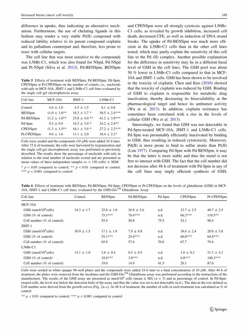
Table 5 shows the percentage of nucleoids with tails in
relation to the total number of nucleoids scored after 72 h of
Fig. 3 Effect of BENSpm, Pd-BENSpm, Pd-Spm, CPENSpm or Pt-
CPENSpm treatment on the polyamine content in L56Br-C1 cells.
After 24 h of treatment with a 10 lM concentration of the
compounds, cells were harvested, counted in a hemocytometer and
then putrescine (a), spermidine (b) and spermine (c) contents were
determined by HPLC. The results are presented as mean values
(n = 3) and bars represent ± SEM. When not visible, the bars are
covered by the symbols. *p \ 0.05 compared to control; **p \ 0.01
compared to control; ***p \ 0.001 compared to control. Control,
BENSpm, Pd-BENSpm, Pd-Spm, CPENSpm, Pt-CPENSpm
Table 2 Spermidine/spermine N1-acetyltransferase activity in
L56Br-C1cells treated with BENSpm, Pd-BENSpm, Pd-Spm,
CPENSpm or Pt-CPENSpm for 24 h
SSAT activity (cpm/106 cells)
Control 200 ± 39
BENSpm 32,654 ± 4,236
Pd-BENSpm 40,004 ± 5,673
Pd-Spm n.d.
CPENSpm 15,050 ± 2,390
Pt-CPENSpm n.d.
After 24 h of treatment with a 10 lM concentration of the com-
pounds, cells were harvested, counted in a hemocytometer and then
the spermidine/spermine N1-acetyltransferase activity was determined
using a radiometric assay. The results are presented as mean values
(n = 6) and bars represent ± SEM
n.d. not detectable
** p \ 0.01 compared to control; *** p \ 0.001 compared to control
346 T. M. Silva et al.
123
treatment. There were fewer nucleoids with tails in control
of MCF-10A and JIMT-1 cells, compared to L56Br-C1
cells. All treatments resulted in an increase in the number of
nucleoids with a tail in all cell lines, although the increase
was most prominent in L56Br-C1 cells. Of the five tested
compounds, Pd-BENSpm was the most efficient in
increasing the number of nucleoids with tails. Online
Resource 2 shows the comets of L56Br-C1 cells.
GSH-GloTM Glutathione assay
Since the toxicity of cisplatin has been shown to be
dependent on the cellular GSH level (Chen and Kuo 2010),
we decided to investigate the GSH level in the cells lines
and effects on the level when treating with the compounds.
Table 6 shows that Pd-Spm treatment reduced the GSH
level in all cell lines. In fact, in Pd-Spm-treated cells, the level
was below the detection limit of the assay and thus the value
was set to not detectable (n.d.) Treatment with BENSpm, Pd-
BENSpm or CPENSpm resulted in a marked reduction of the
cellular GSH levels. The decrease in cellular GSH was most
prominent in the L56Br-C1 cells. Pt-CPENSpm treatment, on
the other hand, only slightly reduced or even increased the
GSH level, depending on the cell line tested.
In addition to the effects of the various treatments on the
GSH level, Table 6 also shows the effects on cell
proliferation (given as % of cells relative to control at 48 h
of treatment). The cell proliferation data are derived from
the growth curves shown in Fig. 1a–c. Comparing the GSH
levels and cell growth, it is clear that there is no obvious
correlation between decrease in GSH and reduction of cell
number in any of the cell lines.
Discussion
Cisplatin was the first Pt-based drug used in cancer therapy
but, unfortunately, it exhibits severe off-target effects and
cancers often acquire resistance to the drug (Lebwohl and
Canetta 1998; Wang and Lippard 2005). In an attempt to
overcome these limitations, new metal-based antitumor
complexes, particularly Pd(II) and Pt(II) polynuclear
complexes using polyamines as bridging ligands, have
been synthesized over the last decades (Lebwohl and
Canetta 1998; Marques et al. 2002; Fiuza et al. 2006, 2011;
Soares et al. 2007; Silva et al. 2012).
In the present study, the cytotoxic effects of the metal-
based polyamine complexes, Pd-BENSpm, Pd-Spm and Pt-
CPENSpm on two breast cancer cell lines (JIMT-1and
L56Br-C1) and one immortalized normal-like breast epi-
thelial cell line (MCF-10A) were analyzed. In general, the
normal-like MCF-10A cells were less affected by treatment
Table 3 Sub-G1 region and cell cycle phase distribution of MCF-10A, JIMT-1 and L56Br-C1 cells treated with BENSpm, Pd-BENSpm, Pd-
Spm, CPENSpm or Pt-CPENSpm for 48 h
Control (%) BENSpm (%) Pd-BENSpm (%) Pd-Spm (%) CPENSpm (%) Pt-CPENSpm (%)
MCF-10A
Sub-G1 4.3 ± 0.6 1.5 ± 0.1 1.7 ± 0.3 4.6 ± 0.5 3.3 ± 0.2 3.5 ± 0.1
G1 73.0 ± 1.1 83.9 ± 0.9 84.9 ± 0.6 63.4 ± 0.9 80.4 ± 0.5 69.3 ± 0.7
S 21.5 ± 0.7 8.0 ± 0.5 7.8 ± 0.4 26.2 ± 0.7 10.5 ± 0.4 22.0 ± 0.8
G2 5.5 ± 0.4 8.1 ± 0.4 7.4 ± 0.4 10.4 ± 0.7 9.1 ± 0.6 8.7 ± 0.3
JIMT-1
Sub-G1 2.6 ± 0.3 2.0 ± 0.1 2.2 ± 0.2 2.7 ± 0.1 2.9 ± 0.2 3.0 ± 0.1
G1 53.6 ± 1.0 61.2 ± 0.6 62.2 ± 0.8 53.6 ± 0.4 68.3 ± 0.7 67.1 ± 0.3
S 34.4 ± 0.9 29.7 ± 0.1 28.3 ± 0.8 38.0 ± 0.2 23.1 ± 0.7 25.2 ± 0.5
G2 12.0 ± 0.1 9.1 ± 0.6 9.5 ± 0.2 8.4 ± 0.2 8.6 ± 0.1 7.7 ± 0.3
L56Br-C1
Sub-G1 28.9 ± 1.6 78.4 ± 0.5 79.7 ± 0.5 29.4 ± 0.5 84.6 ± 0.5 13.3 ± 1.3
G1 56.6 ± 1.2 –a –a 38.3 ± 2.6 –a 53.6 ± 0.7
S 36.4 ± 0.9 –a –a 45.6 ± 0.9 –a 37.4 ± 0.7
G2 7.0 ± 1.3 –a –a 16.1 ± 2.2 –a 9.0 ± 0.2
Twenty-four hours after seeding the cells, BENSpm, Pd-BENSpm, Pd-Spm, CPENSpm or Pt-CPENSpm was added to a final concentration of
10 lM. At 24, 48 and 72 h of treatment, both detached and attached cells were harvested, pooled and fixed in 70 % ice-cold ethanol. The nuclei
were stained with PI and the analysis was performed using flow cytometry. Only results from 48 h of treatment are shown. The results are
presented as mean values (n = 3) ± SEM. The sub-G1 region was evaluated as percentage signals in sub-G1 in relation to all signals in the sub-
G1, G1, S and G2 histograms. The distribution of cells in the G1, S and G2 phases was evaluated in % of all cells in G1, S and G2
a In L56Br-C1 cells treated with BENSpm, Pd-BENSpm or CPENSpm, cell death was so prominent that it was not possible to evaluate the
distribution of cells in G1, S and G2 phases
Increased breast cancer cell toxicity 347
123

with any of the compounds than the cancer cell lines as
shown by the higher IC50 value, the lower effects on cell
proliferation and on comet formation. In addition, of the
very toxic compounds, BENSpm, CPENSpm and Pd-
BENSpm, the latter, i.e., Pd-BENSpm, showed the lowest
relative toxicity in MCF-10A cells, which is promising.
Previous studies have shown that BENSpm and
CPENSpm inhibit cell proliferation of breast cancer cell
lines (Holst and Oredsson 2005; Oredsson et al. 2007;
Cervelli et al. 2010) and our study confirm those data. In
addition, we show that repeated treatment cycles further
suppresses cell proliferation. Interestingly, platination of
CPENSpm clearly induced a marked reduction in toxicity
in all the cell lines analyzed in the present study. In con-
trast, palladination of BENSpm increased the cytotoxic
effect of the compound. One cause for the difference in
toxicity may be related to cellular uptake, which was sig-
nificantly higher for Pd-BENSpm than for Pt-CPENSpm.
Earlier studies on Pd(II)- or Pt(II)-NSpd complexes showed
that the substitution of Pt(II) for Pd(II) increased the
cytotoxicity of the compound (Silva et al. 2013). However,
the difference in cytotoxicity between the Pd(II) complex
and the Pt(II) complex of NSpd was not explained by a
Fig. 4 Effect of BENSpm, Pd-BENSpm, Pd-Spm, CPENSpm or Pt-
CPENSpm treatment on the CD44?CD24- putative cancer stem cell
population in JIMT-1 cells. After 72 h of treatment with a 10 lM
concentration of the compounds, cells were harvested with Accutase and
identified based on their expression of the cell surface markers CD44 and
CD24 by flow cytometry. a Representative cytograms of the flow
cytometric analysis of cell surface-expressed CD44 and CD24 in the
JIMT-1 breast cancer cell line. b Table showing the data obtained with
each treatment. The results are presented as percentage of total
population (n = 9) ± SEM. ***p \ 0.001 compared to control
Table 4 Effects of BENSpm, Pd-BENSpm, Pd-Spm, CPENSpm or
Pt-CPENSpm treatment of JIMT-1 and L56Br-C1 cells on the colony
forming efficiency in soft agar
Cell line JIMT-1 L56Br-C1
Control (%) 29.4 ± 1.0 32.7 ± 1.9
BENSpm (%) 15.1 ± 0.5 (51.2)*** 10.8 ± 0.5 (33.0)***
Pd-BENSpm (%) 12.1 ± 0.3 (41.3)*** 7.8 ± 0.1 (23.7)***
Pd-Spm (%) 20.5 ± 0.3 (69.7)*** 17.0 ± 1.0 (51.8)***
CPENSpm (%) 16.4 ± 0.8 (55.7)*** 11.8 ± 1.0 (36.2)***
Pt-CPENSpm (%) 24.1 ± 0.4 (82.1)*** 24.0 ± 0.5 (73.2)***
Cells were seeded and the compounds (10 lM) were added 24 h later.
After 72 h of treatment, the cells were harvested, counted and
reseeded at cloning density in soft agar. Colonies were counted after
14 days of incubation. The results are presented as mean values
(n = 3) ± SEM and as percentage of control (in brackets)
*** p \ 0.001 compared to control
348 T. M. Silva et al.
123
difference in uptake, thus indicating an alternative mech-
anism. Furthermore, the use of chelating ligands in this
fashion may render a very stable Pt(II) compound with
reduced lability relative to its parent compound cisplatin
and its palladium counterpart and, therefore, less prone to
react with cellular targets.
The cell line that was most sensitive to the compounds
was L56Br-C1, which was also found for NSpd, Pd-NSpd
and Pt-NSpd (Silva et al. 2013). Pd-BENSpm, BENSpm
and CPENSpm were all strongly cytotoxic against L56Br-
C1 cells, as revealed by growth inhibition, increased cell
death, decreased CFE, as well as induction of DNA strand
breaks. The uptake of Pd-BENSpm was much more effi-
cient in the L56Br-C1 cells than in the other cell lines
tested, which may partly explain the sensitivity of this cell
line to the Pd (II) complex. Another possible explanation
for the difference in sensitivity may lie in a different basal
level of GSH in the cell lines. The GSH pool was about
50 % lower in L56Br-C1 cells compared to that in MCF-
10A and JIMT-1 cells. GSH has been shown to be involved
in the toxicity of cisplatin. Chen and Kuo (2010) showed
that the toxicity of cisplatin was reduced by GSH. Binding
of GSH to cisplatin is responsible for metabolic drug
inactivation, thereby decreasing its bioavailability at the
pharmacological target and hence its antitumor activity
(Wu et al. 2013). In addition, cisplatin resistance has
sometimes been correlated with a rise in the levels of
cellular GSH (Wu et al. 2013).
Interestingly, we found that GSH was not detectable in
Pd-Spm-treated MCF-10A, JIMT-1 and L56Br-C1 cells.
Pd-Spm was presumably efficiently inactivated by binding
to GSH, thus resulting in reduced cytotoxicity. Moreover,
Pd(II) is more prone to bind to sulfur atoms than Pt(II)
(Lim 1977). Comparing Pd-Spm with Pd-BENSpm, it may
be that the latter is more stable and thus the metal is not
free to interact with GSH. The fact that the cell number did
not decrease after 48 h of treatment with Pd-Spm in any of
the cell lines may imply efficient synthesis of GSH,
Table 5 Effects of treatment with BENSpm, Pd-BENSpm, Pd-Spm,
CPENSpm or Pt-CPENSpm on the number of comets, i.e., nucleoids
with tails in MCF-10A, JIMT-1 and L56Br-C1 cell lines evaluated by
the single cell gel electrophoresis assay
Cell line MCF-10A JIMT-1 L56Br-C1
Control 4.6 ± 1.0 4.5 ± 1.5 8.1 ± 0.8
BENSpm 11.0 ± 1.6** 18.3 ± 3.7 * 30.5 ± 2.4***
Pd-BENSpm 11.2 ± 1.0** 23.8 ± 4.0 ** 41.2 ± 3.8***
Pd-Spm 9.2 ± 0.9 14.3 ± 3.0 * 24.2 ± 2.4**
CPENSpm 11.3 ± 1.5** 16.1 ± 3.9 * 27.2 ± 2.2***
Pt-CPENSpm 9.0 ± 1.6 11.1 ± 2.0 18.4 ± 2.1*
Cells were seeded and the compounds (10 lM) were added 24 h later.
After 72 h of treatment, the cells were harvested by trypsinization and
the single cell gel electrophoresis assay was performed as previously
described. The results show the percentage of nucleoids with tails in
relation to the total number of nucleoids scored and are presented as
mean values of three independent samples (n = 150 cells) ± SEM
* p \ 0.05 compared to control; ** p \ 0.01 compared to control;
*** p \ 0.001 compared to control
Table 6 Effects of treatment with BENSpm, Pd-BENSpm, Pd-Spm, CPENSpm or Pt-CPENSpm on the levels of glutathione (GSH) in MCF-
10A, JIMT-1 and L56Br-C1 cell lines, evaluated by the GSH-GloTM Glutathione Assay
Cell line Control BENSpm Pd-BENSpm Pd-Spm CPENSpm Pt-CPENSpm
MCF-10A
GSH (nmol/106cells) 34.3 ± 1.7 25.8 ± 1.8 26.9 ± 2.6 n.d. 22.7 ± 2.5 40.7 ± 2.5
GSH (% of control) 75.1*** 78.4*** n.d. 66.2*** 118.5**
Cell number (% of control) 55.4 58.8 75.5 52.1 90.4
JIMT-1
GSH (nmol/106cells) 30.9 ± 1.5 17.1 ± 1.9 7.9 ± 0.8 n.d. 18.6 ± 2.4 20.0 ± 5.8
GSH (% of control) 55.1*** 25.6*** n.d. 60.0*** 64.8***
Cell number (% of control) 65.9 57.6 70.8 67.7 79.4
L56Br-C1
GSH (nmol/106cells) 15.1 ± 1.0 2.8 ± 0.4 0.5 ± 0.5 n.d. 1.0 ± 0.2 21.3 ± 4.2
GSH (% of control) 18.6*** 3.0*** n.d. 6.8*** 140.3***
Cell number (% of control) 19.0 14.9 41.5 20.1 87.0
Cells were seeded in white opaque 96-well plates and the compounds were added 24 h later to a final concentration of 10 lM. After 48 h of
treatment, the plates were removed from the incubator and the GSH-GloTM Glutathione assay was performed according to the instructions of the
manufacturer. The results of the GSH assay are presented as nmol/106 cells (mean ± SD) (n = 3) and as percentage of control. In Pd-Spm-
treated cells, the level was below the detection limit of the assay and thus the value was set to not detectable (n.d.). The data in the row defined as
Cell number were derived from the growth curves (Fig. 2a–c). At 48 h of treatment, the number of cells in each treatment was calculated as % of
control
** p \ 0.01 compared to control; *** p \ 0.001 compared to control
Increased breast cancer cell toxicity 349
123

maintaining at least a low pool, although not detectable, of
GSH sufficient for cell proliferation and survival. However,
this notion has to be further investigated. Treatment with
BENSPM, Pd-BENSpm or CPENSpm lowered the GSH
pools but not to the same extent as Pd-Spm, implying that
these compounds were not inactivated by GSH to the same
extent as was Pd-Spm. The Pd-BENSpm chelate thus
appears to be more stable that its analogue Pd-Spm, pos-
sibly due to the presence of the extra –CH2CH3 groups at
the terminal nitrogens of the polyamine ligand. Moreover,
this additional alkylation renders the complex more lipo-
philic, which can be an advantage for efficiently crossing
the cellular and nuclear membranes in its way to the bio-
logical target (DNA). Regarding the GSH lowering activity
of Pd-Spm, it may be exploited in the search for anticancer
redox chemotherapeutics (Wu et al. 2013).
Pd-Spm has previously been demonstrated to be cyto-
toxic against the breast cancer cell lines MCF-7 and MDA-
MB-231 (Fiuza et al. 2011), as well as the human oral
squamous carcinoma cell line HSC-3 (Soares et al. 2007).
In those studies, the effect of Pd-Spm seemed to be irre-
versible. No recovery was observed after withdrawal of the
drug. Nevertheless, as shown in the present study, the
cytotoxic effect of Pd-Spm was, at least, partly reversible
in the cell lines JIMT-1 and MCF-10A.
Breast cancer is a heterogeneous disease, composed of
tumor cells with different gene expressions and phenotypes
(Kao et al. 2009; Kim et al. 2012). The cell surface markers
CD44 and CD24 are adhesion molecules and
CD44?CD24- cells were suggested to be breast cancer
stem cells (Al-Hajj et al. 2003). We have previous shown
that treatment with the polyamine analogue PG-11047
reduced the putative CD44?CD24- CSC population in
JIMT-1 cells and decreased their CFE (Cirenajwis et al.
2010). BENSpm, which is a polyamine analogue closely
related to PG-11047, also reduced the CSC population
evaluated by FCM and the CSC reducing effect was
retained to a similar degree by Pd-BENSpm treatment (no
significant difference was obtained between BENSpm and
Pd-BENSpm). Although the antiproliferative effect of
CPENSpm treatment was very much similar to that of
BENSpm and Pd-BENSpm treatments, there was no effect
on the putative CD44?CD24- CSC population. In fact, it
increased compared to control. Interestingly, all three
compounds resulted in similar decrease in CFE. Thus, the
CFE was markedly reduced, whereas the putative
CD44?CD24- CSC population was not, after treatment
with CPENSpm. Moreover, Pt-CPENSpm treatment
resulted in an increased putative CD44?CD24- CSC
population, in spite of a decrease in the CFE, compared to
control. Thus, the data support the notion that other factors
than only CD44 positivity and CD24 negativity define
CSCs (Ricardo et al. 2011). Also, we did the CFE in the
presence of FCS, which may support the colony formation
by progenitor cells and not only by CSCs. Only CSCs are
supposed to form colonies under serum free conditions
(Ponti et al. 2005; Fillmore and Kuperwasser 2008),
although our notion is that this is not clearly proven.
The generally accepted mechanism of action for this
type of metal-based antineoplastic agents is the covalent
binding of the metal center [in this case either Pt(II) or
Pd(II)] to DNA, particularly to the N7 of the purine bases.
Thus, a change in structure, and/or in the nature of the
metal center, is expected to affect the compound’s effi-
ciency, as observed in this study. Moreover, a change in
DNA conformation may also affect the binding of the
metal to the DNA. Although, the exact roles of the poly-
amines are not known it is generally believed that they are
important for the DNA conformation. Both BENSpm and
CPENSpm have been shown to efficiently deplete cells of
their polyamines by down-regulating polyamine biosyn-
thetic enzymes as well as inducing the enzyme catalyzing
the initial step in polyamine catabolism, namely SSAT
(Davidson et al. 1999; Wolff et al. 2003; Casero and
Woster 2009). Interestingly, as shown in the present study,
there was a clear correlation between the degree of cyto-
toxicity and capacity to decrease cellular polyamine levels
(and induce SSAT) among the various Pd(II) and Pt(II)
polyamine complexes. Thus, it is conceivable that a
depletion of polyamines may affect DNA conformation in
a way that facilitates the covalent binding of the metal
center to the DNA.
A close statistical comparison (not shown) between
BENSpm and Pd-BENSpm showed that the latter was
indeed somewhat more cytotoxic than the former. The cell
number was significantly lower in Pd-BENSpm-treated
cultures after 48 and 72 h of treatment than in BENSpm-
treated cultures, in all breast cancer cell lines. Pd-BENSpm
treatment reduced the CFE significantly more than
BENSpm treatment did. Pd-BENSpm treatment resulted in
significantly more comets in the SCGE assay than did
BENSpm treatment in the two cancer cell lines JIMT-1 and
L56Br-C1, but not in MCF-10A cells. Pd-BENSpm treat-
ment also reduced the GSH level significantly more than
BENSpm treatment in the two cancer cell lines. Thus,
although the difference between BENSpm and Pd-BENS-
pm is small, Pd-BENSpm showed slightly higher toxicity
against cancer cells and thus may be of some importance
for further design of new metal-based polyamine
analogues.
Conclusion
In conclusion, the present paper demonstrates that the Pd-
BENSpm complex may be regarded as a promising
350 T. M. Silva et al.
123
inorganic agent to be used for the development of new
chemotherapeutic approaches against breast cancer, due to
its slightly higher cancer cell toxicity together with a lower
toxicity in the normal-like cell line.
Acknowledgments The authors wish to thank Ewa Dahlberg for
expert technical assistance and Dr. Patrick Woster for kindly pro-
viding the polyamine analogues BENSpm and CPENSpm. The
authors acknowledge financial support from the Gunnar Nilsson
Cancer Foundation and from the Portuguese Foundation for Science
and Technology—SFRH/BD/46364/2008, Projects PTDC/QUI/
66701/2006 (co-financed by the European Community fund FEDER)
and Pest-OE/Qui/UIOO700/2011.
Conflict of interest The authors declare that they have no conflict
of interests.
Open Access This article is distributed under the terms of the
Creative Commons Attribution License which permits any use, dis-
tribution, and reproduction in any medium, provided the original
author(s) and the source are credited.
References
Al-Hajj M, Wicha, Benito-Hernandez A, Morrison SJ, Clarke MF
(2003) Prospective identification of tumorigenic breast cancer
cells. Proc Natl Acad Sci USA 100(7):3983–3988. doi:10.1073/
pnas.0530291100
Brabec V, Kasparkova J (2005) Modifications of DNA by platinum
complexes. Relation to resistance of tumors to platinum
antitumor drugs. Drug resist updates: reviews and commentaries
in antimicrobial and anticancer. Chemotherapy 8(3):131–146.
doi:10.1016/j.drup.2005.04.006
Casero RA Jr, Woster PM (2009) Recent advances in the develop-
ment of polyamine analogues as antitumor agents. J Med Chem
52(15):4551–4573. doi:10.1021/jm900187v
Cervelli M, Bellavia G, Fratini E, Amendola R, Polticelli F, Barba M,
Federico R, Signore F, Gucciardo G, Grillo R, Woster PM,
Casero RA Jr, Mariottini P (2010) Spermine oxidase (SMO)
activity in breast tumor tissues and biochemical analysis of the
anticancer spermine analogues BENSpm and CPENSpm. BMC
Cancer 10:555. doi:10.1186/1471-2407-10-555
Chen HH, Kuo MT (2010) Role of glutathione in the regulation of
cisplatin resistance in cancer chemotherapy. Met Based Drugs.
doi:10.1155/2010/430939
Cirenajwis H, Smiljanic S, Honeth G, Hegardt C, Marton LJ,
Oredsson SM (2010) Reduction of the putative CD44 ? CD24-
breast cancer stem cell population by targeting the polyamine
metabolic pathway with PG11047. Anticancer Drugs 21(10):
897–906. doi:10.1097/CAD.0b013e32833f2f77
Davidson NE, Hahm HA, McCloskey DE, Woster PM, Casero RA Jr
(1999) Clinical aspects of cell death in breast cancer: the
polyamine pathway as a new target for treatment. Endocr Relat
Cancer 6(1):69–73
Esteban-Fernandez D, Moreno-Gordaliza E, Canas B, Palacios MA,
Gomez-Gomez MM (2010) Analytical methodologies for me-
tallomics studies of antitumor Pt-containing drugs. Metallomics
Integr Biometal Sci 2(1):19–38. doi:10.1039/b911438f
Fillmore CM, Kuperwasser C (2008) Human breast cancer cell lines
contain stem-like cells that self-renew, give rise to phenotypi-
cally diverse progeny and survive chemotherapy. Breast Cancer
Res 10(2):R25. doi:10.1186/bcr1982
Fiuza SM, Amado AM, Oliveira PJ, Sardao VA, Batista De Carvalho
LAE, Marques MPM (2006) Pt(II) vs Pd(II) polyamine com-
plexes as new anticancer drugs: a structure-activity study. Lett
Drug Des Discov 3(3):149–151
Fiuza SM, Holy J, Batista de Carvalho LA, Marques MP (2011)
Biologic activity of a dinuclear Pd (II)-spermine complex toward
human breast cancer. Chem Biol Drug Des 77(6):477–488.
doi:10.1111/j.1747-0285.2011.01081.x
Freiburghaus C, Lindmark-Mansson H, Paulsson M, Oredsson S
(2012) Reduction of ultraviolet light-induced DNA damage in
human colon cancer cells treated with a lactoferrin-derived
peptide. J Dairy Sci 95(10):5552–5560. doi:10.3168/jds.2011-
5279
Hegardt C, Johannsson OT, Oredsson SM (2002) Rapid caspase-
dependent cell death in cultured human breast cancer cells
induced by the polyamine analogue N (1), N (11)-diethylnor-
spermine. Eur J Biochem/FEBS 269(3):1033–1039
Hegmans A, Kasparkova J, Vrana O, Kelland LR, Brabec V, Farrell
NP (2008) Amide-based prodrugs of spermidine-bridged dinu-
clear platinum. Synthesis, DNA binding, and biological activity.
J Med Chem 51(7):2254–2260. doi:10.1021/jm070813z
Holst CM, Oredsson SM (2005) Comparison of three cytotoxicity
tests in the evaluation of the cytotoxicity of a spermine analogue
on human breast cancer cell lines. Toxicol In Vitro Int J Publ
Assoc BIBRA 19(3):379–387. doi:10.1016/j.tiv.2004.10.005
Iacomino G, Picariello G (1823) D’Agostino L (2012) DNA and
nuclear aggregates of polyamines. Biochim Biophys Acta
10:1745–1755. doi:10.1016/j.bbamcr.2012.05.033
Johannsson OT, Staff S, Vallon-Christersson J, Kytola S, Gudjonsson
T, Rennstam K, Hedenfalk IA, Adeyinka A, Kjellen E,
Wennerberg J, Baldetorp B, Petersen OW, Olsson H, Oredsson
S, Isola J, Borg A (2003) Characterization of a novel breast
carcinoma xenograft and cell line derived from a BRCA1 germ-
line mutation carrier. Lab Invest A J Tech Methods Pathol
83(3):387–396
Kao J, Salari K, Bocanegra M, Choi YL, Girard L, Gandhi J, Kwei
KA, Hernandez-Boussard T, Wang P, Gazdar AF, Minna JD,
Pollack JR (2009) Molecular profiling of breast cancer cell lines
defines relevant tumor models and provides a resource for cancer
gene discovery. PLoS One 4(7):e6146. doi:10.1371/journal.
pone.0006146
Kim J, Villadsen R, Sorlie T, Fogh L, Gronlund SZ, Fridriksdottir AJ,
Kuhn I, Rank F, Wielenga VT, Solvang H, Edwards PA,
Borresen-Dale AL, Ronnov-Jessen L, Bissell MJ, Petersen OW
(2012) Tumor initiating but differentiated luminal-like breast
cancer cells are highly invasive in the absence of basal-like
activity. Proc Natl Acad Sci USA 109(16):6124–6129. doi:10.
1073/pnas.1203203109
Lebwohl D, Canetta R (1998) Clinical development of platinum
complexes in cancer therapy: an historical perspective and an
update. Eur J Cancer 34(10):1522–1534
Lim MC (1977) Mixed-ligand complexes of palladium(Ii).1. di-
aqua(ethylenediamine)palladium(Ii) complexes of glycylglycine
and glycinamide. J Chem Soc Dalton 1:15–17. doi:10.1039/
Dt9770000015
Marques MP, Girao T, De Pedroso Lima MC, Gameiro A, Pereira E,
Garcia P (2002) Cytotoxic effects of metal complexes of
biogenic polyamines. I. Platinum(II) spermidine compounds:
prediction of their antitumour activity. Biochim Biophys Acta
1589(1):63–70
Matsui I, Wiegand L, Pegg AE (1981) Properties of spermidine
N-acetyltransferase from livers of rats treated with carbon
tetrachloride and its role in the conversion of spermidine into
putrescine. J Biol Chem 256(5):2454–2459
McCloskey DE, Woster PM, Casero RA Jr, Davidson NE (2000)
Effects of the polyamine analogues N1-ethyl-N11-
Increased breast cancer cell toxicity 351
123
((cyclopropyl)methyl)-4,8-diazaundecane and N1-ethylN-11-
((cycloheptyl)methyl)-4,8-diazaundecane in human prostate can-
cer cells. Clin Cancer Res 6(1):17–23
Miklasova N, Fischer-Fodor E, Lonnecke P, Tomuleasa CI, Virag P,
Schrepler MP, Miklas R, Dumitrescu LS, Hey-Hawkins E (2012)
Antiproliferative effect of novel platinum(II) and palladium(II)
complexes on hepatic tumor stem cells in vitro. Eur J Med Chem
49:41–47. doi:10.1016/j.ejmech.2011.12.001
Oredsson SM, Alm K, Dahlberg E, Holst CM, Johansson VM, Myhre
L, Soderstjerna E (2007) Inhibition of cell proliferation and
induction of apoptosis by N(1), N(11)-diethylnorspermine-
induced polyamine pool reduction. Biochem Soc Trans 35(Pt
2):405–409. doi:10.1042/BST0350405
Palmer AJ, Wallace HM (2010) The polyamine transport system as a
target for anticancer drug development. Amino Acids
38(2):415–422. doi:10.1007/s00726-009-0400-2
Pasini A, Caldarera CM, Giordano E (2013) Chromatin remodeling
by polyamines and polyamine analogs. Amino Acids. doi:10.
1007/s00726-013-1550-9
Pegg AE (1988) Polyamine metabolism and its importance in
neoplastic growth and a target for chemotherapy. Cancer Res
48(4):759–774
Ponti D, Costa A, Zaffaroni N, Pratesi G, Petrangolini G, Coradini D,
Pilotti S, Pierotti MA, Daidone MG (2005) Isolation and in vitro
propagation of tumorigenic breast cancer cells with stem/
progenitor cell properties. Cancer Res 65(13):5506–5511.
doi:10.1158/0008-5472.CAN-05-0626
Ricardo S, Vieira AF, Gerhard R, Leitao D, Pinto R, Cameselle-
Teijeiro JF, Milanezi F, Schmitt F, Paredes J (2011) Breast
cancer stem cell markers CD44, CD24 and ALDH1: expression
distribution within intrinsic molecular subtype. J Clin Pathol
64(11):937–946. doi:10.1136/jcp.2011.090456
Rosenberg B, Vancamp L, Krigas T (1965) Inhibition of cell division
in Escherichia coli by electrolysis products from a platinum
electrode. Nature 205:698–699
Seiler N (2005) Pharmacological aspects of cytotoxic polyamine
analogs and derivatives for cancer therapy. Pharmacol Ther
107(1):99–119. doi:10.1016/j.pharmthera.2005.02.001
Seiler N, Knodgen B (1985) Determination of polyamines and
related-compounds by reversed-phase high-performance liquid-
chromatography—improved separation systems. J Chromatogr
339(1):45–57
Silva TM, Oredsson S, Persson L, Woster P, Marques MP (2012)
Novel Pt(II) and Pd(II) complexes with polyamine analogues:
synthesis and vibrational analysis. J Inorg Biochem 108:1–7.
doi:10.1016/j.jinorgbio.2011.11.021
Silva T, Andersson S, Sukumaran S, Marques M, Persson L, Oredsson
S (2013) Norspermidine and novel Pd(II) and Pt(II) polynuclear
complexes of norspermidine as potential antineoplastic agents
against breast cancer. PLoS One 8(2):e55651. doi:10.1371/
journal.pone.0055651
Soares AS, Fiuza SM, Goncalves MJ, Batista de Carvalho LAE,
Marques MPM, Urbano AM (2007) Effect of the metal center on
the antitumor activity of the analogous dinuclear spermine
chelates (PdCl2)2(spermine) and (PtCl2)2(spermine). Lett Drug
Des Discov 4(7):460–463
Traquete R, Ghani RA, Phanstiel O, Wallace HM (2013) Ant 4,4, a
polyamine-anthracene conjugate, induces cell death and recov-
ery in human promyelogenous leukemia cells (HL-60). Amino
Acids 44:1193–1203. doi:10.1007/s00726-012-1452-2
Uimari A, Keinanen TA, Karppinen A, Woster P, Uimari P, Janne J,
Alhonen L (2009) Spermine analogue-regulated expression of
spermidine/spermine N1-acetyltransferase and its effects on
depletion of intracellular polyamine pools in mouse fetal
fibroblasts. Biochem J 422(1):101–109. doi:10.1042/BJ2009
0411
Ulukaya E, Ari F, Dimas K, Ikitimur EI, Guney E, Yilmaz VT (2011)
Anti-cancer activity of a novel palladium(II) complex on human
breast cancer cells in vitro and in vivo. Eur J Med Chem
46(10):4957–4963. doi:10.1016/j.ejmech.2011.07.055
Wallace HM, Fraser AV, Hughes A (2003) A perspective of
polyamine metabolism. Biochem J 376(Pt 1):1–14. doi:10.
1042/BJ20031327
Wang D, Lippard SJ (2005) Cellular processing of platinum
anticancer drugs. Nat Rev Drug Discov 4(4):307–320. doi:10.
1038/nrd1691
Wolff AC, Armstrong DK, Fetting JH, Carducci MK, Riley CD,
Bender JF, Casero RA Jr, Davidson NE (2003) A Phase II study
of the polyamine analog N1, N11-diethylnorspermine (DENS-
pm) daily for five days every 21 days in patients with previously
treated metastatic breast cancer. Clin Cancer Res 9(16 Pt
1):5922–5928
Wu WJ, Zhang Y, Zeng ZL, Li XB, Hu KS, Luo HY, Yang J, Huang
P, Xu RH (2013) Beta-phenylethyl isothiocyanate reverses
platinum resistance by a GSH-dependent mechanism in cancer
cells with epithelial-mesenchymal transition phenotype. Bio-
chem Pharmacol 85(4):486–496. doi:10.1016/j.bcp.2012.11.017
352 T. M. Silva et al.
123