incidentally detected adrenal lesions - gupea: home · incidentally detected adrenal lesions were...
TRANSCRIPT
Incidentally detected adrenal lesionsRadiological aspects
Lilian Hammarstedt
Department of Radiology Institute of Clinical Sciences
Sahlgrenska Academy at University of Gothenburg
Gothenburg 2011
Incidentally detected adrenal lesionsRadiological aspects
Lilian Hammarstedt
Department of Radiology Institute of Clinical Sciences
Sahlgrenska Academy at University of Gothenburg
Gothenburg 2011
Incidentally detected adrenal lesions© Lilian Hammarstedt [email protected] rights reserved. No part of this publication may be reproduced ortransmitted, in any form or by any means, without written permission.
ISBN ISBN 978-91-628-8370-6
Printed by Geson Hylte Tryck, Göteborg, Sweden 2011
Cover illustration: A cute adrenal lesion found in the Adrenal Study

Vivas ut possis
quando nequis ut velis
Till Ivar, Henry & Tess

ABSTRACT
Incidentally detected adrenal lesions (“adrenal incidentalomas”) have become a growing clinical problem due to increased and refined radiology methods. Autopsy studies show frequencies as high as 7-8% in the elderly. A large part of such lesions give no health problems but some can be related to hormone overproduction or malignant disease. Dedicated radiological imaging can often reveal whether the lesion is benign or malignant, based on e.g. its fat content and wash-out of contrast medium. Our first aim was to determine the frequency of adrenal lesions incidentally detected by radiological methods in a geographically defined region. Secondly, to characterize these lesions biochemically and radiologically in a 2-year follow-up study. Thirdly, the variability of adrenal lesion characterization was investigated. During 18 months all new cases of incidentally detected adrenal lesions were prospective reported from 19 radiology departments in Western Sweden. Included patients were examined with dedicated adrenal computed tomography (CT) and/or magnetic resonance imaging during 2 years follow-up. Biochemical and clinical examinations were performed at detection and after 2 years. To validate the frequency of submitted cases, a re-evaluation of 3.827 abdominal CT-examinations was performed. The characterization of adrenal lesions was validated by an interobserver analysis of 40 adrenal lesions and a phantom study of 8 different CT-machines. In total, 339 patients with adrenal lesions detected at CT were included. The re-evaluation showed that the CT frequency of adrenal lesions varied from 1.8 to 7.1% (mean 4.5%) between hospitals, while the initially reported mean frequency was 0.9%. Follow-up of patients with-out extra-adrenal malignant disease (n=226) revealed no primary adrenal malignancy. Fourteen patients (6.6%) were operated and benign hormone-producing tumours were verified in 3%. All were identified at first examination, and follow-up revealed no additional cases of hormone producing tumours. More than 80% of these patients had radiologically benign lesions. Benign adrenal lesions were found in 74% of patients with history of malignant disease, in 53% of those with concurrent malignancy and in 25% of those with metastatic disease. In patients with two or more adrenal CT-examinations performed over two years 20-25 % of adrenal lesions showed such variations in attenuation values that their classification as “benign” or “indeterminate” would change. Both interobserver analysis and phantom analysis showed some differences in attenuation measurements.
Adrenal lesions detected at radiological examinations are common and frequency figures approach those reported at autopsy. Dedicated imaging and biochemical testing is highly recommended early after detection and further follow-up is recommended for lesions that still are indeterminate after this process. Follow-up should preferably be done with the same CT equipment, including a calibration instrument and the same observer to minimize observer and inter-scanner variability.
Keywords: Incidental finding, Adrenal incidentaloma, Adrenal Gland Neoplasm, Computed Tomography, Magnetic Resonance Imaging, Follow-up Studies, Oncology, Observer Variation, Phantom Studies
ISBN: 978-91-628-8370-6
SAMMANFATTNING PÅ SVENSKA
Binjurarna är två små hormonproducerande organ som är lokaliserade strax ovanför och framför njurarna, en på varje sida. Hormonerna de producerar påverkar metabolismen och salt- och vatten balansen i kroppen. De tillverkar också hormoner vid stress.
Det är mycket vanligt med förändringar/tumörer i binjurarna, obduktionsstudier har visat att frekvensen ökar med åldern och upp till 7-8% av 70-åringar kan ha en binjureförändring. Det har blivit allt vanligare på senare år att man hittar en binjureförändring hos en patient som undersöks för andra än binjurerelaterade symptom, med hjälp av olika radiologiska metoder (datortomografi (DT), magnetkamera undersökning (MR) eller ultraljud). Dessa accidentellt hittade binjureförändringar har även kallats adrenala incidentalom i litteraturen. Att det blivit ett vanligare fynd nuförtiden beror delvis på att upplösningen i bilderna har blivit bättre och att användningen av dessa metoder har ökat kraftigt.
De flesta binjureförändringar ger inga hälsoproblem men en liten del kan vara förknippade med överproduktion av hormoner eller med malignitet. På senare tid har lätt grad av hormonöverproduktion så som subkliniskt Cushings syndrom och subklinisk aldosteronism uppmärksammats som potentiell orsak till symptom som exempelvis högt blodtryck, övervikt och diabetes. Att hitta hormonöverproducerande eller maligna (elakartade) binjure-förändringar ger möjlighet till behandling. Nu för tiden kan man med hjälp av röntgen i form av DT eller med MR till stor del skilja maligna förändringar från de övriga. Man bedömer graden av fettinnehåll i binjureförändringen och finns det tillräcklig mängd fett är förändringen i princip aldrig en cancer.
Denna avhandling är ett samarbetsprojekt mellan radiologi (röntgen) och endokrinkirurgi/-medicin.
Vi studerade patienter med nyupptäckta binjureförändringar som rapporterats in från hela Västra Götaland och norra Halland under en 18-månaders period. Dessa patienter följdes upp under 2 år med upprepade röntgenundersökningar, främst datortomografi, med tanke på tillväxt och utseende av binjureförändringen. Läkarbesök med provtagning utfördes också för att hitta ofysiologiskt hormonproducerande tumörer.
Vi fann att frekvensen av fall med binjureförändringar påvisade med DT som rapporterades in från röntgenavdelningarna var relativt låg, från 0 till 2.4 %. En eftergranskning av ca 3800 DT-bukundersökningar ur samma material visade istället en medelfrekvens på 4.5%, med variation från 1.8 till 7.1% mellan de olika sjukhusen.
Uppföljningen av patienter med binjureförändring som inte hade några tecken på malignitet utanför binjurarna visade att av dessa patienter opererades 6.6% och totalt hade endast 3.1% hormonproducerande tumörer. Ingen primär binjuremalignitet hittades. Alla hormon-producerande binjureförändringar hittades vid första kontrollen och uppföljningen gav inga nya fall. Uppföljningen kan troligen minskas ner betydligt jämfört med tidigare riktlinjer. (De nationella riktlinjerna har redan nu förenklats).
Uppföljningen av patienter med annan malign sjukdom visade att mellan 25-74% av dessa patienter hade benigna (godartade) binjureförändringar, beroende på i vilket stadie av malign sjukdom patienterna var i.
Det påvisades en variation i den radiologiska karaktäriseringen av binjureförändringarna vid uppföljningen. Flera påverkande faktorer verkar spela in såsom bedömare, utrustning (typ av DT-apparat), och hur DT-apparaten används. Om uppföljning behövs vid oklara binjure-förändringar rekommenderas att samma bedömare och samma apparat används varje gång i kombination med ett kalibreringsinstrument för att undvika större variationer i den radiologiska karaktäristiken.

i
LIST OF PAPERS
This thesis is based on the following studies, referred to in the text by their Roman numerals.
I. Hammarstedt L, Muth A, Wängberg B, Björneld L, Sigurjónsdóttir H.A, Götherström G, Almqvist E, Widell H, Carlsson S, Ander S and Hellström M. Adrenal lesion frequency: A prospective, cross-sectional CT study in a defined region, including systematic re-evaluation. Acta Radiol 2010;51:1149-1156
II. Muth A, Hammarstedt L, Hellström M, Sigurjónsdóttir H.A, Almqvist E and Wängberg B. Cohort study of patients with adrenal lesions discovered incidentally. Br J Surg 2011; 98:1383-1391
III. Hammarstedt L, Muth A, Sigurjónsdóttir H.A, Almqvist E, Wängberg B and Hellström M. Adrenal lesions in patients with extra-adrenal malignancy – benign or malignant? Acta Oncol 2011 Aug 31. (e-publication ahead of print)
IV. Hammarstedt L, Thilander-Klang A, Muth A, Wängberg B, Odén A and Hellström M. Adrenal lesions – variability in radiological characterization Manuscript
ii
CONTENT
ABBREVIATIONS ............................................................................................. IV
1 INTRODUCTION ........................................................................................... 1
1.1 Adrenal Gland ....................................................................................... 1
1.1.1 Localization and function .............................................................. 1
1.1.2 Lesions in the adrenals .................................................................. 2
1.1.3 Biochemical characterization of adrenal lesions ........................... 3
1.1.4 Benign adrenal lesions .................................................................. 3
1.1.5 Malignant adrenal lesions .............................................................. 4
1.2 Adrenal characterization in radiology ................................................... 4
1.2.1 Lipid content in adrenal adenomas compared to other lesions ...... 5
1.2.2 CT contrast wash-out in adrenal lesions ........................................ 7
1.2.3 MRI of adrenal lesions .................................................................. 9
1.2.4 Nuclear imaging of adrenal lesions ............................................. 10
1.2.5 Fine needle aspiration/biopsy of adrenal lesions ......................... 11
1.3 Computed tomography (CT) technique .............................................. 12
1.3.1 CT development .......................................................................... 12
2 AIM ........................................................................................................... 14
3 METHODOLOGICAL CONSIDERATIONS ..................................................... 15
3.1 Study region ........................................................................................ 15
3.2 Frequency/incidence analysis ............................................................. 16
3.3 Radiological examinations .................................................................. 18
3.3.1 Dedicated adrenal imaging .......................................................... 18
3.4 Interobserver analysis (Paper I and IV) .............................................. 19
3.5 Phantom analysis (Paper IV) ............................................................... 19
3.6 Ethics ................................................................................................... 19
4 RESULTS AND DISCUSSION ................................................................. 20
4.1 Adrenal lesion frequency .................................................................... 20
4.2 Follow-up data on adrenal lesions ...................................................... 22

iii
4.2.1 Clinical outcome in paper II and III ............................................ 23
4.2.2 Radiologiocal outcome/characterization in paper II and III ........ 25
4.3 Variation of adrenal characterization .................................................. 26
5 CONCLUDING REMARKS ...................................................................... 28
6 FUTURE PERSPECTIVES ............................................................................. 29
ACKNOWLEDGEMENT .................................................................................... 30
REFERENCES .................................................................................................. 32
iv
ABBREVIATIONS
1-mg DST
11C-MTO
ADC
AI
APW
CT
DWI
FDG
HU
mA
kV
MDCT
MRI
NIH
PET
ROI
RPW
SDCT
SPECT
SI
1-mg overnight dexamethasone suppression test
11Carbon-metomidate
apparent diffusion coefficient
adrenal incidentaloma
absolute percentage wash-out
computed tomography
diffusion-weighted imaging
flourodeoxyglucose
Hounsfield units
milliAmpere
kilovolt
multi-detector row computed tomography
magnetic resonance imaging
National Institute of Health (United States)
positron emission tomography
region of interest
relative percentage wash-out
single detector computed tomography
single-photon emission computed tomography
signal intensity index
Lilian Hammarstedt
1
1 INTRODUCTION
This thesis is part of a joint project between the departments of radiology and surgery. The focus in this thesis will be on the radiological aspects of incidentally discovered adrenal lesions. The biochemical and clinical aspects have been reported in the published thesis by Andreas Muth (1).
1.1 Adrenal Gland
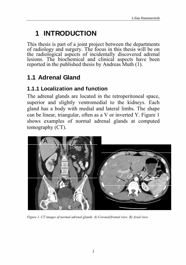
1.1.1 Localization and function The adrenal glands are located in the retroperitoneal space, superior and slightly ventromedial to the kidneys. Each gland has a body with medial and lateral limbs. The shape can be linear, triangular, often as a V or inverted Y. Figure 1 shows examples of normal adrenal glands at computed tomography (CT).
Figure 1. CT images of normal adrenal glands. A) Coronal/frontal view. B) Axial view.
BA
Incidentally detected adrenal lesions
2
The adrenal gland is composed of two separate embryological, functional and morphological units: the cortex and the medulla. Microscopically, the normal adrenal cortex shows three zones: the zona glomerulosa, which produces mineralocorticoids (principally aldosterone); the zona fascicularis and zona reticularis, which produce glucocorticoids (corticosteroids), androgens and estrogens. The adrenal medulla secretes adrenaline and noradrenaline. Most of the cortical tissue is located in the limbs, while the medullary tissue is located in the body of the gland (2). Normal cortical cells contain lipid, and cells with abundant lipid appear “clear” on routine histology, and those with less lipid appear as “compact” cells.
The size of the adrenal glands at axial CT was assessed by Vincent et al in 1994 (3). The mean body width (measured at the junction of the adrenal limbs and the body, perpendicular to the long axis of the body) was 0.61 (range 0.3-1.2) cm in the right and 0.79 (range 0.4-1.2) cm in the left adrenal. The authors also measured normal limb width in each adrenal gland showing a maximum width on both sides of 0.5 cm in the axial plane.
1.1.2 Lesions in the adrenals Due to the increased use and improved technical quality of radiological imaging, incidentally detected adrenal lesions have become an important clinical issue. In the literature the term “adrenal incidentaloma” has often been used for this kind of lesions. For further discussion regarding this term see thesis by A. Muth (1).
In autopsy studies the smallest adrenal lesion classified as adenoma was 2-3 mm in diameter (4-6) and size limits are somewhat difficult to apply since the normal adrenal gland can provide a large variety in shape. Autopsy studies have also shown that the prevalence of adrenal mass lesions varies
Lilian Hammarstedt
3
depending on the age of the population – adrenal lesions are more common in the elderly. In patients over 60 years, adrenal lesions have been found in 8-12 % (4-6).
1.1.3 Biochemical characterization of adrenal lesions
A thorough evaluation of the patient history and a physical examination combined with biochemical screening is important after the detection of adrenal lesions. The rationale behind this is to identify hypersecretory lesions that often require surgical treatment. The most common types of hormone-producing lesions are adrenocortical cortisol-producing tumours (Cushing’s syndrome or subclinical hypercortisolism), adrenocortical aldosterone producing tumours (primary aldosteronism), and pheochromocytomas (catecholamine-secreting tumours originating from the adrenal medulla). The frequency of hypersecreting lesions among incidentally detected adrenal lesions has varied from 5 to 15 % in clinical studies (7-9). The details regarding biochemical and clinical tests are not further evaluated in this thesis but can be studied in the thesis by A. Muth (1).
1.1.4 Benign adrenal lesions The most common lesion in the adrenal is a benign adenoma without evidence of hormone production. The precise frequency of the different adrenal lesions is unclear, as results in reported series vary.
Hematoma and cysts are usually non-neoplastic but may be related to an underlying tumour; e.g. adrenocortical carcinoma, pheochromocytoma or adrenal metastases. The majority of pheochromocytomas are benign but may give serious hormonal symptoms. Other benign lesions include myelolipomas, hemangiomas, lipomas and granulomatous lesions (10, 11).
Incidentally detected adrenal lesions
4
1.1.5 Malignant adrenal lesions Adrenocortical carcinoma and malignant pheochromo-cytoma are the most common primary adrenal malignancies but the most common malignancy in the adrenals is a metastasis from an extra-adrenal primary tumour. Abrams et al (12) found that adrenals were the seventh most common metastasis site, approximately 27% of post-mortem examinations in patients with a malignant neoplasm of epithelial origin showed adrenal metastases. Rich sinusoidal blood supply may be one of the explanations (13). The etiology of metastatic tumours of the adrenal gland is most often lung, stomach/oesophagus, colorectal, breast and pancreatic carcinoma followed by lymphoma (12, 14, 15).
1.2 Adrenal characterization in radiology Adrenal lesions can be detected by different radiology techniques including ultrasound, computed tomography (CT) and magnetic resonance imaging (MRI). Further evaluation of the detected lesion can be performed by dedicated CT, MRI, positron emission tomography (PET) or other scintigraphic methods and by fine needle aspiration or biopsy. The most commonly used method for the evaluation/characterization of adrenal lesions is CT because of its widespread availability, capability, high speed and low cost. The other methods are often used as a complement and will be described in more detail below.
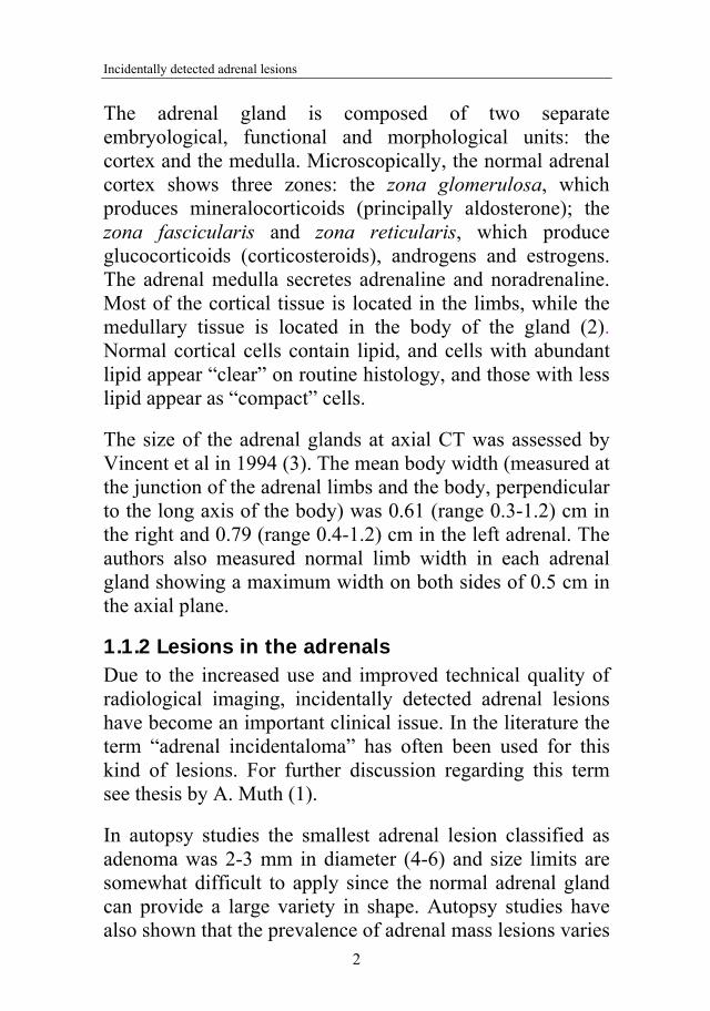
The important radiological question is whether the detected adrenal lesion is benign or malignant, which is the focus of this thesis. However, differentiation between various benign lesions can also be done, using e.g. CT and MRI. Examples of different adrenal lesions found in our studies (Paper I-III) at CT and MRI are shown in Figure 2-4.
Lilian Hammarstedt
5
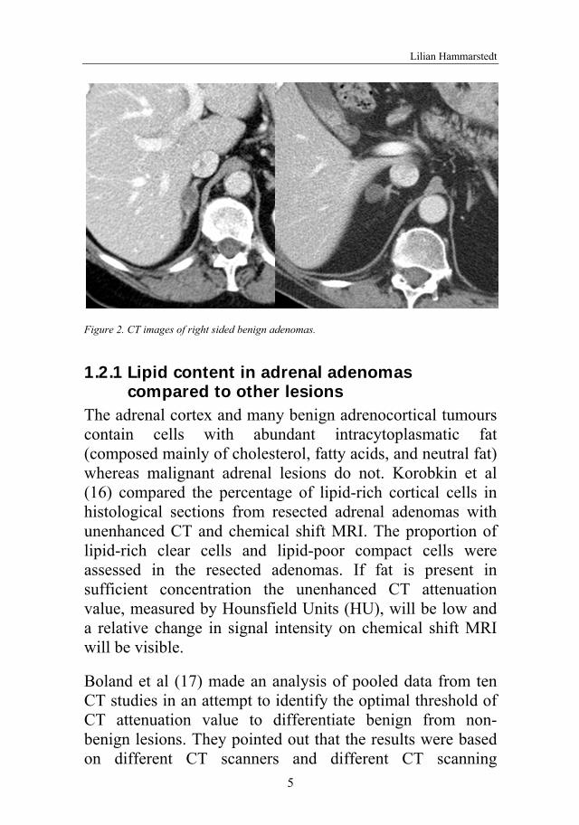
Figure 2. CT images of right sided benign adenomas.
1.2.1 Lipid content in adrenal adenomas compared to other lesions
The adrenal cortex and many benign adrenocortical tumours contain cells with abundant intracytoplasmatic fat (composed mainly of cholesterol, fatty acids, and neutral fat) whereas malignant adrenal lesions do not. Korobkin et al (16) compared the percentage of lipid-rich cortical cells in histological sections from resected adrenal adenomas with unenhanced CT and chemical shift MRI. The proportion of lipid-rich clear cells and lipid-poor compact cells were assessed in the resected adenomas. If fat is present in sufficient concentration the unenhanced CT attenuation value, measured by Hounsfield Units (HU), will be low and a relative change in signal intensity on chemical shift MRI will be visible.
Boland et al (17) made an analysis of pooled data from ten CT studies in an attempt to identify the optimal threshold of CT attenuation value to differentiate benign from non-benign lesions. They pointed out that the results were based on different CT scanners and different CT scanning
B
Incidentally detected adrenal lesions
6
protocols. The thresholds chosen depend on e.g. the patient population and the cost-benefit approach to patient care. With a threshold of 10 HU the sensitivity and specificity in this study was 71 % and 98%, respectively. After the publication of this study, the threshold of 10 HU has been used in clinical practice worldwide. The authors also pointed out that to be sure that a lesion is benign (100% specificity), one should use a threshold value of 2 HU on unenhanced CT. However, this is at the cost of a very low sensitivity (47%).
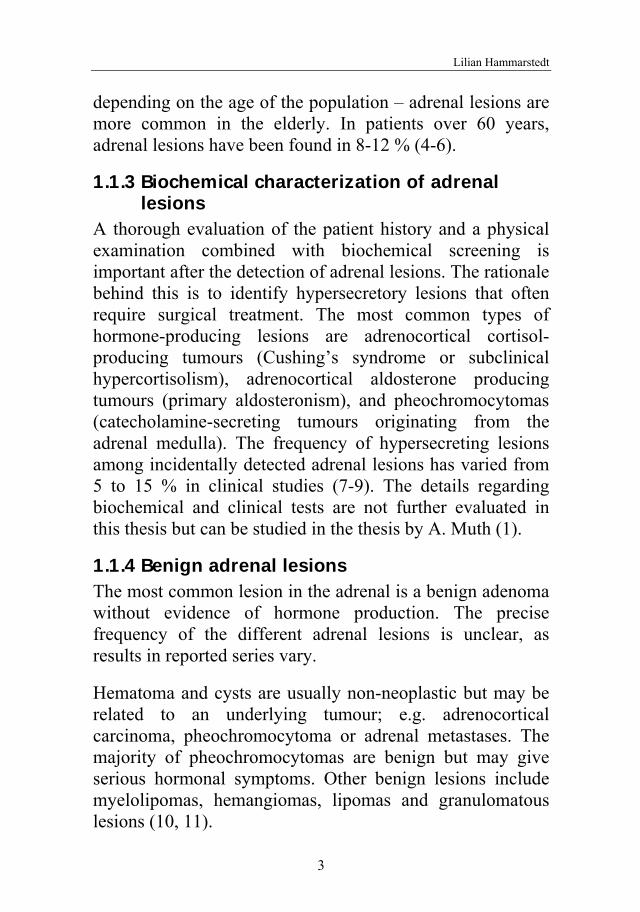
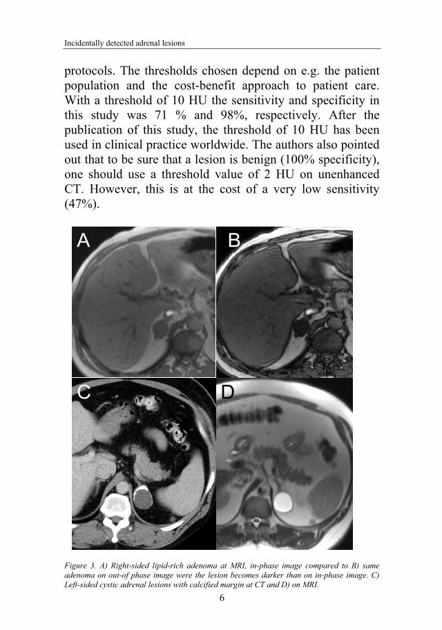
Figure 3. A) Right-sided lipid-rich adenoma at MRI, in-phase image compared to B) same adenoma on out-of phase image were the lesion becomes darker than on in-phase image. C) Left-sided cystic adrenal lesions with calcified margin at CT and D) on MRI.
A B
C D
Lilian Hammarstedt
7
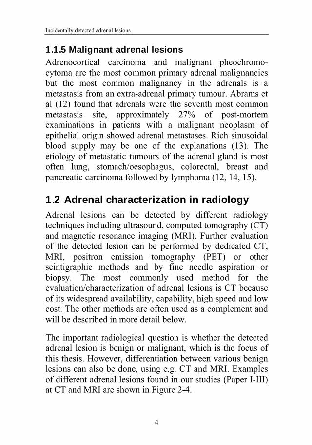
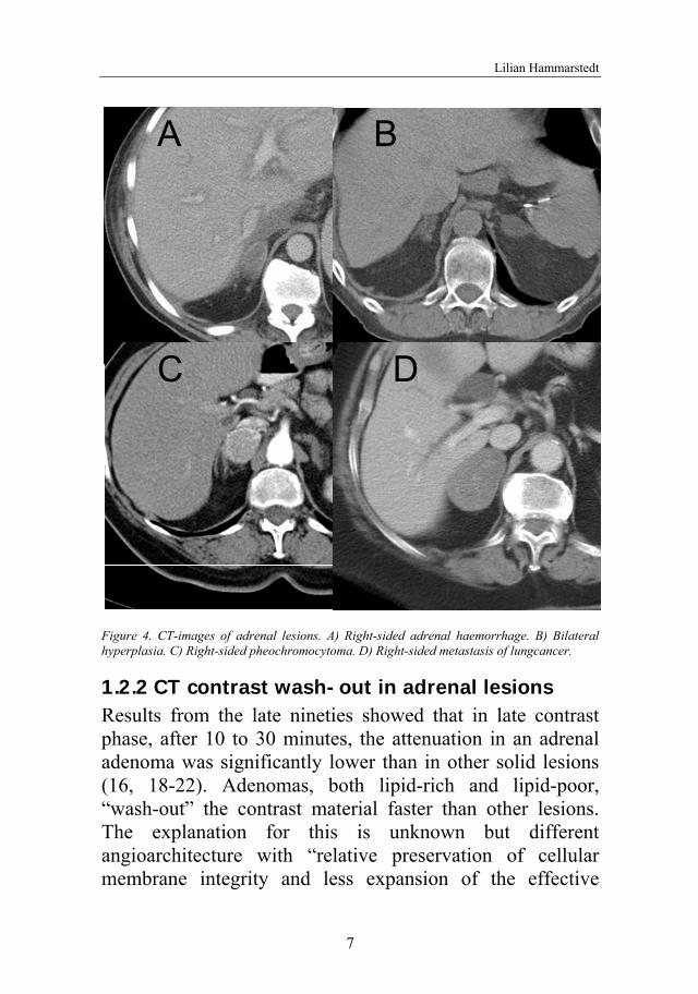
Figure 4. CT-images of adrenal lesions. A) Right-sided adrenal haemorrhage. B) Bilateral hyperplasia. C) Right-sided pheochromocytoma. D) Right-sided metastasis of lungcancer.
1.2.2 CT contrast wash-out in adrenal lesions Results from the late nineties showed that in late contrast phase, after 10 to 30 minutes, the attenuation in an adrenal adenoma was significantly lower than in other solid lesions (16, 18-22). Adenomas, both lipid-rich and lipid-poor, “wash-out” the contrast material faster than other lesions. The explanation for this is unknown but different angioarchitecture with “relative preservation of cellular membrane integrity and less expansion of the effective
A B
C D
Incidentally detected adrenal lesions
8
extracellular space due to tumour infiltration in adrenal adenomas” is a possibility (19).
Using attenuation values from unenhanced, enhanced and late enhanced contrast phases a calculation of absolute (APW) or relative (RPW) percentage wash-out can be done using these formulas:
100
unenhancedenhanced
enhanceddelayedenhanced
valuenAttenuatiovaluenAttenuatio
valuenAttenuatiovaluenAttenuatioAPW
100
enhanced
enhanceddelayedenhanced
valuenAttenuatio
valuenAttenuatiovaluenAttenuatioRPW
In some studies (18, 19), calculation of RPW appears favourable for the diagnosis of adenoma, as compared to APW. Thus, the sensitivity and specificity are slightly higher for RPW than APW, but the RPW still tends to overestimate the true wash-out for adenomas and underestimate the wash-out of nonadenomas (18). Korobkin et al (18) recommended the use of 15-min delay enhanced scan but others have suggested 10-min delay (19, 22, 23). Szolar et al (19) recommended a 30 minute delayed scan if the lesion had low absolute and relative percentage wash-out calculated from 10 min delayed images. Other studies also evaluated 30 min delay, demonstrating sensitivity and specificity as high as 97% and 100%, respectively, for finding lipid-rich adenomas (18, 24). In clinical practice, however, a shorter delay is preferred for logistical reasons. The 10 min delay has lately been questioned because of the reduced sensitivity in calculating the APW and RPW compared to 15-min delay (25).
Lilian Hammarstedt
9
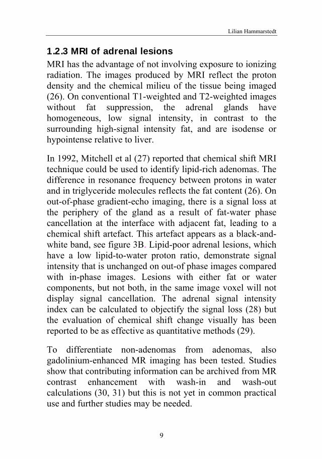
1.2.3 MRI of adrenal lesions MRI has the advantage of not involving exposure to ionizing radiation. The images produced by MRI reflect the proton density and the chemical milieu of the tissue being imaged (26). On conventional T1-weighted and T2-weighted images without fat suppression, the adrenal glands have homogeneous, low signal intensity, in contrast to the surrounding high-signal intensity fat, and are isodense or hypointense relative to liver.
In 1992, Mitchell et al (27) reported that chemical shift MRI technique could be used to identify lipid-rich adenomas. The difference in resonance frequency between protons in water and in triglyceride molecules reflects the fat content (26). On out-of-phase gradient-echo imaging, there is a signal loss at the periphery of the gland as a result of fat-water phase cancellation at the interface with adjacent fat, leading to a chemical shift artefact. This artefact appears as a black-and-white band, see figure 3B. Lipid-poor adrenal lesions, which have a low lipid-to-water proton ratio, demonstrate signal intensity that is unchanged on out-of phase images compared with in-phase images. Lesions with either fat or water components, but not both, in the same image voxel will not display signal cancellation. The adrenal signal intensity index can be calculated to objectify the signal loss (28) but the evaluation of chemical shift change visually has been reported to be as effective as quantitative methods (29).
To differentiate non-adenomas from adenomas, also gadolinium-enhanced MR imaging has been tested. Studies show that contributing information can be archived from MR contrast enhancement with wash-in and wash-out calculations (30, 31) but this is not yet in common practical use and further studies may be needed.
Incidentally detected adrenal lesions
10
Recent focus has been on diffusion-weighted MR imaging (DWI). DWI has been shown to be helpful in the characterization of tumours on the basis of diffusion effects using apparent diffusion coefficient (ADC) measurements, used to assess the mobility of water molecules (32). In theory, malignant tumours have lower ADC values compared with benign lesions, but in a recent study using ADC values, Miller et al (33) were unable to distinguish benign from malignant adrenal lesion.
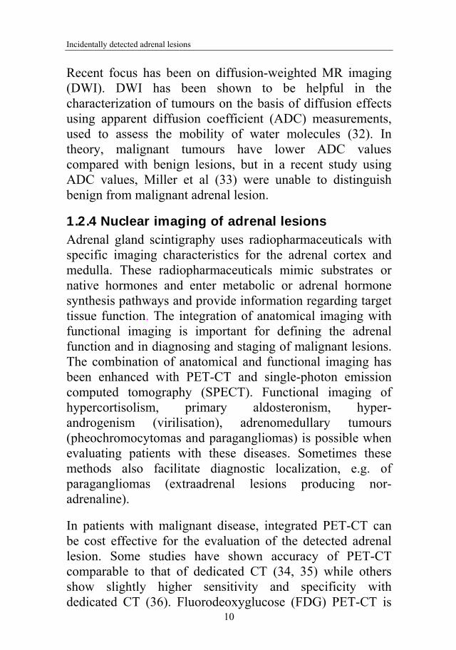
1.2.4 Nuclear imaging of adrenal lesions Adrenal gland scintigraphy uses radiopharmaceuticals with specific imaging characteristics for the adrenal cortex and medulla. These radiopharmaceuticals mimic substrates or native hormones and enter metabolic or adrenal hormone synthesis pathways and provide information regarding target tissue function. The integration of anatomical imaging with functional imaging is important for defining the adrenal function and in diagnosing and staging of malignant lesions. The combination of anatomical and functional imaging has been enhanced with PET-CT and single-photon emission computed tomography (SPECT). Functional imaging of hypercortisolism, primary aldosteronism, hyper-androgenism (virilisation), adrenomedullary tumours (pheochromocytomas and paragangliomas) is possible when evaluating patients with these diseases. Sometimes these methods also facilitate diagnostic localization, e.g. of paragangliomas (extraadrenal lesions producing nor-adrenaline).
In patients with malignant disease, integrated PET-CT can be cost effective for the evaluation of the detected adrenal lesion. Some studies have shown accuracy of PET-CT comparable to that of dedicated CT (34, 35) while others show slightly higher sensitivity and specificity with dedicated CT (36). Fluorodeoxyglucose (FDG) PET-CT is
Lilian Hammarstedt
11
interpreted as positive for malignancy if the FDG uptake in an adrenal lesion is greater than or equal to that of the liver. However, FDG uptake is not tumour specific. It can also be seen in conditions with inflammation and posttraumatic repair, but with the combination of FDG PET and CT most of the false-positive findings can be identified. Still, false-positive cases of adrenal lesions have been reported in 5-6 % at (FDG) PET-CT (35). False-negative cases have also been reported due to hemorrhage, necrosis and lesion size (≤ 10 mm) (34, 37, 38) and metastatic lesions from pulmonary adenocarcinoma can show little FDG uptake (38).
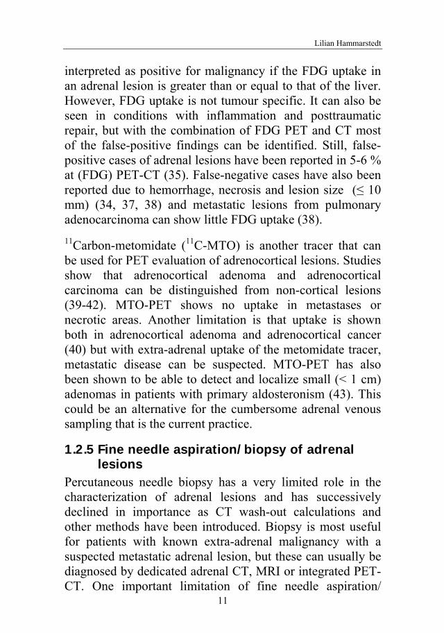
11Carbon-metomidate (11C-MTO) is another tracer that can be used for PET evaluation of adrenocortical lesions. Studies show that adrenocortical adenoma and adrenocortical carcinoma can be distinguished from non-cortical lesions (39-42). MTO-PET shows no uptake in metastases or necrotic areas. Another limitation is that uptake is shown both in adrenocortical adenoma and adrenocortical cancer (40) but with extra-adrenal uptake of the metomidate tracer, metastatic disease can be suspected. MTO-PET has also been shown to be able to detect and localize small (< 1 cm) adenomas in patients with primary aldosteronism (43). This could be an alternative for the cumbersome adrenal venous sampling that is the current practice.
1.2.5 Fine needle aspiration/biopsy of adrenal lesions
Percutaneous needle biopsy has a very limited role in the characterization of adrenal lesions and has successively declined in importance as CT wash-out calculations and other methods have been introduced. Biopsy is most useful for patients with known extra-adrenal malignancy with a suspected metastatic adrenal lesion, but these can usually be diagnosed by dedicated adrenal CT, MRI or integrated PET-CT. One important limitation of fine needle aspiration/
Incidentally detected adrenal lesions
12
biopsy is that it cannot reliably differentiate benign cortical tissue from malignant cortical tissue (44, 45). Fine needle aspiration/biopsy should only be performed if clinical and imaging examinations are inconclusive and if the result of the aspiration/biopsy can be expected to change patient care.
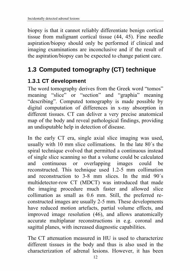
1.3 Computed tomography (CT) technique
1.3.1 CT development The word tomography derives from the Greek word “tomos” meaning “slice” or “section” and “graphia” meaning “describing”. Computed tomography is made possible by digital computation of differences in x-ray absorption in different tissues. CT can deliver a very precise anatomical map of the body and reveal pathological findings, providing an undisputable help in detection of disease.
In the early CT era, single axial slice imaging was used, usually with 10 mm slice collimations. In the late 80´s the spiral technique evolved that permitted a continuous instead of single slice scanning so that a volume could be calculated and continuous or overlapping images could be reconstructed. This technique used 1.2-5 mm collimation and reconstruction to 3-8 mm slices. In the mid 90´s multidetector-row CT (MDCT) was introduced that made the imaging procedure much faster and allowed slice collimation as small as 0.6 mm. Still, the preferred re-constructed images are usually 2-5 mm. These developments have reduced motion artefacts, partial volume effects, and improved image resolution (46), and allows anatomically accurate multiplanar reconstructions in e.g. coronal and sagittal planes, with increased diagnostic capabilities.
The CT attenuation measured in HU is used to characterize different tissues in the body and thus is also used in the characterization of adrenal lesions. However, it has been
Lilian Hammarstedt
13
shown that caution is necessary when using absolute HU values as threshold for diagnosis. Several studies indicate size and attenuation variations due to CT equipment, scanning protocol, examination technique, patient size and other factors (47-49).
Incidentally detected adrenal lesions
14
2 AIM
The general aim of this thesis was to study incidentally detected adrenal lesions, identified at clinically indicated radiological examinations in a defined geographical region.
More specific aims were:
To investigate the frequency of adrenal lesions incidentally detected at radiological examinations during an 18 month observation period in a defined geographical region (Paper I).
To further investigate radiological and biochemical characteristics of the detected adrenal lesions by a 2-year follow-up program in patients with and without extra-adrenal malignant disease (Paper II-III).
To investigate the variability of adrenal lesion characterization over time and factors that may contribute to this variability at computed tomography (Paper IV).
Lilian Hammarstedt
15
3 METHODOLOGICAL CONSIDERATIONS
Detailed description of materials and methods are given in each paper (I-IV), and only those of particular importance are described below.
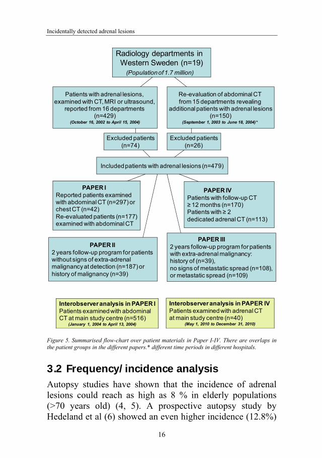
A flow-chart describing the adrenal study population in all papers (I-IV) is shown in Figure 5.
3.1 Study region The studies were performed in the Västra Götaland region including the Northern part of Halland (the region of Western Sweden). Nineteen radiological departments were included (10 local hospitals, 6 county hospitals and one university hospital (main study centre)). Seventeen departments had CT-equipment, 10 had MRI-equipment and all departments had ultrasound service.
Incidentally detected adrenal lesions
16
Figure 5. Summarised flow-chart over patient materials in Paper I-IV. There are overlaps in the patient groups in the different papers.* different time periods in different hospitals.
3.2 Frequency/incidence analysis Autopsy studies have shown that the incidence of adrenal lesions could reach as high as 8 % in elderly populations (>70 years old) (4, 5). A prospective autopsy study by Hedeland et al (6) showed an even higher incidence (12.8%)
Radiology departments in Western Sweden (n=19)
(Population of 1.7 million)
Re-evaluation of abdominal CT from 15 departments revealing
additional patients with adrenal lesions(n=150)
(September 1, 2003 to June 18, 2004)*
Excluded patients(n=74)
Excluded patients(n=26)
Patients with adrenal lesions, examined with CT, MRI or ultrasound,
reported from 16 departments (n=429)
(October 16, 2002 to April 15, 2004)
Included patients with adrenal lesions (n=479)
PAPER IReported patients examinedwith abdominal CT (n=297) or chest CT (n=42) Re-evaluated patients (n=177)examined with abdominal CT
PAPER II2 years follow-up program for patients without signs of extra-adrenal malignancy at detection (n=187) or history of malignancy (n=39)
PAPER III2 years follow-up program for patientswith extra-adrenal malignancy: history of (n=39), no signs of metastatic spread (n=108), or metastatic spread (n=109)
PAPER IVPatients with follow-up CT ≥ 12 months (n=170)Patients with ≥ 2 dedicated adrenal CT (n=113)
Interobserver analysis in PAPER IPatients examined with abdominal CT at main study centre (n=516)
(January 1, 2004 to April 13, 2004)
Interobserver analysis in PAPER IVPatients examined with adrenal CTat main study centre (n=40)
(May 1, 2010 to December 31, 2010)
Lilian Hammarstedt
17
in people aged 60 to 69 years. Clinical studies from Italy and Sweden (8, 9, 50) demonstrated high frequency of serious adrenal findings, 22-38% of incidentally detected adrenal lesions were operated and 5-15% were hormonally active. In the region of Western Sweden the population is 1.7 million inhabitants and approximately 280 000 people are over 65 years old. If calculating with an incidence of 7 % there would be a least 19 600 people with adrenal lesions in our elderly population and, based on the above figures from the literature, 4300 (22%) of them would potentially need an operation. If one instead calculates with the whole adult population of Western Sweden (1.3 million) using the lower overall incidence figures (1.45%) reported by Russi et al (51), about 18850 people would have an adrenal lesion and 4147 of them would be candidates for surgery. In our region approximately 22 patients are operated with adrenalectomi per year, (mean figure from 2002-2005) due to adrenal disease/tumour. The figures do not match. The National Institutes of Health (NIH) consensus on management of the clinically inapparent adrenal mass from 2002 (52) called for prospective studies regarding adrenal lesion prevalence and natural history. To try to determine the frequency of incidentally detected adrenal lesions in our defined geographical region we chose to make a prospective study, reporting cases from the source of diagnosis, i.e. the radiological departments. In order to validate the frequency of reported cases, also a re-evaluation was performed on random samples of cases from departments performing abdominal CT examinations (15 of 16 departments).
It is difficult to study the true frequency of a disease in a population. With this study design we hoped to minimize possible patient selection bias. We chose to study CT examinations as it is the most prevalent abdominal examination demonstrating the adrenals, and as it allowed
Incidentally detected adrenal lesions
18
reliable collection of statistical figures regarding the total number of CT examinations performed in the region.
3.3 Radiological examinations Radiological detection examinations comprised routinely performed CT, MRI and ultrasound examination, designed for the specific clinical situation. Follow-up examinations were, on the other hand, performed according to a predesigned dedicated adrenal CT and/or MRI-protocol. All images were digital and all cases were sent to the main study centre for validation and were stored in a common digital archive. All lesion measurements and calculations could therefore be performed in the same way in papers I-IV using one digital workstation (Centricity RA600; GE Healthcare, Chalfont St. Giles, UK).
3.3.1 Dedicated adrenal imaging At the time of planning our study several studies had shown comforting sensitivity and specificity using 10 minute delayed wash-out imaging (19, 22, 24). It also seemed easier to implement the study protocol in our region with this shorter delay and therefore all dedicated follow-up adrenal imaging with CT was performed with 10 instead of 15 minutes delay. Recent studies have subsequently shown that a longer delay (15 minutes) is preferable, with higher sensitivity and specificity (25). In questionable cases one should consider even longer delay (30 minutes), as suggested by Szolar et al (19). The dedicated imaging protocol used in the study is described in detail in paper II.
In paper IV the interobserver analysis was based on dedicated adrenal imaging with 15 minutes delay wash-out since the adrenal imaging protocol in the main study centre was modified after the completion of the follow-up study (paper II and III) in 2007.
Lilian Hammarstedt
19
3.4 Interobserver analysis (Paper I and IV) In paper I, the re-evaluation frequency figures were validated by an interobserver analysis with two observers. To understand some of the underlying factors contributing to variability in adrenal measurements, a larger interobserver analysis including 5 observers with varying experience was conducted (Paper IV). A large part of the latter statistical analysis was performed by professor Anders Odén, Department of Mathematical Sciences.
3.5 Phantom analysis (Paper IV) A limitation in the adrenal lesion study was the variety of CT-scanners used in the study region and also that patients sometimes were examined at a different hospital during follow-up. Thus, in paper IV the variability of repeated measurement within a scanner, and between different scanners were analysed in a phantom study.
3.6 Ethics The adrenal lesion study was approved by the local Regional Ethical Review Board in Gothenburg (s202-02). Participating patients received oral and written information about the study and gave their consent to participate.
Incidentally detected adrenal lesions
20
4 RESULTS AND DISCUSSION
4.1 Adrenal lesion frequency Paper I firstly describes the frequency of adrenal lesions incidentally detected at CT and subsequently reported to the study centre directly from the local radiologists, and secondly the frequency of adrenal lesions identified in a re-evaluation of a large sample (nearly 4000 abdominal CT examinations) from the same population.
All radiology departments with CT equipment (n=17) in the Västra Götaland region and the northern part of the county of Halland participated in this study and results were reported from 16 of 17 departments. The frequency of incidentally detected adrenal lesions in patients without clinically evident malignant disease reported from the radiology departments was 0.5%. This is comparable to other CT studies reporting frequencies from 0.01 to 0.42 % in similar groups of patients (7, 53, 54). Our frequency figures are based on reported cases divided by the number of examinations done in the region during the study period (n=34 044). The number of examinations is not identical to the number of patients examined during this period because some patients were examined more than once. As patients with adrenal lesions were only included once in the statistics we can assume that our frequency figures somewhat underestimate the true frequency.
The overall reported frequency (including patients with a history of malignancy or present malignant disease) in our study varied between the radiology departments, ranging from 0-2.4 % (mean 0.9 %). No single explanation for this variation could be found. In some departments a low reporting frequency could possibly be explained by the use of large CT slice thickness (10 mm), which may have
Lilian Hammarstedt
21
hampered detection of small adrenal lesions, but other departments used the same slice thickness and reported more lesions. Some small radiology units had low reporting frequency but others had not. The combination of thin slice technique at CT and small size of the department (all reports by the same dedicated radiologist) gained the highest reporting frequency in this study. The re-evaluation performed by one independent radiologist (LH), showed considerably higher frequency figures that varied from 1.8 % to 7.1% (mean 4.6%) between departments. For some units the frequency was rather low even in the re-evaluation, despite the use of thin CT slice technique. The involved radiology departments represented different levels of patient care, and it seems plausible that differences in patient populations regarding age, sex, indications for CT examinations and disease panorama were responsible for parts of the variation in frequency.
In the national Swedish multicentre study by Bülow et al (9) 381 patients with adrenal lesions were reported during 5½ years inclusion time (January 1996 to July 2001). This corresponds to approximately 69 new cases/year in a population of 8.5 million. In our study from a smaller part of Sweden (population of 1.7 million), 151 cases without clinically evident malignancy were reported during 1½ year, corresponding to 101 new cases/year. Our re-evaluation gave fivefold higher figures showing that by focusing on a limited anatomical area at CT (just looking for adrenal lesions), in a non-stressful situation frequency figures will be higher than in a busy clinical situation. Our findings illustrate a potential weakness of multicenter studies that rely on diagnosing and reporting lesions from a large number of participating departments and individuals, in a clinical setting. Participation in a multicentre study is a demanding and time consuming task both for the study centre and the participating units. This must be considered, not only in the initial phase, when exchange and assimilation of study concept, protocols and other information occurs, but also later when continuous reporting of study cases needs to be incorporated into the clinical situation. It is therefore not
Incidentally detected adrenal lesions
22
surprising that frequency figures tend to get higher when a single, dedicated reader performs the analysis. In order to optimize compliance, it is essential to appoint local contact persons at each site in multicentre settings. Our findings also emphasize the importance of validation of data from multicentre studies.
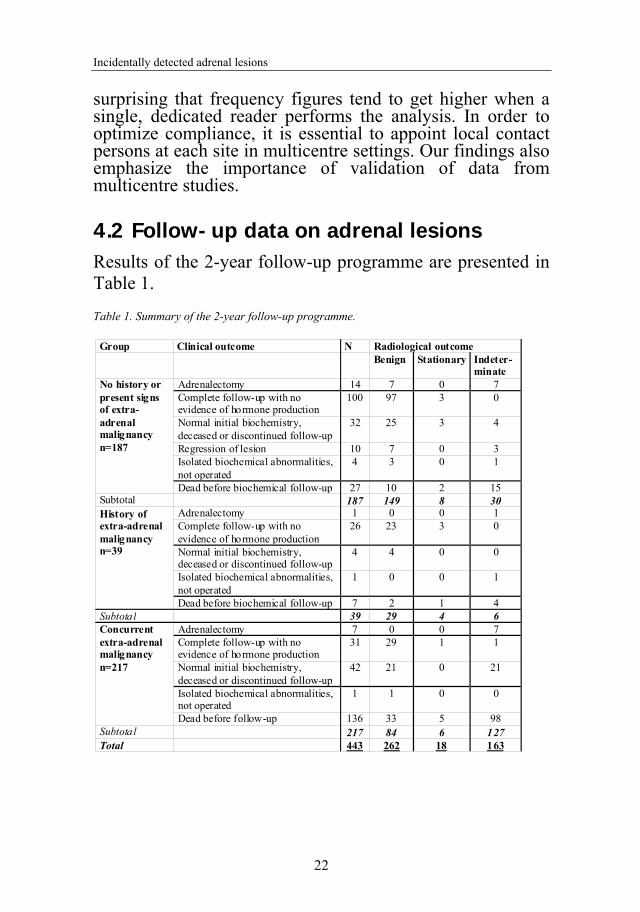
4.2 Follow-up data on adrenal lesions Results of the 2-year follow-up programme are presented in Table 1.
Table 1. Summary of the 2-year follow-up programme.
Group Clinical outcome N Radiological outcome Benign Stationary Indeter-
minate No history or present signs of extra-adrenal malignancy n=187
Adrenalectomy 14 7 0 7 Complete follow-up with no evidence of hormone production
100 97 3 0
Normal initial biochemistry, deceased or discontinued follow-up
32 25 3 4
Regression of lesion 10 7 0 3 Isolated biochemical abnormalities, not operated
4 3 0 1
Dead before biochemical follow-up 27 10 2 15 Subtotal 187 149 8 30 History of extra-adrenal malignancy n=39
Adrenalectomy 1 0 0 1 Complete follow-up with no evidence of hormone production
26 23 3 0
Normal initial biochemistry, deceased or discontinued follow-up
4 4 0 0
Isolated biochemical abnormalities, not operated
1 0 0 1
Dead before biochemical follow-up 7 2 1 4 Subtotal 39 29 4 6 Concurrent extra-adrenal malignancy n=217
Adrenalectomy 7 0 0 7 Complete follow-up with no evidence of hormone production
31 29 1 1
Normal initial biochemistry, deceased or discontinued follow-up
42 21 0 21
Isolated biochemical abnormalities, not operated
1 1 0 0
Dead before follow-up 136 33 5 98 Subtotal 217 84 6 127 Total 443 262 18 163
Lilian Hammarstedt
23
4.2.1 Clinical outcome in paper II and III The NIH-consensus from 2000 (52) recommended a follow-up scheme for incidentally detected adrenal lesions. The major concern was the reported high rate of hormonal hyper-functioning and/or malignant lesions. A review of studies published from 1980 to 2008 by Cawood et al (55) showed that if only studies with “true adrenal incidentalomas” (with-out extra-adrenal malignancy, without adrenal complaints and often with a size limit of > 1 cm) were included, the prevalence figures of malignant and hyper-functional adrenal lesions were considerably lower. Our follow-up study reported in Paper II confirms this. In our study 187 patients without extra-adrenal malignancy and without symptoms related to the adrenals were included for follow-up. One of them had a lesion < 1 cm (8 mm) which leaves 186 patients in our study with “true adrenal incidentaloma” as defined by Cawood. A total of 7.5 % (14/186 patients) were operated and no primary adrenal malignancy was found. Seven of the 186 patients (3.8 %) had signs of hormone over-production. Follow-up revealed no additional hormonally hyper-functioning and/or malignant cases. This is comparable with studies by for example Reincke et al (56) and Tsvetov et al (57) that found no primary adrenal carcinomas but hormonal over-production in 14.7% and 11.9% cases, respectively, in patients with “true adrenal incidentaloma”. Other clinical studies have shown a rate of 1.2 to 4.7 % adrenal carcinomas and hyper-functioning lesions in 4.1 to 14.9 % of the cases (7, 8, 58, 59). A special aspect is the role of subclinical hypercortisolism in patients with adrenal lesions. A limitation of our study is that detailed work-up for revealing such cases was not included and the rate of subclinical hypercortisolism in our study may be underestimated. However, the issue is complicated by the lack of consensus regarding the definition and diagnostic criteria of this condition (1).
Incidentally detected adrenal lesions
24
In the group of 39 patients with a history of previous extra-adrenal malignancy one patient underwent adrenalectomy (2.6%), revealing a metastasis from a previously resected renal cell carcinoma. One patient had elevated chromogranin A possibly indicative of a pheochromocytoma but declined further examinations and no other cases of hormone over-production were revealed.
Patients with signs of ongoing extra-adrenal malignancy are presented in paper III. Six of 217 patients were operated (2.8%) and no primary adrenal malignant or hyper-functional hormonal disease was found among these. Seventy-six patients were tested biochemically and only one patient had signs of subtle cortisol overproduction but was not further evaluated due to advanced tumour disease.
Results from patients with history of or ongoing malignancy show that biochemical evaluation reveals very few cases with treatable adrenal disease and no patients had signs of primary adrenal malignancy. Radiological staging remains important in patients with apparently isolated adrenal metastases to identify patients with potentially resectable disease, as surgery may be associated with prolonged survival (60).
As all cases of hormonal over-production were identified already at first biochemical evaluation, our study results do not support hormonal follow-up unless future studies prove the usefulness of follow-up of subclinical hypercortisolism.
Lilian Hammarstedt
25
4.2.2 Radiological outcome/characterization in paper II and III
As pointed out in the introduction, characterization of adrenal lesions has evolved dramatically from the time when the radiologist almost only described the size of the lesion. First, the ability of unenhanced CT attenuation measurements and MRI chemical shift imaging to show high lipid content of adrenal lesions, allowed differentiation between benign adenoma and non-adenoma. Secondly, the use of contrast media wash-out characteristics at CT improves the distinction of benign from non-benign lesions.
In our adrenal lesion study, lesions were divided into three subgroups using radiological characteristics; benign, stationary indeterminate or indeterminate, se Paper II and III. The follow-up study showed benign lesions in 79.7% (149/187) of patients without extra-adrenal malignancy and in 74.4% (29/39) of patients with a history of malignancy. There was incomplete radiological follow-up in 15.5% (29/187) and 15.4% (6/39) of cases, respectively. Among patients with concurrent extra-adrenal malignancy 38.7 % (84/217) had benign lesions and data was incomplete in 50.7 % (110/217). In other studies 70-94% benign adrenal lesions have been demonstrated in non-oncologic populations (10, 61, 62) and benign lesions occur frequently also in populations with malignancy. In a study by Oliver at al (63) 32% metastatic adrenal lesions were found in patients with non small-cell bronchogenic carcinoma and in a study by Lenert et al (64) 42 % of patients with different kinds of extra-adrenal cancer had metastatic adrenal lesions. Our figures are comparable with these.
A limitation in our studies (Paper II and III) was the use of 10 minutes (instead of 15 minutes) wash-out series in the dedicated adrenal study protocol. The use of this shorter delay and the incompleteness of dedicated radiological
Incidentally detected adrenal lesions
26
follow-up examinations have probably increased the proportion of cases with indeterminate lesions. Dedicated adrenal examination, including thin sections, non-enhanced and 15 minutes post contrast series, with attenuation measurements and wash-out calculation is highly recommended in all patients with adrenal lesions.
4.3 Variation of adrenal characterization In our study of patients with adrenal lesions there were signs of variations in the characterization of the lesions during follow-up. In interobserver studies both high and low concordance between observers has been demonstrated regarding size and attenuation measurements (65-70). Our interobserver analysis showed an overall good agreement between the five observers regarding size measurements, there was a 50 % chance that the observers would differ less than 2.5 mm. When measuring attenuation, maximum difference between observers ranged from 0 to 20 HU (mean 4.6 HU) on unenhanced series. Nevertheless, with characterization based on established threshold values (benign lesion if HU ≤ 10 and/or relative (15 minutes) wash-out > 40 %) the interobserver analysis showed agreement in the classification (benign or indeterminate) in 37 of the 40 lesions (92%).
Other studies have shown that absolute attenuation values measured in Hounsfield units (HU) can vary between different scanners and scanner protocol (47-49, 71, 72). Our results from the phantom study showed a mean intrascanner difference of 2.7 (range 1.8 to 5.1) HU for tap water. Intrascanner attenuation differences for acrylic and polypropylene test tubes showed similar range, from 1.0 to 5.7 HU and 1.0 to 3.7 HU, respectively. The interscanner differences between mean values of tap water were both higher and lower than the intrascanner differences with a
Lilian Hammarstedt
27
range of 0.3 to 8 HU. The data obtained from the interobserver and phantom analyses show that some of the variance of adrenal attenuation can be caused by the scanner and/or observer.
To reduce adrenal attenuation variability when radiological follow-up is needed we recommend the use of the same CT-scanner, identical CT protocol and the same observer. The use of a calibration instrument each time attenuation values are measured on CT is likely to further improve the reproducibility.
When applying established diagnostic criteria for benign versus indeterminate lesions, 50 of 113 patients (56/136 lesions) examined with dedicated adrenal CT could be reclassified, as benign or indeterminate, during follow-up. This means that almost 50% of our studied lesions could have changed classification if the attenuation value would have been the only classification tool. We have to bear in mind that all these lesions showed size stability for at least 6 months and this alone is used in several studies as proof of benign etiology. Absolute CT numbers are only a part of the evaluation of the adrenals and this emphasizes the importance of utilizing all available information in the images. In a study by Gufler et al (73) four radiological features (contour, internal structure, size and attenuation value) of adrenal lesions at unenhanced CT images were used to create a total score. Applying this scoring system with combined parameters yielded a sensitivity of 100% and specificity of 96.8% for differentiation between adenomas and metastases. The validity of such a scoring system for adrenal lesions, with information from both unenhanced and enhanced images, remains to be evaluated. Further studies of routine methods are needed for improved identification of indeterminate lesions in need of further investigations.
Incidentally detected adrenal lesions
28
5 CONCLUDING REMARKS
Adrenal lesions are common findings in patients subjected to radiological examinations. Reported frequencies of adrenal lesions incidentally detected by CT in our study showed figures comparable to other CT-studies, but a re-evaluation of a random sample from the same study population showed considerably higher figures, closer to those from autopsy studies. The re-evaluation demonstrates that underreporting of cases in multicentre settings and probably also in routine clinical settings may be common.
We found that at detection only a small percentage of our adrenal study patients had hypersecretory lesions and no one had a primary adrenal malignancy. Of the patients without any history of malignancy 80% (149/187) had radiologically benign lesions. In the groups of patients with history of or present malignancy and an adrenal lesion, radiologically benign lesions were common. No additional cases of hyper-functioning and/or malignant lesions were found at follow-up. Radiological follow-up of adrenal lesions still has a place for indeterminate lesions but our study does not support the need for follow-up of lesions that can be radiologically characterized early in the process. Guidelines for management of incidentally detected adrenal lesions can probably be simplified both biochemically and radiologically.
We also found that there is variability in attenuation values over time in adrenal lesions, (otherwise stable ≥ 6 months), to some extent due to the usage of different CT scanners and observers. A recommendation is therefore, if follow-up of an adrenal lesions is needed, to mimic the previous examination using the same observer, CT protocol and CT equipment (preferably together with a calibration instrument).
Lilian Hammarstedt
29
6 FUTURE PERSPECTIVES
The combination of a lower threshold value (2 HU) at unenhanced CT and a 15-30 minute delayed imaging may improve the sensitivity and specificity for diagnosing adrenal lesions in patients with or without history of malignant disease. As refereed to earlier, Boland et al (17) showed that the unenhanced threshold value of 2 HU gave 100% specificity but lowered the sensitivity compared to 10 HU threshold. If one takes into account the aspect of variation of attenuation values in each lesion, noticed in our study, a lowered absolute threshold may be supported. When combining this unenhanced threshold value with measurements of late CT wash-out test (> 15 minutes) the sensitivity will rise. Further studies in this area are warranted.
Although we obtained assuring data on the benign course of most lesions over time, the long-term consequences of incidentally detected adrenal lesions is less well known. We are currently performing a long-term (>10 years) follow-up study of patients with adrenal lesions using the study population from the previous, national study, by Bulow et al (9, 74). This will provide unique, long-term radiological and biochemical information on the natural course of incidentally discovered adrenal lesions.
Incidentally detected adrenal lesions
30
ACKNOWLEDGEMENT
I wish to express my deep gratitude to all the people who have helped me through these years and without the great work of all radiology and clinical staff involved in Västra Götaland this thesis would not have been possible. In particular, I would like to thank:
Professor Mikael Hellström, my tutor, for introducing me to the field of research and for sharing great knowledge and enthusiasm. You also tried to introduce me into the intricate world of writing.
Associate Professor Bo Wängberg, my co-tutor, who together with my principal-tutor started this a long time ago. You have always in a very friendly way pushing me forward.
Andreas Muth, my fellow-PhD-student, now already a PhD, thank you for coming into this team and taking so large portion of workload. Our work together has been of great joy.
Lena Björneld, research nurse, for all supporting help in the study as well in other aspects of life.
All my fellow authors, Helga Á. Sigurjónsdóttir, Galina Götherström, Erik Almqvist, Håkan Widell, Sture Carlsson, Stefan Ander and the Adrenal Study Group of Western Sweden, for your advice and work with including and taking care of all study patients. Without you all this had never been done.
All radiology colleagues in the region for all great help with the study.
Associate professor Anne Thilander-Klang, you made my day when I first met you because you made me realise that paper IV was possible. You made the introduction to radiophysics to a great joy.
31
Professor Anders Odén, for introducing me to the advanced world of statistics.
My fellow colleagues in the section of Abdominal and Interventional radiology: Mats Andersson, Mats Asztély, Barbara Bergman, Paul Businge, Per Carlsson, Mårten Falkenberg, Kjell Geterud, Farida Hashimi, Olof Henriksson, Szeréna Horvàth, Henrik Leonhardt, Gerasimos Maroulis, Augustinas Sakinis, Joanna Sternal, Fredrik Thorén, Antonios Tzortzakakis and Karin Zachrisson. I am in deep debt and I appreciate all kinds of help that I have granted from you all.
John Brandberg, my boss, for all help during the study as well as giving me a lot of time making it possible to finish this work.
Ilja Laesser and Charlotte Sandström, my room-mates and friends, for sharing the ups and downs in research, thank you for all creative support.
Pernilla Ekeroth, Eva Michaëlsson and Ewa Strandberg, my dear friends, for all your joyful support along the way.
Above all: My family, my husband Ivar, for all things I can think of; including love, wisdom and joy; and our children Henry and Tess, for being there and showing that life is fantastic.
This thesis was generously supported by the Västra Götaland Clinical Research and Development fund, The Swedish Cancer Society, The Swedish Research Council, The Gothenburg Medical Society, and Government grants under the LUA/ALF Agreement in The Västra Götaland Region.
Incidentally detected adrenal lesions
32
REFERENCES
1. Muth A. Incidentally discovered adrenal tumours, adrenal metastases, and pheochromocytomas [Elektronisk resurs] : clinical and epidemiological aspects. Gothenburg: Department of Surgery, Institute of Clinical Sciences, The Sahlgrenska Academy at the University of Gothenburg; 2011.
2. Dobbie JW, Symington T. The human adrenal gland with special reference to the vasculature. J Endocrinol. 1966 Apr;34(4):479-89.
3. Vincent JM, Morrison ID, Armstrong P, Reznek RH. The size of normal adrenal glands on computed tomography. Clin Radiol. 1994 Jul;49(7):453-5.
4. Commons RR, Callaway CP. Adenomas of the adrenal cortex. Arch Med Interna. 1948 Jan;81(1):37-41.
5. Devenyi I. Possibility of normokalaemic primary aldosteronism as reflected in the frequency of adrenal cortical adenomas. J Clin Pathol. 1967 Jan;20(1):49-51.
6. Hedeland H, Östberg G, Hökfelt B. On the prevalence of adrenocortical adenomas in an autopsy material in relation to hypertension and diabetes. Acta Med Scand. 1968;184:211-4.
7. Herrera MF, Grant CS, van Heerden JA, Sheedy PF, Ilstrup DM. Incidentally discovered adrenal tumors: an institutional perspective. Surgery. 1991 Dec;110(6):1014-21.
8. Mantero F, Terzolo M, Arnaldi G, Osella G, Masini AM, Ali A, et al. A survey on adrenal incidentaloma in Italy. Study Group on Adrenal Tumors of the Italian Society of Endocrinology. J Clin Endocrinol Metab. 2000 Feb;85(2):637-44.
9. Bulow B, Ahren B. Adrenal incidentaloma--experience of a standardized diagnostic programme in the Swedish prospective study. J Intern Med. 2002 Sep;252(3):239-46.
10. Kloos RT, Gross MD, Francis IR, Korobkin M, Shapiro B. Incidentally discovered adrenal masses. Endocr Rev. 1995 Aug;16(4):460-84.
33
11. Boland GW, Blake MA, Hahn PF, Mayo-Smith WW. Incidental adrenal lesions: principles, techniques, and algorithms for imaging characterization. Radiology. 2008 Dec;249(3):756-75.
12. Abrams HL, Spiro R, Goldstein. Metastases in carcinoma. Cancer. 1950 Jan;3(1):74-85.
13. Foster W. Disorders of the adrenal cortex by Bondy. In:Wilson & Foster: Textbook of Endocrinology: Saunders International Edition; 1985.
14. Glomset D. The incidence of metastasis of malignant tumors to the adrenals. The American Journal of Cancer. 1938 Jan;32(1):57-61.
15. Lam KY, Lo CY. Metastatic tumours of the adrenal glands: a 30-year experience in a teaching hospital. Clin Endocrinol (Oxf). 2002 Jan;56(1):95-101.
16. Korobkin M, Giordano TJ, Brodeur FJ, Francis IR, Siegelman ES, Quint LE, et al. Adrenal adenomas: relationship between histologic lipid and CT and MR findings. Radiology. 1996 Sep;200(3):743-7.
17. Boland GW, Lee MJ, Gazelle GS, Halpern EF, McNicholas MM, Mueller PR. Characterization of adrenal masses using unenhanced CT: an analysis of the CT literature. AJR Am J Roentgenol. 1998 Jul;171(1):201-4.
18. Korobkin M, Brodeur FJ, Francis IR, Quint LE, Dunnick NR, Londy F. CT time-attenuation washout curves of adrenal adenomas and nonadenomas. AJR Am J Roentgenol. 1998 Mar;170(3):747-52.
19. Szolar DH, Kammerhuber FH. Adrenal adenomas and nonadenomas: assessment of washout at delayed contrast-enhanced CT. Radiology. 1998 May;207(2):369-75.
20. Szolar DH, Kammerhuber F. Quantitative CT evaluation of adrenal gland masses: a step forward in the differentiation between adenomas and nonadenomas? Radiology. 1997 Feb;202(2):517-21.
21. Boland GW, Hahn PF, Pena C, Mueller PR. Adrenal masses: characterization with delayed contrast-enhanced CT. Radiology. 1997 Mar;202(3):693-6.
Incidentally detected adrenal lesions
34
22. Pena CS, Boland GW, Hahn PF, Lee MJ, Mueller PR. Characterization of indeterminate (lipid-poor) adrenal masses: use of washout characteristics at contrast-enhanced CT. Radiology. 2000 Dec;217(3):798-802.
23. Blake MA, Kalra MK, Sweeney AT, Lucey BC, Maher MM, Sahani DV, et al. Distinguishing benign from malignant adrenal masses: multi-detector row CT protocol with 10-minute delay. Radiology. 2006 Feb;238(2):578-85.
24. Kebapci M, Kaya T, Gurbuz E, Adapinar B, Kebapci N, Demirustu C. Differentiation of adrenal adenomas (lipid rich and lipid poor) from nonadenomas by use of washout characteristics on delayed enhanced CT. Abdom Imaging. 2003 Sep-Oct;28(5):709-15.
25. Sangwaiya MJ, Boland GW, Cronin CG, Blake MA, Halpern EF, Hahn PF. Incidental adrenal lesions: accuracy of characterization with contrast-enhanced washout multidetector CT--10-minute delayed imaging protocol revisited in a large patient cohort. Radiology. 2010 Aug;256(2):504-10.
26. Westbrook, editor. MRI in practice. 3rd ed: Blackwell Science Ltd; 2006.
27. Mitchell DG, Crovello M, Matteucci T, Petersen RO, Miettinen MM. Benign adrenocortical masses: diagnosis with chemical shift MR imaging. Radiology. 1992 Nov;185(2):345-51.
28. Fujiyoshi F, Nakajo M, Fukukura Y, Tsuchimochi S. Characterization of adrenal tumors by chemical shift fast low-angle shot MR imaging: comparison of four methods of quantitative evaluation. AJR Am J Roentgenol. 2003 Jun;180(6):1649-57.
29. Mayo-Smith WW, Lee MJ, McNicholas MM, Hahn PF, Boland GW, Saini S. Characterization of adrenal masses (< 5 cm) by use of chemical shift MR imaging: observer performance versus quantitative measures. AJR Am J Roentgenol. 1995 Jul;165(1):91-5.
30. Slapa RZ, Jakubowski W, Januszewicz A, Kasperlik-Zaluska AA, Dabrowska E, Fijuth J, et al. Discriminatory power of MRI for differentiation of adrenal non-adenomas vs adenomas evaluated by means of ROC analysis: can biopsy be obviated? Eur Radiol. 2000;10(1):95-104.
35
31. Inan N, Arslan A, Akansel G, Anik Y, Balci NC, Demirci A. Dynamic contrast enhanced MRI in the differential diagnosis of adrenal adenomas and malignant adrenal masses. Eur J Radiol. 2008 Jan;65(1):154-62.
32. Koh DM, Collins DJ. Diffusion-weighted MRI in the body: applications and challenges in oncology. AJR Am J Roentgenol. 2007 Jun;188(6):1622-35.
33. Miller FH, Wang Y, McCarthy RJ, Yaghmai V, Merrick L, Larson A, et al. Utility of diffusion-weighted MRI in characterization of adrenal lesions. AJR Am J Roentgenol. 2010 Feb;194(2):W179-85.
34. Metser U, Miller E, Lerman H, Lievshitz G, Avital S, Even-Sapir E. 18F-FDG PET/CT in the evaluation of adrenal masses. J Nucl Med. 2006 Jan;47(1):32-7.
35. Blake MA, Slattery JM, Kalra MK, Halpern EF, Fischman AJ, Mueller PR, et al. Adrenal lesions: characterization with fused PET/CT image in patients with proved or suspected malignancy--initial experience. Radiology. 2006 Mar;238(3):970-7.
36. Park SH, Kim MJ, Kim JH, Lim JS, Kim KW. Differentiation of adrenal adenoma and nonadenoma in unenhanced CT: new optimal threshold value and the usefulness of size criteria for differentiation. Korean J Radiol. 2007 Jul-Aug;8(4):328-35.
37. Kumar R, Xiu Y, Yu JQ, Takalkar A, El-Haddad G, Potenta S, et al. 18F-FDG PET in evaluation of adrenal lesions in patients with lung cancer. J Nucl Med. 2004 Dec;45(12):2058-62.
38. Jana S, Zhang T, Milstein DM, Isasi CR, Blaufox MD. FDG-PET and CT characterization of adrenal lesions in cancer patients. Eur J Nucl Med Mol Imaging. 2006 Jan;33(1):29-35.
39. Hennings J, Hellman P, Ahlstrom H, Sundin A. Computed tomography, magnetic resonance imaging and 11C-metomidate positron emission tomography for evaluation of adrenal incidentalomas. Eur J Radiol. 2009 Feb;69(2):314-23.
40. Hennings J, Lindhe O, Bergstrom M, Langstrom B, Sundin A, Hellman P. [11C]metomidate positron emission tomography of
Incidentally detected adrenal lesions
36
adrenocortical tumors in correlation with histopathological findings. J Clin Endocrinol Metab. 2006 Apr;91(4):1410-4.
41. Zettinig G, Mitterhauser M, Wadsak W, Becherer A, Pirich C, Vierhapper H, et al. Positron emission tomography imaging of adrenal masses: (18)F-fluorodeoxyglucose and the 11beta-hydroxylase tracer (11)C-metomidate. Eur J Nucl Med Mol Imaging. 2004 Sep;31(9):1224-30.
42. Khan TS, Sundin A, Juhlin C, Langstrom B, Bergstrom M, Eriksson B. 11C-metomidate PET imaging of adrenocortical cancer. Eur J Nucl Med Mol Imaging. 2003 Mar;30(3):403-10.
43. Hennings J, Sundin A, Hagg A, Hellman P. 11C-metomidate positron emission tomography after dexamethasone suppression for detection of small adrenocortical adenomas in primary aldosteronism. Langenbecks Arch Surg. 2010 Sep;395(7):963-7.
44. Quayle FJ, Spitler JA, Pierce RA, Lairmore TC, Moley JF, Brunt LM. Needle biopsy of incidentally discovered adrenal masses is rarely informative and potentially hazardous. Surgery. 2007 Oct;142(4):497-502; discussion -4.
45. Paulsen SD, Nghiem HV, Korobkin M, Caoili EM, Higgins EJ. Changing role of imaging-guided percutaneous biopsy of adrenal masses: evaluation of 50 adrenal biopsies. AJR Am J Roentgenol. 2004 Apr;182(4):1033-7.
46. Prokop M. Multislice CT: technical principles and future trends. Eur Radiol. 2003 Dec;13 Suppl 5:M3-13.
47. Baxter BS, Sorenson JA. Factors affecting the measurement of size and CT number in computed tomography. Invest Radiol. 1981 Jul-Aug;16(4):337-41.
48. Levi C, Gray JE, McCullough EC, Hattery RR. The unreliability of CT numbers as absolute values. AJR Am J Roentgenol. 1982 Sep;139(3):443-7.
49. Birnbaum BA, Hindman N, Lee J, Babb JS. Multi-detector row CT attenuation measurements: assessment of intra- and interscanner variability with an anthropomorphic body CT phantom. Radiology. 2007 Jan;242(1):109-19.
37
50. Barzon L, Fallo F, Sonino N, Boscaro M. Development of overt Cushing's syndrome in patients with adrenal incidentaloma. Eur J Endocrinol. 2002 Jan;146(1):61-6.
51. Russi S, Blumenthal H, Gray S. Small adenomas of the adrenal cortex in hypertension and diabetes. Arch of Internal Medicine. 1945;76:284-91.
52. NIH state-of-the-science statement on management of the clinically inapparent adrenal mass ("incidentaloma"). NIH Consens State Sci Statements. 2002 Feb 4-6;19(2):1-25.
53. Caplan RH, Strutt PJ, Wickus GG. Subclinical hormone secretion by incidentally discovered adrenal masses. Arch Surg. 1994 Mar;129(3):291-6.
54. Song JH, Chaudhry FS, Mayo-Smith WW. The incidental adrenal mass on CT: prevalence of adrenal disease in 1,049 consecutive adrenal masses in patients with no known malignancy. AJR Am J Roentgenol. 2008 May;190(5):1163-8.
55. Cawood TJ, Hunt PJ, O'Shea D, Cole D, Soule S. Recommended evaluation of adrenal incidentalomas is costly, has high false-positive rates and confers a risk of fatal cancer that is similar to the risk of the adrenal lesion becoming malignant; time for a rethink? Eur J Endocrinol. 2009 Oct;161(4):513-27.
56. Reincke M, Nieke J, Krestin GP, Saeger W, Allolio B, Winkelmann W. Preclinical Cushing's syndrome in adrenal "incidentalomas": comparison with adrenal Cushing's syndrome. J Clin Endocrinol Metab. 1992 Sep;75(3):826-32.
57. Tsvetov G, Shimon I, Benbassat C. Adrenal incidentaloma: clinical characteristics and comparison between patients with and without extraadrenal malignancy. J Endocrinol Invest. 2007 Sep;30(8):647-52.
58. Bondanelli M, Campo M, Trasforini G, Ambrosio MR, Zatelli MC, Franceschetti P, et al. Evaluation of hormonal function in a series of incidentally discovered adrenal masses. Metabolism. 1997 Jan;46(1):107-13.
59. Emral R, Uysal AR, Asik M, Gullu S, Corapcioglu D, Tonyukuk V, et al. Prevalence of subclinical Cushing's syndrome in 70
Incidentally detected adrenal lesions
38
patients with adrenal incidentaloma: clinical, biochemical and surgical outcomes. Endocr J. 2003 Aug;50(4):399-408.
60. Muth A, Persson F, Jansson S, Johanson V, Ahlman H, Wangberg B. Prognostic factors for survival after surgery for adrenal metastasis. Eur J Surg Oncol. 2010 Jul;36(7):699-704.
61. Gross MD, Shapiro B. Clinical review 50: Clinically silent adrenal masses. J Clin Endocrinol Metab. 1993 Oct;77(4):885-8.
62. Siekavizza JL, Bernardino ME, Samaan NA. Suprarenal mass and its differential diagnosis. Urology. 1981 Dec;18(6):625-32.
63. Oliver TW, Jr., Bernardino ME, Miller JI, Mansour K, Greene D, Davis WA. Isolated adrenal masses in nonsmall-cell bronchogenic carcinoma. Radiology. 1984 Oct;153(1):217-8.
64. Lenert JT, Barnett CC, Jr., Kudelka AP, Sellin RV, Gagel RF, Prieto VG, et al. Evaluation and surgical resection of adrenal masses in patients with a history of extra-adrenal malignancy. Surgery. 2001 Dec;130(6):1060-7.
65. Hopper KD, Kasales CJ, Van Slyke MA, Schwartz TA, TenHave TR, Jozefiak JA. Analysis of interobserver and intraobserver variability in CT tumor measurements. AJR Am J Roentgenol. 1996 Oct;167(4):851-4.
66. Zhao B, James LP, Moskowitz CS, Guo P, Ginsberg MS, Lefkowitz RA, et al. Evaluating variability in tumor measurements from same-day repeat CT scans of patients with non-small cell lung cancer. Radiology. 2009 Jul;252(1):263-72.
67. Macpherson RE, Higgins GS, Murchison JT, Wallace WA, Price A, Gaffney S, et al. Non-small-cell lung cancer dimensions: CT-pathological correlation and interobserver variation. Br J Radiol. 2009 May;82(977):421-5.
68. Nishino M, Guo M, Jackman DM, DiPiro PJ, Yap JT, Ho TK, et al. CT tumor volume measurement in advanced non-small-cell lung cancer: Performance characteristics of an emerging clinical tool. Acad Radiol. 2011 Jan;18(1):54-62.
39
69. Siegel CL, Fisher AJ, Bennett HF. Interobserver variability in determining enhancement of renal masses on helical CT. AJR Am J Roentgenol. 1999 May;172(5):1207-12.
70. Hahn PF, Blake MA, Boland GW. Adrenal lesions: attenuation measurement differences between CT scanners. Radiology. 2006 Aug;240(2):458-63.
71. Stadler A, Schima W, Prager G, Homolka P, Heinz G, Saini S, et al. CT density measurements for characterization of adrenal tumors ex vivo: variability among three CT scanners. AJR Am J Roentgenol. 2004 Mar;182(3):671-5.
72. Nelson JC, Kronmal RA, Carr JJ, McNitt-Gray MF, Wong ND, Loria CM, et al. Measuring coronary calcium on CT images adjusted for attenuation differences. Radiology. 2005 May;235(2):403-14.
73. Gufler H, Eichner G, Grossmann A, Krentz H, Schulze CG, Sauer S, et al. Differentiation of adrenal adenomas from metastases with unenhanced computed tomography. J Comput Assist Tomogr. 2004 Nov-Dec;28(6):818-22.
74. Bulow B, Jansson S, Juhlin C, Steen L, Thoren M, Wahrenberg H, et al. Adrenal incidentaloma - follow-up results from a Swedish prospective study. Eur J Endocrinol. 2006 Mar;154(3):419-23.
