incidence of venous air embolism during myomectomy: … · incidence of venous air embolism during...
TRANSCRIPT

Incidence of venous air embolism during
myomectomy:
the effect of patient position
Jiwon An
Department of Medicine
The Graduate School, Yonsei University

Incidence of venous air embolism during
myomectomy:
the effect of patient position
Jiwon An
Department of Medicine
The Graduate School, Yonsei University

Incidence of venous air embolism during
myomectomy:
the effect of patient position
Directed by Professor Ki Jun Kim
The Master's Thesis
submitted to the Department of Medicine,
the Graduate School of Yonsei University
in partial fulfillment of the requirements for the degree
of Master of Medical Science
Jiwon An
December 2013

This certifies that the Master's Thesis of
Jiwon An is approved.
------------------------------------ Thesis Supervisor: Ki Jun Kim
------------------------------------ Thesis Committee Member #1: Seung Ho Choi
------------------------------------ Thesis Committee Member #2: Ja-Young Kwon
The Graduate School
Yonsei University
December 2013

ACKNOWLEDGEMENTS
This dissertation could not be done without the supports,
advices, and encouragements of people around me and
colleagues.
I would like to express my sincere gratitude to Prof. Ki Jun
Kim for providing continuous guidance, advice, and support
throughout my academic years.
I would also like to thank Prof. Seung Ho Choi and Prof.
Ja-Young Kwon for serving on my supervisory committee and
for their thoughtful comments on this thesis.
I would like to extend my thanks to my colleagues who
supported me to concentrate all my effort on the study. I
greatly thank God for guiding me to this point in my life.
On a more personal note, I would like to thank my father Dae
Woon An and my mother Young Rae Kim, as well as my
loved wife Haerim Kim and my son Youngchan An for their
constant support and encouragement. Without their love, it
would have been extremely difficult for me to complete this
work.

<TABLE OF CONTENTS>
ABSTRACT ······································································ 1
I. INTRODUCTION ····························································· 3
II. MATERIALS AND METHODS ············································ 6
III. RESULTS ·································································· 10
IV. DISCUSSION ······························································ 14
V. CONCLUSION ····························································· 17
REFERENCES ································································· 18
ABSTRACT (IN KOREAN) ················································ 22

LIST OF FIGURES
Figure 1. Externalization of the uterus for excision of a mass of
myoma ································································ 4
Figure 2. Flow diagram ·············································· 6
Figure 3. Venous air emboli detected by transesophageal
echocardiography during myomectomy ·························12
LIST OF TABLES
Table 1. Patient characteristics ·····································10
Table 2. Changes in vital signs ·····································11
Table 3. Maximum grade of venous air embolism (VAE) during
myomectomy ························································12

1
ABSTRACT
Incidence of venous air embolism during myomectomy:
the effect of patient position
Jiwon An
Department of Medicine
The Graduate School, Yonsei University
(Directed by Professor Ki Jun Kim)
Venous air embolism (VAE) is characterized by the entrainment of air or
exogenous gases from broken venous vasculature into the central venous
system. No study exists regarding the effect of patient positioning on the
incidence of VAE during abdominal myomectomy. The purpose of this
study was to assess the incidence and grade of VAE during abdominal
myomectomy in the supine position in comparison to those in the
head-up tilt position using transesophageal echocardiography. In this
study, 84 female patients of American Society of Anesthesiologist
physical status I or II who were scheduled for myomectomy under
general anesthesia were included. Patients were randomly divided into
two groups: supine group and head-up tilt group. Transesophageal
echocardiography images were videotaped throughout the surgery. The
tapes were then reviewed for VAE grading. In the supine group, 10% of
the patients showed no VAE. Moreover, 10% of the patients were
classified as grade I VAE, while 50% were categorized as grade II, 22.5%
as grade III, and 7.5% as grade IV. In the head-up tilt group, no VAE was

2
detected in 43.2% of the patients. In addition, 18.2% of the patients were
classified as grade I VAE, 31.8% as grade II, and 6.8% as grade III; no
patients showed grade IV. VAE grade in the head-up tilt group was
significantly lower than that in the supine group (p<0.001). The
incidence and grade of VAE in the head-up tilt group were significantly
lower than those in the supine group during abdominal myomectomy.
----------------------------------------------------------------------------------------
Key words : venous air embolism, myomectomy, head-up tilt

3
Incidence of venous air embolism during myomectomy:
the effect of patient position
Jiwon An
Department of Medicine
The Graduate School, Yonsei University
(Directed by Professor Ki Jun Kim)
I. INTRODUCTION
Venous air embolism (VAE) is characterized by the entrapment of air (or
exogenous gases, such as carbon dioxide, nitrous oxide, nitrogen, and helium)
from broken venous vasculature in the central venous system.1-7
VAE can
induce systemic effects, such as thromboembolism, to the right heart or
pulmonary artery.
The incidence of VAE has been shown to vary according to type of
medical or surgical procedures.8 In posterior fossa surgery, the incidence of
VAE was 76%.9 On the other hand, neurosurgical procedures in the other
position show an incidence of 15% to 25%.10
Taken together, the position of
patient is considered a major factor in the occurrence of VAE. The key
pathophysiology of this phenomenon involves gravitational gradient. The height
difference between broken venous vasculature and the right heart can create a
negative pressure gradient.11
Moreover, a gravitational gradient as small as 5 cm
has been reported to produce entrapment of large amounts of air that can result
in emboli.10
In this sense, the degree of VAE may be influenced by the position
4
of the patient.
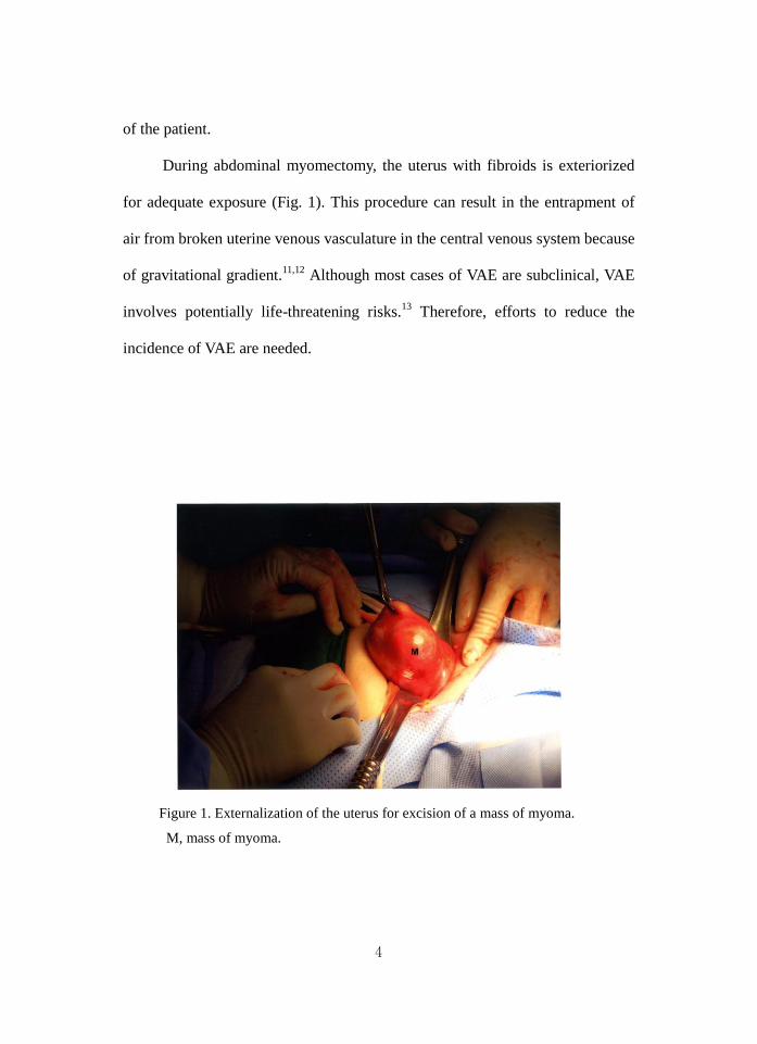
During abdominal myomectomy, the uterus with fibroids is exteriorized
for adequate exposure (Fig. 1). This procedure can result in the entrapment of
air from broken uterine venous vasculature in the central venous system because
of gravitational gradient.11,12
Although most cases of VAE are subclinical, VAE
involves potentially life-threatening risks.13
Therefore, efforts to reduce the
incidence of VAE are needed.
Figure 1. Externalization of the uterus for excision of a mass of myoma.
M, mass of myoma.

5
Several previous studies have reported on altering patient position to
reduce the incidence of VAE. In patients undergoing cesarean section, results
for the effect of a 5-10° head-up tilt position on the incidence of VAE are
conflicting. One study reported that a 5° head-up tilt position reduced the
incidence of VAE from 44% to 1%.14
However, other investigators reported that
there was no significant difference in the incidence of VAE between the supine
position and the 5-10° head-up tilt position.15
Although conflicting data exists
for positioning during cesarean section, studies have yet to compare the effect
of patient positioning on the incidence of VAE during abdominal myomectomy.
Therefore, the purpose of this study was to assess the incidence and grade
of VAE during abdominal myomectomy in the supine position in comparison to
those in the head-up tilt position using transesophageal echocardiography
(TEE).

6
II. MATERIALS AND METHODS
The study was approved by the Institutional Review Board, and written
informed consent was obtained from all patients.
In this study, 84 female patients (20-55 years old) of American Society of
Anesthesiologists physical status I or II who were scheduled for myomectomy
under general anesthesia were included (Fig. 2). Patients with a history of prior
abdominal surgery or cardiovascular or esophageal disease were excluded.
Using a random number sequence, patients were randomly divided into two
groups by a computer generator. Odd-numbered cases were allocated to the
supine group (n=40), and even-numbered cases were allocated to the 10°
head-up tilt group (n=44). All operations were performed by the same surgeon.
Figure 2. Flow diagram.

7
Patients were pre-medicated with midazolam (0.05 mg/kg, intramuscular
injection) 60 minutes before induction of anesthesia. In the pre-anesthetic room,
intravenous loading of lactated Ringer’s solution was started. On arrival in the
operating room, standard monitoring devices, including a three-lead
electrocardiogram, non-invasive blood pressure, and pulse oximetry machines,
were applied to the patient while in the supine position. All patients received 0.2
mg of glycopyrrolate intravenously. Anesthesia was induced with intravenous
propofol (2 mg/kg) and remifentanil (1 μg/kg). Orotracheal intubation was then
performed after intravenous administration of rocuronium (0.6 mg/kg).
Anesthesia was maintained with sevoflurane (1.5-3 vol%) and remifentanil
infusions (0.1 μg/kg/min). Neuromuscular relaxation was maintained with
continuous intravenous infusion of rocuronium (2-5 μg/kg/min).
The patients’ lungs were ventilated with 50% O2/50% air (2 L),
maintaining an end-tidal CO2 partial pressure (PETCO2) of 35-40 mm Hg.
Patients received approximately 1 L of lactated Ringer’s solution just prior to
skin incision. Afterwards, intraoperative fluid was subsequently administered
with an infusion set (Control-A-Flo SetTM
Model FMC5905, Baxter Ltd., Marsa,
Malta) at a rate of 50 mL/hr to avoid echocardiographic artifacts caused by
rapid intravenous infusion.16
After intubation, a 5.0-MHz multiplane TEE probe
(SONOS 4500, Philips, Boeblingen, Germany) was inserted. Just after
externalization of the uterus for myomectomy, patients in the head-up tilt group
were tilted upward by 10°, while position in the supine group was unchanged.

8
The ultrasound gain setting was lowered to minimize artifact
misinterpretation and three TEE views were recorded rapidly for later
interpretation of the stage. The midesophageal (ME) 4-chamber view was
continuously monitored during surgery and videotaped. When a bubble was
detected in the right atrium (RA), the probe was turned to the right side and the
angle was adjusted to the ME bicaval view to confirm its entrance from the
inferior vena cava. Then, the angle was rapidly re-adjusted to view the ME right
ventricle (RV) inflow-outflow view to confirm the extent of air embolism
through the right ventricular outflow tract (RVOT). When gas bubbles filled
more than half the diameter of the RA, RV, and RVOT, the ME 4-chamber, ME
2-chamber, ME long-axis, and transgastric mid short-axis views were obtained
for visual assessment of left ventricular function and RV overload. No
quantitative evaluation was performed. TEE images were videotaped
throughout surgery.17
After surgery, two cardiac anesthesiologists blinded to
patient group assignment reviewed the video tapes and graded VAE: grade I,
single gas bubble in the RA, RV, and RVOT; grade II, gas bubbles filling less
than half the diameter of the RA, RV, and RVOT; grade III, gas bubbles filling
more than half the diameter of the RA, RV, and RVOT; and grade IV, gas
bubbles completely filling the diameter of the RA, RV, and RVOT.18
Blood pressure (systolic, diastolic and mean), SpO2, and PETCO2 were
recorded. Cardiovascular instability was defined as a sudden decrease of more
than 20 mm Hg in the mean arterial blood pressure from the measurement taken

9
5 minutes prior, an acute fall of pulse oximetric saturation below 90%, and/or a
sudden decrease of PETCO2 above 2 mm Hg from the baseline value. EKG was
continuously monitored for changes related to VAE such as ST elevation, ST
depression, paroxysmal supraventricular tachycardia, etc.
Sample size was predetermined using a power analysis based on the
assumptions that 1) the incidence of VAE (above grade II) in patients
undergoing myomectomy in the supine position would be about 70% (based on
preliminary results), 2) the incidence of VAE (above grade II) in patients
undergoing myomectomy in 10° head-up tilt would be decreased to about 40%,
and 3) α=0.05 with a power (1-β) of 0.8. The analysis led us to conclude that 40
patients per group would be sufficient.
Statistical analyses were performed with SAS software (version 6.12,
SAS Institute, Cary, NC, USA). The demographic and vital sign data between
the two groups were compared using Student’s t-test. The data of vital sign
change during surgery in each group were compared using the repeated
measures analysis of variance. Post-hoc analysis was done using the Bonferroni
test. The grades of VAE between the two groups were compared using the
chi-square test and Fisher’s exact test. p-values <0.05 were considered
statistically significant.

10
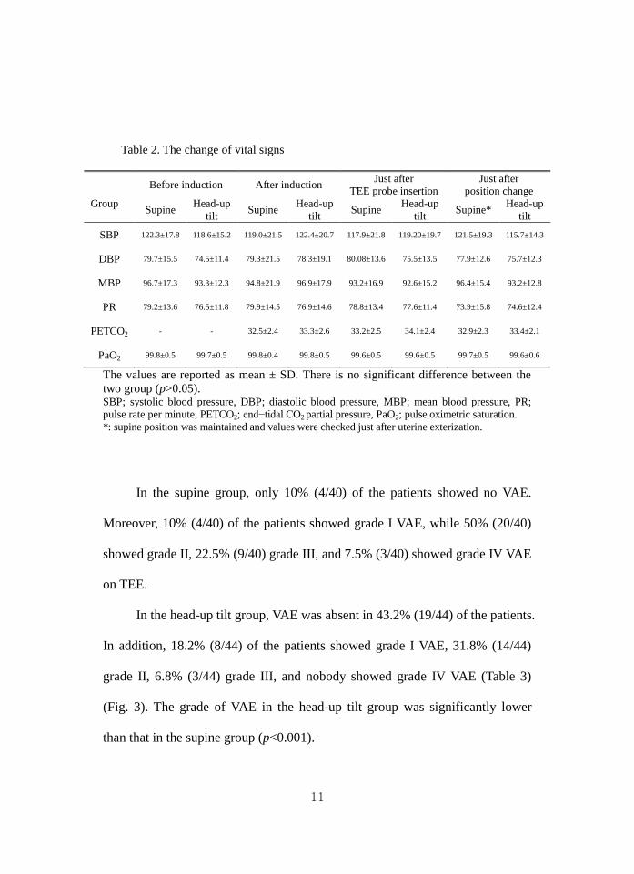
III. RESULTS
All 84 patients completed the protocol. The patient demographic
characteristics were similar for both groups (Table 1). There were no specific
differences in vital signs between the two groups, especially just after uterine
exteriorization with or without position change (Table 2).
Table 1. Patient characteristics
The values are reported as mean ± SD. There was no specific difference between the
two groups.
Supine group Head-up tilt group p-value
(n=40) (n=44)
Age (yrs) 43.2 7.3 41.6 6.9 0.326
Weight (kg) 56.9 7.8 57.4 6.7 0.776
Height (cm) 158.0 8.3 157.7 7.2 0.211
Operative time
(min) 95.2 23.0 99.5 33.4 0.579
Anesthesia time
(min) 108.5 24.4 113.1 35.4 0.508
Weight of myoma
(g) 392.8 106.9 357.5 152.5 0.324
11
Table 2. The change of vital signs
Before induction After induction
Just after
TEE probe insertion
Just after
position change
Group Supine
Head-up
tilt Supine
Head-up
tilt Supine
Head-up
tilt Supine*
Head-up
tilt
SBP 122.3±17.8 118.6±15.2 119.0±21.5 122.4±20.7 117.9±21.8 119.20±19.7 121.5±19.3 115.7±14.3
DBP 79.7±15.5 74.5±11.4 79.3±21.5 78.3±19.1 80.08±13.6 75.5±13.5 77.9±12.6 75.7±12.3
MBP 96.7±17.3 93.3±12.3 94.8±21.9 96.9±17.9 93.2±16.9 92.6±15.2 96.4±15.4 93.2±12.8
PR 79.2±13.6 76.5±11.8 79.9±14.5 76.9±14.6 78.8±13.4 77.6±11.4 73.9±15.8 74.6±12.4
PETCO2 - - 32.5±2.4 33.3±2.6 33.2±2.5 34.1±2.4 32.9±2.3 33.4±2.1
PaO2 99.8±0.5 99.7±0.5 99.8±0.4 99.8±0.5 99.6±0.5 99.6±0.5 99.7±0.5 99.6±0.6
The values are reported as mean ± SD. There is no significant difference between the
two group (p>0.05). SBP; systolic blood pressure, DBP; diastolic blood pressure, MBP; mean blood pressure, PR;
pulse rate per minute, PETCO2; end−tidal CO2 partial pressure, PaO2; pulse oximetric saturation.
*: supine position was maintained and values were checked just after uterine exterization.
In the supine group, only 10% (4/40) of the patients showed no VAE.
Moreover, 10% (4/40) of the patients showed grade I VAE, while 50% (20/40)
showed grade II, 22.5% (9/40) grade III, and 7.5% (3/40) showed grade IV VAE
on TEE.
In the head-up tilt group, VAE was absent in 43.2% (19/44) of the patients.
In addition, 18.2% (8/44) of the patients showed grade I VAE, 31.8% (14/44)
grade II, 6.8% (3/44) grade III, and nobody showed grade IV VAE (Table 3)
(Fig. 3). The grade of VAE in the head-up tilt group was significantly lower
than that in the supine group (p<0.001).

12
Table 3. Maximum grade of venous air embolism (VAE) during myomectomy
Grade Supine group
(n = 40)
Head-up tilt group
(n = 44)
0 4 (10.0%) 19 (43.2%)
1 4 (10.0%) 8 (18.2%)
2 20 (50.0%) 14 (31.8%)
3 9 (22.5%) 3 (6.8%)
4 3 (7.5%) 0 (0%)
The values are the number of patients and incidence (percentage) according to the VAE
grade of each patient. The maximum grade of VAE detected by transesophageal
echocardiography.
*The grades in the head-up tilt group were significantly lower than those in the supine
group (p<0.001).
Figure 3. Venous air emboli detected by transesophageal echocardiography during
myomectomy. (A) Mid-esophageal four-chamber view. (B) Bicaval view. RA, right atrium; RV, right ventricle; LA, left atrium; LV, left ventricle; IVC, inferior vena cava.
In 4 patients in the supine group and 2 patients in the head-up tilt group, a
sudden decrease in PETCO2 above 2 mm Hg from the baseline value was noted.
However, no other cardiovascular instabilities or EKG changes were en-

13
countered in either group. All VAEs in both groups occurred during excision of
myoma from the exposed uterus.

14
IV. DISCUSSION
In addition to abdominal myomectomy, new treatment methods have
become available to patients with symptomatic fibroids including medical
therapy, minimally invasive therapies such as uterine artery embolization or
magnetic resonance-guided focused ultrasound surgery, and laparoscopic or
vaginal myomectomy.19-22
Among these methods, abdominal myomectomy
plays is invaluable because it is not limited to the size and number of fibroids
that can be treated.23
However, abdominal myomectomy also involves the risk
of VAE because the extent of uterine exteriorization necessary to achieve
adequate exposure can produce greater gravitational gradient due to broken
venous vasculature. Therefore, for this study, it was theorized that the grade and
incidence of VAE during abdominal myomectomy could be reduced by
reducing the gravitational gradient through patient positioning. In this study, the
grade of VAE in the head-up tilt group was significantly lower than that in the
supine group.
When VAE occurs in awake patients, the clinical manifestations include
acute dyspnea, cough, and chest pain.24,25
Also, physical examination
demonstrates rales, wheezing, and tachypnea. However, confirmation of the
aforementioned symptoms and signs is not possible in anesthetized patients.
Respiratory functions are therefore continuously monitored in patients under
anesthesia. These monitors can detect decreases in PETCO2 and SpO2 along

15
with hypercapnia; however, the low sensitivity and specificity of these monitors
make it difficult to detect VAE.26
In this study, 4 patients from the supine group
and 2 patients from the head-up tilt group showed a sudden decrease in PETCO2
>2 mm Hg from the baseline value.
TEE, used in this study, is currently the most sensitive monitoring method
for VAE. TEE can detect air volumes as small as 0.02 mL/kg by bolus
injection.27
However, TEE is relatively invasive and is not always obtainable in
actual practice.
VAE can also provoke cardiovascular complications by right ventricular
outflow obstruction. This phenomenon leads to hypotension and right heart
failure. EKG changes generally present as a compromised cardiac status. EKG
changes include ST-T changes that are followed by supraventricular and
ventricular tachyarrhythmias.28
In this study, even a high VAE grade was not
associated with cardiovascular instability, and there were no EKG changes
observed for either group. These results are similar to those of other studies on
the detection of VAE by TEE.16,17,29
However, clinicians should be aware of the
risk of right-to-left shunt through which a direct cerebral air embolism is
possible.
To minimize differences that might arise from having different surgeons
perform the surgeries, in this study, all surgical procedures were performed by
the same surgeon. Despite these efforts, the surgical complexity might have
affected the incidence and stage of VAE. For example, more dissection in a

16
situation of increased surgical complexity could have resulted in a higher degree
of air exposure. In this study, patients with a history of prior abdominal surgery
were excluded.
To avoid echocardiographic artifacts, we administered intra-operative
fluid at a constant rate of 50 mL/hr, based on the study by Derouin, et al.16
We
ascertained that no echocardiographic artifacts were seen on TEE at this
infusion rate.

17
V. CONCLUSION
The grade of VAE in the head-up tilt group was significantly lower than
that in the supine group without fluctuation of vital signs. Therefore, we suggest
that the head up-tilt position during trans-abdominal myomectomy would be
better in reducing VAE than the supine position.
18
REFERENCES
1. Mommerot A, Perrault LP. Carbon dioxide embolism induced by endoscopic
saphenous vein harvesting during coronary artery bypass grafting. J Thorac
Cardiovasc Surg 2006;132:1502.
2. Park EY, Kwon JY, Kim KJ. Carbon dioxide embolism during laparoscopic
surgery. Yonsei Med J 2012;53:459-66.
3. Kama NA. Influence of nitrous oxide anesthesia on venous gas embolism with
carbon dioxide and helium during pneumoperitoneum. Surg Endosc
2001;15:1237-8.
4. Cottin V, Viale JP, Bouffard Y, Delafosse B. Severe nitrous oxide embolism
during venous stripping. Intensive Care Med 1997;23: 1287-8.
5. Boussuges A, Blanc F, Carturan D. Hemodynamic changes induced by
recreational scuba diving. Chest 2006;129:1337-43.
6. Risberg J, Englund M, Aanderud L, Eftedal O, Flook V, Thorsen E. Venous
gas embolism in chamber attendants after hyperbaric exposure. Undersea Hyperb
Med 2004;31:417-29.
7. Mitchell SJ, Benson M, Vadlamudi L, Miller P. Cerebral arterial gas embolism
by helium: an unusual case successfully treated with hyperbaric oxygen and
lidocaine. Ann Emerg Med 2000; 35:300-3.
8. Kim CS, Liu J, Kwon JY, Shin SK, Kim KJ. Venous air embolism during
surgery, especially cesarean delivery. J Korean Med Sci 2008;23:753-61.

19
9. Papadopoulos G, Kuhly P, Brock M, Rudolph KH, Link J, Eyrich K. Venous
and paradoxical air embolism in the sitting position. A prospective study with
transoesophageal echocardiography. Acta Neurochir (Wien) 1994;126:140-3.
10. Albin MS, Carroll RG, Maroon JC. Clinical considerations concerning
detection of venous air embolism. Neurosurgery 1978;3: 380-4.
11. Maroon JC, Goodman JM, Horner TG, Campbell RL. Detection of minute
venous air emboli with ultrasound. Surg Gynecol Obstet 1968;127:1236-8.
12. Lang S. Precordial Doppler diagnosis of haemodynamically compromising
air embolism during caesarean section. Can J Anaesth 1991;38:255-6.
13. Vacanti CA, Lodhia KL. Fatal massive air embolism during transurethral
resection of the prostate. Anesthesiology 1991;74:186-7.
14. Fong J, Gadalla F, Druzin M. Venous emboli occurring caesarean section: the
effect of patient position. Can J Anaesth 1991;38:191-5.
15. Karuparthy VR, Downing JW, Husain FJ, Knape KG, Blanchard J, Solomon
D, et al. Incidence of venous air embolism during cesarean section is unchanged
by the use of a 5 to 10 degree head-up tilt. Anesth Analg 1989;69:620-3.
16. Derouin M, Couture P, Boudreault D, Girard D, Gravel D. Detection of gas
embolism by transesophageal echocardiography during laparoscopic
cholecystectomy. Anesth Analg 1996;82:119-24.
17. Kim CS, Kim JY, Kwon JY, Choi SH, Na S, An J, et al. Venous air embolism
during total laparoscopic hysterectomy: comparison to total abdominal
hysterectomy. Anesthesiology 2009;111:50-4.

20
18. Schmandra TC, Mierdl S, Bauer H, Gutt C, Hanisch E. Transoesophageal
echocardiography shows high risk of gas embolism during laparoscopic hepatic
resection under carbon dioxide pneumoperitoneum. Br J Surg 2002;89:870-6.
19. Seinera P, Arisio R, Decko A, Farina C, Crana F. Laparoscopic
myomectomy: indications, surgical technique and complications. Hum Reprod
1997;12:1927-30.
20. Davies A, Hart R, Magos AL. The excision of uterine fibroids by vaginal
myomectomy: a prospective study. Fertil Steril 1999;71: 961-4.
21. Spies JB, Cooper JM, Worthington-Kirsch R, Lipman JC, Mills BB, Benenati
JF. Outcome of uterine embolization and hysterectomy for leiomyomas: results
of a multicenter study. Am J Obstet Gynecol 2004;191:22-31.
22. Hindley J, Gedroyc WM, Regan L, Stewart E, Tempany C, Hynyen K, et al.
MRI guidance of focused ultrasound therapy of uterine fibroids: early results.
AJR Am J Roentgenol 2004;183:1713-9.
23. Gavai M, Berkes E, Lazar L, Fekete T, Takacs ZF, Urbancsek J, et al. Factors
affecting reproductive outcome following abdominal myomectomy. J Assist
Reprod Genet 2007;24:525-31.
24. Balki M, Manninen PH, McGuire GP, El-Beheiry H, Bernstein M. Venous air
embolism during awake craniotomy in a supine patient. Can J Anaesth
2003;50:835-8.
25. Suarez S, Ornaque I, Fábregas N, Valero R, Carrero E. Venous air embolism
during Parkinson surgery in patients with spontaneous ventilation. Anesth Analg

21
1999;88:793-4.
26. Souders JE. Pulmonary air embolism. J Clin Monit Comput 2000;16:375-83.
27. Furuya H, Suzuki T, Okumura F, Kishi Y, Uefuji T. Detection of air
embolism by transesophageal echocardiography. Anesthesiology 1983;58:124-9.
28. Gildenberg PL, O’Brien RP, Britt WJ, Frost EA. The efficacy of Doppler
monitoring for the detection of venous air embolism. J Neurosurg 1981;54:75-8.
29. Koo BN, Kil HK, Choi JS, Kim JY, Chun DH, Hong YW. Hepatic resection
by the Cavitron Ultrasonic Surgical Aspirator increases the incidence and
severity of venous air embolism. Anesth Analg 2005;101:966-70.

22
ABSTRACT (IN KOREAN)
자궁근종절제술 시행 중 역 트렌델렌버그 자세가 정맥 공기
색전증을 줄일 수 있는가?
<지도교수 김기준>
연세대학교 대학원 의학과
안지원
정맥 공기 색전증은 손상된 정맥을 통해 중심 정맥 계통으로 공기나
의학적으로 사용되는 다른 기체가 유입되어 발생한다. 이러한 정맥
공기 색전증에 대하여 자궁근종절제술 시행 중 환자의 자세에 따른
발생률의 차이를 알아본 연구는 없었다. 본 연구에서는
자궁근종절제술 시행 중 바로누운 자세와 역 트렌델렌버그 자세가
정맥 공기 색전증의 발생률과 정도에 어떤 차이를 보일지 경식도
심초음파를 통해 비교 관찰하였다. 전신마취 하에 자궁근종적출술을
받는 ASA(American Society of Anesthesiologist) I 혹은 II 인
여성 환자 84명을 대상으로 하여 무작위로 바로누운 자세와 역
트렌델렌버그 자세 군을 선정하였으며, 수술 중 경식도 심초음파의
영상을 녹화하였다. 이 영상을 통해 정맥 공기 색전증의 정도를
확인하였다. 바로누운 자세 군에서는 정맥 공기 색전증이 발견되지
않은 환자가 10%, Grade I 은 10%, Grade II 는 50%, Grade III 는

23
22.5%, Grade IV 는 7.5% 였다. 역 트렌델렌버그 자세 군에서는
정맥 공기 색전증이 발견되지 않은 환자가 43.2%, Grade I 은
18.2%, Grade II 는 31.8%, Grade III 는 6.8% 였으며 Grade IV 인
환자는 없었다. 역 트렌델렌버그 자세 군에서 정맥 공기 색전증의
정도는 바로누운 자세 군보다 유의하게 낮았다(p<0.001).
결론적으로 자궁근종절제술 시행 중 역 트렌델렌버그 자세는 정맥
공기 색전증의 발생률과 정도를 바로누운 자세보다 유의하게 낮출 수
있었다.
--------------------------------------
핵심되는 말 : 정맥 공기 색전증, 자궁근종절제술, 역 트렌델렌버그