incidence and distribution of some aerobic bacterial ... · department of poultry diseases, faculty...
TRANSCRIPT

Middle East Journal of Applied Sciences ISSN 2077-4613
Volume : 05 | Issue : 02 | April-June | 2015 Pages: 383-394
Corresponding Author: Eman Anter Morsy, Department of Poultry Diseases, Faculty of Veterinary medicine, Cairo University, Giza-12211, Egypt. E-mail: [email protected], [email protected]
383
Incidence and Distribution of some Aerobic Bacterial Agents Associated with High Chick Mortality in some Broiler Flocks in Egypt Khelfa D.G. and Eman A. Morsy Department of Poultry Diseases, Faculty of Veterinary Medicine, Cairo University, Giza, Egypt
ABSTRACT
A monitoring study was carried out on one hundred two broiler flocks of different breeds with housing capacity ranged from 1000 to 24000 birds located in nine Egyptian governorates (Sharkia, Giza, Gharbia, Fayoum, Beni suef, Menia, Dakahlia, Beheira and Alexandria) in a trial for isolation of responsible aerobic bacterial agents causing high chicks mortality in such flocks. The diagnosis based on history of each examined flock, the observed clinical signs at time of examination, post-mortem (PM) lesions as well as isolation and identification of responsible bacterial agents incriminated in this problem. Two hundred six moribund chicks and one hundred forty one freshly dead chicks were collected for PM examination and samples collection. One hundred two pooled tissue samples include heart, liver, bile, spleen and yolk sac as well as one hundred two cecal content were collected under complete aseptic conditions for bacterial isolation and identification by cultural, biochemical and serological methods. The result of history revealed that examined breeds were Cobb, Hubbard, Saso, Ross, Avian and Balady with age ranged from 1 to 28 days and mortality rate ranged from 0.07 to 6.8%. Observed clinical signs were respiratory signs, omphalitis, lamness, pasty vent and diarrhea. PM lesions were septecaemic picture, fibrinous polyserositis, nodules on heart muscle, pneumonia, distended gall bladder, enteritis, unabsorbed yolk sac and cecal core. The incidence of bacteriologically positive pooled tissue samples were (99%) 101 out of 102 farms and the negative samples were (1%) 1 out of 102 farms, while culturing of pooled cecal content samples for suspected Salmonella infections revealed that 8 out of 102 (7.8%) positive samples and 94 out of 102 (92.2%) negative samples. 146 bacterial isolates were recovered from all bacteriologically cultured samples. Escherichia coli isolates were the predominant strains (74\146) 50.7% followed by Salmonella (29\146)19.8%, Klebsiella pneumonia (20\146)13.7%, Proteus mirabilis (16\146)10.9% and Pseudomonas aeruginosa (7\146) 4.8%.Serological identification of E. coli isolates revealed that both O158 and O78 serotypes were the most predominant isolates (14\74) 18.9%, each, followed by serotype O27 (8\74) 10.8%, serotype O114 (5\74) 6.76% , serotypes O18 and O55 (3\74) 4%, each, serotypes O6, O111 and O159 (2\74) 2.7%, each finally serotypes O8, O15, O26, O44, O119, O142, O153, O166 and O169 (1\74) 1.35%, each. Testing the pathogenicity of E. coli isolates using Congo red test revealed that all isolated E. coli serotypes were pathogenic. Serological identification of Salmonella isolates revealed that S. infantis, S.Virshow, S Entereditis, S, Gallinarum, S. Kentuky and S. Typhimurium were the only isolated Salmonella species with a percentage of (8\29)27.6%, (7\29)24.1%, (6\29) 20.8%, (4\29)13.8%, (3\29)10.3% and (1\29)3.4% respectively.
Key words: Monitoring, Incidence, broiler, chick mortality, aerobic bacteria and Egypt.
Introduction
During the last 40 years the market age of broiler chickens has been reduced by approximately one day every year. This trend is continuing and emphasizing the importance of growth during the first week of life, which presently constitutes 16% of the life span of the broiler (Gyles, 1989). Newly hatched chicks exposed to opportunistic pathogens during the first week post-hatch due to the lack of gastrointestinal flora which makes them highly susceptible to infection so the first few days of age are very important in the life of a broiler chick, (Milner and Shaffer, 1952; Charlton, 1996 and Henderson et al., 1999). Records of mortality during the first few days of brooding have been used to assess the quality of chicks in the broiler industry (Chou et al., 2004). Early chick mortality is one of the most important problems of poultry industry.
Out of total mortality, 30- 50% occurs in this period. Omphalitis, avian encephalomyelitis, brooder pneumonia, spiking mortality, dehydration, ammonia burns and pullorum disease are major problems of the early life (Charlton, 1996). Amongst these problems, omphalitis is the major cause of early chick mortality (Anjum, 1997; Reece and Beddome, 1983 and Ijaz et al., 1994). The major cause of omphalitis is E. coli but other bacteria may be found in single or mixed infections (Zahdeh et al., 1984; Sarma et al. 1985; Linzitto et al. 1988; Jordan 1990; Utomo et al. 1990; Sainsbury 1992; Ali 1993; Choudhury et al. 1993); Deeming, 1995; Rehman et al. 1996; Anjum, 1997; Sharada et al, 1999 and Anonymous 2000). Next frequently found bacteria

Middle East J. Appl. Sci.., 5(2): 383-394, 2015
384
was genus Salmonella (Zahdeh et al. 198; O`-Brien 1988; Mutalib and Hanson 1989; Ali 1993; Choudhury et al. 1993; Rehman et al. 1996; Anjum 1997, Anonymous 2000 and Shivaprasad 2000) Other bacterial genera found to be involved include Pseudomonas (Zahdeh et al., 1984; Sarma et al. 1985; Utomo et al., 1990; Choudhury et al., 1993; Anjum, 1997 and Anonymous, 2000) and Klebsiella (Zahdeh et al., 1984; Choudhury et al., 1993 and Anonymous, 2000). Bacterial diseases are considered as very important (Venkanagouda et al., 1996). Salmonellosis and colibacillosis are known to increase mortality in the initial few weeks of the life of chicks (Shane, 1999). Surveillance studies used to determine the prevalence and cost of different bacterial diseases that allow evaluation of control programs and establishment of research priorities. Such studies may also provide useful epizootiological data on particular diseases. (Brigden and Riddell, 1975).
This study was designed to investigate the common aerobic bacterial agents that are responsible for induction of high broiler chick mortalities among broiler flocks distributed in some Egyptian governorates. Material and methods I. Field diagnosis:
Flock history of each examined broiler flock on day of examination was recorded. This includes breed, age, number of chicks / house, mortality rate, previous vaccination, previous medication, based on flock records. Also observed clinical signs and PM lesions were recorded.
II. Samples a. Samples for post mortem examination:
A total number of 347 broiler chicks including 206 sacrificed moribund chicks and 141 freshly dead chicks were subjected to PM examination for recording of post mortem lesions.
b. Samples for bacterial isolation:
One hundred two tissue samples were collected, each sample represents one farm. The pooled tissues of such farm contain heart, liver, bile, spleen and yolk sac, as well as one hundred two cecal content samples were collected, each sample represents one farm and contains the pooled sampled cecal content of such farm. All samples were taken under complete aseptic conditions for bacteriological investigation.
III. Isolation and purification of suspected aerobic bacterial agents : Samples were Prepared according to (Louise et al., 2008). Bacterial isolation was carried according to (Cruickshank et al., 1975). 1. Culturing in fluid media:
Each prepared pooled tissue sample was inoculated into nutrient broth and buffered peptone water and incubated aerobically for 24 hours and 16-18hr, respectively at 37o C. One inoculum from each positive buffered peptone water broth was sub cultured into Rappaport Vasliadis broth or Selenite F broth and incubated at 42οC and 37οC, respectively for 24 hours. Each cecal content sample was inoculated directly into Selenite F broth without pre-enrichment.
Culturing onto solid media:
Each positive nutrient broth sample was sub-cultured onto different solid media including nutrient, MacConkey's and Eosine methylen blue (EMB) agar media, while each positive selenite-F-broth or Rappaport Vasliadis broth sample was subcultured onto Salmonella Shegilla (S.S) agar and Xylose Lysine Desoxycholate (XLD) agar media. All inoculated media were incubated at 37οC for 24 hours aerobically and examined for bacterial growth. Purified colony from each positive cultured solid media was inoculated into slope agar and positive one stored refrigerated till identification. IV. Identification of obtained bacterial isolates: Morphological, Biochemical and Serological identification of isolated micro-organisms were done. a. Morphological identification:
Cultural characters: The colonial morphological characters of each isolate were identified following (Collee et al., 1996). Staining characters:
Smear from each prepared pure isolate was stained with Gram's stain and examined microscopically under oil emersion lens for recording gram reactivity (Cruickshank et al., 1975).

Middle East J. Appl. Sci.., 5(2): 383-394, 2015
385
Motility character: Motile ability was tested by stabbing of each isolate into semisolid agar. (Cruickshank et al., 1975). b. Biochemical identification: Biochemical identification of prepared pure isolates was done using API 20E plate system. (Nazarzadeh et al., 2014).
Results
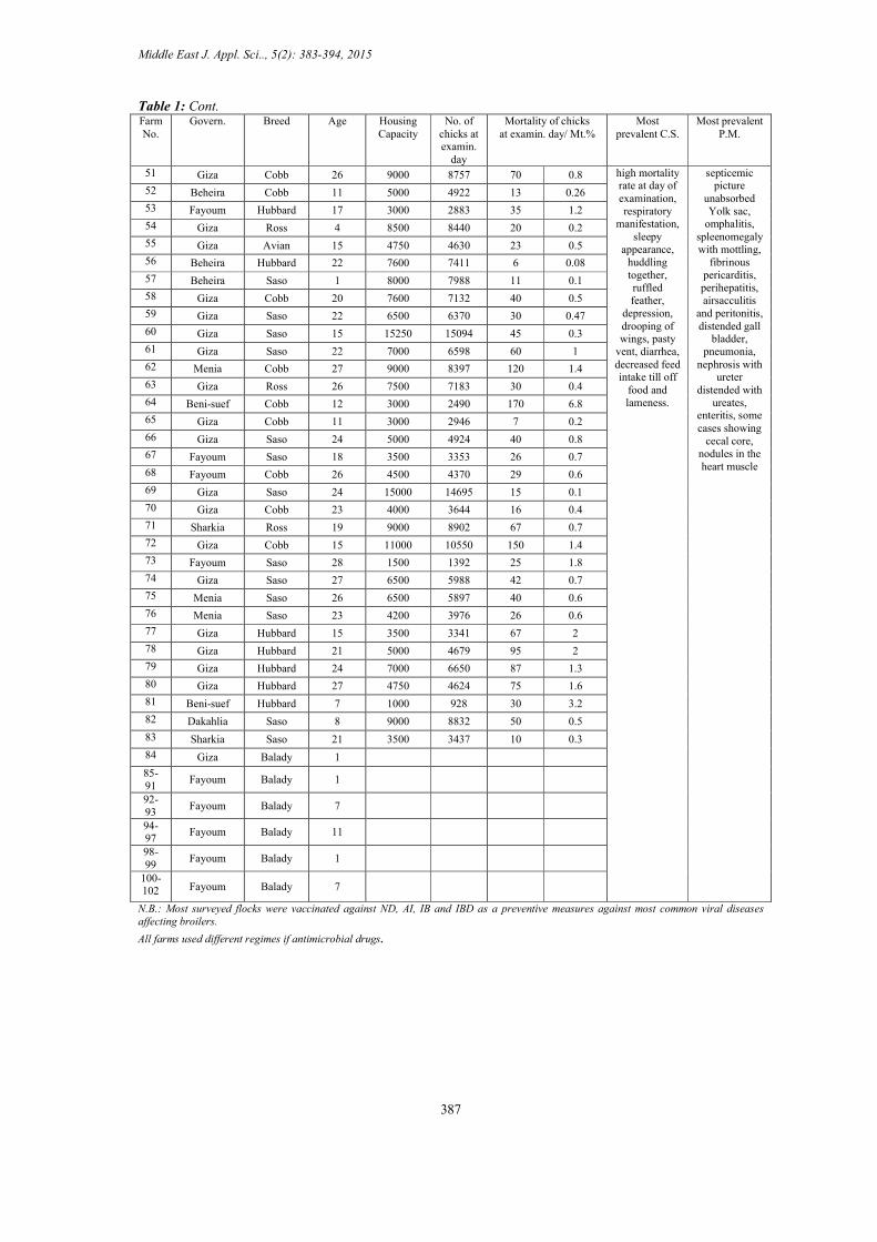
Flock history of examined broiler flocks were illustrated in table (1). The results of history revealed that examined breeds were Cobb, Hubbard, Saso, Avian, Ross and Balady which were located in nine Egyptian governorates including Sharkia, Giza, Gharbia, Fayoum, Beni suef, Menia, Dakahlia, Beheira and Alexandria. The age of such flocks was ranged from 1-28 day old and their housing capacity ranged from 1000-24000 birds. The mortality percent at day of visit ranged from 0.07% - 6.8 %, Cobb breed recorded the highest chick mortality on 12th day of age in Beni suef with a percentage of 6.8% followed by Hubbard breed recording a mortality percent of 3.2% on 7th day of age in Beni suef, Saso breed recorded a mortality percent of 1.8 % on 28th day old in fayoum, Ross breed recorded a mortality percent of 1.2% on 27th day old in Giza, avian breed recorded a mortality percent of 0.5% on 15th day old in Giza and Balady breed recorded mortality percent of 0.17% on 20th day of age in Beni suef.
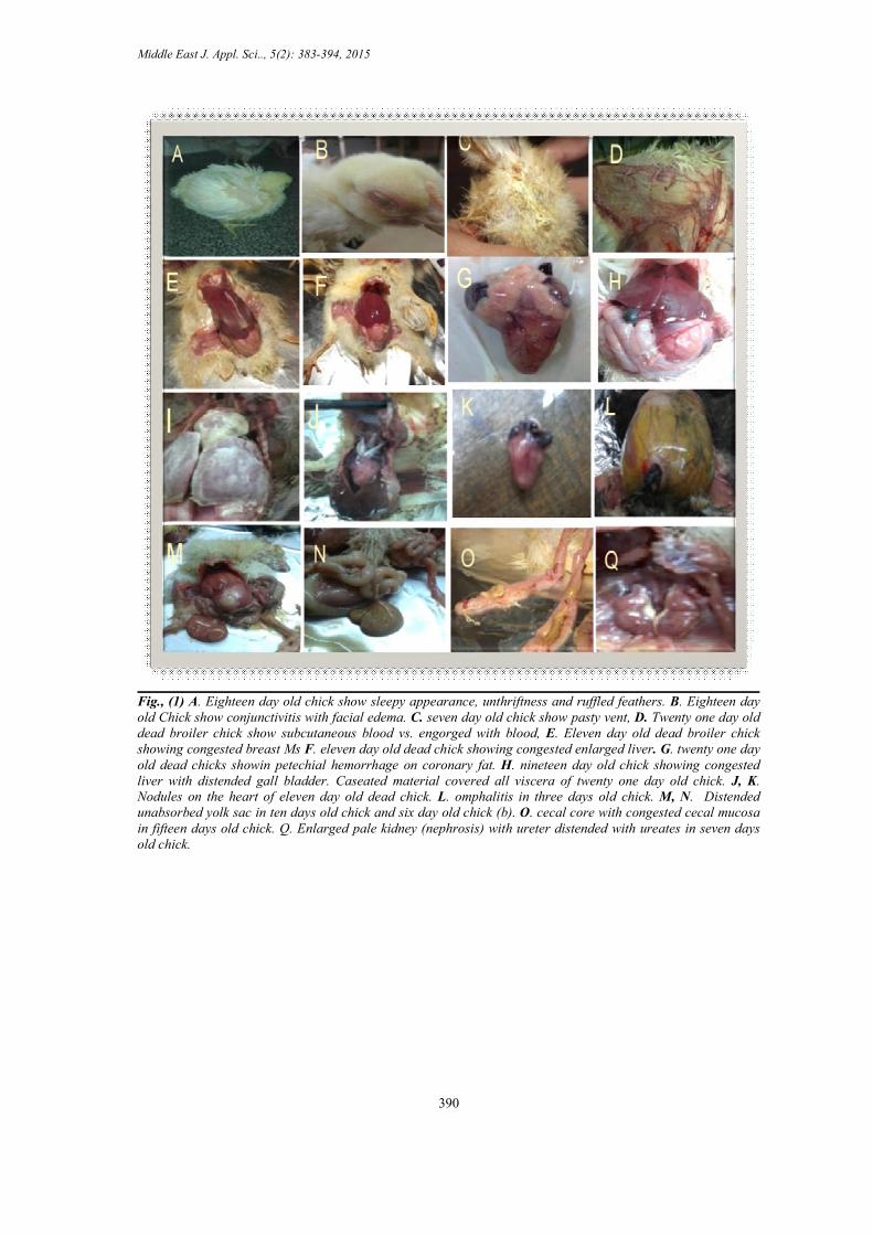
The observed clinical signs of examined broiler flocks, on day of examination, were huddling together, respiratory manifestations, sleepy appearance, ruffled feathers, depression fig., (1A), pasty vent fig., (1C) diarrhea, omphalitis fig.,(1L), conjunctivitis and facial edema fig., (1B) and lameness. On P.M. examination the prevalent lesions were septicaemic picture fig.,(1D), severe congestion of muscle fig.,(1E), kidney, spleen, lung and liver fig.,(1F), petichial hemorrhages on coronary fat fig.,(1G), nodules in the heart muscle fig.,(1J,K), spleenomegaly with mottling, fibrinous pericarditis, perihepatitis, peritonitis and caseous airsacculitis fig.,(1I), distended gall bladder, fig.,(1H), pneumonia, nephrosis with ureter distended with ureates fig.,(1Q), enteritis, unabsorbed Yolk sac fig.,(1M,N) and cecal core fig.,(1O).
The incidence of bacteriologically positive pooled tissue samples were (99%) 101 out of 102 farms and the negative samples were (1%) 1 out of 102 farms, while pooled cecal content samples revealed that 8 out of 102 (7.8%) positive samples and 94 out of 102 (92.2%) negative samples with a total of 146 bacterial isolates were recovered from all bacteriologically cultured samples, 138 isolates from pooled tissue samples and8 isolates from pooled cecal content samples.
The morphological fig., (2 A, B, C, D and E) and biochemical identification, table (2) and fig., (2 H, I, J and K) of isolated organisms indicated that the enterobacteriacae group was the prevalent including Escherichia coli, Salmonella spp., Klebsiella pneumonia and Proteus mirabilis while Pseudomonas aeruginosa was the only none enterobacteriacae isolated bacteria. The Escherichia coli isolates were the predominant organism (74\146) 50.7% followed by Salmonella (29\146)19.9%, Klebsiella pneumonia (20\146)13.7%, Proteus mirabilis (16\146)10.9% and Pseudomonas aeruginosa (7\146) 4.8% table (3).
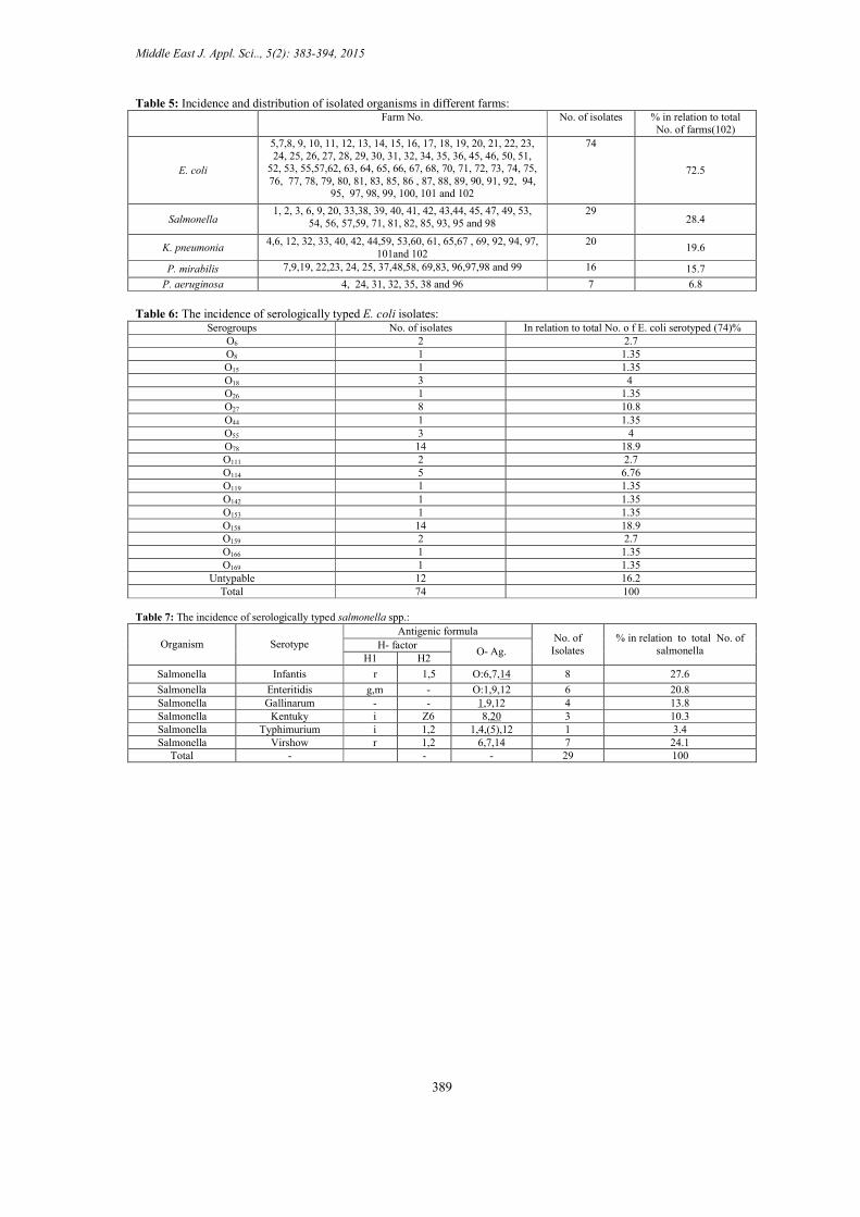
The isolated microorganisms were distributed through the monitored farms in either single or mixed infection with the percentage of 62.4 % and 37.6% respectively table (4) where E. coli isolates were the prevalent 72.5% (74 out of 102 farms) followed by Salmonella 28.4% (29 out of 102 farms), K. pneumonae19.6% (20 out of 102 farms), P. mirabilis 15.7% (16 out of 102 farms) and P. aeruginosa 6.8% (7 out of 102 farms) table. (5).The results proved that Gm-ve bacteria are the main category incriminated in the high chick mortality during 4 weeks of life. Fig., (2 F and G) Farm No. 84 was negative for bacteriological isolation
Culturing of E. coli isolates on Congo red media proved that all tested E. coli isolates were Congo red positive indicating that all E.coli were pathogenic. Fig (2L) E. coli isolates were differentiated serologically into 62 typed isolates where both O158 and O78 serotypes were the most predominant isolates (14\74) 18.9%, each, followed by serotype O27 (8\74) 10.8%, serotype O114 (5\74) 6.76%, serotypes O18 and O55 (3\74) 4%, each, serotypes O6, O111 and O159 (2\74) 2.7%, each finally serotypes O8, O15, O26, O44, O119, O142, O153, O166 and O169 (1\74) 1.35%, each. The remaining E. coli isolates (12\74) 16.2% were untyped, table (6).Salmonella isolates were differentiated serologically into 29 typed isolates where SI were the most predominant isolates (8/29) 27.6% followed by SV (7/29) 24.1%, SE (6/29) 20.8%, SG (4/29)13.8%, SK (3/29)10.3% and ST (1/29) 3.4%, table (7)

Middle East J. Appl. Sci.., 5(2): 383-394, 2015
386
Table 1: Flock history of monitored broiler flocks:
Farm No. Govern. Breed Age Housing Capacity
No. of chicks at
examin. day
Mortality of chicks
at examin. day/ Mt.%
Most prevalent C.S. Most
prevalent P.M.
1 Dakahlia Saso 26 7600 7420 14 0.2 high mortality rate at day of
examination, respiratory
manifestation, sleepy appearance, huddling together,
ruffled feather, depression,
drooping of wings, pasty vent, omphalitis,
diarrhea, decreased feed intake till off
food and lameness.
septicemic picture, unabsorbed Yolk sac, fibrinous pericarditis, perihepatiti, airsacculitis and peritonitis, distended gall bladder, pneumonia, nephrosis with ureter distended with ureates, enteritis, some cases showing cecal core, and nodules on the heart
muscle
2 Gharbia Ross 19 5000 4831 36 0.7 3 Gharbia Cobb 9 5000 4908 20 0.4 4 Sharkia Avian 23 3500 3335 15 0.4 5 Fayoum Cobb 20 24000 23424 320 1.3 6 Gharbia Ross 21 3400 3298 12 0.3 7 Gharbia Cobb 16 6000 5790 46 0.8 8 Beni-suef Balady 20 20000 19693 35 0.17 9 Giza Hubbard 24 9700 9443 63 0.6
10 Giza Cobb 14 15000 14585 54 0.4 11 Giza Saso 27 7500 7237 40 0.5 12 Giza Saso 15 13000 12890 29 0.2 13 Giza Saso 15 10000 9593 42 0.4 14 Menia Cobb 20 5000 4598 200 0.4 15 Giza Saso 3 5000 4968 32 0.6 16 Menia Cobb 13 3000 4916 12 0.2 17 Fayoum Saso 23 7000 6502 57 0.8
18 Giza Saso 11 10000 9891 19 0.2
19 Menia Cobb 19 3000 2903 3 0.1 20 Alexandria Cobb 18 7600 7296 21 0.3
21 Giza Saso 15 4750 4672 24 0.5 22 Dakahlia Cobb 23 3000 2813 9 0.3
23 Giza Saso 15 8000 7860 16 0.2 24 Giza Cobb 12 7000 6849 37 0.5 25 Sharkia Hubbard 25 14000 13515 44 0.3 26 Sharkia Hubbard 19 10000 9690 15 0.1
27 Alexandria Cobb 18 5000 4538 38 0.8 28 Menia Cobb 22 2500 2146 27 1.2 29 Giza Cobb 19 7000 6793 41 0.6 30 Menia Cobb 21 5000 4487 60 1.3 31 Giza Ross 27 9000 8398 105 1.2 32 Giza Cobb 2 5200 5108 50 1 33 Giza Cobb 11 5000 4893 25 0.5 34 Giza Hubbard 16 9000 8914 10 0.1 35 Dakahlia Avian 10 3400 3303 13 0.4 36 Gharbia Saso 5 1700 1616 23 1.4 37 Sharkia Saso 25 7000 6870 5 0.07 38 Beni-suef Cobb 13 4000 3420 35 1.02 39 Giza Cobb 22 1500 1231 12 0.9 40 Dakahlia Cobb 11 3000 2933 9 0.3 41 Fayoum Hubbard 12 1500 1427 15 1 42 Beni-suef
Cobb 15 5000 4797 56 1.1
43 Beni-suef Cobb 9 3500 3413 70 2 44 Giza Saso 15 5000 4872 25 0.5 45 Giza Saso 25 6500 6295 15 0.2 46 Giza Saso 12 7000 6792 20 0.3 47 Giza Saso 17 5400 4891 85 1.7 48 Fayoum Saso 20 7000 6681 14 0.2 49 Giza Ross 15 7000 6827 21 0.3 50 Fayoum Cobb 15 5000 4630 45 1
Middle East J. Appl. Sci.., 5(2): 383-394, 2015
387
Table 1: Cont. Farm No.
Govern. Breed Age Housing Capacity
No. of chicks at examin.
day
Mortality of chicks at examin. day/ Mt.%
Most prevalent C.S.
Most prevalent P.M.
51 Giza Cobb 26 9000 8757 70 0.8 high mortality rate at day of examination, respiratory
manifestation, sleepy
appearance, huddling together, ruffled feather,
depression, drooping of wings, pasty
vent, diarrhea, decreased feed intake till off
food and lameness.
septicemic picture
unabsorbed Yolk sac,
omphalitis, spleenomegaly with mottling,
fibrinous pericarditis, perihepatitis, airsacculitis
and peritonitis, distended gall
bladder, pneumonia,
nephrosis with ureter
distended with ureates,
enteritis, some cases showing
cecal core, nodules in the heart muscle
52 Beheira Cobb 11 5000 4922 13 0.26
53 Fayoum Hubbard 17 3000 2883 35 1.2
54 Giza Ross 4 8500 8440 20 0.2
55 Giza Avian 15 4750 4630 23 0.5
56 Beheira Hubbard 22 7600 7411 6 0.08
57 Beheira Saso 1 8000 7988 11 0.1
58 Giza Cobb 20 7600 7132 40 0.5
59 Giza Saso 22 6500 6370 30 0.47
60 Giza Saso 15 15250 15094 45 0.3
61 Giza Saso 22 7000 6598 60 1
62 Menia Cobb 27 9000 8397 120 1.4
63 Giza Ross 26 7500 7183 30 0.4
64 Beni-suef Cobb 12 3000 2490 170 6.8
65 Giza Cobb 11 3000 2946 7 0.2
66 Giza Saso 24 5000 4924 40 0.8
67 Fayoum Saso 18 3500 3353 26 0.7
68 Fayoum Cobb 26 4500 4370 29 0.6
69 Giza Saso 24 15000 14695 15 0.1
70 Giza Cobb 23 4000 3644 16 0.4
71 Sharkia Ross 19 9000 8902 67 0.7
72 Giza Cobb 15 11000 10550 150 1.4
73 Fayoum Saso 28 1500 1392 25 1.8
74 Giza Saso 27 6500 5988 42 0.7
75 Menia Saso 26 6500 5897 40 0.6
76 Menia Saso 23 4200 3976 26 0.6
77 Giza Hubbard 15 3500 3341 67 2
78 Giza Hubbard 21 5000 4679 95 2
79 Giza Hubbard 24 7000 6650 87 1.3
80 Giza Hubbard 27 4750 4624 75 1.6
81 Beni-suef Hubbard 7 1000 928 30 3.2
82 Dakahlia Saso 8 9000 8832 50 0.5
83 Sharkia Saso 21 3500 3437 10 0.3
84 Giza Balady 1
85-91
Fayoum Balady 1
92-93
Fayoum Balady 7
94-97
Fayoum Balady 11
98-99
Fayoum Balady 1
100-102 Fayoum Balady 7
N.B.: Most surveyed flocks were vaccinated against ND, AI, IB and IBD as a preventive measures against most common viral diseases affecting broilers. All farms used different regimes if antimicrobial drugs.

Middle East J. Appl. Sci.., 5(2): 383-394, 2015
388
Table 2: Biochemical characters of Enterobacteriacae isolated micro-organisms using ApI system. 20E plate system: Biochemical test. Salmonella spp. E. coli Proteus mirabilis Klebsiella pneumonae
ONPG -ve +ve -ve -ve ADH +ve -ve -ve -ve LDC +ve +ve -ve -ve ODC +ve -ve +ve -ve CIT +ve -ve -ve -ve
S2H +ve -ve +ve -ve
URE -ve -ve +ve -ve TDA -ve -ve +ve -ve IND -ve +ve -ve -ve VP -ve -ve -ve -ve
GEL -ve -ve +ve -ve GLU +ve +ve +ve +ve MAN +ve +ve -ve +ve INO +ve ve- ve- +ve SOR +ve +ve -ve +ve RHA +ve +ve -ve -ve SAC -ve -ve -ve +ve MEL +ve -ve -ve -ve AMY -ve -ve -ve +ve ARA +ve +ve -ve -ve OXY -ve -ve -ve -ve
Table 3: Incidence of bacterial agents isolated from both pooled organs (heart, liver, bile, spleen) and pooled cecal content
samples of monitored flocks: Bacterial agents E. coli Salmonella Klebsiella Proteus Pseudomonas
No. %* No. % No. % No. % No. %
74 50.7
29 19.9
20 13.7 16 10.9 7
4.8
P C P C
21 8 14.4 5.4
P: pooled organs, C: cecal content. *: percent in relation to total number of isolates (146 isolates).
Table 4: Incidence of single and mixed infections in monitored broiler chicken flocks: Bacteriological + ve samples
Single infection Mixed infection
Suspected isolates No. of flocks
% * Suspected isolates No. % *
E. coli 47 46.5 E. coli +Salmonella 7 6.9
Salmonella 10 9.9 Klebsiella 2 1.98 E. coli +klebsiella 7 6.9 Proteus 4 3.96 E. coli + proteus 6 5.9 Pseudomonas 0 0 E. coli + pseudomonas 2 1.98
Salmonella + Klebsiella 6 5.9 Salmonella + Proteus 1 0.99 Salmonella + Pseudomonas 1 0.99 klebsiella +Proteus 1 0.99 klebsiella + Pseudomonas 1 0.99 Proteus+Pseudomonas 1 0.99 E. coli +Salmonella+ klebsiella 1 0.99 E. coli + Salmonella+ proteus 1 0.99 E.coli+klebsiella+Pseudomonas 1 0.99 E.coli+Proteus+Pseudomonas 1 0.99 E.coli+klebsiella+Proteus 1 0.99
Total 63 62.4 38 37.6 (*) Percent in relation to total bacteriology positive examined farms (101 farms).
Middle East J. Appl. Sci.., 5(2): 383-394, 2015
389
Table 5: Incidence and distribution of isolated organisms in different farms: Farm No. No. of isolates % in relation to total
No. of farms(102)
E. coli
5,7,8, 9, 10, 11, 12, 13, 14, 15, 16, 17, 18, 19, 20, 21, 22, 23, 24, 25, 26, 27, 28, 29, 30, 31, 32, 34, 35, 36, 45, 46, 50, 51,
52, 53, 55,57,62, 63, 64, 65, 66, 67, 68, 70, 71, 72, 73, 74, 75, 76, 77, 78, 79, 80, 81, 83, 85, 86 , 87, 88, 89, 90, 91, 92, 94,
95, 97, 98, 99, 100, 101 and 102
74
72.5
Salmonella 1, 2, 3, 6, 9, 20, 33,38, 39, 40, 41, 42, 43,44, 45, 47, 49, 53,
54, 56, 57,59, 71, 81, 82, 85, 93, 95 and 98 29
28.4
K. pneumonia 4,6, 12, 32, 33, 40, 42, 44,59, 53,60, 61, 65,67 , 69, 92, 94, 97,
101and 102 20
19.6
P. mirabilis 7,9,19, 22,23, 24, 25, 37,48,58, 69,83, 96,97,98 and 99 16 15.7
P. aeruginosa 4, 24, 31, 32, 35, 38 and 96 7 6.8
Table 6: The incidence of serologically typed E. coli isolates:
% In relation to total No. o f E. coli serotyped (74) No. of isolates Serogroups 2.7 2 6O
1.35 1 O8 1.35 1 O15
4 3 18O 1.35 1 26O 10.8 8 27O 1.35 1 44O
4 3 O55 18.9 14 78O 2.7 2 111O
6.76 5 114O 1.35 1 119O 1.35 1 142O 1.35 1 153O 18.9 14 O158 2.7 2 O159
1.35 1 O166 1.35 1 O169 16.2 12 Untypable 100 74 Total
Table 7: The incidence of serologically typed salmonella spp.:
% in relation to total No. of salmonella
No. of Isolates
Antigenic formula Serotype Organism
O- Ag. H- factor
H2 H1
27.6 8 O:6,7,14 1,5 r Infantis Salmonella
20.8 6 O:1,9,12 - g,m Enteritidis Salmonella 13.8 4 1,9,12 - - Gallinarum Salmonella 10.3 3 8,20 Z6 i Kentuky Salmonella 3.4 1 1,4,(5),12 1,2 i Typhimurium Salmonella
24.1 7 6,7,14 1,2 r Virshow Salmonella 100 29 - -
- Total
Middle East J. Appl. Sci.., 5(2): 383-394, 2015
390
Fig., (1) A. Eighteen day old chick show sleepy appearance, unthriftness and ruffled feathers. B. Eighteen day old Chick show conjunctivitis with facial edema. C. seven day old chick show pasty vent, D. Twenty one day old dead broiler chick show subcutaneous blood vs. engorged with blood, E. Eleven day old dead broiler chick showing congested breast Ms F. eleven day old dead chick showing congested enlarged liver. G. twenty one day old dead chicks showin petechial hemorrhage on coronary fat. H. nineteen day old chick showing congested liver with distended gall bladder. Caseated material covered all viscera of twenty one day old chick. J, K. Nodules on the heart of eleven day old dead chick. L. omphalitis in three days old chick. M, N. Distended unabsorbed yolk sac in ten days old chick and six day old chick (b). O. cecal core with congested cecal mucosa in fifteen days old chick. Q. Enlarged pale kidney (nephrosis) with ureter distended with ureates in seven days old chick.

Middle East J. Appl. Sci.., 5(2): 383-394, 2015
391
Fig., (2)A. E. coli on EMB agar, B. Salmonella spp. on S.S agar, C. salmonella spp. on XlD agar medium, D. Klebsiella isolates on MacConkey agar, E. Pseudomonas isolate on nutrient agar, F,G. Gm-ve coccobacilli bacteria by Gm. Stain H. Biochemical identification of Salmonella spp. by API 20E system, I. Biochemical identification of E. coli by API 20E system J. Biochemical identification of K. pneumonae by API 20E system, K. Biochemical identification of P. mirabilis by API 20E system, L. red colonies of pathogenic E. coli isolates on Congo red medium.
Discussion
Constant increase in daily growth, especially in broiler chicks, makes the length of the fattening period shorter. To achieve this, the first few days of a chick's life are important because this is the basis that leads for optimum growth and health. Chick mortality during the early weeks possesses a great economic challenge to the poultry farmers and is a matter of great concern. Early chick mortality is the result of many interrelated factors, unless tackled carefully from different angles, viz. management, nutrition, disease control, etc., it is difficult to control this problem. Kedar, (2008) Our study was concentrated on investigating the problem of high broiler chick mortality during the first four weeks of their life to stand on the proper and common aerobic bacterial causes of such problem. This was achieved through two tasks. First: monitoring of one hundred two farms

Middle East J. Appl. Sci.., 5(2): 383-394, 2015
392
distributed in nine Egyptian governorates in order to record the history of each flock, the common clinical signs and P.M lesions associated with this problem. Second: trials for isolation and identification of the causative aerobic bacterial agents.
The results of history of investigated broiler farms revealed that, the mortality percent on day of visit ranged from 0.07% - 6.8 %, the highest chick mortality was recorded on 12th day of age and this results was similar to that reported by Kedar et al. (2008) who reported a sudden and unexpected jump in mortality in broilers of 8-16 days of age. Marked variation in mortality percent between the investigated breeds was recorded, Cobb breed recorded the highest percentage of chick mortality in comparing with other breed followed by Hubbard, Saso, Ross, Avian and Balady breed, this may be due to the hypothesis of genetic effect. This hypothesis is supported by the results recorded by Awobajo et al. (2007) who compared the mortality rate between two breeds of broiler bird on brooding stage and showed that the effect of breed is very significant in broiler production as some breed had mortality rate higher than other breed. The prevalent clinical signs in the monitored broiler flocks suffering from high chick mortality problem were huddling together, respiratory manifestations, sleepy appearance, ruffled feathers, depression, pasty vent, diarrhea, omphalitis, conjunctivitis with facial edema and lameness, while the observed P.M. lesions were septicaemic picture, severe congestion of kidney, spleen, lung and liver, petichial hemorrhages on coronary fat, nodules in the heart muscle, spleenomegaly with mottling, fibrinous pericarditis, perihepatitis, peritonitis and caseous airsacculitis, distended gall bladder, pneumonia, nephrosis with ureter distended with ureates, enteritis, unabsorbed Yolk sac and cecal core. The same was recorded by Islam et al. (2004), Fatma et al. (2008) and Kedar et al. (2008).
The clinical signs and P.M. lesions indicates incrimination of many bacterial agents in induction of this problem this was assured by our bacteriological isolation and identification results, where 146 bacterial isolates were obtained including Escherichia coli (74\146) 50.7% followed by Salmonella (29\146)19.9% , Klebsiella pneumonia (20\146)13.7%, Proteus mirabilis (16\146)10.9% and Pseudomonas aeruginosa. This clarify that the enterobacteriacae group of micro-organisms was the prevalent with the prevalence of E. coli isolates. This agreed with results recorded by Agabou, (2009), Venkanagouda et al. (1996) and Andrawis (1980) who proved the presence of different micro-organisms in in broiler poultry farms suffered from elevated chick mortality with the prevalence of enterobacteriacae group. All of them found that E. coli isolates was the prevalent enterobacteriacae one except Andrawis (1980) who stated that the prevalent micro-organism was proteus.
Although the investigated farms used different regimes of antimicrobial therapy, the percentage of bacteriologically positive samples were (99%) 101 out of 102 farms and the negative samples were (1%) 1 out of 102 farms. This is may be interpreted by the presence of bacterial drug resistance. Catrey et al., (2003). The incidence of isolated micro-organisms (single and mixed infections) recovered from surveyed broiler chicken farms was (62.4%) single infections and (37.6%) mixed infections . This may be due to that the most isolated causative agent was E. coli which constituted 74 strains out of 146 with a percentage of 50.7%. As we know E. coli related mainly to many stress factors as hatchery hygiene, transportation and system of housing (opened or semi closed). Similar finding recorded by Zahdeh et al. (1984); Sarma et al. (1985); Linzitto et al. (1988); Jordan (1990); Utomo et al. (1990); Sainsbury (1992); Ali (1993); Choudhury et al. (1993); Deeming, (1995).
The previously proved pathogenic 74 E. coli isolates were differentiated into 62 typed isolates including(O158 and O78 serotypes were the most predominant isolates (14\74) 18.9%, each, followed by serotype O27 (8\74) 10.8%, serotype O114 (5\74) 6.76% , serotypes O18 and O55 (3\74) 4%, each, serotypes O6, O111 and O159 (2\74) 2.7%, each finally serotypes O8, O15, O26, O44, O119, O142, O153, O166 and O169 (1\74) 1.35%, each) and 12 untyped isolates with the most incriminated serotypes were O78 and O158 similar results recorded Abu-El-Azm (2013), Eid (2013), Hassan (2013) and Abd- Elhamed (2014) Serological identification of Salmonella isolates revealed that S. Infantis was the most predominant incriminated serotype in our investigated problem with a percentage of (8/29) 27.6% followed by S. Virshow (7/29) 24.1%, S. Entereditis (6/29) 20.8%, S. Gallinarum (4/29)13.8%, S. Kentukey (3/29) 10.3% and S. Typhimurium (1/29) 3.4% similar results were obtained by Abd El-Fatah (2014), Ahmed (2014) and Ismail (2012) who proved that our isolated Salmonellae species were the commonly isolated serotypes from broiler flocks.
Conclusion
Our results revealed that the problem of high chick mortality resulted from interaction of many interrelated factors, unless tackled carefully from different angles it’s difficult to control the problem. Among these factors many bacterial diseases were involved. Great attention should be taken in prevention of such diseases through vaccination and biosecurity measures.

Middle East J. Appl. Sci.., 5(2): 383-394, 2015
393
References
Abd El-Fatah, Amany Abd El-lattif Mosleh, 2014. Detection of multidrug resistant Salmonellae. Thesis (M.V.Sc.) - Cairo University. Faculty of Veterinary Medicine, Department of Microbiology.
Abd El-Hamed, Ghada Sobhy 2014. Molecular detection of antibiotic resistant genes of avian pathogenic Escherichia coli (APEC). Thesis (Ph.D) - Cairo University Faculty of Veterinary Medicine. Microbilogy Department.
Abu-El-Azm Ehab El-Sayed Nabawy, 2013. Occurrence of antibiotic resistant bacteria in poultry farms at Sharkia Governorate. Thesis (Ph.D.) Zagazig University. Faculty of Veterinary Medicine. Department of Veterinary Public Health.
Agabou, A., 2009. Air-borne bacterial contaminations in two broiler hatcheries in -East of Algeria. Veterinary World, V2n, 2, p, 49-50.
Ahmed, Mohammed Ebrahim Ebrahim Saied, 2014. Immunology and Mycology Studies on recent methods for detection of Salmonellae species isolated from poultry. Thesis (M.Sc.) - Banha university faculty of Veterinary Medicine. Department of Bacteriology.
Ali, M.I., 1993. Studies on isolation and antibiotic sensitivity of bacteria causally associated with omphalitis in broiler chicks. M.V.Sc (Hons.) Thesis, Dept. Vet. Microbiol. Univ. Agri. Faisalabad.
Andrawis, A.H., 1980. Studies of Enterobacteriacea in poultry. (M.V.Sc.) Thesis, Microbiology, Faculty of Veterinary Medicine, Cairo University.
Anjum, A. D., 1997. “Poultry Diseases”, 2nd Ed., Vet Ag Publication, Faisalabad, Pakistan. pp: 178-180. Anonymous, 2000. Controlling yolk sac infection. Int. Hatchery Pract., 14(6): 27-28. Awobajo, O.K., Y.M. Akintan, A.O. Igbosanu, A.A. Mako and O. T. Olatokunbo, 2007.The mortality rate in
the two breeds of broiler on brooding stage. World Applied Sciences Journal, V 2,pt4.p( 304-308). Brigden J. L and C. Riddel 1975. A survey of mortality in four broiler flocks. VET. JOUR., Vol. 16, no. 7. Cartrey, J.A., H. Laevens, L.A. Deveriese, and A. De Kruif, 2003. Antimicrobial resistance in livestock. Journal
of Veterinary Pharmacology and TheraputicsV.26, P (81-93). Charlton, B. R., 1996. Avian Disease Manual, Am. Assoc. Avian Pathologist, Pennsylvania, U.S.A., pp: 220. Chou, C.C., D.D. Jiang, and Y.P. Hung, 2004. Risk factors for cumulative mortality in broiler chicken flocks in
the first week of life in Taiwan. Br. Poult. Sci., V.45: p (573-577). Choudhury, B., A. Chanda, P. Dasgupta, R.K. Dutta, L. Saha, S. Bhuin, L. Saha and S. Bhuin, 1993. Studies on
yolk sac infection in poultry, antibiogram of isolates and correlation between in-vitro and in-vivo drug action. Indian J. Anim. Hlth., 32(1): 21-23.
Collee, J.G., A. G. Fraser, B.P. Marmion and A. Simmons, 1996. Practical medical microbiology.14th Ed., Churchil, livingstone.
Cruickshank R., J. P. Duguid, B. P. Marmion and R. H. A. Swain,1975. Churchil Livingstone, Edinburgh and New York.Medical microbiology, 12th Ed., V.11.
Deeming, D.C., 1995. Possible effect of microbial infection on yolk utilization in ostrich chicks. Vet. Rec., 136(11): 270-271.
Eid, Samah, 2013. Characterization of E. coli associated with high mortality in poultry flocks. Assiut Veterinary Medical Journal, V. 59, No. 139
Fatma, M. Yousseff, Mona, A. Ahmed and Dalia, H. Mansour, 2008. Clinical, Pathological and bacteriological investigations on air sacculitis in Chickens in Ismailia Province, Egypt. Journal of Agricultural and Veterinary Sciences, Qassim University, V.1,2, p (71-79).
Gyles, N.R., 1989. Poultry, people and progress. Poultry Science, 68: 1-8. Hassan, Heba Mohammed, 2013. Molecular studies on the distribution of associated virulence genes with Avian
pathogenic Escherichia coli (APEC). Thesis (Ph.D) - Cairo University. Faculty of Veterinary Medicine. Department of Microbiology.
Henderson, S. C., D. I. Bounous, and M. D. Lee, 1999. Early events in the pathogenesis of avian Salmonellosis. Infection and Immunity, V.67: P.3580-3586.
Ijaz, M., M Arshad, M. Anwar, M. Hussain and M. Iqbal, 1994. Studies on the prevalence of bacterial agents isolated from yolk of broiler chicks suffering from omphalitis. Journal of animal health production, V14:p. 51-54.
Islam, M. T., M. A. Islam, M. A. Samad, and S. M. L. Kabir, 2004. Characterization and antibiogram of Escherchia coli associated with mortality in broilers and duckling in Bangladesh. Bangl. J. Vet. Med. V. 2, pt.1.P.09-14.
Ismail, Nagah Arafat Hussein, 2012. Further Studies on Chicken Salmonellosis. Thesis (Ph.D.) Mansoura University. Faculty of Veterinary Medicine. Department of Poultry Disease.
Jordan, F.T.W., 1990. Poultry Diseases, 3rd Ed., ELBS and Bailliere Tindall, London, U.K., pp: 40. Kedar Karki, 2008. Early chick mortality, www.docstoc.com., interest of animal education V. 10.No. 6

Middle East J. Appl. Sci.., 5(2): 383-394, 2015
394
Kedar Karki, Poornima Manandhar, Tika Ram Neupane, Salina Manandhar and Praggya Koirala, 2008. A preliminary clinical laboratory investigation of endemic spiking mortality syndrome of broiler chickens in Nepal. Veterinary world, V.1, pt.11.p.329-332.
Linzitto, O.R., N.A. Mendenez and H.D. Aberio, 1988. Escherichia coli isolates from fowls: Pathological and bacteriological studies. Therios, 12: 327-330, 332-334.
Louise Dufour-Zavala, David E. Swayne, John R. Glisson, James E. pearson, Willie M. Reed, Mark W. Jackwood, Peter R. Woolcock, 2008. A laboratory manual of the isolation, identification and characterization of avian pathogens. fifth edition. American association of avian pathologists, Inc. Athens, Georgia. Library of Congress Catalog card Number: 2008924452.
Milner, K. C., and M. F. Schaffer, 1952. Bacteriologic studies of experimental Salmonella infection in chicks. J. Infect. Dis. 90:81–96.
Mishra, K., 2000. Occurrence of mortality in brooded chick. Swine and Avian research program, Khumaltar Lalitpur, Nepal.
Mutalib, A.A. and J. Hanson, 1989. Avian Salmonellosis. Canadian Vet. J., 30(2): 178. Nazarzadeh Neda Zaree, Nerssy Nassirabady, Amir Tajbakhsh, 2014. Antimicrobial Susceptibility Profiles of
Environmental Enterobacteriaceae Isolates From Karun River, Iran Int J Enteric Pathog. 2(2):e1683. O`-Brien, J.D.P., 1988. Salmonella enteritidis infection in broiler chickens. Vet. Rec., 122(9): 214. Reece, R. L. and V. D. Beddome, 1983. Causes of culling and mortality in three flocks of broiler chicken in
Victoria during 1979.Vet. Rec., V. 112:p. 450-452. Rehman, R., M. Rabbani, S. A. Khan and C. M. Saleem, 1996. Pathological aspects of early chick mortality due
to bacterial infections. Pakistan J. Sci. Res., 48: 101-107. Sainsbury, D., 1992. Poultry Health and Management, 3rd Ed., Blackwell Scientific Publications, London, U.K.,
pp: 116. Sarma, D. R. L., N. L. Char, M.R.K. Rao, D.I. Khan and G. Narayana, 1985. A comprehensive study on
bacterial flora isolated from yolk sac infection (omphalitis) in chicks. Indian J. Poult. Sci., 20(4): 262-266.
Shane, S., 1999. Promoting chick quality and livability. ASA Technical Bulletin, PO43: 1-3. Sharada, R., G. Kirshnappa, R. Raghavan, R.N.S. Gowda and H.A. Upendra, 1999. Isolation and serotyping of
Escherichia coli from different pathological conditions in poultry. Indian J. Poult. Sci., 34(3): 366-369. Shivaprasad, H.L., 2000. Fowl typhoid and pullorum disease. Review Sci. Technology, 19(2): 405-424. Utomo, B.N., S. Poemomo and Iskander, 1990. Bacteria isolated from chicken yolk sac infection at the Research
Institute for Veterinary Science. Penyakit Hewan, 22(40): 102-105. Venkanagouda, G. Krishnappa and A.S. Upodhye, , 1996. Bacterial etiology of early chick mortality. Indian
Veterinary Journal, v.73,pt.3.p.253-256. Walker, S. E., J. E. Sander, J. L. Cline, and J. S. Helton, 2002. Characterization of Pseudomonas aeruginosa
isolates associated with mortality in broiler chicks. Avian disease, V. 46, pt.4.p.1045-1050. Zahdeh, A. H., A. H. Basioni and M.H. Awaad, 1984. Studies on the prevalence of bacterial agents isolated
from yolk sacs of chicks showing omphalitis. Poultry disease in Near East. Fac. Agriculture Univ. Jordan, Amman, pp: 359-371.