in vivo, in vitro toxicity and in vitro angiogenic...
TRANSCRIPT
567
INTRODUCTION
Age-related macular degeneration (AMD) is the lead-ing cause of visual loss in patients older than 65 years in developed countries,1,2 with risk factors including age and most likely genetics, smoking, and an associ-ated inflammatory process.3,4 The AMD subtype with neovascular membrane development is seen in 10% to 20% of eyes but is the main cause of visual loss in AMD.1,5
In recent studies and clinical practice, inhibiting the vascular endothelial growth factor (VEGF) pathway has been an effective strategy against wet AMD.5,6 When used as a single therapy, monoclonal antibodies against VEGF-A resulted in gains in letters of visual acuity (VA).6–13
Sunitinib malate (Sutent, Pfizer, NY) is a 532-dalton multikinase inhibitor molecule targeting several tyrosine kinase receptors present on the endothelial surface and VEGF molecules responsible
Current Eye Research, 37(7), 567–574, 2012Copyright © 2012 Informa Healthcare USA, Inc.ISSN: 0271-3683 print/1460-2202 onlineDOI: 10.3109/02713683.2011.635916
Received 20 June 2011; revised 02 September 2011; accepted 15 October 2011Correspondence: Mauricio Maia, MD, 901, Otto Ribeiro Avenue, Assis, SP, Brazil 19800-320. Tel: 55-18-3322 2020. Fax: 55-18-3322 2020. E-mail: [email protected]
20 June 2011
15 October 2011
© 2012 Informa Healthcare USA, Inc.
2012
Current Eye Research
0271-36831460-2202
10.3109/02713683.2011.635916
37
567574
7
635916
NCER
ORIGINAL ARTICLE
In Vivo, In Vitro Toxicity and In Vitro Angiogenic Inhibition of Sunitinib Malate
Eduardo Dib1, Mauricio Maia1, Acácio de Souza Lima2, Elaine de Paula Fiod Costa1, Milton Nunes de Moraes-Filho1, Eduardo Büchele Rodrigues1, Fernando Marcondes Penha1,
Larissa Pereira Coppini3, Nilana Meza Tenório de Barros3, Rita de Cassia Sinigaglia Galli Coimbra4, Octaviano Magalhães Júnior1, Tarcisio Guerra1, Bruno de Albuquerque Furlani1,
Edna Freymuller4, and Michel Eid Farah1
1Ophthalmology Department, Vitreoretinal Diseases Sector, São Paulo Federal University (UNIFESP), São Paulo, Brazil, 2Ophthalmology Department, Ocular Pharmacology Sector, Vision Institute (IPEPO),
São Paulo, Brazil, 3Biophysics Department, Federal University of São Paulo, São Paulo, Brazil, and 4Electron Microscopy Center, Federal University of São Paulo, São Paulo, Brazil
ABSTRACT
Purpose: To evaluate the in vivo and in vitro toxicity of sunitinib malate, a multikinase inhibitor molecule.
Design: Experimental, Prospective, Controlled.
Methods: Human retinal pigment epithelial (ARPE-19) and human umbilical vein endothelialcells (HUVECS) were used in a culture toxicity test and exposed to different concentrations of sunitinib malate for 18 hours. The HUVECs also were cultured to evaluate the angiogenesis inhibitory effect of sunitinib malate. Fundus photography and angiographic, electrophysiologic, and histopathologic evaluations with light and electron microscopy were performed in two groups of five rabbits each that received different intravitreal concentra-tions of the drug. Each rabbit received 0.1 ml of sunitinib malate in the right eye (one group with 12.5 mg/ml, the other group with 25 mg/ml); all animals received 0.1 ml of physiologic saline solution in the left eye. After sacrifice, the eyes were enucleated and fixed with modified Karnovsky solution.
Results: No toxicity related to sunitinib malate was observed using an in vitro model with the 12.5 and 25 mg/ml solutions in HUVEC and ARPE cell cultures. No toxicity was observed in the in vivo model with 12.5 mg/ml, but light microscopy showed that the 25 mg/ml solution damaged the photoreceptors layer. No functional changes in the electroretinogram were observed in any group.
Conclusions: Sunitinib malate 12.5 mg/ml caused no toxicity in in vivo and in vitro models, but the 25 mg/ml concentration caused retinal changes suggesting toxicity in the in vivo model. Further research with the drug is needed in models of ocular neovascularization.
Keywords: Angiogenesis, Choroidal neovascularization, Intravitreal injection, Drug toxicity, Tyrosine kinase inhibitor
Cur
r E
ye R
es D
ownl
oade
d fr
om in
form
ahea
lthca
re.c
om b
y L
ulea
Uni
vers
ity O
f T
echn
olog
y on
08/
31/1
3Fo
r pe
rson
al u
se o
nly.
568 E. Dib et al.
Current Eye Research
for inducing neovascularization. Sunitinib malate is a potent oral antiangiogenic drug indicated for gastro-intestinal tumors and metastatic renal cell carcinoma. Its Plasma Protein Binding is 95% and has a half-life of 40–60 hours. Sunitinib is metabolized primarily by the cytochrome P450 enzyme producing its primary active metabolite.14–21 The object of this study was to verify the in vitro and in vivo toxicity of sunitinib malate as well as its in vitro angiogenic inhibition due to avoid cell direct drug toxicity bias and showing that it’s not a pro-drug.
MATERIALS AND METHODS
Drug Preparation
Sunitinib malate was prepared in distilled water to its maximum dilution concentration (25 mg/ml)14 in sterile conditions and controlled Ph (7.4). The solu-tion was titrated in different concentrations according to the in vivo and in vitro experiments. In the in vitro experiments, phosphate buffered saline solution was used to maintain Ph constant during whole process. The solutions used in control group had the same Ph and osmolarity.
Cellular Assays
The cytotoxic effects of sunitinib malate were deter-mined using the human cell lines retinal pigmented epithelium (ARPE-19) and human umbilical vein endothelial cells (HUVECs). The HUVECS and ARPE cells lines were cultured, respectively, in RPMI (Gibco, Carlsbad, CA) containing gentamicin 10 µg/ml and in a mixture of DMEM/F-12 media (Gibco) comprised of 10 units/ml penicillin and 10 µg/ml streptomycin; both cell lines were supplied in 10% fetal bovine serum. Seeded ARPE cells and HUVECs at a concentration of 5 × 10³ cells/well were grown on 96-well, flat-bottom tissue culture plates in 200 µl of culture medium and maintained for 18 hours before the experiments in a humidified incubator in 2.5% CO2 at 37°C. The cells then were incubated for 18 hours with and without different concentrations (0.03, 0.06, 0.13, 0.25, 0.62, 1.5, and 2.5 mg/ml) of sunitinib malate and then washed three times with 200 µl of phosphate buffered saline solution, and MTT reagent (Sigma, St. Louis, MO) was added to each well at a final concentration of 0.5 mg/ml and incubated for 4 hours. Then the supernatant was discarded and 200 µl/well of dimethyl sulfox-ide was added for reagent solubilization. Finally, the results were obtained by absorbance at 570 nm. The measurements were performed in triplicate, and the results were expressed as the mean ± standard devia-tion (SD). Statistical analysis were performed using the Student’s t-test.
The in vitro angiogenesis Matrigel assay (B&D Biosciences, Bedford, MA) was performed according to the methods reported by Paschoalin et al.15 and using BD Matrigel Matrix (B&D Biosciences, Franklin Lakes, NJ). Thawed chilled Matrigel (15 µl/well) was distrib-uted into 96-well plates and then polymerized for 30 minutes at 37°C. HUVECs (5 × 10³ cells/well) in RPMI medium supplemented with 0.2% of fetal bovine serum (FBS) were cultured overnight, after different concen-trations of sunitinib malate (0.03, 0.06, 0.13, 0.25, 0.62, 1.5, and 2.5 mg/ml) were added to 200 µl of 0.2% FCS culture medium. The plates were incubated at 37°C for 18 hours, and images were captured at 8× magnification with a Sony Cyber-shot camera (Sony, Tokyo, Japan) coupled to a light inverted microscope. The numbers of proangiogenic structures (closed rings) were counted in three different wells, and an average value was deter-mined for each sample. As a control, HUVECs were plated on Matrigel alone. The assays were performed in triplicate. Statistical analyses were carried out using the Student’s t-test.
Animals
Ten New Zealand healthy albino rabbits weighing 2.5 to 3.0 kg received unilateral injections of 0.1 ml of 12.5 mg/mL (five eyes) or 25 mg/mL (five eyes) sunitinib malate in the right eye and 0.1 ml of physiologic salt solution (PSS) in the left eye. Any animals with a media opac-ity or retinal damage before the study were excluded. Animals were euthanized on day 14, when histopatho-logic examinations of the eyes were performed. The animals were treated according to the Association for Research in Vision and Ophthalmology Statement for the Use of Animals in Ophthalmic and Vision Research and the declaration of Helsinki in animal studies.16
All animals were anesthetized with ketamine hydrochloride (50 mg/kg) (Phoenix Scientific, Inc., Fort Dodge, IA) and xylazine hydrochloride (5 mg/kg) (Phoenix Scientific, Inc.) before electroretinography (ERG), intravitreal injection, and fluorescein angiog-raphy (FA). Topical proxymetacaine 0.5% also was instilled in each eye. Mydriasis for FA and ERG was achieved by topical application of phenylephrine (5%) (Bausch & Lomb Pharmaceuticals, Inc., Tampa, FL) and tropicamide (0.5%) (Bausch & Lomb Pharmaceuticals, Inc.). At the end of the experiments, the rabbits were sacrificed by an overdose (120 mg/kg) of an intramus-cular sodium pentobarbital injection.
In Vivo Photography and Angiographic Examinations
Retina fundus photography and FA were performed before, 24 hours, and 14 days after the intravitreal injec-tion. A slow intravenous injection (10–30 seconds) of
Cur
r E
ye R
es D
ownl
oade
d fr
om in
form
ahea
lthca
re.c
om b
y L
ulea
Uni
vers
ity O
f T
echn
olog
y on
08/
31/1
3Fo
r pe
rson
al u
se o
nly.
In Vivo and In Vitro Toxicity of Sunitinib Malate 569
© 2012 Informa Healthcare USA, Inc.
0.3 ml 10% sodium fluorescein (0.3 ml) (Ophthalmos, São Paulo, Brazil) was performed into the marginal ear vein to avoid iatrogenic breakdown of the blood-retina barrier.17 Fundus photographs were obtained with a fundus camera (TRC50×, Topcon Inc., Tokyo, Japan). The fundus photographs began 5 to 7 seconds after the injection and then sequentially at 20-second intervals for 10 minutes (late frames).
ERG
ERG using the Ephiós handheld system (Ephiós AB, Rejmyre, Switzerland) was performed at baseline and 14 days after injection. The rabbits were dark-adapted for at least 40 minutes after complete mydriasis. Contact lens positive electrodes (ERG-JET, Madison, WI) were placed on the cornea with methylcellulose. The refer-ence electrode gold cup was placed in the cutaneous space between the eye and ear after trichotomy and the ground electrode was clipped to the ipsilateral earlobe; both were held in place using conductive paste. The fellow eye was patched to avoid stimulation. After obtaining scotopic responses, the animals were light-adapted for 10 minutes under a backlight scotopic (rod and combined rod-cone) and photopic (cone and 30-Hz flicker). ERG responses were obtained based on International Society for Clinical Electrophysiology of Vision (ISCEV) parameters.18
ERG analysis was based on the amplitude and latency of the a- and b-waves at each stimulus intensity. To minimize the effect of individual and daily variations in the ERG results, the mean b-wave ratio (experimental eye/control eye) was used as an index of retinal func-tion in the experimental eye as previously described.19 This ratio also was calculated for a-wave amplitude and a- and b-wave latencies.20–22 The two-tailed paired Student’s t-test was used to calculate potential ERG dif-ferences between the control and study eyes before and after injection. A P value > 0.05 was considered statisti-cally significant.
Light and Electron Microscopy
The rabbits were euthanized with an intramuscular injection of 120 mg/kg sodium pentobarbital 14 days after the intravitreal injection. After sacrifice, the eyes were enucleated. All eyes were sectioned in half and fixed at 4°C in a mixture of 2.5% glutaraldehyde and 4% paraformaldehyde in 0.1 mole/L phosphate buffer (pH 7.4). Samples were postfixed with 2% osmium tetroxide and embedded in Epon (Electron Microscopy Sciences, Hatfield, PA). Semi-thin sections were stained with 1% toluidine blue and examined with a Nikon Optiphot-2™ microscope (Nikon, Tokyo, Japan). Ultra-thin sections were stained with uranyl acetate and lead citrate and observed with a JEOL 1200 EXII electron microscope at
80 kV (Jeol, Tokyo, Japan). Samples were collected from two areas of all the eyes that received the intravitreal injection (12.5, 25 mg/mL and control) in three serial sections: 500 µm inferior to the optic nerve and 4 mm from the optic nerve at the temporal-inferior quadrant. A horizontal diameter of the retinal surface of 1100 µm was used for detailed analysis of retinal toxicity. To avoid bias, two masked examiners analyzed the eyes regarding the control and the two concentrations.
Histologic damage of the ganglion cell layer (GCL) and inner nuclear layer (INL) was characterized by edema and cytoplasmic vesiculation. The outer nuclear layer (ONL) injury was identified when signs of pykno-sis were found, such as chromatin condensation and reduced size of the nuclei. The damage to the outer photoreceptors segment (POS) and the inner photo-receptor (PIS) segment were characterized as rupture, twisting, swelling, or structural disorganization of the photoreceptor segments observed by light microscopy (LM) and confirmed in detail by transmission electron microscopy (TEM). RPE damage was characterized by cytoplasmic vesiculation or chromatin condensation based on TEM.23–26
Statistical Analysis
Statistical analyses were performed using Graphpad Prism 5 software (Graphpad, La Jolla, CA). The Student’s t-test was used for statistical analysis of the in vitro and in vivo changes results during follow-up and a P value of less than 0.05 was considered to be statistically significant.
RESULTS
In Vitro Model
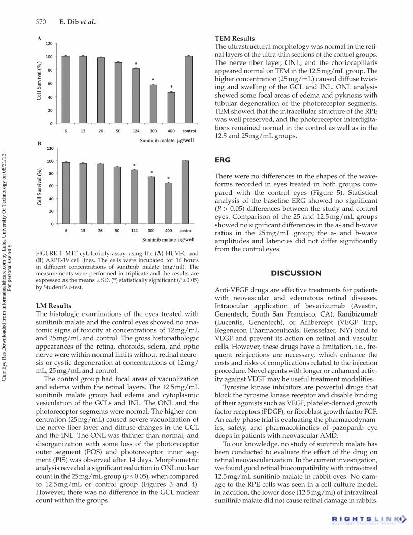
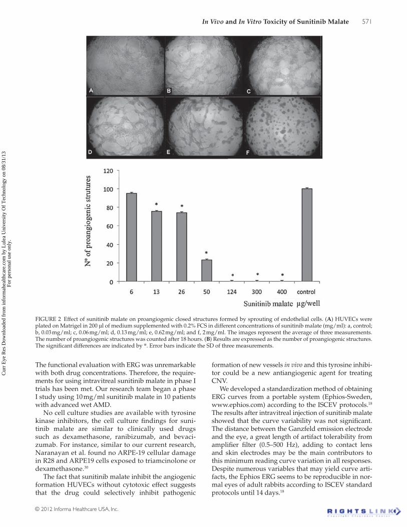
The Matrigel assay showed strong inhibition of proangiogenic structures on the HUVECs (Figures 1A and 2), which indicated the important effect of sunitinib malate.27–29 In addition, effective concentrations of suni-tinib malate that inhibited the proangiogenic structures were non-cytotoxic to the HUVECS and ARPE cell lines (Figure 1A and 1B). However, after administration of 0.62 mg/ml (124 µg/well), the viability of both cell lines was inhibited significantly.
In Vivo Model
Fundus Images and FANo alterations in the fundus images were seen in any study eyes in any group at baseline, 24 hours, and 14 days after injections, and there were no changes in FA in any time during study. No changes were seen in the ret-ina or choroidal and retinal vessels during follow-up.
Cur
r E
ye R
es D
ownl
oade
d fr
om in
form
ahea
lthca
re.c
om b
y L
ulea
Uni
vers
ity O
f T
echn
olog
y on
08/
31/1
3Fo
r pe
rson
al u
se o
nly.
570 E. Dib et al.
Current Eye Research
LM ResultsThe histologic examinations of the eyes treated with sunitinib malate and the control eyes showed no ana-tomic signs of toxicity at concentrations of 12 mg/mL and 25 mg/mL and control. The gross histopathologic appearances of the retina, choroids, sclera, and optic nerve were within normal limits without retinal necro-sis or cystic degeneration at concentrations of 12 mg/mL, 25 mg/mL and control.
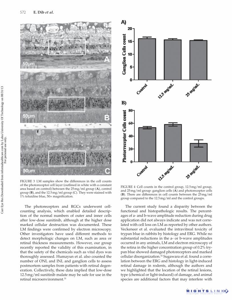
The control group had focal areas of vacuolization and edema within the retinal layers. The 12.5 mg/mL sunitinib malate group had edema and cytoplasmic vesiculation of the GCLs and INL. The ONL and the photoreceptor segments were normal. The higher con-centration (25 mg/mL) caused severe vacuolization of the nerve fiber layer and diffuse changes in the GCL and the INL. The ONL was thinner than normal, and disorganization with some loss of the photoreceptor outer segment (POS) and photoreceptor inner seg-ment (PIS) was observed after 14 days. Morphometric analysis revealed a significant reduction in ONL nuclear count in the 25 mg/mL group (p ≤ 0.05), when compared to 12.5 mg/mL or control group (Figures 3 and 4). However, there was no difference in the GCL nuclear count within the groups.
TEM ResultsThe ultrastructural morphology was normal in the reti-nal layers of the ultra-thin sections of the control groups. The nerve fiber layer, ONL, and the choriocapillaris appeared normal on TEM in the 12.5 mg/mL group. The higher concentration (25 mg/mL) caused diffuse twist-ing and swelling of the GCL and INL. ONL analysis showed some focal areas of edema and pyknosis with tubular degeneration of the photoreceptor segments. TEM showed that the intracellular structure of the RPE was well preserved, and the photoreceptor interdigita-tions remained normal in the control as well as in the 12.5 and 25 mg/mL groups.
ERG
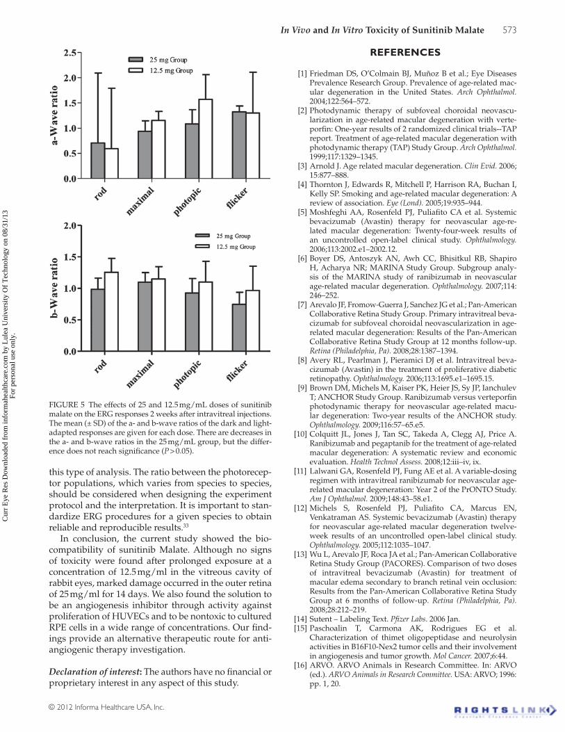
There were no differences in the shapes of the wave-forms recorded in eyes treated in both groups com-pared with the control eyes (Figure 5). Statistical analysis of the baseline ERG showed no significant (P > 0.05) differences between the study and control eyes. Comparison of the 25 and 12.5 mg/mL groups showed no significant differences in the a- and b-wave ratios in the 25 mg/mL group; the a- and b-wave amplitudes and latencies did not differ significantly from the control eyes.
DISCUSSION
Anti-VEGF drugs are effective treatments for patients with neovascular and edematous retinal diseases. Intraocular application of bevacizumab (Avastin, Genentech, South San Francisco, CA), Ranibizumab (Lucentis, Genentech), or Aflibercept (VEGF Trap, Regeneron Pharmaceuticals, Rensselaer, NY) bind to VEGF and prevent its action on retinal and vascular cells. However, these drugs have a limitation, i.e., fre-quent reinjections are necessary, which enhance the costs and risks of complications related to the injection procedure. Novel agents with longer or enhanced activ-ity against VEGF may be useful treatment modalities.
Tyrosine kinase inhibitors are powerful drugs that block the tyrosine kinase receptor and disable binding of their agonists such as VEGF, platelet-derived growth factor receptors (PDGF), or fibroblast growth factor FGF. An early-phase trial is evaluating the pharmacodynam-ics, safety, and pharmacokinetics of pazopanib eye drops in patients with neovascular AMD.
To our knowledge, no study of sunitinib malate has been conducted to evaluate the effect of the drug on retinal neovascularization. In the current investigation, we found good retinal biocompatibility with intravitreal 12.5 mg/mL sunitinib malate in rabbit eyes. No dam-age to the RPE cells was seen in a cell culture model; in addition, the lower dose (12.5 mg/ml) of intravitreal sunitinib malate did not cause retinal damage in rabbits.
FIGURE 1 MTT cytotoxicity assay using the (A) HUVEC and (B) ARPE-19 cell lines. The cells were incubated for 16 hours in different concentrations of sunitinib malate (mg/ml). The measurements were performed in triplicate and the results are expressed as the means ± SD. (*) statistically significant (P ≤ 0.05) by Student’s t-test.
Cur
r E
ye R
es D
ownl
oade
d fr
om in
form
ahea
lthca
re.c
om b
y L
ulea
Uni
vers
ity O
f T
echn
olog
y on
08/
31/1
3Fo
r pe
rson
al u
se o
nly.
In Vivo and In Vitro Toxicity of Sunitinib Malate 571
© 2012 Informa Healthcare USA, Inc.
The functional evaluation with ERG was unremarkable with both drug concentrations. Therefore, the require-ments for using intravitreal sunitinib malate in phase I trials has been met. Our research team began a phase I study using 10 mg/ml sunitinib malate in 10 patients with advanced wet AMD.
No cell culture studies are available with tyrosine kinase inhibitors, the cell culture findings for suni-tinib malate are similar to clinically used drugs such as dexamethasone, ranibizumab, and bevaci-zumab. For instance, similar to our current research, Naranayan et al. found no ARPE-19 cellular damage in R28 and ARPE19 cells exposed to triamcinolone or dexamethasone.30
The fact that sunitinib malate inhibit the angiogenic formation HUVECs without cytotoxic effect suggests that the drug could selectively inhibit pathogenic
formation of new vessels in vivo and this tyrosine inhibi-tor could be a new antiangiogenic agent for treating CNV.
We developed a standardization method of obtaining ERG curves from a portable system (Ephios-Sweden, www.ephios.com) according to the ISCEV protocols.18 The results after intravitreal injection of sunitinib malate showed that the curve variability was not significant. The distance between the Ganzfeld emission electrode and the eye, a great length of artifact tolerability from amplifier filter (0.5–500 Hz), adding to contact lens and skin electrodes may be the main contributors to this minimum reading curve variation in all responses. Despite numerous variables that may yield curve arti-facts, the Ephios ERG seems to be reproducible in nor-mal eyes of adult rabbits according to ISCEV standard protocols until 14 days.18
FIGURE 2 Effect of sunitinib malate on proangiogenic closed structures formed by sprouting of endothelial cells. (A) HUVECs were plated on Matrigel in 200 µl of medium supplemented with 0.2% FCS in different concentrations of sunitinib malate (mg/ml): a, control; b, 0.03 mg/ml; c, 0.06 mg/ml; d, 0.13 mg/ml; e, 0.62 mg/ml; and f, 2 mg/ml. The images represent the average of three measurements. The number of proangiogenic structures was counted after 18 hours. (B) Results are expressed as the number of proangiogenic structures. The significant differences are indicated by *. Error bars indicate the SD of three measurements.
Cur
r E
ye R
es D
ownl
oade
d fr
om in
form
ahea
lthca
re.c
om b
y L
ulea
Uni
vers
ity O
f T
echn
olog
y on
08/
31/1
3Fo
r pe
rson
al u
se o
nly.
572 E. Dib et al.
Current Eye Research
The photoreceptors and RGCs underwent cell-counting analysis, which enabled detailed descrip-tion of the normal numbers of outer and inner cells after low-dose sunitinib, although at the higher dose marked cellular destruction was documented. These LM findings were confirmed by electron microscopy. Other investigators have used different methods to detect morphologic changes on LM, such as area or retinal thickness measurements. However, our group recently reported the validity of this examination, in that the safety of the chemicals such as vital dyes was thoroughly assessed. Humayun et al. also counted the number of ONL and INL and ganglion cells to assess postmortem samples from patients with retinal degen-eration. Collectively, these data implied that low-dose 12.5 mg/ml sunitinib malate may be safe for use in the retinal microenvironment.31
The current study found a disparity between the functional and histopathologic results. The percent-ages of a- and b-wave amplitude reduction during drug application did not always indicate and was not corre-lated with cell loss on LM as reported by other authors. Veckeneer et al. evaluated the intravitreal toxicity of trypan blue in rabbits by histology and ERG. While no substantial reductions in the a- or b-wave amplitudes occurred in any animals, LM and electron microscopy of the retina in the higher concentration group of 0.2% try-pan blue showed damaged photoreceptors and marked cellular disorganization.32 Sugawara et al. found a corre-lation between the ERG and histology in light-induced retinal damage in rodents, although the authors and we highlighted that the location of the retinal lesions, type (chemical or light-induced) of damage, and animal species are additional factors that may interfere with
FIGURE 3 LM samples show the differences in the cell counts of the photoreceptor cell layer (outlined in white with a constant area based on control) between the 25 mg/ml group (A), control group (B), and the 12.5 mg/ml group (C). They were stained with 1% toluidine blue, 50× magnification.
FIGURE 4 Cell counts in the control group, 12.5 mg/ml group, and 25 mg/ml group: ganglion cells (A) and photoreceptor cells (B). There are differences in cell counts between the 25 mg/ml group compared to the 12.5 mg/ml and the control groups.
Cur
r E
ye R
es D
ownl
oade
d fr
om in
form
ahea
lthca
re.c
om b
y L
ulea
Uni
vers
ity O
f T
echn
olog
y on
08/
31/1
3Fo
r pe
rson
al u
se o
nly.
In Vivo and In Vitro Toxicity of Sunitinib Malate 573
© 2012 Informa Healthcare USA, Inc.
this type of analysis. The ratio between the photorecep-tor populations, which varies from species to species, should be considered when designing the experiment protocol and the interpretation. It is important to stan-dardize ERG procedures for a given species to obtain reliable and reproducible results.33
In conclusion, the current study showed the bio-compatibility of sunitinib Malate. Although no signs of toxicity were found after prolonged exposure at a concentration of 12.5 mg/ml in the vitreous cavity of rabbit eyes, marked damage occurred in the outer retina of 25 mg/ml for 14 days. We also found the solution to be an angiogenesis inhibitor through activity against proliferation of HUVECs and to be nontoxic to cultured RPE cells in a wide range of concentrations. Our find-ings provide an alternative therapeutic route for anti-angiogenic therapy investigation.
Declaration of interest: The authors have no financial or proprietary interest in any aspect of this study.
REFERENCES
[1] Friedman DS, O’Colmain BJ, Muñoz B et al.; Eye Diseases Prevalence Research Group. Prevalence of age-related mac-ular degeneration in the United States. Arch Ophthalmol. 2004;122:564–572.
[2] Photodynamic therapy of subfoveal choroidal neovascu-larization in age-related macular degeneration with verte-porfin: One-year results of 2 randomized clinical trials--TAP report. Treatment of age-related macular degeneration with photodynamic therapy (TAP) Study Group. Arch Ophthalmol. 1999;117:1329–1345.
[3] Arnold J. Age related macular degeneration. Clin Evid. 2006; 15:877–888.
[4] Thornton J, Edwards R, Mitchell P, Harrison RA, Buchan I, Kelly SP. Smoking and age-related macular degeneration: A review of association. Eye (Lond). 2005;19:935–944.
[5] Moshfeghi AA, Rosenfeld PJ, Puliafito CA et al. Systemic bevacizumab (Avastin) therapy for neovascular age-re-lated macular degeneration: Twenty-four-week results of an uncontrolled open-label clinical study. Ophthalmology. 2006;113:2002.e1–2002.12.
[6] Boyer DS, Antoszyk AN, Awh CC, Bhisitkul RB, Shapiro H, Acharya NR; MARINA Study Group. Subgroup analy-sis of the MARINA study of ranibizumab in neovascular age-related macular degeneration. Ophthalmology. 2007;114: 246–252.
[7] Arevalo JF, Fromow-Guerra J, Sanchez JG et al.; Pan-American Collaborative Retina Study Group. Primary intravitreal beva-cizumab for subfoveal choroidal neovascularization in age-related macular degeneration: Results of the Pan-American Collaborative Retina Study Group at 12 months follow-up. Retina (Philadelphia, Pa). 2008;28:1387–1394.
[8] Avery RL, Pearlman J, Pieramici DJ et al. Intravitreal beva-cizumab (Avastin) in the treatment of proliferative diabetic retinopathy. Ophthalmology. 2006;113:1695.e1–1695.15.
[9] Brown DM, Michels M, Kaiser PK, Heier JS, Sy JP, Ianchulev T; ANCHOR Study Group. Ranibizumab versus verteporfin photodynamic therapy for neovascular age-related macu-lar degeneration: Two-year results of the ANCHOR study. Ophthalmology. 2009;116:57–65.e5.
[10] Colquitt JL, Jones J, Tan SC, Takeda A, Clegg AJ, Price A. Ranibizumab and pegaptanib for the treatment of age-related macular degeneration: A systematic review and economic evaluation. Health Technol Assess. 2008;12:iii–iv, ix.
[11] Lalwani GA, Rosenfeld PJ, Fung AE et al. A variable-dosing regimen with intravitreal ranibizumab for neovascular age-related macular degeneration: Year 2 of the PrONTO Study. Am J Ophthalmol. 2009;148:43–58.e1.
[12] Michels S, Rosenfeld PJ, Puliafito CA, Marcus EN, Venkatraman AS. Systemic bevacizumab (Avastin) therapy for neovascular age-related macular degeneration twelve-week results of an uncontrolled open-label clinical study. Ophthalmology. 2005;112:1035–1047.
[13] Wu L, Arevalo JF, Roca JA et al.; Pan-American Collaborative Retina Study Group (PACORES). Comparison of two doses of intravitreal bevacizumab (Avastin) for treatment of macular edema secondary to branch retinal vein occlusion: Results from the Pan-American Collaborative Retina Study Group at 6 months of follow-up. Retina (Philadelphia, Pa). 2008;28:212–219.
[14] Sutent – Labeling Text. Pfizer Labs. 2006 Jan.[15] Paschoalin T, Carmona AK, Rodrigues EG et al.
Characterization of thimet oligopeptidase and neurolysin activities in B16F10-Nex2 tumor cells and their involvement in angiogenesis and tumor growth. Mol Cancer. 2007;6:44.
[16] ARVO. ARVO Animals in Research Committee. In: ARVO (ed.). ARVO Animals in Research Committee. USA: ARVO; 1996: pp. 1, 20.
FIGURE 5 The effects of 25 and 12.5 mg/mL doses of sunitinib malate on the ERG responses 2 weeks after intravitreal injections. The mean (± SD) of the a- and b-wave ratios of the dark and light-adapted responses are given for each dose. There are decreases in the a- and b-wave ratios in the 25 mg/mL group, but the differ-ence does not reach significance (P > 0.05).
Cur
r E
ye R
es D
ownl
oade
d fr
om in
form
ahea
lthca
re.c
om b
y L
ulea
Uni
vers
ity O
f T
echn
olog
y on
08/
31/1
3Fo
r pe
rson
al u
se o
nly.
574 E. Dib et al.
Current Eye Research
[17] Price N, Gottfried MR, Clary E et al. Safety and efficacy of India ink and indocyanine green as colonic tattooing agents. Gastrointest Endosc. 2000;51:438–442.
[18] Marmor MF, Fulton AB, Holder GE, Miyake Y, Brigell M, Bach M; International Society for Clinical Electrophysiology of Vision. ISCEV Standard for full-field clinical electroretin-ography (2008 update). Doc Ophthalmol. 2009;118:69–77.
[19] Zemel E, Loewenstein A, Lei B, Lazar M, Perlman I. Ocular pigmentation protects the rabbit retina from gentamicin-in-duced toxicity. Invest Ophthalmol Vis Sci. 1995;36:1875–1884.
[20] Kwok AK, Lai TY, Yeung CK, Yeung YS, Li WW, Chiang SW. The effects of indocyanine green and endoillumination on rabbit retina: An electroretinographic and histological study. Br J Ophthalmol. 2005;89:897–900.
[21] Inan UU, Avci B, Kusbeci T, Kaderli B, Avci R, Temel SG. Preclinical safety evaluation of intravitreal injection of full-length humanized vascular endothelial growth fac-tor antibody in rabbit eyes. Invest Ophthalmol Vis Sci. 2007;48:1773–1781.
[22] Rodrigues EB, Penha FM, Farah ME et al. Preclinical investigation of the retinal biocompatibility of six novel vital dyes for chromovitrectomy. Retina (Philadelphia, Pa). 2009;29:497–510.
[23] Penha FM, Maia M, Farah ME et al. Morphologic and clinical effects of subretinal injection of indocyanine green and infracyanine green in rabbits. J Ocul Pharmacol Ther. 2008;24:52–61.
[24] Penha FM, Maia M, Eid Farah M et al. Effects of subretinal injections of indocyanine green, trypan blue, and glucose in rabbit eyes. Ophthalmology. 2007;114:899–908.
[25] Maia M, Kellner L, de Juan E Jr et al. Effects of indocyanine green injection on the retinal surface and into the subretinal space in rabbits. Retina (Philadelphia, Pa). 2004;24:80–91.
[26] Rodrigues EB, Penha FM, de Paula Fiod Costa E et al. Ability of new vital dyes to stain intraocular membranes and tissues in ocular surgery. Am J Ophthalmol. 2010;149:265–277.
[27] Korpanty G, Sullivan LA, Smyth E, Carney DN, Brekken RA. Molecular and clinical aspects of targeting the VEGF path-way in tumors. J Oncol. 2010;2010:652320.
[28] Ropert S, Vignaux O, Mir O, Goldwasser F. VEGF pathway inhibition by anticancer agent sunitinib and susceptibil-ity to atherosclerosis plaque disruption. Invest New Drugs. 2011;29:1497–1499.
[29] Sablin MP, Dreyer C, Colichi C et al. Benefits from phar-macological and pharmacokinetic properties of sunitinib for clinical development. Expert Opin Drug Metab Toxicol. 2010;6:1005–1015.
[30] Narayanan R, Mungcal JK, Kenney MC, Seigel GM, Kuppermann BD. Toxicity of triamcinolone acetonide on retinal neurosensory and pigment epithelial cells. Invest Ophthalmol Vis Sci. 2006;47:722–728.
[31] Maia M, Margalit E, Lakhanpal R et al. Effects of intravitreal indocyanine green injection in rabbits. Retina (Philadelphia, Pa). 2004;24:69–79.
[32] Veckeneer M, van Overdam K, Monzer J et al. Ocular toxicity study of trypan blue injected into the vitreous cavity of rabbit eyes. Graefes Arch Clin Exp Ophthalmol. 2001;239:698–704.
[33] Sugawara T, Sieving PA, Bush RA. Quantitative relationship of the scotopic and photopic ERG to photoreceptor cell loss in light damaged rats. Exp Eye Res. 2000;70:693–705.
Cur
r E
ye R
es D
ownl
oade
d fr
om in
form
ahea
lthca
re.c
om b
y L
ulea
Uni
vers
ity O
f T
echn
olog
y on
08/
31/1
3Fo
r pe
rson
al u
se o
nly.