in vivo identification of regulators of cell invasion...
TRANSCRIPT

(120), ra35. [DOI: 10.1126/scisignal.2000654] 3Science SignalingWeiss and David R. Sherwood (4 May 2010) David Q. Matus, Xiao-Yan Li, Sarah Durbin, Daniel Agarwal, Qiuyi Chi, Stephen J.MembranesIn Vivo Identification of Regulators of Cell Invasion Across Basement
This information is current as of 29 May 2010. The following resources related to this article are available online at http://stke.sciencemag.org.
Article Tools http://stke.sciencemag.org/cgi/content/full/sigtrans;3/120/ra35
Visit the online version of this article to access the personalization and article tools:
MaterialsSupplemental
http://stke.sciencemag.org/cgi/content/full/sigtrans;3/120/ra35/DC1 "Supplementary Materials"
Related Content http://stke.sciencemag.org/cgi/content/abstract/sigtrans;3/121/pc10
's sites:ScienceThe editors suggest related resources on
References http://stke.sciencemag.org/cgi/content/full/sigtrans;3/120/ra35#BIBL
1 article(s) hosted by HighWire Press; see: cited byThis article has been
http://stke.sciencemag.org/cgi/content/full/sigtrans;3/120/ra35#otherarticlesThis article cites 39 articles, 11 of which can be accessed for free:
Glossary http://stke.sciencemag.org/glossary/
Look up definitions for abbreviations and terms found in this article:
Permissions http://www.sciencemag.org/about/permissions.dtl
Obtain information about reproducing this article:
the American Association for the Advancement of Science; all rights reserved. byAssociation for the Advancement of Science, 1200 New York Avenue, NW, Washington, DC 20005. Copyright 2008
(ISSN 1937-9145) is published weekly, except the last week in December, by the AmericanScience Signaling
on May 29, 2010
stke.sciencemag.org
Dow
nloaded from

R E S E A R C H A R T I C L E
C E L L B I O L O G Y
In Vivo Identification of Regulators of Cell InvasionAcross Basement MembranesDavid Q. Matus,1 Xiao-Yan Li,2,3 Sarah Durbin,1 Daniel Agarwal,1* Qiuyi Chi,1
Stephen J. Weiss,2,3 David R. Sherwood1†
(Published 4 May 2010; Volume 3 Issue 120 ra35)
Dow
nloade
Cell invasion through basement membranes during development, immune surveillance, and metastasisremains poorly understood. To gain further insight into this key cellular behavior, we performed an in vivoscreen for regulators of cell invasion through basement membranes, using the simple model of Caeno-rhabditis elegans anchor cell invasion, and identified 99 genes that promote invasion, including the genesencoding the chaperonin complex cct. Notably, most of these genes have not been previously implicatedin invasive cell behavior. We characterized members of the cct complex and 11 other gene products,determining the distinct aspects of the invasive cascade that they regulate, including formation of aspecialized invasive cell membrane and its ability to breach the basement membrane. RNA interference–mediated knockdown of the human orthologs of cct-5 and lit-1, which had not previously been implicated incell invasion, reduced the invasiveness of metastatic carcinoma cells, suggesting that a conserved geneticprogram underlies cell invasion. These results increase our understanding of the genetic underpinnings ofcell invasion and also provide new potential therapeutic targets to limit this behavior.
d fro
on May 29, 2010 stke.sciencem
ag.orgm
INTRODUCTIONThe ability of cells to traverse the barriers imposed by the basement mem-brane (BM), the dense sheet-like extracellular matrix that surrounds mosttissues, is a critical cellular behavior that occurs during both normal on-togeny and immune system function (1). For example, trophoblast cellsbreach the endometrium BM to establish the placenta (2), and leukocytescross the perivascular BM to reach sites of infection and injury (3). Un-controlled invasive cell activity is also associated with various human dis-eases, most notably cancer, where transformed cells are thought to hijackdevelopmental invasive programs to metastasize (4, 5). Despite its impor-tance in both development and human disease, cell invasion through theBM remains poorly understood (1, 3, 6–8). Most of the work on cell in-vasion has been limited to in vitro models, which do not reflect the in vivomicroenvironment and endogenous BM architecture (9, 10). Althoughrecent advances in imaging technology are providing new insights intocell invasion in vertebrates (11), it remains challenging to perform func-tional perturbations in these models and simultaneously visualize thecomplex, dynamic process of cell invasion through BMs.
Anchor cell (AC) invasion in C. elegans is a simple model of cellinvasion through BMs that combines forward genetics with single-cellvisual analysis. During C. elegans larval development, the AC, a spe-cialized gonadal cell, breaches the gonadal and ventral epidermal BMsto contact the central primary-fated vulval precursor cells (1° VPCs), ini-tiating uterine-vulval connection (12, 13). AC invasion is a regulated androbust process, which occurs invariantly before the P6.p four-cell stage inwild-type animals (Fig. 1A) (12). During the L2-L3 molt (approximately
1Biology Department, Duke University, Durham, NC 27708, USA. 2Divisionof Molecular Medicine and Genetics, Department of Internal Medicine,University of Michigan, Ann Arbor, MI 48109, USA. 3Life Sciences Institute,University of Michigan, Ann Arbor, MI 48109, USA.*Present address: Weill Cornell Medical College, 1300 York Avenue, NewYork, NY 10065, USA.†To whom correspondence should be addressed. E-mail: [email protected]
6 hours before invasion), a specialized invasive cell membrane, rich inF-actin and actin regulators, is established in the AC through coordi-nation of netrin (14) and integrin (15) signaling at the interface of theAC and BM (Fig. 1A). AC invasion is stimulated by an unidentifiedchemotactic cue secreted by the underlying 1° VPCs (Fig. 1A). The abil-ity of the AC to breach the underlying BMs in response to this cue isdependent on two oncogenes, the bZIP transcription factor (TF) fos-1a(13) and zinc finger TF, egl-43L, the C. elegans ortholog of vertebrateEVI1 and MEL paralogs (16, 17). Together, these TFs regulate the ex-pression of the zinc metalloproteinase zmp-1 as well as other targets en-coding pro-invasive factors in the AC (13).
Toward the goal of comprehensively identifying regulators of cellinvasion through the BM in vivo, we have performed a focused whole-genome RNA interference (RNAi) screen. Here, we report the identifi-cation of 99 regulators of AC invasion, most of which have not beenpreviously implicated in invasion or metastasis. We have further charac-terized the genes encoding the most robust pro-invasive factors, includingmembers of the cct complex and 11 others, encompassing both knownoncogenes and previously unknown regulators of cell invasion. Notably,small interfering RNA (siRNA)–mediated knockdown of two of thesepro-invasive genes reduced the invasiveness of metastatic carcinomacells, suggesting that our approach has identified conserved regulatorsthat might be potential therapeutic targets in halting cancer progression.
RESULTS
An RNAi screen identifies 99 regulators of AC invasionA defect in AC invasion disrupts uterine-vulval attachment and results inan easily observed Protruded vulva (Pvl) and Egg-laying defective (Egl)phenotype. We have taken advantage of data collected from a number ofwhole-genome RNAi screens that have identified the complement ofgenes giving a Pvl or Egl phenotype after RNAi depletion (18–20). Usingthese data, we examined 539 genes that produce a Pvl or Egl phenotype
www.SCIENCESIGNALING.org 4 May 2010 Vol 3 Issue 120 ra35 1

R E S E A R C H A R T I C L E
Dow
nl
after RNAi knockdown, using Nomarski optics at the time of AC invasion(table S1). We identified 99 genes whose reduction results in a failure ofthe AC to breach the BM, as evidenced by an unbroken phase-dense lineunderneath the AC (Fig. 1B and tables S2 and S3). Ninety-five percent(94 of 99) of these genes have human orthologs as determined byBLASTP analysis, of which 90% (85 of 94 genes) have not been previouslyimplicated in cell invasion or cancer metastasis (table S2). Validating thespecificity and rigor of this approach, we identified components of ge-netic pathways known to promote AC invasion, including the TFs fos-1a(13) and egl-43L (16, 17); the netrin receptor unc-40 (14); and the integrina subunit ina-1 (15) (Table 1 and table S2).
Eleven pro-invasive genes and the cct complexregulate the AC’s ability to breach the BMTo focus on the most crucial regulators of AC invasion, we further char-acterized genes whose RNAi depletion gave a robust AC invasion defect,which we set at >30%, a degree of penetrance similar to invasion defectsresulting from RNAi directed against members of known AC invasionpathways, including FOS-1A and EGL-43L activity, as well as INA-1and UNC-40 signaling (Table 1 and tables S2 and S3). This list includesgenes with human orthologs that have been implicated directly in cell
invasion and metastasis [hda-1, a histone deactylase (21), and cdc-37,a co-chaperone of hsp90 (22)], as well as previously unknown regulators(mep-1, C48A7.2, cacn-1, F28F8.5, T20B12.1, lit-1, T03F1.8, hbl-1, unc-62,and the cct complex) (Table 1). The genes whose RNAi knockdown gavethe most robust AC invasion defect (>79%) included seven of the eight-member cct chaperonin complex (Table 1 and tables S2 and S3). Be-cause AC invasion defects were similar for all cct genes tested (Table1 and table S2), we further characterized several members of this com-plex, cct-2, cct-5, cct-6, and cct-7 (henceforth referred to as cct). Consist-ent with our RNAi results, AC invasion defects were detected in putativenull alleles for mep-1, hda-1, C48A7.2, and cct-6 (table S4 and fig. S1).For genes lacking putative null alleles or whose loss of function preventedanimals from surviving to the time of AC invasion, we observed AC inva-sion defects by using multiple double-stranded RNA (dsRNA) constructs,ruling out off-target RNAi effects (23) (table S4). To verify that the observedAC invasion defect was specifically because of failure to breach the under-lying BM, we examined the integrity of laminin, a key structural componentof BM. After RNAi depletion targeting cct and the remaining 11 genes,LAM-1::GFP (laminin) remained intact under the AC after a failure to in-vade, confirming that the invasion defects were due to an inability to breakthrough the BM (Fig. 1B and fig. S2).
on May 29, 2010
stke.sciencemag.org
oaded from
A
B
EmptyVector
hda-1(RNAi)
cdc-37(RNAi)
Nomarski Fluorescence Overlay
LAMININ::GFPAC
VNC
wild-type L2-L3 molt
Netrin and integrinpolarization
AC
BM
AC
BM
wild-type late L3
Contact with 1O VPCs(Invasion complete)
AC
BM1 VPC
wild-type mid-to-late L3
O
Basement membrane removal and response
to vulval cue
FOS-1A/EGL-43L
P6.p 1-cell stage P6.p 2-cell stage P6.p 4-cell stage
1 VPCO1 VPCO
Fig. 1. Summary of C. elegans AC inva-sion and invasion defects after RNAi de-pletion. (A) Schematic representation ofthe known mechanisms underlying AC in-vasion. At the L2-L3 molt (P6.p one-cellstage; left), approximately 6 hours beforeinvasion, UNC-6 (netrin) secretion (yellowarrows) from the ventral nerve cord (VNC)and integrin signaling polarize the AC’sbasal cell membrane by recruiting F-actin,actin regulators, and the netrin receptor(UNC-40; orange) toward the juxtaposedBMs (shown in green) (14, 15). Duringthe mid-to-late L3 stage (P6.p two-cellstage; middle), an unidentified cue fromthe 1° VPCs (blue arrows) stimulates in-vasive protrusions from the AC that re-quire the activity of the TFs FOS-1A andEGL-43L to breach the BM. During thelate L3 stage (P6.p four-cell stage), theAC contacts the P6.p granddaughters,initiating the connection between thedeveloping uterine and the vulval epi-thelium. (B) Nomarski image (left), corre-sponding fluorescence image (middle),and overlay (right). Anterior is left and ven-tral is down. In wild-type or worms fed con-trol RNAi (L4440 empty vector; top panel),the AC (magenta, zmp-1>mCherry; whitearrowhead) breaches the BM (green,LAM-1::GFP) and contacts the central pri-mary fated P6.p granddaughters (bracket;P6.p four-cell stage). In contrast, after RNAi-mediated knockdown of hda-1 and cdc-37(middle and bottom panels, respectively),the BMs remain intact after a failure in ACinvasion (table S3). Scale bar, 5 mm.
www.SCIENCESIGNALING.org
4 May 2010 Vol 3 Issue 120 ra35 2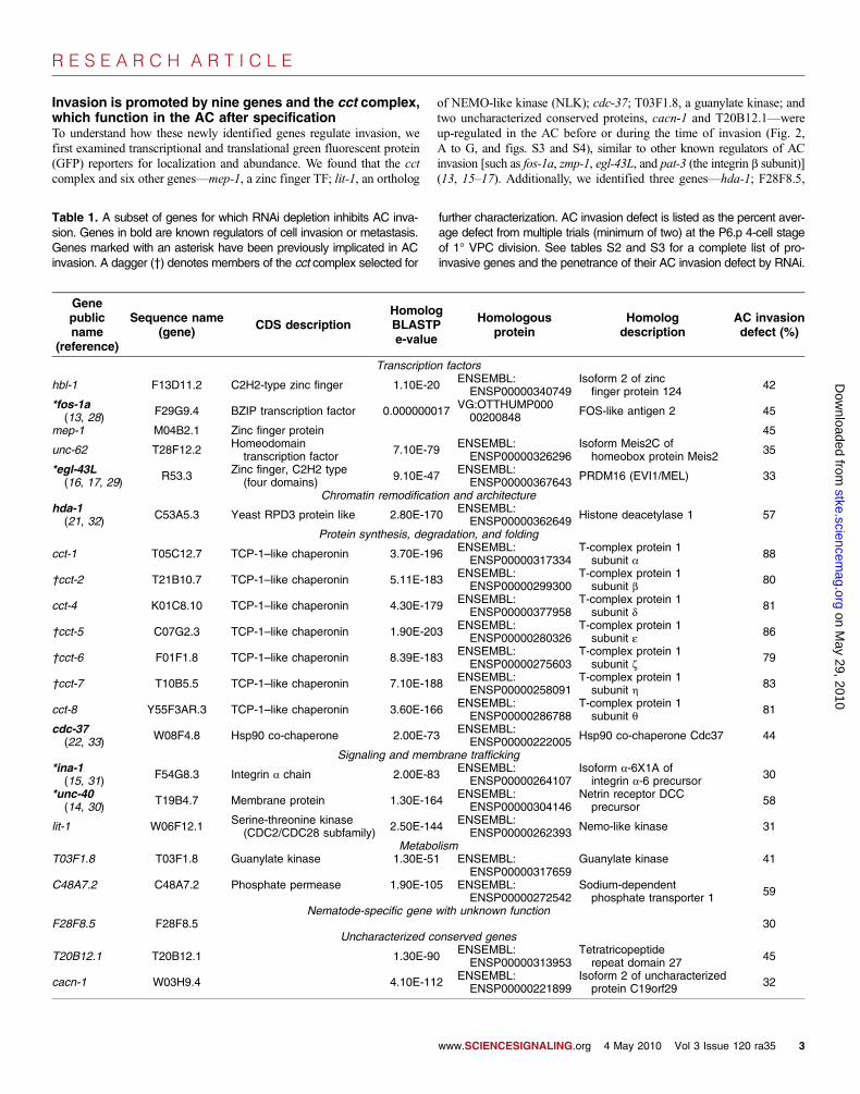
R E S E A R C H A R T I C L E
Invasion is promoted by nine genes and the cct complex,which function in the AC after specificationTo understand how these newly identified genes regulate invasion, wefirst examined transcriptional and translational green fluorescent protein(GFP) reporters for localization and abundance. We found that the cctcomplex and six other genes—mep-1, a zinc finger TF; lit-1, an ortholog
of NEMO-like kinase (NLK); cdc-37; T03F1.8, a guanylate kinase; andtwo uncharacterized conserved proteins, cacn-1 and T20B12.1—wereup-regulated in the AC before or during the time of invasion (Fig. 2,A to G, and figs. S3 and S4), similar to other known regulators of ACinvasion [such as fos-1a, zmp-1, egl-43L, and pat-3 (the integrin b subunit)](13, 15–17). Additionally, we identified three genes—hda-1; F28F8.5,
Table 1. A subset of genes for which RNAi depletion inhibits AC inva-sion. Genes in bold are known regulators of cell invasion or metastasis.Genes marked with an asterisk have been previously implicated in ACinvasion. A dagger (†) denotes members of the cct complex selected for
further characterization. AC invasion defect is listed as the percent aver-age defect from multiple trials (minimum of two) at the P6.p 4-cell stageof 1° VPC division. See tables S2 and S3 for a complete list of pro-invasive genes and the penetrance of their AC invasion defect by RNAi.
Genepublicname
(reference)
Sequence name(gene)
CDS descriptionHomologBLASTPe-value
ww
Homologousprotein
w.SCIENCESIGNALING.
Homologdescription
org 4 May 2010 Vol 3 Issue
AC invasiondefect (%)
Transcription factors
Do
hbl-1 F13D11.2 C2H2-type zinc finger 1.10E-20w
ENSEMBL:ENSP00000340749
Isoform 2 of zincfinger protein 124
42nloa
*fos-1a(13, 28)
F29G9.4 BZIP transcription factor 0.000000017 VG:OTTHUMP00000200848
FOS-like antigen 2 45d
mep-1 M04B2.1 Zinc finger protein 45 ed f unc-62 T28F12.2r
Homeodomaintranscription factor
7.10E-79 ENSEMBL:ENSP00000326296
Isoform Meis2C ofhomeobox protein Meis2
35som
*egl-43L(16, 17, 29)
R53.3 Zinc finger, C2H2 type(four domains)
9.10E-47 ENSEMBL:ENSP00000367643 PRDM16 (EVI1/MEL) 33tke
Chromatin remodification and architecture.sci
hda-1(21, 32)
C53A5.3 Yeast RPD3 protein like 2.80E-170 ENSEMBL:ENSP00000362649
Histone deacetylase 1 57en
Protein synthesis, degradation, and foldingcem
cct-1 T05C12.7 TCP-1–like chaperonin 3.70E-196 ENSEMBL:ENSP00000317334T-complex protein 1subunit a
88ag.
†cct-2 T21B10.7 TCP-1–like chaperonin 5.11E-183 oENSEMBL:ENSP00000299300
T-complex protein 1subunit b
80org
cct-4
K01C8.10 TCP-1–like chaperonin 4.30E-179n
ENSEMBL:ENSP00000377958
T-complex protein 1subunit d
81Ma
†cct-5 C07G2.3 TCP-1–like chaperonin 1.90E-203y
ENSEMBL:ENSP00000280326
T-complex protein 1subunit e
8629
†cct-6 F01F1.8 TCP-1–like chaperonin 8.39E-183,
ENSEMBL:ENSP00000275603
T-complex protein 1subunit z
7920
†cct-7 T10B5.5 TCP-1–like chaperonin 7.10E-1881
ENSEMBL:ENSP00000258091
T-complex protein 1subunit h
830
cct-8 Y55F3AR.3 TCP-1–like chaperonin 3.60E-166 ENSEMBL:ENSP00000286788T-complex protein 1subunit q
81cdc-37(22, 33)
W08F4.8 Hsp90 co-chaperone 2.00E-73 ENSEMBL:ENSP00000222005
Hsp90 co-chaperone Cdc37 44Signaling and membrane trafficking
*ina-1(15, 31)
F54G8.3 Integrin a chain 2.00E-83 ENSEMBL:ENSP00000264107Isoform a-6X1A ofintegrin a-6 precursor
30*unc-40(14, 30)
T19B4.7 Membrane protein 1.30E-164 ENSEMBL:ENSP00000304146
Netrin receptor DCCprecursor
58lit-1
W06F12.1 Serine-threonine kinase(CDC2/CDC28 subfamily) 2.50E-144 ENSEMBL:ENSP00000262393
Nemo-like kinase 31Metabolism
T03F1.8 T03F1.8 Guanylate kinase 1.30E-51 ENSEMBL:ENSP00000317659
Guanylate kinase 41C48A7.2
C48A7.2 Phosphate permease 1.90E-105 ENSEMBL:ENSP00000272542Sodium-dependentphosphate transporter 1
59Nematode-specific gene with unknown function
F28F8.5 F28F8.5 30Uncharacterized conserved genes
T20B12.1
T20B12.1 1.30E-90 ENSEMBL:ENSP00000313953Tetratricopeptiderepeat domain 27
45cacn-1
W03H9.4 4.10E-112 ENSEMBL:ENSP00000221899Isoform 2 of uncharacterizedprotein C19orf29
32120 ra35 3

R E S E A R C H A R T I C L E
an uncharacterized nematode-specific gene; and C48A7.2, a sodium andphosphate transporter—which were expressed in most cells, includingthe AC (Fig. 2, H to J, and figs. S3 and S4). hbl-1 and unc-62 (theC. elegans orthologs of the TFs hunchback and homothorax, respective-ly) were not detected in the AC before or during the time of invasion, butwere expressed in the underlying vulval cells (unc-62>GFP) or in theventral nerve cord and underlying vulval cells at earlier stages of devel-
opment (hbl-1::GFP) (Fig. 2, K and L, and fig. S5). The absence oflocalization in the AC and expression in the vulval cells suggested thathbl-1 and unc-62 might act in VPCs to promote invasion. A summary ofthe localization of the proteins encoded by newly identified pro-invasivegenes is shown in Fig. 2M.
AC invasion relies on the proper specification of the AC and theunderlying 1° VPCs, which generate the invasive cue (12). To determine
on May 29, 2010
stke.sciencemag.org
Dow
nloaded from
O
CDC-37, LIT-1, T20B12.1,CACN-1, GUK-1, CCT-7
C48A7.2
F-ACTIN, MIG-2, UNC-40, UNC-34, ZMP-1, CDH-3, INA-1, PAT-3 HEMICENTIN
FOS-1A, EGL-43L, HDA-1, LIT-1, F28F8.5, CACN-1, T03F1.8, CCT-7CACN-1
Cytosol
Plasma membrane
Invasivemembrane
ECM
Nucleus
Nucleolus
H
IB
D
G
J
E
F
ANomarski Fluorescence
C
K
L
Nomarski Fluorescence
AC
BM
1 VPC
Wild-type mid-to-late L3
UNC-62
UNC-6 / HBL-1
M
MEP-1::GFP
HDA-1::GFP
F28F8.5::GFP
CDC-37::YFP
T20B12.1::GFP
C48A7.2::GFP
T03F1.8::YFP
cacn-1>GFP
CCT-7::GFP
GFP::LIT-1
unc-62 >GFP
HBL-1::GFP
Fig. 2. Transgene reporter localization of newly identified genes that reg-ulate AC invasion. Nomarksi image, left; corresponding fluorescenceimage, right, at the P6.p one-cell stage. Anterior is left and ventral is down.All images are confocal z slices except (C) and (D), which are wide-fieldfluorescence images. (A to J) Translational (::) and transcriptional (>)reporter constructs for the cct complex (as shown by cct-7::GFP) and9 of the 11 newly identified pro-invasive genes showed AC-enriched(arrow) GFP localization in various subcellular compartments beforeand during AC invasion. (K) A transcriptional reporter for unc-62 (unc-
62>GFP) showed VPC expression before and during the time of AC in-vasion (white brackets). (L) A translational reporter for HBL-1 (hbl-1::GFP)was detected in the cell bodies of the ventral nerve cord (black arrow-heads) before and during the time of AC invasion. Although HBL-1::GFPis not localized to the VPCs at the time of invasion, it is expressed inVPCs hours before invasion (fig. S5). (M) Summary diagram of the sub-cellular localization of proteins in the AC, underlying 1° VPCs, and VNCduring invasion. Genes identified in this study are bolded. Scale bar,5 mm.
www.SCIENCESIGNALING.org 4 May 2010 Vol 3 Issue 120 ra35 4

R E S E A R C H A R T I C L E
whether these newly identified genes regulate invasion by causing defects inAC or 1° VPC cell specification, we examined the expression of lin-3>GFPand egl-17>GFP, markers of AC and 1° VPC cell fate, respectively(24, 25). lin-3>GFP was detected in the AC after RNAi-mediated deple-tion of the cct complex and the remaining 11 genes, indicating that AC
specification was normal despite reduction of activity of these genes(fig. S6). The expression of egl-17>GFP in the underlying vulval cellswas lost only after depletion of unc-62 by RNAi, consistent with a rolefor unc-62 in regulating invasion by controlling 1° VPC specification(fig. S7). Additionally, RNAi knockdown of hbl-1 resulted in precocious
on May 29, 2010
stke.sciencemag.org
Dow
nloaded from
L444
0(e
mpt
y ve
ctor
)hd
a-1(
RN
Ai)
cacn
-1(R
NA
i)T
03F
1.8(
RN
Ai)
zmp-1>CFP
Downstreamtarget of
fos-1a pathway
FOS-1A::YFP
Regulatefos-1a pathway
F-actinmCherry::moesinABD
Invasivemembrane
AB
C
** **
**
** ** * * *
**
1
1.5
2
2.5
3
3.5
4
Vul
cct-
5
cct-
7
hda-
1
mep
-1
fos-
1a
C48
A7.
2
cacn
-1
F28
F8.
5
T20
B12
.1
lit-1
cdc-
37
T03
F1.
8
Perturbs F-actin localization
* * * *
F-actin polarity
Average polarity ratio
L444
0
cct-
5
cct-
7
hda-
1
mep
-1
fos-
1a
C48
A7.
2
cacn
-1
F28
F8.
5
T20
B12
.1
lit-1
cdc-
37
T03
F1.
80
25
5075
100
125
150
175
200
225
Upstream of fos-1aUpstream of zmp-1
FOS-1A::YFP zmp-1>CFP
% F
luor
esce
nce
Inte
nsity
fos-1a pathway reporter analysis
Fig. 3. Newly identified regulators of AC invasion function withinestablished as well as previously uncharacterized pathways to promoteinvasion. (A) Confocal z slice fluorescence images of F-actin probe (left;mCherry::moeABD) and wide-field fluorescence images of FOS-1A::YFP(middle) and zmp-1>CFP (right) in empty vector control and hda-1,cacn-1, and T03F1.8 RNAi-depleted animals at the time of invasion. Asingle-factor ANOVA followed by Tukey’s post hoc test for significancewas used in all cases. For this and all subsequent figures, n refers to thenumber of animals per genotype. The top panel shows localization orexpression for all three reporters in wild-type ACs. RNAi depletion ofhda-1 resulted in a loss of F-actin at the invasive membrane (n = 18;P < 0.05) and reduced FOS-1A::YFP fluorescence in the AC to 57% ofthat in wild-type animals (n = 11; P < 0.05). In contrast, RNAi depletion ofcacn-1 (n = 11) or T03F1.8 (n = 17) did not affect polarization of F-actinat the invasive front (arrowhead) or significantly alter FOS-1A::YFP fluo-rescence in the AC (n >10; P > 0.05). zmp-1>CFP expression was sig-nificantly reduced after RNAi depletion of hda-1 or cacn-1 (n = 12 forboth; P < 0.05), but not of T03F1.8 (n = 10; P > 0.05). (B and C) Quan-tification of an F-actin probe (B) and FOS-1A::YFP/zmp-1>CFP (C) in
Vulvaless (Vul), empty vector control (L4440), and RNAi-depletedanimals in which the AC failed to invade (n >10 animals for each RNAidepletion). *P < 0.05. (B) RNAi depletion of the cct complex, hda-1, andmep-1 blocked polarization of F-actin at the invasive membrane (graybar). In Vul animals, AC invasion is blocked, but polarity is maintained(14). Knockdown of the other genes caused defects in AC invasion,although the ACs were still polarized. Loss of C48A7.2 resulted in reducedAC size, which could account for an F-actin polarity ratio that is greaterthan in Vul control animals. (C) RNAi knockdown of the cct complex,hda-1, and mep-1 decreased the expression of FOS-1A::YFP (P < 0.05;white bars) and its downstream target, zmp-1>CFP (P < 0.05; black bars)in the AC (denoted by the double asterisks). RNAi targeting five genes(C48A7.2, cacn-1, F28F8.5, T20B12.1, and lit-1) reduced zmp-1>CFP ex-pression in the AC (P < 0.05), but did not affect FOS-1A::YFP expression(denoted by the single asterisk). cdc-37 and T03F1.8 RNAi depletion didnot significantly decrease the expression of either fos-1a pathway reporter(P > 0.05). Although cdc-37(RNAi) increased zmp-1>CFP expression, wehave previously shown that increased zmp-1 expression does not alterinvasion (13). Error bars report the SEM. Scale bar, 5 mm.
www.SCIENCESIGNALING.org 4 May 2010 Vol 3 Issue 120 ra35 5

R E S E A R C H A R T I C L E
Dow
nloaded from
egl-17>GFP expression and division of the VPCs (figs. S5B and S7), aheterochronic phenotype that leads to inability of the AC to respond tothe early release of the vulval cue (12). Together with the transgene lo-calization of unc-62>GFP and hbl-1::GFP, these results suggest thatunc-62 and hbl-1 promote AC invasion by regulating 1° VPC specifica-tion, and that the remaining nine pro-invasive genes and cct complex appearto influence postspecification aspects of the invasive process.
To further examine the function of this subset of genes from ourscreen that promote invasion, we used a C. elegans strain in which onlyuterine tissue, including the AC, is sensitive to RNAi (15) (table S5).RNAi depletions targeting cct and nine other genes, all of which showedtransgene localization in the AC (Fig. 2, A to J), blocked invasion (tableS5). Because uterine cells do not contribute to AC invasion (12), an ACinvasion defect in this background indicates that the gene functions inthe AC. In contrast, RNAi targeting hbl-1 and unc-62, which were notexpressed in the AC (Fig. 2, K and L) showed normal invasion in theuterine-specific RNAi-sensitive background (table S5), consistent withthe data above suggesting that these genes function within the vulvalcells. We conclude that most of the genes identified in our screen thatblock invasion after reduction of activity function within the AC.
Genes that function within the AC regulatemultiple aspects of invasionBefore invasion, netrin and integrin signaling in the AC establish a spe-cialized F-actin–rich invasive cell membrane that contacts the underlying
BM (14, 15). To determine whether any of the pro-invasive genes thatfunction in the AC regulate the formation of this invasive cell membranedomain, we used a probe containing the F-actin–binding domain of themoesin gene (mCherry::moeABD) to visualize F-actin at the invasive mem-brane (14). RNAi targeting cct, hda-1, or mep-1 resulted in loss of polarizedF-actin, indicating a failure to form the invasive cell membrane (Fig. 3,A and B, and fig. S8). Consistent with this finding, both the cct complex(26) and the histone deacetylases (HDACs) (21, 27) regulate the actincytoskeleton and cell motility in mammalian cells. To determine whetherthese four genes regulate invasive membrane formation through inter-actions with netrin or integrin signaling, we examined the localizationof the netrin receptor (UNC-40::GFP) and integrin b subunit PAT-3(PAT-3::GFP) after RNAi knockdown. Localization of the netrin receptor(fig. S9) or of integrin (fig. S10) at the invasive membrane was not alteredafter reduction of cct, hda-1, or mep-1, which suggests that these genesfunction independently of netrin and integrin receptor localization to reg-ulate the polarization of the F-actin cystoskeleton.
Independent of invasive membrane formation, the ability of the AC tobreach the underlying BM relies on at least two TFs, fos-1a and egl-43L,which function in the AC to regulate the expression of downstream tar-gets, including the zinc metalloproteinase zmp-1 (13, 16, 17). To deter-mine whether any of the genes functioning within the AC regulate thefos-1a−egl-43L pathway, we quantified the fluorescence of a translationalreporter for fos-1a (fos-1a::YFP) and a transcriptional reporter for zmp-1(zmp-1>CFP). Loss of five of the genes (C48A7.2, cacn-1, F28F8.5,
www.SCIENCESIGNALING.org 4 May 2010 Vol 3 Issue 120 ra35 6
s
on May 29, 2010 tke.sciencem
ag.org
C
E
BA
D
FOS-1A
EGL-43L
ZMP-1
LIT-1
CDC-37
F-ACTIN
T03F1.8
T20B12.1
CACN-1
F28F8.5
C48A7.2
UNC-6/HBL-1
HBL-1
UNC-62
HBL-1
?
removal
HDA-1
MEP-1
CCT’s
basement membrane
siCCT5 siNLK
ScrCAM
0
4
8
10
16
18
14
scr siCCT5 siNLK
F
* ** *
Mea
n n
um
ber
(10
6 ) t
um
or
cel
ls c
ross
ing
CA
M
siRNA treatment
2
6
12
HCT116MDA-MB-231
UNC-62
Fig. 4. Identification of conserved regulators of cell invasion through BM.(A) Newly identified AC invasion–promoting genes were mapped ontopreexisting molecular pathways governing AC invasion. Two TFs, hbl-1and unc-62, appear to indirectly affect invasion by regulating 1° VPCspecification. Upstream regulators (hda-1, mep-1, and the cct com-plex; black box) control both the establishment of the AC invasive mem-brane and the fos-1a transcriptional pathway underlying BM removal.Additionally, genes were identified that function within (lit-1) and parallelto (cacn-1, F28F8.5, T20B12.1, C48A7.2) the fos-1a pathway. Lastly, wehave identified two genes, cdc-37 and T03F1.8, that act in distinct as-pects of AC invasion beyond the establishment of the invasive membraneand fos-1a pathway. (B to F) CCT5 and NLK are required for tissue-invasive activity of breast and colon tumor cells in vivo. GFP-labeledMDA-MB-231 cells were transfected with a control siRNA (SCR) or siRNAdirected against CCT5 (siCCT5) or NLK (siNLK). The transfectants werecultured for 3 days on top of the CAM of 11-day-old chicks. Chick BM andcell nuclei in cross sections were visualized by staining with an antibody
directed against type IV collagen (red) and with 4,6-diamidino-2-phenylindole(DAPI; blue), respectively. (B) Cross section of chick CAM in the absenceof overlying cancer cells shows the ectodermal BM (white arrowheads)at the CAM surface (position indicated by the black arrow). Vascularstructures, which are surrounded by type IV collagen, can also be ob-served along with the endodermal BM along the lower edge of the CAM.(C) GFP-labeled MDA-MB-231 cells electroporated with scrambled siRNA(Scr) breach the chick BM. Invading cancer cells are outlined with whitedashed lines. Areas of BM degradation are demarcated with yellowarrows. White arrowheads mark areas with intact BM. (D and E) MDA-MB-231 cells electroporated with siRNAs directed against CCT5 or NLKfail to breach the BM. (F) Invasion is quantified as the number of breast(MDA-MD-231) and colon (HCT116) tumor cells (106) that cross the CAMsurface (mean invasion ± SEM of three or more experiments) after siRNAknockdown of CCT5 and NLK (*P < 0.001) relative to control (Scr) siRNAtreatment, using a single-factor ANOVA followed by Tukey’s post hoctest for significance. Scale bar, 100 mm.

R E S E A R C H A R T I C L E
on May 29, 2010
stke.sciencemag.org
Dow
nloaded from
T20B12.1, and lit-1) reduced the expression of zmp-1>CFP, but not theabundance of FOS-1A::YFP, and loss of two genes (cdc-37 and T03F1.8)did not decrease the expression of either reporter (Fig. 3, A and C, andfigs. S11 and S12). RNAi depletion of cct, hda-1, and mep-1 decreasedboth FOS-1A::YFP abundance and zmp-1>CFP expression. These resultssuggest that HDACs may play a conserved role in the transcriptional reg-ulation of fos family oncogenes during invasion, because in mammaliancell culture, HDAC inhibition represses invasion in v-fos transformed cellsand leads to the reexpression of normally suppressed genes (21). Notably,loss of the cct genes, hda-1, and mep-1 also perturbed F-actin polarity,revealing a set of upstream regulators that control both fos-1a–dependentBM removal and invasive membrane formation (13–15).
Of the five genes that promoted zmp-1>CFP expression, only the abun-dance of the translational reporter for lit-1 (GFP::lit-1) was decreased byfos-1 RNAi depletion, indicating that lit-1 is a target of fos-1a (figs. S13and S14A). Additionally, lit-1 RNAi depletion did not affect FOS-1A::YFPabundance or egl-43L>GFP expression in the AC (fig. S14), which sug-gests that it acts downstream of fos-1a and egl-43L but upstream ofzmp-1 to promote AC invasion (Fig. 4A). The remaining four genes thatpromote zmp-1>CFP expression appear to act parallel to the fos-1apathway, because fos-1 RNAi depletions did not affect the abundance oftheir reporters (fig. S13). Together, these experiments identify three maingroups of genes that promote AC invasion: (i) upstream regulators control-ling both the establishment of the invasive membrane and BM removalthrough the fos-1a pathway (cct complex members, mep-1, and hda-1);(ii) genes that are downstream (lit-1) or parallel (C48A7.2, F28F8.5,T20B12.1, and cacn-1) to the fos-1a pathway; and (iii) two genes (cdc-37 and T03F1.8) that fail to regulate either invasive membrane formationor components of the fos-1a pathway, which suggests that they controldistinct aspects of AC invasion (Fig. 4A).
cct-5/CCT5 and lit-1/NLK are required forinvasion in carcinoma cellsThe human orthologs of genes that promote AC invasion [fos-1a/FOS fam-ily members (28), egl-43L/EVI-1 (29), netrin (30), and integrin (31)] reg-ulate the tissue-invasive activity of mammalian cells. Similarly, several ofthe human orthologs to genes identified in our screen have been directlyimplicated in controlling cell invasion or metastasis, including HDA1 andCDC37, both of which are targets of anticancer therapeutics (32, 33). Todetermine whether any of the other newly identified genes also regulate theinvasive behavior of mammalian cells, we used siRNA to knock down humanorthologs of two genes, cct-5/CCT5 and lit-1/NLK, in transformed humanbreast cancer cells (MDA-MB-231) and colon carcinoma cells (HCT116)explanted onto a chick chorioallantoic membrane (CAM), an ex vivo sys-tem used to assay the ability of cells to cross an endogenous BM (34) (Fig.4B). Although not linked previously to tissue-invasive activity, members ofthe CCT–TCP-1 complex are thought to function as chaperones for variousproteins, including actin and tubulin monomers (26), and NLK is a negativeregulator of the canonical Wnt signaling pathway (35). After electroporationwith scrambled siRNA controls, breast and colon carcinoma cells couldbreach the BM of the chick CAM (Fig. 4C). Strikingly, siRNA-mediatedknockdown of either CCT5 or NLK reduced invasion in both cancer celllines, leaving the BM intact under the proliferating tumor mass (Fig. 4, Dand E, and fig. S15, B and C). SiRNA-mediated depletion of CCT5 andNLK did not significantly affect breast carcinoma cell proliferation, migra-tion, or apoptosis (fig. S15, E and F), suggesting that these genes specifi-cally affect the BM transmigration activity of the cancer cells. Together,these results suggest that the genes identified that control AC invasion mightalso be components of a conserved mechanism used by mammalian cells tobreach BM barriers.
DISCUSSION
BM invasion is a critical process that occurs during development, im-mune system surveillance, and the spread of metastatic cancer. Here,we report the identification of 99 genes that promote AC invasion duringC. elegans larval development through a loss-of-function screen to iden-tify cell invasion regulators in vivo. We have identified genes with in-creased expression in the AC (cdc-37 and lit-1), as well as several thatwere ubiquitously expressed (hda-1, F28F8.5, and C48A7.2), criticallyimportant genes that might be missed in expression-based studies toidentify genes that regulate invasion. Remarkably, 90% of the pro-invasive genes that have human orthologs have not been previously im-plicated in cell invasion or cancer metastasis. We further characterizedthe most robust regulators of AC invasion, identifying lit-1 as a newmember of the fos-1a pathway, as well as a set of genes (cct complex,hda-1, and mep-1) that function to regulate multiple aspects of invasion,including the establishment of a specialized invasive membrane (14, 15)and the ability to remove underlying BM (13).
The basement membrane, which is an interwoven network of extra-cellular matrix molecules, is an ancient metazoan structure underlyingthe basal surface of epithelia and endothelia. The predominant compo-nents of the BM that provide structural support and barrier function arethe meshwork of type IV collagen and laminin, which are evolutionarilyconserved from sponges to humans (8, 36). Similar to other cell biologicalprocesses (such as stem cell determination and maintenance, apoptosis,regulation of cell division, and epithelial-to-mesenchymal transition), ithas been suggested that the genetic networks controlling cell invasionduring development are also conserved and redeployed during tumor in-vasion (1, 8, 34, 36, 37). Here, we provide additional evidence for theconservation of cell invasion programs during development and diseaseshowing that the human orthologs of several newly identified regulatorsof AC invasion, including the genes encoding the cct complex and lit-1,which encodes an intracellular kinase, also function during carcinoma cellinvasion. This suggests that much of the required invasion machinery isshared and that the regulators of cell invasion identified here might offerpotent new therapeutic targets to modulate invasive cell behavior in devel-opment and human diseases such as cancer.
MATERIALS AND METHODS
Worm handling and strainsWild-type nematodes were strain N2. Strains were reared at 15°, 20°,and 25°C under standard conditions (38). In the text and figures, wedesignate linkage to a promoter with the (>) symbol and linkages thatfuse open reading frames with the (::) annotation (14). Vulvaless animalswere created with the strain lin-3(n1059)/lin-3(n378) (12). The followingtransgenes and alleles were used for experiments performed in this paper:qyIs42[pat-3::GFP, ina-1], qyIs49[T03F1.8::YFP], qyIs67[cdh-3>unc-40::GFP], qyIs68[cdh-3>unc-40::GFP], qyIs69[C48A7.2::YFP],qyIs72[cdc-37::YFP], qyIs91[egl-43L::GFP], qyIs93[hda-1::GFP],qyIs96[cacn-1>GFP], qyIs99[unc-62>GFP], qyIs100[T20B12.1::GFP],qyIs102[fos-1a>rde-1], qyIs114[cdh-3>cacn-1::GFP], qyEx171[cct-7::GFP], UL906[F28F8.5::GFP], sEx10433[cct-2>GFP], sEx12510[cct-7>GFP], ctIs39[hbl-1::GFP], neEx1[lit-1::GFP], cgc5338Is1mep-1::GFP];LGI: ayIs4[egl-17>GFP], dpy-5(e907); LGII: cacn-1(tm3042), cacn-1(tm(3126), cct-2(ok3438), qyIs17[zmp-1>mCherry], rrf-3(pk1426), rol-6(n1270); LGIII: cct-6(ok2904), lit-1(ok649), unc-119(ed4), syIs107[lin-3>GFP];LGIV: eri-1(mg366), mep-1(n3702), mep-1(q660), mep-1(ok421),syIs68[zmp-1>CFP], dpy-20(e1282), lin-3(n1059)/lin-3(n378); LGV:hda-1(e1795), hda-1(ok1595), rde-1(ne219), qyIs50[cdh-3>mCherry::moeABD];
www.SCIENCESIGNALING.org 4 May 2010 Vol 3 Issue 120 ra35 7

R E S E A R C H A R T I C L E
on May 29, 2010
stke.sciencemag.org
Dow
nloaded from
LGX: hbl-1(mg285), hbl-1(ve18), lin-15B(n744), syIs123[fos-1a::YFP],qyIs66[cdh-3>unc-40::GFP], qyIs7[lam-1::GFP].
Molecular biology and generation of transgenic animalsTranslational reporter constructs fusing coding sequences for GFP tocomplementary DNAs (cDNAs) encoding UNC-40, laminin, and the actin-binding domain of moesin have been described previously (14, 15). Weused polymerase chain reaction (PCR) fusion (13) to generate promoter>GFPand PROTEIN::GFP constructs to the genes listed in table S6. AC-specificpromoter fusions were generated with the cdh-3mk62-63 AC-specific reg-ulatory element (13). Templates and specific primer sets for each promoterand reporter gene are listed in table S7. Transgenic worms were created bytransformation with co-injection markers pPDMM016B (unc-119+) intothe germline of unc-119(ed4) worms. These expression constructs wereinjected with Eco RI–digested salmon sperm DNA and pBSSK-DNA at50 to 100 ng/µl serving as carrier DNA along with serial dilutions of theGFP fusion construct to optimize expression levels and avoid toxicity.Transgenic extrachromosomal (Ex) lines and integrated strains (Is) gener-ated in this study are listed in table S6. Integrated strains were generated asdescribed previously (13).
RNA interferencedsRNA for the 539 genes listed in table S1 was delivered by feeding to rrf-3(pk1426) worms by means of the RNAi protocol that was originally usedto produce a Pvl or Egl phenotype (L4 or L1 plating) (18–20). RNAi vec-tors from the Simmer et al. (18) and Rual et al. (20) dsRNA libraries weresequenced to verify the correct insert. One hundred–base pair dsRNAconstructs were designed and cloned into L4440 to minimize any potentialoff-target RNAi affects with the Web portal dsCheck (http://dscheck.rnai.jp/) (table S4) and fed to rrf-3(pk1426); lam-1::GFP worms to verifyAC invasion defects. Uterine-specific RNAi sensitivity was generated byrestoring RDE-1 protein to the cells of the somatic gonad under the controlof the fos-1a promoter in rde-1(ne219) mutant animals, using rde-1(ne219);fos-1a>rde-1; rrf-3(pk1426) worms (15).
Image acquisition, processing, and analysesImages were acquired by means of a Zeiss AxioImager A1 microscopewith a 100× plan-apochromat objective and a Zeiss AxioCam MRmcharge-coupled device camera, controlled by Zeiss Axiovision software(Zeiss Microimaging), or with a Yokogawa spinning disk confocal mountedon a Zeiss AxioImager A1 microscope using IVision software (BiovisionTechnologies). Images were processed in ImageJ (NIH Image) and over-laid with Photoshop CS3 (Adobe Systems).
AC invasion scoring, polarity, and fluorescenceintensity measurementsAC invasion was evaluated in relation to the timing of P6.p descendant di-visions, the L3 molt, gonad reflection, and ventral uterine (VU) celldivisions (13). For the initial RNAi screen, all 539 genes (table S1) targetedby feeding RNAi were scored with Nomarski optics for AC invasiondefects and the production of adult Pvl and Egl phenotypes to confirmthe effectiveness of the RNAi clone used. At least two independentdsRNA feeding experiments were performed for those genes that showedan AC invasion defect (defined as a minimum of two out of nine animalshaving a block in AC invasion) upon RNAi depletion (tables S2 and S3).Polarity measurements and ratios for F-actin (mCherry::moeABD) andUNC-40::GFP in wild-type, mutant, and RNAi-targeted strains were de-termined with ImageJ (NIH Image) v.1.4 by comparing the average fluo-rescence intensity from five-pixel–wide line scans drawn along the invasiveand noninvasive membranes of mCherry::moeABD and UNC-40::GFP in
wild-type and RNAi-depleted animals. A polarity ratio was generated bydividing the fluorescence density of the invasive membrane by the fluores-cence density of the noninvasive membrane (n >10 animals were scored foreach genotype with an observable defect in AC invasion at the P6.p four-cell stage) (14, 15). Fluorescence intensity measurements of AC GFP abun-dance for translational reporters fos-1a::YFP (syIs123), T20B12.1::GFP(qyIs100), GFP::lit-1 (neEx1), F28F8.5::GFP (UL906), cdc-37::YFP(qyIs72), and T03F1.8::YFP (qyIs49) and AC GFP expression of transcrip-tional reporters zmp-1>CFP (qyIs17), egl-43L>GFP (qyIs91), cacn-1>GFP(qyIs96)were determined with ImageJ (NIH Image) v.1.4 (15) (n >10 animalsfor each with an observable defect in AC invasion). In all cases, exceptquantification of GFP::LIT-1 abundance (by an unpaired Student’s t test),a single-factor analysis of variance (ANOVA), followed by Tukey’s posthoc test was used to determine statistical significance of changes in GFPabundance or polarity.
Tissue culture, siRNA electroporation, andCAM invasion assayHuman breast cancer cells (MDA-MB-231) or colon cancer cells (HCT116),marked with GFP, were grown in Dulbecco’s modified Eagle’s medium(DMEM) with 10% fetal bovine serum (FBS). Cells were transfectedwith 50 pmol each of control, CCT5, or NLK siRNA (Invitrogen) byelectroporation with Amaxa Cell Line Nucleofector Kit V according tothe manufacturer’s instructions (Lonza). Silencing efficiency was estab-lished 48 hours after electroporation by reverse transcription PCR (RT-PCR)(fig. S15A). Transfected cells (~1 × 10⁶) were cultured atop the CAM of11-day-old chicken embryos for 3 days as described previously (34, 39).Invasion was monitored in cross sections of the fixed CAM by fluores-cent microscopy with BM integrity assessed with a chick-specific mousemonoclonal antibody directed against type IV collagen (provided by J. Fitchand T. Linsenmayer, Tufts University). Invasion is expressed as the meannumber of tumor cells (106) below the CAM surface; statistical signifi-cance was calculated by a single-factor ANOVA followed by Tukey’spost hoc test. CAM invasion results obtained with a pool of CCT5 siR-NAs or a single NLK siRNAwere confirmed with at least two individualCCT5 siRNAs or a second independent siRNA directed against NLK(fig. S15B). Cancer cell migration, proliferation, and apoptosis (fig. S15,E and F) were quantified as described (34, 39).
SUPPLEMENTARY MATERIALSwww.sciencesignaling.org/cgi/content/full/3/120/ra35/DC1Fig. S1. Putative null alleles of mep-1, hda-1, cct-6, C48A7.2, and hbl-1 show AC invasiondefects.Fig. S2. LAM-1::GFP (laminin) is intact following RNAi targeting the cct complex and 11other pro-invasive genes at the P6.p 4-cell stage of VPC division.Fig. S3. Transgene reporter localization of newly identified pro-invasive genes at the P6.p2-cell stage of VPC division.Fig. S4. Transgene reporter localization of newly identified pro-invasive genes at the P6.p4-cell stage of VPC division.Fig. S5. hbl-1 functions in VPC specification to promote AC invasion.Fig. S6. The AC is correctly specified, as shown by lin-3>GFP, following RNAi depletion ofthe newly identified regulators of AC invasion.Fig. S7. 1° VPC specification following RNAi depletion of newly identified AC invasion genes.Fig. S8. Identification of regulators of invasive membrane formation.Fig. S9. Regulators of invasive membrane formation function independently of netrin receptorlocalization.Fig. S10. Regulators of invasive membrane formation function independently of integrinreceptor localization.Fig. S11. Identification of regulators of the fos-1a pathway.Fig. S12. Identification of genes that act within or parallel to the fos-1a pathway.Fig. S13. fos-1a RNAi depletion identifies lit-1 as a new member of the fos-1a pathway.Fig. S14. lit-1 functions downstream of fos-1a and egl-43L during AC invasion.
www.SCIENCESIGNALING.org 4 May 2010 Vol 3 Issue 120 ra35 8

R E S E A R C H A R T I C L E
on May 29, 2010
stke.sciencemag.org
Dow
nloaded from
Fig. S15. CCT5 and NLK siRNA-mediated knockdown specifically affect BM transmigrationactivity.Table S1. 539 Pvl and Egl genes targeted by RNAi for AC invasion defects.Table S2. Identification of 99 regulators of AC invasion.Table S3. RNAi depletion of 99 genes results in an AC invasion defect.Table S4. Timing and degree of AC invasion into the vulval epithelium: mutant analysisand off-target RNAi controls.Table S5. Timing and degree of AC invasion into the vulval epithelium: uterine-specificRNAi.Table S6. Extrachromosomal array and integrated strain generation.Table S7. Primer sequences and templates used for PCR fusions and restriction enzymecloning.
REFERENCES AND NOTES1. R. G. Rowe, S. J. Weiss, Breaching the basement membrane: Who, when and how?
Trends Cell Biol. 18, 560–574 (2008).2. P. Duc-Goiran, T. M. Mignot, C. Bourgeois, F. Ferré, Embryo-maternal interactions at
the implantation site: A delicate equilibrium. Eur. J. Obstet. Gynecol. Reprod. Biol. 83,85–100 (1999).
3. R. Yadav, K. Y. Larbi, R. E. Young, S. Nourshargh, Migration of leukocytes throughthe vessel wall and beyond. Thromb. Haemost. 90, 598–606 (2003).
4. M. R. Stratton, P. J. Campbell, P. A. Futreal, The cancer genome. Nature 458, 719–724(2009).
5. G. P. Gupta, J. Massagué, Cancer metastasis: Building a framework. Cell 127, 679–695(2006).
6. B. Podbilewicz, How does a cell anchor and invade an organ? Dev. Cell 5, 5–7(2003).
7. D. Hanahan, R. A. Weinberg, The hallmarks of cancer. Cell 100, 57–70 (2000).8. A. Srivastava, J. C. Pastor-Pareja, T. Igaki, R. Pagliarini, T. Xu, Basement membrane
remodeling is essential for Drosophila disc eversion and tumor invasion. Proc. Natl.Acad. Sci. U.S.A. 104, 2721–2726 (2007).
9. S. Even-Ram, K. M. Yamada, Cell migration in 3D matrix. Curr. Opin. Cell Biol. 17,524–532 (2005).
10. F. Sabeh, R. Shimizu-Hirota, S. J. Weiss, Protease-dependent versus -independentcancer cell invasion programs: Three-dimensional amoeboid movement revisited. J. CellBiol. 185, 11–19 (2009).
11. E. Sahai, Illuminating the metastatic process. Nat. Rev. Cancer 7, 737–749 (2007).12. D. R. Sherwood, P. W. Sternberg, Anchor cell invasion into the vulval epithelium in
C. elegans. Dev. Cell 5, 21–31 (2003).13. D. R. Sherwood, J. A. Butler, J. M. Kramer, P. W. Sternberg, FOS-1 promotes basement-
membrane removal during anchor-cell invasion in C. elegans. Cell 121, 951–962(2005).
14. J. W. Ziel, E. J. Hagedorn, A. Audhya, D. R. Sherwood, UNC-6 (netrin) orients theinvasive membrane of the anchor cell in C. elegans. Nat. Cell Biol. 11, 183–189(2009).
15. E. J. Hagedorn, H. Yashiro, J. W. Ziel, S. Ihara, Z. Wang, D. R. Sherwood, Integrinacts upstream of netrin signaling to regulate formation of the anchor cell’s invasivemembrane in C. elegans. Dev. Cell 17, 187–198 (2009).
16. B. J. Hwang, A. D. Meruelo, P. W. Sternberg, C. elegans EVI1 proto-oncogene, EGL-43,is necessary for Notch-mediated cell fate specification and regulates cell invasion.Development 134, 669–679 (2007).
17. I. Rimann, A. Hajnal, Regulation of anchor cell invasion and uterine cell fates by theegl-43 Evi-1 proto-oncogene in Caenorhabditis elegans. Dev. Biol. 308, 187–195(2007).
18. F. Simmer, C. Moorman, A. M. van der Linden, E. Kuijk, P. V. van den Berghe,R. S. Kamath, A. G. Fraser, J. Ahringer, R. H. Plasterk, Genome-wide RNAi ofC. elegans using the hypersensitive rrf-3 strain reveals novel gene functions. PLoS Biol.1, E12 (2003).
19. R. S. Kamath, A. G. Fraser, Y. Dong, G. Poulin, R. Durbin, M. Gotta, A. Kanapin, N. Le Bot,S. Moreno, M. Sohrmann, D. P. Welchman, P. Zipperlen, J. Ahringer, Systematic func-tional analysis of the Caenorhabditis elegans genome using RNAi. Nature 421, 231–237(2003).
20. J. F. Rual, J. Ceron, J. Koreth, T. Hao, A. S. Nicot, T. Hirozane-Kishikawa, J. Vandenhaute,S. H. Orkin, D. E. Hill, S. van den Heuvel, M. Vidal, Toward improving Caenorhabditiselegans phenome mapping with an ORFeome-based RNAi library. Genome Res. 14,2162–2168 (2004).
21. L. C. McGarry, J. N. Winnie, B. W. Ozanne, Invasion of v-FosFBR-transformed cells isdependent upon histone deacetylase activity and suppression of histone deacetylaseregulated genes. Oncogene 23, 5284–5292 (2004).
22. T. Zhang, A. Hamza, X. Cao, B. Wang, S. Yu, C. G. Zhan, D. Sun, A novel Hsp90 inhib-itor to disrupt Hsp90/Cdc37 complex against pancreatic cancer cells.Mol. Cancer Ther. 7,162–170 (2008).
23. Y. Naito, T. Yamada, T. Matsumiya, K. Ui-Tei, K. Saigo, S. Morishita, dsCheck: Highlysensitive off-target search software for double-stranded RNA-mediated RNA interfer-ence. Nucleic Acids Res. 33, W589–W591 (2005).
24. C. Chang, A. P. Newman, P. W. Sternberg, Reciprocal EGF signaling back to theuterus from the induced C. elegans vulva coordinates morphogenesis of epithelia.Curr. Biol. 9, 237–246 (1999).
25. R. D. Burdine, C. S. Branda, M. J. Stern, EGL-17(FGF) expression coordinates theattraction of the migrating sex myoblasts with vulval induction in C. elegans. Development125, 1083–1093 (1998).
26. J. Grantham, K. I. Brackley, K. R. Willison, Substantial CCT activity is required for cellcycle progression and cytoskeletal organization in mammalian cells. Exp. Cell Res.312, 2309–2324 (2006).
27. X. Zhang, Z. Yuan, Y. Zhang, S. Yong, A. Salas-Burgos, J. Koomen, N. Olashaw,J. T. Parsons, X. J. Yang, S. R. Dent, T. P. Yao, W. S. Lane, E. Seto, HDAC6 mod-ulates cell motility by altering the acetylation level of cortactin. Mol. Cell 27, 197–213(2007).
28. H. Hasegawa, T. Senga, S. Ito, T. Iwamoto, M. Hamaguchi, A role for AP-1 in matrixmetalloproteinase production and invadopodia formation of v-Crk-transformed cells.Exp. Cell Res. 315, 1384–1392 (2009).
29. K. Mitani, Molecular mechanisms of leukemogenesis by AML1/EVI-1. Oncogene 23,4263–4269 (2004).
30. S. Rodrigues, O. De Wever, E. Bruyneel, R. J. Rooney, C. Gespach, Opposing rolesof netrin-1 and the dependence receptor DCC in cancer cell invasion, tumor growthand metastasis. Oncogene 26, 5615–5625 (2007).
31. D. E. White, W. J. Muller, Multifaceted roles of integrins in breast cancer metastasis.J. Mammary Gland Biol. Neoplasia 12, 135–142 (2007).
32. D. C. Drummond, C. O. Noble, D. B. Kirpotin, Z. Guo, G. K. Scott, C. C. Benz, Clinicaldevelopment of histone deacetylase inhibitors as anticancer agents. Annu. Rev.Pharmacol. Toxicol. 45, 495–528 (2005).
33. P. J. Gray Jr., T. Prince, J. Cheng, M. A. Stevenson, S. K. Calderwood, Targeting theoncogene and kinome chaperone CDC37. Nat. Rev. Cancer 8, 491–495 (2008).
34. I. Ota, X. Y. Li, Y. Hu, S. J. Weiss, Induction of a MT1-MMP and MT2-MMP-dependent basement membrane transmigration program in cancer cells by Snail1.Proc. Natl. Acad. Sci. U.S.A. 106, 20318–20323 (2009).
35. C. E. Rocheleau, J. Yasuda, T. H. Shin, R. Lin, H. Sawa, H. Okano, J. R. Priess, R. J. Davis,C. C. Mello, WRM-1 activates the LIT-1 protein kinase to transduce anterior/posteriorpolarity signals in C. elegans. Cell 97, 717–726 (1999).
36. A. Aouacheria, C. Geourjon, N. Aghajari, V. Navratil, G. Deleage, C. Lethias, J. Y. Exposito,Insights into early extracellular matrix evolution: Spongin short chain collagen-relatedproteins are homologous to basement membrane type IV collagens and form a novelfamily widely distributed in invertebrates. Mol. Biol. Evol. 23, 2288–2302 (2006).
37. R. G. Rowe, X. Y. Li, Y. Hu, T. L. Saunders, I. Virtanen, A. Garcia de Herreros, K. F. Becker,S. Ingvarsen, L. H. Engelholm, G. T. Bommer, E. R. Fearon, S. J. Weiss, Mesenchymalcells reactivate Snail1 expression to drive three-dimensional invasion programs. J. CellBiol. 184, 399–408 (2009).
38. S. Brenner, The genetics of Caenorhabditis elegans. Genetics 77, 71–94 (1974).39. F. Sabeh, I. Ota, K. Holmbeck, H. Birkedal-Hansen, P. Soloway, M. Balbin, C. Lopez-Otin,
S. Shapiro, M. Inada, S. Krane, E. Allen, D. Chung, S. J. Weiss, Tumor cell traffic throughthe extracellular matrix is controlled by the membrane-anchored collagenase MT1-MMP.J. Cell Biol. 167, 769–781 (2004).
40. Acknowledgments: We are grateful to P. Sternberg in whose laboratory this study wasinitiated, E. Cram for cacn-1(tm3042) and cacn-1(tm3082), A. Puoti for the mep-1::GFPstrain, I. Hope for the F28F8.5::GFP strain, N. Poulson for the cdc-37::GFP strain,F. Mason for T03F1.8::GFP strain, A. Hajnal for egl-43L primers, C. Hu for help with theCAM assay, J. Ziel for help with statistical analyses, the Caenorhabditis GeneticsCenter for providing additional strains, and E. Hagedorn, M. Yang, J. Ziel, N. Matus,and D. Killebrew for comments on the manuscript. Funding: D.Q.M. is a Robert BlackFellow of the Damon Runyon Cancer Research Foundation (DRG-1949-07). This workwas supported by NIH grants CA088308 and CA116516 and the Breast CancerResearch Foundation (to S.J.W.) and a Basil O’Connor Award, Pew Scholars Awardand NIH grants GM079320 and K01 CA098316-01 (to D.R.S). Author contributions:D.Q.M., S.D., S.J.W., and D.R.S. participated in the experimental design; D.Q.M., X.-Y.L.,S.D., D.A., Q.C., and D.R.S. participated in data acquisition and analysis; D.Q.M., S.J.W.,and D.R.S. wrote the manuscript.
Submitted 18 September 2009Accepted 16 April 2010Final Publication 4 May 201010.1126/scisignal.2000654Citation: D. Q. Matus, X.-Y. Li, S. Durbin, D. Agarwal, Q. Chi, S. J. Weiss, D. R. Sherwood, Invivo identification of regulators of cell invasion across basement membranes. Sci. Signal. 3,ra35 (2010).
www.SCIENCESIGNALING.org 4 May 2010 Vol 3 Issue 120 ra35 9