in vivo and in vitro radiation dose measurements in radio ... · 557 in vivo and in vitro radiation...
TRANSCRIPT

557
IN VIVO AND IN VITRO RADIATION DOSE MEASUREMENTS IN RADIO-IODINE THERAPY OF THYROID CANCER
H.I.Faraga,F.Ahmadb, S.A.Gaafarc, Noha E. andb,H.M.Eissab aNational Cancer Institute, Cairo. b National Institute for Standards, Cairo. cBiophysics Department, Cairo
University
ABSTRACT Accurate measurements of radiation dose are necessary if radiation treatment with I-131 is to be used effectively at an early phase of disease. The goal of the dosimetric studies is to help relating the therapy technique to the outcome. In Vivo measurements of iodine-131 absorbed dose by using thermoluminescence dosimeters (LiF-700) as performed. This work was undertaken on 44 patients with differentiated thyroid cancer, who received I-131 for ablation of post surgical thyroid remnants at unit of nuclear medicine of Egypt National Cancer Institute, Cairo University. The results indicate the validity of in vivo measurements of Iodine-131 concentration for each individual human tumor sites. In Vitro measurements of I-131 absorbed dose were performed on ART phantom and dose distribution were plotted by the help of computer. Using this data we could roughly estimate the brain absorbed dose, which was found to be 2.478 (mGy/mCi.hr) cm3 for 26 mCi I-131 per 22 hour. Good agreement was found between in vivo and in vitro measurements of I-131 concentration. Then we can predict the absorbed dose at any site in-patients preferring only to physical means. ---------------------------------------------------------------------------------------------------------------------------------------- Keywords: Radioiodine therapy, thyroid Cancer, thermoluminescence dosimeters, and human phantom
INTRODUCTION Radioiodine therapy with I-131 has been used as a clinical technique since before 1947, for both the treatment of thyrotoxicosis and differentiated cancer of thyroid. In-patients with differentiated cancer of the thyroid a total thyroidectomy is recommended followed by I-131 ablation of any remaining normal tissue. After this, the Radioiodine concentrates in the functioning primary or secondary cancer of the thyroid destroying the tumor. Remnant destruction had been shown to reduce the recurrence rate and improve survival rate, but the dose of radioactive iodine required destroying remnant remains controversial. In general, side effects of low I-131 doses are mild and self-limited and sever complications are rare enough that the benefit of therapy typically outweights the risk. However, with large doses of I-131 life threatening sequela such as leukemia, severe bone marrow depression followed by hemorrhagic or infectious complications, and /or radiation pneumonitis/pulmonary fibrosis have been reported. We believe that it is important to perform careful I-131 dosimetric studies prior to therapy for metastatic disease in order to maximize the dose delivered to tumor sites, while limiting the whole body exposure and minimizing the risks of complications (Schlesinger et al, 1989.Sgouros et al, 1999). Problems with I-131 clearance may develop and lead to excessive radiation exposure and associated complications. The purpose of the study was a preliminary study in thyroid cancer patients to determine if there are significant differences in the absorbed doses among individual patients. If such differences exist, then dosimetry calculations could be effective in radioiodine treatment. Dosimetry is required by the clinician for several reasons. First, treatment is often limited by the dose delivered to vital organs, e.g. Bone marrow. Second, dosimetry is required to prescribe the correct activity of radioiodine. It is also important to relate the response of a treatment to the dose that was delivered in order to prescribe the correct activity in future. In Vivo measurements of I-131 absorbed dose obtained by using thermoluminescent dosimeters (LiF-700), (York et al, 1993). This technique is well-established in medical and health physics. This work was undertaken on 50 patients with differentiated thyroid cancer, who received I-131 for ablation of post surgical thyroid remnants at unit of nuclear medicine of Egypt National Cancer Institute, Cairo University. This work began on Jan 1997 and terminated on Jan 1999. In this investigation we tried to obtain reliable measurements, and good qualities data. For this purpose calibration of all used detectors had been performed. So high quality data sets are obtained.
H. I. Farag et al. / ICEHM2000, Cairo University, Egypt, 2000, page 557- 568

558
THEORETICAL ASPECTS
Some theoretical aspects about thyroid gland, thyroid cancer, I-131 ablation in thyroid neoplasm, benefits and complications of I-131 therapy, and phantoms are represent Thyroid Gland: The thyroid gland is an endocrine organ, which is composed of two fairly large lateral lobes and a connecting portion, the isthmus, the weight of the gland in the adult is somewhat variable but is usually about 30 gm. The thyroid is located immediately below the larynx on either side and anterior to the trachea. The sole function of the thyroid gland is the production and secretion of thyroid hormones, which are thyroxin, triiodothyronine, and much smaller quantities of several other closely related iodinated hormones that have a profound effect on the metabolism of the body. It also secretes calcitonin, a hormone that is important for calcium metabolism. Classification of Thyroid Tumors: The human thyroid gland contains follicular cells, parafollicular cells, connective tissue cells, and some lymphoid cells particularly in elderly woman. The most important primary malignant tumors are those derived from: 1- Follicular cells. 2- Parafollicular cells. 3- Lymphoid cells. Thyroid cancer is classified according to its histogenetic origin into epithelial and non-epithelial.
Treatment of Papillary and Follicular Thyroid Carcinoma Surgical Treatment: There is general agreement that surgery, whenever feasible, is the primary treatment of choice. The spectrum of surgery ranges from local excision of a nodule, lobotomy, subtotal thyroidectomy and total thyroidectomy with modified neck dissection Patient preparation for thyroid ablation and therapy: It is generally recommended that following near total thyroidectomy, thyroid replacement, thyroxin, all iodine containing drugs and food be withheld for 4 weeks until serum TSH rises sufficiently to stimulate 131Iuptake by functioning metastases. Radioactive Iodine [131I] Therapy:
In order to ablate the thyroid bed in post surgical patients, between 30000 to100000 rads is needed to be delivered to the remaining thyroid tissue. Two important determinants of the success of thyroid ablation are the mass of the remaining thyroid tissue in the neck, and the initial dose rate to this tissue. Although it is generally recommended to limit treatment to yearly intervals, if necessary, therapy for mets may be repeated as every 3 to 6 months for up to 5 to 10 treatments.
MATERIALS Patients: Patients were 20 male and 24 females. who were referred to nuclear medicine unit in the National cancer institute, Cairo University. Table (1) represented the pathological type of cancer for the patients under investigation, while table (2) summarized the therapeutic dose received by them.
Papillary carcinoma Follicular carcinoma
Differentiated Undifferentiated
Follicular cell origin
Medullary carcinoma
Parafollicular cell origin
Thyroid Carcinoma
H. I. Farag et al. / ICEHM2000, Cairo University, Egypt, 2000, page 557- 568

559
Table (1): Pathological type of cancer for patients under investigation
Type of cancer Papillary Follicular
Papillary follicular
Medullary Others
Number of patients 27 14 1 1 1
Table (2): The administrated dose for patients under investigation
The administered dose 100
mCi 80
mCi 70
mCi 50
mCi Number of patients 1 32 4 7
Alderson phantom: The Alderson therapy phantom (ART) is the world wide standard for quality assurance for radiation therapy. The ART has been refined and improved in both design and materials. Irradiation Facilities: A-131- I- sodium iodide solution for diagnosis and therapy: Sodium iodide is readily absorbed in the gastrointestinal tract. Following absorption, iodide is distributed primarily within the plasma and extracellular fluid of the body, reaching a steady state in 4 hours. It is concentrated and transformed into organic iodine by the thyroid, where it is subsequently incorporated into the tyrosine residues of thyroglobulin giving rise to the precursors of the thyroid hormones. Maximum thyroid uptake of I-131 sodium iodide takes place 24 hours after administration. The kidneys excrete sodium iodide very quickly. I-131 -S-1 sodium iodide is a non-injectable solution in PBS buffer at pH 7.4, containing sodium thiosulphate, which must be administered orally. It is available with radioactivity ranging from 0.037 to 11.1 GBq (1-300mCi), supplied by Sorin, Italy. We receive it completely sterilized and pyrogen free.
B- lithium fluoride (LiF): LiF is the most commonly used TLD material. It provides a good compromise between the desired dosimetric properties. Its effective atomic number (Zeff) LiF is sufficiently near to that (Zeff) of tissue [(Zeff) LiF=8.14, (Zeff) tissue=7.4]. In this work Harshow chemical company, in Ohio, USA, manufactured the used lithium fluoride crystals (LiF-700). The crystal dimensions were 3.2x3.2 x 0.9 mm3.
METHODS A. In vivo measurements: This work took place in the department of nuclear medicine, National Cancer Institute (NCI) Egypt. In this work, firstly, we tried to measure accurately the absorbed dose delivered to some vital organ in-patients suffering from different types of thyroid cancer and undergoing treatment with 131I for ablation. However the iodine is concentrated in the thyroid tissue, and its physical half-life is 8.08 days. Absorbed dose were measured by means of Thermo-luminescence technique (TLD) (O’Connor 1979 and Yorke 1993) at five chosen positions, which were thyroid gland, Right and left temporal mandibular joint (RTMJ) (LTMJ), front forehead, and hind neck. These positions expressing the doses to thyroid gland, right and left eyes, brain, and bone marrow respectively. The crystals are left for about one day, and the time of their exposure is recorded accurately.
B. In vitro measurements:
We measured in vitro the absorbed dose from 131I in Anderson Rando radiation treatment (ART) phantom by TLD technique, and the dose distribution was plotted. Eventually, human brain absorbed dose was estimated roughly.
1. TLD crystals are positioned at surface of the ART phantom as that placed on patients. 2. Radioactive I-131 had been kept in a suitable glass bottle in the thyroid position of the 3. TLD crystals exposed to radiation emitted from I-131 for about 22 hours. 4. Absorbed doses recorded in the TL crystals were obtained Three dimension curves and
contour maps were plotted.
H. I. Farag et al. / ICEHM2000, Cairo University, Egypt, 2000, page 557- 568
560
5. From these curves the absorbed dose of human brain was estimated roughly, derived from the total absorbed dose of head and neck phantom.
6. Comparison between in vivo and in vitro measurements.
RESULTS A- in Vivo TLD Measurements:
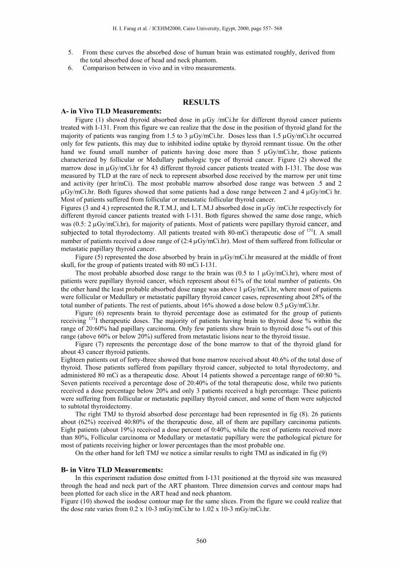
Figure (1) showed thyroid absorbed dose in µGy /mCi.hr for different thyroid cancer patients treated with I-131. From this figure we can realize that the dose in the position of thyroid gland for the majority of patients was ranging from 1.5 to 3 µGy/mCi.hr. Doses less than 1.5 µGy/mCi.hr occurred only for few patients, this may due to inhibited iodine uptake by thyroid remnant tissue. On the other hand we found small number of patients having dose more than 5 µGy/mCi.hr, those patients characterized by follicular or Medullary pathologic type of thyroid cancer. Figure (2) showed the marrow dose in µGy/mCi.hr for 43 different thyroid cancer patients treated with I-131. The dose was measured by TLD at the rare of neck to represent absorbed dose received by the marrow per unit time and activity (per hr/mCi). The most probable marrow absorbed dose range was between .5 and 2 µGy/mCi.hr. Both figures showed that some patients had a dose range between 2 and 4 µGy/mCi hr. Most of patients suffered from follicular or metastatic follicular thyroid cancer. Figures (3 and 4.) represented the R.T.M.J, and L.T.M.J absorbed dose in µGy /mCi.hr respectively for different thyroid cancer patients treated with I-131. Both figures showed the same dose range, which was (0.5: 2 µGy/mCi.hr), for majority of patients. Most of patients were papillary thyroid cancer, and subjected to total thyrodectomy. All patients treated with 80-mCi therapeutic dose of 131I. A small number of patients received a dose range of (2:4 µGy/mCi.hr). Most of them suffered from follicular or metastatic papillary thyroid cancer.
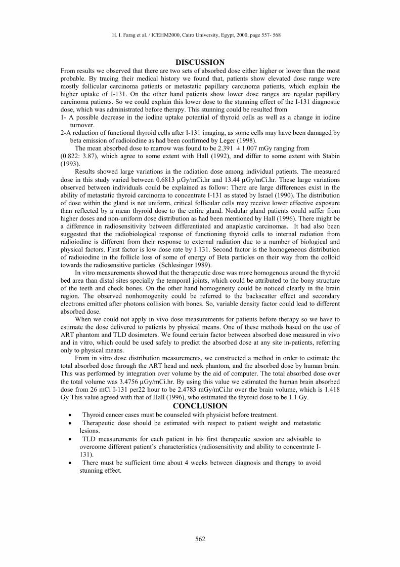
Figure (5) represented the dose absorbed by brain in µGy/mCi.hr measured at the middle of front skull, for the group of patients treated with 80 mCi I-131.
The most probable absorbed dose range to the brain was (0.5 to 1 µGy/mCi.hr), where most of patients were papillary thyroid cancer, which represent about 61% of the total number of patients. On the other hand the least probable absorbed dose range was above 1 µGy/mCi.hr, where most of patients were follicular or Medullary or metastatic papillary thyroid cancer cases, representing about 28% of the total number of patients. The rest of patients, about 16% showed a dose below 0.5 µGy/mCi.hr.
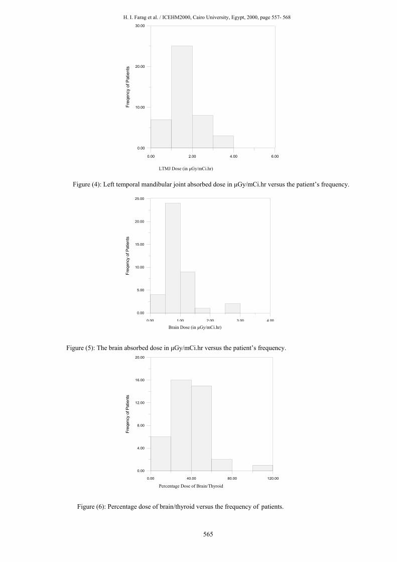
Figure (6) represents brain to thyroid percentage dose as estimated for the group of patients receiving 131I therapeutic doses. The majority of patients having brain to thyroid dose % within the range of 20:60% had papillary carcinoma. Only few patients show brain to thyroid dose % out of this range (above 60% or below 20%) suffered from metastatic lisions near to the thyroid tissue.
Figure (7) represents the percentage dose of the bone marrow to that of the thyroid gland for about 43 cancer thyroid patients. Eighteen patients out of forty-three showed that bone marrow received about 40.6% of the total dose of thyroid. Those patients suffered from papillary thyroid cancer, subjected to total thyrodectomy, and administered 80 mCi as a therapeutic dose. About 14 patients showed a percentage range of 60:80 %. Seven patients received a percentage dose of 20:40% of the total therapeutic dose, while two patients received a dose percentage below 20% and only 3 patients received a high percentage. These patients were suffering from follicular or metastatic papillary thyroid cancer, and some of them were subjected to subtotal thyroidectomy.
The right TMJ to thyroid absorbed dose percentage had been represented in fig (8). 26 patients about (62%) received 40:80% of the therapeutic dose, all of them are papillary carcinoma patients. Eight patients (about 19%) received a dose percent of 0:40%, while the rest of patients received more than 80%, Follicular carcinoma or Medullary or metastatic papillary were the pathological picture for most of patients receiving higher or lower percentages than the most probable one.
On the other hand for left TMJ we notice a similar results to right TMJ as indicated in fig (9)
B- in Vitro TLD Measurements: In this experiment radiation dose emitted from I-131 positioned at the thyroid site was measured
through the head and neck part of the ART phantom. Three dimension curves and contour maps had been plotted for each slice in the ART head and neck phantom. Figure (10) showed the isodose contour map for the same slices. From the figure we could realize that the dose rate varies from 0.2 x 10⁻³ mGy/mCi.hr to 1.02 x 10⁻³ mGy/mCi.hr.
H. I. Farag et al. / ICEHM2000, Cairo University, Egypt, 2000, page 557- 568

561
Table (3): The minimum and maximum absorbed doses measured per hour per administered mCi for each slice of the head and neck of the ART phantom.
Slice No
Minimum absorbed Dose (µGy/mCi.hr)
Maximum absorbed Dose (µGy/mCi.hr)
1 0.202 1.023 2 0.092 1.432 3 0.497 2.247 4 1.192 2.47 5 0.927 3.636 6 3.788 6.993 7 4.702 23.865 8 6.033 20.788 9 4.044 54.441
C- Estimation of Total Dose over Head and Neck Phantom:
To estimate the total absorbed dose distributed throughout the head and neck of the ART phantom, we contracted the following steps: 1- Calculate the total dose of each slice of the phantom individually by integration of dose Dn
(xn,yn) function , which is distributed throughout the whole slices by the use of surfer program. 2- Calculate total dose of every slice and plotted it against the values of z coordinate, which is
the distance from source to the indicated slice. Figure (11) shows the relation between the total approximated dose over area of 6 cm x 6 cm with depth z. D = 594.819 exp (-0.165269z) (1) Coeff. Of determination, R-squared = 0.985631 Residual mean square, Sigma-hat-sq’d = 0.022193 Total dose (DT) can be given by (2)
=3475.59 (µGy/mCi.hr) cm3
3- whenever the total volume of the studied head and neck phantom slices was 734.4865 cm3, then the dose per unit volume is 4.732 µGy/mCi.hr.
4- Assuming that, brain is enclosed in the upper 4 slices, then by performing the above procedure the total dose become 313.896 (µGy/mCi.hr) cm3 , giving 1.13978 µGy/mCi.hr in unit volume.
5- In order to estimate the total dose absorbed in the brain, the brain volume must be multiplied by 1.139. When the average dimensions of human brain are 14 cm width, 16.7 cm length, 9.3 cm height (Blinkov 1968, and Nieuwenhuys et al 1998). The absorbed dose becomes 2.478-mGy/mCi hr.
6- For 26 mCi I-131 dose at 22 hour the total absorbed dose become 1.412 Gy. D- Intercomparison Between Invivo and Invitro Measurements: The absorbed dose obtained by means of TLD detectors, which was placed on ART phantom at the same positions on patients (thyroid, RTMJ, LTMJ, Skull, Neck). The results showed an agreement with patients with little variable deviations at the positions of RTMJ, LTMJ, and Skull, as showed in table (4). Table (4): In vivo and In vitro absorbed dose measured at different positions.
Site of TLD In vivo In vitro Conversion factor Neck 1.8009 ± 0.1207 3.705 ± 0.177 0.486073 RTMJ 1.5076 ± 0.28 1.901 ± 0.136 0.79306 LTMJ 1.2305 ± 0.127 2.526 ± 0.091 0.48711
Average TMJ 1.36905 ± 0.196 2.2135 ± 0.44 0.6185 Skull 0.6547 ± 0.098 1.125 ± 0.092 0.58194
dzzDDT ∫=4.20
0
)(
H. I. Farag et al. / ICEHM2000, Cairo University, Egypt, 2000, page 557- 568
562
DISCUSSION
From results we observed that there are two sets of absorbed dose either higher or lower than the most probable. By tracing their medical history we found that, patients show elevated dose range were mostly follicular carcinoma patients or metastatic papillary carcinoma patients, which explain the higher uptake of I-131. On the other hand patients show lower dose ranges are regular papillary carcinoma patients. So we could explain this lower dose to the stunning effect of the I-131 diagnostic dose, which was administrated before therapy. This stunning could be resulted from 1- A possible decrease in the iodine uptake potential of thyroid cells as well as a change in iodine
turnover. 2-A reduction of functional thyroid cells after I-131 imaging, as some cells may have been damaged by
beta emission of radioiodine as had been confirmed by Leger (1998). The mean absorbed dose to marrow was found to be 2.391 ± 1.007 mGy ranging from
(0.822: 3.87), which agree to some extent with Hall (1992), and differ to some extent with Stabin (1993).
Results showed large variations in the radiation dose among individual patients. The measured dose in this study varied between 0.6813 µGy/mCi.hr and 13.44 µGy/mCi.hr. These large variations observed between individuals could be explained as follow: There are large differences exist in the ability of metastatic thyroid carcinoma to concentrate I-131 as stated by Israel (1990). The distribution of dose within the gland is not uniform, critical follicular cells may receive lower effective exposure than reflected by a mean thyroid dose to the entire gland. Nodular gland patients could suffer from higher doses and non-uniform dose distribution as had been mentioned by Hall (1996). There might be a difference in radiosensitivity between differentiated and anaplastic carcinomas. It had also been suggested that the radiobiological response of functioning thyroid cells to internal radiation from radioiodine is different from their response to external radiation due to a number of biological and physical factors. First factor is low dose rate by I-131. Second factor is the homogeneous distribution of radioiodine in the follicle loss of some of energy of Beta particles on their way from the colloid towards the radiosensitive particles (Schlesinger 1989).
In vitro measurements showed that the therapeutic dose was more homogenous around the thyroid bed area than distal sites specially the temporal joints, which could be attributed to the bony structure of the teeth and check bones. On the other hand homogeneity could be noticed clearly in the brain region. The observed nonhomogenity could be referred to the backscatter effect and secondary electrons emitted after photons collision with bones. So, variable density factor could lead to different absorbed dose.
When we could not apply in vivo dose measurements for patients before therapy so we have to estimate the dose delivered to patients by physical means. One of these methods based on the use of ART phantom and TLD dosimeters. We found certain factor between absorbed dose measured in vivo and in vitro, which could be used safely to predict the absorbed dose at any site in-patients, referring only to physical means.
From in vitro dose distribution measurements, we constructed a method in order to estimate the total absorbed dose through the ART head and neck phantom, and the absorbed dose by human brain. This was performed by integration over volume by the aid of computer. The total absorbed dose over the total volume was 3.4756 µGy/mCi.hr. By using this value we estimated the human brain absorbed dose from 26 mCi I-131 per22 hour to be 2.4783 mGy/mCi.hr over the brain volume, which is 1.418 Gy This value agreed with that of Hall (1996), who estimated the thyroid dose to be 1.1 Gy.
CONCLUSION • Thyroid cancer cases must be counseled with physicist before treatment. • Therapeutic dose should be estimated with respect to patient weight and metastatic
lesions. • TLD measurements for each patient in his first therapeutic session are advisable to
overcome different patient’s characteristics (radiosensitivity and ability to concentrate I-131).
• There must be sufficient time about 4 weeks between diagnosis and therapy to avoid stunning effect.
H. I. Farag et al. / ICEHM2000, Cairo University, Egypt, 2000, page 557- 568

563
REFERENCES 1. Blinkov SM, and Glezer II. The human brain in figures and tables. A quantitative handbook,
1968; Plenum press, New York, 1968.
2. Hall P, Boice JD, Berg G, Bjelkengren G, Ericsson UB, Hallquist A, Lidberg M, Lundell G, Mattsson A, and Tennvall J. Leukaemia incidence after iodine-131 exposure. Lancet 1992; 4; 340(8810): 1-4.
3. Hall P, Mattsson A, and Boice JD. Thyroid cancer after diagnostic administration of Iodine-131. Radiat Res 1996; 145 : 86-92.
4. Israel O, Iosilevsky G, Front D, Bettman L, Frenkel A, Ish-Shalom S, Steiner M, Harush BM, and Kolodny GM. SPECT quantitation of iodine-131 concentration in phantoms and human tumors. J Nucl Med 1990; 31(12): 1945-1949.
5. Leger FA, Izembart M, Dagousset F, Baaitault L, Baillet G, Chevalier A, Clerc J. Decreased uptake of therapeutic doses of iodine-131 after 185- MBq iodine-131 diagnostic imaging for thyroid remnants in differentiated thyroid carcinoma. Euro J Nucl Med 1998; 25: 242-246.
6. Neiuwenhuys R, Ten Donkelaar HJ, and Nicholson C. The central nervous system of vertebrates. 1998; Vol. 3; Springer; Berlin.
7. O’Connor MK, Cullen MJ, and Malone JF. The value of a tracer dose in predicting the kinetics of therapeutic doses of I-131 in thyrotoxicosis. British Journal of Radiology 1979; 52: 719-726.
8. Schlesinger T, Flower MA, and McCready. Radiation dose assessments in radioiodine (131I) therapy. 1. The necessity for in vivo quantitation and dosimetry in the treatment of carcinoma of the thyroid. Radiotherapy and Oncology 1989; 14: 35-41.
9. Sgouros G, Barest G, Thekkumthala J, Chui C, Mohan R, Bigler R, and Zanzonico P. Treatment planning for internal radionuclide therapy: three dimensional dosimetry for nonuniformly distributed radionuclide. J Nucl Med, 1999; 31(11): 1884- 1891.
10. Stabin MG. Radiation doses to the upper spine from therapeutic administration of iodine-131-sodium iodide. J Nucl Med 1993; 34: 695-696.
11. Yorke ED, Williams LE, Demidecki AJ, Heidorn DB, Roberson PL, and Wessels BW. Multicellular dosimetry for beta –emitting radionuclide: Autoradiography, thermoluminescent dosimetry and three and three-dimensional dose calculations. Med Phys. 1993; 20(2); 2: 543-550.
H. I. Farag et al. / ICEHM2000, Cairo University, Egypt, 2000, page 557- 568

564
0.00 4.00 8.00 12.00 16.00
0.00
4.00
8.00
12.00
16.00
Freq
uenc
y of
Pat
ient
s
Thyroid Dose (in µGy/mCi.hr)
Figure (1): Thyroid absorbed dose in µGy/mCi.hr versus the patient’s frequency.
0.00 4.00 8.00 12.00 16.00
0.00
5.00
10.00
15.00
20.00
25.00
Freq
ency
of P
atie
nts
Marrow Dose (in µGy/mCi.hr)
Figure (2): Marrow absorbed dose in µGy/mCi.hr versus the patient’s frequency.
0.00 2.00 4.00 6.00
0.00
10.00
20.00
30.00
Freq
uenc
y of
Ptie
nts
RTMJ Dose (in µGy/mCi.hr)
Figure (3): Right temporal mandibular joint absorbed dose in µGy/mCi.hr versus the patient’s frequency.
H. I. Farag et al. / ICEHM2000, Cairo University, Egypt, 2000, page 557- 568
565
0.00 2.00 4.00 6.00
0.00
10.00
20.00
30.00
Freq
ency
of P
atie
nts
LTMJ Dose (in µGy/mCi.hr)
Figure (4): Left temporal mandibular joint absorbed dose in µGy/mCi.hr versus the patient’s frequency.
0.00 1.00 2.00 3.00 4.00
0.00
5.00
10.00
15.00
20.00
25.00
Freq
ency
of P
atie
nts
Brain Dose (in µGy/mCi.hr)
Figure (5): The brain absorbed dose in µGy/mCi.hr versus the patient’s frequency.
0.00 40.00 80.00 120.00
0.00
4.00
8.00
12.00
16.00
20.00
Freq
ency
of P
atie
nts
Percentage Dose of Brain/Thyroid
Figure (6): Percentage dose of brain/thyroid versus the frequency of patients.
H. I. Farag et al. / ICEHM2000, Cairo University, Egypt, 2000, page 557- 568

566
0.00 50.00 100.00 150.00 200.00 250.00 300.00 350.00
0.00
4.00
8.00
12.00
16.00
20.00
Freq
uenc
y of
Pat
ient
s
Percentage Dose of Marrow/Thyroid
Figure (7): Percentage dose of Marrow/thyroid versus the frequency of patients.
0.00 40.00 80.00 120.00 160.00
0.00
4.00
8.00
12.00
16.00
Freq
uenc
y of
Pat
ient
s
Percentage Dose of RTMJ/Thyroid
Figure (8): Percentage dose of Right Temporal Mandibular Joint (RTMJ)/thyroid versus the frequency of patients.
Figure (9): Percentage dose of left temporal Mandibular Joint (LTMJ)/thyroid versus the frequency of patients.
Percentage Dose of LTMJ/Thyroid
0.00 40.00 80.00 120.00 160.00
0.00
4.00
8.00
12.00
16.00
Fr e q u e n c y ofP ati
e nt s
H. I. Farag et al. / ICEHM2000, Cairo University, Egypt, 2000, page 557- 568

567
Figure (10): The isodose contour map in µGy/mCi.hr overslices no. 1-9 of the ART phanton
-2 0 2
x-axis
-8
-6
-4
-2
0
2
4
6
8
y-axis
-6 -5 -4 -3 -2 -1 0 1 2 3 4 5 6
x-
-6
-5
-4
-3
-2
-1
0
1
2
3
4
5
6
yaxis
-3 -2 -1 0 1 2 3
X-axis
-6
-5
-4
-3
-2
-1
0
1
2
3
4
5
6
y-ax
is
-8 -6 -4 -2 0 2 4 6 8x-axis
-8
-6
-4
-2
0
2
4
6
8
y-ax
is
-6 -5 -4 -3 -2 -1 0 1 2 3 4 5 6
x-axis
-6
-5
-4
-3
-2
-1
0
1
2
3
4
5
6
y-ax
is
-3.0 -2.5 -2.0 -1.5 -1.0 -0.5 0.0 0.5 1.0 1.5 2.0 2.5 3.0
x-axis
-3.0
-2.5
-2.0
-1.5
-1.0
-0.5
0.0
0.5
1.0
1.5
2.0
2.5
3.0
y-ax
is
-6 -5 -4 -3 -2 -1 0 1 2 3 4 5 6
x-axis
-6
-5
-4
-3
-2
-1
0
1
2
3
4
5
6
y-ax
is
-6 -5 -4 -3 -2 -1 0 1 2 3 4 5 6x-axis
-6
-5
-4
-3
-2
-1
0
1
2
3
4
5
6
y - a x i s
- 3 - 2 - 1 0 1 2x- a x i s
- 6
- 5
- 4
- 3
- 2
- 1
0
1
2
3
4
5
6
y-ax
is
H. I. Farag et al. / ICEHM2000, Cairo University, Egypt, 2000, page 557- 568

568
0.00 4.00 8.00 12.00 16.00 20.00Depth (cm)
0.00
200.00
400.00
600.00
800.00
T ot al A p pr o xim at e d D o s e o v er 3 x 3 cm ar e a
Figure (11) : Relation between total approximated dose over 3 x 3 cmarea and depth in cm .
2
2
H. I. Farag et al. / ICEHM2000, Cairo University, Egypt, 2000, page 557- 568