in vitro evaluation of determinants bactericidal activity...
TRANSCRIPT
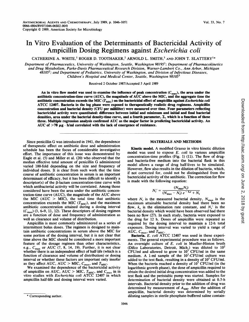
Vol. 33, No. 7ANTIMICROBIAL AGENTS AND CHEMOTHERAPY, JUlY 1989, P. 1046-10510066-4804/89/071046-06$02.00/0Copyright C 1989, American Society for Microbiology
In Vitro Evaluation of the Determinants of Bactericidal Activity ofAmpicillin Dosing Regimens against Escherichia coli
CATHERINE A. WHITE,1 ROGER D. TOOTHAKER,2 ARNOLD L. SMITH,3 AND JOHN T. SLATTERYl*Department of Pharmaceutics, University of Washington, Seattle, Washington 981951; Department ofPharmacokineticsand Drug Metabolism, Parke-Davis Pharmaceutical Research Division, Warner-Lambert Co., Ann Arbor, Michigan
481052; and Department of Pediatrics, University of Washington, and Division of Infectious Diseases,Children's Hospital and Medical Center, Seattle, Washington 981053
Received 2 October 1987/Accepted 5 April 1989
An in vitro flow model was used to examine the influence of peak concentration (Cmax), the area under theantibiotic concentration-time curve (AUC), the magnitude of AUC above the MIC, and the aggregate time theantibiotic concentration exceeds the MIC (TMIC) on the bactericidal effect of ampicillin against Escherichia coliATCC 12407. Bacteria in the log phase were exposed to therapeutically realistic drug regimens. Ampicillinconcentration and bacterial density (CFU per milliliter) were measured over time. Four parameters reflectingbactericidal activity were quantitated: difference between initial and minimum and initial and final bacterialdensities, area under the bacterial density-time curve, and a fourth parameter, Z, which is a function of thesethree. Multiple regression analysis confirmed AUC as the major factor in predicting bactericidal activity. AnAUC of >70 ug- h/ml correlated with the lack of emergence of resistance.
Since penicillin G was introduced in 1941, the dependenceof therapeutic -effect on antibiotic dose and administrationschedule has been the focus of considerable investigativeeffort. The importance of this issue was demonstrated byEagle et al. (5) and Miller et al. (20) who observed that themedian effective total amount of penicillin G administeredvaried 100-fold depending on the size and frequency ofindividual doses. It is clear from such work that the timecourse of antibiotic concentration in serum is an importantdeterminant of efficacy, but it has been difficult to identify adescriptor(s) of the P-lactam concentration-time curve withwhich antibacterial activity will be correlated. Among thoseconsidered have been the area under the antibiotic concen-tration-time curve (AUC), the magnitude of the AUC abovethe MIC (AUC > MIC), the total time that antibioticconcentration exceeds the MIC (TMIC), and the maximumantibiotic concentration attained during a dosing interval(Cmax) (3, 6-9, 11, 12). These descriptors of dosing regimenare a function of dose and frequency of administration aswell as clearance and volume of distribution.
Ampicillin is most commonly administered as a series ofintermittent bolus doses. The regimen is designed to main-tain antibiotic concentrations in serum above the MIC forsome portion of the dosing interval, but it is not clear thattime above the MIC should be considered a more importantfeature of the dosage regimen than other characteristics,e.g., Cmax or AUC (7, 8, 14, 19). Further, it is not clearwhether there is an independent effect of half-life (which is afunction of clearance and volume of distribution) or dosinginterval or whether these factors are important only insofaras they affect AUC, AUC > MIC, Cmax, and TMIC.We examined the dependence of the bactericidal activity
of ampicillin on AUC, AUC > MIC, TMIC, and Cmax in invitro studies with Escherichia coli ATCC 12407 in whichampicillin half-life and dosing interval were varied.
* Corresponding author.
MATERIALS AND METHODS
Kinetic model. A modified Grasso in vitro kinetic dilutionmodel was used to expose E. coli to various ampicillinconcentration-time profiles (Fig. 1) (11). The flow of drug-and bacteria-free medium into the bacterial flask in thismodel allows a range of drug half-lives to be simulated.However, flow also results in the dilution of bacteria, which,if not corrected for, could not be distinguished from thebactericidal activity of the antibiotic. The correction for flowis made with the following equation:
I' = (NmaxNt)Nt' (Nmax - N,) e - ket + N,where N, is the measured bacterial density, Nmax is themaximum attainable bacterial density had there been noflow, ke is the elimination rate constant, and N,' is thebacterial density which would have been observed had therebeen no flow (27). In each study, bacteria were exposed tothe drug for 12 h. Doses of ampicillin were repeated asrequired by the dosing interval to maintain a 12-h totalexposure. Dosing interval was varied to yield a range ofAUC, Cmax, and TMIC.
Bacteria. E. coli ATCC 12407 was used in these experi-ments. The general experimental procedure was as follows.An overnight culture of E. coli in Mueller-Hinton broth(Difco Laboratories, Detroit, Mich.) was diluted to 102CFU/ml and allowed to grow to 10i CFU/ml in the samemedium. A 1-ml sample of the 107-CFU/ml culture wasadded to the test flask, resulting in a density of 105 CFU/ml.When the bacteria reached a density of 107 CFU/ml (in thelogarithmic growth phase), the dose of ampicillin required toobtain the desired initial drug concentration was added to thetest flask and the peristaltic pump was started. Samples fordetermination of bacterial density were obtained at 0.5-hintervals. Bacterial density prior to the addition of drug wasdetermined by measurement of A6.. After the addition ofampicillin, bacterial density was determined by seriallydiluting samples in sterile phosphate-buffered saline contain-
1046
on June 2, 2018 by guesthttp://aac.asm
.org/D
ownloaded from

DOSING REGIMEN AND AMPICILLIN ACTIVITY 1047
D
I I
FIG. 1. Schematic representation of in vitro dilution model.Medium is transferred from the reservoir flask (A) to the test flask(B) by the pump (C). Flask B maintains a constant volume; bacteria,drug, and medium are sent to waste (D).
121ing 0.1% gelatin. Triplicate 10-pI aliquots of each dilutionand 100 ,ul of undiluted sample were streaked onto nutrientagar plates (Difco). The plates were incubated at 37°C for a
minimum of 12 h. Preliminary studies determined that thesmall amount of ampicillin in the sample had no effect on theestimation of bacterial density (data not shown), most prob-ably because of dilution of the drug during preparation of thesample prior to streaking on the plates. The MIC and MBCof ampicillin were determined by broth dilution by standardmethods (25, 26). The MIC is defined as that ampicillinconcentration which inhibited visibly detectable growth inbroth containing 5 x 10⁵ CFU. The MBC was the ampicillinconcentration which resulted in killing of .99.9% of thesame inoculum. The MBC was the same as the MIC whentested as described above and at an inoculum of 103 CFU.Rejection values of 3 and 11 were used for the inocula of 5 x
105 and 1 x 10³ CFU, respectively (22).Determination of bactericidal parameters. These studies
required quantitative measures of bactericidal activity. Thefour parameters used were ANmax, ANtot, BAUC, and X,which (except for E) are illustrated in Fig. 2. The valuesreported for each parameter were determined over the entire12-h period of exposure of bacteria to antibiotic. ANmax isthe difference between the initial and minimum bacterial
I-
zuJ
a
-J
CiuJ
mcc
ot
----T
/< __ __ANtot
- --------
--7-- -- --------
O AN
'-BAUC- ---
0 T1 2T'
1max
TIMEFIG. 2. Illustration of the bactericidal effect parameters: ANmax,
ANto,, and BAUC. X is calculated from the three parametersdepicted here as described in the text. BAUC is the shaded areaunder the solid curve of bacterial density. The stippled lines identifythe bacterial density-time points used to calculate the differencesANmax and ANtot. t is the interval between doses of antibiotic.
10E
0
0
o
C:
~0
-o
a)0
co
8
6
4
2
I
0 2 4 6 8 10 12
Time, hoursFIG. 3. Time course of bacterial density and ampicillin concen-
tration in a representative study with E. coli ATCC 12407. Ampicil-lin was dosed at 0 and 6 h and removed by flow with a 1.0-h half-life.Drug concentration exceeded the MIC for 6 h of the 12-h study.Arrows identify times at which ampicillin was added.
densities and represents the initial decrease in viable celldensity associated with antibiotic addition. ANtot is thedifference between the bacterial density at the beginning andend of the 12-h study and represents the regrowth phase (inwhich the emergence of resistance becomes apparent) overtwo or more dosing intervals. BAUC is the area under thebacterial concentration-time curve divided by the initialinoculum size. BAUC is a time-integrated parameter and isaffected by cell death, stationary, and regrowth phases. Theparameter L is introduced in an attempt to obtain a singleparameter which is a function of the three fundamentalparameters. l is calculated by assigning a rank of 10 to thelowest value of each respective fundamental parameter(ANmax, ANtot) and BAUC) observed in all studies con-ducted with a given strain of bacteria. A score of 1 isassigned to the maximum value of each parameter. Scoresare assigned to other values of a given parameter by linearinterpolation between these extremes. l is the sum of theranks thus assigned to the values of the respective parame-ters. A value of E is calculated for each exposure condition.
Ampicillin assay. Ampicillin concentration was determinedby a standard disk diffusion microbiological method (24).Sarcina lutea ATCC 9341 or Bacillus subtilis (Difco) wasused as the assay organism (2, 15). Ampicillin standards(Sigma Chemical Co., St. Louis, Mo.) were prepared in
-Es_
E
VOL. 33, 1989
on June 2, 2018 by guesthttp://aac.asm
.org/D
ownloaded from

ANTIMICROB. AGENTS CHEMOTHER.
TABLE 1. Reproducibility of results with E. coli 12407
Study Dosing t1/2 Cmax TMIC AUC AUC > MIC ANmax ANtot BAUC Iinterval (h) (h)" (,ug/ml) (h) (pig h/ml) (pLg. h/ml) (log CFU/mi) (log CFU/ml) (log CFU * h/mi)
1 6 0.87 10.1 5.8 25.0 17.0 -3.6 +1.9 106 10.72 6 0.88 9.1 5.6 23.0 15.0 -3.3 +1.9 113 9.53 6 0.9 9.0 5.6 22.9 15.0 -3.3 +1.9 113 9.54 6 1.1 7.2 6.0 21.2 12.7 -2.2 +2.4 125 6.6
a t12, Half-life.
Mueller-Hinton broth. The lower limit of detection was 0.5,ug/ml, and the coefficient of variation of the slope of thestandard curve was 6.6% over a 2-year period.
Determination of pharmacokinetic parameters. The in vitrosystem is designed such that antibiotic concentration willdecline monoexponentially. The actual elimination rate con-stant, ke and maximum concentration, Cmax, were deter-mined by linear regression. AUC and AUC > MIC werecalculated by the trapezoidal rule over the 12-h period ofeach study (10). The aggregate time during which concentra-tions were above the MIC for the initial inoculum wascalculated by TMIC = kI-1 ln(Cmax/MIC).
Statistical analysis. Multiple regression analysis was per-formed to determine the importance of each of the descrip-tors of the antibiotic concentration-time profile as a deter-minant of ANmax, A&N,01, BAUC, and Z. Multiple regressionwas performed by the stepwise linear regression procedurewith a significance level of 0.05 (17). Stepwise regression isan improved version of forward regression which permitsreexamination at every step of the variables incorporated inthe model in previous steps. At each step, a partial F test foreach variable presently in the model is made, treating it asthough it were the most recent variable entered. This pro-cedure allows removal of variables that were entered at anearly stage which have become superfluous because of theirrelationship with other variables subsequently evaluated.
RESULTS
The goal of this investigation required the simulation of alarge number of dosing regimens. The general strategy wasto evaluate ranges of dosing interval and half-life which areencountered clinically after intravenous ampicillin adminis-tration. Cmax was varied from approximately 1 to 100 times
the MIC. Bacterial density and ampicillin concentration-timecurves from a representative study are shown in Fig. 3;parameter values calculated from these raw data are sum-marized in row 3 of Table 1. The dosing interval was 6 h andthe half-life of ampicillin was approximately 1 h; this alloweddrug concentration to exceed the MIC for 6 h of the 12-hstudy. Ampicillin was effective in killing bacteria (Cmax wasnine times the MIC and ANmax was -3.3 log CFU/ml, i.e.,10-3-3 CFU/ml). However, there was a net growth of bacte-ria over the period of exposure; AN,., was 1.9 log CFU/ml.Net growth began 2 h after the first dose and 1 h after thesecond dose. Table 1 shows the reproducibility of theseresults in a series of four replicate studies. The replicatesshown represent data collected over 1 year.The results obtained from the various exposure conditions
are summarized in Table 2. Data from individual studieswere analyzed by multiple linear regression to determine thedescriptor(s) of the antibiotic concentration-time profilemost strongly associated with bactericidal activity. Logtransformations of Cmax and AUC, referred to as log Cmaxand log AUC, were used in the analysis because the logtransformations improved the correlation between theseparameters and bactericidal efficacy. Log transformation didnot improve the correlation between measures of efficacyand any other descriptors of dosage regimen (e.g., TMIC).The results of the regression analysis are shown in Table
3. A value of 0 for a coefficient means that inclusion of theparameter with which it is associated does not improve theoverall correlation. The absolute value of the regressioncoefficient is a reflection of the relative importance of a
parameter as a determinant of the bactericidal activity of aregimen. The relationship between the various bactericidalparameters and log AUC for E. coli is shown in Fig. 4.The apparent emergence of resistance was encountered in
TABLE 2. Influence of ampicillin concentration-time profile on bactericidal activity against E. coli ATCC 12407
Dosing t112 Cmax TMIc AUC AUC > MIC ANmax ANt., BAUCinterval (h) (h) (pig/mi) (h) (pLg. h/ml) (pig. h/ml) (log CFU/mi) (log CFU/mi) (log CFU h/ml)
2a 1.0 2.5 7.5 15.9 5.2 -1.5 +1.7 124 6.52a 1.1 25.5 12.0 169 157 -7.9 -5.0 60 26.52 1.1 62.0 12.0 413 201 -7.9 -7.9 53 30.02 4.0 1.3 8.2 13.2 1.7 -2.6 +0.6 118 9.330 0.9 5.2 8.5 24.0 13.2 -1.9 +2.1 118 7.33a 1.0 8.8 11.2 42.0 30.0 -3.7 +1.8 104 11.23 10.0 8.4 12.0 91.0 99.0 -5.5 -5.2 69 23.06 1.1 4.8 4.7 14.0 9.6 -3.6 +1.1 106 11.46a 0.9 8.8 5.8 23.0 14.9 -3.1 +2.0 114 9.16 1.1 80.4 12.0 253 241 -7.9 -3.1 64 24.66 4.1 2.1 8.3 15.3 3.9 -2.5 +0.3 118 9.56 5.8 8.4 12.0 72.0 60.0 -5.1 -5.1 74 21.912 1.0 10.2 3.3 14.4 11.1 -5.0 +1.3 101 13.3120 1.0 80.4 6.1 111 105 -7.5 -1.2 71 21.7
Data represent the mean of two to four individual incubations when dosing interval and half-life U1,2) were constant and peak concentration varied less than10%. n = 1 if no superscript.
1048 WHITE ET AL.
on June 2, 2018 by guesthttp://aac.asm
.org/D
ownloaded from

DOSING REGIMEN AND AMPIC1LL1N ACTIVITY 1049
TABLE 3. Multiple regression summary for E. coli ATCC 12407
Regression coefficient"Parameter
bh C d ,
ANnlrx 2.26 -5.78 0 0.30 0.83ANto,t 13.0 -18.9 10.2 0.84 0.83BAUC 176 -48.9 0 0 0.84M; -10.1 15.2 0 0 0.81
"General regression equation: parameter = a + b log AUC + ( log Cnflax +dTMIc. Regression coefficient is 0 when inclusion of the respective descriptordid not improve the value of r2. Parameters not listed, i.e., half-life, dosinginterval, and AUC > MIC, always had coefficients of 0.
more than half of the studies conducted. The frequency ofmutation for resistance to 10 p.g of ampicillin per ml was 6.18X 10⁻⁷ for E. coli ATCC 12407, as determined by themodified Luria-Delbruck fluctuation test (19). The frequencyof mutation for resistance to ampicillin at 20 jig/ml was 1.1 x10'. Thus, resistance to ampicillin at 10 jig/ml apparentlydeveloped as a consequence of the emergence of a resistantclone present in the original inoculum of E. coli ATCC 12407(23). The MIC was regularly determined over the studyperiod. The initial population of E. coli ATCC 12407 (107CFU/ml) was tested to determine what fraction of thepopulation was resistant to ampicillin concentrations equalto the MIC, twice the MIC, four times the MIC, and eighttimes the MIC. Of the initial bacterial population, 0% wasresistant at these ampicillin concentrations.Table 4 shows that the emergence of resistance (defined by
a minimum of a threefold increase in MIC) was associatedwith an ampicillin AUC of <70 [Lg h/mi. In variants of the
3-1
1.
0
c) -3'0
z
.7-
C.R0
0)0
-m
m
TABLE 4. Relationship between emergence of resistanceand ampicillin AUC
Presence of resistant strains ofAmpicillin AUC E. coli ATCC 12407
(p.g h/ml)"No. positive No. negative
>70 0 7<70 12 1
Resistance identified by MIC. Presence of resistance was positive whenbacteria achieved an MIC three times the original.
Grasso model, regrowth of bacteria has been shown undercertain conditions by Haag et al. (13) to be a consequence ofadherence of bacteria to the wall of the incubation flask,forming a thin film. The film sheds bacteria into the medium,resulting in apparent regrowth. We evaluated the potentialrole of this artifact in our system by conducting (in duplicate)incubations without drugs and with a medium flow rate suchthat the drug half-life would have been 15 min. Results (meanvalues of the duplicate studies) are shown in Fig. 5. Bacterialdensity declined log linearly for the first 4 h of the incubationand then became constant at approximately 3.5 log CFU/ml.The results suggest that some bacteria adhere to the model(in that bacterial density should have been reduced to 1 logCFU/ml at this flow) but that adherence per se cannotaccount for the regrowth observed in our studies withampicillin. In addition, growth rate constants at ampicillinconcentrations below the MIC were dependent on mediumflow rate. The growth rate constant at a medium flow rate of1.2 ml/min was 0.58 ± 0.13 h-1 (range, 0.3 to 0.74 h-<), andat a medium flow rate of 0.25 ml/min, it was 1.0 ± 0.16 h-'
-1
0
0
0
.2 -3C.)0' -40
-5
E -6Z
-7
-810 0
100 100oo - 1
30T0
8oo
I 0 00
0
0000
.0%o0I0
0
00
0
OD 0 0
1o0000
25t
20c
15
10
5o
0 0
00 00
1 oon
00
o O
0°
0
0
(;o 00
0
1-10(
8
100 100oo
AUC, gg - hr/mlFIG. 4. Scatter plots illustrating the correlation between the various measures of bactericidal efficacy and log AUC.
VOL. 33, 1989
I
0
on June 2, 2018 by guesthttp://aac.asm
.org/D
ownloaded from

ANTIMICROB. AGENTS CHEMOTHER.
E 8-
CD)0)6-
C)w
0
< C° I
m 0 2 4 6 8 10 12TIME, hours
FIG. 5. Time course of bacterial density in an incubation withoutampicillin and a flow rate such that drug half-life (had drug beenincorporated) would be 15 min. Data shown are mean values ofduplicate incubations.
(range, 0.8 to 1.2 h-1). When the film accounted for theapparent resistance in the studies by Haag et al. (13), thegrowth rate constant was independent of medium flow rate.
DISCUSSIONThe analysis of data obtained in dynamic in vitro studies of
antibiotic effect has been complicated by the lack of quanti-tative measures with which to characterize bactericidalactivity. We defined four parameters with which this objec-tive can be accomplished. ANmax, AN,,, and BAUC (de-fined in Materials and Methods) reflect different facets of thebactericidal activity of a dosing regimen. The term X was
introduced to provide a method to sum all these measures ina single term and to serve as an overall index of bactericidalactivity. However, a high value of E may not translate intothe most effective regimen in vivo, as the characteristics ofthe bactericidal activity of a regimen reflected in ANmax,ANtot, and BAUC are likely not to be of equal importancewhen other factors such as host defense contribute to theremoval of bacteria.
In our studies with ampicillin, AUC was determined to bethe most consistent index of bactericidal activity (Table 3,Fig. 3) in agreement with the in vitro results of others (7, 9,16). Studies in mice have also shown a correlation betweenAUC and bactericidal activity (3). However, TMIC has alsobeen suggested to be strongly associated with bactericidalactivity in vitro and in vivo (5, 6, 19). In our studies, TMICdid not covary strongly with AUC for E. coli (r2 = 0.36).Thus, a contribution of TMIC to bactericidal activity was notmasked by the correlation obtained with AUC. It is possiblethat TMIC was not identified as an important characteristic ofthe dosage regimen in our studies owing to the high fre-quency (>50%) with which resistant strains emerged. WhenMIC increases during a study, the period that the antibioticconcentration exceeds the MIC for the original inoculumbecomes irrelevant. In support of this view, Eagle et al. (5)noted that a longer TMIC was required to cure mice infectedwith group A Streptococcus species when regrowth was
observed between doses. However, their in vivo studiesexamined the relationship between concentration of antibi-otic in plasma and bactericidal effect in tissue, and therelationship between concentration in tissue and concentra-tion in plasma was not known.
In in vitro studies with several strains of bacteria andantibiotics, Blaser et al. (4) observed that dosage regimenswhich produced a peak antibiotic concentration eightfoldgreater than the MIC effected substantial cell killing andinhibited the emergence of resistance. In our studies, Cmaxwas identified as an important determinant (although second-ary to AUC) of AN,0, the bactericidal parameter which ismost directly affected by the emergence of resistance. Cmax,of course, is simply the ampicillin concentration at a singlepoint in time and provides virtually no information concern-ing concentrations after that time. A Cmax of >10 times theMIC can result in a net gain in bacterial concentration(positive AN,0t,) if the half-life is short, whereas the sameCmax coupled with a longer elimination half-life can result ina net loss of bacteria (negative ANt,d) (Table 2). The differentsigns of the coefficients of AUC and Cmax (Table 3) areapparently due to the frequent association of a high Cmaxwith a short half-life. A contribution of Cmax to the otherbactericidal parameters may have been masked by thecorrelation between Cmax and AUC, r- = 0.56.AUC was also found to be an important determinant of the
emergence of resistance, defined by a threefold increase inMIC or by a positive value of ANt,,, (Table 2). There was nosuch result with Cmax. This observation is consistent withthat reported by Grasso et al. (11).As mentioned above, Haag et al. (13) showed that the
emergence of resistance could be an artifact of in vitrosystems owing to the adherence of bacteria to the glass wallsof incubation flasks. A similar experiment conducted with E.coli ATCC 12407 indicated that the regrowth observed inthese experiments was not an artifact of the in vitro system(Fig. 5). In addition, resistance was documented by changesin MIC, and the growth rate constants at an ampicillinconcentration of <1 p.g/ml (the MIC for the original inocu-lum) in our studies were a function of medium flow rate (i.e.,ampicillin half-life), whereas Haag et al. (13) pointed out thata constant growth rate as a function of medium flow rate is ahallmark of apparent resistance owing to adherence ofbacteria to glass.Although the emergence of resistance is a problem en-
countered more commonly in vitro than in vivo, suboptimumantibiotic therapy has been shown to allow the emergence ofresistant strains in animal studies and in certain patients (1,18, 21). The emergence of resistance is of particular concernwhen the host is immunocompromised (9). In immunocom-promised patients, the results of kinetic in vitro studies maybe directly applicable. The observation that resistance isleast likely to emerge when AUC is high means that resis-tance will be least likely when relatively large doses are
given frequently. A large dose, frequent administration, or a
long half-life alone may be insufficient.Maintenance of antibiotic concentration above the MIC
for the initial inoculum also appears to be insufficient toprevent the emergence of resistance of E. coli to ampicillin.Indeed, a constant ampicillin concentration just above theMIC is likely to allow the emergence of resistant E. coli,whether present initially or arising from a mutation duringexposure to the antibiotic. The results of this and otherstudies (4) suggest that the emergence of resistance will bediscouraged if a dosage regimen is adopted which is designedto eradicate the least susceptible organism present at thebeginning of therapy.
ACKNOWLEDGMENT
C.A.W. was supported by training grant GM07750 from theNational Institutes of Health.
1050 WHITE ET AL.
on June 2, 2018 by guesthttp://aac.asm
.org/D
ownloaded from

DOSING REGIMEN AND AMPICILLIN ACTIVITY 1051
LITERATURE CITED1. Bayer, A. S., D. Norman, and K. S. Kim. 1985. Efficacy of
amikacin and ceftazidime in experimental aortic valve en-docarditis due to Pseudomonas aeruginosa. Antimicrob.Agents Chemother. 8:781-785.
2. Bennett, J. V., J. L. Brodie, E. J. Benner, and W. M. M. Kirby.1966. Simplified accurate method for antibiotic assay of clinicalspecimens. Appl. Microbiol. 14:170-177.
3. Bergeron, M. G., B. M. Nguyen, and L. Gauvreau. 1978.Influence of constant infusion versus bolus injections of antibi-otics on in vivo synergy. Infection 6(Suppl.):38-46.
4. Blaser, J., B. B. Stone, M. G. Groner, and S. H. Zinner. 1987.Comparative study with enoxacin and netilmicin in a pharma-codynamic model to determine importance of ratio of antibioticpeak concentration to MIC for bactericidal activity and emer-gence of resistance. Antimicrob. Agents Chemother. 31:1054-1060.
5. Eagle, H., R. Fleishman, and M. Levy. 1953. Continuous vs.discontinuous therapy with penicillin. N. Engl. J. Med. 248:481-488.
6. Eagle, H., R. Fleishman, and A. Musselman. 1950. Effect ofschedule of administration on the therapeutic efficacy of peni-cillin. Am. J. Med. 9:280-299.
7. Gerber, A. U., H. Brugger, C. Feller, T. Stritzko, and B. Stalder.1986. Antibiotic therapy of infections due to Pseudomonasaeruginosa in normal and granulocytopenic mice: comparisonof murine and human pharmacokinetics. J. Infect. Dis. 153:90-97.
8. Gerber, A. U., W. A. Craig, H. Brugger, C. Feller, A. P. Vastola,and J. Brandel. 1983. Impact of dosing intervals on activity ofgentamicin and ticarcillin against Pseudomonas aeruginosa ingranulocytopenic mice. J. Infect. Dis. 147:910-917.
9. Gerber, A. U., P. Wiprachtiger, U. Stettler-Spichiger, and G.Lebek. 1982. Constant infusions vs. intermittent doses of gen-tamicin against Pseudomonas aeruginosa in vitro. J. Infect. Dis.145:554-560.
10. Gibaldi, M., and D. Perrier. 1982. Pharmacokinetics, 2nd ed., p.445-449. Marcel Dekker, Inc., New York.
11. Grasso, S., G. Menardi, I. deCarneri, and V. Tamassia. 1978.New in vitro model to study the effect of antibiotic concentra-tion and rate of elimination on antibacterial activity. Antimi-crob. Agents Chemother. 13:570-576.
12. Guggenbichler, J. P., E. Semenitz, and P. Konig. 1985. Killkinetics and regrowth pattern of bacteria exposed to antibioticconcentrations simulating those observed in vivo. J. Antimi-crob. Chemother. 15(Suppl. A):139-146.
13. Haag, R., P. Lexa, and I. Werkhauser. 1986. Artifacts in dilutionpharmacokinetic models caused by adherent bacteria. Antimi-
crob. Agents Chemother. 29:765-768.14. Hunter, P. A., G. N. Rolinson, and D. A. Witting. 1973.
Comparative activity of amoxycillin and ampicillin in an exper-imental bacterial infection in mice. Antimicrob. Agents Chemo-ther. 4:285-293.
15. Jallings, B., A. S. Malmberg, A. Lindman, and L. 0. Boreus.1972. Evaluation of a micromethod for determination of antibi-otic concentration in plasma. Eur. J. Clin. Pharmacol. 4:150-157.
16. Klaus, U., W. Henninger, P. Jacobi, and B. Wiedemann. 1981.Bacterial elimination and therapeutic effectiveness under dif-ferent schedules of amoxicillin administration. Chemotherapy27:200-208.
17. Kleinbaum, D., and L. Krupper. 1978. Applied regressionanalysis and other multivariable methods. Duxbury Press,North Scituate, Mass.
18. Maugh, T. H., II. 1981. A new wave of antibiotics builds.Science 214:1225-1228.
19. Merrikin, D., and G. N. Rollinson. 1979. Antibiotic levels inexperimentally infected mice in relation to therapeutic effectand antibacterial activity in vitro. J. Antimicrob. Chemother.5:423-429.
20. Miller, A. K., D. L. Wilmer, and W. F. Verwey. 1950. Effect ofpenicillin dosage schedule on treatment of experimental thyroidinfections in mice. Proc. Soc. Exp. Biol. Med. 74:62-64.
21. Olson, B., R. A. Weinstein, C. Nathan, W. Chamberlin, andS. A. Kabins. 1985. Occult aminoglycoside resistance in Pseu-domonas aeruginosa: epidemiology and implication for therapyand control. J. Infect. Dis. 152:769-774.
22. Pearson, R. D., R. T. Steigbigel, H. T. Davis, and S. W.Chapman. 1980. Method for reliable determination of minimallethal antibiotic concentrations. Antimicrob. Agents Chemo-ther. 18:699-708.
23. Sherris, J. C., and B. H. Minshew. 1980. Mutational antibioticresistance, p. 418-432. In V. Lorian (ed.), Antibiotics in labo-ratory medicine. The Williams & Wilkins Co., Baltimore, Md.
24. Simon, H. J., and E. J. Yin. 1970. Microbioassay of antimicro-bial agents. Appl. Microbiol. 19:573-579.
25. Thrupp, L. D. 1980. Susceptibility testing of antibiotics in liquidmedia, p. 73-113. In V. Lorian (ed.), Antibiotics in laboratorymedicine. The Williams & Wilkins Co., Baltimore, Md.
26. Waterworth, P. M. 1978. Quantitative methods for bacterialsensitivity testing, p. 34-38. In D. S. Reeves, I. Phillips, J. D.Williams, and R. Wise (ed.), Laboratory methods in antimicro-bial chemotherapy. Churchill Livingstone, Inc., New York.
27. White, C. A., R. D. Toothaker, A. L. Smith, and J. T. Slattery.1987. Correction for bacterial loss in in vitro dilution models.Antimicrob. Agents Chemother. 31:1859-1860.
VOL. 33, 1989
on June 2, 2018 by guesthttp://aac.asm
.org/D
ownloaded from