in vitro activity of local plants from …eprints.utar.edu.my/1848/1/in_vitro_actvity_of_local...ii...
TRANSCRIPT
i
IN VITRO ACTIVITY OF LOCAL PLANTS FROM MALAYSIA
AGAINST CHIKUNGUNYA VIRUS
By
ARVIND DEVAR RAMACHENDRIN
A project report submitted to the Department of Biomedical Science
Faculty of Science
Universiti Tunku Abdul Rahman
in partial fulfillment of the requirements for the degree of
Bachelor of Science (Hons) Biomedical Science
September 2015

ii
ABSTRACT
IN VITRO ACTIVITY OF LOCAL PLANTS FROM MALAYSIA
AGAINST CHIKUNGUNYA VIRUS
Arvind Devar Ramachendrin
Chikungunya, is an acute febrile illness that has been identified in over 60
countries worldwide, and is associated with severe debilitating athralgias and
rash. It is caused by Chikungunya virus (CHIKV), an arbovirus that is
transmitted through the bite of an infected Aedes mosquito. So far there are no
commercially available vaccines or antiviral drugs for the prevention and
treatment. This study was conducted to investigate the activity of local plants
namely Ipomoea aquatica, Persicaria odorata, Rhapis excelsa, Rhoeo
spathacea and Vernonia amygdalina better known as ‘kang kung’, ‘daun
kesum’, lady palm, boat lily and ‘daun bismillah’ respectively against CHIKV.
The leave or aerial parts of the plants were selected and a total of 30 crude
extracts were tested against the virus infected African green monkey kidney
(Vero) cells in triplicates. Cytotoxic test was performed using the neutral red
uptake (NRU) assay to determine the half-maximal cytotoxic concentration
(CC50) and the maximal non-cytotoxic concentration (MNCC) of the various
plant extracts as a prerequisite for the antiviral assay. Based on the NRU assay,
the highest cytotoxicity was found in the ethanol extract of Rhapis excelsa
(CC50 = 51.67 ± 2.89 µg/mL), followed by ethyl acetate extract of Vernonia
amygdalina (CC50 = 86.87 ± 2.89 µg/mL) and methanol extract of Rhapis

iii
excelsa (CC50 = 91.67 ± 2.89 µg/mL). For the post-inoculation antiviral assay
two-fold serial dilutions of each extracts were prepared from the MNCC. All
tested extracts did not achieve the half-maximal effective concentration (EC50).
However, potential results were obtained from the water and hexane extracts of
Vernonia amygdalina at the concentrations of 20 and 80 μg/mL. This study
showed that all tested extracts had an activity potential below EC50, and among
them the water and hexane extracts of Vernonia amygdalina demonstrated
potential antiviral activity on CHIKV. Further studies should be conducted to
identify the plant bioactive compounds, phytochemistry as well as to quantify
the viral load after the treatment.

iv
ACKNOWLEDGEMENTS
I wish to express my sincere gratitude to Dr. Sit Nam Weng who always
worked hard to make sure that this work was a success. His contribution in
correction of this thesis made the completion of this work possible. Not to be
forgotten is Miss Chan Yik Sin and Mr. Tie Shin Wei for their technical
assistance throughout the project.
I greatly appreciate the entire Ramachendrin family: my parents Ramachendrin
and Susila Davi and my siblings Kumara Devar and Menaga for their moral
and financial support.
I thank Universiti Tunku Abdul Rahman through the Department of
Biomedical Science for providing us the exposure of conducting and presenting
our very own project. Lastly, I thank the Almighty God for giving me good
health and allowing me to complete this work.
v
DECLARATION
I hereby declare that the project report is based on my original work except for
quotations and citations which have been duly acknowledged. I also declare
that it has not been previously or concurrently submitted for any other degree
at UTAR or other institutions.
_________________________________
ARVIND DEVAR RAMACHENDRIN

vi
APPROVAL SHEET
This project report entitled “IN VITRO ACTIVITY OF LOCAL PLANTS
FROM MALAYSIA AGAINST CHIKUNGUNYA VIRUS (CHIKV)” was
prepared by ARVIND DEVAR RAMACHENDRIN and submitted as partial
fulfillment of the requirements for the degree of Bachelor of Science (Hons)
Biomedical Science at Universiti Tunku Abdul Rahman.
Approved by
____________________________
(Dr. SIT NAM WENG) Date:................................
Supervisor
Department of Biomedical Science
Faculty of Science
Universiti Tunku Abdul Rahman
vii
FACULTY OF SCIENCE
UNIVERSITI TUNKU ABDUL RAHMAN
Date:_________________
PERMISSION SHEET
It is hereby certified that ARVIND DEVAR RAMACHENDRIN
(ID No: 13ADB00584) has completed this final year project entitled “IN
VITRO ACTIVITY OF LOCAL PLANTS FROM MALAYSIA AGAINST
CHIKUNGUNYA VIRUS” supervised by Dr. SIT NAM WENG from the
Department of Biomedical Science, Faculty of Science.
I hereby give permission to the University to upload the softcopy of my final
year project in pdf format into the UTAR Institutional Repository, which may
be made accessible to the UTAR community and public.
Yours truly,
_________________________________
(ARVIND DEVAR RAMACHENDRIN)
viii
TABLE OF CONTENTS
Page
ABSTRACT ii
ACKNOWLEDGEMENTS iv
DECLARATION v
APPROVAL SHEET vi
PERMISSION SHEET vii
TABLE OF CONTENTS viii
LIST OF TABLES x
LIST OF FIGURES xi
LIST OF ABBREVIATIONS xiii
CHAPTER
1 INTRODUCTION 1
2 LITERATURE REVIEW 4
2.1 Chikungunya Disease
2.1.1 History and Epidemiological Features 4
2.1.2 Chikungunya Virus 6
2.1.3 Vector and Natural Reservoir 10
2.1.4 Pathogenesis 11
2.1.5 Clinical Features 12
2.1.6 Diagnosis 13
2.1.7 Treatment and Prevention 14
2.2 Plant of the Study: Ipomoea aquatica 15
2.2.1 Description 15
2.2.2 Chemical Constituent and Medicinal Uses 16
2.3 Plant of the Study: Persicaria odorata 17
2.3.1 Description 17
2.3.2 Chemical Constituent and Medicinal Uses 18
2.4 Plant of the Study: Rhapis excelsa 19
2.4.1 Description 19
2.4.2 Chemical Constituent and Medicinal Uses 20
2.5 Plant of the Study: Rhoeo spathacea 20
2.5.1 Description 20
2.5.2 Chemical Constituent and Medicinal Uses 22
2.6 Plant of the Study: Vernonia amygdalina 22
2.6.1 Description 22
2.6.2 Chemical Constituent and Medicinal Uses 23
2.7 Extraction of Medicinal Plants 24
2.8 Vero Cell Line 26
2.8.1 Description 26
2.8.2 Morphology and Structure 26
2.8.3 Susceptibility and Resistance 27
ix
2.9 Cytotoxicity Assay 28
2.9.1 Neutral Red Uptake (NRU) Assay 29
2.10 TCID50 Assay 29
2.11 Antiviral Assay 30
3 MATERIALS AND METHODS 32
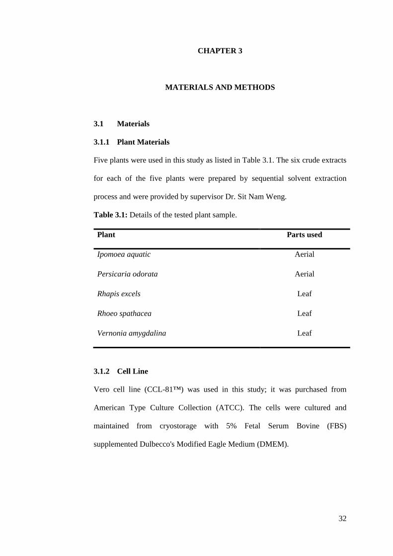
3.1 Materials 32
3.1.1 Plant Materials 32
3.1.2 Cell Line 32
3.1.3 Chikungunya Virus (CHIKV) 33
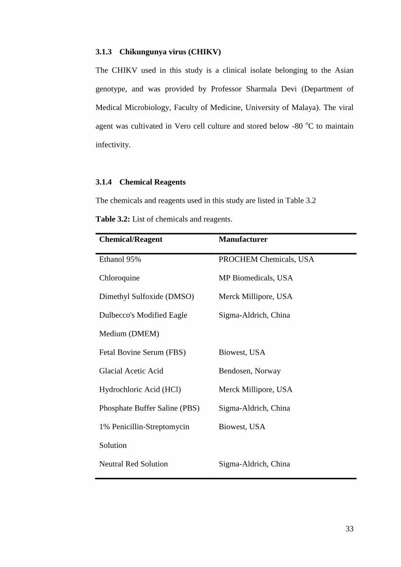
3.1.4 Chemical Reagents 33
3.1.5 Equipment and Labwares 34
3.2 Methodology 36
3.2.1 Preparation of Plant Extract 36
3.2.2 Preparation of Reagents 36
3.2.3 Vero Cell Culture 38
3.2.4 CHIKV Culture 41
3.2.5 NRU Cytotoxicity Assay 42
3.2.6 TCID50 Assay 45
3.2.7 Post-Inoculation Antiviral Assay 47
3.3 Data Analysis 51
3.3.1 NRU Cytotoxicity Assay 51
3.3.2 Post-Inoculation Antiviral Assay 52
4 RESULTS 53
4.1 NRU Cytotoxicity Assay 53
4.2 Post-Inoculation Antiviral Assay 59
5 DISCUSSION 63
5.1 Preparation of Plant Extract 63
5.2 NRU Cytotoxicity Assay 64
5.3 Post-Inoculation Antiviral Assay 65
5.4 Limitations 69
5.5 Future Perspectives 69
6 CONCLUSION 70
REFERENCES 71
x
LIST OF TABLES
Tables Page
2.1 Specific chemical compounds tested against CHIKV 9
2.2 Diagnostic criteria for Chikungunya Disease 14
2.3 Virus species susceptible and resistance to Vero cell line 27
3.1 Details of the tested plant sample 32
3.2 List of chemicals and reagents 33
3.3 List of equipments and labwares 34
4.1 Cytotoxicity of Ipomoea aquatica, Persicaria odorata, 57
Rhapis excelsa, Rhoeo spathacea and Vernonia amygdalina
plant extracts on Vero cells expressed as CC50 and MNCC
obtained in 72 hour NRU assay.
4.2 Antiviral activity of Chloroquine against CHIKV in 72 62
hour post-inoculation antiviral assay

xi
LIST OF FIGURES
Figure Page
2.1 Global distribution of the three CHIKV strains 6
2.2 Illustration of CHIKV virion 7
2.3 CHIKV genome organization 8
2.4 Pathogenesis of Chikungunya disease 12
2.5 Ipomoea aquatica 16
(A) Leaves and flower (B) Branch and disected flower
2.6 Persicaria odorata 18
(A) Leaf and stem (B) Flowers, stem, seeds and bark
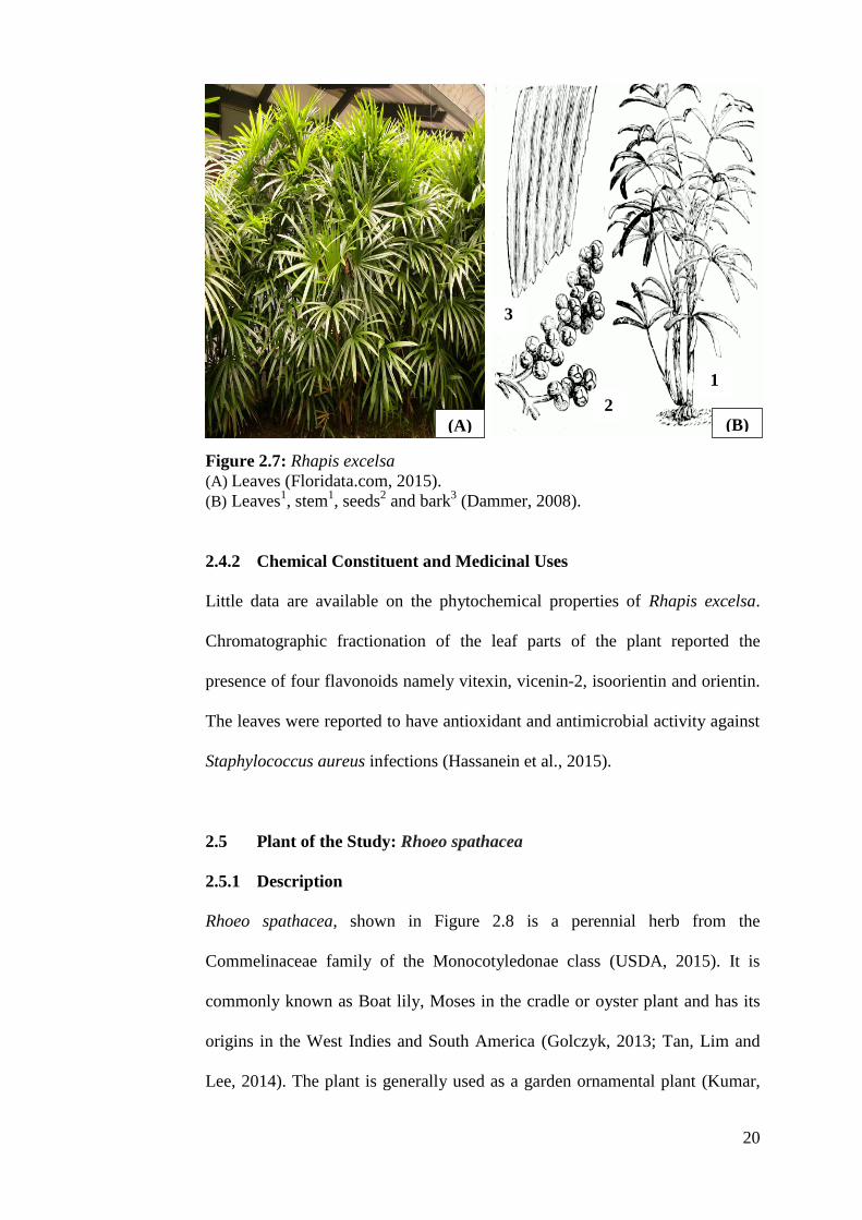
2.7 Rhapis excelsa 20
(A) Leaves (B) Leaves, stem and seeds
2.8 Rhoeo spathacea 21
(A) Leaves (B) Leaves, flowering branch, flower and root
2.9 Vernonia amygdalina 23
(A) Leaves and stems
(B) Leaf, flowering branch, flowering head and fruit
2.10 Vero Cell Line 27
(A) 100x Microscopy image of Vero cells and
(B) 400x Microscopy image of Vero cells
3.1 Four cell counting grids where viable cells were enumerated 40
3.2 Illustration of two-fold serial dilution of each plant extract 43
stock solution for NRU assay
3.3 Layout of 96-well plate for NRU assay 44
3.4 Illustration of two-fold serial dilution of CHIKV stock 46
suspension for TCID50 assay
3.5 Layout of 96-well plate for TCID50 assay 47
3.6 Illustration of two-fold serial dilution of Chloroquine stock 48
solution for post-inoculation antiviral assay
xii
3.7 Layout of 96-well plate for post-inoculation antiviral 50
assay
4.1 Cytotoxic activity of Ipomoea aquatica extracts on Vero 54
cells at different concentrations in 72 hour NRU assay
4.2 Cytotoxic activity of Persicaria odorata extracts on Vero 54
cells at different concentrations in 72 hour NRU assay
4.3 Cytotoxic activity of Rhapis excelsa extracts on Vero cells at 55
different concentrations in 72 hour NRU assay
4.4 Cytotoxic activity of Rhoeo spathacea extracts on Vero 55
cells at different concentrations in 72 hour NRU assay
4.5 Cytotoxic activity of Vernonia amygdalina extracts on Vero 56
cells at different concentrations in 72 hour NRU assay
4.6 Antiviral activity of Ipomoea aquatica extracts at 60
different concentrations against CHIKV in 72 hour
post-inoculation antiviral assay
4.7 Antiviral activity of Persicaria odorata extracts at 60
different concentrations against CHIKV in 72 hour
post-inoculation antiviral assay
4.8 Antiviral activity of Rhapis excelsa extracts at different 61
concentrations against CHIKV in 72 hour
post-inoculation antiviral assay
4.9 Antiviral activity of Rhoeo spathacea extracts at 61
different concentrations against CHIKV in 72 hour
post-inoculation antiviral assay
4.10 Antiviral activity of Vernonia amygdalina extracts at 62
different concentrations against CHIKV in 72 hour
post-inoculation antiviral assay
xiii
LIST OF ABBREVIATIONS
ATCC American Type Culture Collection
ANOVA Analysis of variance
CC50 Half-maximal cytotoxic concentration
CHIKV Chikungunya virus
CO2 Carbon dioxide
CPE Cytopathic effect
DNA Deoxyribonucleic acid
DMEM Dulbecco's Modified Eagle Medium
DMSO Dimethyl sulfoxide
ECSA East-Central-South African
EC50 Half-maximal effective concentration
EDTA Ethylenediaminetetraacetic acid
E1 Virus envelope 1
E2 Virus envelope 2
ELISA Enzyme-linked immunosorbent assay
FBS Fetal bovine serum
IFN γ Interferon gamma
IgM Immunoglobulin M
IgG Immunoglobulin G
IL Interleukin
LDH Lactate dehydrogenase
MNCC Maximal non-cytotoxic concentration
MOI Multiplicity of infection
xiv
NHP Non-human primate
NRU Neutral red uptake
nSP Non-structural protein
ORF Open reading frame
PBS Phosphate buffered saline
PCR Polymerase chain reaction
RdRp RNA-dependent RNA polymerase
RNA Ribonucleic acid
SD Standard deviation
RT-PCR Reverse transcription polymerase chain reaction
TCID50 Tissue Culture Infectious Dose, 50%.
UTR Untranslated region
WHO World Health Organization
1
CHAPTER 1
INTRODUCTION
Chikungunya is a disease of viral origin that is characterised as a debilitating
viral fever (Mudurangaplar, 2015), the etiological agent behind the disease is
an alphavirus that is transmitted primarily to humans through the bite of an
infected female Aedes mosquito (Raquin et al., 2015). The virus, Chikungunya
(CHIKV) since its first isolation back in the year 1952 in Tanzania (LaBeaud et
al., 2015) had caused massive outbreaks and is currently identified in over 60
countries and territories worldwide (Staples, Breiman and Powers, 2009). In
India alone, it was approximated more than 1.4 million of its population would
have been affected between the years of 2006 to 2008 (Weaver, 2014).
Meanwhile, in Southeast Asia, the number of reported cases went up to an
estimated 1.9 million patients from the year 2005 (Coffey, Failloux and
Weaver, 2014). Following the autochronous transmission of the virus to the
Western hemisphere, more than 1.2 million probable cases have been reported
in the year 2014 (CDC, 2015; Javelle et al., 2015).
Notably in Malaysia it had caused several outbreaks following the 2005
epidemic in the Indian Ocean islands. A total of 6 314 confirmed cases were
reported from the year 2006 to 2009 throughout the country with most
incidence coming from Johor state contributing 43.6% to the total cases
reported (Chua, 2010; Azami et al., 2013). The outbreak in Kenya in the year
2004 initiated the resurgence of the virus (Robinson et al., 2014) and during

2
this period the viral strain of East/Central/South African (ECSA) lineage was
reported to have undergone a mutation on its E1 envelope glycoprotein (Ala-
226-Val) that resulted in the transition of its principal transmission vector
Aedes aegypti to Aedes albopictus and an efficient transmission of the disease
to humans (Lee and Chu, 2015; Yoon et al., 2015). In spite of the increase in
global incidence, to date there are no licensed vaccines or therapeutics
available for its treatment and prophylaxis (Raquin et al., 2015). The therapy
given is generally symptomatic with bed rest, rehydration and administration of
analgesics (Hrnjakovic-Cvjetkovic et al., 2015).
Plant natural products, since the primordial days were used by all global
traditions as the principal source of medicine. Presently, it is notable that
around 80-85% of the world population depends on traditional herbal therapy
for their health benefits and requirements (Rasingam, 2012; Prakash et al.,
2013). Extracts from plants are an essential source of novel pharmacologically
active products with many drugs being continuously acquired both directly and
indirectly from plant sources (Veeresham, 2012). According to a study
conducted in the year 2007 by Newman and Cragg, more than 44% of
antivirals approved between the years of 1981 to 2006 were derived based on
natural-product pharmacophores.
The inhibitory effects of medicinal plants extracts on the replication of viruses
were reported in the past six decades (Liu and Du, 2012). Plant extracts have
been shown to possess antiviral potential against viral strains that were
resistant to conventional antiviral agents (Serkedjieva and Hay, 1998; Tolo et
3
al., 2006). The antiviral activities of natural products, including ingredients,
fractions and extracts, has to be evaluated by various antiviral models,
including in vitro and in vivo models (Liu and Du, 2012). With the advent of
new and powerful screening assays and prediction tools, the idea of a drug to
efficiently treat viral infections by blocking specific host functions has re-
bloomed (Martinez et al., 2015).
This study which is a part of a screening project, was aimed to investigate the
antiviral activity of extracts from five local plants namely Ipomoea aquatica,
Persicaria odorata, Rhapis excelsa, Rhoeo spathacea and Vernonia
amygdalina. A total of thirty plant extracts were obtained and examined for
activity against CHIKV.
Therefore the objectives of this project are:
I) To perform cytotoxicity assay of the various plant extracts against
Vero cell line in order to calculate the half-maximal cytotoxic
concentration (CC50) and the maximal non-cytotoxic concentration
(MNCC) as a pre-requisite for the post-inoculation antiviral assay.
II) To analyse the antiviral activity of the prepared plant extracts
against CHIKV using the post-inoculation antiviral assay and to
calculate half-maximal effective concentrations (EC50).
4
CHAPTER 2
LITERATURE REVIEW
2.1 Chikungunya Disease
Chikungunya is an emerging viral disease, which is often associated with acute
febrile and sometimes eruptive polyarthritis (Javelle et al., 2015). The
causative agent, Chikungunya virus (CHIKV) is an arbovirus that spreads
through the bite of an infected Aedes mosquito (Aubry et al., 2015). The name
‘Chikungunya’ originates from the Swahili language which refers to the
stooped posture developed as a result of the arthritic symptoms of the disease
(Mudurangaplar, 2015). The disease has been reported in over 60 countries
mainly from parts of Africa, Asia and recently from some parts of Europe
(Hrnjakovic-Cvjetkovic et al., 2015; Khan et al., 2015).
2.1.1 History and Epidemiological Features
CHIKV was first isolated in Africa during an outbreak in the year 1952 from
the Mokande Plateau of Tanzania (Stamm, 2015). In Asia, the disease was first
recorded in the year 1958 following the isolation from Bangkok, Thailand
(Moyen et al., 2014). Since then, the Asian genotype of CHIKV continued to
cause several major outbreaks such as the outbreak that took place in South
Asia from the year 1963 and 1964 in the city of Kolkata and Solapur, India
(Mudurangaplar, 2015). Concurrently, the virus continued to be active in
Thailand (Lam et al., 2001) and until then it had disappeared for three decades.
Following the year 2004 the virus re-emerged causing an outbreak in Kenya
5
leading to the massive widespread of the disease in the Indian Ocean islands
particularly in the French islands of Mayotte and La Re´union (Moyen et al.,
2014; Robinson et al., 2014) with more than 1,400 000 to 6, 500 000 cases
recorded in India and 300 000 cases in the island of La Re´union in the year
2006 (Hrnjakovic-Cvjetkovic et al., 2015). During this outbreak, the virus
seems to have acquired mutations in glycoprotein E1, which is important for
membrane fusion and virion assembly (Hamer and Chen, 2014). Travelers
from India to Europe introduced chikungunya, resulting in local transmission
in France and Italy with 207 confirmed cases reported from the year 2007-
2010. However, in the year 2013, through autochronous transmission, CHIKV
was reported in over 44 countries of the North, Central and South American
countries and the viral strain responsible for the growing epidemic was
reported to be the Asian genotype (Weaver, 2014).
In Malaysia, Chikungunya was first recorded in Port Klang between the year
1998 and 1999 (Lam et al., 2001; AbuBakar et al., 2007). Following the
outbreak, the virus was silent for seven years till the next outbreak which took
place in the coastal village of Bangan Panchor, Perak with a population of 200
villagers affected (AbuBakar et al., 2007), the third outbreak took place in
Ipoh, Perak on the same year (Azami et al., 2013). Both of these outbreaks
occurred synchronously with the ongoing epidemic then in the Indian Ocean
islands (Paganin et al., 2006) however it was caused by the Asian genotype
rather than the ECSA Indian Ocean lineage (Weaver, 2014). Later in the year
2008, fourth outbreak was reported from Johor state which then spread to other
territories of Malaysia (Sam et al., 2009; Rozilawati et al., 2015). From then

6
there was a nationwide outbreak resulted in 10 000 cases, with no fatalities
reported (Ministry of Health, Malaysia, 2010).
2.1.2 Chikungunya Virus (CHIKV)
2.1.2.1 Classification and Phylogenesis
CHIKV is a 12 kb positive sense single stranded RNA virus belonging to the
genus alphavirus from the togaviridae family (Pun, Bastola and Shah, 2014;
Yoon et al., 2015). It belongs to the antigenic complex IV (Semliki forest
serocomplex) subgroup of the genus alphavirus (Pialoux et al., 2007;
Hrnjakovic-Cvjetkovic et al., 2015). The phylogenetic study of CHIKV strains
by Powers et al. (2000), identified three variant strains namely the West
African (WAf), East/Central/South African (ECSA), and Asian genotypes
(Volk et al., 2010; Vega-Rúa et al., 2015). Figure 2.1 illustrates the distribution
of the three CHIKV strains.
Figure 2.1: Global distribution of the three CHIKV strains (Volk et al., 2010).

7
2.1.3.2 Morphology and Genome Structure
CHIKV as shown in Figure 2.2, is a spherical-enveloped virus with a diameter
of 60 to 70 nm that consists of its major structural proteins Capsid, E1 and E2
(Lim and Chu, 2014) and its viral genome shown in Figure 2.3 has two ORFs:
structural and nonstructural that begins with a UTR at the 5’ terminal end and
followed by the coding regions for the nonstructural proteins (nsP1 to 4) which
occupy two-third of its genome. Subsequently, the remaining one-third of the
coding region encodes for the structural protein Capsid-E3-E2-6K-E, and a 3’-
terminal poly-A-tail (Hussain and Chu, 2011). The nsPs are responsible for the
different roles in viral replication where nsP1 is involved in viral RNA
synthesis initiation and RNA capping (Ahola et al., 1997). The nsP2 possesses
protease and RNA helicase activities (Gomez de Cedrón et al., 1999; Frolova et
al., 2006) meanwhile the nsP3, composed of three domains, is required for the
formation and localization of replication complexes. Lastly, nsP4 functions to
possess RNA-dependent RNA polymerase (RdRp) activity which is important
for replication and synthesis of the viral genome (Frolova et al., 2006; Tomar
et al., 2006).
Figure 2.2: Illustration of CHIKV virion (3DCIENCIA.com, 2015).

8
Figure 2.3: CHIKV genome organization (3DCIENCIA.com, 2015).
2.1.2.3 Viral Replication
The transmission of CHIKV requires infection of a female mosquito through
viremic blood meal and following a suitable extrinsic incubation period,
transmission to another vertebrate host during subsequent feeding (Solignat et
al., 2009). Upon transmission, virus entry is mediated through the clathrin-
mediated endocytosis as soon as the virus binds to its receptors on the host
cells (Kielian, Chanel-Vos and Liao, 2010). The viral particle then undergoes
disassembly and releases its genomic RNA into the cytosol of the infected cell.
The viral genome is then translated into structural and non-structural proteins
(Glanville et al., 1976) and undergoes cleavage of its non-structural protein,
during the process of cleavage; it forms the P123 and nsP4 (De Groot et al.,
1990; Takkinen, Peranen and Kaariainen, 1991) peptides that causes the
synthesis of negative strand RNA (Strauss and Strauss, 1994) by forming an
unstable initiation complex. Soon after the cleavage of the non-structural
proteins into nsP 1-4, the virus switches its synthesis of the negative-strand
RNA to genomic and sub-genomic positive strand RNA synthesis (Iemm et al.,
1994; Shirako, Strauss and Strauss, 2000). Upon the availability of C protein, it
associates with the newly synthesized RNA and recognizes the specific signals
for packaging in the 5’ half of the genome, so that only RNA of full length is
packaged (Owen and Kuhn, 1996; Weiss et al., 2015). The synthesized E2 and
E3 glycoprotein interacts with each other and forms heterodimers which is then
9
transported to the cell surface via the Golgi complex from the endoplasmic
reticulum. The cleavage of the precursor protein PE2 to generate mature E2
and E3 proteins causes conformational change and weakening of the
interaction within the heterodimers (Wahlberg, Boere and Garoff, 1989)
subsequently resulting in the priming of the fusion peptide for activation upon
the exposure to a low pH. Through the interactions between C protein and the
cytoplasmic domain of the E2 protein the budding process is initiated, with E1-
E2 heterodimers forming an envelope around nucleocapsid-like particles
(Ziemiecki, Garoff and Simons, 1980; de Curtis and Simons, 1988; Sariola,
Saraste and Kuismanen, 1995). The virons acquire a phospholipid bilayed
derived from the host cell membrane upon release from the host cell (Laine,
Soderlund and Renkonen, 1973; Vogel et al., 1986; Fuller, 1987; Leung, Ng
and Chu, 2011).
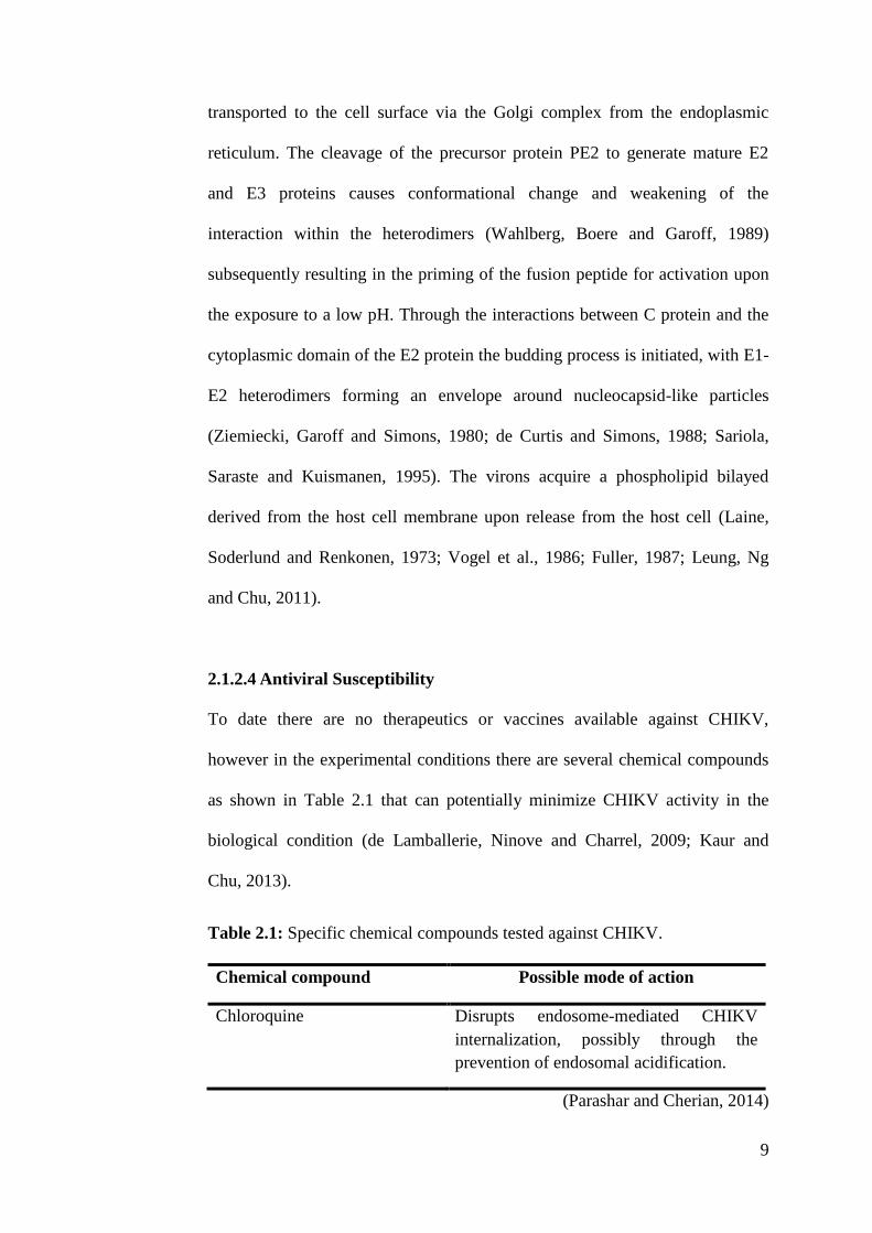
2.1.2.4 Antiviral Susceptibility
To date there are no therapeutics or vaccines available against CHIKV,
however in the experimental conditions there are several chemical compounds
as shown in Table 2.1 that can potentially minimize CHIKV activity in the
biological condition (de Lamballerie, Ninove and Charrel, 2009; Kaur and
Chu, 2013).
Table 2.1: Specific chemical compounds tested against CHIKV.
Chemical compound Possible mode of action
Chloroquine Disrupts endosome-mediated CHIKV
internalization, possibly through the
prevention of endosomal acidification.
(Parashar and Cherian, 2014)

10
Table 2.1(continued): Specific chemical compounds tested against CHIKV.
Chemical compound Possible mode of action
Ribavirin Can interact with the intracellular viral RNA
production.
6-Azauridine Inhibition of orotidine monophosphate
decarboxylase, an enzyme involved in the de
novo biosynthesis of pyrimidine, cytidine, and
thymidine.
Arbidol Inhibition of virus mediated fusion and
blocking of the viral entry into the target cells
through inhibition of glycoprotein
conformational changes that are essential for
the fusion process.
Harringtonine Affects CHIKV RNA production inside the
infected cell as well as viral protein expression
such as the nsP3 and the E2 proteins.
(Parashar and Cherian, 2014)
2.1.3 Vector and Natural Reservoir
The strains from different geographical distributions relatively circulate in
dissimilar ecological cycles. The strain in the African tropics exist in an
enzootic cycle primarily between mosquitoes in the forest and non-human
primates (NHP) which serves as the principle reservoir and amplication hosts
in the cycle. Meanwhile in Asia and other continents affected, transmission
primarily exists from the infected mosquitoe to humans (Eldridge and Edman,
2004; Weaver et al., 2012; Coffey, Failloux and Weaver, 2014). However,
study conducted by Apandi et al. (2009) reported the possibility of the
existence of such an Asian enzootic cycle. There are several mosquito species
that can transmit CHIKV to humans; however the primary vectors in the large

11
human outbreaks were from the genus Aedes particularly Ae.(Stegomyia)
albopictus and Ae.(Stegomyia) aegypti (Weaver et al., 2012; Parashar and
Cherian, 2014; Tretyakova et al., 2014). These two urban species are globally
spread and have drastically increased the incidence of viral spread to new
regions where the environmental conditions were permissible for transmission
(Vega-Rúa et al., 2015). Meanwhile, the 2005 to 2006 La Re´union outbreak,
suggested the possibility of transmission vertically from an infected pregnant
mother to her child, and the transmission is most likely to occur short before
delivery (Thiboutot et al., 2010).
2.1.4 Pathogenesis
Following bite of an infected Aedes mosquito on the skin, CHIKV travels
through the bloodstream and disseminates in the liver, muscles, lymph nodes
and spleen where primary replication takes place (Schwartz and Albert, 2010;
Miranda, Oliveira and Poian, 2013). As displayed on Figure 2.4 the incubation
period for CHIKV is 2-4 days and is followed by the onset of clinical phases
with no prodromal period. It is during this stage, infected individuals
experience various life-debilitating symptoms which includes severe
incapacitating joint pain. The acute phase of infection normally lasts days to
weeks, and severity of this infection is solely based on the viral load (Roques
and Gras, 2010). The onset of disease coincides with increasing virus titer and
results in the activation of innate immune response with the characteristic
production of type I interferons (IFN’s) and pro-inflammatory cytokines and
chemokines which includes IFN γ inducible protein 10, monocyte
chemoattractant protein, and IL 8 are elevated (Sebastian, Lodha and Kabra,
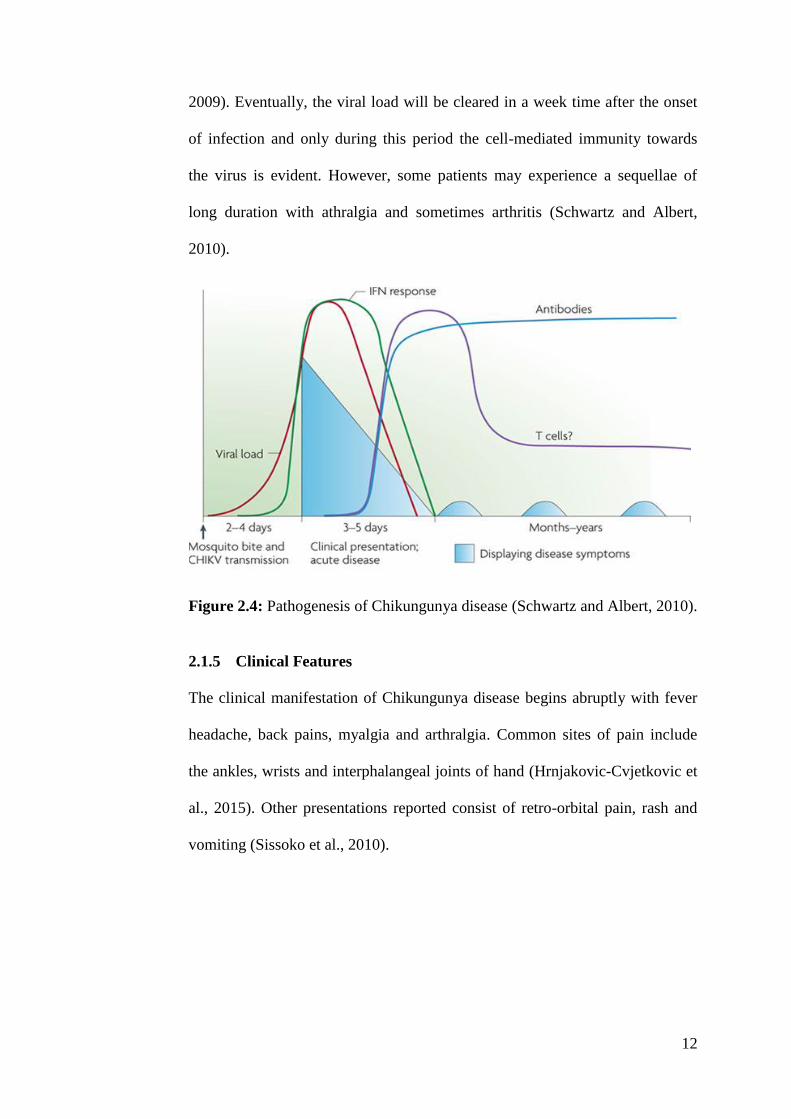
12
2009). Eventually, the viral load will be cleared in a week time after the onset
of infection and only during this period the cell-mediated immunity towards
the virus is evident. However, some patients may experience a sequellae of
long duration with athralgia and sometimes arthritis (Schwartz and Albert,
2010).
Figure 2.4: Pathogenesis of Chikungunya disease (Schwartz and Albert, 2010).
2.1.5 Clinical Features
The clinical manifestation of Chikungunya disease begins abruptly with fever
headache, back pains, myalgia and arthralgia. Common sites of pain include
the ankles, wrists and interphalangeal joints of hand (Hrnjakovic-Cvjetkovic et
al., 2015). Other presentations reported consist of retro-orbital pain, rash and
vomiting (Sissoko et al., 2010).

13
2.1.6 Diagnosis
The gold standard for detection of CHIKV disease is through virus culture and
isolation (Pialoux et al., 2007; Powers and Logue, 2007; Chevillon et al., 2008;
Simon, Savini and Parola, 2008; Sudeep and Parashar, 2008); which requires
the collection of patient’s blood samples that will be subjected to PCR for
further downstream analyses (WHO, 2015). However due to its limited
availability, other diagnostic methods are exercised. The standard diagnostic
procedures recommended by the World Health Organization (WHO), other
than virus isolation is serological testing. The serological test, Enzyme-linked
immunosorbent assay (ELISA) is widely available and is relatively economical
(Dash, Mohanty and Padhi, 2011). The principle is based on the detection of
IgM and IgG antibodies against the CHIKV antigens. CHIK IgM antibodies
become detectable during the fifth day of infection and remain the highest in
patient’s sera for 2-3 months period of illness (Pialoux et al., 2007; Hrnjakovic-
Cvjetkovic et al., 2015). Meanwhile, the IgG antibodies are only evident during
the 10th
-14th
day of disease (Dash, Mohanty and Padhi, 2011). Other
diagnostic procedures that are currently used include indirect
immunofluorescent, hemagglutination inhibition, neutralization techniques or
genotyping (Lakshmi et al., 2008; WHO, 2009; Hrnjakovic-Cvjetkovic et al.,
2015). Table 2.2 demonstrates the criteria’s for a successful diagnosis of
Chikungunya disease.

14
Table 2.2: Diagnostic criterias for Chikungunya Disease.
Diagnostic Criteria for CHIK Disease
Suspected case
A patient presenting with acute onset of fever usually with
chills/rigors, which lasts for 3-5 days with multiple joint
pains/swelling of extremities that may continue for weeks to
months
Probable case
A suspected case (see above) with any one of the following:
History of travel or residence in areas reporting outbreaks
Ability to exclude malaria, dengue and any other known
cause for fever with joint pains
Confirmed case
Any patient who meets one or more of the following findings
irrespective of the clinical presentation
Virus isolation in cell culture or animal inoculations from
acute phase sera
Presence of viral RNA in acute phase sera by RT-PCR
Presence of virus-specific IgM antibodies in single serum
sample in acute or convalescent stage
Fourfold increase in virus-specific IgG antibody titer in
samples collected at least three weeks apart
Footnote: RNA: Ribonucleic acid; RT-PCR: Reverse transcription polymerase
chain reaction; IgM: Immunoglobulin M; IgG: Immunoglobulin G.
(Mohan et al., 2010)
2.1.7 Treatment and Prevention
The absence of therapeutics for the treatment of Chikungunya disease (WHO,
2015; Yoon et al., 2015) poses a challenge for physicians in identifying and
administering the optimal treatment to prevent the progression and
perpetuation of the infection into a possibly life-decapitating course of disease
(Javelle et al., 2015).

15
Following the outbreak in the Re´union Island, analgesic drugs were the most
preferred option in alleviating the debilitating arthralgia’s associated with the
infection, among these a combination of paracetamol and nonsteroidal anti-
inflammatory drugs (NSAID’s) was a regular choice. Corticosteroids were
prescribed to some patients with incapacitating forms of the disease (Michault
and Staikowsky, 2009). Prevention of disease transmission is mainly focused
on the eradication of mosquito breeding sites and mobilization of those
affected (Hamer and Chen, 2014; WHO, 2015) to prevent the transmission of
disease to other healthy counterparts.
2.2 Plant of the Study: Ipomoea aquatica
2.2.1 Description
Ipomoea aquatic, shown in Figure 2.5 is also known as water spinach, swamp
morning glory (USDA, 2015) or simply ‘kangkung’ in the Malay language. It
is an aquatic plant and an important food crop belonging to the family
Convolvulaceae (Austin, 2007) and is thought to have originated in China
where it is consumed as a green leafy vegetable (Alkiyumi et al., 2012). The
plant is a perennial herb that is widely distributed throughout Africa, Australia,
South and Southeast Asia (Manvar and Desai, 2013) growing abundantly in
muddy stream banks, freshwater pond and lakes (Hamid et al., 2011). Ipomoea
aquatica is a vine that trails and creeps, moreover it has the ability to climb,
overtop and twine around other plants. Its stems are usually thick and spongy,
and rooting occurs at the nodes (Ogunwenmo and Oyelana, 2009). The leaves
are 5-12.5 cm long and 3.2-7.5 cm broad, acute, cordate or hastate with
rounded or acute lobes. Meanwhile, its flowers are infundibuliform, solitary

16
and consists of five free sepals, five united pale purple petals, unequal five
stamens with spiny pollens, and two-celled glabrous ovary with two ovules in
each cell. The fruits are capsuler with 1-4 seeds, 8 mm long capsules, ovoid,
and minutely pubescent (Manvar and Desai, 2013).
Figure 2.5: Ipomoea aquatica
(A) Leaves and flower (Medicalhealthguide.com, 2015).
(B) Branch1 and disected flower
2 (Datta and Saha, 1974).
2.2.2 Chemical Constituent and Medicinal Uses
Phytochemical studies of the plant reported the presence of carotenes such as
cryptoxanthin, lutein, lutein epoxide, violoxanthin and neoxanthin (Tee and
Lim, 1991), flavonoids such as mycertin, quercetin, luteolin and apigenin
(Daniel, 1989) and some alkaloids (Tofern et al., 1999). In addition, studies on
the leaf parts of the plant was revealed the presence of adequate quantities of
essential amino acids such as aspartic acid, glycine, alanine and leucine
(Hamid et al., 2011). The plant is traditionally used in the treatment of nervous
and general debility, piles, worm infections, leucoderma, leprosy, jaundice and
1 2 (A) (B)

17
liver disorders (Alkiyumi et al., 2012). Furthermore, its leaf extracts are used to
reduce blood sugar levels and as an antibiotic against Escherichia coli,
Pseudomonas aeruginosa and Bacillus subtilis infections (Hamid et al., 2011).
2.3 Plant of the Study: Persicaria odorata
2.3.1 Description
The plant Persicaria odorata, synonymously known as Polygonum odoratum
is a herb that has its origin in the tropics of Southeast Asia. It is a rampant
member of the Polygonaceae families that are collectively named as
smartweeds or pinkweeds, and is popularly known as ‘Daun Kesum’, laksa leaf
or Vietnamese coriander among the locals (Seidemann, 2005; Starkenmann et
al., 2006). As displayed in Figure 2.6, the plant is an evergreen stoloniferous
soft-wooded perennial herb that grows best to a height of 30-35 cm (Orr, 2014)
in the tropical and subtropical zones (Ridzuan, 2013). It has a characteristic
pointed leaves with dark purple distinctive features on the centre, its top is dark
green in colour and its bottom is in burgundy red, besides the plant’s stem is
joined at the leaf. The flowers are white and normally flowers during the
periods of early to late summer. The plant is routinely used in cooking where
the leaves of the plant are added into local dishes (Sasongko, Laohankunjit and
Kerdchoechuen, 2011) mainly for its peppery and mint flavor as a replacement
for the regular cilantro (Saad et al., 2014). The herb is usually propagated by
cutting the parts of the plant, and the roots are developed simply from its
nodes.
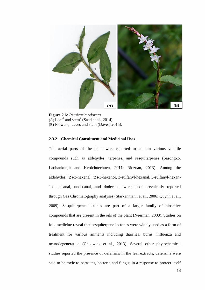
18
Figure 2.6: Persicaria odorata
(A) Leaf1 and stem
2 (Saad et al., 2014).
(B) Flowers, leaves and stem (Daves, 2015).
2.3.2 Chemical Constituent and Medicinal Uses
The aerial parts of the plant were reported to contain various volatile
compounds such as aldehydes, terpenes, and sesquiterpenes (Sasongko,
Laohankunjit and Kerdchoechuen, 2011; Ridzuan, 2013). Among the
aldehydes, (Z)-3-hexenal, (Z)-3-hexenol, 3-sulfanyl-hexanal, 3-sulfanyl-hexan-
1-ol, decanal, undecanal, and dodecanal were most prevalently reported
through Gas Chromatography analyses (Starkenmann et al., 2006; Quynh et al.,
2009). Sesquiterpene lactones are part of a larger family of bioactive
compounds that are present in the oils of the plant (Neerman, 2003). Studies on
folk medicine reveal that sesquiterpene lactones were widely used as a form of
treatment for various ailments including diarrhea, burns, influenza and
neurodegeneration (Chadwick et al., 2013). Several other phytochemical
studies reported the presence of defensins in the leaf extracts, defensins were
said to be toxic to parasites, bacteria and fungus in a response to protect itself
(A) (B)
1 2

19
from infections (Saad et al., 2014). The plant is widely regarded to have a
variety of medicinal properties especially in managing fever and coughs,
reducing thirst, application in stomach and lung injuries, diabetes mellitus and
as a anti-inflammatory, insect antifeedant and antimicrobial activities
(Shavandi, Haddadian and Ismail, 2015).
2.4 Plant of the Study: Rhapis excelsa
2.4.1 Description
Rhapis excelsa, shown in Figure 2.7 is commonly known as lady palm,
broadleaf lady palm or raffia palm (da Luz, de Oliveira Paiva and Tavares,
2008). It is a garden and indoor ornamental plant from the Arecaceae family
and is distributed from southern China to Southeast Asia (Dransfield and Uhl,
1998). Rhapis excelsa grows up to 4 m in height and 30 mm in diameter in
multi-stemmed clumps with glossy leaves. It can be cultivated in a variety of
soils and is represented as small under-growth palms of the tropical rainforest
(Uhl and Dransfield 1998; Hastings 2003; Averyanov, Nguyen and Phan,
2006).
20
Figure 2.7: Rhapis excelsa
(A) Leaves (Floridata.com, 2015).
(B) Leaves1, stem
1, seeds
2 and bark
3 (Dammer, 2008).
2.4.2 Chemical Constituent and Medicinal Uses
Little data are available on the phytochemical properties of Rhapis excelsa.
Chromatographic fractionation of the leaf parts of the plant reported the
presence of four flavonoids namely vitexin, vicenin-2, isoorientin and orientin.
The leaves were reported to have antioxidant and antimicrobial activity against
Staphylococcus aureus infections (Hassanein et al., 2015).
2.5 Plant of the Study: Rhoeo spathacea
2.5.1 Description
Rhoeo spathacea, shown in Figure 2.8 is a perennial herb from the
Commelinaceae family of the Monocotyledonae class (USDA, 2015). It is
commonly known as Boat lily, Moses in the cradle or oyster plant and has its
origins in the West Indies and South America (Golczyk, 2013; Tan, Lim and
Lee, 2014). The plant is generally used as a garden ornamental plant (Kumar,
(A) (B)
1
2
3

21
Nagpal and Arun, 2011) and is grown widely in households of tropical
countries like Malaysia. Parts of the plant are traditionally taken to treat
various ailments meanwhile it is also popularly consumed as a favourite
beverage in South American countries (Tan, Lee and Lim, 2013). Other
synonyms of Rhoeo spathacea include Tradescantia spathacea and Rhoeo
discolor (USDA, 2015). The plant is fleshy/succulent, short and can grow to a
height of 20 cm; it has two colours on its leaves: green with yellow stripes on
the top and purple on the bottom (Golczyk, 2013; Tan et al., 2014). They are
large, imbricated and takes the shape of a lance with spiral pattern that closely
overlaps (Parivuguna et al., 2008). The plant is immensely clumped with
vibrant and lengthy leaves stemming out from its trunk (Bercu, 2013). The
flowers are small, white and clustered within a folded bract protruding from the
leaf axils with a short stalk; it has three petals and six stamens, produced
throughout the year and adds features to the decorative elements of the leaves
(National Parks, 2015)
Figure 2.8: Rhoeo spathacea
(A) Leaves (USDA, 2015).
(B) Leaves1, flowering branch
2, flower
3 and root
4 (Tramil.net, 2015).
(A) (B)
1
3
4
2
22
2.5.2 Chemical Constituent and Medicinal Uses
Little data are available on the phytochemical properties of Rhoeo spathacea.
A study by Parivuguna et al. (2008) through preliminary analysis on leaf
extracts reported the abundance of alkaloids, flavonoids, steroids, saponins,
cardiac glycoside, terpenoids, tannins, phenolic compounds and oil (Nikam et
al., 2013). This plant has been traditionally used by communities in Mexico
and Southeast Asia to treat cancer, superficial mycoses, coughs, colds, and
dysentery (Rosales-Reyes et al., 2008; Joash et al., 2014). Furthermore, it was
reported to have insecticidal, anti-inflammatory and anti-fertility properties
(Siriwanthana et al., 2007).
2.6 Plant of the Study: Vernonia amgydalina
2.6.1 Description
Vernonia amygdalina as shown in Figure 2.9 is a short wooden shrub that
grows to a height of 1 to 5 m and belongs to the family Asteraceae (Adesanoye
et al., 2012). It originates in African tropics and is commonly used for culinary
purposes (Ajibola, Eleyinmi and Aluko, 2011). It was reported being consumed
by chimpanzees in the wild for self-deparasitization (Huffman, 2001; Nweze,
Ogidi and Ngongeh, 2013). The plant is internationally recognized as African
bitter leaf (Grubben, 2004) and locally as ‘daun bismillah’ (Mohd Abd Razak
et al., 2014) which literally means sacred leaves in the local Malay language.
The plant was only recently introduced in Malaysia, and is cultivated in many
parts of the country for its medicinal values (Atangwho et al., 2013). The plant
is moderately branched with a trunk up to the size of 40 cm in diameter, the
barks are in grey to brown colour with a smooth texture and are fissured, and

23
however young branches are highly pubescent (Grubben, 2004). The leaves of
the plant are simple, obovate-oblanceolate, display few lateral nerves and are
finely glandular at the bottom. The flowers are white, fragrant, normally
infested by bees and occur in copious corymbose panicles (Iwu, 2013). The
stem and seeds of the plant were used for medicinal purposes whereas the
leaves were used for both medicinal and cooking purposes (Mueller and
Mechler, 2005). It can be harvested by simply by cutting the leafy shoots and
allowing the new side shoots to develop, which normally takes a few weeks.
Once the plant is established in the garden, the leaves and young shoots can be
harvested for a period of 7 years (Grubben, 2004).
Figure 2.9: Vernonia amygdalina
(A) Leaves and Stems (Yeap et al., 2010).
(B) Leaf1, flowering branch
2, flowering head
3 and fruit
4 (Grubben, 2004).
2.6.2 Chemical Constituent and Medicinal Uses
The characteristic bitter tastes of the leaves were ascribed to the presence of
anti-nutritive constituents for instance alkaloids, glycosides, tannins and
saponins (Adiukwu et al., 2013). However, the main constituents of the leaves
(A) (B)
1
3 4
2

24
that contributes to its medicinal properties include saponin vernonin,
sesquiterpene lactones such as vernodalin, vernolide, hydroxyvernolide,
vernomydin and vernodal and the kaempferol flavonoid (Ademola and Eloff,
2010; Iwu, 2013). The pharmacological activities observed from this plant are
attributed to the presence of various biologically active constituents in the
leave. The vernonin from the plant is said to elicit strong antitumoral response
in leukaemic cells. Meanwhile, the strong antioxidant activities of the plant are
evident due to the presence of ubiquitous flavonoids (Adaramoye, 2008).
Besides that, the antimalarial activity of Vernonia amygdalina can be related to
the presence of flavonoids, saponins and alkaloids (Oyugi et al., 2009). The
sequesterpene lactones and flavonoids produced by the leaf parts of the plant
adds anti-phlogistic and anti-nociceptive effects to the plant’s bioactive
properties (Nangendo et al., 2002; Favi et al., 2008; Iroanya, Okpuzor and
Mbagwu, 2010). In addition, the leaves of Vernonia amygdalina were highly
reputed in the traditional African medicine to be effective in the treatment of
gastrointestinal disorders, diarrhea and hepatitis (Wan, 2012; Nweze, Ogidi and
Ngongeh, 2013).
2.7 Extraction of Medicinal Plants
Extraction is the pharmaceutical term used to define the approach used for the
separation of the therapeutically desired compound from the other unwanted
insoluble substances with the treatment of selective solvents (Kothari, Gupta
and Naraniwal, 2012). Diverse solvent systems are available to extract
bioactive compounds, selections are solely based on the target compounds, for
instance an extraction of hydrophilic compound uses polar solvents such as
25
acetone, ethanol and methanol (Wendakoon, Calderon and Gagnon, 2012)
whereas the extraction of a more lipophilic compound uses solvents like
dichloromethane or a mixture of dichloromethane/methanol in ratio of 1:1.
Conventional methods of bioactive compound extraction techniques are
heating, maceration, refluxing, solid liquid extraction (Soxhlet) and steam
distillation or cold press (Martins and da Conceição, 2015) which principally
relies on the correct use of organic solvent and the use of agitation and/or heat
to intensify the solubility of the bioactive compounds. However, these methods
produces lower product yields and have minimal selectivity. Besides that, it
uses large volumes of organic solvents which poses potential environmental
and health risks (Abdel-Azim et al., 2013). The demand for a more efficient
extraction process with maximal purity and with no loss of activity has led to
the augmentation of newer extraction processes (Santos, Vardanega and De
Almeida, 2014) collectively named as modern extraction techniques which
includes the microwave assisted extraction (MAE), solid phase micro
extraction (SPME) and Soxhwave ultrasonification assisted extraction (UAE)
(Gupta, Naraniwal and Kothari, 2012). These modern sample preparation
techniques resulted in the significant reduction in the consumption of organic
solvent and in minimizing degradation of samples (Kothari, Gupta and
Naraniwal, 2012) and are therefore advantageous over the conventional
methods of extraction.
26
2.8 Vero Cell Line
2.8.1 Description
The Vero cell line is a continuous highly anchorage-dependent cell line that is
derived from the African green monkey (Cercopithecus aethiops) kidney
epithelium by two scientists Y. Yasumura and Y. Kawakita from Chiba
University, Japan on March 27, 1962 (Osada et al., 2014). It is widely used for
the production of vaccines and determination of virus contaminations,
primarily due to its production of clear cytopathic effect (CPE) and wide range
of susceptibility to human viruses (Cao et al., 2013). Besides that, the cell line
is also used to evaluate mammalian cell susceptibility to bacterial toxins and in
the study of intracellular bacterial cell propagation (Ammerman, Beier-Sexton
and Azad, 2005). The Vero cell line is considered advantageous over other cell
lines because they are widely available, require no extensive culture conditions
and grow faster in cultures; also they can be used in suspension and
microcarrier cultures for large scale production in bioreactors (Chen and Chen,
2009).
2.8.2 Morphology and Structure
The Vero cells as shown in Figure 2.10 have a cuboidal epithelial morphology
growing in monolayer on a glass or treated plastic surface. Vero cells possess a
pseudo-diploid karyotype (ATCC, 2015) which refers to a cell that has 46
chromosomes with one or more structural abnormalities, gain-loss of the whole
chromosome, or both types of abnormality patterns (Williams et al., 1982) and
they are non-oncogenic when a cell passage was not prolonged (Osada et al.,
2014).

27
Figure 2.10: Vero Cell Line
(A) 100x Microscopy image of Vero cell.
(B) 400x Microscopy image of Vero cell.
2.8.3 Susceptibility and Resistance
The Vero cells have been classified as the most extensively used cell line in the
culture of viruses. Table 2.3 shows list of viruses that are susceptible and
resistant to the cell line.
Table 2.3: Virus species susceptible and resistant to Vero cell line.
Susceptibility/ Resistance Viral Species
Virus susceptibility Chikungunya virus
Human poliovirus 1, 2, 3
Getah virus
Pixuna virus
Ross River virus
Semliki Forest virus
Kokobera virus
Modoc virus
Guaroa virus
Tacaribe virus , Tacaribe virus
SV-5 (parainfluenza type 2), SV40
Measles virus
(ATCC, 2015)
(A) (B)

28
Table 2.3 (continued): Virus species susceptible and resistant to Vero cell
line.
Susceptibility/ Resistance Viral Species
Virus susceptibility Rubella virus , Rubella virus
Reovirus type 2, 3
Simian adenovirus 3, 17, 11, 1 , 20, 18, 16,
8, 17, 19, 21, 25, 22, 23, 38, 37, 27, 39, 32,
34, 31, 33, 36
Virus resistance Apeu; Ossa virus
(ATCC, 2015)
2.9 Cytotoxicity Assay
Cytotoxicity testing provides useful information in understanding the actions of
chemicals on cells (Li et al., 2013). They utilize various parameters associated
with proliferation and cell death, of them the most common is the Neutral red
(3-amino-m-dimethylamino-2-methylphenazine hydrochloride) uptake assay
which was developed in Rockfeller University as a tool for cell viability
chemosensitivity determination, the assay quantifies the number of uninjured
and viable cells through its ability to incorporate the supravital dye in its
lysosomes (Repetto, del Peso and Zurita, 2008). Other cytotoxicity assays
commonly in practice include the 3-(4,5-dimethylthiazol-2-yl)-2,5-diphenyl-
2H-tetrazolium bromide (MTT) assay, a measure of the reductive activity of
dehydrogenase enzyme present in the mitochondria of viable cells and its
ability to convert the reactant tetrazolium compound into a water insoluble
formazan crystals (van Tonder, Joubert and Cromarty, 2015). The Lactate
dehydrogenase (LDH) activity assay is based on the integrity of cell plasma

29
membrane. It is a measure of the cytoplasmic enzyme, LDH activity released
by cells upon the damage of cell membrane (Weyermann, Lochmann and
Zimmer, 2005).
2.9.1 Neutral Red Uptake (NRU) Asssay
Neutral red dye is frequently used in the evaluation of cell viability in cytotoxic
assays, the principle behind the staining of cells by neutral red lies in the ability
of the dye to penetrate the cell membrane through a non-ionic passive
diffusion, it concurrently accumulate within the lysosomes by binding with
anion and phosphate groups of the lysosomal matrix through electrostatic
hydrophobic bonds. Further, evaluation of its mechanism reveals that its uptake
principally relies on the capacity of the cell to establish equilibrium of its pH
gradients through the production of ATP. At a pH of 7.4, the dye’s net charge
is maintained near zero; which enables the dye to penetrate the cell membrane.
Meanwhile within the lysosomes, the action of a proton gradient lowers its pH
to that of the cytoplasm causing the dye to be charged and retained within the
lysosomes (Repetto, del Peso and Zurita, 2008). The dye retained can be
extracted out from the cells via an acidified ethanol solution, and the amount of
accumulated dye can be quantified by reading its absorbance at a wavelength
of 540 nm (Fotakis and Timbrell, 2006). On the contrary if a cell dies or when
the pH gradient was reduced, the dye will not be retained within the lysosomes.
2.10 TCID50 Assay
The median tissue culture infective dose (TCID50) also known as the endpoint
dilution assay is one of the two most widely used methods of virus
30
quantification, the other is the plaque forming units (PFU) assay. These
methods are built on serial dilutions of the virus samples and observation of the
development of a cytopathic effect (CPE) in a cell monolayer (Grigorov et al.,
2011). The TCID50 assay is a quantal assay which determines the dilution of
the sample at which 50% of the fractions have infectious virus whereas the
PFU assay is a quantitative assay which determines the figure of infectious
units of virus of interest in a sample (Lee et al., 2014). TCID50 virus titers can
be calculated by one of the two methods available namely, the Spearman-
Kaerber and Reed–Muench calculation methods. Considering the TCID50
method which is laborious and time consuming, newer variation of virus
quantification techniques have been developed to replace the conventional cell
culture based techniques, of these the real-time PCR technique is on the height
due to its rapid and efficient quantification (Jonsson, Gullberg and Lindberg,
2009). Furthermore, it is also being used extensively in studies involving virus
isolation, amplification and characterization. Other modern techniques for virus
quantification include flow cytometry, tunable resistive pulse sensing (TRPS),
and enzyme-linked immunosorbent assay (ELISA) (Pankaj, 2013).
2.11 Antiviral Assay
The conventional methods to analyze antiviral activity of a sample include both
in vitro and in vivo techniques (Jassim and Naji, 2003; Esimone et al., 2005). In
vitro studies to evaluate compounds with antiviral potential usually involve the
methods such as the plaque inhibition assay, plaque reduction assay, inhibition
of virus-induced cytopathic effect, virus yield reduction assay, end point titer
determination assay, reduction or inhibition of the synthesis of virus-specific
31
polypeptides, immunological assays detecting viral antigens and viral enzyme
inhibition-based assays (Vlietinck and Vanden Berghe, 1991; Cowan, 1999). In
the in vivo techniques, the samples were tested on laboratory mice, ferrets,
cotton rats and chickens to measure a few parameters to the extent of inhibition
of the infection. There are two methods of propagation and isolation of virus
namely the embryonated egg method (Härtl et al., 2004; Wang et al., 2008) and
the cell culture based (Nwodo et al., 2011).
32
CHAPTER 3
MATERIALS AND METHODS
3.1 Materials
3.1.1 Plant Materials
Five plants were used in this study as listed in Table 3.1. The six crude extracts
for each of the five plants were prepared by sequential solvent extraction
process and were provided by supervisor Dr. Sit Nam Weng.
Table 3.1: Details of the tested plant sample.
Plant Parts used
Ipomoea aquatic Aerial
Persicaria odorata Aerial
Rhapis excels Leaf
Rhoeo spathacea Leaf
Vernonia amygdalina Leaf
3.1.2 Cell Line
Vero cell line (CCL-81™) was used in this study; it was purchased from
American Type Culture Collection (ATCC). The cells were cultured and
maintained from cryostorage with 5% Fetal Serum Bovine (FBS)
supplemented Dulbecco's Modified Eagle Medium (DMEM).
33
3.1.3 Chikungunya virus (CHIKV)
The CHIKV used in this study is a clinical isolate belonging to the Asian
genotype, and was provided by Professor Sharmala Devi (Department of
Medical Microbiology, Faculty of Medicine, University of Malaya). The viral
agent was cultivated in Vero cell culture and stored below -80 oC to maintain
infectivity.
3.1.4 Chemical Reagents
The chemicals and reagents used in this study are listed in Table 3.2
Table 3.2: List of chemicals and reagents.
Chemical/Reagent Manufacturer
Ethanol 95%
Chloroquine
Dimethyl Sulfoxide (DMSO)
Dulbecco's Modified Eagle
Medium (DMEM)
Fetal Bovine Serum (FBS)
Glacial Acetic Acid
Hydrochloric Acid (HCl)
Phosphate Buffer Saline (PBS)
1% Penicillin-Streptomycin
Solution
Neutral Red Solution
PROCHEM Chemicals, USA
MP Biomedicals, USA
Merck Millipore, USA
Sigma-Aldrich, China
Biowest, USA
Bendosen, Norway
Merck Millipore, USA
Sigma-Aldrich, China
Biowest, USA
Sigma-Aldrich, China
34
Table 3.2 (continued): List of chemicals and reagents.
Chemical/Reagent Manufacturer
Sodium Bicarbonate (NaHCO3) Merck Millipore, USA
Trypan blue Thermo Fisher Scientific, USA
0.25% Trypsin:EDTA Sigma-Aldrich, China
3.1.5 Equipment and Labwares
The equipment and labwares used in this study are listed in Table 3.3.
Table 3.3: List of equipment and labwares.
Equipments/Labwares Manufacturer
Aluminium foil DIAMOND
Autoclave machine HICLAVETM
HVE-50, HIRAYAMA
Bench-top centrifuge machine Sigma-Aldrich, USA
Centrifuge tube CELLSTAR®
Class IIB biological safety cabinet TELSTAR®, Spain
Cryovial Greiner Bio-one, USA
Freezer HAIER®, China
Hemacytometer HIRSCHMANN®, Germany
Incubator Binder, Germany
Inverted microscope OLYMPUS®, Japan
Laboratory film Parafilm ―M‖ ®, Pechiney Plastic
Packaging
35
Table 3.3 (continued): List of equipment and labwares.
Equipments/Labwares Manufacturer
Laboratory oven Memmert, Germany
Microcentrifuge tubes AXYGEN, INC., Union City, USA
Micropipette WATSON BioLab, Japan
Micropipette tips AXYGEN, INC., Union City, USA
Microplate reader TECAN M200®, USA
Multichannel pipette Gilson, France
Pasteur pipette AXYGEN, INC., Union City, USA
Portable bunsen CAMPINGAZ® LABOGAZ 206
Refrigerator TOSHIBA®, Japan
Sample vial SAMCO®, United Kingdom
Glass bottle SCHOTT DURAN®, Germany
Serological pipettes Greiner bio-one, Austria
Serological pipetters Thermo Fischer Scientific, USA
Syringe (3 mL/5 mL) Terumo Medical Corporation, Japan
Syringe filter (0.45 μm) Sartorius Minisart®
Flat-bottomed polystyrene
96-well plate
Thermo Fisher Scientific, USA
Tissue-culture flask SPL Life Science, South Korea
Vacuum pump Eppendorf, Concentrator plus, Germany
Vortex mixer VELP® SCIENTICA, Europe
Weighing scale KERN & SOHN®, Germany

36
3.2 Methodology
3.2.1 Preparation of Plant Extract
In order to prepare plant extract stock solution, 0.1024 mg of each extract was
dissolved in 400 µL of DMSO: ethanol (6:4, v/v) to achieve the concentration
of 256 mg/mL. The extract solutions were then filtered using a 0.45 µm syringe
filter into sample vials and stored at 4 oC until use.
3.2.2 Preparation of Reagents
3.2.2.1 Phosphate Buffered Saline (PBS)
To prevent contamination, all the subsequent steps were performed within
Class IIB biological safety cabinet. Phosphate Buffered Saline (PBS) solution
was prepared by dissolving 4.8 g of powdered PBS into 500 mL distilled water.
The solution was autoclaved at 121 oC for 20 minutes and stored at 4
oC prior
usage.
3.2.2.2 Cell Freezing Medium
Cell freezing medium was used for the cryopreservation of Vero cells. For its
preparation, 150 mL of FBS and 50 mL of DMSO was added into 300 mL
serum negative DMEM. The medium was then sealed tightly and stored at 4 oC
prior usage
3.2.2.3 1% and 5% FBS Supplemented DMEM
The 1% and 5% FBS supplemented DMEM was intended to be used in assays
and for the maintenance of Vero cells respectively. For its preparation 13.4 g of
powdered DMEM and 3.7 g of sodium bicarbonate was weighed and

37
transferred into a 1 L glass bottle, then 500 mL of distilled water was added to
dissolve the powder. The solution was made up to 1 L with distilled water. The
pH of the solution was adjusted between 7.40 to 7.49 by adding 1M
Hydrochloric acid solution. Next, 10 mL of 1% Penicillin-Streptomycin
solution was added and the medium was filtered using 0.45 µm filter unit. A
volume of 5 mL (1% FBS supplemented DMEM) or 25 mL (5% FBS
supplemented DMEM) FBS was added into the prepared medium depending
on the type of growth medium being prepared. The occurrence of
contamination was examined by aspiring 2 mL of the prepared medium into a
petri plate. The plates were incubated at 37 oC for 72 hours in a CO2
humidified incubator. Finally, the glass bottle containing prepared medium was
sealed tightly with parafilm and stored at 4 oC prior usage.
3.2.2.4 Low Glucose Medium
Low glucose medium was intended to be used in the NRU assay and was
prepared similarly as the 1% and 5% FBS supplemented DMEM (as mentioned
in section 3.2.2.3, page 36), however a low glucose formulation of DMEM was
used and the medium was supplemented with 5 mL of FBS.
3.2.2.5 Chloroquine
Chloroquine which served as the positive control was prepared by dissolving
chloroquine powder into 100 mL of distilled water to achieve the concentration
of 62 mg/mL. The solution was filtered using 0.45 µm syringe filter and stored
away from light at 4 oC prior usage.

38
3.2.2.6 Neutral Red
In order to prepare Neutral red (NR) solution, 582 µL of NR stock solution (3.3
g/L, w/v) was diluted into 47.42 mL of low glucose medium to achieve the
concentration of 40 µg/mL. The solution was stored away from light at room
temperature prior usage.
3.2.2.7 Neutral Red Destain Solution
Neutral red destain solution was prepared by adding 10 mL of glacial acetic
acid and 500 mL of 95% ethanol into 490 mL of distilled water. The solution
was stored at room temperature prior usage.
3.2.3 Vero Cell Culture
3.2.3.1 Propagation from Frozen Stock
For the propagation of Vero cells from frozen stock, the cells were removed
from liquid nitrogen tank and thawed in water bath at 37 oC with gentle
swirling. The thawed cells were diluted with 5% FBS supplemented DMEM to
remove the cryopreservant, prior to the transfer into a 75 cm2
tissue culture
flask containing 6 mL of 5% FBS supplemented DMEM.
The cells were then incubated for 6 hours at 37 oC in a 5% CO2 humidified
incubator. After that, the cell suspension was transferred into a 15 mL
centrifuge tube and spinned at (1, 500) rpm for 5 minutes at room temperature.
The resulting pellet was re-suspended in 5 mL of 1% FBS supplemented
DMEM and seeded into a new 75 cm2 tissue culture flask containing 10 mL of
39
5% FBS supplemented DMEM. The cells were incubated, and monitored daily
to obtain 70-80% cell confluency.
3.2.3.2 Maintenance of Cells
Vero cells were maintained by replenishing the exhausted cell culture medium
2 to 3 times in a week (ATCC, 2015) depending on the cell confluency. The
cells require regular inspection under an inverted phase-contrast microscopy to
ensure it is free from any forms of contamination.
3.2.3.3 Subculture of Cells
Vero cells are normally subcultured when it reaches a percentage of 70-80%
cell confluency (ATCC, 2015). In order to subculture Vero cells, the cell
culture medium in the culture flask was removed. Then, the adherent cells in
culture flask were washed twice with 8 mL of prepared PBS (as mentioned in
section 3.2.2.1, page 36) and discarded after washing. These steps were
performed to remove traces of FBS which contains trypsin inhibitors that may
interfere in the process of enzymatic cell disaggregation (Ammerman, Beier-
Sexton and Azad, 2005). Next, 1.5 mL of 0.25% Trypsin: EDTA solution was
added and the cells were incubated at 37 oC in a 5% CO2 humidified incubator
for 5 minutes.
After incubation, gentle shaking and tapping was performed to aid in cell
detachment. The degree of cell detachment was examined under an inverted
phase contrast microscope. Then, to inactivate the activity of trypsin: EDTA,
an equivalent quantity of 1% FBS supplemented DMEM was added. The cell
suspension in the flask was transferred into a 15 mL centrifuge tube and
40
centrifuged at (1, 500) rpm for 5 minutes at room temperature. Once
centrifugation was complete, the supernatant was discarded and the obtained
pellet was re-suspended with 4 mL of 1% FBS supplemented DMEM. The
suspension was divided evenly into two tissue culture flask and topped up with
10 mL of 5% FBS supplemented DMEM. The flasks containing the cells were
incubated at 37 oC in a 5% CO2 humidified incubator and monitored daily to
ensure it is free from any forms of contamination.
3.2.3.4 Cell Count
Vero cell concentration in a cell suspension was established by cell count
procedure using a hemacytometer. Following the re-suspending of cell pellet
with 1% FBS supplemented DMEM (as mentioned in section 3.2.3.3, page 39),
10 µL of cell suspension was pipetted into a 1.5 mL microcentrifuge tube and
thoroughly mixed with 10 µL of 0.4% trypan blue. Next, 10 µL of the mixed
suspension was loaded onto a haemacytometer and via an inverted phase
contrast microscope at 100x magnification, the cells which were located in the
counting grids as shown in Figure 3.1 were enumerated carefully. Based on the
formula given below, the cell suspension concentration (cells/mL) was
calculated.
Cell concentration (cells/mL)= (A C D
4) x 2 x 104
x ml
Where, V is the volume of 1% FBS supplemented DMEM used to re-suspend
the pellet.

41
Figure 3.1: Four cell counting grids where viable cells were enumerated.
3.2.3.5 Cryostorage of Cells
Maintenance of frozen cell stocks is essential during the culture of cell lines.
Frozen cell stocks were routinely prepared shortly after the initiation of
cultures from the previously frozen stocks (Ammerman, Beier-Sexton and
Azad, 2005). For the cryopreservation of Vero cells, cells from the culture
flask were trypsinized and transferred into a 15 mL centrifuge tube. Next, it
was centrifuged at (1, 500) rpm for 5 minutes at room temperature. Once
centrifugation was complete, the supernatant was discarded and the obtained
pellet was re-suspended with 1 mL of prepared cell freezing medium. The
suspension was transferred into a 1.5 mL cryovial and frozen slowly to -80 oC
for 24 hours. On the following day, the cryovial containing the suspension was
transferred into a liquid nitrogen tank for long term storage at -196 oC.
3.2.4 CHIKV Culture
3.2.4.1 Thawing of CHIKV
Thawing of CHIKV stock was performed prior to virus titer assessment and
antiviral assays primarily to maintain stability of the virus. The cryovial
containing the virus stock was removed from ultrafreezer and thawed on a
37 oC water bath with gentle agitation.
42
3.2.4.2 Cultivation and Storage of CHIKV
To cultivate CHIKV through Vero cell culture, Vero cells were passaged into
two 25 cm2 tissue culture flasks containing 6 mL of 5% FBS supplemented
DMEM. The cells were incubated, and monitored daily to obtain 70-80% cell
confluency. Meanwhile, the thawed virus stock was diluted to a ratio of 1:10
with 1% FBS supplemented DMEM. Once the desired confluency was
obtained, 500 µL of the diluted virus suspension was inoculated into the two
culture flasks. The flasks were incubated at 37 oC in a 5% CO2 humidified
incubator. Next, Vero cells were examined daily under an inverted phase
contrast microscope for the development of cytopathic effect (CPE). Once the
desired degree of CPE was produced, the cell-virus suspension in the flask was
transferred into a 15 mL centrifuge tube and centrifuged at (1, 500) rpm for 5
minutes at room temperature. The resulting supernatant which contains the new
virus stock was transferred into 1.5 mL cryovials and stored in an ultrafreezer
at -80 oC prior to virus titer determination (Lennette and Schmidt, 1979;
Rovozzo and Burke, 1982; Burleson, Chambers and Wiedbrauk, 1992).
3.2.5 NRU Cytotoxicity Assay
In order to perform the NRU cytotoxicity assay, all sterile 96-well plates were
labeled correctly as shown in Figure 3.3. Following the cell count procedure
(as mentioned in section 3.2.3.4, page 40), 100 µL of cell suspension consisting
of 4 x 10⁴ cells/mL were seeded into the test and cell control wells of 96-well
plates. All plates were covered and sealed properly with parafilm and incubated
at 37 °C in a 5 % CO2 humidified incubator for 24 hours.
43
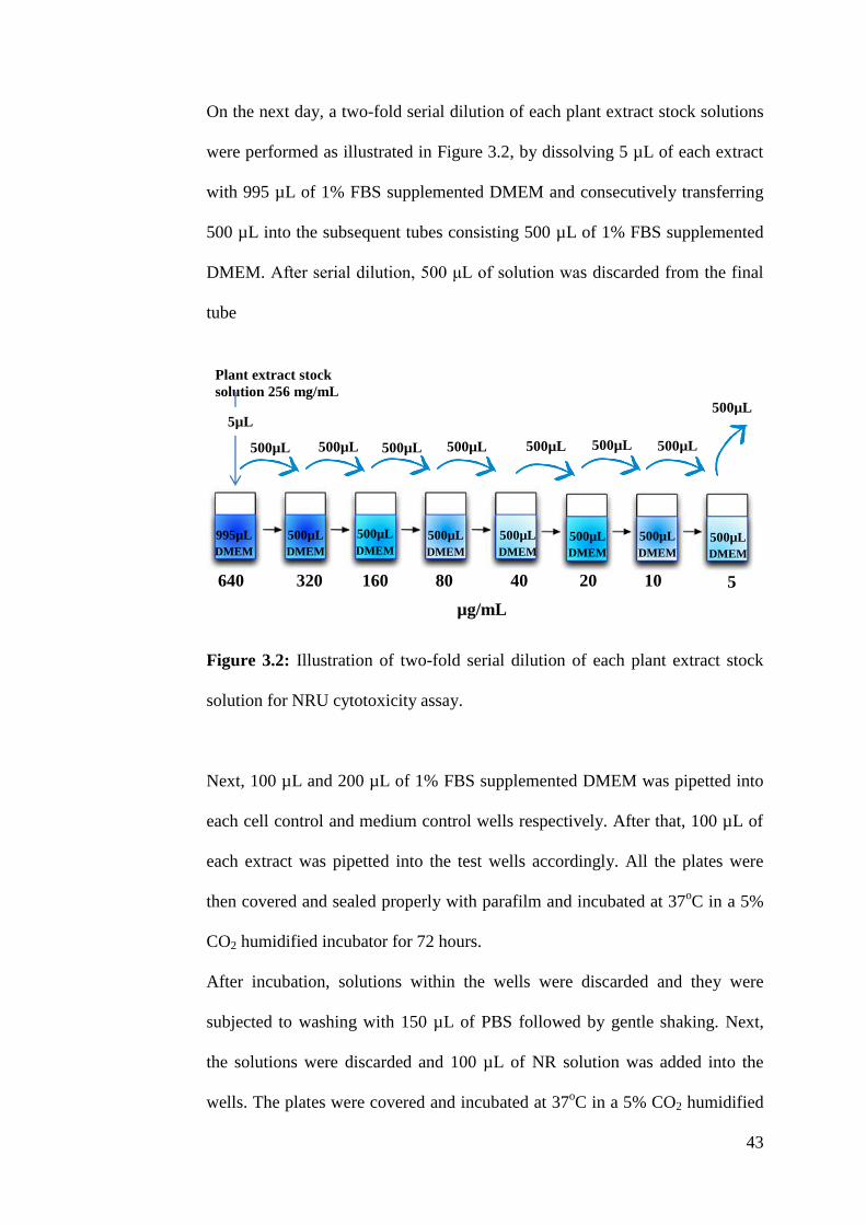
On the next day, a two-fold serial dilution of each plant extract stock solutions
were performed as illustrated in Figure 3.2, by dissolving 5 µL of each extract
with 995 µL of 1% FBS supplemented DMEM and consecutively transferring
500 µL into the subsequent tubes consisting 500 µL of 1% FBS supplemented
DMEM. After serial dilution, 500 μL of solution was discarded from the final
tube
Figure 3.2: Illustration of two-fold serial dilution of each plant extract stock
solution for NRU cytotoxicity assay.
Next, 100 µL and 200 µL of 1% FBS supplemented DMEM was pipetted into
each cell control and medium control wells respectively. After that, 100 µL of
each extract was pipetted into the test wells accordingly. All the plates were
then covered and sealed properly with parafilm and incubated at 37oC in a 5%
CO2 humidified incubator for 72 hours.
After incubation, solutions within the wells were discarded and they were
subjected to washing with 150 µL of PBS followed by gentle shaking. Next,
the solutions were discarded and 100 µL of NR solution was added into the
wells. The plates were covered and incubated at 37oC in a 5% CO2 humidified
640 320 160 80 40 20 10 5
µg/mL
500µL 500µL 500µL 500µL 500µL 500µL 500µL
500µL
500µL
DMEM
500µL
DMEM
500µL
DMEM
500µL
DMEM
500µL
DMEM
500µL
DMEM
500µL
DMEM
995µL
DMEM
Plant extract stock
solution 256 mg/mL
5µL

44
incubator for 2 hours. Once incubation was complete, NR solution were
replaced with 150 µL of NR destain solution for colour development. The
wells that were stained red indicate the presence of viable cells. The
observations were recorded and the absorbances of the all wells were read
using a multiplate reader at a wavelength of 540 nm. The NRU cytotoxicity
assay was repeated in quadruplicate for each plant extract.
Plate 1
Plate 2
640 320 160 80 40 20 10 5
HE
640 320 160 80 40 20 10 5
HE
CE
CE
EAE
EAE
EE
EE
ME
ME
WE
WE

45
Footnote: HE=Hexane extract; CE=Chloroform extract; EAE=Ethyl acetate
extract; EE=Ethanol extract; ME=Methanol extract; WE=Water extract; Row
1= Plant extract concentration used in the wells of the respective column
(µg/mL).
Figure 3.3: Layout of 96-well plates for NRU cytotoxicity assay.
3.2.6 TCID50 Assay
The tissue culture infectious dose (TCID50) assay was used to determine the
titer of CHIKV stock suspension (as mentioned in 3.2.4.2, page 41). For
TCID50 assay, all sterile 96-well plates were labeled correctly as shown in
Figure 3.5. Following the cell count procedure (as mentioned in section 3.2.3.4,
page 40), 100 µL of cell suspension consisting of 4 x 10⁴ cells/mL were seeded
into the test and cell control wells of 96-well plates. All plates were covered
and sealed properly with parafilm and incubated at 37 °C in a 5% CO2
humidified incubator for 24 hours.
On the next day, the thawed virus stock was diluted to a ratio of 1:10 by
dissolving 5 µL of the thawed CHIKV suspension with 995 µL of 1% FBS
supplemented DMEM and consecutively a two-fold serial dilution of CHIKV
suspension was prepared as illustrated in Figure 3.4, by dissolving 100 µL of
the diluted suspension with 900 µL of 1% FBS supplemented DMEM to
achieve CHIKV dilutions of 10-1
to 10-10
.
Test wells [consist of 100 µL of cells and 100 µL of plant extract]
Cell control [consist of 100 µL of cells and 100 µL of medium]
Medium control [consist of 200 µL of medium]

46
Figure 3.4: Illustration of two-fold serial dilution of CHIKV stock suspension
for TCID50 assay.
After incubation, 100 µL of 1% FBS supplemented DMEM was pipetted into
the cell control wells which serve as the negative control. Then, 100 µL of
prepared CHIKV dilutions were introduced into the test wells accordingly. All
plates were covered and sealed properly with parafilm and incubated at 37 oC
in a 5% CO2 humidified incubator for 72 hours.
Once incubation was complete, Vero cells were examined under an inverted
phase contrast microscope for the development of CPE, the observation were
recorded and the resulting virus titer and MOI was determined using the Reed-
Meunch method. The TCID50 assay was repeated in quadruplicate for CHIKV
stock suspension in each cryovial. Based on the formula given below, the
CHIKV titer was calculated (Reed and Meunch, 1938).
X = log10 dilution factor ( )
10-1
100µL
900µL
DMEM
CHIKV 1:10 diluted
suspension
100 µL
10-2
10-3
10-4
10-5
10-6
10-7
10-8
10-9
100µL 100µL 100µL 100µL 100µL 100µL 100µL 100µL
900µL
DMEM
900µL
DMEM
900µL
DMEM
900µL
DMEM
900µL
DMEM
900µL
DMEM
900µL
DMEM
900µL
DMEM
900µL
DMEM
10-10
% infection at next dilution above 50%-50%
% infection at next dilution above 50% - % of infection
at next dilution below 50%

47
Footnote: Row 1=CHIKV dilutions used in the wells of the respective column.
Figure 3.5: Layout of 96-well plate for TCID50 assay.
3.2.7 Post-Inoculation Antiviral Assay
In order to perform the post-inoculation antiviral assay, all sterile 96-well
plates were labeled correctly as shown in Figure 3.7. Following the cell count
procedure (as mentioned in section 3.2.3.4, page 40), 50 µL of cell suspension
consisting of 4 x 10⁴ cells/mL were seeded into the test, antiviral control, virus
control, cell control and cytotoxic control wells of 96-well plates. All plates
were covered and sealed properly with parafilm and incubated at 37 °C in a
5% CO2 humidified incubator for 24 hours.
10-1
10-2
10-3
10-4
10-5
10-6
10-7
10-8
10-9
Test wells [consist of 100 µL of cells and 100 µL of virus inoculum]
Cell control [consist of 100 µL of cells and 100 µL of medium]
10-10
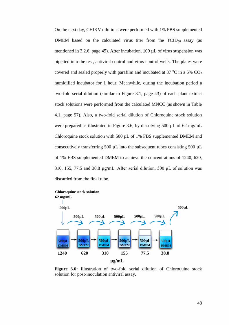
48
On the next day, CHIKV dilutions were performed with 1% FBS supplemented
DMEM based on the calculated virus titer from the TCID50 assay (as
mentioned in 3.2.6, page 45). After incubation, 100 µL of virus suspension was
pipetted into the test, antiviral control and virus control wells. The plates were
covered and sealed properly with parafilm and incubated at 37 oC in a 5% CO2
humidified incubator for 1 hour. Meanwhile, during the incubation period a
two-fold serial dilution (similar to Figure 3.1, page 43) of each plant extract
stock solutions were performed from the calculated MNCC (as shown in Table
4.1, page 57). Also, a two-fold serial dilution of Chloroquine stock solution
were prepared as illustrated in Figure 3.6, by dissolving 500 µL of 62 mg/mL
Chloroquine stock solution with 500 µL of 1% FBS supplemented DMEM and
consecutively transferring 500 µL into the subsequent tubes consisting 500 µL
of 1% FBS supplemented DMEM to achieve the concentrations of 1240, 620,
310, 155, 77.5 and 38.8 µg/mL. After serial dilution, 500 μL of solution was
discarded from the final tube.
Figure 3.6: Illustration of two-fold serial dilution of Chloroquine stock
solution for post-inoculation antiviral assay.
1240 620 310 155 77.5 38.8
µg/mL
500µL 500µL 500µL 500µL 500µL
500µL
500µL
DMEM
500µL
DMEM
500µL
DMEM
500µL
DMEM
500µL
DMEM
500µL
DMEM
Chloroquine stock solution
62 mg/mL
500µL

49
Once incubation was complete, 100 µL of each extract and 100 µL of the
prepared Chloroquine solutions were pipetted into the test and antiviral control
wells accordingly. Finally, 100 µL, 250 µL, 200 µL and 100 µL of 1% FBS
supplemented DMEM was pipetted into the virus control, medium control, and
cell control, and cytotoxic control wells respectively. All the plates were then
covered and sealed properly with parafilm and incubated at 37 oC in a 5% CO2
humidified incubator for 72 hours.
After incubation, the plates were subjected to washing and measurement of cell
viability at 540 nm using NRU assay (as mentioned in section 3.2.5, page 42).
The post-inoculation antiviral assay was repeated in triplicate for each plant
extract.
50
Plate 1
Plate 2
Antiviral control [consist of 50 µL of cells, 100 µL of virus
inoculum and 100 µL of Chloroquine]
Test wells [consist of 50 µL of cells, 100 µL of virus inoculum
and 100 µL of plant extract]
HE
HE
CE
CE
EAE
EAE
EE
EE
ME
ME
WE
WE
1240
620
310
155
77.5
38.8
1240
620
310
155
77.5
38.8
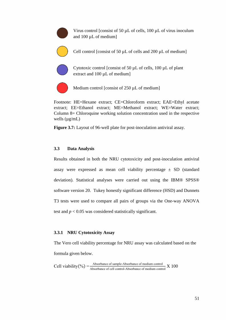
51
Footnote: HE=Hexane extract; CE=Chloroform extract; EAE=Ethyl acetate
extract; EE=Ethanol extract; ME=Methanol extract; WE=Water extract;
Column 8= Chloroquine working solution concentration used in the respective
wells (µg/mL)
Figure 3.7: Layout of 96-well plate for post-inoculation antiviral assay.
3.3 Data Analysis
Results obtained in both the NRU cytotoxicity and post-inoculation antiviral
assay were expressed as mean cell viability percentage ± SD (standard
deviation). Statistical analyses were carried out using the IBM® SPSS®
software version 20. Tukey honestly significant difference (HSD) and Dunnets
T3 tests were used to compare all pairs of groups via the One-way ANOVA
test and p < 0.05 was considered statistically significant.
3.3.1 NRU Cytotoxicity Assay
The Vero cell viability percentage for NRU assay was calculated based on the
formula given below.
Cell viability( ) =Absorbance of sample-Absorbance of medium control
Absorbance of cell control-Absorbance of medium control X 100
Medium control [consist of 250 µL of medium]
Cytotoxic control [consist of 50 µL of cells, 100 µL of plant
extract and 100 µL of medium]
Cell control [consist of 50 µL of cells and 200 µL of medium]
Virus control [consist of 50 µL of cells, 100 µL of virus inoculum
and 100 µL of medium]

52
3.3.2 Post-Inoculation Antiviral Assay
The Vero cell viability percentage for post-inoculation antiviral assay was
calculated based on the formula given below.
Cell viability( ) =Absorbance of sample-Absorbance of virus control
Absorbance of cell control-Absorbance of medium control X 100
53
CHAPTER 4
RESULTS
4.1 NRU Cytotoxicity Assay
In order to distinguish antiviral activity from cytotoxic effect, the
concentrations of tested extracts that reduced viability of Vero cells by half
(CC50) and the maximal non-cytotoxic concentration (MNCC) were determined
via the neutral red uptake (NRU) assay. Figures 4.1 - 4.5, display cytotoxic
activity of Ipomoea aquatica, Persicaria odorata, Rhapis excelsa, Rhoeo
spathacea and Vernonia amygdalina extracts at varying concentrations of 5,
10, 20, 40, 80, 160, 320 and 640 µg/mL on Vero cells in a 72 hour NRU assay.
Based on Figures 4.1 to 4.5, Vero cells treated with ethanol extract of Rhapis
excelsa between the concentrations of 80-640 µg/mL were demonstrated to be
the most toxic, and exhibited the lowest cell viability percentage following 72
hours of incubation, ranging from 8.86% ± 0.02 to 2.24% ± 0 (CC50 = 51.67 ±
2.89 µg/mL), this was followed by ethyl acetate extract of Vernonia
amygdalina (CC50 = 86.87 ± 2.89 µg/mL) and methanol extract of Rhapis
excelsa (CC50 = 91.67 ± 2.89 µg/mL) from the concentrations of 320 to 640
µg/mL and 160 to 640 µg/mL correspondingly. The NRU assay further
reported that Vero cells with the treatment of chloroform and ethyl acetate
extracts of Ipomoea aquatica and Vernonia amygdalina resulted in more than
50% and 90% cell death respectively between the concentrations of 320 to 640
µg/mL. On the other hand, treatment of hexane, chloroform and ethyl acetate
extracts of Persicaria odorata exhibited more than 70% cell death between the
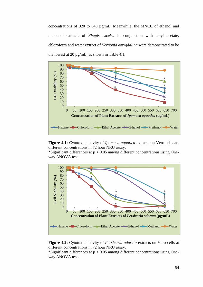
54
concentrations of 320 to 640 µg/mL. Meanwhile, the MNCC of ethanol and
methanol extracts of Rhapis excelsa in conjunction with ethyl acetate,
chloroform and water extract of Vernonia amygdalina were demonstrated to be
the lowest at 20 µg/mL, as shown in Table 4.1.
Figure 4.1: Cytotoxic activity of Ipomoea aquatica extracts on Vero cells at
different concentrations in 72 hour NRU assay.
*Significant differences at p < 0.05 among different concentrations using One-
way ANOVA test.
Figure 4.2: Cytotoxic activity of Persicaria odorata extracts on Vero cells at
different concentrations in 72 hour NRU assay.
*Significant differences at p < 0.05 among different concentrations using One-
way ANOVA test.
0102030405060708090
100
0 50 100 150 200 250 300 350 400 450 500 550 600 650 700
Cel
l V
iab
ilit
y (
%)
Concentration of Plant Extracts of Ipomoea aquatica (µg/mL)
Hexane Chloroform Ethyl Acetate Ethanol Methanol Water
-100
102030405060708090
100
0 50 100 150 200 250 300 350 400 450 500 550 600 650 700
Cel
l V
iab
ilit
y (
%)
Concentration of Plant Extracts of Persicaria odorata (µg/mL)
Hexane Chloroform Ethyl Acetate Ethanol Methanol Water
*
*
*
*
*
* *
* *
*
*
*
*
*
*
*
* * *
*
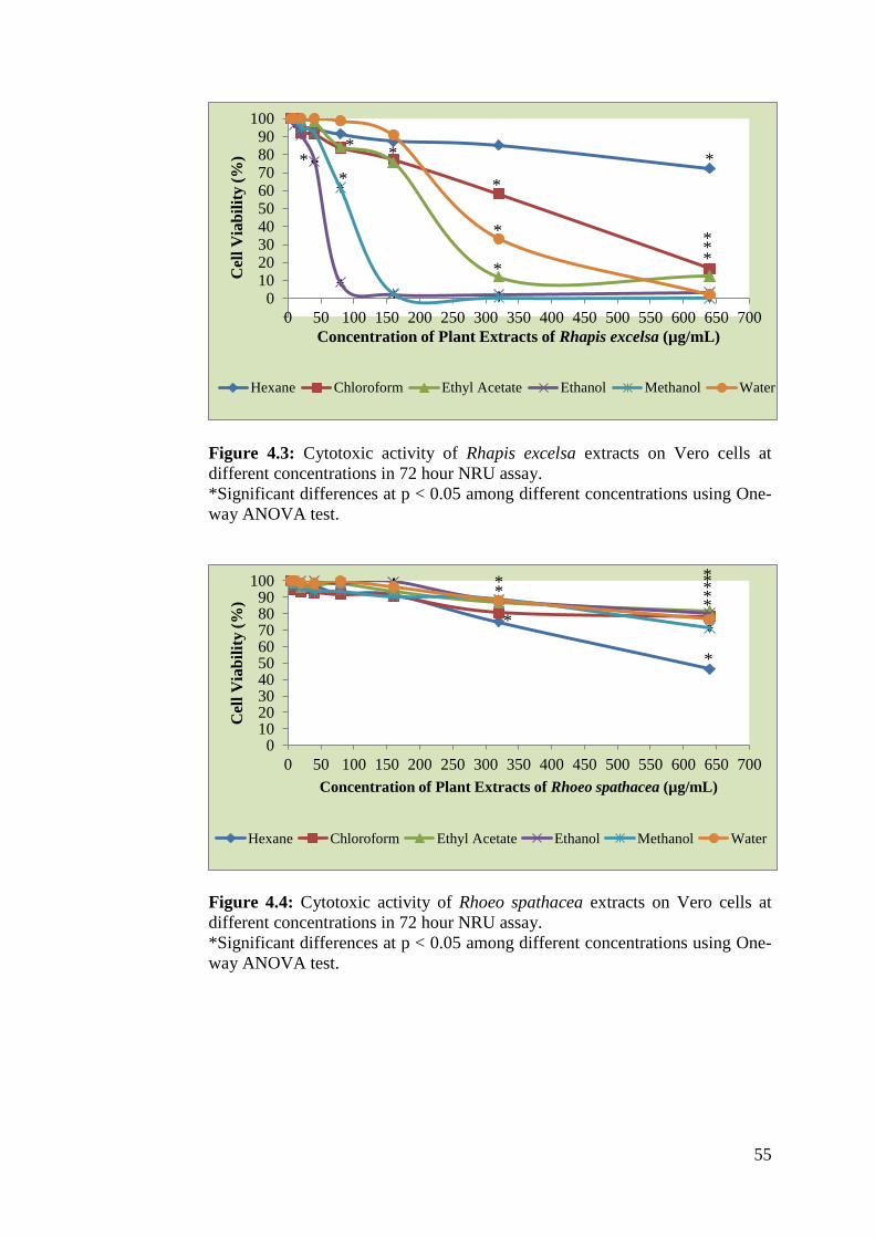
55
Figure 4.3: Cytotoxic activity of Rhapis excelsa extracts on Vero cells at
different concentrations in 72 hour NRU assay.
*Significant differences at p < 0.05 among different concentrations using One-
way ANOVA test.
Figure 4.4: Cytotoxic activity of Rhoeo spathacea extracts on Vero cells at
different concentrations in 72 hour NRU assay.
*Significant differences at p < 0.05 among different concentrations using One-
way ANOVA test.
-100
102030405060708090
100
0 50 100 150 200 250 300 350 400 450 500 550 600 650 700
Cel
l V
iab
ilit
y (
%)
Concentration of Plant Extracts of Rhapis excelsa (µg/mL)
Hexane Chloroform Ethyl Acetate Ethanol Methanol Water
0102030405060708090
100
0 50 100 150 200 250 300 350 400 450 500 550 600 650 700
Cel
l V
iab
ilit
y (
%)
Concentration of Plant Extracts of Rhoeo spathacea (µg/mL)
Hexane Chloroform Ethyl Acetate Ethanol Methanol Water
*
*
*
* *
*
*
* *
* *
*
* *
* * * * * *

56
Figure 4.5: Cytotoxic activity of Vernonia amygdalina extracts on Vero cells
at different concentrations in 72 hour NRU assay.
*Significant differences at p < 0.05 among different concentrations using One-
way ANOVA test.
-100
102030405060708090
100
0 50 100 150 200 250 300 350 400 450 500 550 600 650 700
Cel
l V
iab
ilit
y (
%)
Concentration of Plant Extract of Vernonia amygdalina (µg/mL)
Hexane Chloroform Ethyl Acetate Ethanol Methanol Water
*
* * * *
* *
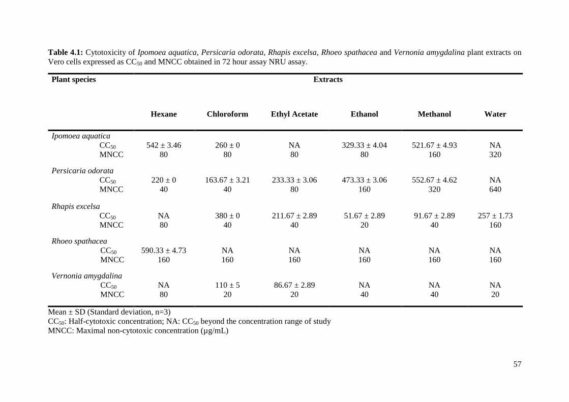
57
Table 4.1: Cytotoxicity of Ipomoea aquatica, Persicaria odorata, Rhapis excelsa, Rhoeo spathacea and Vernonia amygdalina plant extracts on
Vero cells expressed as CC50 and MNCC obtained in 72 hour assay NRU assay.
Plant species Extracts
Hexane Chloroform Ethyl Acetate Ethanol Methanol Water
Ipomoea aquatica
CC50
MNCC
542 ± 3.46
80
260 ± 0
80
NA
80
329.33 ± 4.04
80
521.67 ± 4.93
160
NA
320
Persicaria odorata
CC50
MNCC
220 ± 0
40
163.67 ± 3.21
40
233.33 ± 3.06
80
473.33 ± 3.06
160
552.67 ± 4.62
320
NA
640
Rhapis excelsa
CC50
MNCC
NA
80
380 ± 0
40
211.67 ± 2.89
40
51.67 ± 2.89
20
91.67 ± 2.89
40
257 ± 1.73
160
Rhoeo spathacea
CC50
MNCC
590.33 ± 4.73
160
NA
160
NA
160
NA
160
NA
160
NA
160
Vernonia amygdalina
CC50
MNCC
NA
80
110 ± 5
20
86.67 ± 2.89
20
NA
40
NA
40
NA
20
Mean ± SD (Standard deviation, n=3)
CC50: Half-cytotoxic concentration; NA: CC50 beyond the concentration range of study
MNCC: Maximal non-cytotoxic concentration (µg/mL)

58
4.2 Post-Inoculation Antiviral Assay
Antiviral activity of the various test extracts were determined via the post-
inoculation antiviral assay, where the maximal non-cytotoxic concentration
(MNCC) as indicated in Table 4.1 (page 57) was used as the initial
concentration for the assays and were incorporated after the inoculation of
CHIKV. The antiviral activity of each extract is displayed in Figures 4.6-4.10.
Based on Figures 4.6 to 4.10, all tested extracts did not achieve the half-
maximal effective concentration (EC50) with cell viability percentage ranging
from 1.00 to 23.13%. Among these, the water and hexane extracts of Vernonia
amygdalina exhibited potential activity against CHIKV infection at the
concentrations of 20 µg/mL and 80 µg/mL respectively with a mean cell
viability percentages of 23.13 ± 0 and 19.09 ± 0 correspondingly. Moreover,
among the various extracts of plants studied, hexane extract of Ipomoea
aquatica and Rhapis excelsa together with hexane, ethyl acetate, and
chloroform extracts of Persicaria odorata had notable activity against CHIKV
infection at MNCC among the respective plant groups.
As for the antiviral control group, the drug Chloroquine displayed highest
activity against CHIKV at the concentrations of 620 to 1240 µg/mL with mean
cell viability percentage ranging from 65.47 ± 0.02 to 80.87 ± 0.06. Table 4.2
shows the Vero cell viability percentage after the treatment with Chloroquine
following the inoculation of CHIKV in 72 hours post-inoculation antiviral
assay.
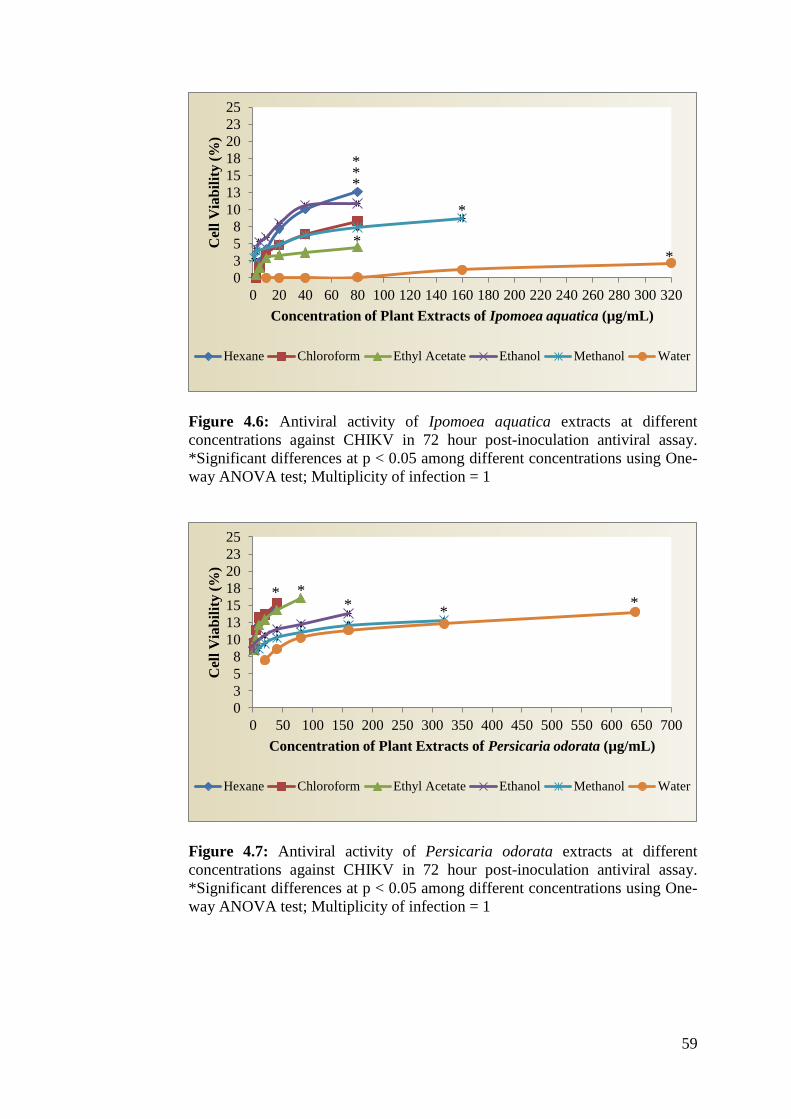
59
Figure 4.6: Antiviral activity of Ipomoea aquatica extracts at different
concentrations against CHIKV in 72 hour post-inoculation antiviral assay.
*Significant differences at p < 0.05 among different concentrations using One-
way ANOVA test; Multiplicity of infection = 1
Figure 4.7: Antiviral activity of Persicaria odorata extracts at different
concentrations against CHIKV in 72 hour post-inoculation antiviral assay.
*Significant differences at p < 0.05 among different concentrations using One-
way ANOVA test; Multiplicity of infection = 1
0
3
5
8
10
13
15
18
20
23
25
0 20 40 60 80 100 120 140 160 180 200 220 240 260 280 300 320
Cel
l V
iab
ilit
y (
%)
Concentration of Plant Extracts of Ipomoea aquatica (µg/mL)
Hexane Chloroform Ethyl Acetate Ethanol Methanol Water
0
3
5
8
10
13
15
18
20
23
25
0 50 100 150 200 250 300 350 400 450 500 550 600 650 700
Cel
l V
iab
ilit
y (
%)
Concentration of Plant Extracts of Persicaria odorata (µg/mL)
Hexane Chloroform Ethyl Acetate Ethanol Methanol Water
*
*
* *
*
*
* * *
* *

60
Figure 4.8: Antiviral activity of Rhapis excelsa extracts at different
concentrations against CHIKV in 72 hour post-inoculation antiviral assay.
*Significant differences at p < 0.05 among different concentrations using One-
way ANOVA test; Multiplicity of infection = 1
Figure 4.9: Antiviral activity of Rhoeo spathacea extracts at different
concentrations against CHIKV in 72 hour post-inoculation antiviral assay.
*Significant differences at p < 0.05 among different concentrations using One-
way ANOVA test; Multiplicity of infection = 1
0
3
5
8
10
13
15
18
20
23
25
0 20 40 60 80 100 120 140 160
Cel
l V
iab
ilit
y (
%)
Concentration of Plant Extracts of Rhapis excelsa (µg/mL)
Hexane Chloroform Ethyl Acetate Ethanol Methanol Water
0
3
5
8
10
13
15
18
20
23
25
0 20 40 60 80 100 120 140 160
Cel
l V
iab
ilit
y (
%)
Concentration of Plant Extracts of Rhoeo spathacea (µg/mL)
Hexane Chloroform Ethyl Acetate Ethanol Methanol Water
* *
* *
*
* * * * * *
*
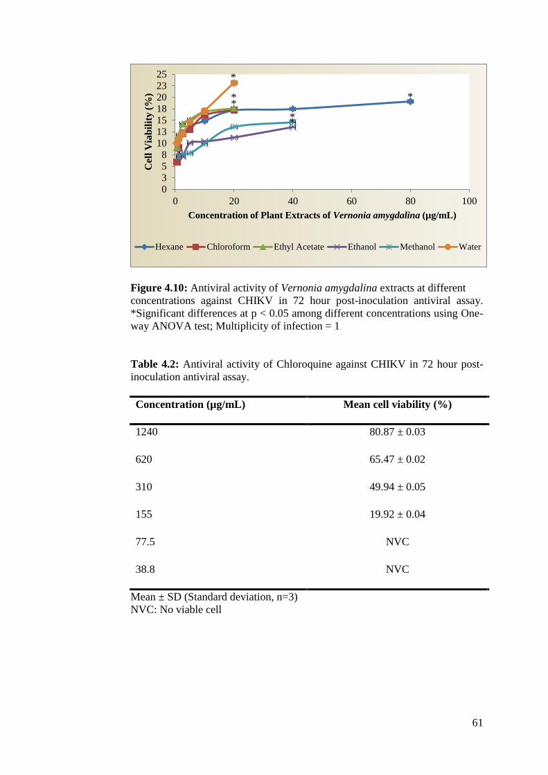
61
Figure 4.10: Antiviral activity of Vernonia amygdalina extracts at different
concentrations against CHIKV in 72 hour post-inoculation antiviral assay.
*Significant differences at p < 0.05 among different concentrations using One-
way ANOVA test; Multiplicity of infection = 1
Table 4.2: Antiviral activity of Chloroquine against CHIKV in 72 hour post-
inoculation antiviral assay.
Concentration (µg/mL) Mean cell viability (%)
1240 80.87 ± 0.03
620 65.47 ± 0.02
310 49.94 ± 0.05
155 19.92 ± 0.04
77.5 NVC
38.8 NVC
Mean ± SD (Standard deviation, n=3)
NVC: No viable cell
0358
10131518202325
0 20 40 60 80 100
Cel
l V
iab
ilit
y (
%)
Concentration of Plant Extracts of Vernonia amygdalina (µg/mL)
Hexane Chloroform Ethyl Acetate Ethanol Methanol Water
*
* *
*
* *

62
CHAPTER 5
DISCUSSION
5.1 Preparation of Plant Extract
Most biological agents are poorly soluble in water and tend to be more soluble
in lipophilic solvents (Kelava, Cavar and Culo, 2011). The stock solutions of
various plant extracts were prepared by diluting crude extracts into the
hydrophobic solvents DMSO:Ethanol at a ratio of 6:4, v/v. The solvents
ethanol and DMSO are extensively applied both in vitro and in vivo assays as
carrier solvents for hydrophobic chemicals and to a lesser extent hydrophilic
chemicals (Adefolaju, Theron and Hosie, 2015). Studies suggests that the
solvent DMSO has tendencies to increase the diffusion of pharmacologically
active compounds through cell membrane by disruption of cell barrier function.
This phenomenon can be explained through the aprotic interactions that DMSO
mediate with the intercellular lipids and through reversible distortion of lipid
head groups of the phospholipid bilayer in order to produce a greater
permeable packing arrangement (Capriotti and Capriotti, 2012).
Inspite the fact that DMSO is comprehensively employed in cell-based assays,
this solvent however does not dissolve all compounds and frequently, other
solvents are used in combination to achieve sufficient solubility. Ethanol is a
principal carrier in pharmacology and biochemistry (Maes et al., 2012). Studies
shows that its lipophilic carbon tail, has the capacity to concentrate at the
interface between lipid and surrounding water forming hydrogen bonds with

63
the hydrophilic head groups of bilayer giving rise to an increase in area per
lipid, contributing to overall fluidity of the membrane to deliver hydrophilic
chemicals more easily in unison with DMSO (Patra et al., 2006; Gurtovenko
and Anwar, 2009).
Studies on the effects of DMSO:ethanol on Vero cells by Chan (2013) revealed
that at the concentration of 1% and below, cytotoxic effects on the Vero cells
was not apparent and cell viability remained at 100%. In this study, the
concentration of DMSO:Ethanol used on Vero cells were 0.5% and below.
5.1 NRU Cytotoxicity Assay
According to Betancur-Galvis et al. (2002), the half-maximal cytotoxic
concentration (CC50) is the concentration of test extracts that reduces the
absorbance value at 540 nm of treated uninfected cells to 50% of that of
untreated uninfected cells. According to Anyango (2011), definition of
cytotoxicity on Vero cells is as follows: CC50 < 10 μg/ml, high toxicity; CC50
11-50 μg/ml, moderate toxicity; CC50 51-100 μg/ml, mild toxicity; and CC50 >
100 μg/ml not toxic.
In this study, none of the extracts tested was highly toxic though mild toxicity
was observed in the ethanol and methanol extract of Rhapis excelsa and ethyl
acetate extract of Vernonia amygdalina. No previous studies on all tested
plants were recorded against Vero cells. However, a search through the
literatures showed that the ethanol extract of Calamus gibbsianus belonging to
the same family as Rhapis excelsa (Arecaceae) demonstrated moderate to high

64
toxicity against Vero cells. Meanwhile, ethyl acetate extract of the plant
Vernonia zollingerianoides belonging to same genus as Vernonia amygdalina
(Asteraceae) revealed moderate toxicity on Vero cells (Siti et al. 2012;
Roshida, 2014) .
On the contrary, MNCC of test extracts is defined as the highest concentration
tolerated by the treated uninfected cells without toxic symptoms (Orsine et al.,
2012). The ethanol extract of Rhapis excelsa together with chlorofom, ethyl
acetate and water extract of Vernonia amygdalina demonstrated the lowest
MNCC value of 20 µg/mL. However, no previous studies were reported on the
MNCC of these plant extracts on Vero cells.
5.2 Post-Inoculation Antiviral Assay
By comparing the Figures 4.6 to 4.10 (page 60-62) it is apparent that all 30
tested extracts from the various plants of study did not achieve the half-
maximal effective concentration (EC50). However, the water and hexane
extracts from the leaves of Vernonia amygdalina has a potential activity
against CHIKV than the aerial and leaf extracts of all other tested plants. This
was assumed to be due to presence of plant-derived secondary metabolites with
various viral targets ranging from adsorption of the virus to the host cell to
release of the virus from the cells. Some common plant metabolites in this
category include alkaloids, flavonoids, and steroid saponins (Wink, 1999).
Flavonoids are polyphenolic compounds that are present in different plants.
They are well known for various biological properties including: antimicrobial

65
activity, anti-inflammatory activity, anti-allergic activity, and antitumor
activity (Lani et al., 2015). Phytochemical screenings of leaf parts of the plant
Vernonia amygdalina and Ipomoea aquatica revealed the presence of
flavonoids especially luteolin, luteolin 7-O-glucosides and luteolin 7-O-
glucuronide in Vernonia amygdalina (Farombi and Owoeye, 2011) and
mycertin, quercetin, luteolin and apigenin in aerial parts of the plant Ipomoea
aquatica (Igwenyi et al., 2011). According to Zandi et al. (2011), antiviral
activities of flavonoids have been demonstrated against numerous viruses
including human cytomegalovirus (HCMV), Herpes Simplex Virus-1 and 2
and some types of human adenoviruses. A recent study conducted by Murali et
al. (2015) on the activity of luteolin and apigenin rich fractions from ethanolic
extract of Cynodon dactylon against CHIKV revealed potent, 98% virus
inhibition activity at the concentration of 50 µg/mL. The mechanisms of
antiviral activity were reported to revolve mainly in interfering with viral
nucleic acid synthesis by binding to the viral polymerases, preventing viral
entry and enhancing activity of interferon (Lani et al., 2015; Murali et al.,
2015).
Alkaloids are important chemical compounds that serve as a rich reservoir for
drug discovery. Alkaloids such as nortropane alkaloids constitute as the major
secondary metabolite in the aerial parts of the plant Ipomoea aquatica
(Malakar and Choudhury, 2015). Studies shows that nortropane alkaloids
possess glycosidase inhibitory activity and thus has a potential utility as an
antiviral agent (Rahman, 2012). The viral envelope glycoproteins are often
essential for virion assembly and secretion and infectivity. Compounds that

66
interfere with the glycosylation processes of viral glycoproteins such as
nortropane alkaloids may have significant effects in the inhibition of infectivity
(Asano, 2003). Alkaloids were also reported as a major constituent of the leaf
parts of the plants Vernonia amygdalina and Rhoeo spathacea (Farombi and
Owoeye, 2011; Nikam et al., 2013). However, limited studies been reported on
the type of alkaloid present in these two plants.
The presence of terpenes such as triterpene lupenone in Rhoeo spathacea was
revealed by Nikam et al. (2013). Leyssen et al. (2014) reported that triterpene
lupenone had moderate to significant antiviral activity against CHIKV and
other alphavirus, the mode of action revolves mainly in the inhibition of viral
replications. Other plant secondary metabolites that are reported to possess
potent antiviral activity include sesquiterpene lactones that is demonstrated in
Persicaria odorata (Sasongko, Laohankunjit and Kerdchoechuen, 2011;
Ridzuan, 2013) and saponins such as vemoniosides D and E that are present in
Vernonia amygdalina which exhibits virucidal mechanism especially by
coating the viral binding sites and preventing attachments in Rotavirus
infections (Adiukwu et al., 2013; Arthanari et al., 2013). Meanwhile,
sesquiterpene lactones such as chlorohyssopifolin A, chlorojanerin and 13-
acetyl solstitialin A were reported to have activity against herpes simplex virus
type 1 (HSV-1) (Özçelik et al., 2009). However, the mechanism may not be
similar in CHIKV and other alphaviruses since their genome is composed of a
positive sense single stranded RNA virus (Pun, Bastola and Shah, 2014).

67
Our results of Chloroquine matched the data reported in the study conducted by
Khan et al. (2010). Chloroquine being a weak base is thought to possess
multiple mechanism of action that differs predominantly based on the type of
pathogen being targeted (Farias et al., 2014). In the case of viral infection, its
therapeutic value lies in its ability to accumulate in the acidic vesicles of a cell
such as the endosome, lysosome vesicles and the Golgi apparatus consequently
increasing the pH therein. The increase of pH prevents the low pH-induced
fusion of viral envelope and cell endosome membranes, successfully blocking
the entry of viruses into the cytosol (Farias, Machado and da Fonseca, 2013;
Farias et al., 2014).
Chloroquine is a highly lipophilic drug in nature (Day, 2005). According to
Adams and Merluzzi (1993) hydrophobicity of compounds is an essential
criterion to achieve high antiviral activity. The hydrophobic property of
chloroquine explains its ability to penetrate the lipid membrane and accumulate
in the acidic vesicles of infected cells. In this study, the notable activity
demonstrated by the less polar extracts such as the hexane extract of Ipomoea
aquatica and Rhapis excelsa and hexane, chloroform and ethyl acetate extracts
of Persicaria odorata was assumed due to their non-polar nature. However,
this finding contradicts with studies conducted by Sangeetha and Rajarajan
(2015) on five medicinal plants from the genus alpinia, andrographis and
azadirachta against CHIKV that reports polar extracts such as water and
ethanol had more prominent activity against the virus. However, the activity
demonstrated by certain plants at a more polar solvent might not be the same
for all other plants (Anyango, 2011).
68
5.3 Limitations
Despite the presence of various secondary metabolites with significant antiviral
activity, all tested plant extracts did not achieve the half-maximal effective
concentration (EC50). This may be due to limitations on crude extract, which
often contain various compounds that may interfere with the bioassay
antagonistically.
5.4 Future Perspectives
Other parts of the plants such as the roots and stem of Ipomoea aquatica and
Persicaria odorata and the fruit, roots and stem of Rhapis excelsa, Rhoeo
spathacea and Vernonia amygdalina can be used for screenings against
CHIKV in future. The various extracts can also be assessed on other viruses
such as dengue virus, an endemic virus in Malaysia.
Meanwhile, the active secondary metabolites in the water and hexane extracts
of Vernonia amygdalina can be isolated and purified using the high
performance liquid chromatography, column chromatography and thin layer
chromatography. Furthermore, CHIKV viral load can be measured using the
real-time polymerase chain reaction technique in order to study the mode of
action of the tested extracts.
69
CHAPTER 6
CONCLUSION
The NRU assay demonstrated the lowest CC50 for ethanol (51.67 ± 2.89
µg/mL) extract of Rhapis excelsa followed by ethyl acetate (86.87 ± 2.89 µg/
mL) extract of Vernonia amygdalina and methanol (91.67 ± 2.89 µg/mL)
extract of Rhapis excelsa. Moreover, all the three extracts together with
chlorofom and water extracts of Vernonia amygdalina were reported to have
the lowest MNCC at 20 µg/mL.
In addition, all tested extracts did not achieve the half-maximal effective
concentration (EC50), with cell viability percentage ranging from 1.00 to
23.13%. Among them the water and hexane extracts of Vernonia amygdalina
exhibited potential activity against CHIKV infection at the concentrations of 20
µg/mL and 80 µg/mL with a mean cell viability percentage of 23.13% ± 0 and
19.09% ± 0 respectively.
Furthermore, among the various extracts of plants studied, hexane extract of
Ipomoea aquatica and Rhapis excelsa together with hexane, ethyl acetate and
chloroform extracts of Persicaria odorata had notable activity against CHIKV
at MNCC.

70
REFERENCES
Abdel-Azim, N., Shams, K., El-Missery, M., Ismail, S. and Hammouda, F.,
2013. Introducing green technology for extraction of medicinal and aromatic
plants to Egypt. Planta Medica, 79(13), pp. 78-79.
Abid, N., 2012. Assessment of the cytotoxic effect and in vitro evaluation of
the anti-enteroviral activities of plants rich in flavonoids. Journal of Applied
Pharmaceutical Science, 2(5), pp. 74-78.
AbuBakar, S., Sam, I., Wong, P., Hooi, P. and Roslan, N., 2007. Reemergence
of endemic chikungunya, Malaysia. Emerging Infectious Disease Journal,
13(1), pp. 147-149.
Adams, J. and Merluzzi, V., 1993. The Search for antiviral drugs. Boston:
Birkhauser.
Adaramoye, O., 2008. Lipid-lowering effects of methanolic extract of
Vernonia amygdalina leaves in rats fed on high cholesterol diet. Journal of
Vascular Health and Risk Management, 4(1), pp. 235-241.
Ademola, I. and Eloff, J., 2010. Anthelminthic activity of acetone extract and
fractions of Vernonia amygdalina against Haemonchus contortus eggs and
larvae. Tropical Animal Health and Production, 43(2), pp. 521-527.
Adesanoye, O., Molehin, O., Delima, A., Adefegha, A. and Farombi, E., 2012.
Modulatory effect of methanolic extract of Vernonia amygdalina (MEVA) on
tert-butyl hydroperoxide-induced erythrocyte haemolysis. Cell Biochemistry
and Function, 31(7), pp. 34-38.
Adiukwu, P., Kayanja, F., Nambatya, G., Rugera, S., Ezeonwumelu, J.,
Tanayen, J., Murokore, B., Twikirize, O., Twinomujuni, S., Byamugisha, D.
and Imanirampa, L., 2013. Antipyretic and antinociceptive properties of the
aqueous extract and saponin from an edible vegetable: Vernonia amygdalina
leaf. African Journal of Food, Agriculture, Nutrition and Development, 13(2),
pp. 7588-7589.

71
Aguilar, J., Roy, D., Ghazal, P. and Wagner, E., 2002. Dimethyl sulfoxide
blocks herpes simplex virus-1 productive infection in vitro acting at different
stages with positive cooperativity. Application of micro-array analysis. BMC
Infectious Diseases, 2(1), pp. 9.
Ahola, T., Laakkonen, P., Vihinen, H. and Kääriäinen, L., 1997. Critical
residues of Semliki Forest virus RNA capping enzyme involved in
methyltransferase and guanylyltransferase-like activities. Journal of Virology,
71(1), pp. 392-397.
Ajibola, C., Eleyinmi, A. and Aluko, R., 2011. Kinetics of the inhibition of
renin and angiotensin I converting enzyme by polar and non-polar
polyphenolic extracts of Vernonia amygdalina and Gongronema latifolium
leaves. Plant Foods for Human Nutrition, 66(4), pp. 320-327.
Alkiyumi, S., Abdullah, M., Alrashdi, A., Salama, S., Abdelwahab, S. and
Hadi, A., 2012. Ipomoea aquatica extract shows protective action against
thioacetamide-induced hepatotoxicity. Molecules, 17(12), pp. 6146-6155.
Ammerman, N., Beier-Sexton, M. and Azad, A., 2005. Growth and
maintenance of Vero cell lines. Current Protocols in Microbiology, 1(1), pp. 2-
10
Anil, K., Ashmi, M., Maheshwar, S. and Madhuri, S., 2014. Antimalarial
activity of yaoundamine a naphthyl iso-quinoline alkaloid, extracted from stem
of Ancistrocladus heyneanus. Annals of Biological Science, 2(2), pp. 40-44.
Anyango, O., 2011. Anti-malarial activity and phytochemical studies of
cissamp. Masters. Kenyatta University.
Apandi, Y., Lau, S., Izmawati, N., Amal, N., Faudzi, Y., Mansor, W., Hani, M.
and Zaina, S., 2010. Identification of Chikungunya virus strains circulating in
Kelantan, Malaysia in 2009. Southeast Asian Journal of Tropical Medicine
Public Health, 41(6), pp. 1374-1380.
Apandi, Y., Nazni, W. and Noor Azleen, Z., 2009. The first isolation of
Chikungunya virus from nonhuman primates in Malaysia. Journal of General
and Molecular Virology, 1(1), pp. 35-39.
72
Arriaga-Alba, M., Blasco, J., Ruíz-Pérez, N., Sánchez-Navarrete, J., Rivera-
Sánchez, R. and González-Avila, M., 2011. Antimutagenicity mechanisms of
the Rhoeo discolor ethanolic extract. Experimental and Toxicologic Pathology,
63(3), pp. 243-248.
Arthanari, S., Vanitha, J., Krishnaswami, V., Renukadevi, P., Deivasigamani, K.
and De Clercq, E., 2013. In vitro antiviral and cytotoxic screening of
methanolic extract of Cassia auriculata flowers in HeLa, Vero, CRFK and
HeLa cell lines. Drug Invention Today, 5(1), pp. 28-31.
Asano, N., 2003. Glycosidase inhibitors: update and perspectives on practical
use. Glycobiology, 13(10), pp. 93-104.
Assunção-Miranda, I., Cruz-Oliveira, C. and Da Poian, A., 2013. Molecular
mechanisms involved in the pathogenesis of alphavirus-induced arthritis.
BioMed Research International, 1(1), pp. 1-11.
Atangwho, I., Egbung, G., Ahmad, M., Yam, M. and Asmawi, M., 2013.
Antioxidant versus anti-diabetic properties of leaves from Vernonia
amygdalina Del. growing in Malaysia. Food Chemistry, 141(4), pp. 3428-
3434.
ATCC, 2015. Vero ATCC ® CCL-81™ Cercopithecus aethiops kidney normal.
[online] Available at: http://www.atcc.org/products/all/CCL-
81.aspx#culturemethod [Accessed 19 Jul. 2015].
Aubry, M., Teissier, A., Roche, C., Richard, V., Yan, A., Zisou, K., Rouault,
E., Maria, V., Lastère, S., Cao-Lormeau, V. and Musso, D., 2015.
Chikungunya outbreak, French Polynesia, 2014. Emerging Infectious Diseases,
21(4), pp. 724-726.
Audu, S., Taiwo, A., Ojuolape, A., Sani, A., ukola, A. and Mohammed, I.,
2012. A study review of documented phytochemistry of ernonia amygdalina
(Family Asteraceae) activity of plant extract. Journal of Natural Sciences
Research, 2(7), pp. 2224-3186.
Austin, D., 2007. Water Spinach (Ipomoea aquatica, Convolvulaceae) A food
gone wild. Ethnobotany Research & Applications, 5(1), pp. 123-146.

73
Ayu, S., Lai, L., Chan, Y., Hatim, A., Hairi, N., Ayob, A. and Sam, I., 2010.
Seroprevalence survey of Chikungunya virus in agan Panchor, Malaysia.
American Journal of Tropical Medicine and Hygiene, 83(6), pp. 1245-1248.
Averyanov, L., Nguyen, T. and Phan, K., 2006. Rhapis vidaliia new papyrus
likepalm from Vietnam. Palms, 50, pp. 11-12.
Azami, N., Salleh, S., Shah, S., Neoh, H., Othman, Z., Zakaria, S. and Jamal,
R., 2013. Emergence of chikungunya seropositivity in healthy Malaysian adults
residing in outbreak-free locations: Chikungunya seroprevalence results from
the Malaysian Cohort. BMC Infectious Diseases, 13(1), p. 67.
Barrett, P., Portsmouth, D. and Ehrlich, H., 2013. Accelerated
biopharmaceutical development: Vero cell technology and pandemic influenza
vaccine production. Modern Biopharmaceuticals, 1(1) pp. 349-386.
Bedoya-Arias, J., Murillo-García, D., Bolaños-Muñoz, E., Hurtado-Hurtado,
N., Ramírez-Jaramillo, V., Granados-Õlvarez, S. and Rodriguez-Morales, A.,
2015. Healthcare students and workers' knowledge about epidemiology and
symptoms of chikungunya fever in two cities of Colombia. The Journal of
Infection in Developing Countries, 9(03), pp. 330-332.
Bercu, R., 2013. Histoanatomical study on the vegetative organs of
Tradescantia spathacea (Commelinaceae). Botanica Serbica, 37(2), pp. 121-
126.
Betancur-Galvis, L., Morales, G., Forero, J. and Roldan, J., 2002. Cytotoxic
and antiviral activities of Colombian medicinal plant extracts of the euphorbia
genus. Memórias do Instituto Oswaldo Cruz, 97(4), pp. 541-546.
Burleson, F., Chambers, T. and Wiedbrauk, D., 1992. Virology. San Diego:
Academic Press.
Cao, S., Dong, G., Tang, J., Li, J., Liu, J., Shi, L., Li, C. and Wang, J., 2013.
Development of a Vero cell DNA reference standard for residual DNA
measurement in China. Human Vaccines & Immunotherapeutics, 9(2), pp. 413-
419.
CDC, 2015. Chikungunya - Chapter 3 - 2016 Yellow book travelers' health
CDC. Available at: http://wwwnc.cdc.gov/travel/yellowbook/2016/infectious-
diseases-related-to-travel/chikungunya [Accessed 31 Aug. 2015].
74
CDC, 2015. Geographic Distribution | Chikungunya virus | CDC. [online]
Available at: http://www.cdc.gov/chikungunya/geo/ [Accessed 31 Jul. 2015].
Chan, S., 2013. In vitro quantitative assessment of antiviral activity of
medicinal plants against Chikungunya virus. Masters. Universiti Tunku Abdul
Rahman.
Chen, T. and Chen, K., 2009. Investigation and application progress of Vero
cell serum-free culture. International Journal of Biology, 1(2), p. 41.
Chevillon, C., Briant, L., Renaud, F. and Devaux, C., 2008. The Chikungunya
threat: an ecological and evolutionary perspective. Trends in Microbiology,
16(2), pp. 80-88.
Chua, K., 2010. Epidemiology of Chikungunya in Malaysia: 2006-2009.
Medical Journal of Malaysia, 65, pp. 278-273.
Coffey, L., Failloux, A. and Weaver, S., 2014. Chikungunya virus-vector
interactions. Viruses, 6(11), pp. 4628-4663.
Colegate, S. and Molyneux, R., 2008. Bioactive natural products. oca Raton:
CRC Press.
Cowan, M., 1999. Plant products as antimicrobial agents. Clinical
Microbiology Reviews, 12(4), pp. 564-582.
da Luz, P., de Oliveira Paiva, P. and Tavares, A., 2008. Effect of foliar and
substrate fertilization on Lady Palm seedling growth and development. Journal
of Plant Nutrition, 31(7), pp. 1313-1320.
Dammer, U., 2008. Zimmerblattpflanzen. Berlin: Echo Library.
Daniel, M., 1989. Polyphenols of some Indian vegetables. Current Science,
58(1), pp. 1332-1334.
Dash, M., Mohanty, I. and Padhi, S., 2011. Laboratory diagnosis of
Chikungunya virus: Do we really need it? Indian Journal of Medical Sciences,
65(3), p. 83.

75
Datta, P. and Saha, N., 1974. Development of leaf forms in Ipomoea reptans
(Linn.) Poir. Oxford Journals, 40(4), pp. 837-843.
Daves, H., 2015. PlantFiles: Vietnamese Coriander. [online] Available at:
http://davesgarden.com/guides/pf/go/310/#b [Accessed 13 Sep. 2015].
Day, R., 2005. Antirheumatic therapy. Basel: Birkhauser.
de Curtis, I. and Simons, K., 1988. Dissection of Semliki Forest virus
glycoprotein delivery from the trans-golgi network to the cell surface in
permeabilized BHK cells. Proceedings of the National Academy of Sciences,
85(21), pp. 8052-8056.
de Groot, R., Reef Hardy, W., Shirako, Y. and Strauss, J., 1990. Cleavage-site
preferences of Sindbis virus polyproteins containing the non-structural
proteinase. Evidence for temporal regulation of polyprotein processing in vivo.
EMBO Journal, 9(8), pp. 2631-2638.
de Lamballerie, X., Ninove, L. and Charrel, R., 2009. Antiviral treatment of
Chikungunya virus infection. Infectious Disorders - Drug Targets, 9(2), pp.
101-104.
Dransfield, J. and Uhl, N., 1998. The families and genera of vascular plants.
IV. flowering plants, monocotyledons: Alismatanae and commelinanae, except
gramineae. Springer Berlin, 1(1), pp. 306-389.
Eldridge, B. and Edman, J., 2004. Medical Entomology. Dordrecht: Springer.
Esimone, C., Grunwald, T., Wildner, O., Nchinda, G., Tippler, B., Proksch, P.
and Uberla, K., 2005. In vitro pharmacodynamic evaluation of antiviral
medicinal plants using a vector-based assay technique. Journal of Applied
Microbiology, 99(6), pp. 1346-1355.
Farias, K., Machado, P. and da Fonseca, B., 2013. Chloroquine inhibits dengue
virus type 2 replication in Vero cells but not in C6/36 cells. The Scientific
World Journal, 1(1), pp. 1-5.
Farias, K., Machado, P., de Almeida Junior, R., de Aquino, A. and da Fonseca,
B., 2014. Chloroquine interferes with dengue-2 virus replication in U937 cells.
Microbiology and Immunology, 58(6), pp. 318-326.

76
Farombi, E. and Owoeye, O., 2011. Antioxidative and chemopreventive
properties of Vernonia amygdalina and Garcinia biflavonoid. International
Journal of Environmental Research and Public Health, 8(6), pp. 2533-2555.
Favi, F., Cantrell, C., Mebrahtu, T. and Kraemer, M., 2008. Leaf peltate
glandular trichomes of Vernonia galamensis ssp. galamensis var. ethiopica
gilbert: development, ultrastructure, and chemical composition. International
Journal of Plant Sciences, 169(5), pp. 605-614.
FLEPPC, 2015. FLEPPC - Florida Exotic Pest Plant Council. [online]
Available at: http://www.fleppc.org/ [Accessed 27 Jul. 2015].
Floridata, 2015. Rhapis excelsa Plant Profile. [online] Mobile.floridata.com.
Available at: http://mobile.floridata.com/Plants/Arecacea/Rhapis%20excelsa
/235 [Accessed 13 Sep. 2015].
Fotakis, G. and Timbrell, J., 2006. In vitro cytotoxicity assays: comparison of
LDH, neutral red, MTT and protein assay in hepatoma cell lines following
exposure to cadmium chloride. Toxicology Letters, 160(2), pp. 171-177.
Frolova, E., Gorchakov, R., Garmashova, N., Atasheva, S., Vergara, L. and
Frolov, I., 2006. Formation of nsP3-specific protein complexes during sindbis
virus replication. Journal of Virology, 80(8), pp. 4122-4134.
Fuller, S., 1987. The T=4 envelope of Sindbis virus is organized by
interactions with a complementary T=3 capsid. Cell, 48(6), pp. 923-934.
Ghamba, P., alla, H., Goje, L., Halidu, A. and Dauda, M., 2014. In vitro
antimicrobial activities of ernonia amygdalina on selected clinical isolates.
International Journal of Current Microbiology and Applied Sciences, 3(4), pp.
1103-1113.
Glanville, N., Ranki, M., Morser, J., Kaariainen, L. and Smith, A., 1976.
Initiation of translation directed by 42S and 26S RNAs from Semliki Forest
virus in vitro. Proceedings of the National Academy of Sciences, 73(9), pp.
3059-3063.
Golczyk, H., 2013. Tradescantia spathacea (syn. Rhoeo spathacea). A review
Tradescantia spathace (syn. Rhoeo spathacea). Annales UMCS, Biologia,
68(1), pp. 39-53.
77
Gomez de Cedrón, M., Ehsani, N., Mikkola, M., García, J. and Kääriäinen, L.,
1999. RNA helicase activity of Semliki Forest virus replicase protein NSP2.
FEBS Letters, 448(1), pp. 19-22.
González-Avila, M., Arriaga-Alba, M., de la Garza, M., del Carmen
HernándezPretelín, M., Domínguez-Ortíz, M., Fattel-Fazenda, S. and Villa-
Treviño, S., 2003. Antigenotoxic, antimutagenic and ROS scavenging activities
of a Rhoeo discolor ethanolic crude extract. Toxicology in Vitro, 17(1), pp. 77-
83.
Goto, T., Takahashi, N., Hirai, S. and Kawada, T., 2010. Various terpenoids
derived from herbal and dietary plants function as PPAR modulators and
regulate carbohydrate and lipid metabolism. PPAR Research, 1(1), pp. 20-28.
Grigorov, B., Rabilloud, J., Lawrence, P. and Gerlier, D., 2011. Rapid titration
of measles and other viruses: optimization with determination of replication
cycle length. PLoS ONE, 6(9), pp. 24-25.
Grubben, G., 2004. Vegetables. Wageningen, Netherlands: Backhuys.
Gupta, A., Naraniwal, M. and Kothari, V., 2012. Modern extraction methods
for preparation of bioactive plant extracts. International Journal of Applied and
Natural Sciences, 1(1), pp. 8-26.
Hamer, D. and Chen, L., 2014. Chikungunya: Establishing a new home in the
Western Hemisphere. Annals of Internal Medicine, 161(11), p. 827.
Hamid, K., Ullah, M., Sultana, S., Howlader, M., Basak, D., Nasrin, F. and
Rahman, M., 2011. Evaluation of the leaves of Ipomoea aquatica for its
hypoglycemic and antioxidant activity. Journal of Pharmaceutical Sciences
and Research, 3(7), pp. 1330-1333.
Härtl, A., Sauerbrei, A., Stelzner, A. and Wutzler, P., 2004. Influenza infection
of the embryonated hen's egg/an alternative model for in vivo evaluation of
antiviral compounds. Arzneimittelforschung, 54(2), pp. 130-134.
Hassanein, H., Elsayed, W., Abreu, A., Simões, M. and Abdelmohsen, M.,
2015. Polyphenolic constituents and antimicrobial activity of Rhapis excelsa
(Arecaceae, Coryphoideae). Research Journal of Pharmaceutical, Biological
and Chemical Sciences, 6(1), pp. 1714-1720.

78
Hastings, L., 2003. A revision of Rhapis, the Lady Palms. Palms, 47(2), pp. 62-
67.
Hrnjakovic-Cvjetkovic, I., Cvjetkovic, D., Patic, A., Nikolic, N., Stefan-Mikic,
S. and Milosevic, V., 2015. Chikungunya - a serious threat for public health.
Public health medical review, 68(3-4), pp. 122-125.
Huffman, M., 2001. Self-Medicative behavior in the African great apes: An
evolutionary perspective into the origins of human traditional medicine.
BioScience, 51(8), p. 651.
Hussain, K. and Chu, J., 2011. Insights into the interplay between
Chikungunya virus and its human host. Future Virology, 6(10), pp. 1211-1223.
Iemm, J., Rümenapf, T., Strauss, E., Strauss, J. and Rice, C., 1994. Polypeptide
requirements for assembly of functional Sindbis virus replication complexes: A
model for the temporal regulation of minus- and plus-strand RNA synthesis.
EMBO Journal, 13(12), pp. 2925-2934.
Igile, G., Olenszek, W., Jurzysta, M., Aquino, R., de Tommasi, N. and Pizza,
C., 1995. Vemoniosides D and E, two novel saponins from ernonia
amygdalina. Journal of Natural Products, 58(9), pp. 1438-1443.
Igwenyi, I., Offor, C., Ajah, D., Nwankwo, O., Ukaomah, J. and Aja, P., 2011.
Chemical compositions of Ipomea aquatica (green kangkong). International
Journal of Pharma and Bio Sciences, 2(4), pp. 593-595.
Iroanya, O., Okpuzor, J. and Mbagwu, H., 2010. Anti-nociceptive and anti-
phlogistic actions of a polyherbal decoction. International Journal of
Pharmacology, 6(1), pp. 31-36.
Iwu, M., 2013. Handbook of African Medicinal Plants, Second Edition.
Hoboken: Taylor and Francis.
Jassim, S. and Naji, M., 2003. Novel antiviral agents: a medicinal plant
perspective. Journal of Applied Microbiology, 95(3), pp. 412-427.

79
Javelle, E., Ribera, A., Degasne, I., Gaüzère, B., Marimoutou, C. and Simon,
F., 2015. Specific management of post-Chikungunya rheumatic disorders: A
retrospective study of 159 cases in Reunion island from 2006-2012. PLOS
Neglected Tropical Diseases, 9(3), pp. 360-363.
Joash, B., Yap, W., Tan, S., Lim, Y. and Lee, S., 2014. Antioxidant content,
antioxidant activity, and antibacterial activity of five plants from the
Commelinaceae family. Antioxidants Molecular Diversity Preservation
International Journal, 3, pp. 758-769.
Jonsson, N., Gullberg, M. and Lindberg, A., 2009. Real-time polymerase chain
reaction as a rapid and efficient alternative to estimation of picornavirus titers
by tissue culture infectious dose 50% or plaque forming units. Microbiology
and Immunology, 53(3), pp. 149-154.
Kaur, P. and Chu, J., 2013. Chikungunya virus: an update on antiviral
development and challenges. Drug Discovery Today, 18(19-20), pp. 969-983.
Khan, M., Santhosh, S., Tiwari, M., Lakshmana Rao, P. and Parida, M., 2010.
Assessment of in vitro prophylactic and therapeutic efficacy of chloroquine
against chikungunya virus in vero cells. Journal of Medical irology, 82(5), pp.
817-824.
Khan, S., Dutta, P., Topno, R., Bohra, J., Chowdhury, P. and Mahanta, J.,
2015. Chikungunya outbreak in Garo Hills, Meghalaya: an epidemiological
perspective. Indian Journal of Medical Research, 141, pp. 591-597.
Kielian, M., Chanel-Vos, C. and Liao, M., 2010. Alphavirus entry and
membrane fusion. Viruses, 2(4), pp. 796-825.
Kothari, V., Gupta, A. and Naraniwal, M., 2012. Comparative study of various
methods for extraction of antioxidant and antibacterial compounds from plant
seeds. Journal of Natural Remedies, 12(2), pp. 162-173.
Kuete, V., 2013. Medicinal plant research in Africa. London: Elsevier Ltd.
Kumar, K., Nagpal, R. and Arun, A., 2011. Spectrum of chromosomal
configurations in pollen mother cells of Rhoeo spathacea (Swartz) Stearn.
Caryologia, 64(2), pp. 135-146.

80
Kumar, S. and Pandey, A., 2013. Chemistry and biological activities of
flavonoids: An overview. The Scientific World Journal, 1(1), pp. 1-16.
LaBeaud, A., Banda, T., Brichard, J., Muchiri, E., Mungai, P., Mutuku, F.,
Borland, E., Gildengorin, G., Pfeil, S., Teng, C., Long, K., Heise, M., Powers,
A., Kitron, U. and King, C., 2015. High Rates of O’nyong-onyong and
Chikungunya virus transmission in coastal Kenya. PLOS Neglected Tropical
Diseases, 9(2), pp. 343.
Laekeman, G., De Clerck, F., Vlietinck, A. and Herman, A., 1985. Vernolepin:
An antiplatelet compound of natural origin. Naunyn-Schmiedeberg's Archives
of Pharmacology, 331(1), pp. 108-113.
Laine, R., Söderlund, H. and Renkonen, O., 1973. Chemical composition of
Semliki Forest virus. Intervirology, 1(2), pp. 110-118.
Lakshmi, V., Neeraja, M., Subbalaxmi, M., Parida, M., Dash, P., Santhosh, S.
and Rao, P., 2008. Clinical features and molecular diagnosis of Chikungunya
fever from South India. Clinical Infectious Diseases, 46(9), pp. 1436-1442.
Lam, S., Chua, K., Hooi, P., Rahimah, M., Kumari, S., Tharmaratnam, M.,
Chuah, S., Smith, D. and Sampson, I., 2001. Chikungunya infection--an
emerging disease in Malaysia. Southeast Asian Journal of Tropical Medicine
and Public Health, 32(3), pp. 447-451.
Lamballerie, X., Boisson, V., Reynier, J., Enault, S., Charrel, R., Flahault, A.,
Roques, P. and Grand, R., 2008. On Chikungunya acute infection and
chloroquine treatment. Vector-Borne and Zoonotic Diseases, 8(6), pp. 837-840.
Lani, R., Hassandarvish, P., Chiam, C., Moghaddam, E., Chu, J., Rausalu, K.,
Merits, A., Higgs, S., Vanlandingham, D., Abu Bakar, S. and Zandi, K., 2015.
Antiviral activity of silymarin against Chikungunya virus. Scientific Reports, 5,
pp. 1-4
Lee, R. and Chu, J., 2015. Proteomics profiling of Chikungunya-infected Aedes
albopictus C6/36 cells reveal important mosquito cell factors in virus
replication. PLOS Neglected Tropical Diseases, 9(3), pp. 1-3
Lee, W., Chen, Y., Wang, W. and Mosser, A., 2014. Infectivity assays of
Human Rhinovirus-A and -B serotypes. Methods in Molecular Biology, 1(1),
pp. 71-81.

81
Lennette, E. and Schmidt, N., 1979. Diagnostic procedures for viral, rickettsial
and chlamydial infections. Washington: American Public Health Association.
Leung, J., Ng, M. and Chu, J., 2011. Replication of alphaviruses: A review on
the entry process of alphaviruses into cells. Advances in Virology, 1(1), pp. 1-9.
Leyssen, P., Smadja, J., Rasoanaivo, P., Gurib-Fakim, A., Mahomoodally, M.,
Canard, B., Guillemot, J., Litaudon, M. and Gurib, F., 2014. Novel Plant
Bioresources: Applications in Food, Medicine and Cosmetics. John Wiley &
Sons, Ltd, 2(1), pp. 151-158.
Li, X., Peng, B., Nie, C., Shang, P. and Liu, H., 2013. Evaluation of the
cytotoxicity of cigarette smoke total particulate matter using three in vitro
assays and two types of cells. Toxicology Mechanisms and Methods, 23(4), pp.
240-246.
Lim, P. and Chu, J., 2014. A polarized cell model for Chikungunya virus
infection: Entry and egress of virus occurs at the apical domain of polarized
cells. PLOS Neglected Tropical Diseases, 8(2), pp. 1-3.
Ling, A., Khoo, B., Seah, C., Foo, K., Cheah, R., Chye, S. and Koh, R., 2015.
Inhibitory activities of methanol extracts of Andrographis paniculata and
Ocimum sanctum against dengue-1 virus. International Conference on
Biological, Environment and Food Engineering, 1(1), pp. 47-52.
Liu, A. and Du, G., 2012. Antiviral properties of phytochemicals. Dietary
Phytochemicals and Microbes, 1(1), pp. 93-126.
Lo Presti, A., Lai, A., Cella, E., Zehender, G. and Ciccozzi, M., 2014.
Chikungunya virus, epidemiology, clinics and phylogenesis: A review. Asian
Pacific Journal of Tropical Medicine, 7(12), pp. 925-932.
Lu, J., Bao, J., Chen, X., Huang, M. and Wang, Y., 2012. Alkaloids isolated
from natural herbs as the anticancer agents. Evidence-Based Complementary
and Alternative Medicine, 3(2), pp. 315-318.
Maha, M., Susilarini, N. and Hariastuti, N., 2015. Chikungunya virus mutation,
Indonesia, Emerging Infectious Diseases Journal., 21(2), pp. 379-381.
82
Malakar, C. and Choudhury, P., 2015. Pharmacological potentiality and
medicinal uses of Ipomoea aquatica Forsk: a review. Asian Journal of
Pharmaceutical and Clinical Research, 8(2), pp. 60-63.
Manvar, M. and Desai, T., 2013. Phytochemical and pharmacological profile of
Ipomoea aquatica. Indian Journal of Medical Sciences, 67(3), p. 49.
Mareková, M., 2000. Ethanol induced apoptosis in human HL-60 cells.
National Centre for Biotechnology Information. 4(3), pp. 2-7.
Martinez, J., Sasse, F., Brönstrup, M., Diez, J. and Meyerhans, A., 2015.
Antiviral drug discovery: broad-spectrum drugs from nature. Natural Product
Reports, 32(1), pp. 29-48.
Martins, F. and da Conceição, E., 2015. Evaluation of extraction method on the
chemical composition in Apeiba tibourbou Aubl′s extracts. Pharmacognosy
Magazine, 11(42), p. 368.
MedicalHealthGuide, 2015. Kangkong Herbal Medicine, Health enefits, Side
Effects. [online] Available at: http://www.medicalhealthguide.com
/articles/kangkong.htm [Accessed 31 Aug. 2015].
Mekseepralard, C., Kamkaen, N. and Wilkinson, J., 2010. Antimicrobial and
antioxidant activities of traditional Thai herbal remedies for aphthous ulcers.
Phytotherapy Research, 24(10), pp. 1514-1519.
Ministry of Health, 2010. Current situation of dengue and Chikungunya in
Malaysia for epidemiological week 18, 2010. Kuala Lumpur: Ministry of
Health, pp. 10.
Mizui, T., Yamashina, S., Tanida, I., Takei, Y., Ueno, T., Sakamoto, N.,
Ikejima, K., Kitamura, T., Enomoto, N., Sakai, T., Kominami, E. and
Watanabe, S., 2009. Inhibition of hepatitis C virus replication by chloroquine
targeting virus-associated autophagy. Journal of Gastroenterology, 45(2), pp.
195-203.
Mohan, A., Kiran, D., Manohar, C. and Kumar, P., 2010. Epidemiology,
clinical manifestations, and diagnosis of chikungunya fever: Lessons learned
from the re-emerging epidemic. Indian Journal of Dermatology, 55(1), pp. 53-
54.

83
Momoh, M., Muhamed, U., Agboke, A., Akpabio, E. and Osonwa, U., 2012.
Immunological effect of aqueous extract of ernonia amygdalina and a known
immune booster called immunaciation and their admixtures on HIV/AIDS
clients: a comparative study. Asian Pacific Journal of Tropical Biomedicine,
2(3), pp. 181-184.
Moyen, N., Thiberville, S., Pastorino, B., Nougairede, A., Thirion, L.,
Mombouli, J., Dimi, Y., Leparc-Goffart, I., Capobianchi, M., Lepfoundzou, A.
and de Lamballerie, X., 2014. First reported Chikungunya fever outbreak in the
Republic of Congo. PLoS ONE, 9(12), pp. 1-4.
Mudurangaplar, B., 2015. Seroepidemiological survey of Chikungunya in and
around the regions of Bijapur (Vijayapura - North Karnataka). Journal of
Clinical and Diagnostic Research, 9(5), pp. 1-2
Mueller, M. and Mechler, E., 2005. Medicinal plants in tropical countries.
Stuttgart: Thieme.
Murali, K., Sivasubramanian, S., Vincent, S., Murugan, S., Giridaran, B.,
Dinesh, S., Gunasekaran, P., Krishnasamy, K. and Sathishkumar, R., 2015.
Anti-chikungunya activity of luteolin and apigenin rich fraction from Cynodon
dactylon. Asian Pacific Journal of Tropical Medicine, 1(1), pp. 352-358.
Naithani, R., Huma, L., Holland, L., Shukla, D., McCormick, D., Mehta, R.
and Moriarty, R., 2008. Antiviral activity of phytochemicals: A comprehensive
review. Mini-Reviews in Medicinal Chemistry, 8(11), pp. 1106-1133.
National Parks, 2015. Flora Fauna Web - plant detail. [online]
Florafaunaweb.nparks.gov.sg. Available at:https://florafaunaweb.nparks.gov.sg
/special-pages/plant-detail.aspx?id=2524 [Accessed 29 Jul. 2015].
Newman, D. and Cragg, G., 2007. Natural products as sources of new drugs
over the last 25 years. Journal of Natural Products, 70(3), pp. 461-477.
Nikam, M., Mundada, P., Kadam, D., Jadhav, S. and Aparadh, V., 2013.
Comparative screening of various solvent for phytochemical testing using
some commelinaceae members. International Research Journal of
Pharmaceutical and Applied Science, 3(2), pp. 18-20.
84
Nweze, N., Ogidi, A. and Ngongeh, L., 2013. Anthelmintic potential of three
plants used in Nigerian ethnoveterinary medicine. Pharmaceutical Biology,
51(3), pp. 311-315.
Nwodo, U., Ngene, A., Iroegbu, C., Onyedikachi, O., Chigor, V. and Okoh, A.,
2011. In vivo evaluation of the antiviral activity of Cajanus cajan on measles
virus. Archives of Virology, 156(9), pp. 1551-1557.
Oboh, G., 2005. Effect of blanching on the antioxidant properties of some
tropical green leafy vegetables. LWT - Food Science and Technology, 38(5),
pp. 513-517.
Offor, C., 2014. Comparative chemical analyses of Vernonia amygdalina and
Azadirachta indica Leaves. IOSR Journal of Pharmacy and Biological
Sciences, 9(5), pp. 73-77.
Ogunwenmo, K. and Oyelana, O., 2009. Biotypes of Ipomoea aquatica
Forssk. (Convolvulaceae) exhibit ecogeographic and cytomorphological
variations in Nigeria. Plant Biosystems - An International Journal Dealing with
all Aspects of Plant Biology, 143(1), pp. 71-80.
Orr, S., 2014. The new American herbal. New York: Clarkson
Potter/Publishers.
Orsine, J., da Costa, R., da Silva, R., Santos, M. and Novaes, M., 2012. The
acute cytotoxicity and lethal concentration (LC50) of Agaricus sylvaticus
through hemolytic activity on human erythrocyte. International Journal of
Nutrition and Metabolism, 4(11), pp. 19-23.
Osada, N., Kohara, A., Yamaji, T., Hirayama, N., Kasai, F., Sekizuka, T.,
Kuroda, M. and Hanada, K., 2014. The genome landscape of the African green
monkey kidney-derived Vero cell line. DNA Research, 21(6), pp. 673-683.
Owen, K. and Kuhn, R., 1996. Identification of a region in the Sindbis virus
nucleocapsid protein that is involved in specificity of RNA encapsidation.
Journal of Virology, 70(5), pp. 2757-2763.
Oyugi, D., Luo, X., Lee, K., Hill, B. and Izevbigie, E., 2009. Activity markers
of the anti-breast carcinoma cell growth fractions of Vernonia amygdalina
extracts. Experimental Biology and Medicine, 234(4), pp. 410-417.

85
Özçelik, ., Gürbüz, I., Karaoglu, T. and Yeşilada, E., 2009. Antiviral and
antimicrobial activities of three sesquiterpene lactones from Centaurea
solstitialis. Microbiological Research, 164(5), pp. 545-552.
Pankaj, K., 2013. Methods for rapid virus identification and quantification.
Materials and Methods, 3(1), pp. 2-5.
Parashar, D. and Cherian, S., 2014. Antiviral perspectives for Chikungunya
virus. BioMed Research International, 2014, pp. 1-11.
Parivuguna, V., Gnanaprabhal, R., Dhanabalan, R. and Dhoss, A., 2008.
Antimicrobial properties and phytochemical constituents of Rheo discolor
Hance. Ethnobotanical Leaflets, 12, pp. 841-845.
Pialoux, G., Gaüzère, B., Jauréguiberry, S. and Strobel, M., 2007.
Chikungunya, an epidemic arbovirosis. The Lancet Infectious Diseases, 7(5),
pp. 319-327.
PlantsWorld, 2015. Plants World. [online] Available at: http://www.plants-
world.in/palm6.html [Accessed 31 Aug. 2015].
Powers, A., Brault, A., Tesh, R. and Weaver, S., 2000. Re-emergence of
Chikungunya and O’nyong-nyong viruses: evidence for distinct geographical
lineages and distant evolutionary relationships. Journal of General Virology,
81, pp. 471-479.
Powers, A. and Logue, C., 2007. Changing patterns of chikungunya virus: re-
emergence of a zoonotic arbovirus. Journal of General Virology, 88(9), pp.
2363-2377.
Prakash, O., Kumar, A., Kumar, P. and Ajeet, A., 2013. Anticancer potential of
plants and natural products: A review. American Journal of Political Science,
1(6), pp. 104-115.
Pun, S., Bastola, A. and Shah, R., 2014. First report of Chikungunya virus
infection in Nepal. The Journal of Infection in Developing Countries, 8(6), pp.
1-3.

86
Quynh, C., Iijima, Y., Morimitsu, Y. and Kubota, K., 2009. Aliphatic aldehyde
reductase activity related to the formation of volatile alcohols in vietnamese
coriander leaves. Bioscience, Biotechnology and Biochemistry, 73(3), pp. 641-
647.
Rahman, A. (2012). Studies in natural products chemistry. Amsterdam:
Elsevier.
Raquin, V., Valiente Moro, C., Saucereau, Y., Tran, F., Potier, P. and
Mavingui, P., 2015. Native wolbachia from Aedes albopictus blocks
Chikungunya virus infection in cellulo. PLOS ONE, 10(4), pp 1-3.
Rasingam, L., 2012. Ethnobotanical studies on the wild edible plants of Irula
tribes of Pillur Valley, Coimbatore district, Tamil Nadu, India. Asian Pacific
Journal of Tropical Biomedicine, 2(3), pp. 1493-S1497.
Razak, M., Afzan, A., Ali, R., Jalaluddin, N., Wasiman, M., Zahari, S.,
Abdullah, N. and Ismail, Z., 2014. Effect of selected local medicinal plants on
the asexual blood stage of chloroquine resistant Plasmodium falciparum. BMC
Complementary and Alternative Medicine, 14(1), p. 492.
Reed, L. and Meunch, H., 1938. A simple method of estimating fifty per cent
end points. The American Journal of Hygiene, 27(3), pp. 493-495.
Renapurkar, D., 2011. Efficacy of chloroquine in management of
Chikungunya: A phase IV clinical trial. International Journal of Pharma and
Bio Sciences, 2(1), pp. 407-411.
Repetto, G., del Peso, A. and Zurita, J., 2008. Neutral red uptake assay for the
estimation of cell viability/cytotoxicity. Nature Protocols, 3(7), pp. 1125-1131.
Rianthavorn, P., Prianantathavorn, K., Wuttirattanakowit, N., Theamboonlers,
A. and Poovorawan, Y., 2010. An outbreak of chikungunya in southern
Thailand from 2008 to 2009 caused by African strains with A226V mutation.
International Journal of Infectious Diseases, 14, pp. 161-165.
Ridzuan, P., 2013. Antibacterial and antifungal properties of Persicaria
odorata leaf against pathogenic bacteria and fungi. The Open Conference
Proceedings Journal, 4(1), pp. 71-74.

87
Robinson, M., Conan, A., Duong, V., Ly, S., Ngan, C., Buchy, P., Tarantola,
A. and Rodó, X. (2014). A model for a Chikungunya outbreak in a rural
Cambodian setting: Implications for disease control in uninfected areas. PLoS
Negl Trop Dis, 8(9), pp. 312.
Roques, P. and Gras, G., 2010. Chikungunya fever: Focus on peripheral
markers of pathogenesis. Journal of Infectious Diseases, 203(2), pp. 141-143.
Rosales-Reyes, T., Garza, M., Arias-Castro, C., Rodríguez-Mendiola, M.,
Fattel-Fazendra, S., Arce-Popoca, E., Hernández-García, S. and Villa-Treviño,
S., 2008. Aqueous crude extract of Rhoeo discolor, a Mexican medicinal plant,
decreases the formation of liver preneoplastic foci in rats. Journal of
Ethnopharmacology, 115(3), pp. 381-386.
Roshida, P., 2014. Cytotoxic effect of n-hexane, ethylacetate and ethanol
extracts of Plectranthus amboinicus (Lour Spreng.) on HeLa and Vero cells
lines. International Journal of Pharmaceutical Technological Research, 6(6),
pp. 1806-1809.
Rost, T., 2006. Plant biology. Southbank, Vic., Australia:
Thomson/ rooks/Cole.
Rovozzo, and Burke, 1982. A manual of basic virological techniques. Taipei:
Yi Hsien.
Rozilawati, H., Faudzi, A., Siti Rahidah, A., Nor Azlina, A., Abdullah, A.,
Amal, N., Wan Mansor, H., Hani, H., Apandi, Y., Noor, F., Nazni, W., Zairi, J.
and Lee, H., 2015. Entomological study of chikungunya infections in the State
of Kelantan, Malaysia. Indian Journal of Medical Research, 133, pp. 670-673.
Saad, R., Khan, J., Krishnanmurthi, V., Asmani, F. and Yusuf, E., 2014. Effect
of different extraction techniques of Persicaria odorata extracts utilizing anti-
bacterial bioassay. British Journal of Pharmaceutical Research, 4(18), pp.
2146-2154.
Sam, I., Chan, Y., Chan, S., Loong, S., Chin, H., Hooi, P., Ganeswrie, R. and
AbuBakar, S., 2009. Chikungunya virus of Asian and Central/East African
genotypes in Malaysia. Journal of Clinical Virology, 46(2), pp. 180-183.

88
Sam, I., Loong, S., Michael, J., Chua, C., Wan Sulaiman, W., Vythilingam, I.,
Chan, S., Chiam, C., Yeong, Y., AbuBakar, S. and Chan, Y., 2012. Genotypic
and phenotypic characterization of Chikungunya virus of different genotypes
from Malaysia. PLoS ONE, 7(11), pp. 5-6.
Sangeetha, K. and Rajarajan, S., 2015. In vitro activity of Indian medicinal
plants to ECSA African lineage of Chikungunya virus. International Journal of
Pharmaceutical Science, 6(2), pp. 692-697.
Santos, D., Vardanega, R. and De Almeida, M., 2014. Intensification of
bioactive compounds extraction from medicinal plants using ultrasonic
irradiation. Pharmacognosy Reviews, 8(16), pp. 89-90.
Sariola, M., Saraste, J. and Kuismanen, E., 1995. Communication of post-
Golgi elements with early endocytic pathway: Regulation of endoproteolytic
cleavage of Semliki Forest virus p62 precursor. Journal of Cell Science,
108(6), pp. 2465-2475.
Sasidharan, S., Chen, Y., Saravanan, D., Sundram, K. and Latha, L., 2010.
Extraction, isolation and characterization of bioactive compounds from plants’
extracts. African Journal of Traditional, Complementary and Alternative
Medicines, 8(1), pp. 40-43.
Sasongko, P., Laohankunjit, L. and Kerdchoechuen, O., 2011. Evaluation of
physicochemical properties of plant extracts from Persicaria
odorata. Agricultural Science Journal, 42(2), pp. 333-336.
Schuffenecker, I., Iteman, I., Michault, A., Murri, S., Frangeul, L., Vaney, M.,
Lavenir, R., Pardigon, N., Reynes, J., Pettinelli, F., Biscornet, L., Diancourt,
L., Michel, S., Duquerroy, S., Guigon, G., Frenkiel, M., Bréhin, A., Cubito, N.,
Desprès, P., Kunst, F., Rey, F., Zeller, H. and Brisse, S., 2006. Genome
microevolution of Chikungunya viruses causing the Indian Ocean outbreak.
PLoS Medicine, 3(7), pp. 263.
Schwartz, O. and Albert, M., 2010. Biology and pathogenesis of chikungunya
virus. Nature Reviews Microbiology, 8(7), pp. 491-500.
Seidemann, J. (2005). World spice plants. Berlin: Springer.
89
Serkedjieva, J. and Hay, A., 1998. In vitro anti-influenza virus activity of a
plant preparation from Geranium sanguineum Antiviral Research, 37(2), pp.
121-130.
Shavandi, M., Haddadian, Z. and Ismail, M., 2015. Eryngium foetidum,
coriandrum sativum and Persicaria odorata: A review. Journal of Asian
Scientific Research, 2(8), pp. 410-426.
Shirako, Y., Strauss, E. and Strauss, J., 2000. Suppressor mutations that allow
Sindbis virus RNA polymerase to function with nonaromatic amino acids at the
n-terminus: Evidence for interaction between nsP1 and nsP4 in minus-strand
RNA synthesis. Virology, 276(1), pp. 148-160.
Simon, F., Savini, H. and Parola, P., 2008. Chikungunya: A paradigm of
emergence and globalization of vector-borne diseases. Medical Clinics of
North America, 92(6), pp. 1323-1343.
Siriwanthana, B., Treesangsri, W., Boriboontrakul, B., Niumsakul, S. and
Chavalittumrong, P., 2007. In vitro effects of Thai medicinal plants on human
lymphocyte activity. Songklanakarin Journal of Science and Technology, 29,
pp. 17-28.
Sissoko, D., Ezzedine, K., Moendandza, A., Giry, C., Renault, P. and Malvy,
D., 2010. Field evaluation of clinical features during chikungunya outbreak in
Mayotte, 2005-2006. Tropical Medicine & International Health, 15(5), pp.
600-607.
Siti, M., Nurhanan, M., Hafiz, J., Ilham, A., Getha, K., Asiah, O., Norhayati, I.,
Lili, H. and Anee, S., 2012. Study findings from M.M.S. Sharifah et al provide
new insights into forestry. In: Q. Ashdon, ed., Advances in Cell Line Research
and Application, 1st ed. Atlanta: Scholarly Editions.
Solignat, M., Gay, B., Higgs, S., Briant, L. and Devaux, C., 2009. Replication
cycle of chikungunya: A re-emerging arbovirus. Virology, 393(2), pp. 183-197.
Soumahoro, M., Boelle, P., Gaüzere, B., Atsou, K., Pelat, C., Lambert, B., La
Ruche, G., Gastellu-Etchegorry, M., Renault, P., Sarazin, M., Yazdanpanah,Y.,
Flahault, A., Malvy, D. and Hanslik, T., 2011. La Réunion Island in 2005–
2006: A Cost-of-Illness Study. PLoS Neglected Tropical Diseases, 5(6), pp. 45.
90
Sebastian, M., Lodha, R. and Kabra, S., 2009. Chikungunya infection in
children. Indian Journal of Pediatrics, 76(2), pp. 185-189.
Srikanth, P., Sarangan, G., Mallilankaraman, K., Nayar, S., Barani, R., Mattew,
T., Selvaraj, G., Sheriff, K., Palani, G. and Muthumani, K., 2010. Molecular
characterization of Chikungunya virus during an outbreak in South India.
Indian Journal of Medical Microbiology, 28(4), p. 299-305.
Stamm, L., 2015. Chikungunya. JAMA Dermatology, 151(3), p. 257-263.
Staples, J., reiman, R. and Powers, A., 2009. Chikungunya fever: An
epidemiological review of a reermerging infectious disease. Clinical Infectious
Diseases, 49(6), pp. 942-948.
Starkenmann, C., Luca, L., Niclass, Y., Praz, E. and Roguet, D., 2006.
Comparison of volatile constituents of Persicaria odorata (Lour) Soják
(Polygonum odoratum Lour.) and Persicaria hydropiper L. Spach ( Polygonum
hydropiper L.). Journal of Agricultural Food Chemistry, 54(8), pp. 3067-3071.
Strauss, J. and Strauss, E., 1994. The alphaviruses: Gene expression,
replication, and evolution. Microbiological Reviews, 58(3), pp. 491-562.
Nangendo, G., Stein, A., Gelens, M., de Gier, A. and Albricht, R., 2002.
Quantifying differences in biodiversity between a tropical forest area and a
grassland area subject to traditional burning. Forest Ecology and Management,
164(1-3), pp. 109-120.
Sudeep, A. and Parashar, D., 2008. Chikungunya: an overview. Journal of
Biosciences, 33(4), pp. 443-449.
Takkinen, K., Peranen, J. and Kaariainen, L., 1991. Proteolytic processing of
Semliki Forest virus-specific non-structural polyprotein. Journal of General
Virology, 72(7), pp. 1627-1633.
Talib, W., 2010. Antiproliferative activity of plant extracts used against cancer
in traditional medicine. Scientia Pharmaceutica, 78(1), pp. 33-45.

91
Tan, J., Lim, Y. and Lee, S., 2014. Rhoeo spathacea (Swartz) Stearn leaves, a
potential natural food colorant. Journal of Functional Foods, 7, pp. 443-451.
Tan, J., Yap, W., Tan, S., Lim, Y. and Lee, S., 2014. Antioxidant content,
antioxidant activity, and antibacterial activity of five plants from the
Commelinaceae family. Antioxidants, 3(4), pp. 758-769.
Tee, E. and Lim, C., 1991. Carotenoid composition and content of Malaysian
vegetables and fruits by the AOAC and HPLC methods. Food Chemistry,
41(3), pp. 309-339.
Thiboutot, M., Kannan, S., Kawalekar, O., Shedlock, D., Khan, A., Sarangan,
G., Srikanth, P., Weiner, D. and Muthumani, K., 2010. Chikungunya: A
potentially emerging epidemic?. PLOS Neglected Tropical Diseases, 4(4), pp.
623.
Tofern, B., Mann, P., Kaloga, M., Jenett-Siems, K., Witte, L. and Eich, E.,
1999. Aliphatic pyrrolidine amides from two tropical convolvulaceous species.
Phytochemistry, 52(8), pp. 1437-1441.
Tolo, F., Rukunga, G., Muli, F., Njagi, E., Njue, W., Kumon, K., Mungai, G.,
Muthaura, C., Muli, J., Keter, L., Oishi, E. and Kofi-Tsekpo, M., 2006. Anti
viral activity of the extracts of a Kenyan medicinal plant Carissa edulis against
herpes simplex virus. Journal of Ethnopharmacology, 104(1-2), pp. 92-99.
Tomar, S., Hardy, R., Smith, J. and Kuhn, R., 2006. Catalytic core of
alphavirus nonstructural protein nsP4 possesses terminal adenylyltransferase
activity. Journal of Virology, 80(20), pp. 9962-9969.
Tönnesmann, E., Kandolf, R. and Lewalter, T., 2013. Chloroquine
cardiomyopathy – a review of the literature. Immunopharmacology and
Immunotoxicology, 35(3), pp. 434-442.
Tramil.net, 2015. The TRAMILibrary. [online] Available at:
http://www.tramil.net/fototeca/imageDisplay.php?id_elem=351&lang=en[Acce
ssed 13 Sep. 2015].
Tretyakova, I., Hearn, J., Wang, E., Weaver, S. and Pushko, P., 2014. DNA
vaccine initiates replication of live attenuated Chikungunya virus in vitro and
elicits protective immune response in mice. Journal of Infectious Diseases,
209(12), pp. 1882-1890.
92
Tsetsarkin, K., Vanlandingham, D., McGee, C. and Higgs, S., 2007. A single
mutation in Chikungunya virus affects vector specificity and epidemic
potential. PLoS Pathogens, 3(12), pp. 201-209.
USDA, 2015. A Invasive Species: Aquatic Species - Water Spinach (Ipomoea
aquatica). [online] Available at: http://www.invasivespeciesinfo.gov
/aquatics/waterspinach.shtml [Accessed 18 Jul. 2015].
USDA, 2015. Plants Profile for Tradescantia spathacea (boatlily). [online]
Available at: http://plants.usda.gov/core/profile?symbol=TRSP8 [Accessed 27
Jul. 2015].
vanTonder, A., Joubert, A. and Cromarty, A., 2015. Limitations of the 3-(4,5-
dimethylthiazol-2-yl)-2,5-diphenyl-2H-tetrazolium bromide (MTT) assay when
compared to three commonly used cell enumeration assays. BMC Research
Notes, 8(1), p. 47.
Veeresham, C., 2012. Natural products derived from plants as a source of
drugs. Journal of Advanced Pharmaceutical Technology & Research, 3(4), p.
200-205.
Vega-Rúa, A., Lourenço-de-Oliveira, R., Mousson, L., Vazeille, M., Fuchs, S.,
Yébakima, A., Gustave, J., Girod, R., Dusfour, I., Leparc-Goffart, I.,
Vanlandingham, D., Huang, Y., Lounibos, L., Mohamed Ali, S., Nougairede,
A., de Lamballerie, X. and Failloux, A., 2015. Chikungunya virus transmission
potential by local Aedes mosquitoes in the Americas and Europe. PLoS
Neglected Tropical Diseases, 9(5), pp. 1-4.
Vigerust, D. and McCullers, J., 2007. Chloroquine is effective against
influenza A virus in vitro but not in vivo. Influenza Respiratory Viruses, 1(5-6),
pp. 189-192.
Vlietinck, A. and Vanden Berghe, D., 1991. Can ethnopharmacology
contribute to the development of antiviral drugs?. Journal of
Ethnopharmacology, 32(1-3), pp. 141-153.
Vogel, R., Provencher, S., von Bonsdorff, C., Adrian, M. and Dubochet, J.,
1986. Envelope structure of Semliki Forest virus reconstructed from
cryoelectron micrographs. Nature, 320(6062), pp. 533-535.
93
Volk, S., Chen, R., Tsetsarkin, K., Adams, A., Garcia, T., Sall, A., Nasar, F.,
Schuh, A., Holmes, E., Higgs, S., Maharaj, P., Brault, A. and Weaver, S., 2010.
Genome-scale phylogenetic analyses of Chikungunya virus reveal independent
emergences of recent epidemics and various evolutionary rates. Journal of
Virology, 84(13), pp. 6497-6504.
Wahlberg, J., Boere, W. and Garoff, H., 1989. The heterodimeric association
between the membrane proteins of Semliki Forest virus changes its sensitivity
to low pH during virus maturation. Journal of Virology, 63(12), pp. 4991-4997.
Wan, Ho., 2012. In vitro and In vivo antioxidant activity of Vernonia
amygdalina water extract. African Journal of Biotechnology., 11(17), pp. 1-7.
Wang, J., Zhou, J., Yang, Q., Chen, Y., Li, X., Piao, Y. and Li, H., 2008. An
improved embryonated chicken egg model for the evaluation of antiviral drugs
against influenza A virus. Journal of Virological Methods, 153(2), pp. 218-
222.
Watson, R. and Preedy, V., 2008. Botanical medicine in clinical practice.
Wallingford, UK: CA I.
Weaver, S., 2014. Arrival of Chikungunya Virus in the new world: Prospects
for spread and impact on public health. PLOS Neglected Tropical Diseases,
8(6), pp. 292-298.
Weaver, S., Osorio, J., Livengood, J., Chen, R. and Stinchcomb, D., 2012.
Chikungunya virus and prospects for a vaccine. Expert Review of Vaccines,
11(9), pp. 1087-1101.
Weiss, B., Nitschko, H., Ghattas, I., Wright, R. and Schlesinger, S., 2015.
Evidence for specificity in the encapsidation of Sindbis virus RNAs. Journal of
Virology, 63(12), pp. 5310-5318.
Wendakoon, C., Calderon, P. and Gagnon, D., 2012. Evaluation of selected
medicinal plants extracted in different ethanol concentrations for antibacterial
activity against human pathogens. Journal of Medicinally Active Plants, 1(2),
pp. 60-68.
Weyermann, J., Lochmann, D. and Zimmer, A., 2005. A practical note on the
use of cytotoxicity assays. International Journal of Pharmaceutics, 288(2), pp.
369-376.
94
WHO, 2009. Guidelines for prevention and control of Chikungunya fever. New
Delhi: World Health Organization, Regional Office for South-East Asia, pp.
13-16.
WHO, 2015. WHO | Chikungunya. [online] Available at: http://www.who.int
/mediacentre/factsheets/fs327/en/ [Accessed 26 Jul. 2015].
Williams, D., Tsiatis, A., Brodeur, G., Look, A., Melvin, S., Bowman, W.,
Kalwinsky, D., Rivera, G. and Dahl, G., 1982. Prognostic importance of
chromosome number in 136 untreated children with acute lymphoblastic
leukemia. Blood Journal, 60, pp. 864-871.
Wink, M., 1999. Functions of plant secondary metabolites and their
exploitation in biotechnology. Sheffield, England: Sheffield Academic Press.
Wulff, N., Tzatzaris, M. and Young, P., 2012. Monte Carlo simulation of the
Spearman-Kaerber TCID50. Journal of Clinical Bioinformatics, 2(1), p.5.
Yeap, S., Ho, W., Beh, B., Liang, W., Ky, H., Yousr, A. and Alitheen, N.,
2010. Vernonia amygdalina, an ethnoveterinary and ethnomedical used green
vegetable with multiple bioactivities. Journal of Medicinal Plants Research,
4(25), pp. 2787-2812.
Yoon, I., Alera, M., Lago, C., Tac-An, I., Villa, D., Fernandez, S.,
Thaisomboonsuk, B., Klungthong, C., Levy, J., Velasco, J., Roque, V., Salje,
H., Macareo, L., Hermann, L., Nisalak, A. and Srikiatkhachorn, A., 2015. High
rate of subclinical Chikungunya virus infection and association of neutralizing
antibody with protection in a prospective cohort in The Philippines. PLOS
Neglected Tropical Diseases, 9(5), pp. 3-7.
Zandi, K., Teoh, B., Sam, S., Wong, P., Mustafa, M. and AbuBakar, S., 2011.
Antiviral activity of four types of bioflavonoid against dengue virus type-2.
Virology Journal, 8(1), p. 560-565.
Zhou, X., Peng, J., Fan, G. and Wu, Y., 2005. Isolation and purification of
flavonoid glycosides from Trollius ledebouri using high-speed counter-current
chromatography by stepwise increasing the flow-rate of the mobile phase.
Journal of Chromatography A, 1092(2), pp. 216-221.

95
Ziemiecki, A., Garoff, H. and Simons, K., 1980. Formation of the semliki
forest virus membrane glycoprotein complexes in the infected cell. Journal of
General Virology, 50(1), pp. 111-123.
Zim, M., Sam, I., Omar, S., Chan, Y., AbuBakar, S. and Kamarulzaman, A.,
2013. Chikungunya infection in Malaysia: Comparison with dengue infection
in adults and predictors of persistent arthralgia. Journal of Clinical Virology,
56(2), pp. 141-145.