in-utero mother-to-child sars-cov-2 transmission: viral
TRANSCRIPT

Page 1/14
In-Utero Mother-to-Child SARS-CoV-2 Transmission: Viral Detectionand Fetal Immune ResponseClaudio Fenizia
University of Milan https://orcid.org/0000-0003-0735-8331Mara Biasin
Department of Biomedical and CLinical Science L. Sacco, University of MilanIrene Cetin
University of MilanPatrizia Vergani
San Gerardo Hospital, University of Milan-BicoccaDavide Mileto
ASST Fatebenefratelli SaccoArsenio Spinillo
IRCCS Fondazione Policlinico San Matteo. University of PaviaMaria Rita Gismondo
L. Sacco HospitalFrancesca Perrotti
IRCCS Fondazione Policlinico San MatteoClelia Callegari
San Gerardo HospitalAlessandro Mancon
ASST Fatebenefratelli SaccoSelene Cammarata
ASST Fatebenefratelli Sacco, University of MilanIlaria Beretta
San Gerardo Hospital, ASST MonzaManuela Nebuloni Daria Trabattoni
Department of Biomedical and CLinical Science L. Sacco, University of MilanMario Clerici
University of MilanValeria Savasi ( [email protected] )
ASST Fatebenefratelli Sacco, University of Milan
Article
Keywords: in-utero vertical transmission, SARS-CoV-2, fetal immune response, obstetric management
Posted Date: July 28th, 2020
DOI: https://doi.org/10.21203/rs.3.rs-45729/v1
License: This work is licensed under a Creative Commons Attribution 4.0 International License. Read Full License
Version of Record: A version of this preprint was published at Nature Communications on October 12th, 2020. See the published version athttps://doi.org/10.1038/s41467-020-18933-4.

Page 2/14
AbstractPregnancy is known to increase the risk of severe illnesses in response to viral infections. Therefore, the impact of SARS-CoV-2 infectionduring gestational ages might be detrimental and the potential vertical transmission should be thoroughly studied.
Herein, we investigated whether SARS-CoV-2 vertical transmission is possible and, in case, whether this results in a fetal involvement.Additionally, we analyzed the role of the antibody and the in�ammatory responses in placenta and plasma from SARS-CoV-2-positivepregnant women and fetuses.
31 SARS-CoV-2 pregnant women were enrolled. Real-time PCR was performed to detect the virus on maternal and newborns’nasopharyngeal swabs, vaginal swabs, maternal and umbilical cord plasma, placenta and umbilical cord biopsies, amniotic �uids andmilk. Maternal and umbilical cord plasma, and milk were tested for speci�c anti-SARS-CoV-2 antibodies. RNA expression quanti�cation ofgenes involved in the in�ammatory response was performed on four selected placentas. On maternal and umbilical cord plasma of thesame subjects, secreted cytokines/chemokines were quanti�ed.
SARS-CoV-2 is found in at-term placentae and in the umbilical cord blood, in the vaginal mucosa of pregnant women and in milk.Furthermore, we report the presence of speci�c anti-SARS-CoV-2 IgM and IgG antibodies in the umbilical cord blood of pregnant women, aswell as in milk specimens. Finally, a speci�c in�ammatory response is triggered by SARS-CoV-2 infection in pregnant women at bothsystemic and placental level, and in umbilical cord blood plasma.
Our data strongly support the hypothesis that in-utero vertical transmission is possible in SARS-CoV-2 positive pregnant women. This isessential for de�ning proper obstetric management of COVID-19 pregnant women, or putative indications for mode and timing of delivery.
IntroductionCOVID-19 pandemic is currently spreading worldwide. The number of con�rmed cases currently exceeding 11.5 million people, about590,000 deaths, and Italy representing one of the most affected countries1–3. Severe COVID-19 cases exhibit a dysfunctional immuneresponse characterized by higher blood plasma levels of IL-1β, IL-2, IL-6, IL-7, IL-10, granulocyte colony- stimulating factor (G-CSF), IP-10(CXCL10), MCP1 (CCL2), MIP1α (CCL3) and tumor necrosis factor (TNF), that mediate widespread lung in�ammation and fail tosuccessfully eradicate the pathogen1, 4–7.
Maternal physiological adaptations to pregnancy are known to increase the risk of developing severe illness in response to viral infections,such as in�uenza; preliminary data suggest that the prognosis of SARS-CoV-2 infection could be more severe as well in pregnant women8.Vertical transmission of SARS-CoV and MERS, the two other animal coronaviruses known to infect humans, was never documented tooccur. However, the number of reported cases of infected pregnant women was very low and not su�cient to draw �rm conclusions (12reported cases for SARS-CoV and 11 for MERS)9,10. Conversely, as the number of SARS-CoV-2-positive patients is rising worldwide,multiple reports focus on SARS-CoV-2-positive pregnant women11–16. No trace of the virus was detected by real-time PCR, sofar11,12,14,15,17; however, two independent manuscripts described elevated SARS-CoV-2-speci�c IgG and IgM antibody levels in the blood ofthree newborns of SARS-CoV-2 infected mothers18,19. As IgG, but not IgM, are normally transferred across the placenta, this is suggestiveof in-utero infection18,19. Moreover, placental sub-membrane and cotyledon was reported positive to the virus in a 20 weeks miscarriage ofa SARS-CoV-2-positive pregnant woman20.
As recently reported, the two known SARS-CoV-2 receptors Angiotensin Converting Enzyme 2 (ACE2) and Transmembrane Protease Serine2 (TMPRSS2) are widely spread in speci�c cell types of the maternal-fetal interface21. Therefore, the impact of the virus on placenta andthe potential of vertical transmission of SARS-CoV-2 need to be further carefully addressed.
Herein we investigated: 1) whether SARS-CoV-2 vertical transmission is possible, 2) how the production of antibodies occurs in the motherand possibly in the fetus and 3) the in�ammatory pro�le in COVID-19-positive pregnant women and fetuses. Answering these questions isessential for understanding virus biological behavior during pregnancy, and for de�ning proper obstetric management of COVID-19pregnant women.
Materials And MethodsStudy population
Page 3/14
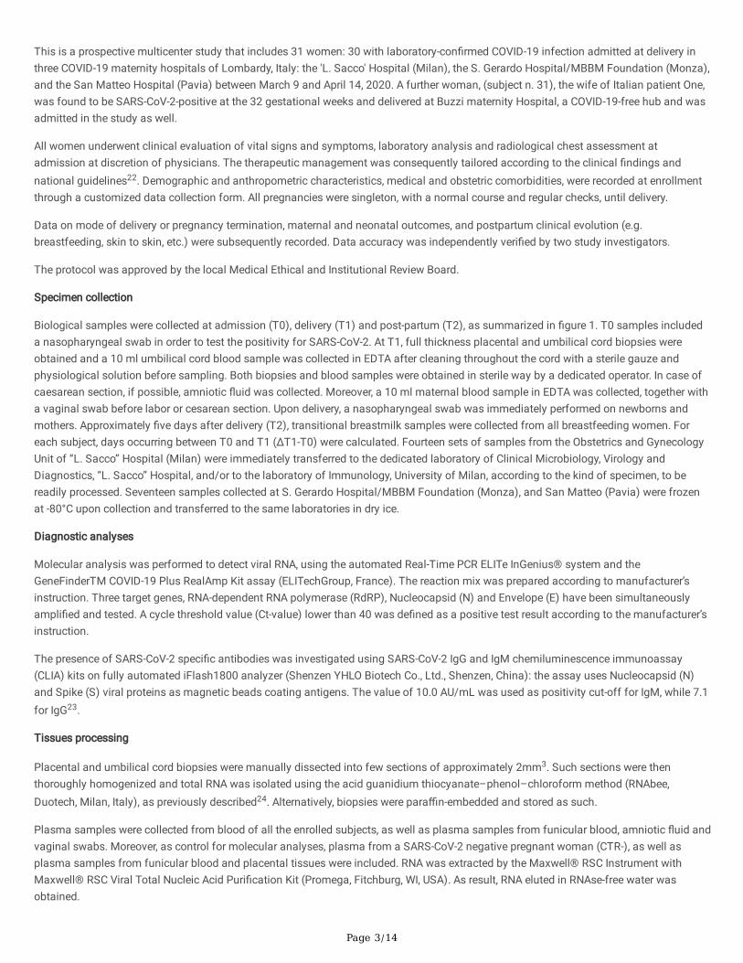
This is a prospective multicenter study that includes 31 women: 30 with laboratory-con�rmed COVID-19 infection admitted at delivery inthree COVID-19 maternity hospitals of Lombardy, Italy: the 'L. Sacco' Hospital (Milan), the S. Gerardo Hospital/MBBM Foundation (Monza),and the San Matteo Hospital (Pavia) between March 9 and April 14, 2020. A further woman, (subject n. 31), the wife of Italian patient One,was found to be SARS-CoV-2-positive at the 32 gestational weeks and delivered at Buzzi maternity Hospital, a COVID-19-free hub and wasadmitted in the study as well.
All women underwent clinical evaluation of vital signs and symptoms, laboratory analysis and radiological chest assessment atadmission at discretion of physicians. The therapeutic management was consequently tailored according to the clinical �ndings andnational guidelines22. Demographic and anthropometric characteristics, medical and obstetric comorbidities, were recorded at enrollmentthrough a customized data collection form. All pregnancies were singleton, with a normal course and regular checks, until delivery.
Data on mode of delivery or pregnancy termination, maternal and neonatal outcomes, and postpartum clinical evolution (e.g.breastfeeding, skin to skin, etc.) were subsequently recorded. Data accuracy was independently veri�ed by two study investigators.
The protocol was approved by the local Medical Ethical and Institutional Review Board.
Specimen collection
Biological samples were collected at admission (T0), delivery (T1) and post-partum (T2), as summarized in �gure 1. T0 samples includeda nasopharyngeal swab in order to test the positivity for SARS-CoV-2. At T1, full thickness placental and umbilical cord biopsies wereobtained and a 10 ml umbilical cord blood sample was collected in EDTA after cleaning throughout the cord with a sterile gauze andphysiological solution before sampling. Both biopsies and blood samples were obtained in sterile way by a dedicated operator. In case ofcaesarean section, if possible, amniotic �uid was collected. Moreover, a 10 ml maternal blood sample in EDTA was collected, together witha vaginal swab before labor or cesarean section. Upon delivery, a nasopharyngeal swab was immediately performed on newborns andmothers. Approximately �ve days after delivery (T2), transitional breastmilk samples were collected from all breastfeeding women. Foreach subject, days occurring between T0 and T1 (ΔT1-T0) were calculated. Fourteen sets of samples from the Obstetrics and GynecologyUnit of “L. Sacco” Hospital (Milan) were immediately transferred to the dedicated laboratory of Clinical Microbiology, Virology andDiagnostics, “L. Sacco” Hospital, and/or to the laboratory of Immunology, University of Milan, according to the kind of specimen, to bereadily processed. Seventeen samples collected at S. Gerardo Hospital/MBBM Foundation (Monza), and San Matteo (Pavia) were frozenat -80°C upon collection and transferred to the same laboratories in dry ice.
Diagnostic analyses
Molecular analysis was performed to detect viral RNA, using the automated Real-Time PCR ELITe InGenius® system and theGeneFinderTM COVID-19 Plus RealAmp Kit assay (ELITechGroup, France). The reaction mix was prepared according to manufacturer’sinstruction. Three target genes, RNA-dependent RNA polymerase (RdRP), Nucleocapsid (N) and Envelope (E) have been simultaneouslyampli�ed and tested. A cycle threshold value (Ct-value) lower than 40 was de�ned as a positive test result according to the manufacturer’sinstruction.
The presence of SARS-CoV-2 speci�c antibodies was investigated using SARS-CoV-2 IgG and IgM chemiluminescence immunoassay(CLIA) kits on fully automated iFlash1800 analyzer (Shenzen YHLO Biotech Co., Ltd., Shenzen, China): the assay uses Nucleocapsid (N)and Spike (S) viral proteins as magnetic beads coating antigens. The value of 10.0 AU/mL was used as positivity cut-off for IgM, while 7.1for IgG23.
Tissues processing
Placental and umbilical cord biopsies were manually dissected into few sections of approximately 2mm3. Such sections were thenthoroughly homogenized and total RNA was isolated using the acid guanidium thiocyanate–phenol–chloroform method (RNAbee,Duotech, Milan, Italy), as previously described24. Alternatively, biopsies were para�n-embedded and stored as such.
Plasma samples were collected from blood of all the enrolled subjects, as well as plasma samples from funicular blood, amniotic �uid andvaginal swabs. Moreover, as control for molecular analyses, plasma from a SARS-CoV-2 negative pregnant woman (CTR-), as well asplasma samples from funicular blood and placental tissues were included. RNA was extracted by the Maxwell® RSC Instrument withMaxwell® RSC Viral Total Nucleic Acid Puri�cation Kit (Promega, Fitchburg, WI, USA). As result, RNA eluted in RNAse-free water wasobtained.
Page 4/14
Once RNA was reverse transcribed into cDNA24, real-time PCR was performed on a CFX96 (Bio-rad, CA, USA) using TaqMan probesspeci�cally designed to target two regions of the nucleocapsid (N) gene of SARS-CoV-2. For such application, we employed the 2019-nCoVCDC qPCR Probe Assay emergency kit (IDT, Iowa, USA) which include also primers and probes that target the human RNase P gene.
Expression analyses of in�ammatory response
The in�ammatory response was analysed in four selected RNA samples extracted from placenta biopsies of one negative control (CTR-),one SARS-CoV-2 recovered (subject n. 31) subject and two SARS-CoV-2 positive subjects (subjects n. 17 and 25). Subjects n. 17 and 25gave birth to SARS-CoV-2 positive newborns, according to the �rst nasopharyngeal swab (T1). Analyses were performed by a PCR arraythat include a set of 84 optimized real-time PCR primers plus 5 housekeeping genes on a 96-well plates; the procedures suggested by themanufacturer were followed (Qiagen, Hilden, Germania). Undetermined raw CT values were set to 35. Only variables with at least a two-fold increase in their value are presented and discussed in the manuscript.
Concentration of 27 cytokines was assessed in maternal and funicular plasma samples from the very same four subjects usingimmunoassays formatted on magnetic beads (Bio-rad, CA, USA) according to manufacturer’s protocol via Luminex 100 technology(Luminex, Texas, USA).
Statistics
For the study variables, medians and ranges were reported for quantitative variables. The analyses were performed using SPSS Statistics,Version 26.0 (IBM Corp. Armonk, NY) together with GraphPad Prism 8.
All the procedures were carried out in accordance with the GLP guidelines adopted in our laboratories.
ResultsPopulation
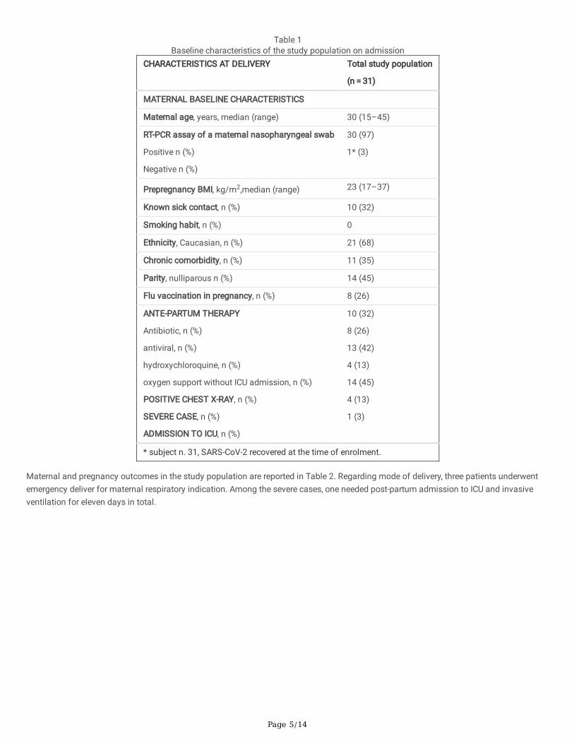
Four patients were classi�ed as severe cases, de�ned by the need of urgent delivery for the deterioration of maternal conditions or byICU/sub-intensive care admission. A radiological con�rmation of interstitial pneumonia was obtained on admission or antepartum for allthe severe cases and in 10 (32%) of the mild cases. Pharmacological treatment during the antepartum period of hospitalization is reportedin Table 1. In the only severe case of pre-term labor (subject n. 17), corticosteroids for RDS-prophylaxis were administered.
Page 5/14
Table 1Baseline characteristics of the study population on admission
CHARACTERISTICS AT DELIVERY Total study population
(n = 31)
MATERNAL BASELINE CHARACTERISTICS
Maternal age, years, median (range) 30 (15–45)
RT-PCR assay of a maternal nasopharyngeal swab
Positive n (%)
Negative n (%)
30 (97)
1* (3)
Prepregnancy BMI, kg/m2,median (range) 23 (17–37)
Known sick contact, n (%) 10 (32)
Smoking habit, n (%) 0
Ethnicity, Caucasian, n (%) 21 (68)
Chronic comorbidity, n (%) 11 (35)
Parity, nulliparous n (%) 14 (45)
Flu vaccination in pregnancy, n (%) 8 (26)
ANTE-PARTUM THERAPY
Antibiotic, n (%)
antiviral, n (%)
hydroxychloroquine, n (%)
oxygen support without ICU admission, n (%)
POSITIVE CHEST X-RAY, n (%)
SEVERE CASE, n (%)
ADMISSION TO ICU, n (%)
10 (32)
8 (26)
13 (42)
4 (13)
14 (45)
4 (13)
1 (3)
* subject n. 31, SARS-CoV-2 recovered at the time of enrolment.
Maternal and pregnancy outcomes in the study population are reported in Table 2. Regarding mode of delivery, three patients underwentemergency deliver for maternal respiratory indication. Among the severe cases, one needed post-partum admission to ICU and invasiveventilation for eleven days in total.

Page 6/14
Table 2Maternal and pregnancy outcomes in the study population.
Total study population
n = 31
Delivery mode
- vaginal, n (%)
- caesarean section, n (%)
25 (81)
6 (19)
GA at delivery, weeks median (range) 39 (34.4–41.4)
Induction of delivery related to COVID-19, n (%) 6 (19)
Caesarean section for severe maternal illness related COVID-19, n (%) 3 (9)
Preterm delivery, n (%) 1 (3)
Fetal gender, male, n (%) 18 (58)
Birth weight, g, median (range) 3200 (2180–4165)
Umbilical artery pH, median (range) 7.31 (7.14–7.43)
APGAR score 5' < 7, n (%) 1 (3)
Newborn COVID, positive, n (%) 2 (6)
NICU admission, n (%) 2 (6)
Skin to Skin, n (%) 4 (13)
Breastfeeding, n (%) 29 (94)
Subject n. 31 became negative at week 35 of pregnancy and delivered spontaneously at week 38. Except in one case (subject n. 17), allpregnancies were full term. Subject n. 17 was admitted preterm at 33 + 6 weeks with fever and dyspnea and delivered spontaneously at 34 + 4 weeks. A female baby was born, weighing 2180 g, with an Apgar Score of 9 and 10 at 1 and 5 minutes, respectively, with a pH ofumbilical artery of 7.14. The newborn was diagnosed with COVID-19 infection through a nasopharyngeal swab and was admitted to NICUfor prematurity. Subject n. 25 spontaneously delivered at week 39 + 2. A male baby was born, weighing 3340 g, with an Apgar Score of 9and 10 at 1 and 5 minutes, respectively, and the umbilical artery pH of was 7.14. The newborn was diagnosed with SARS-CoV-2 infectionthrough a nasopharyngeal swab upon delivery, while he tested negative 48 h later. Except for the two above-mentioned cases, no othernewborns resulted positive to SARS-CoV-2 detection by nasopharyngeal swab. Except for two cases, all newborns were breastfed. All thenewborns were healthy, including the two SARS-CoV-2 positive ones.
Virus and antibody detection
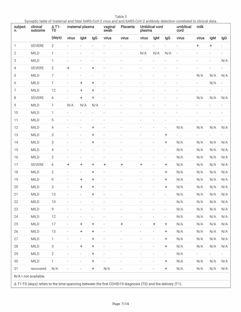
We investigated the presence of SARS-CoV-2 in the collected specimens, as shown in Table 3 and summarized in Table 4a. We detectedthe virus in two (6%) maternal plasma samples (subjects n. 4 and 17), both of them characterized by a severe clinical outcome.Remarkably, we detected the presence of SARS-CoV-2 in vaginal swab, placental tissue and cord plasma from subject n. 17. Moreover, wedetected SARS-CoV-2 in placental tissue from subject n. 25. We detected SARS-CoV-2 in one milk specimen only, from a severe clinicaloutcome case (subject n. 1). None of the tested six amniotic �uids, nor the twelve umbilical cords, resulted positive (Tables 3 and 4a).
Page 7/14
Table 3Synoptic table of maternal and fetal SARS-CoV-2 virus and anti-SARS-CoV-2 antibody detection correlated to clinical data.
subjectn.
clinicaloutcome
Δ T1-T0
(days)
maternal plasma vaginalswab
Placenta Umbilical cordplasma
umbilicalcord
milk
virus IgM IgG virus virus virus IgM IgG virus virus IgM IgG
1 SEVERE 2 - - - - - - - - - + + -
2 MILD 1 - - - - - N/A N/A N/A - - - -
3 MILD 1 - - - - - - - - - - - N/A
4 SEVERE 2 + - + - - - - - - - - -
5 MILD 7 - - - - - - - - - N/A N/A N/A
6 MILD 1 - + + - - - - - - - N/A -
7 MILD 12 - + + - - - - - - - - -
8 SEVERE 6 - + + - - - - - - N/A N/A N/A
9 MILD 1 N/A N/A N/A - - - - - - - - -
10 MILD 1 - - - - - - - - - - - -
11 MILD 5 - - - - - - - - - - - -
12 MILD 4 - - + - - - - - N/A N/A N/A N/A
13 MILD 3 - - + - - - - + - - - -
14 MILD 3 - - + - - - - + N/A N/A N/A N/A
15 MILD 4 - - - - - - - - N/A N/A N/A N/A
16 MILD 2 - - - - - - - - N/A N/A N/A N/A
17 SEVERE 6 + + + + + + - + N/A N/A N/A N/A
18 MILD 2 - - + - - - - + N/A N/A N/A N/A
19 MILD 9 - + + - - - - + N/A N/A N/A N/A
20 MILD 3 - + + - - - - + N/A N/A N/A N/A
21 MILD 13 - - + - - - - - N/A N/A N/A N/A
22 MILD 10 - - - - - - - - N/A N/A N/A N/A
23 MILD 9 - - - - - - - - N/A N/A N/A N/A
24 MILD 12 - - - - - - - - N/A N/A N/A N/A
25 MILD 17 - + + - + - + + N/A N/A N/A N/A
26 MILD 13 - + + - - - - + N/A N/A N/A N/A
27 MILD 1 - - + - - - - + N/A N/A N/A N/A
28 MILD 3 - + + - - - - + N/A N/A N/A N/A
29 MILD 2 - - + - - - - - N/A - - -
30 MILD 1 - - + - - - - + N/A N/A N/A N/A
31 recovered N/A - - + N/A - - - + N/A N/A N/A N/A
N/A = not available.
Δ T1-T0 (days) refers to the time spanning between the �rst COVID19 diagnosis (T0) and the delivery (T1).
Page 8/14
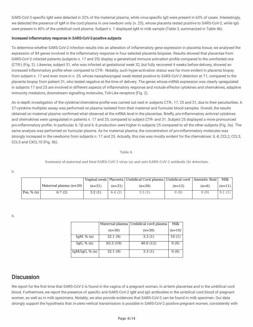
SARS-CoV-2-speci�c IgM were detected in 32% of the maternal plasma, while virus-speci�c IgG were present in 63% of cases. Interestingly,we detected the presence of IgM in the cord plasma in one newborn only (n. 25), whose placenta tested positive to SARS-CoV-2, while IgGwere present in 40% of the umbilical cord plasma. Subject n. 1 displayed IgM in milk sample (Table 3, summarized in Table 4b).
Increased in�ammatory response in SARS-CoV-2-positive subjects
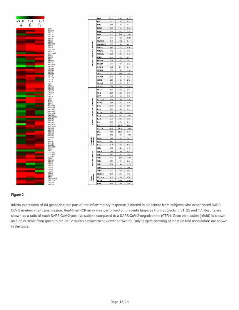
To determine whether SARS-CoV-2 infection results into an alteration of in�ammatory gene expression in placenta tissue, we analysed theexpression of 84 genes involved in the in�ammatory response in four selected placenta biopsies. Results showed that placentae fromSARS-CoV-2 infected patients (subjects n. 17 and 25) display a generalized immune activation pro�le compared to the uninfected one(CTR-) (Fig. 2). Likewise, subject 31, who was infected at gestational week 32, but fully recovered 4 weeks before delivery, showed anincreased in�ammatory pro�le when compared to CTR-. Notably, such hyper-activation status was far more evident in placenta biopsyfrom subject n. 17 and even more in n. 25, whose nasopharyngeal swab tested positive to SARS-CoV-2 detection at T1, compared to theplacenta biopsy from patient 31, who tested negative at the time of delivery. The genes whose mRNA expression was clearly upregulatedin subjects 17 and 25 are involved in different aspects of in�ammatory response and include effector cytokines and chemokines, adaptiveimmunity mediators, downstream signalling molecules, Toll-Like receptors (Fig. 2).
An in-depth investigation of the cytokine/chemokine pro�le was carried out next in subjects CTR-, 17, 25 and 31, due to their peculiarities. A27-cytokine multiplex assay was performed on plasma isolated from their maternal and funicular blood samples. Overall, the resultsobtained on maternal plasma con�rmed what observed at the mRNA level in the placentae. Brie�y, pro-in�ammatory antiviral cytokinesand chemokines were upregulated in patients n. 17 and 25, compared to subject CTR- and 31. Subject 25 displayed a more pronouncedpro-in�ammatory pro�le. In particular, IL-1β and IL-6 production were higher in subjects 25 compared to all the other subjects (Fig. 3a). Thesame analysis was performed on funicular plasma. As for maternal plasma, the concentration of pro-in�ammatory molecules wasstrongly increased in the newborns from subjects n. 17 and 25. Actually, this rise was mostly evident for the chemokines: IL-8, CCL2, CCL3,CCL5 and CXCL10 (Fig. 3b).
Table 4.
Summary of maternal and fetal SARS-CoV-2 virus (a) and anti-SARS-CoV-2 antibody (b) detection.
a.
Maternal plasma (n=30)
Vaginal swab
(n=31)
Placenta
(n=31)
Umbilical Cord plasma
(n=30)
Umbilical cord
(n=12)
Amniotic fluid
(n=6)
Milk
(n=11)
Pos, % (n) 6.7 (2) 3.2 (1) 6.4 (2) 3.3 (1) 0 (0) 0 (0) 9.1 (1)
b.
Maternal plasma
(n=30)
Umbilical cord plasma
(n=30)
Milk
(n=10)
IgM, % (n) 32.1 (9) 3.3 (1) 10 (1)
IgG, % (n) 63.3 (19) 40.0 (12) 0 (0)
IgM/IgG, % (n) 32.1 (9) 3.3 (1) 0 (0)
DiscussionWe report for the �rst time that SARS-CoV-2 is found in the vagina of a pregnant woman, in at-term placentae and in the umbilical cordblood. Furthermore, we report the presence of speci�c anti-SARS-CoV-2 IgM and IgG antibodies in the umbilical cord blood of pregnantwomen, as well as in milk specimens. Notably, we also provide evidences that SARS-CoV-2 can be found in milk specimen. Our datastrongly support the hypothesis that in-utero vertical transmission is possible in SARS-CoV-2 positive pregnant women, consistently with
Page 9/14
how previously reported25. Finally, this is the �rst report describing the in�ammatory response triggered by SARS-CoV-2 infection inpregnant women at both systemic and placental level.
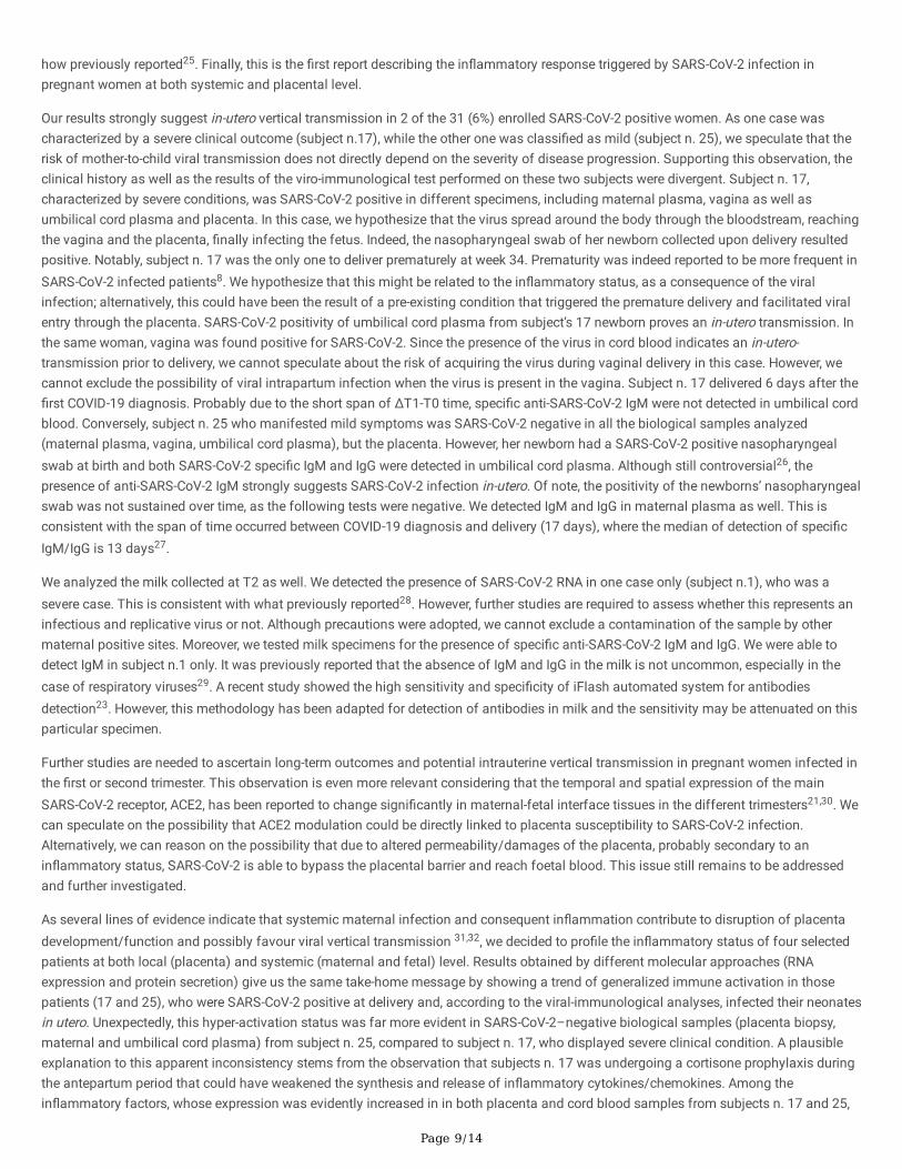
Our results strongly suggest in-utero vertical transmission in 2 of the 31 (6%) enrolled SARS-CoV-2 positive women. As one case wascharacterized by a severe clinical outcome (subject n.17), while the other one was classi�ed as mild (subject n. 25), we speculate that therisk of mother-to-child viral transmission does not directly depend on the severity of disease progression. Supporting this observation, theclinical history as well as the results of the viro-immunological test performed on these two subjects were divergent. Subject n. 17,characterized by severe conditions, was SARS-CoV-2 positive in different specimens, including maternal plasma, vagina as well asumbilical cord plasma and placenta. In this case, we hypothesize that the virus spread around the body through the bloodstream, reachingthe vagina and the placenta, �nally infecting the fetus. Indeed, the nasopharyngeal swab of her newborn collected upon delivery resultedpositive. Notably, subject n. 17 was the only one to deliver prematurely at week 34. Prematurity was indeed reported to be more frequent inSARS-CoV-2 infected patients8. We hypothesize that this might be related to the in�ammatory status, as a consequence of the viralinfection; alternatively, this could have been the result of a pre-existing condition that triggered the premature delivery and facilitated viralentry through the placenta. SARS-CoV-2 positivity of umbilical cord plasma from subject’s 17 newborn proves an in-utero transmission. Inthe same woman, vagina was found positive for SARS-CoV-2. Since the presence of the virus in cord blood indicates an in-utero-transmission prior to delivery, we cannot speculate about the risk of acquiring the virus during vaginal delivery in this case. However, wecannot exclude the possibility of viral intrapartum infection when the virus is present in the vagina. Subject n. 17 delivered 6 days after the�rst COVID-19 diagnosis. Probably due to the short span of ΔT1-T0 time, speci�c anti-SARS-CoV-2 IgM were not detected in umbilical cordblood. Conversely, subject n. 25 who manifested mild symptoms was SARS-CoV-2 negative in all the biological samples analyzed(maternal plasma, vagina, umbilical cord plasma), but the placenta. However, her newborn had a SARS-CoV-2 positive nasopharyngealswab at birth and both SARS-CoV-2 speci�c IgM and IgG were detected in umbilical cord plasma. Although still controversial26, thepresence of anti-SARS-CoV-2 IgM strongly suggests SARS-CoV-2 infection in-utero. Of note, the positivity of the newborns’ nasopharyngealswab was not sustained over time, as the following tests were negative. We detected IgM and IgG in maternal plasma as well. This isconsistent with the span of time occurred between COVID-19 diagnosis and delivery (17 days), where the median of detection of speci�cIgM/IgG is 13 days27.
We analyzed the milk collected at T2 as well. We detected the presence of SARS-CoV-2 RNA in one case only (subject n.1), who was asevere case. This is consistent with what previously reported28. However, further studies are required to assess whether this represents aninfectious and replicative virus or not. Although precautions were adopted, we cannot exclude a contamination of the sample by othermaternal positive sites. Moreover, we tested milk specimens for the presence of speci�c anti-SARS-CoV-2 IgM and IgG. We were able todetect IgM in subject n.1 only. It was previously reported that the absence of IgM and IgG in the milk is not uncommon, especially in thecase of respiratory viruses29. A recent study showed the high sensitivity and speci�city of iFlash automated system for antibodiesdetection23. However, this methodology has been adapted for detection of antibodies in milk and the sensitivity may be attenuated on thisparticular specimen.
Further studies are needed to ascertain long-term outcomes and potential intrauterine vertical transmission in pregnant women infected inthe �rst or second trimester. This observation is even more relevant considering that the temporal and spatial expression of the mainSARS-CoV-2 receptor, ACE2, has been reported to change signi�cantly in maternal-fetal interface tissues in the different trimesters21,30. Wecan speculate on the possibility that ACE2 modulation could be directly linked to placenta susceptibility to SARS-CoV-2 infection.Alternatively, we can reason on the possibility that due to altered permeability/damages of the placenta, probably secondary to anin�ammatory status, SARS-CoV-2 is able to bypass the placental barrier and reach foetal blood. This issue still remains to be addressedand further investigated.
As several lines of evidence indicate that systemic maternal infection and consequent in�ammation contribute to disruption of placentadevelopment/function and possibly favour viral vertical transmission 31,32, we decided to pro�le the in�ammatory status of four selectedpatients at both local (placenta) and systemic (maternal and fetal) level. Results obtained by different molecular approaches (RNAexpression and protein secretion) give us the same take-home message by showing a trend of generalized immune activation in thosepatients (17 and 25), who were SARS-CoV-2 positive at delivery and, according to the viral-immunological analyses, infected their neonatesin utero. Unexpectedly, this hyper-activation status was far more evident in SARS-CoV-2–negative biological samples (placenta biopsy,maternal and umbilical cord plasma) from subject n. 25, compared to subject n. 17, who displayed severe clinical condition. A plausibleexplanation to this apparent inconsistency stems from the observation that subjects n. 17 was undergoing a cortisone prophylaxis duringthe antepartum period that could have weakened the synthesis and release of in�ammatory cytokines/chemokines. Among thein�ammatory factors, whose expression was evidently increased in in both placenta and cord blood samples from subjects n. 17 and 25,

Page 10/14
the chemokines, CXCL10, CXCL8, CCL5, CCL3, CCL2, could have played a major role in favouring vertical transmission. Indeed, they couldhave created a chemotactic gradient between villi and the inter-villous space, where maternal lymphocyte circulate, thus favouring viraldissemination33. To perform such molecular analyses, only four subjects (CTR-, n. 17, n. 25 and n. 31) were chosen due to theirpeculiarities. However, further experiments are envisaged, in order to con�rm this distinctive pro�le.
In conclusion, for the �rst time SARS-CoV-2 was detected in umbilical cord plasma, indicating that in-utero mother-to-child transmission,although rare, is possible and apparently related to a high maternal in�ammatory state. Concerningly, such maternal in�ammatory state ismirrored in the fetus. Although further studies are needed, this should be taken into consideration in the management of COVID-19pregnant women.
DeclarationsAcknowledgements
We would like to thank Paolo Quaini, Francesco Leone, Federica Fusè, Irma Saulle, Claudia Fusetti, Margherita Longo, Alberto Rizzo,Francesca Romeri, Federica Brunetti and Francesca Sabbatini for their support and contribution to the project. This study was supportedby COVID-19 donation to Obstetrics and Gynecology and to Laboratory of Immunology, Department of Biomedical and Clinical Sciences,University of Milan, Italy. and by a grant from Flak Renewables.
Authors’ contributions
VS conceived the presented idea. MB further developed the project with the help of CF. VS, IC, PV, AS, FP, CC and SC performed subjectenrolment and clinical management, as well as samples collection. IB was involved in sample collection and management. CF and MBconceived, planned, performed and analysed the experiments on SARS-CoV-2 detection on plasma, biopsies and vaginal swab, andexperiments on the in�ammatory response. DT helped with the interpretation of the data. DM and AM conceived, planned and performedthe experiments on speci�c antibody detection and experiments on milk, under the supervision of MG. CF, MB and VS discussed the dataand wrote the manuscript. MC, IC and PV critically reviewed the manuscript. CF and MB equally contributed to the manuscript.
References1. Huang, C. et al. Clinical features of patients infected with 2019 novel coronavirus in Wuhan, China. The Lancet 395, 497–506 (2020).
2. Grasselli, G. et al. Baseline Characteristics and Outcomes of 1591 Patients Infected With SARS-CoV-2 Admitted to ICUs of theLombardy Region, Italy. JAMA 323, 1574–1581 (2020).
3. Dong, E., Du, H. & Gardner, L. An interactive web-based dashboard to track COVID-19 in real time. The Lancet Infectious Diseases 20,533–534 (2020).
4. Fu, Y., Cheng, Y. & Wu, Y. Understanding SARS-CoV-2-Mediated In�ammatory Responses: From Mechanisms to Potential TherapeuticTools. Virol Sin 1–6 (2020) doi:10.1007/s12250-020-00207-4.
5. Tay, M. Z., Poh, C. M., Rénia, L., MacAry, P. A. & Ng, L. F. P. The trinity of COVID-19: immunity, in�ammation and intervention. NatureReviews Immunology 1–12 (2020) doi:10.1038/s41577-020-0311-8.
�. Qin, C. et al. Dysregulation of immune response in patients with COVID-19 in Wuhan, China. Clin Infect Dis (2020)doi:10.1093/cid/ciaa248.
7. Zhou, Y. et al. Pathogenic T cells and in�ammatory monocytes incite in�ammatory storm in severe COVID-19 patients. Natl Sci Revdoi:10.1093/nsr/nwaa041.
�. Savasi, V. et al. Clinical Findings and Disease Severity in Hospitalized Pregnant Women With Coronavirus Disease 2019 (COVID-19).Obstretrics and Gynecology.
9. Wong, S. F. et al. Pregnancy and perinatal outcomes of women with severe acute respiratory syndrome. Am J Obstet Gynecol 191,292–297 (2004).
10. Alfaraj, S. H., Al-Taw�q, J. A. & Memish, Z. A. Middle East Respiratory Syndrome Coronavirus (MERS-CoV) infection during pregnancy:Report of two cases & review of the literature. J Microbiol Immunol Infect 52, 501–503 (2019).
11. Chen, H. et al. Clinical characteristics and intrauterine vertical transmission potential of COVID-19 infection in nine pregnant women: aretrospective review of medical records. The Lancet 395, 809–815 (2020).
12. Rasmussen, S. A., Smulian, J. C., Lednicky, J. A., Wen, T. S. & Jamieson, D. J. Coronavirus Disease 2019 (COVID-19) and pregnancy:what obstetricians need to know. Am J Obstet Gynecol (2020) doi:10.1016/j.ajog.2020.02.017.

Page 11/14
13. Zhu, H. et al. Clinical analysis of 10 neonates born to mothers with 2019-nCoV pneumonia. Translational Pediatrics 9, 51-60–60(2020).
14. Wang, X. et al. A case of 2019 Novel Coronavirus in a pregnant woman with preterm delivery. Clin Infect Dis (2020)doi:10.1093/cid/ciaa200.
15. Lu, Z. et al. Analysis of the pregnancy outcomes in pregnant women with COVID-19 in Hubei Province. Chinese Journal of Obstetricsand Gynecology 55, E009–E009 (2020).
1�. Ferrazzi, E. et al. Vaginal delivery in SARS-CoV-2 infected pregnant women in Northern Italy: a retrospective analysis. BJOG: AnInternational Journal of Obstetrics & Gynaecology n/a,.
17. Peng, Z. et al. Unlikely SARS-CoV-2 vertical transmission from mother to child: A case report. J Infect Public Health 13, 818–820(2020).
1�. Zeng, H. et al. Antibodies in Infants Born to Mothers With COVID-19 Pneumonia. JAMA (2020) doi:10.1001/jama.2020.4861.
19. Dong, L. et al. Possible Vertical Transmission of SARS-CoV-2 From an Infected Mother to Her Newborn. JAMA (2020)doi:10.1001/jama.2020.4621.
20. Baud, D. et al. Second-Trimester Miscarriage in a Pregnant Woman With SARS-CoV-2 Infection. JAMA (2020)doi:10.1001/jama.2020.7233.
21. Li, M., Chen, L., Zhang, J., Xiong, C. & Li, X. The SARS-CoV-2 receptor ACE2 expression of maternal-fetal interface and fetal organs bysingle-cell transcriptome study. PLOS ONE 15, e0230295 (2020).
22. Clinical management of severe acute respiratory infection when COVID-19 is suspected. https://www.who.int/publications-detail/clinical-management-of-severe-acute-respiratory-infection-when-novel-coronavirus-(ncov)-infection-is-suspected.
23. Infantino, M. et al. Diagnostic accuracy of an automated chemiluminescent immunoassay for anti-SARS-CoV-2 IgM and IgGantibodies: an Italian experience. J. Med. Virol. (2020) doi:10.1002/jmv.25932.
24. Saulle, I. et al. Endoplasmic Reticulum Associated Aminopeptidase 2 (ERAP2) Is Released in the Secretome of Activated MDMs andReduces in vitro HIV-1 Infection. Front. Immunol. 10, (2019).
25. Vivanti, A. J. et al. Transplacental transmission of SARS-CoV-2 infection. Nature Communications 11, 3572 (2020).
2�. Kimberlin, D. W. & Stagno, S. Can SARS-CoV-2 Infection Be Acquired In Utero?: More De�nitive Evidence Is Needed. JAMA 323, 1788–1789 (2020).
27. Long, Q.-X. et al. Antibody responses to SARS-CoV-2 in patients with COVID-19. Nature Medicine 1–4 (2020) doi:10.1038/s41591-020-0897-1.
2�. Groß, R. et al. Detection of SARS-CoV-2 in human breastmilk. The Lancet 395, 1757–1758 (2020).
29. Dixon, D.-L. The Role of Human Milk Immunomodulators in Protecting Against Viral Bronchiolitis and Development of ChronicWheezing Illness. Children (Basel) 2, 289–304 (2015).
30. Valdés, G. et al. Distribution of Angiotensin-(1-7) and ACE2 in Human Placentas of Normal and Pathological Pregnancies. Placenta 27,200–207 (2006).
31. Lee, J. K., Oh, S.-J., Park, H. & Shin, O. S. Recent Updates on Research Models and Tools to Study Virus–Host Interactions at thePlacenta. Viruses 12, 5 (2020).
32. Heerema‐McKenney, A. Defense and infection of the human placenta. APMIS 126, 570–588 (2018).
33. Kim, C. J., Romero, R., Chaemsaithong, P. & Kim, J.-S. Chronic In�ammation of the Placenta: De�nition, Classi�cation, Pathogenesis,and Clinical Signi�cance. Am J Obstet Gynecol 213, S53–S69 (2015).
Figures

Page 12/14
Figure 1
Specimen collection timeline. List of specimens collected at admission to the study (T0), at delivery (T1) and during lactation (T2) for eachenrolled subject.
Page 13/14
Figure 2
mRNA expression of 84 genes that are part of the in�ammatory response is altered in placentae from subjects who experienced SARS-CoV-2 in-utero viral transmission. Real-time PCR array was performed on placenta biopsies from subjects n. 31, 25 and 17. Results areshown as a ratio of each SARS-CoV-2-positive subject compared to a SARS-CoV-2-negative one (CTR-). Gene expression (nfold) is shownas a color scale from green to red (MEV multiple experiment viewer software). Only targets showing at least >2-fold modulation are shownin the table.

Page 14/14
Figure 3
Protein secretion of 27 cytokines/chemokines that are part of the in�ammatory response is altered in maternal and umbilical cord plasmafrom subjects who experienced SARS-CoV-2 in-utero viral transmission. Multiplex array was performed on maternal and umbilical plasmafrom subjects n. 31, 25 and 17. As reference, a SARS-CoV-2-negative plasma is shown (CTR-). Protein concentration is shown as pg/ml ofplasma.