in this chapter - ace | certified personal trainer · pdf fileanatomy, and physiology to...
TRANSCRIPT
Communication With the Medical Team
Initial Interview
Objective Evaluation
Professional Impression
Plan
Client Population
Rehabilitation ProtocolsProtocol for Rehabilitation After an Arthroscopic Partial Meniscectomy
Concepts of Healing
Systematic Progression of Programming
Increasing Range of Motion and FlexibilityImproving Aerobic ConditionReturning to Physical ActivitiesBuilding Strength and Power
Case StudiesCase Study 1Case Study 2
Summary
In This Chapter
A b o u t T h e A u t h o r John R. Martínez, P.T., M.P.T., is the owner and president of
Executive Operations Management, L.L.C., a medical consulting
firm, and Physical Therapy Experts, P.L.L.C., a private medical
practice, both in New York City. He is a teacher of neurology,
anatomy, and physiology to undergraduate students in Manhattan.
Martinez received his Bachelor of Arts and teaching certification in
1988 from Swarthmore College and has taught elementary through
graduate school students and a variety of topics in science,
recreation, wellness, and exercise. In 1997, Martinez received his
Bachelor of Science and Master of Physical Therapy degrees from
the Philadelphia College of Pharmacy and Science.

C h A p T e r 1 7
Considering the rather sophisticated health insurance require-ments and restrictions, as well as their increasing costs, clients who need further attention from these medical profession-als may have to rely on the ACE-AHFS for their continued rehabilitation. Thus, the principles of post-orthopedic reha-bilitation have changed over the past few decades, requiring fitness professionals to add a strong clinical component to their educational foundation. The ability to maintain current knowledge of these medical principles and apply that knowl-edge successfully with the appropriate clients will be the key to an ACE-AHFS becoming an invaluable member of the modern medical team.
Communication With the Medical Team
It is impossible to overstate the importance of consistent communication with the medical team, which includes the physician or physicians involved in the medical care of the
client, the physical and/or occupational therapist, any other medical or integrative medicine professional treating the client (e.g., chiropractor, acupuncturist, nutritionist), and the client him- or herself, who will have acquired a tremendous amount of information from all of these sources. Including the client as
a member of the medical team is often overlooked. However, the client’s understanding of his or her own body and its func-tions, as well as the fact that the client is in attendance at all the treatments with each of the medical professionals, makes him or her a crucial source of information regarding health history and healthcare needs. Obtaining as much information, both subjective and objective, as possible regarding the cli-ent’s health will help establish a strong foundation for making exercise program development successful for the client. In particular, obtaining information from the new medical team members regarding the precautions and contraindications related to exercise is crucial, especially in orthopedic rehabilita-tion. Doing so will help protect the client from re-injury or any regression of the medical condition, which could damage the ACE-AHFS’s relationship with not only the medical team, but also the client and future referred clients. The concept of “protection” is an important one for the ACE-AHFS to understand. Often, immediately following an acute orthopedic injury or surgical intervention, the client is placed on a status of “maximal protection” (e.g., non-weightbearing after a hip replacement). This type of information is crucial to protect the client during a time when he or she is at high risk for re-injury or worsening the medical condition. As the individual pro-gresses, his or her status progresses to “moderate protection” (e.g., toe-touch weightbearing); to “minimal protection” (e.g.,
The fine line between exercise for healthy individuals and therapeutic exer-
cise for individuals needing rehabilitation after injury, disease, illness, or other
pathology can be difficult to determine. An ACE-certified Advanced Health &
Fitness Specialist (ACE-AHFS) must know when it is appropriate to proceed with exer-
cise program development for a client, rather than referring him or her to a licensed
medical professional, such as a physical therapist, occupational therapist, or physician.
Principles of Post-orthopedic Rehabilitation
John Martínez
A C E A D v A n C E D H E A l T H & F I T n E S S S P E C I A l I S T M A n u A l
Chapter Seventeen Principles of Post-orthopedic Rehabilitation
A C E A D v A n C E D H E A l T H & F I T n E S S S P E C I A l I S T M A n u A l
422
weightbearing as tolerated); and, lastly, to “unre-stricted protection” (e.g., full weightbearing as risk decreases with physical recovery, both anatomically and physiologically). A clear understanding of the concept of protection and how it relates to the performance of activities that are contraindicated (higher risk) and to activities that are precautions (lower risk) is mandatory for the ACE-AHFS. The request for this specific information, along with the collection of the client’s general medical history, will help to build the trust and confidence necessary from the entire medical team that will support the ACE-AHFS’s work.
Initial Interview
Acquiring accurate information from any new client during the first meeting is critical to establishing a reliable history of
the client’s health. Clients can be a valuable source of personal health information, but since their reports often come from their untrained medical perspective, this information can have a variety of inaccuracies. Discerning which pieces of infor-mation are more or less useful requires focused listening along with targeted questioning from the ACE-AHFS. Bringing a skillful investigatory technique and style to an initial interview is crucial to the success of exercise program development for the post-orthopedic rehabilitation client. This process will provide the ACE-AHFS with the health information necessary to make informed decisions on the frequency, intensity, type, and duration of exercise that is best for the client. This is particularly true if the client must discontinue his or her medical treatment but wants to con-tinue the recovery from injury, disease, or illness with an ACE-AHFS. To further aid in bridging the gap between the clinical and fitness settings, the ACE-AHFS should also consult with the cli-ent’s rehabilitation specialist as described in the previous section.
Questioning a client regarding his or her level of pain or discomfort during this initial interview is also extremely important when developing safe exercise programming. A client’s description of his or her pain will help the ACE-AHFS decipher if it is due to normal physical stress (such as muscle
soreness), injury, disease, or other type of pathol-ogy. It will also lead to a discussion regarding what type of activities or circumstances exacerbate the pain or discomfort versus ones that provide relief. This type of exchange will provide valuable information on what exercises can be tolerated by the client. During post-orthopedic rehabilita-tion, respecting pain is important and protecting clients from further injury or exacerbation of their signs and symptoms must be a primary goal. Of course, upon exacerbation of pain or other signs and symptoms, immediate adjustments are neces-sary, and communication with the medical team is mandatory to formulate a plan for continued safe participation in an exercise program.
Objective Evaluation
Post-orthopedic rehabilitation clients will often come to the ACE-AHFS just short of returning to their pre-injury
health status. Given a limited amount of sessions with the rehabilitation team due to health insur-ance and cost restrictions, clients will often be discharged from these medical services with the functional ability to return to their activities of daily living (ADL), but not to their normal level of activity. It is therefore important to measure a client’s physical abilities before initiating a train-ing program to establish a baseline of objective physical capabilities that can be referred to as the client progresses. It is also necessary to continue to document the client’s progress overall in the tran-sition from one medical team member to another. Documenting the role of the ACE-AHFS in the overall picture of a client’s health recovery is an important component of the fitness professional’s responsibilities as a member of the medical team.
The ACE-AHFS’s assessment of the client’s physical condition must be objective and some-what comparable to the discharge assessment of the rehabilitation medical professionals. Acquiring an understanding of the rehabilitation special-ist’s measuring and assessment approaches will provide the ACE-AHFS with a solid foundation from which to build his or her own unique evalu-ation. Performing careful measurements from objective fitness testing techniques and activities

423
A C E A D v A n C E D H E A l T H & F I T n E S S S P E C I A l I S T M A n u A lA C E A D v A n C E D H E A l T H & F I T n E S S S P E C I A l I S T M A n u A l
Principles of Post-orthopedic Rehabilitation Chapter Seventeen 423
will greatly benefit the client and the rest of the medical team by providing a consistent method for reviewing the client’s progress over time and from one professional to another. Allowing the client’s subjective and objective goals to guide the assessment will make the evaluation more efficient. Assessments of generalized and local joint range of motion (ROM), muscle endurance and strength, and the ability to perform important functional activities (e.g., ambulation, balance) will form a baseline of a client’s initial health status. This information is crucial as the ACE-AHFS builds an effective exercise program that will continue to show rehabilitative progress compared to baseline measurements. Adding recreational activities and movements to these programs will increase a client’s motivation and enthusiasm to remain compliant and show objective advances on a regu-lar basis through comparison to a well-established evaluation, which will also make the client more committed to the program.
Professional Impression
Once the initial evaluation, interview, and testing have taken place, the ACE-AHFS’s review and opinion of
all this information is what makes him or her a true professional. Determining the important health issues to focus on and recognizing when a client’s health status is outside one’s scope of expertise are important components of any fit-ness professional’s standard of care. Most often, the medical team’s summaries are the only component of a medical report that other team members will review, given their time restraints. Trust in, and respect for, the knowledge, skills, and expertise of the ACE-AHFS will make him or her a valuable and accepted member of the team. The ACE-AHFS can develop that trust through thorough assessments of each new client’s health status and professional commu-nication to the medical team.
These assessment summaries should briefly state the client’s age, gender, medical diagnosis, and history, and how the client presented to the ACE-AHFS with regard to the most important physical findings.
Sample Assessment SummaryThe client is a 5'7"(1.7 m) 63-year-old
female weighing 110 lb (50 kg) with a diag-nosis of osteoporosis. She has had back pain for the past 3 years. She comes to the ACE-AHFS after completion of three months of physical therapy, which resulted in minimal back pain. She has a short-term goal to return to running 3–4 days per week as she did prior to her back pain and a long-term goal to run a marathon one more time.
The client’s aerobic condition is dimin-ished, with tolerance of only 10 minutes of walking on the treadmill at 3.0 mph resulting in RR 25, HR 155, BP 140/90, and RPE 18. upper- and lower-extremity anaerobic condition is also decreased, with an ability to perform only 6–8 repetitions on all tests before fatigue and occasional loss of balance with self-correcting. Minimal back pain was reported by the client during and after testing.
The client will benefit from exercise pro-gramming to increase her aerobic condition with cardiovascular activities, elevate her extremity strength levels with resistance train-ing, stabilize her balance during recreational activities to reduce the risk of falls through coordination and agility activities, and improve her bone strength with closed-chain exercises. Referral to a nutritional expert has been made.
Note: RR = Respiration rate; HR = Heart rate; BP = Blood pressure; RPE = Ratings of perceived exertion
These summaries provide the medical team with all the necessary information regarding an ACE-AHFS’s findings and how the client will benefit from his or her services. If the medical team members disagree or feel that the ACE-AHFS needs to know additional information that will change the assessment, they will con-tact the ACE-AHFS with that information. This report gives them the opportunity to do so and makes them aware that their patient is actively trying to improve his or her health with the help of a qualified fitness professional. If the medical team members would like to see more

Chapter Seventeen Principles of Post-orthopedic Rehabilitation
A C E A D v A n C E D H E A l T H & F I T n E S S S P E C I A l I S T M A n u A l
424
details, they will be able to review the rest of the report, which should contain all of the objective activities that occurred. Therefore, the entire report should be sent to the medical team mem-bers, including a plan of what the ACE-AHFS intends to do to help the client achieve his or her goals. Additionally, the ACE-AHFS should be sure to include his or her contact informa-tion and a statement expressing a willingness to communicate further with the clinician about the client.
Plan
When preparing to work with a new post-orthopedic rehabilitation client, the ACE-AHFS should keep
the process of reaching the established goals in mind. The initial evaluative report should include a written summary of the steps the ACE-AHFS plans to take to help the client reach his or her goals. Taking a broad perspec-tive is important in visualizing how the entire program will begin, progress, and end. This will make the day-to-day planning more efficient and effective. Forecasting the exercise programming is a skill that the ACE-AHFS will develop over time with each plan he or she creates. Short-term goals that are measurable, specific, and concrete, along with long-term goals that are functional, will be helpful and can be included either in the plan or the assessment portion of the initial evaluation. Remember, health status changes and new information continues to arise. Therefore, after each month of working with a client, a new plan should be created, updating the short-term goals and fine-tuning the long-term goals in an effort to move as close as possible to a successful outcome. Signing and dating this report gives it authenticity and helps establish a level of pro-fessionalism. The ACE-AHFS should not be overly concerned about specifically achieving the original plan, as it is generally understood that components and factors may change and lead to different outcomes. It is the documentation that is important to the medical team and what makes the ACE-AHFS valuable in the post-orthopedic rehabilitation process of recovery.
Client Population
As medical technology, procedures, and skills advance, people are living longer and more functionally than ever before. This
increasing lifespan results in a general population that is living with chronic illnesses, diseases, inju-ries, and other pathologies. The considerably large “Baby Boomer” population is now seeking medical attention for myriad health conditions in an effort to move forward with their active lives both with and beyond traditional medical therapies. Thus, these clients may continue seeking relief from the signs and symptoms of their conditions after considering all that the medical therapies can offer. The recommendation to significantly decrease work and recreational activities is no longer a real-istic or acceptable option, creating the need for the special skills of an ACE-AHFS who can help these clients recover their functional capabilities and remain active for years into the future.
understanding the basic medical concepts of chronic conditions in general, as well as some specific pathologies that are more common than others, will provide the ACE-AHFS with the foundation he or she will need to address clients’ ongoing wellness goals. The other chapters in this manual address these needs and are the structure upon which an ACE-AHFS can build this foundation. In general, these clients will be highly ambulatory (i.e., walking in their homes and communities), and be able to accomplish functional ADl (e.g., eating, dressing, grocery shopping). They may present to the ACE-AHFS with some loss of general conditioning, complaints of consistent pain, discomfort, or paraesthesia from a cause either known or unknown, weakness of some muscular groups, and/or loss of ROM or flexibility. Frequently, these signs and symptoms will be affecting their daily function to some degree and be preventing them from performing at the levels they have in the past. These clients will have a strong desire, motivation, and enthusiasm to return to their past levels of function and often will have already begun this rehabilitation through participation in programs and therapies with other medical team members. An ACE-AHFS’s clients usually arrive having achieved general improvement in

A C E A D v A n C E D H E A l T H & F I T n E S S S P E C I A l I S T M A n u A l
Principles of Post-orthopedic Rehabilitation Chapter Seventeen 425
their physical condition to a certain level, either through rehabilitation with medical experts or through their own efforts, but wish to continue advancing toward higher physical goals.
Rehabilitation Protocols
Many orthopedic medical profession-als have developed rehabilitation treatment protocols that have sup-
port from scientific research regarding their effectiveness with patients. These protocols serve as treatment guidelines for the gradual process of recovery through which they and other medical team members should proceed to meet patients’ rehabilitation goals. Surgeons, sports medicine physicians, physical therapists, and occupational therapists have all established such protocols for rehabilitation from a variety of diagnoses that will require their patients to exercise if they are to return to their normal ADl. Additionally, generalized protocols can be found in both the research and academic literature, particularly in the field of physical therapy rehabilitation.
Protocol for Rehabilitation After an Arthroscopic Partial Meniscectomy
OverviewTypically, damage to the meniscus results
in a tear. As the torn piece begins to move in an abnormal fashion inside the joint, it can cause a great deal of pain and limitation in the knee, thereby limiting activity tolerance. Depending on the type and size of the tear, arthroscopic surgery may be recommended. The options are to perform a repair of the meniscus or a meniscectomy, where the damaged meniscus is removed to prevent further irritation. Since the meniscus has such a poor blood supply, a meniscectomy is often performed. Generally, following knee arthroscopy, a fairly aggressive approach can be taken and ROM and strength are pro-gressed as tolerated.
Phase I (Weeks 1–4)The emphasis is on regaining full knee
extension so the patient can ambulate with a normal gait pattern. This requires facilitating neuromuscular control of the quadriceps, controlling swelling, emphasiz-ing normal gait pattern, and achieving knee ROM of 0 to 90 degrees.
Strengthening: Quad sets (isometric quad-riceps contractions); straight-leg raise (SlR) in all planes of motion; standing heel raises on the Total Gym®; stretching (pain-free range) of the hamstrings, gastrocnemius, ili-otibial band (ITB), and piriformis
ROM: Manual patellar mobilizations; heel slides using a towel or wall if needed; prone hangs as needed to gain full extension
Balance: Weight shifting; single-limb stance
Gait: Move to single crutch when the patient is able and then discontinue (D/C) the use of crutches when the patient is able to ambulate with a normal gait pattern
Modalities: Electrical muscle stimula-tion (EMS) may be needed to facilitate the quadriceps if voluntary muscle contraction is difficult. Ice should be used following exercise and initially every hour for 20 minutes. A clinically directed home exercise program (HEP) should be performed three times a day.
Phase II (Weeks 5–11)The criteria to progress to this phase is
minimal pain and swelling to allow sufficient healing, full weightbearing with normalized gait mechanics, and good control of lower-extremity musculature. By the end of this phase, the patient should independently ambulate with a normal gait, have good quad-riceps control and controlled swelling, and be able to ascend and descend stairs.
Strengthening: Quad sets should be con-tinued until swelling is gone and quadriceps tone is restored. SlR in all planes should be continued with progression to ankle weights when ready. leg presses, both bilateral and unilateral, should be performed with the

Chapter Seventeen Principles of Post-orthopedic Rehabilitation
A C E A D v A n C E D H E A l T H & F I T n E S S S P E C I A l I S T M A n u A l
426
body weight on the heels to avoid too much load on the patellar tendon. Step-ups, step-overs, wall slides, mini squats, calf raises, and hamstring curls are also appropriate strength-ening exercise choices.
ROM: Biking should not be performed until 110 degrees of knee flexion is achieved. Patients must not use the bike to gain ROM. Biking should be performed daily with a focus on increasing resistance as the patient is able to work the quadriceps.
Stretching: Continue with hamstring, calf, iliotibial band, and piriformis stretch-ing. The goal for ROM is 0 to 125 degrees. Additionally, aggressive scar massage at inci-sion sites, prone hangs, and seated or supine heel slides are appropriate for stretching enhancement.
BalanceSingle-leg stance on even and uneven
surfaces focusing on knee flexion; medicine ball toss; lateral cone walking with single-leg balance between each cone; foam roller or biomechanical ankle platform system (BAPS) board balance work
Gait: Cone walking forward and lateral; D/C crutches when normal gait pattern is achieved
Modalities: Continue to use ice after exercise
Phase III (Weeks 12–18)The criteria to progress to this phase
includes a good tolerance for the previous phase, full ROM, normal muscle strength, good closed-chain control in linear and multidirectional activities, and isokinetic strength of 70% of the uninvolved extrem-ity. Goals for this phase are full quadriceps control and good quadriceps tone, abil-ity to perform ADl without difficulty, a return to pre-injury sport and recreational activities, and the establishment of an ongoing training program.
Note: Exercises will be progressed based on the patient’s quadriceps tone. A client
who continues to have poor quadriceps tone must not be advanced to activities that require high quadriceps strength, such as squats and lunges.
Strengthening: Continue with the previ-ous exercises, increasing the intensity as much as the client can tolerate. Appropriate exercises during this phase include slow and controlled forward and lateral step-ups using dumbbells as needed to increase inten-sity; free squats or squats using the Smith machine; forward and reverse lunges using dumbbells as needed to increase intensity; hip flexion with elastic resistance; single-leg squats and single-leg wall squats; and Russian dead lifts (unilateral and bilateral).
An ACE-AHFS can often obtain these proto-cols through literature reviews, a simple request with a written letter, or even a telephone conversa-tion with medical team members. Maintaining a current file of these protocols by diagnosis will be an invaluable resource to the ACE-AHFS, who can learn from them and refer to them while working with clients. Rehabilitation protocols provide structure for a client’s exercise program, as well as guidance for progressing the program at any point in a client’s recovery. Additionally, these protocols can effectively serve as conversation “ice-breakers” with medical professionals if a client is not able to see the medical professionals due to a lack of insurance or because of other prohibitive reasons. Requesting advice and accepting it from fellow medical team members by obtaining these protocols promotes trust and support and dem-onstrates a commitment to the best interests of the client. The ACE-AHFS will earn respect from all involved and will receive an increase in client referrals because of the high level of professional-ism displayed by adopting such an approach.
Concepts of Healing
During any phase of these protocols, or at any time during the ACE-AHFS’s relationship with a post-orthopedic
rehabilitation client, there may be an active

A C E A D v A n C E D H E A l T H & F I T n E S S S P E C I A l I S T M A n u A l
Principles of Post-orthopedic Rehabilitation Chapter Seventeen 427
process of healing taking place within the client’s body. Whether the client comes to exercise train-ing with a chronic disease, acute injury, new illness diagnosis, or existing condition, the body is always in a process of trying to achieve a state of balance, or homeostasis (Marieb & Hoehn, 2006). Simply in the course of improving a client’s health through the development of flexibility, endurance, and strength, the body will be involved in a type of healing and rebuilding process as it adjusts to new activities (Table 17-1). One of the most common signs of healing from a tissue injury is inflammation, which usually brings symptoms of pain, redness, swelling, and warmth. This inflammation occurs in either a specific or general area of the body due to increased blood flow that brings in oxygen and nutrients and removes harmful wastes. This is one way the body signifies that healing is taking place and, although it often evokes concern, it is gener-ally a positive process and a normal component of healing. The ACE-AHFS should immediately recognize these signs and symptoms and decide if they are significant enough to warrant a minor or major change in the course of the program. Resting the affected area is usually a beneficial option, as is controlling the inflammatory response so that it does not overly restrict the mobility of a joint or body part. Although joint movement and light muscular activity can sometimes enhance the
healing process, the general rule of rest, ice, com-pression, and elevation (RICE) of an injured body part is the most appropriate action until further evaluation can be made by the client’s primary care physician. However, this often does not restrict the continuation of a client’s exercise program for the uninvolved parts of the body since maintenance of physical activity may produce positive physiological systemic effects.
Tissue injury and the advancement of a disease process, infection, illness, or other type of pathol-ogy can result in a variety of responses from the body’s immune system. Given this fact, when the ACE-AHFS observes a noticeable objective change in a client physically, biologically, psy-chologically, emotionally, or even subjectively by report from him or her directly, the ACE-AHFS should consider making some change to the pro-gramming for that session. This change can simply be a discontinuation of one particular exercise, a decrease in the intensity and/or duration of the session, an adjustment of focus from one type of conditioning to another, or a complete cancella-tion of all exercises for that session. The range of options available to the ACE-AHFS is wide, but documentation of the change in the client and the response by the fitness professional is mandatory to facilitate comparison to past and future ses-sions and for communication to the medical team.
Table 17-1 Phases of Tissue Healing
Phase Description Objective Duration
Inflammation Immediately post-injury, Care for injury 1 day–1 week the area shows signs of and control warmth, redness, inflammation swelling, and pain Proliferation Development of scar tissue Clear necrotic tissue; 1–4 weeks that lays down with random begin tissue and cell orientation; increased girth regeneration to improve due to edema circulation Remodeling Scar tissue edema decreases, Reestablish function of 1–12 months but density increases; tissue, skeletal muscle, signs and symptoms reduce; and joint in the area tissue fully fuses Source: Denegar, C.R., Saliba, E., & Saliba, S. (2005). Therapeutic Modalities for Musculoskeletal Injuries (2nd ed.). Champaign, Ill.: Human Kinetics.

Chapter Seventeen Principles of Post-orthopedic Rehabilitation
A C E A D v A n C E D H E A l T H & F I T n E S S S P E C I A l I S T M A n u A l
428
Although exercise is healthy for the body and its functions in general, under certain conditions it can place the immune system at a disadvantage. For example, exercising an upper extremity that is hosting a blood infection may result in an increase in blood flow to the area of infection, resulting in a more rapid spread throughout the body. Thus, the ability to recognize changing conditions in clients and then respond appropriately is a critical skill.
Systematic Progression of Programming
Following injury, disease, illness, or other pathology, clients regularly experience a decline in a series of physiological func-
tions that affect their lives. Typically, a loss of normal joint ROM is the most debilitating to cli-ents, as it affects their daily functional movements. The inability to perform these natural move-ments, which are often taken for granted, is of significant concern. The next most noticeable loss is a decline in an individual’s general condition, resulting in feelings of malaise and fatigue. This occurs due to the decrease in activity secondary to injury or disease and will affect the individual’s recreational activities in addition to his or her normal ADl. Often, this issue is combined with a psychological component of longing for a return to these activities. Finally, muscular weakness becomes evident to the client as everyday activities that require some strength become difficult or impossible to perform. During medical rehabilita-tion treatments, clients will regain some function in these areas, but most likely not return to the full functional state that was enjoyed prior to diag-nosis. Following a basic process toward improved biomechanical function to address these physi-ological issues will help the ACE-AHFS achieve successes with clients in a systematic way.
Increasing Range of Motion and Flexibility
Developing programs to initially address losses in a client’s active range of motion should be an early focus of the ACE-AHFS. Regaining age-related normal joint range of motion through
stretching and exercise will result in quick gains in functional activities that are challenging for the client due simply to loss of movement. Increasing a client’s flexibility usually involves stretching of the tissues surrounding the affected joints. These tissues often have become shortened due to a decrease in activities that would normally stretch the tissues on a regular basis. Healthy tissue with less structural stiffness responds differently to stretching activities than unhealthy contracture, or scar, tissue. In general, tissue (e.g., muscle, con-nective tissue, and skin) increases in length when it undergoes a static stretch of low magnitude for a prolonged period of time (15 to 30 seconds), when it reaches the plastic range and remodels to a new length. Shorter-duration stretches result in a return of this tissue to its original, pre-stretch length, as it only reaches its elastic range. Tissue that has been lacerated, either surgically or non-surgically, and has scar tissue forming around it will benefit from a significantly longer period of low-load, static stretching lasting minutes, due to increased bonding of collagen fibers. This effect on the tissue—called “creep”—elongates the tissue over time, influencing the scar tissue to deform permanently, resulting in greater flexibil-ity. A tissue generally responds best to stretching when its temperature is elevated (Kisner & Colby, 2007)—a concept often overlooked by less skilled fitness professionals. Thus, an ACE-AHFS’s understanding of the mechanical, physical, and neurological properties of tissue will prepare him or her to skillfully apply a variety of stretching techniques to clients’ programs and quickly have a positive impact on their lives.
The phase of recovery an exercise client is cur-rently experiencing will determine the types of stretching activities an ACE-AHFS may choose. The safest type of activity to increase a client’s flex-ibility is a static stretch, which is a prolonged (15 to 30 seconds), low-resistance hold of a position that will bring the tissues surrounding a joint into the plastic range and change their length. Other techniques commonly used by fitness profes-sionals include passive, active-assisted, and active range of motion activities. These techniques are effective at maintaining the current degree of movement and length of tissues at a joint, as well

A C E A D v A n C E D H E A l T H & F I T n E S S S P E C I A l I S T M A n u A l
Principles of Post-orthopedic Rehabilitation Chapter Seventeen 429
as maintaining joint health by promoting the progress of fluids in and out of the joint. Ballistic stretching involves a quick, dynamic, bouncing movement at the end range of joint motion and is often used by athletes to prepare them to achieve this extended flexibility during a sporting event for a short time period. Ballistic stretching has a much higher risk for injury to the tissues sur-rounding the joint and is usually not used during a post-orthopedic rehabilitation phase. Another popular stretching technique that is appropriate for clients during their recovery is active inhibi-tion/proprioceptive neuromuscular facilitation (PNF). The body’s neuromuscular system works to balance the activities of agonist and antago-nist muscle groups. For example, overloading an agonist muscle using an isometric contraction to the point of fatigue causes its antagonist muscle to readily contract while the agonist relaxes after the isometric hold (Kisner & Colby, 2007) (Figure 17-1). An astute ACE-AHFS can take advantage of this principle to stretch a client’s ago-nist muscle, joint, and tissues by facilitating this response and holding the agonist in a stretched position (contract-relax), and even asking the client to help by contracting the antagonist during the hold (contract-relax-contract). This broader knowledge of techniques to improve a client’s
flexibility through increased range of motion will help the ACE-AHFS in his or her preparations to work with a wide range of clients, including those undergoing post-orthopedic rehabilitation.
Improving Aerobic ConditionIncreased flexibility sets a strong foundation for
further fitness improvements by reestablishing a normalized physical structure upon which to build. Greater elasticity of both skeletal and smooth muscle tissue lends itself to a greater capacity to gen-erate increasing forces (Marieb & Hoehn, 2006). Specifically, if the smooth muscle of the heart is able to contract with greater force, more blood will be pumping through the body with fewer heart beats per minute, bringing more oxygen and nutri-ents throughout the body more efficiently. Thus, improving a client’s basic cardiovascular condition through a variety of aerobic endurance exercises will improve the health of various physiologic systems (e.g., cardiovascular, digestive, immune, respira-tory). In addition, the client’s heart rate, blood pressure, and respiratory rate will decrease, while muscle tone, energy storage, and aerobic system capacity will increase. This aerobic enhancement will also improve a client’s perception of well-being by increasing his or her functional abilities, as well as psychological and emotional stability.
Figure 17-1Proprioceptive neuromuscular facilitation (PNF): contraction followed by relaxation and a slow, passive stretch

Chapter Seventeen Principles of Post-orthopedic Rehabilitation
A C E A D v A n C E D H E A l T H & F I T n E S S S P E C I A l I S T M A n u A l
430
Returning to Physical ActivitiesAfter seeing improvements in a client’s flexibility
and aerobic condition, an ACE-AHFS should con-tinue advancing physical activities through exercise programming that distinctively mimics these move-ments and focuses on building the strength of the specific muscles used in performing them. Clients will benefit significantly from being able to return to their favorite recreations more often and with greater ease, whether it is bicycling , walking with a pet, or playing in the park with their children. The proprioceptive component of the body’s nervous system supports the movements of a client’s favor-ite activities through receptors in the joints (i.e., proprioceptors). Additionally, this system, through these peripherally (joints of the extremities) and centrally (vertebrae) located receptors, assists in the coordination of movement. using activities requir-ing balance and agility, and coordinated movement patterns such as quick and repetitive upper- or lower-extremity movements, will enhance proprio-ception and make it easier for clients to participate in their recreational activities. Gradually building these abilities, for example, by having a client stand on one foot for 15 seconds and then progressing to a goal of standing for 60 seconds while toss-ing a ball, will build a client’s self-confidence and clearly demonstrate a progression toward overall goals. A successful return to recreational activi-ties is one of the most important components of exercise programming for a client with any injury, illness, disease, or other pathology, as it symbolizes a regaining of “normalcy.” Improving clients’ flex-ibility, aerobic condition, and recreational function will often effectively return them to their health status prior to diagnosis. At this point, continuing to enhance their health through strength-training activities will promote injury prevention and a life of improved wellness.
Building Strength and Powerlastly, building the strength and power neces-
sary in competitive sports and in some personal and career activities will offer clients the option of taking on new challenges in their lives or advancing their skills and talents in current activities. Some resistance-training activities may have been started earlier to slowly rebalance the strength of very
specific muscles surrounding a client’s diagnosis. These exercises may have been part of a treatment program directed by one of his or her rehabilitation medical professionals. Reviewing the home exercise program provided to the client by one of the medi-cal team members will reveal a basic structure for building a strength program. In general, isometric exercises are safe to improve strength early after a post-orthopedic rehabilitation program and can be used at various degrees in the full range of motion of a joint. Since the tissues are not moving with resis-tance, there is less chance for irritation. The next step would be active-assisted resistance exercise in which the ACE-AHFS would assist the client with the resistance exercise, followed by active resistance exercise during which the client moves on his or her own. However, if the client has received any medi-cal therapy treatments at all, he or she would likely come to the ACE-AHFS with the ability to safely and effectively perform light resistive exercise.
understanding the various philosophies of strength exercise is important so that an appro-priate program can be developed to avoid any exacerbations of signs and symptoms of a client’s condition. For example, a post-surgical client may need to avoid a specific program of eccentric con-tractions to avoid tearing the surgical repair due to the higher stress on muscle fibers that are contract-ing while they are elongating. Additionally, longer periods of soreness may prevent clients from using one approach versus another, therefore requiring the ACE-AHFS to review and adjust the program more frequently. Even activities that are oriented toward a closed kinetic chain (i.e., upper and lower extremities in contact with a stable surface) need to be considered carefully and applied with purpose. using alternative exercise equipment and environments may be necessary to create the most appropriate surroundings for the continued rehabilitation of the post-orthopedic client. Yoga or tai chi classes can improve flexibility and static strength, Pilates can help a client develop core stability, and aquatic exercise classes can be used to introduce resistance exercises in a slower, more controlled medium. A detailed understanding of the principles and applications of strength training is crucial to the successful development of the final phases of rehabilitation for post-orthopedic clients
A C E A D v A n C E D H E A l T H & F I T n E S S S P E C I A l I S T M A n u A l
Principles of Post-orthopedic Rehabilitation Chapter Seventeen 431
upon inquiry into her physical therapy treat-ment history and her precautions after surgery, it is discovered that Dorothy had been discharged from inpatient physical therapy and referred to out-patient physical therapy, with full hip active range of motion contraindications (i.e., no hip flexion past 90 degrees, no hip adduction beyond midline, and no hip internal rotation beyond neutral). She entered the health and wellness center seeking the “P.T.” with whom she was given an appoint-ment. Even though her remarkable presentation at this time is extremely impressive, Dorothy’s hips have not fully healed and she is at risk for ruining her surgery if she neglects her contraindica-tions and is progressed too intensely. This client should be referred to outpatient physical therapy immediately.
On the other hand, consider a situation in which this individual comes to the ACE-AHFS, who sub-sequently discovers that she does not have health insurance. Dorothy is able to pay for personal-train-ing services and not physical therapy, in which case the ACE-AHFS may need to take on the client. Communication with Dorothy’s physical therapist and medical doctor is crucial. Physical therapists are extremely helpful in these situations and contact-ing the physician’s nurse with Dorothy’s written permission may be the fastest way to get her surgi-cal information from the physician. Additionally, contacting physical therapists in situations such as this is a great way to develop a relationship with them for future clients. Obey all contraindications [i.e., avoid sitting in low chairs to keep hip flexion (or trunk flexion) less than 90 degrees, avoid cross-ing legs with exercise to not adduct the hip past midline]; be mindful of the client getting in and out of machines or exercise positions so that hip internal rotation does not go beyond the neutral point; follow advice from the physical therapist; progress slowly while being mindful of pain, sore-ness, and tightness; and keep everyone informed with monthly progress reports and secure email or telephone communication. The ACE-AHFS should begin by focusing on increasing passive and active range of motion as allowed and gradually increasing Dorothy’s strength to perform func-tional activities such as walking, carrying groceries, climbing stairs, and standing for prolonged periods
and their transition back into exercise as healthy individuals.
Overall, progressing post-orthopedic rehabili-tation clients through a system of activities and exercises that do the following provides a consistent structure that will be highly successful for both the client and the ACE-AHFS:
• Improve the body’s active range of motion and flexibility
• Enhance general conditioning and endurance• Reintegrate clients into physical activity, recre-
ation programs, and wellness• Improve strength for competitive sports and
manual labor challenges
Case Studies
Case Study 1Having recently been discharged from physical
therapy, Dorothy makes an appointment to see a “P.T.” (physical therapist) to continue her recovery after surgery to simultaneously replace both of her hips. She ambulates into the facility at a pace within normal limits for a woman 62 years of age, using a cane but not heavily relying on it. She is energetic, friendly, and excited to get started on an exercise program that will help her return to her daily walks, recreational activities, and even occasional jogging.
upon reviewing Dorothy’s medical history (from standard forms that she was asked to com-plete), the ACE-AHFS discovers that her surgery was only six weeks ago and that she is living with her daughter in the immediate urban neighbor-hood where the exercise facility is located. She has been extremely active all of her life and a consistent exerciser through a variety of activities, including running. After physical testing, it becomes evident that she is more limited in her strength, cardiovas-cular endurance, and active range of motion than initially perceived. She also complains of pain, tightness, and fatigue that seem inconsistent with her presentation.
The first “red flag” in the initial assessment of this client are the inconsistencies with what she can achieve and how she initially presents. The ACE-AHFS should ask more questions and seek more information regarding this client.

Chapter Seventeen Principles of Post-orthopedic Rehabilitation
A C E A D v A n C E D H E A l T H & F I T n E S S S P E C I A l I S T M A n u A l
432
Summary
Clients who utilize the services of an ACE-AHFS in continuing their rehabilitation after traditional
medical treatments require an approach that is more attentive to the subtle, yet significant, changes that their recovering bodies present. Progressing any of the components of an exer-cise program too quickly can lead to re-injury, relapse, or exacerbation of signs and symptoms in the post-orthopedic rehabilitation client. The human body requires energy and physi-ologic support in the process of recovery and appropriate levels of stretching and exercise can help meet this requirement. Increases in the function of the cardiovascular system will enhance the transportation of oxygen and nutrients to the recovering areas of the body, and mild loading of the musculature involved will also assist in developing strength and health. However, excessive increases in stress to these systems due to overloading can rob the body of the energy it needs, resulting in a slowing of the healing process. The ACE-AHFS must understand that working with post-orthopedic rehabilitation clients involves a skillfully patient approach to exercise pro-gramming that requires recognition of subtle external changes that may represent significant internal reactions. A gradual progression and sound application of knowledge and skills, with focused attention on a client’s physiologic response followed by sensitive and reasoned adjustments in programming, will result in the ACE-AHFS being singled out as an expert for post-orthopedic rehabilitation clientele.
Developing the ability to communicate this expertise through verbal and written interaction with the traditional medical team will result in the ACE-AHFS becoming a respected and trusted member. Having this support structure will lead to the opportunity to help more cli-ents with medical conditions achieve a higher health status. Bringing enhanced wellness to post-orthopedic rehabilitation clients using these higher-level skills will enrich the lives of the cli-ents, as well as the ACE-AHFS’s life and career.
of time. As Dorothy achieves these goals, she can progress to more intense community and recre-ational activities.
Case Study 2Steven, a 63-year-old retired banker, enters the
club and requests information about the services of an ACE-AHFS. Since he retired 10 years ago, he has been an avid golfer, going to the course about three times a week. However, a recent pain-ful low-back episode has prevented him from participating in many of the physical activities he enjoys. His primary care physician diagnosed him with a severe muscle strain of the lumbar region and prescribed six weeks of physical therapy to reduce pain and improve the faulty posture habits that presumably caused the incident. Steven com-pleted his physical therapy last week and has been cleared for regular physical activity. He claims to be pain free, but is hesitant to start golfing again due to his fear of becoming re-injured.
A program of general fitness should be initiated with Steven considering his goals of resuming golf and becoming more physically active again. Careful attention should be paid to Steven’s subjective assessment of his pain and function during his exercise sessions with the ACE-AHFS. low-back pain sufferers often have a strong fear of experiencing another painful episode, so this psychological component should be factored into the program by making it clear that Steven can address any concerns about any of the exercises with the ACE-AHFS.
Initially, the program should focus on func-tional range-of-motion activities that will enhance his ability to perform ADl and eventually play golf. Of high importance is conditioning Steven’s core posture muscles and reeducating him about proper spinal alignment during all activities. After reviewing the home exercise program prescribed by his physical therapist, the ACE-AHFS can build the exercises through increases in complexity and intensity. As the client begins to feel strong and stable, reintroduction of golf-specific move-ments should be a priority, along with exercises to address the opposite muscles to maintain a healthy musculoskeletal balance (e.g., practicing golf swings with the opposite upper extremity).

A C E A D v A n C E D H E A l T H & F I T n E S S S P E C I A l I S T M A n u A l
Principles of Post-orthopedic Rehabilitation Chapter Seventeen 433
Fox, S.I. (2010). Human Physiology (12th ed.). New York: McGraw-Hill.
Frownfelter, D. & Dean, E. (1996). Principles and Practice of Cardiopulmonary Physical Therapy (3rd ed.). St. Louis: Mosby.
Kendall, F., McCreary, E. & Provance, P. (2005). Muscles: Testing and Function with Posture and Pain (5th ed.). Philadelphia: Lippincott Williams & Wilkins.
Kisner, C. & Colby, L.A. (2007). Therapeutic Exercise Foundations and Techniques (5th ed.). Philadelphia: F.A. Davis Company.
Levangie, P.L. & Norkin, C.C. (2011). Joint Structure & Function (5th ed.). Philadelphia: F.A. Davis Company.
Magee, D. (2008). Orthopedic Physical Assessment (5th ed.). Philadelphia: W.B. Saunders.
McArdle, W.D., Katch, E.L., & Katch, V.L. (2009). Exercise Physiology: Energy, Nutrition and Human Performance (7th ed.). Wolters Kluwer/Lippincott Williams & Wilkins.
O’Sullivan, S.B. & Schmitz, T.J. (2006). Physical Rehabilitation Assessment and Treatment (5th ed.). Philadelphia: F.A. Davis Company.
Reid, D.C. (2008). Sports Injury Assessment and Rehabilitation (2nd ed.). New York: Churchill Livingstone.
Torg, J.S. & Shephard, R.J. (1995). Current Therapy in Sports Medicine (3rd ed.). St. Louis: Mosby.
ReferencesDenegar, C.R., Saliba, E., & Saliba, S. (2005). Therapeutic Modalities for Musculoskeletal Injuries (2nd ed.). Champaign, Ill.: Human Kinetics.
Kisner, C. & Colby, L.A. (2007). Therapeutic Exercise Foundations and Techniques (5th ed.). Philadelphia: F.A. Davis Company.
Marieb, E. & Hoehn, K. (2006). Human Anatomy & Physiology (7th ed.). San Francisco: Pearson Benjamin Cummings.
Suggested ReadingAmerican Council on Exercise (2010). ACE Personal Trainer Manual (4th ed.). San Diego: American Council on Exercise.
American Council on Exercise (2007). Clinical Exercise Specialist Manual. San Diego: American Council on Exercise.
Brimer, M. & Moran, M. (2003). Clinical Cases in Physical Therapy (2nd ed.). Boston: Butterworth-Heinemann.
Brotzman, S.B. & Manske, R.C. (2011). Clinical Orthopaedic Rehabilitation (3rd ed.). St. Louis: Elsevier Mosby.
Callahan, L. (2004). The Fitness Factor. New York: Lyons

Screening the Client
Principles of Restorative ExerciseFlexibilityStrengtheningFunctional Integration
Hip Pathologies The Iliotibial Band ComplexHip OsteoarthritisTotal Hip Replacement
Knee PathologiesPatellofemoral Pain SyndromeMeniscal InjuriesAnterior Cruciate Ligament InjuriesTotal Knee Replacement
Ankle and Foot PathologiesAnkle SprainsPlantar FasciitisAchilles TendinopathyShin Splints
Muscle StrainsManagement
Structural Abnormalities
Case StudiesCase Study 1Case Study 2
Summary
In This Chapter
A b o u t T h e A u t h o r Scott Cheatham, DPT, OCS, ATC, CSCS, is owner of
Bodymechanix Sports Medicine & PT in Torrance, Calif. He
taught previously at Chapman University and is currently a national
presenter. Dr. Cheathasm has authored various manuscripts and
has served on the exam committee for the national PT Board Exam
and the National Athletic Training Certification Exam. He is also an
ACE Master Practical Trainer, a reviewer for the Journal of Athletic
Training and the Strength & Conditioning Journal, and is on the
review board for National Strength and Conditioning Association’s
Performance Training Journal.

C h A p T e r 1 8
This chapter focuses on common musculoskeletal injuries of the lower extremity. Particular attention will be placed on recognition, management, and restorative exercise guidelines for the selected topics. A thorough understanding of common non-operative and post-operative musculoskeletal conditions is necessary to make accurate assessments and to know when to refer to other healthcare professionals.
Screening the Client
In addition to the general health information obtained from questionnaires such as the Physical Activity Readiness Questionnaire (PAR-Q), more specific screen-
ing questions are needed to obtain a complete history from the client. It is important to understand what interventions have been done and at what stage in the healing process the client is currently. The following screening questions are rec-ommended prior to designing a restorative program:
How did the injury happen (i.e., the mechanism of •injury)? Did the client see his or her physician? If yes, what treat-•ment has been done (e.g., surgery, physical therapy, oral medications, cortisone injection)? Did the physician issue any exercise precautions or con-•traindications (e.g., limit walking to 15 minutes)?
What type of symptoms is the client feeling (e.g., “sharp” •pain when walking on the treadmill)? Does the client have any functional limitations (e.g., •unable to lift objects overhead)?What is the client’s tolerance to activity (e.g., “feeling •fatigue” after 10 minutes of treadmill walking)?
These questions will help guide the ACE-AHFS in answering the single most important question: Is this client appropriate for exercise at this time?
Principles of Restorative Exercise
The design of a restorative exercise program needs to be specific to the client’s goals and functional abilities. Typically, when a client is recovering from an injury
or is post-surgical, restorative exercise programs can help him or her regain flexibility, strength, proprioception, and endur-ance, and provide positive progress toward more functional or sport-specific activities. There are many different approaches to designing a restorative exercise program. The most effective programs take into account the individual’s functional abili-ties, recovery status (e.g., stage of healing), prior activity level, comorbidities (e.g., diabetes), and goals (Brotzman & Wilk, 2003). If a post-injury or post-surgical client undergoes rehabilitation, the physical therapist typically addresses
The fitness industry has evolved tremendously in recent years due to changes in
America’s healthcare system. Patients are being discharged from rehabilitation
early and are being referred to fitness professionals for further guidance. The
current demands require the ACE-certified Advanced Health & Fitness Specialist (ACE-
AHFS) to have a broad base of knowledge about common medical and post-operative
conditions to create safe, effective programs.
Musculoskeletal Injuries of the Lower Extremity
Scott Cheatham

A C E A D v A n C E D H E A L T H & F I T n E S S S P E C I A L I S T M A n u A L
Chapter eighteen Musculoskeletal Injuries of the Lower Extremity
A C E A D v A n C E D H E A L T H & F I T n E S S S P E C I A L I S T M A n u A L
436
these principles. Typically, the role of the ACE-AHFS is to progress what has been done in rehabilitation and help the client transition back to full function. The timelines given for returning to fitness activities are general rec-ommendations and may be different among individuals due to the doctor’s guidelines. In fact, the ACE-AHFS may see these clients earlier in the timeline based on their unique situation. For each topic discussed in this chap-ter, exercise recommendations are categorized into flexibility, strengthening, and functional integration. These categories are given for orga-nization and ease of reference.
FlexibilityFlexibility is defined as the range of motion
(ROM) of a joint, which can be limited by joint structure, neuromuscular coordination, muscle strength of opposing groups, and the mobility of the soft tissues (e.g., muscles, ligaments, and connective tissue) associated with the joint (Brotzman & Wilk, 2003). Most flexibility programs utilize various forms of stretching and myofascial release to achieve the desired level of flexibility. Common techniques include static stretching, proprioceptive neuromuscu-lar facilitation (PNF), and myofascial release using a foam roller.
StrengtheningStrengthening of the post-injury or post-
surgical client is very important to the success of the program. When an individual is recover-ing, there may be a decline in neuromuscular control, muscular strength, and local muscular endurance. utilizing progressive resistive exer-cises (PREs) will ensure adequate progression of strength and endurance. This technique uses the overload principle to challenge the client as he or she gets stronger. Increasing the weight by 5% with each set is an example of PREs. The goal is to safely overload the tissue in a progres-sive fashion.
Strengthening exercises can be classified into two main categories: open kinetic chain (OKC) and closed kinetic chain (CKC). OKC exercises are non-weightbearing, with the distal end (e.g.,
the foot) free, and involve isolating a specific muscle group. The leg extension machine and sidelying hip abduction are examples of open chain activities. CKC exercises have the distal end fixed and are typically more functional. Examples include squats and lunges. CKC exercises are often thought to be superior due to joint compression, muscle co-contraction, and increased functionality (Manske, 2006).
Functional Integration Functional training describes specific activi-
ties that help to train the body for activities performed in life (Brotzman & Wilk, 2003). This term is used here to describe the integra-tion of restorative exercise principles, which include flexibility, strength training, and proprioception.
Proprioception can be defined as a per-son’s awareness of his or her body in space. Proprioception is part of the sensory system that detects joint movement (kinesthesia) and joint position (proprioception). Balance is dependent on sensory receptors, which are located in muscles, skin, tendons, ligaments, and joints. The central nervous system (CNS) receives input from these receptors along with visual and vestibular input, which are used to control body position and balance (Anderson, Hall, & Parr, 2008). When injury occurs, these pathways can be diminished due to trauma or disuse, which leads to poor balance and increased risk for injury. Retraining these pathways is necessary to maintain adequate neuromuscular control during functional and athletic activities. Proprioceptive exercises must be specific to the activity and should follow a graduated progression that includes the follow-ing principles: slow to fast, low force to high force, and controlled to uncontrolled move-ment (Anderson, Hall, & Parr, 2008).
Therefore, functional integration represents exercises that are specific to the activity or sport and reflect the client’s physical abilities and per-formance goals. Specific functional integration strategies are discussed along with cardiovas-cular recommendations for the specific topics covered in this chapter.
437
A C E A D v A n C E D H E A L T H & F I T n E S S S P E C I A L I S T M A n u A LA C E A D v A n C E D H E A L T H & F I T n E S S S P E C I A L I S T M A n u A L
Musculoskeletal Injuries of the Lower Extremity Chapter eighteen 437
Hip Pathologies
The Iliotibial Band ComplexThe iliotibial band (ITB) complex is a band of
fibrous connective tissue (fascia) on the outside of the femur that goes from the hip to the knee. Proximally, the gluteals and tensor fasciae latae (TFL) both blend into the upper fibers of the ITB. This is the region where trochanteric bur-sitis occurs. The lower fibers of the ITB attach distally to the proximal anterolateral tibia (Gerdy’s tubercle) and also attach to the patella and biceps femoris via fascial connections (Brotzman & Wilk, 2003). This is also the region where ili-otibial band friction syndrome (ITBFS) occurs. The function of the ITB complex is to serve as a shock absorber and lateral stabilizer. Problems in this complex are common among both active and sedentary individuals (Brotzman & Wilk, 2003). Acute or repetitive overuse can tighten the ITB complex, resulting in microtears of the fascia that can lead to scar tissue and functional shortening of the ITB over time (Brotzman & Wilk, 2003; Foye & Stitik, 2006).
Trochanteric BursitisTrochanteric bursitis is characterized by painful
inflammation of the trochanteric bursa between the greater trochanter of the femur and the glu-teus medius/iliotibial complex (Bierma-Zeinstra et al., 1999). This condition is becoming more common; approximately 10 to 20% of patients seeing their doctors for hip problems have pain over the trochanteric region (Bierma-Zeinstra et al., 1999). This condition is more common in female runners, cross country skiers, and ballet dancers (Lievense, Bierma-Zeinstra, & Schouten, 2005; Anderson, Hall, & Parr, 2008). Inflammation of the bursa may be due to an acute incident or repetitive (cumulative) trauma. Acute incidents may include trauma from falls, contact sports (e.g., football), and other sources of impact. Repetitive trauma may be due to excessive friction by the ITB. Factors such as prolonged running, an increase or change in activity, leg-length discrepancy, and lateral hip surgery have been described as causes of repetitive trauma (Foye & Stitik, 2006). Research shows a higher prevalence
rate of trochanteric bursitis with low-back pain and osteoarthritis of the hip (Lievense, Bierma-Zeinstra, & Schouten, 2005; Foye & Stitik, 2006).
Iliotibial Band Friction SyndromeIliotibial band friction syndrome (ITBFS) is a
repetitive overuse condition that occurs when the distal portion of the iliotibial band rubs against the lateral femoral epicondyle (Brotzman & Wilk, 2003; Anderson, Hall, & Parr, 2008). As the knee moves from full extension to approximately 30 degrees of flexion, the ITB moves from an ante-rior position to the lateral femoral epicondyle to a posterior position. The repeated flexion and extension of the knee causes the ITB to pass back and forth over the lateral femoral epicondyle, leading to irritation and inflammation (Brotzman & Wilk, 2003). ITBFS is common among active individuals 15 to 50 years of age and is primarily caused by training errors during running, cycling, playing volleyball, and weightlifting (Martinez & Honsik, 2006; Anderson, Hall, & Parr, 2008). Risk factors may include overtraining, changes in running surface, structural abnormalities (pes planus, bow-legs, and leg-length discrepancy), muscle imbalance, and muscle tightness (Martinez & Honsik, 2006; Brotzman & Wilk, 2003). Signs and symptoms, precautions, and restorative exer-cise strategies for both pathologies are discussed in the following sections.
Signs and Symptoms of Trochanteric Bursitis and ITBFS
Trochanteric bursitis pain and/or parasthe-sias (i.e., tingling, prickling, and numbness) often radiate from the greater trochanter to the posterior lateral hip, down the iliotibial tract, to the lateral knee (Little, 1979). Symptoms are most often related to an increase in activity or repetitive overuse. Aggravating activities may include lying on the affected side, prolonged walking/running, and certain hip movements (internal and external rotation). Deficits in hip strength, ROM, and gait may be present secondary to the pain. The client may walk with a limp (i.e., Trendelenburg gait) due to pain or weakness. He or she may develop a compen-sation pattern through the painful limb that

Chapter eighteen Musculoskeletal Injuries of the Lower Extremity
A C E A D v A n C E D H E A L T H & F I T n E S S S P E C I A L I S T M A n u A L
438
directly affects the lower kinetic chain. This may result in decreased muscle length (e.g., in the quadriceps or hamstrings), myofascial tightness (e.g., in the ITB complex), and weak, inhibited muscles.
Clients with ITBFS often report a gradual onset of tightness, burning, or pain at the lat-eral aspect of the knee during activity. The pain may be localized, but generally radiates to the outside of the knee and/or up the outside of the thigh. Snapping, popping, or pain may be felt at the lateral knee when it is flexed and extended (Brotzman & Wilk, 2003; Anderson, Hall, & Parr, 2008; Martinez & Honsik, 2006). Aggravating factors may include any repetitive activity such as running (especially downhill) or cycling. Symptoms often resolve with rest but can increase in intensity and frequency if not properly treated. The client may present with weakness in the hip abductors, ITB shortening, and tender-ness throughout the ITB complex (Martinez & Honsik, 2006; Brotzman & Wilk, 2003).
PrecautionsThere are no direct precautions for either
trochanteric bursitis or ITBFS. Clients are advised to avoid any aggravating activities and return to activity in a slow, systematic manner. When a client is ready to return to fitness activities, a written clearance from his or her physician may be necessary. More spe-cifically, clarification from the physician or physical therapist regarding what the client can and cannot do would help guide the ACE-AHFS when designing the restorative exercise program.
Early InterventionConservative treatment of trochanteric
bursitis and ITBFS often includes avoid-ing aggravating activities, physical therapy, modalities (e.g., ice, heat), assistive devices (e.g., a cane), oral anti-inflammatory medica-tion, cortisone injections, or surgery (Foye & Stitik, 2006). Once the client is cleared for more advanced activity, the restorative exercise program should progress from what has already been done in treatment and rehabilitation.
Restorative Exercise Program for Trochanteric Bursitis and ITBFS
When designing the program, the ACE-AHFS should include client education. Important components include proper train-ing techniques, appropriate footwear, and early injury recognition. The client should be pain free with activity and should be reminded to use ice after the workouts to prevent any latent discomfort or inflammation. The fol-lowing restorative exercise principles are recommended.
Flexibility
For trochanteric bursitis and ITBFS, muscle tightness and myofascial restrictions should be addressed to restore proper length and sym-metry to the hip and thigh region. Particular emphasis should be placed on the ITB complex and the surrounding muscles. Due to their fascial connections, tightness or decreased length in the biceps femoris, vastus lateralis, and gluteus medius can directly impair mobil-ity. Tightness often leads to friction over the proximal greater trochanteric bursa or the distal femoral epicondyle. These muscle and fascial connections are often called the mechani-cal interface to the ITB complex. Stretching should target these areas and may include static stretching, assisted PnF stretching, and myo-fascial release of the ITB complex using a foam roller (Figure 18-1).
Figure 18-1Self–myofascial release of the ITB complex with foam roller

A C E A D v A n C E D H E A L T H & F I T n E S S S P E C I A L I S T M A n u A L
Musculoskeletal Injuries of the Lower Extremity Chapter eighteen 439
Strengthening
For both conditions, the focus of strengthen-ing should be to restore proper neuromuscular control throughout the hip region and abdominal core. The gluteals, hip abductors, adductors, and external rotators should be the focus of strengthening. At this point, isolated open-chain strengthening may still be necessary due to local weakness, endurance deficits, and poor muscle recruitment. Examples of isolated hip exercises include side-lying abduction and adduction, and side-lying hip abduction/exter-nal rotation “clams” (Figure 18-2).
Functional Integration
For both pathologies, the functional program should focus on challenging the abdominal core and hip complex. CKC exercise can be introduced to integrate more functional activity, which can be progressed in all planes of motion (Table 18-1). Challenging the client through functional exercise will help to prepare him or her for more advanced activity or sport-specific training.
Deficits in general balance may be evident due to disuse of the kinetic chain. Basic progression of balance activities can be combined with CKC activities to challenge the client. For example, a single-leg squat on an air-filled disc combines CKC and proprioceptive exercise. Simply com-bining an unstable surface with different modes of exercise can be an efficient way of challenging a client (Table 18-2).
Cardiovascular conditioning is essential for recovery and overall health. The client should return to cardiovascular activity in a slow, pro-gressive manner. Running, prolonged walking, and cycling have been associated with both tro-chanteric bursitis and ITBFS. Cardiovascular activities such as riding a stationary bike or
using an elliptical trainer can be alternatives until the client is cleared to continue with higher-loading activities.
Hip Osteoarthritis
Osteoarthritis FactsOsteoarthritis (OA), or degenerative joint
disease, is the most common form of arthritis. Buckwalter and Martin (2006) report that approximately 20 million Americans have OA, and the World Health Organization (WHO)
Figure 18-2Side-lying “clams” for hip external rotator muscles
Table 18-1 Suggested Close Kinetic Chain Progression for the Lower Extremity
Plane of Motion Exercise Progression (Easy gHard)
Sagittal plane Leg press machine g wall squats with ball g forward lunges g stair walking g bilateral squats on a foam pad g bilateral squats on air-filled discs or a BOSU® g single-leg squats on the ground g single-leg squats on a foam pad g single-leg squats on an air filled disc or BOSU
Frontal plane Side stepping on a level surface g side stepping up onto a step g side stepping with bands g side stepping (fast) with ball passing g slide board
Combined planes Multidirectional lunges g single-leg balance with multidirectional toe touch g single-leg reach g multidirectional hops (bilateral) g multidirectional hops (single leg)
Table 18-2 Suggested Balance Progressions
Difficulty Level Exercise Progression (EasygHard)
Level I (bilateral balance)
Ground g mini-trampoline g foam pad g air-filled discs g BOSU® g wobble board
Level II (single limb—basic)
Ground g mini-trampoline g foam pad g air-filled discs g BOSU g wobble board
Level III (single limb—advanced)
Level II progression with ball tossing g head turning (up/down or side/side) g head diagonals g eyes closed
Manipulate time and speed of movement

Chapter eighteen Musculoskeletal Injuries of the Lower Extremity
A C E A D v A n C E D H E A L T H & F I T n E S S S P E C I A L I S T M A n u A L
440
estimates that about 10% of the world’s popula-tion over the age of 60 has the disease. The disease affects men and women equally; however, women tend to have earlier, more severe symptoms (Lawrence et al., 1998).
OA develops from the degeneration of joint cartilage and supporting structures, and changes in the underlying bone structure. This leads to stiffness, pain, mobility problems, and limited physical activity [Arthritis Foundation/Centers for Disease Control & Prevention (CDC), 1999]. This degeneration is caused by a physio-logic imbalance between the stress applied to the joint and the ability of the joint to endure the stress. Simply put, osteoarthritis develops when breakdown (i.e., catabolism) exceeds regrowth (i.e., cartilage synthesis).
OA commonly affects joints of the hand, knee, hip, foot, and spine. The true or cause of osteoar-thritis is unknown. However, certain risk factors are present (Hinton et al., 2002):
Obesity•Prior injury•Age (older than 50)•Immobilization•Hypermobile or unstable joints•Peripheral neuropathy (e.g., from diabetes)•Muscle weakness•Prolonged mechanical joint stress (e.g., sports •or occupational)
Signs and Symptoms of Hip ArthritisA client with hip arthritis may complain of
a “deep aching” pain in the anterior hip with weightbearing activity and “stiffness” after inac-tivity (less than 30 minutes). The client may have activity limitations due to restricted, pain-ful motion or a feeling of instability. The hip joint may be tender to touch, swollen, and have crepitation (i.e., grinding or crackling sensa-tion) (Brotzman & Wilk, 2003).
Precautions These clients must limit prolonged weight-
bearing activities, shock loading (e.g., running), and repetitive squatting. Specific activities to avoid include deep squats or lunges, knee extensions, and plyometric activity. Light-to-moderate activity is
recommended due to the diminished shock-absorbing capacity of the joint.
Early InterventionEarly intervention includes patient educa-
tion, physical therapy (e.g., ROM exercises, strengthening), weight loss, supportive devices (e.g., cane or bracing), oral anti-inflammatory medication, cortisone injections, and modalities (e.g., heat, ice) (Brotzman & Wilk, 2003).
Restorative Exercise ProgramManagement of hip OA includes progress-
ing what was done in the early intervention. The focus of the program should be on light- to moderate-loading exercises that are specific to the client’s needs.
Flexibility
Due to the stiffness of the hip joint and surrounding tissues, clients may have global restrictions, as opposed to restrictions related to one specific movement such as hip internal or external rotation. Flexibility exercises should be done at a level that does not elicit pain and is within a comfortable ROM. Stretching should focus on the surrounding hip muscles, includ-ing the gluteals, hamstrings, hip adductors, hip abductors, and hip external rotators.
Strengthening
The focus of strengthening should be to restore proper strength throughout the hip region and abdominal core. Specific OKC exercises, such as side-lying hip abduction, side-lying hip adduction, clams, prone hip extension, and seated internal or external rotation with a band can help to isolate the muscles that control the hip (Figures 18-3 and 18-4). CKC exercises should be progressed with caution. As mentioned earlier, light- to moderate-loading exercises are best for these clients. Exercises such as deep squatting or lunging can exces-sively load the joint and elicit pain. Midrange activity such as partial squats or lunges may be tolerable and can be progressed to single-leg movements.

A C E A D v A n C E D H E A L T H & F I T n E S S S P E C I A L I S T M A n u A L
Musculoskeletal Injuries of the Lower Extremity Chapter eighteen 441
Functional Integration
The combination of adequate flexibility, strength, and aerobic conditioning is vital for the success of the client. Functional activity should integrate all of these principles, but needs to follow the precautions mentioned earlier. Aquatic exercise is a great way to integrate basic functional activity while de-weighting the joint. The warmth and buoyancy of the water creates a great medium for exercise for these clients. A greater under-standing of the science behind aquatic exercise is essential for the ACE-AHFS when working with individuals who have arthritis (Bonelli, 2001).
Deficits in general balance may be evident due to disuse of the kinetic chain. Basic progression of balance activities would be appropriate if no pain is elicited. Table 18-2 highlights a progressive pro-gram for balance.
Cardiovascular activity should be included to build cardiovascular and local muscular endur-ance. The bike or elliptical trainer is preferred over treadmill walking due to their mild-to-moderate joint loading. Other low-loading activities include swimming and water walking.
Total Hip ReplacementTotal hip replacement or total hip arthroplasty
is a surgical procedure where the head of the femur and the surface of the acetabulum are replaced with a prosthetic “ball and socket.” The “ball” replaces the head of the femur and the “socket” is the cup-shaped form of the acetabulum. Total hip replacement is one of the most common surgical procedures performed in the united States. In 2004, 234,000 procedures were done, with patients at an average of 66 years of age (u.S. Department of Health and Human Services, 2004).
This procedure is commonly done to correct intractable damage from osteoarthritis, rheuma-toid arthritis, hip fractures, avascular necrosis, and cerebral palsy (Maxey & Magnusson, 2007). Contraindications, or factors that would prevent the surgical procedure, may include osteoporosis, ligament laxity, infection, medical risk factors (e.g., diabetes), and poor patient motivation (Maxey & Magnusson, 2007). It is important for the ACE-AHFS to understand that a total hip replacement is an end-stage procedure for the client. Typically, the client has suffered with a painful, stiff joint for some time and may have tried conservative treatment such as oral medica-tions, injections, and physical therapy. When these conservative approaches have failed, replac-ing the joint is often the best option. The primary goals of the procedure are to replace the diseased joint and to decrease or eliminate pain.
There are three commonly used procedures conducted by surgeons. First, primary total hip replacementis when the whole joint is replaced with three components: a synthetic cup that replaces the acetabulum (plastic, ceramic, or metal); a ball that replaces the femoral head (highly polished metal or ceramic material); and a metal stem that is secured in the medullary canal of the proximal femur (Maxey & Magnusson, 2007). Second, a hemiarthroplasty, or partial hip
Figure 18-3Seated hip external rotation
Figure 18-4Seated hip internal rotation

Chapter eighteen Musculoskeletal Injuries of the Lower Extremity
A C E A D v A n C E D H E A L T H & F I T n E S S S P E C I A L I S T M A n u A L
442
replacement, involves only half of the joint and includes replacing the ball portion of the joint, but not the socket portion. This procedure is commonly used to treat hip fractures or avascular necrosis of the hip (Maxey & Magnusson, 2007). Third, for younger active individuals (less than 55 years of age), hip resurfacing can be done. This procedure includes resurfacing and reshaping only the femoral head with a shell or cap. Hip resurfac-ing is a common alternative to primary total hip replacement because it leaves more of the bone in place and does not remove the femoral neck shaft. Therefore, the procedure may give the patient more time before having to replace the whole joint (Cioppa-Mosca et al., 2006).
Primary total hip replacement is the most common among the three procedures. The success of the surgery depends on factors such as surgical technique, patient selection, type of implant, and method of fixation (Cioppa-Mosca et al., 2006). There are three main surgical procedures for primary total hip replacement currently used by surgeons. It is important for the ACE-AHFS to have a working knowledge of these procedures.
Posterior Lateral ApproachThis technique includes cutting the hip exter-
nal rotators (i.e., piriformis, gemelli, obturators, quadratus femoris, and gluteus maximus) and posterior hip capsule with an incision between the gluteus maximus and medius (Maxey & Magnusson, 2007). This technique spares the hip abductors but makes the hip susceptible to poste-rior dislocation, because the posterior supporting structures are cut to perform the surgery. Due to this trauma, surgeons require individuals to follow specific movement precautions. In general, the individual should avoid the following (Maxey & Magnusson, 2007):
Hip flexion greater than 90 degrees•Hip adduction past the midline of the body •Hip internal rotation past neutral•
Typically, these precautions are followed for the first eight weeks, but can last up to one year depending on the individual and the surgeon’s preference (Cioppa-Mosca et al., 2006). The reported benefits of this approach include preservation of the hip abductors and surgeon
familiarity. The primary risk of this procedure is posterior dislocation (Maxey & Magnusson, 2007).
Anterior Lateral ApproachThis surgical procedure utilizes a lateral curved
incision that cuts through the gluteus minimus, gluteus maximus, tensor fasciae latae, vastus lat-eralis, and anterior capsule. The technique spares the posterior elements of the hip (i.e., hip external rotators, posterior capsule), but does violate the hip abductors (Maxey & Magnusson, 2007). Movement restrictions also apply with this pro-cedure. In general, the patient should avoid the following (Maxey & Magnusson, 2007):
Combined hip external rotation and flexion•Hip adduction past the midline of the body •Hip internal rotation beyond neutral•
As with the posterior lateral approach, these restrictions are followed for the first eight weeks, but can last up to one year depending on the individual and the surgeon’s preference (Maxey & Magnusson, 2007). The reported benefits of this procedure are preservation of the posterior ele-ments and a decreased dislocation rate (Maxey & Magnusson, 2007). However, the risks include the onset of a post-operative limp due to disruption of the abductor tendon or injury to the superior gluteal nerve (Maxey & Magnusson, 2007).
Anterior ApproachThis surgical procedure is more current and has
fewer post-operative restrictions. The procedure utilizes an anterior incision between the tensor fasciae latae and sartorius, which affects only the anterior capsule (Kennon et al., 2004; Matta, Shahrdar, & Ferguson, 2005). The anterior inci-sion does not violate the contractile (e.g., hip external rotators and abductors) and connective tissues (e.g., hip capsule) around the hip, except for the surgical site. The procedure is done on a special table that positions the patient supine, allowing clear access to the hip joint. This proce-dure has two general movement precautions. The patient should avoid the following (Kennon et al., 2004; Matta, Shahrdar, & Ferguson, 2005):
Hyperextension of the hip •Extreme hip external rotation•

A C E A D v A n C E D H E A L T H & F I T n E S S S P E C I A L I S T M A n u A L
Musculoskeletal Injuries of the Lower Extremity Chapter eighteen 443
These precautions may only be relative depend-ing on the surgeon. In fact, some surgeons have given no post-operative precautions with this procedure. The reported benefits include preser-vation of the hip muscles, decreased dislocation rate, normal hip mechanics, and true pelvic and leg alignment. negligible post-operative complications have been reported with this pro-cedure (Kennon et al., 2004; Matta, Shahrdar, & Ferguson, 2005).
PrecautionsHigh-impact activities such as running, foot-
ball, basketball, soccer, karate, waterskiing, and racquetball should be avoided following total hip replacement (Maxey & Magnusson, 2007). These activities may cause abnormal stress to the prosthetic joint. As mentioned earlier, there may be certain movement restrictions depend-ing on the procedure.
Early Intervention Typically, the client has been discharged from
the hospital and is transitioning from home therapy to outpatient rehabilitation. Outpatient physical therapy will help the client move back to more functional activities. It is not uncommon for clients to be severely deconditioned and have post-operative pain during this phase. The focus is on improving basic strength, functional ability, range
of motion, and endurance within the precau-tions prescribed by the physician. Management of the scar and soft-tissue restrictions will often be addressed through massage, stretching, and myo-fascial release.
Restorative Exercise ProgramThe ACE-AHFS may begin to see the client
between six and 12 weeks after surgery. Typically, the client may do a combination of physical therapy and fitness activities. Prior to working with these clients, it is important for the ACE-AHFS to talk with the physician and/or physical therapist to find out the client’s status. Specific questions to ask include the following:
Does the client have any movement •restrictions or medical precautions?Did the client attend physical therapy? If yes, •what types of exercises were performed for aerobic and anaerobic activity? Does the client have any functional •limitations?
A restorative program needs to progress systematically to avoid unnecessary pain or possible re-injury. When the client is still under movement restrictions, it is important to program exercises that do not violate the prescribed hip precautions (Table 18-3). If pre-cautions are lifted, the client may be progressed as tolerated. The client should be monitored
Table 18-3 Movement Restrictions Following Total Hip Replacement
Hip Precautions Posterior Lateral Approach Anterior Lateral Approach Anterior Approach
Flexion >90 degrees
Deep squats, lunges, yoga poses
Deep squats, lunges, yoga poses —
Adduction(past midline)
Side-lying adduction, stretching the leg across midline
Side-lying adduction, stretching the leg across midline
—
Internal rotation Yoga poses Yoga poses —
External rotation — Yoga poses, seated groin stretch, sitting with legs crossed
Yoga poses, seated groin stretch, sitting with legs crossed
Hyperextension — — Lunges, prone hip extension

Chapter eighteen Musculoskeletal Injuries of the Lower Extremity
A C E A D v A n C E D H E A L T H & F I T n E S S S P E C I A L I S T M A n u A L
444
for surgical-site pain during and after training sessions. This pain is often described as “sharp or stabbing” rather than the typical low-grade “muscle ache” [i.e., delayed onset muscle sore-ness (DOMS)] that is often felt following a vigorous workout session.
The ACE-AHFS must remember that fac-tors such as age, pre-existing medical conditions, nutritional status, prior fitness level, and client motivation will influence the client’s program. There are many published post-operative protocols for primary total hip replacement. However, most surgeons have developed their own protocols based on the type of surgery, their own preferences, and available research. Reviewing specific protocols is beyond the scope of this text. The specific recom-mendations presented here for each category are based on the idea that the client has no motion restrictions and is cleared for fitness activities.
Flexibility
General stretching should be included to address any muscle tightness or myofascial restrictions. Static stretching, assisted PnF stretching, and self–myofascial release may be done at a mild-to-moderate level that does not stress the surgical site or hip prosthesis. A good rule is for the client to stretch into “slight or mild discomfort” but not into “pain.” Particular emphasis should be placed on the gluteals, ITB complex, hamstrings, and quadriceps. Self–myofascial release should not cause “pain” that is more intense that the typical “discomfort” that is felt with this technique. Muscle groups that should be targeted include the gluteals/external rotators (Figure 18-5), ITB complex (see Figure 18-1), hamstrings (Figure 18-6), and quadriceps (Figure 18-7).
Strengthening
These clients are generally deconditioned and may need an initial program that focuses on build-ing local strength and endurance throughout the hip region and abdominal core. Early fitness activity may include isolated hip open-chain strengthening using lighter resistance with higher repetitions to improve local strength, endurance, and muscle recruitment. The goal should be to restore proper strength and neuromuscular con-trol prior to advancing to functional activity.
Functional Integration
The functional program should challenge the abdominal core and lumbo-pelvic-hip complex in all planes of motion, but must be progressed from basic functional activities. The post-operative client may be at a lower functional level than a relatively healthy client. Basic functional tasks such as sit-to-stand, rolling in bed, stair climbing, and picking up objects may still be difficult. The client may need to master these basic skills prior to progressing with the CKC exercises.
Deficits in general balance may be evident due to disuse of the kinetic chain. Remember, basic functional ability should be obtained prior to
Figure 18-5Myofascial release for gluteals/external rotators
Figure 18-6Myofascial release for the hamstrings
Figure 18-7Myofascial release for the quadriceps

A C E A D v A n C E D H E A L T H & F I T n E S S S P E C I A L I S T M A n u A L
Musculoskeletal Injuries of the Lower Extremity Chapter eighteen 445
stress (Dixit et al., 2007). Pes planus, or flat foot, has been associated with PFPS because it alters the alignment of the knee. Loss of the medial arch flattens the foot, causing a compensatory internal rotation of the tibia or femur that alters the dynamics of the patellofemoral joint (Dixit et al., 2007). Conversely, pes cavus, or high arches, causes less cushioning compared to a normal foot. This leads to excessive stress to the patellofemoral joint, particularly with loading activities such as running (Dixit et al., 2007). Also, an abnormally large Q-angle has been associated with PFPS. The Q-angle is the angle formed by lines drawn from the anterior superior iliac spine (ASIS) to the central patella and from the central patella to the tibial tubercle. The Q-angle is an estimate of the effective angle at which the quadriceps group pulls on the patella (Brotzman & Wilk, 2003). A normal Q-angle is considered to be below 12 degrees, and angles greater than 15 degrees are considered pathological (Dixit et al., 2007; Brotzman & Wilk, 2001). Note: On average, the Q-angle is several degrees greater in women than in men. It is believed that this increased Q-angle places more stress on the knee joint and leads to increased foot pronation in women (naslund et al., 2006).
Muscle Dysfunction
Muscle tightness and length deficits have been associated with PFPS. Tightness in the ITB com-plex (e.g., gluteals) causes an excessive lateral force to the patella via its fascial connection. Tightness in the hamstrings can cause a posterior force on the knee, leading to increased contact between the patella and femur. Also, tightness in the gastroc-nemius/soleus complex can lead to compensatory pronation and excessive posterior force that result in increased patellofemoral contact pressure (Juhn, 1999; Brotzman & Wilk, 2001).
Muscle weakness in the quadriceps and hip external rotators have been associated with PFPS. In particular, quadriceps weakness has been associ-ated with patellofemoral maltracking. For years, weakness of the vastus medialis oblique (vMO) muscle has been thought to cause patellar maltrack-ing and increased patellofemoral contact pressure. This theory of vMO weakness has been questioned
implementing balance activity. Early balance activ-ity can be combined with basic functional tasks. For example, the client can do the sit-to-stand exercise with a foam pad under his or her feet or pick up objects while standing on two air-filled discs. When appropriate, the client can be pro-gressed with balance activity.
Cardiovascular conditioning is essential for recovery in the post-operative client. Cardiovascular activity should be within physician guidelines and general precautions. Low-loading activities such as water aerobics, stationary cycling, and elliptical training are all good alternatives to higher-loading activities such as running.
Knee Pathologies
Patellofemoral Pain SyndromePatellofemoral pain syndrome (PFPS) is
often called “anterior knee pain” or “runner’s knee.” This syndrome has been found to be the most common knee diagnosis in the outpatient setting and to have the highest prevalence among runners. In fact, PFPS makes up 16 to 25% of all running injuries (Dixit et al., 2007). The etiology of PFPS is often considered multifactorial and can be classified into three primary categories: overuse, biomechanical, and muscle dysfunction.
Mechanism of Injury
Overuse
PFPS is often classified as an overuse syndrome when repetitive loading activities (e.g., climbing and/or descending stairs or hills) or sports (e.g., run-ning) are the cause of symptoms. These repetitive activities cause abnormal stress to the knee joint, which leads to pain and dysfunction. The excessive loading exceeds the body’s physiological balance, which leads to tissue trauma, injury, and pain (Dixit et al., 2007). Recent changes in intensity, frequency, duration, and training environment (e.g., surface) may contribute to this condition.
Biomechanical
Biomechanical abnormalities can alter tracking of the patella and/or increase patellofemoral joint

Chapter eighteen Musculoskeletal Injuries of the Lower Extremity
A C E A D v A n C E D H E A L T H & F I T n E S S S P E C I A L I S T M A n u A L
446
Knee bracing•Arch supports•Foot orthotics•Patient education•Oral anti-inflammatory medication•Modalities (e.g., ice, heat) •
If non-surgical intervention fails, surgery would be the next option and is often considered the last resort (Juhn, 1999).
Restorative Exercise ProgramWith regard to the knee, the choice between
closed and open kinetic chain activity has been debated for years. The primary concern is the stresses that are imposed on the knee joint and patella during exercise. With CKC exercises, the patellofemoral contact pressure increases as the knee bends closer to 90 degrees of flexion. In fact, the joint force begins to rise between 30 and 60 degrees and peaks at 90 degrees of flexion (Manske, 2006). These findings support the current stan-dard of avoiding exercises that force the knee to bend beyond 90 degrees of flexion (e.g., squats). Extreme ranges of knee flexion can put the client at risk for injury due to the increased joint stress. Thus, CKC exercises between 0 and 45 degrees have been suggested as a safe range for clients with knee pathology (Kisner & Colby, 2007).
Consequently, when the foot is not fixed (as in open kinetic chain activity) the opposite occurs—the lowest force across the patella is at 90 degrees of flexion. As the knee moves toward extension, the joint forces increase and the patellar contact area decreases, producing a large increase in joint stress. The joint stress peaks between 25 and 0 degrees (Manske, 2006). These findings also support the standard that OKC exercises, such as the leg extension, need to be done with caution for certain knee pathologies [e.g., PFPS, post-oper-ative anterior cruciate ligament (ACL) injury]. Some experts have suggested that an exercise range between 90 and 60 degrees may be safe due to low joint stress (Manske, 2006). OKC exercises that are done with the knee straight are the best option. Examples include the straight-leg raise and prone hip extension. These exercises isolate the muscle groups that cross the knee but do not impose any abnormal stress.
based on more recent evidence. The current thought points to training the quadriceps as a group, versus isolated training of the vMO. In fact, studies that have tried to isolate the vMO resulted in the entire quadriceps group being strengthened instead of just the vMO (Manske, 2006). Also, weakness of the hip external rotators can cause femoral internal rotation, abnormal knee valgus, and compensatory foot pronation (Juhn, 1999; Robinson & nee, 2007; Brotzman & Wilk, 2003). Weakness in the external rotators and all of the resultant malalignments can affect patellofemoral tracking, which may lead to pain and dysfunction. Other contributing factors to consider include improper footwear, a history of injuries, patellar instability, direct trauma, and prior surgery (Juhn, 1999; Brotzman & Wilk, 2003).
Signs/SymptomsCommonly reported symptoms include pain
with running, stair climbing, squatting, or pro-longed sitting (e.g., theater sign). The client will typically describe a gradual “achy” pain that occurs behind or underneath the knee cap and may be immediate if trauma has occurred (Dixit et al., 2007). Clients may also report knee stiffness, giving way, clicking, or a popping sensation during movement (Juhn 1999; Brotzman & Wilk, 2003).
Precautions The client is encouraged to avoid high-stress
activities such as running, repetitive squatting, prolonged sitting, and stair climbing (Brotzman & Wilk, 2003; Manske 2006). Also, certain OKC exercises (e.g., leg extensions) have been known to cause abnormal stress on the patellofemoral joint.
Early InterventionEarly intervention for PFPS includes the fol-
lowing (Brotzman & Wilk, 2003; Dixit et al., 2007):
Avoiding aggravating activities (e.g., •prolonged sitting, deep squats, and running)Modifying training techniques (e.g., •frequency, intensity)Proper footwear•Physical therapy•Patellar taping•
A C E A D v A n C E D H E A L T H & F I T n E S S S P E C I A L I S T M A n u A L
Musculoskeletal Injuries of the Lower Extremity Chapter eighteen 447
This results in a tight, shortened muscle group that is unable to contract or relax through a full range of joint motion.
Strengthening
The focus should be to restore proper strength and neuromuscular control throughout the hip, knee, and ankle. Strengthening the quadriceps group should be the priority. The ACE-AHFS should use a combination of OKC and CKC exercises to train the quadriceps group. OKC exercises can be used to isolate the quadriceps muscle group and are often utilized in the early stages. Examples of OKC exercises include straight-leg raises and leg extensions from 90 to 60 degrees. Once local strength is obtained, CKC exercises can be introduced to progress toward more functional movements. Examples of CKC exercises include bilateral quarter squats, single-leg squats, step-ups, and side-stepping. The ACE-AHFS is again reminded to have the client do these exercises in a pain-free ROM to avoid re-injury. Exercises for the hip and ankle complex should be included due to their effects on the knee joint. Improving femoral control through strengthening of the hip muscles will help to control the forces imposed on the knee joint. The muscles that control the ankle complex may need to be strengthened if they are to have distal con-trol. See “Ankle and Foot Pathologies” later in this chapter for further discussion.
Functional Integration
For a client with PFPS, the return to function should be a systematic process that follows the precautions mentioned earlier in this chapter. The client should have adequate flexibility, strength, and neuromuscular control prior to progress-ing toward more advanced movements. Slowly returning to full activity while monitoring for changes in symptoms (e.g., pain) is recommended. Refer to Table 18-1 for further examples of func-tional CKC exercises.
Deficits in general balance may be evident due to disuse of the kinetic chain. Basic progression of balance activities would be appropriate if no pain is elicited. Table 18-2 highlights a progressive program for balance. Low-loading cardiovascular
There has been some debate in the literature regarding the application of OKC and CKC exercises. A study by Cohen et al. (2001) mea-sured the knee joint forces in subjects while they were doing OKC exercises (i.e., knee extension 90 to 0 degrees) and CKC exercises (partial squat 20 to 60 degrees). The authors found that during OKC exercises joint stresses were not significantly higher than with CKC exer-cises. Another study series by Witvrouw et al. (2000) looked at subjects with patellofemoral pain after a five-week intervention program, and again at a five-year follow-up. The goal was to assess the efficacy of OKC versus CKC exercises. At five weeks, both the open and closed kinetic chain groups showed improved function and decreased pain. At the five-year follow-up, both groups still reported functional improvements and decreased pain. The authors concluded that both OKC and CKC exercises have long-term benefits in individuals with patellofemoral pain. These studies lack clear-cut evidence regarding which type of exercise is the best. The choice to use CKC or OKC exercises should be based on a thorough assessment, the client’s physical abilities, exercise tolerance, and physician clearance. Further studies are needed to confirm which exercises are safer and more effective for specific populations.
The focus of the restorative program is to progress what was done in the early stages. The ACE-AHFS must remember that restoring proper strength and flexibility is the key with PFPS.
Flexibility
Deficits in muscle length and myofascial mobility have been associated with PFPS. More specifically, addressing tightness in the ITB complex through stretching and myofascial release (e.g., on a foam roller) can have a major impact on the dynamics of the patellofemoral joint. Stretching of the hamstrings and calves will also help to restore muscle-length bal-ance across the knee joint. Clients with PFPS may have tightness in these muscle groups from compensatory patterns that developed in response to pain. For example, the client may limp or avoid certain movements due to pain.

Chapter eighteen Musculoskeletal Injuries of the Lower Extremity
A C E A D v A n C E D H E A L T H & F I T n E S S S P E C I A L I S T M A n u A L
448
the meniscus that exceeds the strength of the tissue, resulting in injury (Manske, 2006). Older individuals with degenerative menisci are more predisposed to meniscal tears (Goldstein & Zuckerman, 2000). Meniscal tears can also occur with other traumatic injuries such as acute ACL tears (e.g., lateral meniscus) or medial collateral ligament injury (e.g., medial meniscus) (Manske, 2006).
Signs and SymptomsWhen a client has a meniscal tear, he or she
may complain of symptoms during activity. Commonly reported symptoms include stiff-ness, clicking or popping with joint loading, giving way, catching, and locking (in more severe tears). Other signs include joint pain, swelling, and muscle weakness (e.g., quadriceps) (Manske, 2006; Baker & Lubowitz, 2006).
Precautions Frequently with non-operative manage-
ment, clients will be cleared to resume activity once symptoms have diminished, but they are encouraged to avoid deep squats, cutting, pivoting, or twisting for as long as symptoms are present (Manske, 2006). Post-surgical procedures have specific precautions that are discussed later in this chapter.
Non-operative Management Indications for non-operative management
include absent or diminished symptoms, and small or degenerative tears (Manske, 2006). Typically, the client will be sent to physical ther-apy to improve strength and ROM. Modalities (e.g., ice, heat), compression, bracing, and oral anti-inflammatory medication often accompany physical therapy. If conservative management fails, surgical intervention may be the next step.
Surgical ConsiderationsIn the past, total meniscectomy (removal of the
greater part of the meniscus) was commonly done to relieve symptoms. Over time, this procedure has become less popular due to the progressive joint degeneration that it causes. Arthroscopic (e.g., with a camera) partial meniscectomy and
activity such as water aerobics, riding a stationary bike, and using an elliptical trainer is preferred over higher-loading activities such as running or treadmill walking.
Meniscal InjuriesMeniscal tears are one of the most com-
monly reported knee injuries. Meniscal tears can be either acute or degenerative. Studies have reported that acute meniscal tears occur in 61 of out every 100,000 people. In individuals older than 65 years, the prevalence of degen-erative meniscal tears is 60%. The primary age range for meniscal tears for males is 31 to 40 years and for females is 11 to 20 years (Bhagia et al., 2006; Baker & Lubowitz, 2006).
The menisci have an important role within the knee through their multiple functions. First, both the medial and lateral menisci act as shock absorbers and assist with load bearing of the joint. Second, the menisci work together to assist with joint congruency of the femur and tibia during motion. Third, they act as second-ary restraints to give the joint more stability. Fourth, the menisci assist with joint lubrica-tion by helping to maintain a synovial layer inside the joint. Fifth, nerve endings within the menisci are thought to give proprioceptive feed-back during motion and compression (Manske, 2006; Bhagia et al., 2006; Baker & Lubowitz, 2006). It is important to note that the menisci only receive blood in 10 to 25% of the outer periphery, which is called the vascular zone. Due to its blood supply, this region may heal better that the non-vascular inner region of the meniscus (Manske, 2006; Bhagia et al., 2006). This can be a factor in determining when sur-gery is necessary.
Mechanism of Injury Meniscal injuries often occur from trauma
or degeneration. Traumatic injuries can occur from a combination of loading and twisting of the joint. For example, a tear can occur when an individual suddenly decelerates and twists on a flexed knee during running. The com-bination of axial loading with pivoting of the femur on the tibia causes a shear force across

A C E A D v A n C E D H E A L T H & F I T n E S S S P E C I A L I S T M A n u A L
Musculoskeletal Injuries of the Lower Extremity Chapter eighteen 449
Restorative Exercise Program The ACE-AHFS may begin to see the client
for fitness activities as soon as four weeks after a meniscectomy and eight to 12 weeks after a meniscal repair (Manske, 2006; Brotzman & Wilk, 2003). The client may do a combina-tion of physical therapy and fitness activities. As noted earlier, it is important to consult with the doctor or physical therapist regarding exercise precautions or contraindications. The strate-gies presented in the following sections take into account clearance by the physician and the client’s ability to load the knee with no symptoms.
Flexibility
At this point, the client will have done stretch-ing for a period of time. However, fitness activities can be more demanding than general rehabilita-tion on a weakened lower extremity. With both the partial meniscectomy and meniscal repair, pro-gressive stretching of the muscle groups that cross the knee should be done. Specifically, stretching of the quadriceps and hamstrings should be empha-sized to help maintain adequate flexibility.
Strengthening
When recommending exercises after both the meniscectomy and meniscal repair, exercises that require deep squatting, cutting, or pivoting should be avoided until cleared by the client’s physi-cian. Examples of exercises to avoid include bar squats, leg presses or lunges with greater than 90 degrees of flexion, full ROM on the leg-extension machine, and plyometric or agility drills that include cutting or pivoting. These exercises may impart high shear forces to the healing tissues, which can result in re-injury. In fact, deep squat-ting and hyperflexion of the knee are discouraged for the first six months following a meniscal repair (Manske, 2006).
Most exercises are safe if progressed appropri-ately by the ACE-AHFS. CKC activities such as squats, leg presses, and lunges can be performed initially from 0 to 45 degrees and progressed to 90 degrees once the client is cleared by his or her physician. OKC activities such as the straight-leg raise, side-lying abduction, and side-lying adduction are encouraged to isolate the
meniscal repairs are now the two most common procedures. When choosing which procedure is appropriate, the surgeon must consider several factors, including age, location, severity, associ-ated ligament injury, and type of tear (Maxey & Magnusson, 2007).
With a partial meniscectomy, the surgeon only removes the unstable, torn fragments and leaves the viable, healthy tissue intact. This is typically done when there is a large tear that enters the avas-cular inner zone (Maxey & Magnusson, 2007). The goal is to preserve as much of the meniscus as possible and allow the remaining meniscus to still serve its function without causing early degeneration.
A meniscal repair involves suturing the torn fragment back in place. The ideal location for repair is a tear that occurs in the outer vascular zone. This procedure preserves the meniscal tissue, but requires a slower rehabilitation due to heal-ing of the repair versus extracting the torn tissue. Common candidates include active individuals under the age of 50 who have a small tear in the outer vascular zone (Baker & Lubowitz, 2006).
Early InterventionEarly intervention after partial meniscectomy
or meniscal repair may involve specific precau-tions for the first two to eight weeks, depending on the surgeon’s preference. With partial menis-cectomy, there is no anatomical structure that needs to be protected, so rehabilitation can be progressed more aggressively with immediate par-tial or full weightbearing. The client may still have to use crutches and a brace. The meniscal repair often involves a slower progression with partial or non-weightbearing activities with crutches and a brace. The client may also have ROM restrictions for knee flexion (e.g., 60 degrees) for the first four to six weeks to protect the healing tissue (Manske, 2006; Brotzman & Wilk, 2003).
For both procedures, the patient is typically sent to outpatient physical therapy for six to 12 weeks, depending on the physician’s plan of care and the patient’s insurance constraints. During this time, the goal is to increase ROM, improve lower-extremity strength, control pain and swelling, and progress to more functional activity.

Chapter eighteen Musculoskeletal Injuries of the Lower Extremity
A C E A D v A n C E D H E A L T H & F I T n E S S S P E C I A L I S T M A n u A L
450
Mechanism of Injury The mechanism of injury often involves a
maneuver of deceleration combined with twist-ing, pivoting, or side-stepping. The combined multiplanar movements cause a traumatic shearing force that exceeds the tensile strength of the ACL, resulting in injury (Griffin, 2000; Kirkendall & Garret, 2000; Yu et al., 2002; Colby et al., 2002).
Signs/SymptomsAn ACL injury is often traumatic. The client
will often report hearing a “pop” during the activity, followed by immediate swelling, insta-bility, decreased ROM, and pain. This typically requires immediate medical care to immobilize and protect the joint, followed by a visit to the orthopedic doctor for further diagnosis and intervention (e.g., non-operative versus opera-tive approaches) (Maxey & Magnusson, 2006).
Non-operative Management non-operative treatment may be beneficial
for older, sedentary individuals, but it may be problematic for younger, active individu-als. The ACL-deficient knee may still cause instability with activity and may lead to further injury to knee structures such as the menisci or articular cartilage (Brotzman & Wilk, 2003). The focus of treatment is to maintain adequate ROM, gait, proprioception, and strength of the muscles around the knee. Specifically, strengthening the hamstrings has been shown to help prevent anterior translation of the tibia. Modalities including ice and compres-sion wrapping may be used to control swelling (Brotzman & Wilk, 2003).
Non-operative Precautions With non-operative management, the
client may be cleared to slowly resume activity once symptoms have diminished, but may be restricted from performing jumping, cutting, pivoting, or twisting motions (Manske, 2006). Wearing a protective knee brace is recom-mended to protect the deficient knee during activity. After rehabilitation, some individu-als attempt to return to their activity or sport
hip musculature. Initially, knee extensions are advised from 90 to 60 degrees and progressed once cleared. More advanced, double- and single-leg multiplanar activity should be safe once adequate healing has taken place and proper clearance is obtained.
Functional Integration
Functional integration back into athletic activ-ity should be relatively easy for these clients. In general, a client with a partial meniscectomy can return to basic activity after two to four weeks and return to athletic and sports activity between six and 12 weeks after surgery. For a client with a meniscal repair, running may begin at three to four months after surgery, and full return to athletic and sports activity may begin five to six months after surgery (Manske, 2006).
Deficits in general balance may be evident due to disuse of the kinetic chain. Basic progression of balance activities would be appropriate at this stage (see Table 18-2).
Anterior Cruciate Ligament InjuriesACL injuries are the most common sports-
related injury of the knee (Boden & Garrett, 1996). Seventy to 80% of ACL injuries are non-contact, with only 20 to 30% resulting from direct contact (Griffin, 2000). Injuries often occur in relatively young athletic individuals 15 to 45 years of age (Mankse, 2006). Females are two to eight times more likely to injure their ACL than males (Arendt & Dick, 1995). Also, female basketball players are 7.8 times more likely to injure their ACL than male basketball players (Pearl, 1993).
The ACL has a primary role in preventing anterior translation of the tibia on the femur. The ACL and posterior cruciate ligament (PCL) work together to control excessive rotary motion (Manske, 2006). An intact ACL can resist forces between 1725 and 2195 newtons (n) prior to failing. Typical running and cutting maneuvers only create approximately 1700 n of force (Brotzman & Wilk, 2003; Manske, 2006). However, injury will occur if the forces imposed on the knee exceed its strength.

A C E A D v A n C E D H E A L T H & F I T n E S S S P E C I A L I S T M A n u A L
Musculoskeletal Injuries of the Lower Extremity Chapter eighteen 451
semitendinosus to reconstruct the ACL. Surgeons also use additional tendons from the gracilis muscle, which creates a combined four-strand tendon graft that has an esti-mated failure rate of 4108 n (Manske, 2006; Brotzman & Wilk, 2003). This procedure may be especially beneficial for younger patients who still have open growth plates. With the hamstring tendon graft, there are no graft bone ends that could violate the growth plate and stimulate early closure, as may occur with a patellar graft (Brown, 2007). This procedure has fewer problems with pain behind the knee-cap, better cosmesis (no anterior incision), decreased post-operative stiffness, and faster recovery (Manske, 2006). Reported problems with the procedure include increased laxity of the new ligament due to graft elongation (stretching), slower healing of the tendon graft, and loosening of the graft at the anchoring site in the bone (Manske, 2006).
The Allograft
Surgeons also use cadaveric or allograft grafts from the Achilles tendon, tibialis anterior, and patellar tendon to replace the torn ACL (Manske, 2006; noyes & Barber-Westin,1996). The allograft procedure may be beneficial for patients who have failed prior ACL reconstruction or who have mul-tiple ligaments that need repair. Advantages include decreased morbidity at the donor site, decreased surgical time, and less post-operative pain. Problems with the allograft procedure include risk of infec-tion and graft elongation (Maxey &Magnusson, 2003; noyes & Barber-Westin, 1996; nikolaou et al., 1986). Note: The client’s physician is always in the best position to make the most appropriate recommendation regarding the choice of a given surgical technique.
Post-operative Precautions It is common for clients who return to
higher-level activity to develop anterior knee pain. The prevalence ranges from 15 to 25%, with reported incidences as high as 55% (Manske, 2006). The healing patellar graft has been linked to anterior knee pain. The knee should be gradually introduced to activity to
despite the presence of instability. If this proves unsuccessful, surgery may be the next option. Post-surgical procedures have specific precau-tions that will be discussed in subsequent sections. Prior to surgery, the physician may prescribe pre-operative rehabilitation to restore ROM, muscle strength, and proper gait.
Surgical ConsiderationsThere are several procedures currently used by
surgeons to repair the ACL. Surgery involving the medial third of the patellar tendon and the medial hamstring (i.e., semitendinosus) are the two most common procedures. Both of the procedures have good short- and long-term functional outcomes (Aglietti et al., 1994; Marder et al., 1991; Eriksson et al., 2001; Spindler et al., 2004).
The Patellar Tendon Graft
This procedure involves taking the middle third of the patellar tendon (autograft) to replace the damaged ACL. This procedure, which has been done since the 1920s, has been referred to as the “gold standard” (Maxey & Magnusson, 2003). The procedure has consistently demon-strated excellent surgical outcomes with a 90 to 95% success rate in individuals returning to pre-injury levels of activity (Maxey & Magnusson, 2003; Spindler et al., 2004). The procedure is recommended for athletes in high-demand sports and individuals with occupations that do not require large amounts of kneeling or squat-ting (Spindler et al., 2004). This procedure may not be indicated for people with a history of patellofemoral pain, arthritis, or patellar tendi-nitis, or for smaller individuals with a narrow patellar tendon (Allen, 2007). Reported prob-lems with the procedure include post-operative pain behind the kneecap, pain with squatting, and a low risk of patellar fractures (Freedman et al., 2003; Sachs et al., 1989). The patellar graft has an initial failure rate of 2300 n, which is stronger than a healthy, intact ACL (Manske, 2006, Brotzman & Wilk, 2003).
The Hamstring Tendon Graft
With this procedure, the surgeon typically harvests strands of tendons from the medial
Chapter eighteen Musculoskeletal Injuries of the Lower Extremity
A C E A D v A n C E D H E A L T H & F I T n E S S S P E C I A L I S T M A n u A L
452
protecting the graft through bracing. Researchers found that early, safe activities that loaded the graft site helped to stimulate healing (Manske, 2006). Most current protocols are based on milestones that the individual must meet before continuing to the next phase. For example, the individual needs to have adequate quadriceps strength, proprioception, and ROM before being able to unlock the brace and walk without crutches. Common among protocols is a return to functional activity between 12 and 16 weeks after surgery and a return to sporting activities around six months (Manske, 2006; Maxey & Magnusson, 2003; Brotzman & Wilk, 2001; Cioppa-Mosco et al., 2006). Most orthopedic surgeons have developed their own protocols based on the type of surgery, personal preferences and experience, and available research. Reviewing specific protocols is beyond the scope of this text, though the following sections cover specific recommendations within each category that are based on the idea that the client is cleared to do fitness activities.
Restorative Exercise Program The ACE-AHFS may begin to see the client as
soon as 12 to 16 weeks after surgery. The client may do a combination of physical therapy and fit-ness activities. It is important for the ACE-AHFS to consult the physician or physical therapist regarding what procedure was done and the post-operative protocol, as well as to obtain clearance for fitness activities.
Flexibility
Stretching the muscles around the knee is a priority for the client. One important principle is that weak muscles can become tight and tight muscles can become weak. Weakened muscles may not be able to generate adequate force due to poor strength and endurance. This may create tightness due to the inability to generate the needed force for movement. A tight muscle has a poor length-tension relationship and cannot generate adequate force for movement. Specifically, the quadriceps, hamstrings, and calves should be targeted to maintain adequate flexibility around the knee.
allow adaptation and adequate healing. To protect the graft, the physician may have the client wear a protective brace for the first year after surgery or permanently during activity. Activity should be stopped if any of the follow-ing occurs: increased pain at the surgical site, increased swelling, loss of ROM, and increased exercise pain (Manske, 2006).
Early InterventionTypically, the client will be in physical
therapy for the first three to four months, depending on the physician’s preferences and the client’s insurance constraints. The client is generally able to perform full weightbearing with crutches and a brace for the first two to six weeks. The client may also get a custom brace later on to wear during workouts and athletic activity (Manske, 2006).
During the first six to 12 weeks after sur-gery, the fixation of the graft into the bone is the weakest point (Brotzman & Wilk, 2003; Manske, 2006). Exercise programming during this time must take this weakness into account. Also, the graft goes through a sequence of avascular necrosis (i.e., breakdown), revas-cularization, and remodeling. This sequential process helps to change the properties, or “ligamentize,” the graft so that it will eventually resemble the original ACL that was replaced. The implanted graft begins to resemble the original ACL after around six months to one year. Full maturation has been reported to occur after one year (Brotzman & Wilk, 2003; Manske, 2006).
There are a vast amount of published protocols on ACL rehabilitation. Early protocols developed in the 1970s and 1980s stressed more protection of the knee, limited weightbearing, and immo-bilization with a cast or brace for the first six to 12 weeks. The client was then slowly progressed with strength and ROM, and then began run-ning between nine and 12 months post-surgery (Manske, 2006). As researchers began to under-stand more about the ACL, the protocols began to mature between the late 1980s and the 1990s with the development of the “accelerated proto-col” in which early mobility is stressed while still
A C E A D v A n C E D H E A L T H & F I T n E S S S P E C I A L I S T M A n u A L
Musculoskeletal Injuries of the Lower Extremity Chapter eighteen 453
training, is preferred over higher-loading activity until the client is cleared by his or her physician.
Total Knee ReplacementTotal knee replacements (TKR), or total
knee arthroplasty, were first performed in 1968. Since then, improvements in surgical materials and procedures have greatly increased its success. Approximately 300,000 knee replacements are per-formed annually in the united States (Brotzman & Wilk, 2003). TKR is indicated when conserva-tive treatment fails to restore mobility or reduce arthritic pain, chronic knee inflammation, or swelling. Similar to a client coping with an arthritic hip, the TKR client has suffered with a painful joint for some time and may have tried conserva-tive treatment such as oral medication, injections, and physical therapy (Maxey & Magnusson, 2007; Brotzman & Wilk, 2003). When these conserva-tive approaches have failed, replacing the joint is often the best option. With joint replacement, the primary goals are to replace the diseased joint and eliminate knee pain (Maxey & Magnusson, 2007). Contraindication to the procedure may include osteoporosis, ligament laxity, infection, medical risk factors, morbid obesity, and poor patient com-pliance (Maxey & Magnusson, 2007). There are two common procedures conducted for total knee replacement: primary TKR and partial knee replacement.
Primary Total Knee ReplacementPrimary TKR commonly consists of three
components: the femoral component (e.g., highly polished metal), the tibial component (e.g., durable plastic often held in a metal tray), and the patellar component (e.g., durable plas-tic). The aggregate of these components make up the prosthetic knee joint. Primary TKR is recommended for candidates who have arthritis throughout the knee joint, as well as for young, active people. The primary TKR has the ability to withstand high levels of activity (Maxey & Magnusson, 2007; Brotzman & Wilk, 2003).
Partial Knee ReplacementAnother alternative to primary TKR
is the partial, or unicompartmental, knee
Strengthening
The choice between closed and open kinetic chain activity is of utmost importance for the client who is post-surgical. The goal is to progres-sively strengthen the leg without risking injury to the graft site. It is recommended that OKC knee extension be limited to 90 to 30 degrees, while flexion can be full ROM. With CKC exercise, 0 to 60 degrees is recommended as a safe range (Manske, 2006). It is important to understand that limiting the ROM helps protect the healing graft by preventing excessive force to the joint. OKC exercises with the knee straight and CKC exercises within the appropriate ranges are recom-mended to protect the surgical site. The ROM precautions can be lifted once adequate healing has taken place.
Strengthening of the quadriceps, hamstrings, and hip musculature is important. Both the hamstrings and quadriceps play a key role in pre-vention of further injury. Training should focus on developing symmetrical strength between muscle groups, which has been shown to be effec-tive in preventing ACL tears (Manske, 2006). Hip strengthening should be implemented due to its effect on the knee joint during CKC exercises.
Functional Integration
Functional integration may begin with basic activity as early as 12 weeks post-surgery and can be progressed toward athletic activity between four and six months. The goal during this time (i.e., 12 weeks to six months) is to safely load the knee in all planes of motion without compro-mising the graft site. Refer to Table 18-1 for a description of CKC progression.
Clients who have undergone ACL recon-struction will have deficits in balance. Balance activity should have been implemented in the early stages of rehabilitation. At the time of fitness activity, the client should have good balance with basic single-leg activities. Progressively challenging the knee in multiple planes will help prepare the joint for higher-level activity (see Table 18-2).
Low-loading cardiovascular activity, such as water aerobics, stationary cycling, and elliptical
Chapter eighteen Musculoskeletal Injuries of the Lower Extremity
A C E A D v A n C E D H E A L T H & F I T n E S S S P E C I A L I S T M A n u A L
454
Restorative Exercise Program The client may be cleared to return to progres-
sive fitness activities as soon as six to eight weeks after surgery. The client may still be attending physical therapy during this time. It is important to consult the physician or physical therapist regarding any post-operative guidelines and obtain clearance for exercise.
Flexibility
Post-operative muscle tightness is common with patients who underwent TKR. Stretching the muscles around the knee will be important to restore adequate flexibility. In particular, the quadriceps group can become tight at the incision site and throughout the muscle group. Specific stretching and myofascial release with a foam roller has shown to be beneficial for restoring flexibility (Brotzman & Wilk, 2003). General stretching and myofascial release of the hip muscles, hamstrings, and calves will also help to maintain flexibility throughout the kinetic chain. Flexibility exercises should be done at a level that does not elicit pain and is within a comfortable ROM.
Strengthening
Most exercises are safe, if they are gradually progressed. OKC exercises can still be imple-mented for isolated strengthening. Particular attention should be placed on the quadriceps and hamstrings to regain knee stability. Basic guidelines for OKC exercises can be applied. The ACE-AHFS must remember that even though the client has a prosthetic knee, he or she still has a patella. Therefore, OKC exercises with the knee straight or from 90 to 60 degrees will prevent excessive loading of the patella. CKC exercises (see Table 18-1) can be progressed appropriately within the acceptable range of 0 to 45 degrees with a progression to 90 degrees. General conditioning of the hip and ankle mus-cles should be included to address any deficits.
Functional Integration
The client should have adequate strength, ROM, and basic proprioception before progress-ing to more functional activities. Functional
replacement. The knee joint consists of three compartments: the medial compartment, the lateral compartment, and the patellofemoral compartment. This procedure is used for a knee joint that is relatively healthy with only one damaged (e.g., arthritic) compartment. If two or more compartments are damaged, partial knee replacement is not recommended [American Academy of Orthopedic Surgeons (AAOS), 2007; Manske, 2006). This technique is used to replace the medial or lateral compart-ment, but not the patellar component. This procedure is generally not recommended for younger, active individuals because the partial components tend to have less durability than the primary components and can break down faster. Candidates tend to be older individuals with a fairly sedentary lifestyle. Only six to eight out of 100 patients with arthritic knees are appropriate candidates for this procedure (AAOS, 2007).
Precautions There are no specific movement precautions
for these procedures. The client is encouraged to avoid high-stress activities such as jogging, skiing, tennis, racquetball, jumping, repetitive squatting, and contact sports (e.g., football, basketball). until cleared, lifting is typically limited to no more than 40 pounds (18 kg) and heavy weightlifting is discouraged (AAOS, 2007; Brotzman & Wilk, 2003; Maxey & Magnusson, 2007).
Early Intervention The client is typically sent to outpatient
physical therapy for six to 12 weeks, depend-ing on the physician’s plan of care and the client’s insurance constraints. During this time, the goal is to increase ROM, improve lower-extremity strength, enhance balance, control pain and swelling, and progress to more functional activity (AAOS, 2007; Brotzman & Wilk, 2003; Maxey & Magnusson, 2007). It is not uncommon for these clients to still have post-surgical knee pain and be deconditioned. The recommendations in the following sections are based on the idea that the client has been cleared for fitness activities.

Table 18-4 Grading System for Lateral Ankle Sprains
Grade I Grade II Grade III
Ligament involved ATFL ATFL and CFL One or more
Stretched or ruptured Stretched Stretched Ruptured
Pain/swelling Mild Moderate Severe
Weightbearing Full Partial Unable
Note: ATFL = Anterior talofibular ligament; CFL = Calcaneofibular ligament
A C E A D v A n C E D H E A L T H & F I T n E S S S P E C I A L I S T M A n u A L
Musculoskeletal Injuries of the Lower Extremity Chapter eighteen 455
Mechanism of InjuryLateral, or inversion, ankle sprains are the
most common type of ankle sprain. In fact, 85% of ankle sprains are to the lateral structures of the ankle (Garrick, 1982; Balduini & Tetzlaff, 1982). The mechanism is typically inversion with a plantarflexed foot. The lateral ankle liga-ments are the most common structures involved, including the anterior talofibular ligament (ATFL), calcaneofibular ligament (CFL), and posterior talofibular ligament (PTFL).
Medial, or eversion, ankle sprains account for approximately 10 to 15% of all ankle injuries and result from forced dorsiflexion and eversion of the ankle. The medial deltoid ligament is the most common structure involved and injury often requires further examination to rule out a fracture (Anderson, Hall, & Parr, 2008).
Signs/SymptomsWith lateral ankle sprains, the individual can
often recall the mechanism and hearing a “pop” or “tearing” sound. Specific signs and symptoms for lateral ankle sprains are described in the next section. Medial ankle sprains rarely happen in isolation. The individual is often unable to recall the specific mechanism, but can reproduce dis-comfort by dorsiflexing and everting the ankle. There may be medial swelling with tenderness over the deltoid ligament (Anderson, Hall, & Parr, 2008).
Classification Lateral ankle sprains are often described
using a specific grading system (Table 18-4). A Grade I (first degree) ankle sprain involves
activity should progress what has been done in rehabilitation and must follow any precautions. The client should master basic functional skills before progressing to higher-level activity. For these clients, aquatic exercise is a great way to progress functional activity while de-weighting the joint. Aquatic exercise often is used to tran-sition clients to higher-intensity land-based activity. The buoyancy of the water unloads the joint, allowing for more activity with lower amounts of pain.
Deficits in general balance may be evident due to disuse of the kinetic chain. Basic progression of balance activities would be appropriate if no pain is elicited (see Table 18-2). Low-level car-diovascular activity is indicated for these clients. Exercising on a bike or elliptical trainer is pre-ferred over jogging or walking long distances.
Ankle and Foot Pathologies
Ankle SprainsAnkle sprains are very common in the athletic
population. They account for 10 to 30% of sports-related injuries in young athletes. Each year, an estimated 1 million people visit the physician with an acute ankle injury and 20 to 40% develop chronic problems (Brotzman & Wilk, 2003; Perlman et al., 1987). Ankle sprains are most common in sports such as basketball, volleyball, soccer, and ice skating (Ivins, 2006). There is little data regarding risk factors for ankle sprains, though a history of ankle sprains has been found to be a risk factor. Foot type, general laxity, and gender have also been linked to the incidence of ankle sprains (Ivins, 2006).
Chapter eighteen Musculoskeletal Injuries of the Lower Extremity
A C E A D v A n C E D H E A L T H & F I T n E S S S P E C I A L I S T M A n u A L
456
Elevating the ankle 6 to 10 inches (15 to •25 cm) above the level of the heart will also help to control swelling. This is done to reduce hemorrhage, inflammation, swelling, and pain.
Most often, individuals with lateral and medial ankle sprains are referred to a medical doctor for further diagnosis and treatment. Grade I and II lateral sprains are often immobi-lized with an ankle brace for several days. Grade III lateral sprains are often immobilized with a removable walking boot for up to three weeks (Brotzman & Wilk, 2003).
Early intervention can begin one to three weeks after injury unless a severe ankle sprain has occurred that may require further immobi-lization (Brotzman & Wilk, 2003). The client may be sent to physical therapy to improve strength, flexibility, proprioception, and endur-ance, and to control swelling.
Restorative Exercise Program The ACE-AHFS may begin to see the client
for fitness training as soon as one to two weeks post-injury for Grade I ankle sprains, two to three weeks post-injury for Grade II ankle sprains, and three to six weeks for Grade III ankle sprains (Brotzman & Wilk, 2003). During this time, the client may still be in physical ther-apy and be ready to transition to fitness activities. The client should be progressed according to his or her tolerance to exercise. In other words, pain should be the guide. It is common for the injured ankle to have mild to moderate discomfort and swelling after increased activity.
Flexibility
Stretching of the gastrocnemius and soleus may be beneficial if the client has tightness and decreased length after immobilization. Stretching the ankle in motions that stress the injured ligaments is not recommended. For example, stretching the ankle into inversion or eversion can stretch the healing ligaments, resulting in local pain and irritation. General stretches for the lower extremity should be included to maintain adequate flexibility throughout the whole kinetic chain.
the ATFL ligament, with pain and mild swell-ing over the lateral aspect of the ankle. Typically, weightbearing is tolerable after injury. A Grade II (second degree) ankle sprain involves both the ATFL and CFL ligaments, with more severe pain and swelling over the lateral ankle. Weightbearing may be limited due to pain. A Grade III (third degree) ankle sprain is considered a complete tear of one or more of the lateral ligaments. Rapid, severe pain, swelling, and discoloration occur and individuals are unable to bear weight (Anderson, Hall, & Parr, 2008).
Medial ankle sprains are often associated with fibular fractures, severe lateral ankle sprains, and fractures to the medial malleolus. To date, there is no specific grading system for medial ankle sprains (Anderson, Hall, & Parr, 2008).
Precautions The client may be cleared to slowly resume
activity once symptoms have diminished. He or she is encouraged to wear the appropriate ankle bracing and to avoid lateral and multi-plane movements until cleared by a physician. These movements may put the client at risk for further injury and should be introduced when appropriate (Brotzman & Wilk, 2003).
Early Intervention Early intervention often includes medical
management. The acronym PRICE—protection, restricted activity, ice, compression, and eleva-tion—describes a safe early intervention strategy for an acute ankle sprain (Anderson, Hall, & Parr, 2008).
Protection• includes protecting the injured ankle with the use of crutches and appropriate ankle bracing. Restricted• activity includes limiting weightbearing activity until the client is cleared by the physician. Ice should be applied every two hours for 10 •to 15 minutes. Compression• can be done by applying an elastic wrap to the area. This helps to minimize local swelling.

A C E A D v A n C E D H E A L T H & F I T n E S S S P E C I A L I S T M A n u A L
Musculoskeletal Injuries of the Lower Extremity Chapter eighteen 457
Strengthening
Strengthening the muscles of the kinetic chain will be beneficial, with particular emphasis on the muscles that control the foot and ankle (Table 18-5). OKC exercises using resistive bands are a good way to isolate the muscles that control the ankle (Figure 18-8).
Targeting the peroneal group for inversion ankle sprains is essential for prevention. The peroneal reflex with muscle contraction has been considered the first mechanism for dynamic joint stability. Therefore, with sudden inversion ankle movements, the peroneals will be the first muscles to contract and attempt to stabilize the ankle. Delayed action of this mechanism has been associated with inversion ankle sprains (Brotzman & Wilk, 2003). Experts recommend training these muscle groups eccentrically to improve the stabilizing effect. Eccentric loading creates higher tension levels versus isometric or concentric activities at specific joint ranges. (Kaminski & Hartsell, 2002). Strength gains have been reported as soon as six weeks after initiation of a program of progressive resistive exercise (Kaminski & Hartsell, 2002). Clients with eversion ankle sprains may have more global deficits due to the fact that eversion ankle sprains are rarely seen in isolation; often they follow other trauma such as a fracture. CKC exercise progression should emphasize dynamic single-leg strengthening activities that challenge the lower kinetic chain. Strengthening programs for these injuries are often individualized and are determined by the injury. As mentioned in previous sections, exer-cises for the hip and knee should be included to address any strength deficits and to help maintain control of the kinetic chain during activity.
Functional Integration
Functional activity can begin once the client has adequate strength, ROM, and most impor-tantly, proprioception. Clients who have suffered ankle sprains may have deficits in balance. The ligaments are a major source of proprioceptive feedback for balance and joint position sense throughout the kinetic chain. If ligament trauma occurs, the feedback can be lost, which results in a higher risk of reinjury (Brotzman & Wilk,
Table 18-5 Suggested Exercises for Muscles That Control the Foot and Ankle
Muscle Group Exercises
Gastrocnemius Calf raise with knee straight
Soleus Calf raise with knee bent
Tibialis anterior Resistive ankle dorsiflexion
Tibialis posterior Resistive ankle inversion
Peroneal group Resistive ankle eversion
Foot intrinsic group Towel crunches with toes
Figure 18-8Resistive band exercises for the ankle

Chapter eighteen Musculoskeletal Injuries of the Lower Extremity
A C E A D v A n C E D H E A L T H & F I T n E S S S P E C I A L I S T M A n u A L
458
Mechanism of InjuryThere have been several intrinsic and extrin-
sic risk factors associated with this condition. Intrinsic factors include pes planus (excessive pronation or low arch height), pes cavus (high arch height), and decreased strength and poor flexibility of the calf muscles. Extrinsic factors include overtraining, improper footwear, obesity, and unforgiving and hard surfaces (Buchbinder, 2004). Any of these factors can cause excessive loading of the plantar fascia, leading to pain and dysfunction.
Signs and SymptomsTypically, individuals report pain on the plan-
tar, medial heel at its calcaneal attachment that worsens after rest, but improves after 10 to 15 minutes of activity (Buchbinder, 2004). In partic-ular, clients will commonly report excessive pain during the first few steps in the morning. Clients may also have stiffness and muscle spasms in the lower leg with tightness in the Achilles tendon (Buchbinder, 2004).
PrecautionsIndividuals with plantar fasciitis may be limited
in their activity due to pain. Activities that exces-sively load the fascia, such as running or jumping, should be avoided due to exacerbation of the con-dition. The condition can be challenging due to the pain relief that occurs with basic activity and the recurrence of symptoms after rest. The ACE-AHFS needs to monitor changes in symptoms and refer the client to the appropriate medical professional, if necessary.
Early Intervention Management of this condition may include
modalities (e.g., ice), oral anti-inflammatory medication, heel pad or plantar arch, stretching, and strengthening exercises. The medical doctor may prescribe physical therapy, a night splint, or orthotics, or inject the area with cortisone (Buchbinder, 2004; Cole, Seto, & Gazewood, 2005). Conservative treatment of this condition has shown good long-term outcomes. A study by Wolgin and colleagues (2004) found that 80% of patients treated with conservative therapies had
2003). In fact, chronic lateral ankle sprains have been associated with muscle weakness (e.g., in the hip abductors, peroneal group), delayed activa-tion patterns in the hip and knee, and diminished postural control (Friel et al., 2006; van Deun et al., 2007; Evans, Hertel, & Sebastianelli, 2004). Based on this evidence, one can appreciate the interaction between the muscular and propriocep-tive system. If injury occurs in the lower extremity, the information to the central nervous system from the lower extremity changes, which results in delayed muscle activation patterns, weakness, and overall compensation.
The functional progression for these clients should include a combination of CKC and bal-ance activities to achieve the optimal benefits. The client should be safely progressed though the pro-gram while wearing his or her protective bracing. OKC exercises can still be used to isolate specific muscle groups as needed. CKC exercises that integrate balance are a key element in challenging the kinetic chain and reestablishing the reactive feedback loop that is required for multiplanar activity (see Tables 18-1 and 18-2). A commonly used progression is challenging the kinetic chain with double- or single-leg activity in the sagittal plane first, the frontal plane second, and com-bined planes last. The frontal and combined plane activities may need to be slowly introduced to prevent re-injury.
Cardiovascular activity such as biking and ellipti-cal training can be added to the program to build or maintain basic cardiovascular fitness. Higher-level activity such as running, sprints, or agility drills should follow the progression just described.
Plantar FasciitisPlantar fasciitis is an inflammatory condition
of the plantar aponeurosis, or fascia, of the foot. This condition has been reported to be the most common cause of heel pain and accounts for 10% of running injuries. The prevalence is highest among individuals 40 to 60 years old. up to one-third of injured individuals have pain in both feet. Plantar fasciitis is more common in obese indi-viduals and people who are on their feet for long periods of time (Buchbinder, 2004; Cole, Seto, & Gazewood, 2005).
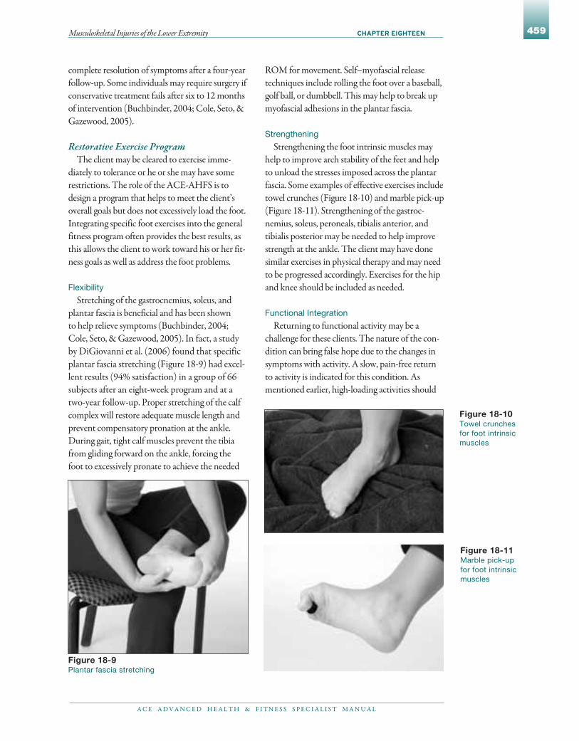
A C E A D v A n C E D H E A L T H & F I T n E S S S P E C I A L I S T M A n u A L
Musculoskeletal Injuries of the Lower Extremity Chapter eighteen 459
ROM for movement. Self–myofascial release techniques include rolling the foot over a baseball, golf ball, or dumbbell. This may help to break up myofascial adhesions in the plantar fascia.
Strengthening
Strengthening the foot intrinsic muscles may help to improve arch stability of the feet and help to unload the stresses imposed across the plantar fascia. Some examples of effective exercises include towel crunches (Figure 18-10) and marble pick-up (Figure 18-11). Strengthening of the gastroc-nemius, soleus, peroneals, tibialis anterior, and tibialis posterior may be needed to help improve strength at the ankle. The client may have done similar exercises in physical therapy and may need to be progressed accordingly. Exercises for the hip and knee should be included as needed.
Functional Integration
Returning to functional activity may be a challenge for these clients. The nature of the con-dition can bring false hope due to the changes in symptoms with activity. A slow, pain-free return to activity is indicated for this condition. As mentioned earlier, high-loading activities should
complete resolution of symptoms after a four-year follow-up. Some individuals may require surgery if conservative treatment fails after six to 12 months of intervention (Buchbinder, 2004; Cole, Seto, & Gazewood, 2005).
Restorative Exercise Program The client may be cleared to exercise imme-
diately to tolerance or he or she may have some restrictions. The role of the ACE-AHFS is to design a program that helps to meet the client’s overall goals but does not excessively load the foot. Integrating specific foot exercises into the general fitness program often provides the best results, as this allows the client to work toward his or her fit-ness goals as well as address the foot problems.
Flexibility
Stretching of the gastrocnemius, soleus, and plantar fascia is beneficial and has been shown to help relieve symptoms (Buchbinder, 2004; Cole, Seto, & Gazewood, 2005). In fact, a study by DiGiovanni et al. (2006) found that specific plantar fascia stretching (Figure 18-9) had excel-lent results (94% satisfaction) in a group of 66 subjects after an eight-week program and at a two-year follow-up. Proper stretching of the calf complex will restore adequate muscle length and prevent compensatory pronation at the ankle. During gait, tight calf muscles prevent the tibia from gliding forward on the ankle, forcing the foot to excessively pronate to achieve the needed
Figure 18-9Plantar fascia stretching
Figure 18-10Towel crunches for foot intrinsic muscles
Figure 18-11 Marble pick-up for foot intrinsic muscles
Chapter eighteen Musculoskeletal Injuries of the Lower Extremity
A C E A D v A n C E D H E A L T H & F I T n E S S S P E C I A L I S T M A n u A L
460
may include both an inflammatory and degenera-tive process of the tendon.
Mechanism of Injury various intrinsic and extrinsic factors are
associated with this condition. Intrinsic factors include age, bodyweight, pes cavus, pes planus, leg-length discrepancies, and lateral ankle instabil-ity. Extrinsic factors include errors in training, prior injuries, poor footwear, muscle weakness, and poor flexibility. (Paavola et al., 2002; Kader et al., 2002; Mazzone, 2002). The extrinsic factors are typically responsible for acute tendon trauma. Overuse and chronic injuries are often multifacto-rial and include a combination of intrinsic and extrinsic factors (Paavola et al., 2002).
Signs and Symptoms Individuals often complain of pain that is 0.75
to 2.25 inches (2 to 6 cm) above the tendon inser-tion into the calcaneus. The typical pattern is initial morning pain that is “sharp” or “burning,” as well as pain with more vigorous activity. Rest will often alleviate the pain, but as the condition becomes worse the pain becomes more constant and begins to interfere with activities of daily living (Mazzone, 2002; Brotzman & Wilk, 2003).
Precautions Clients with this condition are encouraged
to stop all aggravating activity and seek proper treatment for the condition. High-loading activities such as jumping, running, and stair climbing should be avoided until the condition has improved.
Early InterventionEarly intervention includes controlling pain
and inflammation by using modalities (e.g. ice, ultrasound), rest, and oral anti-inflammatory medication (Mazzone, 2002). Management of the condition may include modified rest and the addressing of specific risk factors. Modified rest allows the injured body part to rest while the client is exercising the uninjured parts of the body (e.g., using an upper-body ergometer). Proper training techniques, losing weight, proper foot-wear, orthotics, strengthening, and stretching can
be limited to avoid further exacerbation of the injury. With these clients, balance may be an issue and should be addressed, if needed. Low-loading cardiovascular activities such as biking, ellipti-cal training, or water aerobics are preferred over higher-loading activities such as running.
Achilles TendinopathyInjury to the Achilles tendon is common in
athletes and the active population. The prevalence of the condition is highest among runners, gym-nasts, and dancers. Other sports where this injury is common include track and field, volleyball, basketball, and soccer. Typically, older athletes are more affected by the condition than teens or children (Mazzone, 2002). This condition can eventually lead to rupture if not addressed appro-priately. Achilles ruptures primarily occur in males 30 to 50 years of age. The prevalence rate has increased in recent years due to the fact that more people are exercising (Mazzone, 2002).
The classification of Achilles injuries has been quite confusing due to the many names given to the condition. Terms commonly used in the past, including Achilles tendinitis, tenosynovi-tis, and tendonosis, have become questionable due to subsequent findings (Paavola et al., 2002; Maffulli, Khan, & Puddu, 1998; Mazzone, 2002). These studies have not found any clear sign of an inflammatory reaction in the tendon, which is often described as tendinitis and tenosynovitis. Researchers do acknowledge that there may be a prior inflammatory reaction, but no evidence has been found to date. Tendonosis or tendon degeneration is often used, but may be incorrect if not confirmed by the proper medical testing, such as biopsy, radiographic imaging, or surgical exploration (Paavola et al., 2002; Maffulli, Khan, & Puddu, 1998). Tendonosis causes a diffuse thickening of the tendon with no evidence of inflammation. This can develop from repeated microtrauma, aging, and vascular problems (Mazzone, 2002).
This confusion has led to the term Achilles tendinopathy. Maffulli, Khan, and Puddu (1998) suggested this term to describe the combination of pain, swelling, and poor function that accompa-nies this condition. Thus, Achilles tendinopathy

A C E A D v A n C E D H E A L T H & F I T n E S S S P E C I A L I S T M A n u A L
Musculoskeletal Injuries of the Lower Extremity Chapter eighteen 461
Kotsko, 2007). Therefore, progressively loading the Achilles tendon with eccentric activity can benefit the client, but may be even more benefi-cial when combined with other interventions. Examples of eccentric activity include slowly lowering the calf while standing on a step or per-forming a single-leg squat with an emphasis on slowly lowering the leg.
Functional Integration
Returning to basic functional and sports activities may be challenging for these clients. The nature of the condition can bring changes in symptoms with activity. A gradual, pain-free return to activity is indicated for this condition. Modifications in training techniques and the training environment should be addressed, with an emphasis on client education. CKC exercise combined with eccentric loading can progres-sively challenge the Achilles tendon, but should not create pain. Any deficits in balance should be addressed as needed. Simply adding an unstable surface such as a foam pad to CKC exercises can challenge the client’s balance (see Tables 18-1 and 18-2). Cardiovascular activity should be progressed with caution. Low-loading activity should precede higher-loading activity to avoid pain or reinjury.
Shin Splints“Shin splints” is a general term used to describe
exertional leg pain (Brotzman & Wilk, 2003). Shin splints are typically classified as two specific conditions: medial tibial stress syndrome (MTSS) and anterior shin splints (Figure 18-12).
MTSS, also called posterior shin splints, is an overuse injury that occurs in the active popula-tion. MTSS is an exercise-induced condition that is often triggered by a sudden change in activity (Brotzman & Wilk, 2003; Anderson, Hall, & Parr, 2008). MTSS is actually periostitis, or inflammation of the periosteum (connec-tive tissue covering) of the bone. Originally, this condition was thought to be caused by posterior tibial tendinitis. It has since been related to a traction periostitis at the distal insertion of the soleus muscle or from the flexor digitorum longus muscle (Brotzman & Wilk, 2003; Anderson,
help alleviate pain and prevent progression of the condition (Paavola et al., 2002; Mazzone, 2002). Also, the client may be sent to physical therapy to address the factors mentioned earlier. If conserva-tive treatment fails after four to six months, then surgical intervention is indicated (Brotzman & Wilk, 2003; Mazzone, 2002).
Restorative Exercise Program The client may be cleared to exercise imme-
diately to tolerance, or he or she may have some activity restrictions. The role of the ACE-AHFS is to design a program that helps to meet the client’s overall goals but does not exacerbate the condition. Consulting the medical doctor and physical therapist can give key information about how the client is responding to treat-ment and what he or she is currently doing for exercise. The following sections summarize strategies for management of this condition.
Flexibility
Restoring proper length and elasticity to the calf muscles can reduce strain to the muscle-tendon unit and decrease symptoms (Kader et al., 2002). Studies regarding stretching have suggested that stretching does stimulate the healing response (Mazzone, 2002). The goal of stretching should be to restore general lower-body flexibility, with an emphasis on calf mobility. The client should be cautioned to stretch to tolerance and avoid overexertion. Overstretching of the Achilles tendon can cause irritation to the muscle-tendon unit and should be avoided.
Strengthening
Eccentric strengthening of the calf complex has been shown to be beneficial for relieving symptoms. In fact, Wasielewski and Kotsko (2007) conducted a systematic review of research from 1980 to 2006 on eccentric strengthening of the calf muscles. Their analysis revealed that eccentric exercise may reduce pain and improve strength in Achilles tendinopathy. However, eccentric training has not been shown to be superior over other forms of therapeutic inter-ventions for this condition (Wasielewski &
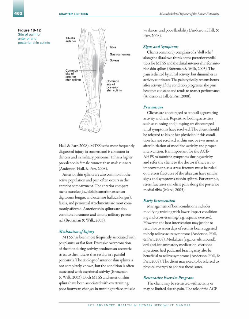
Chapter eighteen Musculoskeletal Injuries of the Lower Extremity
A C E A D v A n C E D H E A L T H & F I T n E S S S P E C I A L I S T M A n u A L
462
weakness, and poor flexibility (Anderson, Hall, & Parr, 2008).
Signs and Symptoms Clients commonly complain of a “dull ache”
along the distal two thirds of the posterior medial tibia for MTSS and the distal anterior shin for ante-rior shin splints (Brotzman & Wilk, 2003). The pain is elicited by initial activity, but diminishes as activity continues. The pain typically returns hours after activity. If the condition progresses, the pain becomes constant and tends to restrict performance (Anderson, Hall, & Parr, 2008).
PrecautionsClients are encouraged to stop all aggravating
activity and rest. Repetitive loading activities such as running and jumping are discouraged until symptoms have resolved. The client should be referred to his or her physician if this condi-tion has not resolved within one or two months after initiation of modified activity and proper intervention. It is important for the ACE-AHFS to monitor symptoms during activity and refer the client to the doctor if there is no improvement, as a stress fracture must be ruled out. Stress fractures of the tibia can have similar signs and symptoms as shin splints. For example, stress fractures can elicit pain along the posterior medial tibia (Metzl, 2005).
Early InterventionManagement of both conditions includes
modifying training with lower-impact condition-ing and cross-training (e.g., aquatic exercise). However, the best intervention may just be to rest. Five to seven days of rest has been suggested to help relieve acute symptoms (Anderson, Hall, & Parr, 2008). Modalities (e.g., ice, ultrasound), oral anti-inflammatory medication, cortisone injections, heel pads, and bracing may also be beneficial to relieve symptoms (Anderson, Hall, & Parr, 2008). The client may need to be referred to physical therapy to address these issues.
Restorative Exercise Program The client may be restricted with activity or
may be limited due to pain. The role of the ACE-
Hall, & Parr, 2008). MTSS is the most frequently diagnosed injury in runners and is common in dancers and in military personnel. It has a higher prevalence in female runners than male runners (Anderson, Hall, & Parr, 2008).
Anterior shin splints are also common in the active population and pain often occurs in the anterior compartment. The anterior compart-ment muscles (i.e., tibialis anterior, extensor digitorum longus, and extensor hallucis longus), fascia, and periosteal attachments are most com-monly affected. Anterior shin splints are also common in runners and among military person-nel (Brotzman & Wilk, 2003).
Mechanism of Injury MTSS has been most frequently associated with
pes planus, or flat foot. Excessive overpronation of the foot during activity produces an eccentric stress to the muscles that results in a painful periostitis. The etiology of anterior shin splints is not completely known, but the condition is often associated with exertional activity (Brotzman & Wilk, 2003). Both MTSS and anterior shin splints have been associated with overtraining, poor footwear, changes in running surface, muscle
Figure 18-12Site of pain for anterior and posterior shin splints
Tibialis anterior
Tibia
Soleus
Common site of anteriorshin splints Common
site of posteriorshin splints
Gastrocnemius

A C E A D v A n C E D H E A L T H & F I T n E S S S P E C I A L I S T M A n u A L
Musculoskeletal Injuries of the Lower Extremity Chapter eighteen 463
the client is pain free. Balance exercises can also be integrated into the program as needed.
Cross-training can be utilized throughout the program to maintain adequate cardiovas-cular fitness. Examples of low-impact activities include water jogging, stationary biking, and elliptical training. Clients should avoid run-ning, jumping, and shock-loading activities that stress the affected region.
Muscle Strains
Muscle strains are injuries in which a muscle works beyond its capac-ity, resulting in a tear of the muscle
fibers. In mild strains, the client may report
AHFS should be to slowly progress the client back to full unrestricted activity without exacerbat-ing the symptoms. Cross-training to maintain adequate levels of fitness is indicated in the early stages. Consulting the client’s physician and physi-cal therapist can give key information about how the client is responding to treatment and what he or she is currently doing for exercise. The follow-ing sections summarize strategies for management of this condition.
Flexibility
Pain-free stretching of the calf muscles, espe-cially the soleus, has been shown to be effective in relieving symptoms related to MTSS. Stretching the anterior compartment has been shown to help relieve the symptoms of anterior shin splints (Figures 18-13 and 18-14) (Anderson, Hall, & Parr, 2008; Brotzman & Wilk, 2003). The goal of stretching should be to restore proper length and elasticity to the muscle and reduce strain in the muscle-tendon unit (Kader et al., 2002). A general lower-body stretching program should accompany more specific stretching to address any secondary muscle-length deficits that may affect the foot and ankle.
Strengthening
Rest and modified activity are the primary interventions for symptom relief. However, there may be some residual strength deficits in the muscles that control the ankle. Targeting the muscles that control the ankle is the goal, especially the calf and anterior tibialis muscles. Exercises for the hip and knee should be added as needed.
Functional Integration
A gradual return to athletic and sports activity is best for these clients. Strengthening exercises should be related to the client’s func-tional goals, but needs to be low-impact to avoid any excessive stress. Both OKC and CKC exercises can be integrated throughout the program as tolerated by the client. Progression should begin with low-loading activities and systematically progressed to higher-loading activities such as jumping or running, as long as
Figure 18-13Sitting stretch for the anterior compartment
Figure 18-14Standing “toe drag” stretch for the anterior compartment

Chapter eighteen Musculoskeletal Injuries of the Lower Extremity
A C E A D v A n C E D H E A L T H & F I T n E S S S P E C I A L I S T M A n u A L
464
Risk factors include muscle fatigue, fluid and electrolyte depletion, forced knee extension while the foot is dorsiflexed, or forced dorsiflex-ion while the knee is extended.
Management If a muscle strain does occur, all aggravating
activity should be stopped and the RICE prin-ciple (rest, ice, compression, elevation) should be applied immediately. The ACE-AHFS should administer basic first-aid procedures and then refer the client to the appropriate health-care professional. It is beyond the scope of the ACE-AHFS to attempt to diagnose a client’s problem and make decisions regarding his or her care.
Structural Abnormalities
It is important for the ACE-AHFS to understand that certain structural changes in the lower-extremity kinetic chain have
been associated with various musculoskeletal conditions. First, pes planus, or overprona-tion, is considered to be a flat mobile foot, which offers little structural support. It has been associated with plantar fasciitis, Achilles tendinopathy, posterior tibial tendinitis, shin splints, ITBFS, and patellofemoral pain. Second, pes cavus, or high arches, is consid-ered to be a rigid foot that offers little shock absorption. It has been associated with ITBFS, plantar fasciitis, stress fractures of the tarsal and metatarsal bones of the foot, and peroneal tendinitis. Third, leg-length discrepancies have been associated with hip problems such as trochanteric bursitis and ITB syndrome (Brotzman & Wilk, 2003; Manske, 2006; Anderson, Hall, & Parr, 2008).
It is important for the ACE-AHFS to note any structural deviations, as they may be con-tributing factors to the pathologies discussed in this chapter. Attempting to correct these biomechanical deviations is beyond the scope of the ACE-AHFS. If an ACE-AHFS suspects that a deviation may be contributing to an undiagnosed problem, then referral to the appropriate medical professional is indicated.
tightness or tension (Anderson, Hall, & Parr, 2008). In more severe cases, the client may report feeling a sudden “tear” or “pop” that leads to immediate pain and weakness in the muscle. Swelling, discoloration (ecchymosis), and loss of function often occur after the injury (Anderson, Hall, & Parr, 2008). Strains of the lower extremity primarily occur in the ham-strings, groin muscles, and calves.
Muscle strains of the hamstring group are often caused by a severe stretch to the muscle or a rapid, forceful contraction (e.g., during sprinting). The hamstrings have the highest fre-quency of strains in the body; hamstring strains are common in running and jumping sports (Anderson, Hall, & Parr, 2008). The client will often report a “sharp” posterior thigh pain that occurs after insult. Pain may also be felt with contraction or stretching of the muscle. Risk factors include poor flexibility; poor posture; muscle imbalance among the gluteals, quad-riceps, and hamstrings; improper warm-up; errors in training; and prior injury (Anderson, Hall, & Parr, 2008).
Groin or adductor strains are common in sports such as ice hockey and figure skating that require explosive acceleration, deceleration, and change of direction. With injury, the client may report an initial “pull” of the groin muscles, fol-lowed by intense pain and loss of function. Pain may be felt with contraction of the muscles with the hip in adduction, passive stretching with the hip in abduction, or when crossing the leg over the midline of the body. In more severe cases, increased pain and weakness occurs, which may limit specific motions. Jogging straight may be tolerable, but any side-to-side motion tends to elicit pain. Muscle imbalance between the adductors and abductors is the most prevalent risk factor for this type of injury (Anderson, Hall, & Parr, 2008).
Calf strains are common in most running and jumping sports. With injury, the client will often report a sudden and painful “tear-ing” sensation in the medial muscle belly or at the junction between the muscle belly and the Achilles tendon. Pain, swelling, ecchymosis, and loss of function often occur after the injury.

A C E A D v A n C E D H E A L T H & F I T n E S S S P E C I A L I S T M A n u A L
Musculoskeletal Injuries of the Lower Extremity Chapter eighteen 465
Fitness AssessmentAt the time of fitness assessment, the client
demonstrated problems with basic movements. In particular, the client demonstrated immediate pain with getting out of the chair and walking down stairs. At this time, the session was stopped and no further action was taken.
ManagementInspection of the knee revealed it to be swollen
and painful to bend. The client was immediately referred back to the medical doctor for further evaluation. The ACE-AHFS recommended that the client use the RICE principle until he or she sees the doctor and receives proper clearance to resume activity. The goal was to make sure the client did not re-injure the ACL or damage any other structures. upon hearing back from his patient, the doctor diagnosed him as having patel-lar tendinitis and general joint irritation. The client is restricted from fitness activity for two weeks. upon clearance to resume activity, the fol-lowing actions should be taken with this client:
Client education: CKC vs. OKC exercises, •overtraining, injury recognition Training modification: Reestablish program •goals and training schedule Fitness evaluation: Baseline data for •flexibility, muscular strength and endurance, body composition, and cardiovascular fitnessExercise program: Slow progression of sets, •repetitions, and time 3Flexibility: Focus on stretching/
myofascial release for the ITB complex, quadriceps, hamstrings, and calves
3Strengthening: Focus on the quadriceps and hamstrings, and general hip strengthening
3Balance: Progress to multiplane activity with a knee brace
3Functional: Focus on CKC exercises within pain-free limits with a progression toward sport-specific movements. After 16 weeks, begin basic agility and sport-specific movements.
Safety •3Brace worn during workouts 3Recommend icing after workouts
Case Studies
Case Study 1
Client History An ACE-AHFS was referred a 32-year-old
male recreational soccer player who had a right knee ACL repair 12 weeks ago with a patellar tendon graft. The mechanism of injury was a planting and twisting maneuver. The client has been in physical therapy for the past 12 weeks and treatment has included a combination of strengthening, cardiovascular, and balance exer-cises. The physical therapist has also been doing massage, stretching, and myofascial release. The physical therapist reports that the client has responded well to physical therapy and is highly motivated. He or she is on schedule with the physician’s protocol, but because of insurance constraints has been transitioned to fitness activity. Review of medications reveals the fol-lowing: vicodin® (pain) and Advil® as needed.
Client Interview upon meeting with the client for initial
assessment, the ACE-AHFS notices that he is highly motivated and immediately talks about returning to soccer. The client does have writ-ten clearance by the medical doctor, with the precautions of wearing a brace during activity and no running until 16 weeks. His health history reveals no major medical problems or comorbities. The client does have a history of recurrent right ankle sprains and occasional low-back pain. The client is an engineer and works at a desk most of the day. His fitness goals are to return to soccer, running, and weightlifting.
When asked about his knee, the client reveals that he has been getting anterior knee pain with increased swelling for the past two weeks while working out at the gym. The client is doing 45 minutes of cardiovascular exercise on the elliptical trainer and general upper-body strengthening. Further questioning reveals that he has been doing lunges, leg presses, and leg extensions three to five times per week, includ-ing during his physical therapy program.

Chapter eighteen Musculoskeletal Injuries of the Lower Extremity
A C E A D v A n C E D H E A L T H & F I T n E S S S P E C I A L I S T M A n u A L
466
Movement screen: Limited due to weakness •and fall risk Flexibility: Tightness in the ITB complex, •hamstrings, and calves Functional testing: Weakness noted in the •hip with sit-to-stand, side-stepping, quarter lunges, and partial-ROM step-ups Cardiovascular testing: unable to do •submaximal testing on the bike or step due to weakness and deconditioning Muscular strength: Able to establish baseline •weights on exercise machines and resistive bands
ManagementDue to the client’s low functional level, a
modified restorative program should be cre-ated that focuses on hip and abdominal core strengthening, balance, and cardiovascular endurance. The following actions should be taken with this client.
Client education•3 Basic training principles, CKC vs. OKC
exercises, injury recognition3 Precautions and program modifications
for osteoporosis and hypertension Training modification: Reestablish program •goals and training schedule Fitness evaluation: Baseline data for flexibility, •muscular strength and endurance, body composition, and cardiovascular fitness when tolerated by the clientExercise program: Slow progression of sets, •repetitions, and time 3Flexibility: Focus on stretching/myofascial
release for the hip muscles, ITB complex, quadriceps, hamstrings, and calves
3Strengthening: Focus on the gluteals, hip external rotators, quadriceps, and hamstrings
3Balance: Slowly progress multiplane activity under close supervision
3Functional: Focus on CKC exercises within pain-free limits with a progression toward multiplanar functional activities • Basic movements: Sit-to-stand• Gait: Walking over even and uneven
terrain (e.g., cement vs. grass)
3Monitor for increased symptoms: redness, pain, and swelling
3Monitor for signs and symptoms of overtraining
Case Study 2
Client History An ACE-AHFS is working with a 72-year-old
female who is three months post-operative after a left total hip replacement via the posterior lateral approach. The woman had a history of hip arthri-tis and finally elected to have the procedure. She has been attending physical therapy for strengthen-ing, endurance, and balance activities. The physical therapist reports that the client is doing well with ROM but still has hip weakness that makes functional and balance activities difficult. Due to insurance constraints, the client has been transi-tioned to fitness activity.
Client Questions upon meeting with the client, she brings in
written clearance from the medical doctor with precautions noted to avoid high-impact activity such as running. Her health history reveals hyper-tension, osteoporosis, and osteoarthritis. The client did have a left TKR three years ago with no reported problems. Review of medications are as follows: diuretic (hypertension), Fosamax® (osteo-porosis), and Celebrex® (osteoarthritis). The client is retired but volunteers at the local school, where she is on her feet most of the day. Her fitness goals are to return to walking and swimming.
When asked about her right hip, the client reveals having mild to moderate pain after physi-cal therapy that goes away after icing. The client reports being cleared by the medical doctor to resume full ROM of the hip with no restrictions. Further questioning reveals that she is still having trouble with functional movements, such as sit-to-stand actions and picking up objects.
Fitness AssessmentAt this time, the fitness assessment was limited
due to the global hip weakness that the client was having. The following information was obtained from the modified assessment:

A C E A D v A n C E D H E A L T H & F I T n E S S S P E C I A L I S T M A n u A L
Musculoskeletal Injuries of the Lower Extremity Chapter eighteen 467
is required to have a deeper knowledge about non-operative and post-operative musculosk-eletal conditions. This chapter has focused on the recognition, management, and restorative exercise guidelines for common musculoskel-etal injuries and post-operative conditions of the lower extremity. The reader is encouraged to continue the study of these conditions to effectively design safe restorative programs for different conditions and populations.
Safety •3Precautions for osteoporosis and
hypertension 3Choosing exercises that are safe 3Recommend icing after workouts and
monitor for change in symptoms
Summary
Due to the changes in the healthcare system, the role of fitness profession-als has expanded. The ACE-AHFS

Chapter eighteen Musculoskeletal Injuries of the Lower Extremity
A C E A D v A n C E D H E A L T H & F I T n E S S S P E C I A L I S T M A n u A L
468
Cohen, Z. et al. (2001). Patellofemoral stresses during open and closed kinetic chain exercises: An analysis using computer simulation. American Journal of Sports Medicine, 29, 480–487.
Colby, S. et al. (2002). Electromyographic and kinematic analysis of cutting maneuvers: Implications for anterior cruciate ligament injury. American Journal of Sports Medicine, 28, 2, 234–240.
Cole, C., Seto, C., & Gazewood, J. (2005). Plantar fasciitis: Evidence-based review of diagnosis and therapy. American Family Physician, 72, 11, 2237–2243.
DiGiovanni, B.F. et al. (2006). Chronic plantar fasciitis: A prospective clinical trial with two-year follow-up. Journal of Bone & Joint Surgery, 88-A, 8, 1–15.
Dixit, S. et al. (2007). Management of patellofemoral pain syndrome. American Family Physician, 75,194–204.
Eriksson, K. et al. (2001). A comparison of quadruple semitendinosus and patellar tendon grafts in reconstruction of the anterior cruciate ligament. Journal of Bone Joint Surgery, 83, 348–354.
Evans, T., Hertel, J., & Sebastianelli, W. (2004). Bilateral deficits in postural control following lateral ankle sprain. Foot & Ankle International, 25, 11, 833–839.
Foye, P.M. & Stitik, T.P. (2006). Trochantaric bursitis. E-Medicine Online Journal (Web MD), Dec 21, 1–14. www.emedicine.com.
Freedman, K.B. et al. (2003). Arthroscopic anterior cruciate ligament reconstruction: A meta-analysis comparing patellar tendon and hamstring tendon autografts. American Journal of Sports Medicine, 31, 1, 2–11.
Friel, K. et al. (2006). Ipsilateral hip abductor weakness after inversion ankle sprain. Journal of Athletic Training, 41, 1, 74–78.
Garrick, J.G. (1982). Epidemiologic perspective. Clinical Sports Medicine, 1, 13–18.
Goldstein, J. & Zuckerman, J.D. (2000). Selected orthopedic problems in the elderly. Rheumatic Disease Clinics of North America, 26, 3, 593–616.
Griffin, L.Y. (2000). Noncontact anterior cruciate ligament injuries: Risk factors and prevention strategies. Journal of American Academy of Orthopedic Surgeons, 8, 141–150.
Hinton, R. et al. (2002). Osteoarthritis: Diagnosis and therapeutic considerations. American Family Physician, 65, 841–848.
Ivins, D. (2006). Acute ankle sprains: An update. American Family Physician, 74, 1714–1720.
Juhn, M. (1999). Patellofemoral pain syndrome: A review and guidelines for treatment. American Family Physician, 60, 2012–2022.
ReferencesAglietti, P. et al. (1994). Patellar tendon versus doubled semitendinosus and gracilis tendons for anterior cruciate ligament reconstruction. American Journal of Sports Medicine, 22, 211–218.
Allen, C. (2007). ACL injury: Does it require surgery? American Academy of Orthopedic Surgeons. www.orthoinfo.aaos.org.
American Academy of Orthopedic Surgeons (2007).Your orthopedic connection: Total knee replacement. www.orthoinfo.aaos.org.
Anderson, M.K., Hall, S.J., & Parr, G.P. (2008).Foundations of Athletic Training: Prevention, Assessment, and Management (4th ed.). Baltimore, Md.: Lippincott Williams & Wilkins.
Arendt, E. & Dick, R. (1995). Knee injury patterns among men and women in collegiate basketball and soccer: NCAA data and review of literature. American Journal of Sports Medicine, 23, 6, 694–701.
Arthritis Foundation & Centers for Disease Control and Prevention (1999). National Arthritis Action Plan: A Public Health Strategy. www.cdc.gov/nccdphp/pdf/naap.pdf.
Baker, B. & Lubowitz, J. (2006). Meniscal injuries. E-Medicine Online Journal (Web MD), 1–14. www.emedicine.com.
Balduini, F.C. & Tetzlaff, J. (1982). Historical perspectives on injuries of the ligaments of the ankle. Clinical Sports Medicine, 1, 1, 3–12.
Bhagia, S.M. et al. (2006). Meniscal tears. E-Medicine Online Journal (Web MD), 1–14. www.emedicine.com.
Bierma-Zeinstra, S. et al. (1999). Validity of American College of Rheumatology criteria for diagnosing hip osteoarthritis in primary care research. Journal of Rheumatology, 26, 1129–1133.
Boden, B.P. & Garrett, W.E. (1996). Mechanism of injuries to the anterior cruciate ligament. Medicine & Science in Sports & Exercise, 28, 5, 156–168.
Bonelli, S. (2001). Aquatic Exercise, San Diego, Calif.: American Council on Exercise.
Brotzman, B. & Wilk, K. (2003). Clinical Orthopedic Rehabilitation (2nd ed.). St. Louis, Mo.: Mosby.
Brown, D.W. (2007). Anterior cruciate ligament reconstruction techniques. Orthopedic Associates. www.orthoassociates.com.
Buchbinder, R. (2004). Plantar fasciitis. New England Journal of Medicine, 350, 21, 2159–2167.
Buckwalter, J.A. & Martin, J.A. (2006). Osteoarthritis. Advanced Drug Delivery Reviews, 58, 150–167.
Cioppa-Mosca, J.M. et al. (2006). Postsurgical Rehabilitation Guidelines for the Orthopedic Clinician. St. Louis, Mo.: Mosby.

A C E A D v A n C E D H E A L T H & F I T n E S S S P E C I A L I S T M A n u A L
Musculoskeletal Injuries of the Lower Extremity Chapter eighteen 469
Naslund, J. et al. (2006). Comparison of symptoms and clinical findings in subgroups of individuals with patellofemoral pain. Physiotherapy Theory & Practice, 22, 5, 105–118.
Nikolaou, P. et al. (1996). Anterior cruciate ligament allograft transplantation: Long-term function, histology, revascularization, and operative technique. American Journal of Sports Medicine, 14, 5, 348–360.
Noyes, F.R. & Barber-Westin, S.D. (1996). Reconstruction of the anterior cruciate ligament with human allograft: Comparison of early and later results. Journal of Bone and Joint Surgery,78A, 524–537.
Paavola, M. et al. (2002). Current concepts review: Achilles tendinopathy. Journal of Bone & Joint Surgery, 84-A, 11, 2062–2076.
Pearl, A.J. (1993). American Orthopedic Society for Sports Medicine: The Athletic Female. Champaign, Ill.: Human Kinetics.
Perlman, M. et al. (1987). Inversion lateral ankle trauma: Differential diagnosis, review of the literature, and prospective study. Journal of Foot Surgery, 26, 95–135.
Robinson, R.L. & Nee, R.J. (2007). Analysis of hip strength in females seeking physical therapy treatment for unilateral patellofemoral pain syndrome. Journal of Orthopedic and Sports Physical Therapy, 37, 5, 232–238.
Sachs, R. et al. (1989). Patellofemoral problems after anterior cruciate ligament reconstruction. American Journal of Sports Medicine, 17, 6, 760–765.
Spindler, K.P. et al. (2004). Anterior cruciate ligament reconstruction autograft choice: Bone-tendon-bone versus hamstring: Does it really matter? A systematic review. American Journal of Sports Medicine, 32, 8, 1986–1995.
U.S. Department of Health and Human Services; Centers for Disease Control and Prevention; National Center for Health Statistics (2004). Number of Patients, Number of Procedures, Average Patient Age, Average Length of Stay. National Hospital Discharge Survey 1998–2004. www.cdc.gov/nchs/
Van Deun, S. et al. (2007). Relationship of chronic ankle instability to muscle activation patterns during the transition from double-leg to single-leg stance. American Journal of Sports Medicine, 35, 274–281.
Wasielewski, N.J. & Kotsko, K.M. (2007). Does eccentric exercise reduce pain and improve strength in physically active adults with symptomatic lower extremity tendinosis? A systematic review. Journal of Athletic Training, 42, 3, 409–422.
Witvrouw, E. et al. (2000). Open versus closed kinetic chain exercises for patellofemoral pain: A prospective, randomized study. The American Journal of Sports Medicine, 28, 687–694.
Kader, D. et al. (2002). Achilles tendinopathy: Some aspects of basic science and clinical management. British Journal of Sports Medicine, 36, 239–249.
Kaminski, T.W. & Hartsell, H.D. (2002). Factors contributing to chronic ankle instability: A strength perspective. Journal of Athletic Training, 37, 4, 394–405.
Kennon, R. et al. (2004). Anterior approach for total hip arthroplasty: Beyond the minimally invasive technique. Journal of Bone and Joint Surgery, 86, 91–97.
Kirkendall, D.T. & Garrett, W.E. (2000). The anterior cruciate ligament enigma: Injury mechanisms and prevention. Clinical Orthopedics, 372, 64–68.
Kisner, C. & Colby, L. (2007). Therapeutic Exercise: Foundations and Techniques (5th ed.). Philadelphia: F.A. Davis Company.
Lawrence R.C. et al. (1998). Estimates of the prevalence of arthritis and selected musculoskeletal disorders in the United States. Arthritis and Rheumatology, 41, 778–799.
Lievense, A., Bierma-Zeinstra, S., & Schouten, B. (2005). Prognosis of trochantaric pain in primary care. British Journal of General Practice, 55, 512, 199–204.
Little, H. (1979). Trochanteric bursitis: A common cause of pelvic girdle pain. Canadian Medical Association Journal, 120, 456–458.
Maffulli, N., Khan, K.M., & Puddu, G. (1998). Overuse tendon conditions: Time to change a confusing terminology. Arthroscopy, 14, 840–843.
Manske, R.C. (2006). Postsurgical Orthopedic Sports Rehabilitation: Knee and Shoulder. St. Louis, Mo.: Mosby.
Marder, R.A. et al. (1994). Prospective evaluation of arthroscopically assisted anterior cruciate ligament reconstruction: Patellar tendon versus semitendinosus and gracilis tendons. American Journal of Sports Medicine,19, 2478–2484.
Martinez, J.M. & Honsik, K. (2006). Iliotibial band syndrome. E-Medicine Online Journal (Web MD). Dec 6, 1–14. www.emedicine.com.
Matta, J.M., Shahrdar, C., & Ferguson, T. (2005). Single-incision anterior approach for total hip arthroplasty on an orthopedic table. Clinical Orthopedics and Related Research, 441,115–124.
Maxey, L. & Magnusson, J. (2007). Rehabilitation for the Post Surgical Orthopedic Patient (2nd ed.). St. Louis, Mo.: Mosby.
Mazzone, M. (2002). Common conditions of the achilles tendon. American Family Physician, 65, 1805–1810.
Metzl, J. (2005). A case-based look at shin splints. Patient Care, Nov, 39–46.
Chapter eighteen Musculoskeletal Injuries of the Lower Extremity
A C E A D v A n C E D H E A L T H & F I T n E S S S P E C I A L I S T M A n u A L
470
Wolgin, M. et al. (2004). Conservative treatment of plantar heel pain: Long-term follow-up. Foot & Ankle International, 5, 97–102.
Yu, B. et al. (2002). Lower extremity motor control-related and other risk factors for noncontact anterior cruciate ligament injuries. Instructional Course Lecture, 51, 315–325
Suggested ReadingAnderson, M.K., Hall, S.J., & Parr, G.P. (2008). Foundations of Athletic Training: Prevention, Assessment, and Management (4th ed.). Baltimore: Lippincott Williams & Wilkins.
Brotzman, B. & Wilk, K. (2003). Clinical Orthopedic Rehabilitation (2nd ed.). St. Louis, Mo.: Mosby.
Kisner, C. & Colby, L. (2007). Therapeutic Exercise: Foundations and Techniques (5th ed.). Philadelphia: F.A. Davis Company.
Manske, R.C. (2006). Postsurgical Orthopedic Sports Rehabilitation: Knee and Shoulder. St. Louis, Mo.: Mosby

Acromioclavicular Joint Injuries
Shoulder Instability
Rotator Cuff Pathology
Lateral Epicondylitis
Medial Epicondylitis
Hand and Wrist Injuries
Case Studies Case Study 1 Case Study 2
Summary
In This Chapter
A b o u t T h e A u t h o r Michael Levinson, P.T., CSCS, has been at the Hospital for
Special Surgery since 1984, where he is a clinical supervisor
of the Rehabilitation Department. Levinson is also a physical
therapist for the New York Mets Baseball Club. He is certified by
the National Strength and Conditioning Association as a Strength
and Conditioning Specialist. In addition, Levinson is on the faculty
of the Columbia University Physical Therapy Program.

C h A p T e r 1 9
therapist and physician can be extremely valuable in prevent-ing re-injury. Symptoms such as recurrent pain, instability, or loss of ROM should be communicated.
Acromioclavicular Joint Injuries
The acromioclavicular joint consists of the articulation of the distal end of the clavicle and the acromion, which is a portion of the scapula. The joint is covered
with cartilage and is stabilized by the coracoclavicular and acromioclavicular ligaments. The clavicle rotates upward 40 to 50 degrees as the arm is fully elevated. Without rotation of the clavicle, it has been demonstrated that the arm can only be elevated to approximately 110 degrees. Injuries to the acro-mioclavicular joint can present as either traumatic or chronic. The most common mechanism of injury is a direct force on the point of the shoulder or a fall on an outstretched arm. If the clavicle does not fracture, the acromion is driven inferiorly and medially in relation to the clavicle. The ligaments are then stretched or torn, depending on the severity of the injury. This injury is often referred to as a “separated shoulder” and occurs commonly in contact sports such as football, hockey, lacrosse, and rugby. The injury is classified by six different degrees of severity (Rockwood & Matsen, 2004). Patients with acro-mioclavicular joint pathology often present with pain during
passive horizontal adduction or have pain during an O’Brien active compression test, which consists of resistance of the shoulder in flexion, internal rotation, and horizontal adduc-tion (O’Brien et al., 1998) (Figure 19-1). More severe cases will present with a “step-off” deformity where the separation of the clavicle and the acromion can be seen.
Treatment varies greatly and is partially dependent on the severity of the injury. The current trend is toward conserva-tive treatment without surgery. However, in certain severe cases, a surgical procedure is the treatment of choice. Surgical procedures vary greatly and are utilized for individuals that present with persistent pain, joint instability, or an undesir-able and visible deformity at the joint sight. Especially among the female population, cosmesis, or a concern for appearance, is often a rationale for surgery. In addition, athletes who perform overhead movements and individuals who perform a great deal of highly physical work may find conservative treatment unsatisfactory. There are numerous surgical tech-niques available. However, there is no true “gold standard.” Surgical treatment includes resection of the distal clavicle, ligament transfer, ligament reconstruction, and internal fixation. Patients undergoing clavicle resection often must modify certain activities such as push-ups and the bench press. Reconstructions that are successful can theoretically return
When training an individual following an upper-extremity musculoskeletal
injury, it is important that the ACE-certified Advanced Health & Fitness
Specialist (ACE-AHFS) is aware of several factors, including the mecha-
nism of injury, the structures involved, the healing constraints of the structures involved,
exacerbating activities, and range-of-motion (ROM) issues. Pain is the most important
guideline when designing a training program. Communication with the client’s physical
Musculoskeletal Injuries of the Upper Extremity
Michael Levinson

Chapter NINeteeN Musculoskeletal Injuries of the Upper Extremity
A C E A d v A n C E d H E A l T H & F I T n E S S S P E C I A l I S T M A n U A l
474
the individual to a normal exercise level. However, screws or pins utilized for internal fixation run the risk of breaking.
Injury to the acromioclavicular joint can lead to chronic degenerative changes at the joint. Abnormal mechanics or instability at the joint can result in a wearing away of the cartilage and arthrosis. In addition, people with poor mechanics who have done a great deal of bench pressing or push-ups can cause degenerative changes at the joint.
Pathomechanics following injury to this joint vary. First, there may be a loss of clavicle rotation, which can result in a loss of shoulder elevation. Secondly, the structural suspension of the entire shoulder girdle may be compromised. Most criti-cal are the pathomechanics of the scapula. The scapula provides a stable platform for shoulder motion and any deviation may result in other shoulder problems. Inferior and medial rotation of the scapula is often a consequence of this injury.
Conservative treatment of this injury varies, but the trend is moving away from extended periods of immobilization. Initially, an immobilizer may be utilized for pain control, reduction of muscle spasm, and reduction of soft-tissue damage. Reduction of the injury with strict immobilization for extended periods has not been demonstrated to be an effective treatment plan. Patient compli-ance has often been a limiting factor. Cryotherapy
for reduction of pain, swelling, and spasm is a key component of the early stages of recovery.
The goal of the initial stages of recovery is to restore pain-free ROM. Manske (2006) advises avoiding active-ROM and passive-ROM exercises in the supine position. The rationale is that the client’s body weight prevents scapula ROM and thus results in greater clavicle rotation, which may result in exacerbation of the injury. Seated or standing is the preferred position.
Strengthening should be initiated with submax-imal isometrics for abduction, flexion, extension, internal rotation, and external rotation. When progressing a strengthening program, several pre-cautions should be followed:
• Traction through the shoulder joint should be avoided or minimized. For example, when a client is performing shrugs or curls, the ACE-AHFS should provide a weight that can be controlled. Also, weights should not be carried around the gym. Beginning exer-cise with elastic resistance or tubing is often the safest choice.
• Resistive exercises in horizontal abduction or adduction should be avoided or minimized secondary to stress on the joint.
• When performing scapula strengthening exercises, extremes of scapula retraction and protraction should be avoided.
• Internal and external rotation exercises for the rotator cuff are tolerated best with the arm in adduction.
• Overhead resistive activity, such as the mili-tary press and incline bench press, should be minimized or avoided. These activities should be initiated only when the client is asymptomatic and has a good proximal strength base.
Shoulder Instability
Shoulder instability is a very common pathology. The glenohumeral joint consists of the head of the humerus and the glenoid
fossa, which is a portion of the scapula. Together they form a “ball and socket.” Unlike the true ball-and-socket joint of the hip formed primarily by bony structures, the motion at the glenohumeral joint is controlled by the capsule and ligaments
Figure 19-1O’Brien active compression test

A C E A d v A n C E d H E A l T H & F I T n E S S S P E C I A l I S T M A n U A l
Musculoskeletal Injuries of the Upper Extremity Chapter NINeteeN 475
that surround the joint and the four rotator cuff muscles. This joint requires a great deal of ROM to perform many athletic activities. For this reason, the glenohumeral joint is an inherently unstable joint. Shoulder instability can be a result of an acute, traumatic event such as a dislocation. It can also be a chronic condition that results from overuse activities, especially overhead activities such as when throwing or playing tennis or volleyball, where the shoulder experiences various forces related to acceleration and deceleration. These powerful repetitive activities can cause excessive laxity in the capsule and ligaments that surround the shoulder joint. In addition, certain individuals are born with congenital joint laxity, which may predispose them to shoulder instability.
When the head of the humerus actually comes out of the socket, it is considered a dislocation. At times, it will go back in by itself. However, it often has to be reduced by a physician in the emergency room or on the field. Resultant trauma can cause soft-tissue damage and muscle spasm. The shoul-der capsule and a structure called the labrum are often injured. The labrum is fibrocartilage that helps to increase the stability of the shoulder joint (levine, 2000). Bony damage or loss can also result from recurrent instability. In addition, with instability, the humerus may translate excessively, but not completely come out of the socket. This is referred to as a subluxation, which is often a chronic condition. The most common instability
is anterior. It usually occurs during some combi-nation of shoulder external rotation, abduction, and extension. Common mechanisms of injury are falling on an outstretched arm, planting a ski pole and falling forward, or trying to arm-tackle someone. In each case, the humeral head is levered out the front of the shoulder. The recurrence rate for shoulder dislocations is extremely high, espe-cially in the younger population (Hovelius, 1987). Clients with anterior shoulder instability may present with a positive apprehensive sign. That is, the client may become apprehensive about, or not allow the joint to be brought into, abduction and external rotation (Figure 19-2). When perform-ing this test, the ACE-AHFS must be cautious to avoid dislocating the joint.
Following an initial dislocation, there is a period of immobilization in a sling. This allows for soft-tissue healing and reduction of pain and spasm. The trend is moving toward shorter periods of immobilization to prevent loss of ROM and exces-sive muscle atrophy.
during the initial phase of recovery, the goals are to decrease pain, inflammation, and spasm and gradually restore shoulder ROM in a safe manner. Positions of abduction, external rotation, and exten-sion are avoided. Elevation of the arm is initiated in the plane of the scapula. This plane is 30 to 45 degrees anterior to the frontal plane (Figure 19-3). This plane provides the greatest amount of joint
Figure 19-2Anterior apprehension test
Figure 19-3Diagonal arm raise in the plane of the scapula

Chapter NINeteeN Musculoskeletal Injuries of the Upper Extremity
A C E A d v A n C E d H E A l T H & F I T n E S S S P E C I A l I S T M A n U A l
476
congruity and the least amount of stress to the shoulder capsule (Saha, 1983).
When initiating a strengthening program, sub-maximal, pain-free isometrics are performed for the rotator cuff and the deltoid to help reestablish stability of the shoulder joint. Precautions should be taken for the rotator cuff when performing internal and external rotation (IR/ER) exercises. The rotator cuff is often inflamed with a shoulder dislocation or instability. Isolated IR/ER exercises can increase the inflammation and thus reflexively inhibit the rotator cuff (Timm, 1998). As external rotation ROM improves and inflammation is reduced, isotonic IR/ER exercises may be incorpo-rated using elastic resistance.
A key component of restoring shoulder sta-bility is to restore the strength of the muscles associated with the scapula. A normal scapula provides a stable base for shoulder rotation and maintains the proper length-tension relationship of the rotator cuff and deltoid muscles. When initiating scapula musculature strengthening, the ACE-AHFS should continue to protect the shoulder capsule and labrum. External rotation and extension should be limited to neutral. For individuals with anterior instability, closed-chain exercises are often utilized. These exercises are performed with the distal end of the limb fixed and provide a compressive load to the shoulder joint and promote stability (Tippett, 1992). Examples include ball stabilization, wall push-ups, quadruped stabilization, and dips or seated press-ups (Figures 19-4 through 19-8). As the
client becomes less symptomatic and more stable, open-chain exercises are incorporated for scapula musculature strengthening. These exercises are performed with the distal end of the limb free and can be considered more functional, as most activities of daily living (ADL) take place in this mode. Scapula-muscle exercises may include rowing (retraction), shrugs (elevation), and serratus punches (protraction) (Figures 19-9 through 19-11). In addition, these exercises have
Figure 19-5Wall push-ups
Figure 19-6 Quadruped shoulder stablization
Figure 19-4Shoulder stablization on stability ball

A C E A d v A n C E d H E A l T H & F I T n E S S S P E C I A l I S T M A n U A l
Musculoskeletal Injuries of the Upper Extremity Chapter NINeteeN 477
Figure 19-7 Bench dips
Figure 19-8 Seated press-ups
Figure 19-9Seated rowing
Figure 19-10Shoulder shrugs
Figure 19-11Serratus punches

Chapter NINeteeN Musculoskeletal Injuries of the Upper Extremity
A C E A d v A n C E d H E A l T H & F I T n E S S S P E C I A l I S T M A n U A l
478
been shown to indirectly strengthen the rotator cuff. The scapula initially sets during the first 60 degrees of elevation of the arm. Following this phase, there should be a 2:1 ratio of gle-nohumeral motion to scapulothoracic motion. Any deviation from this ratio may manifest itself as shoulder pathology.
The latissimus dorsi also contributes sig-nificantly to the stability of the shoulder by providing a compressive force to the gle-nohumeral joint (Bassett et al., 1988). When initiating strengthening, the ACE-AHFS should limit the ROM from below 90 degrees of forward flexion to neutral extension. This may be accomplished with elastic resistance or a cable column (Figure 19-12 ). When pro-gressing, the lat pull-down should never be performed in the behind-the-neck position. This position places the shoulder in abduction and external rotation, thus increasing the stress on the shoulder capsule and ligaments (Gross et al., 1993). The pull-down should be performed in front and in a reclined position with the trunk in slight extension (Figure 19-13). The bar is pulled down to the chest. Aside from reducing the chance of injury, this position provides a greater mechanical advantage for the latissimus dorsi and the scapular retractors (Fees et al., 1998).
A client with shoulder instability may return to performing biceps curls, a very popular exercise in most health clubs and gyms. However, the ACE-AHFS must be aware that the long head of the biceps has an attachment at the labrum. One particular labral tear is referred to as a SlAP lesion (superior labrum from anterior to posterior) (Snyder et al., 1990). This injury occurs in the region where the biceps originate. If there has been any damage to the labrum, excessive biceps activ-ity may cause traction and exacerbate the injury. In addition, the ACE-AHFS should monitor the client for any increased anterior/superior shoulder pain. Pain in this region with resistive forearm supination or resistive shoulder forward flexion with the forearm in supination may be an indica-tion of bicipital tendinitis (Figure 19-14). In this case, the activity should be stopped and the physi-cian or physical therapist should be informed.
Figure 19-12Shoulder extension
Figure 19-13Lat pull-down

A C E A d v A n C E d H E A l T H & F I T n E S S S P E C I A l I S T M A n U A l
Musculoskeletal Injuries of the Upper Extremity Chapter NINeteeN 479
Biceps curls performed in a seated, supported position may reduce the chances of exacerbation. Also, avoiding end ranges of elbow extension may reduce the traction on the labrum. In addition, performing curls with a neutral forearm position will reduce the load on the biceps.
Modifications should be made for a client who wants to return to the bench press. First, there should be a mandatory “handoff” and spot. Second, shoulder position should be limited to below 90 degrees of forward flexion, 45 degrees of abduction, and neutral external rotation (Fees et al., 1998). These restrictions eliminate perfor-mance of the incline bench press, which would increase the stress on the capsule and ligaments. Repetitions should also be limited to avoid exces-sive fatigue, which can result in a loss of dynamic shoulder stability. Finally, weight machines such as a chest press, in which range of motion can be controlled, may be a safer option.
The shoulder press or military press is another popular exercise. It is best to discourage clients with shoulder instability from performing this exercise. An effective initial strategy for the ACE-AHFS is to advise the client to substitute other exercises in its place. Those who want to continue the shoulder press must avoid the behind-the-neck position. This position places significant stress on the shoulder capsule and ligaments and places the shoulder in a tenuous position for insta-bility. Bringing the shoulder into a more anterior position or closer to the scapular plane signifi-cantly reduces the stress to the shoulder capsule
and ligaments and provides better joint confor-mity between the humeral head and the glenoid fossa. Again, weight machines may provide a safer alternative to free weights.
Posterior instability is less common, but it does occur. Posterior dislocations are rare. The mechanism of injury is usually a fall on an outstretched hand in a position of shoulder flexion, adduction, and internal rotation. Other mechanisms include seizures, car accidents, and electric shock. More common are subluxations, or excessive translation, often related to overhead activities such as throwing or tennis. Clients will often complain of pain while following through during these activities. Also, repetitive activities such as bench pressing or push-ups can stretch the posterior shoulder capsule.
during the early stages of rehabilitation, the goals and treatment are similar to those used when working with a client with anterior instability. Again, the rotator cuff may be inflamed and care should be taken in restoring IR/ER strength. Conversely, positions of shoulder flexion, inter-nal rotation, and horizontal adduction must be avoided or minimized. When restoring strength, the program is often biased to the posterior mus-culature to provide secondary restraints to the posterior stabilizers of the shoulder. Rowing with scapular retraction, external rotation, shoulder extension, and horizontal abduction are impor-tant exercises with posterior instability.
Contrary to anterior instability, closed-chain or weightbearing activities must be minimized or modified to avoid excessive stretch to the posterior capsule. Activities such as push-ups, which drive the humeral head posteriorly, are often contraindicated. Any exercises that may force the humeral head posteriorly should be performed with posterior support or in the plane of the scapula to avoid excessive stretching of the capsule (Figure 19-15).
Bench pressing is often contraindicated. However, clients who want to continue per-forming this exercise should use a wider grip and avoid full elbow extension. This will limit the amount of horizontal adduction and decrease stress on the posterior capsule.
Figure 19-14Resistive forearm supination
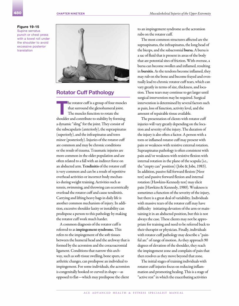
Chapter NINeteeN Musculoskeletal Injuries of the Upper Extremity
A C E A d v A n C E d H E A l T H & F I T n E S S S P E C I A l I S T M A n U A l
480
Rotator Cuff Pathology
The rotator cuff is a group of four muscles that surround the glenohumeral joint. The muscles function to rotate the
shoulder and contribute to stability by forming a dynamic “sling” for the joint. They consist of the subscapularis (anteriorly), the supraspinatus (superiorly), and the infraspinatus and teres minor (posteriorly). Injuries of the rotator cuff are common and may be chronic conditions or the result of trauma. Traumatic injuries are more common in the older population and are often related to a fall with an indirect force on an abducted arm. Tendinitis of the rotator cuff is very common and can be a result of repetitive overhead activities or incorrect body mechan-ics during weight training. Activities such as tennis, swimming, and throwing can eccentrically overload the rotator cuff and cause tendinitis. Carrying and lifting heavy bags in daily life is another common mechanism of injury. In addi-tion, excessive shoulder laxity or instability can predispose a person to this pathology by making the rotator cuff work much harder.
A common diagnosis of the rotator cuff is referred to as impingement syndrome. This refers to the impingement of the soft tissues between the humeral head and the archway that is formed by the acromion and the coracoacromial ligament. Conditions that narrow this arch-way, such as soft-tissue swelling, bone spurs, or arthritic changes, can predispose an individual to impingement. For some individuals, the acromion is congenitally hooked or curved in shape—as opposed to flat—which may predispose the client
to an impingement syndrome as the acromion rubs on the rotator cuff.
The most common structures affected are the supraspinatus, the infraspinatus, the long head of the biceps, and the subacromial bursa. A bursa is a sac of fluid that is present in areas of the body that are potential sites of friction. With overuse, a bursa can become swollen and inflamed, resulting in bursitis. As the tendons become inflamed, they may rub on the bone and become frayed and even-tually lead to chronic rotator cuff tears, which can vary greatly in terms of size, thickness, and loca-tion. These tears may continue to get larger until surgical intervention may be required. Surgical intervention is determined by several factors such as pain, loss of function, activity level, and the amount of repairable tissue available.
The presentation of clients with rotator cuff injuries will vary greatly depending on the loca-tion and severity of the injury. The duration of the injury is also often a factor. A person with a torn or inflamed rotator cuff may present with pain or weakness with resistive external rotation. Supraspinatus pathology is often consistent with pain and/or weakness with resistive flexion with internal rotation in the plane of the scapula (i.e., the “empty can” position) (Jobe & Jobe, 1983). In addition, passive full forward flexion (neer test) and passive forward flexion and internal rotation (Hawkins-Kennedy test) may elicit pain (Hawkins & Kennedy, 1980). Weakness is sometimes a function of the severity of the injury, but there is a great deal of variability. Individuals with massive tears of the rotator cuff may have difficulty initiating elevation of the arm or main-taining it in an abducted position, but this is not always the case. These clients may not be appro-priate for training and need to be referred back to their therapist or physician. Finally, individuals with rotator cuff pathology may describe a “pain-ful arc” of range of motion. As they approach 90 degrees of elevation of the shoulder, they reach the impingement zone and complain of pain that then resolves as they move beyond that zone.
The initial stages of training individuals with rotator cuff injuries focus on reducing inflam-mation and promoting healing. This is a stage of “active rest” in which the exacerbating activities
Figure 19-15 Supine serratus punch or chest press with a towel roll under the shoulder to avoid excessive posterior translation

A C E A d v A n C E d H E A l T H & F I T n E S S S P E C I A l I S T M A n U A l
Musculoskeletal Injuries of the Upper Extremity Chapter NINeteeN 481
are eliminated or modified. Common causes of injury are overhead sports, military press, incline bench press, and lateral raises in the frontal plane. Restoring flexibility is also an important goal of this phase. Individuals with rotator cuff pathology often lose flexibility of the posterior structures of the shoulder. loss of horizontal adduction is often an indication of a contracture of the posterior capsule, while loss of internal rotation is often an indication of a contracture of the posterior rotator cuff. Both of these situations can contribute to an increased chance of rotator cuff impingement. Flexibility exercises are initiated to restore range of motion (Figures 19-16 and 19-17). As always, the ACE-AHFS should have the client avoid ranges that are painful.
As in the case of shoulder instability, strengthening should be initiated with the scapula, especially in the case of a significantly inflamed rotator cuff. Any deviation in scapu-lar function can have a negative effect on the shoulder. For example, if the scapula is elevated too high, the mechanical advantage of the rota-tor cuff is altered. By restoring normal scapula function, the proper length-tension relation-ship of the rotator cuff is restored. In addition, many of the scapula strengthening exercises (e.g., rowing, shrugs, serratus punches, push-ups with a plus) indirectly strengthen the rotator cuff (Hintermeister et al., 1998).
As inflammation decreases, IR/ER exercises may be cautiously introduced. As mentioned previously, the ACE-AHFS should carefully monitor symptoms to avoid an inhibition of the rotator cuff. It should be noted that not everyone can tolerate these exercises. Strengthening can be introduced as submaximal isometrics. Clients can then progress to using elastic resistance. When performing external rotation exercises, the client can position a towel roll at his or her side, which places the shoulder in a slightly abducted posi-tion (Figure 19-18). This will improve the blood supply to the shoulder and enhance the mechani-cal advantage of the external rotators.
For those who want to continue deltoid strengthening, scapular plane elevation is pre-ferred to performing lateral raises in the frontal plane (Figure 19-19). The exercise in the scapular
plane affords the least amount of stress on the shoulder. It is also a more functional plane in which to work. Finally, this exercise also recruits much of the scapula musculature, and to some extent the supraspinatus (Moseley, Jobe, & Pink, 1994). The “empty can” position described
Figure 19-16Posterior capsule stretch
Figure 19-17Medial rotation stretch using a towel
Figure 19-18External rotation

Chapter NINeteeN Musculoskeletal Injuries of the Upper Extremity
A C E A d v A n C E d H E A l T H & F I T n E S S S P E C I A l I S T M A n U A l
482
by Jobe and Jobe (1983) for strengthening the supraspinatus is not advised, as the internally rotated position significantly increases the chance of shoulder impingement and is a common source of shoulder pain.
When designing a strength-training program, the ACE-AHFS should consider that many ath-letic or functional demands require a significant amount of eccentric muscle activity. Therefore, the eccentric or negative phase of each exercise should also be emphasized. However, the ACE-AHFS should closely monitor these exercises, as they are often a cause of delayed onset muscle soreness (DOMS). Many clients with rotator cuff injuries will want to return to overhead activities such as tennis, swimming, or throwing. In such cases, multijoint activities such as proprioceptive neuromuscular facilitation (PNF) patterns are useful to reproduce these demands. In particular, the d2 flexion pattern, which consists of shoul-der flexion, abduction, and external rotation, reproduces the neuromuscular demands of many overhead activities (Figure 19-20).
When a client is returning to performing a bench press, a narrower hand spacing should be utilized to minimize the peak shoulder torque in the pressing motion and reduce the rotator cuff and biceps tendon requirements for stabilization of the humeral head (Fees et al., 1998).
Lateral Epicondylitis
Lateral epicondylitis is often referred to as “tennis elbow.” It results from the repeti-tive tension overloading of the wrist and
finger extensors that originate at the lateral epi-condyle. Traditionally, the mechanism of injury takes place during the backhand of a novice tennis player who has poor mechanics. For example, not getting the racquet back fast enough and hitting the ball in front of the body or having a poor weight shift can result in greater stresses on the lateral aspect of the elbow. A change in the frequency of activity or a poorly fitted racquet can also contribute to injury. Tennis players who have a deficit in their proximal strength, such as in the scapula muscles or the rotator cuff, may be more susceptible to developing lateral epicondylitis. A lack of proximal stability may manifest itself further down the chain at the elbow. In addition, poor mechanics reduces the use of the lower body and core in the tennis stroke. This can result in increased stress on the elbow.
“Tennis elbow” is often a misnomer, as this injury it is not always a result of tennis. Carrying heavy bags or performing manual labor, especially with the elbow in extension, can result in lateral
Figure 19-19 Shoulder abduction in scapular plane
Figure 19-20 D2 flexion pattern

A C E A d v A n C E d H E A l T H & F I T n E S S S P E C I A l I S T M A n U A l
Musculoskeletal Injuries of the Upper Extremity Chapter NINeteeN 483
stretching should be performed slowly and gradu-ally, and be maintained in a pain-free range. Slow, progressive stretching allows the muscle to relax instead of reflexively guarding the area. These types of stretches have a longer-lasting effect.
When initiating a strengthening program, the ACE-AHFS should assess the shoulder and scap-ula for any underlying deficits. With tennis elbow, proximal strength deficits, especially in the shoul-der rotators, are often found. Initially, attempting to isolate the wrist extensors can exacerbate the symptoms. The client should be relatively asymptomatic in normal Adl prior to perform-ing wrist extension exercises. Tolerance to a firm handshake has been described as a prerequisite to these exercises. When initiating wrist extension strengthening, the elbow should be supported and be in flexion to reduce the stress (Figure 19-23).
One approach to initiating a strengthening pro-gram is to use functional, multijoint exercises such as rowing, shrugs, lat pull-downs, and PnF pat-terns. These exercises allow some strengthening of the wrist and forearm without trying to isolate them. They also provide a more global approach to strengthening the entire upper extremity and establishing proximal strength. These exercises should be performed while avoiding the end ranges of elbow extension. When a client is per-forming any activity, increasing the grip size of resistive equipment or a tennis racquet can reduce the amount of wrist extensor activity and the amount of stress on the lateral epicondyle.
As symptoms subside, the wrist extensors and forearm supinators can gradually be exercised in greater degrees of elbow extension. Movement patterns such as a tennis stroke or a golf swing can be reproduced using elastic resistance. novice
epicondylitis. In addition, excessive computer work can lead to increased stress to the extensor tendons. Ergonomic adjustments are often a key aspect of treatment.
Regardless of the mechanism of injury, the overload can result in inflammation of the ten-dons that attach at the lateral epicondyle. In later stages, a mass may form in the tendon and even result in a tear. This injury is often very resistant to treatment, as it is not often detected until latter stages of the pathology. At those stages, clients will complain about activities such as shaking hands, holding a coffee cup, or carrying something with the elbow in extension. Pain is elicited with resistive wrist extension, especially with the elbow in extension and passive wrist flexion (Figure 19-21).
The goals of the initial stage of treatment are to reduce the symptoms and promote healing. various modalities are utilized to reduce symp-toms and a period of active rest is encouraged. The causative activity must be eliminated or modi-fied. For example, a client may be encouraged to avoid tennis or make ergonomic adjustments. In the gym, lifting weights is avoided or modified, depending on the severity of symptoms. lifting weights with the elbow extended is certainly to be avoided. A wrist splint may be used to rest the extensor mechanism. In addition, a counterforce brace may be used around the elbow to dissipate forces away from the injured site and reduce pain.
As symptoms begin to subside, the ACE-AHFS should help the client restore normal flexibility. loss of passive wrist flexion is a common finding, and can be restored with passive wrist flexion with the elbow in extension (Figure 19-22). As always,
Figure 19-21Resistive wrist extension
Figure 19-22 Passive wrist flexion

Chapter NINeteeN Musculoskeletal Injuries of the Upper Extremity
A C E A d v A n C E d H E A l T H & F I T n E S S S P E C I A l I S T M A n U A l
484
Remember, the goals of the early stages of reha-bilitation are to reduce symptoms and promote healing. during these stages, causative activities are modified or eliminated, golfers are encouraged to take lessons, throwing mechanics are reviewed, and swimming strokes are assessed. Proximal shoulder and scapular strength are assessed for any underly-ing deficits.
Strengthening is again initiated with multijoint, functional exercises, as opposed to isolating the wrist flexors and forearm pronators. Exercises such as biceps curls may be better tolerated in a neutral forearm position than in a pronated position. Full elbow extension should be avoided when perform-ing resistive exercises. Flexibility should be initiated by stretching into wrist extension and forearm supination (Figure 19-24). Once again, the range should be basically pain free. Isolated wrist flexion and forearm pronation exercises should be avoided until the client is asymptomatic. When initiat-ing these exercises, they should be done with the elbow supported and in flexion. The ACE-AHFS should proceed cautiously with isolated pronation exercises, as they can often be a source of pain or injury. Strengthening can often be achieved with multijoint exercises. Prior to a return to activity, functional exercises such as PnF patterns may be helpful. note that repetitive activity at the computer can result in medial epicondylitis. The etiology of this injury is rapid, repetitive finger flex-ion with a fixed wrist as the individual clicks and drags the mouse.
Hand and Wrist Injuries
There are numerous types of pathologies of the wrist and hand. Two of the most common injuries are carpal tunnel syn-
drome and De Quervain’s syndrome. Carpal tunnel syndrome occurs when the median nerve, which extends from the forearm into the hand, becomes compressed at the wrist. The carpal tunnel is formed by ligaments and bones at the base of the hand. Thickened tendons or other swelling can cause the nerve to become impinged or compressed. Some people are congenitally pre-disposed to this condition. However, common causes are wrist trauma, arthritis, work stress, and fluid retention. Symptoms include burning,
tennis players are encouraged to take lessons to improve mechanics. As weight training is progressed, the ACE-AHFS should continue to closely monitor the client for any recurrence of lateral elbow pain. Finally, when perform-ing cardiovascular activities, the client should avoid gripping the apparatus too tightly with the affected hand.
Medial Epicondylitis
Medial epicondylitis occurs due to an overload of the wrist flexors and fore-arm pronators. Golf, throwing, and
swimming are common mechanisms of injury. Overuse or poor mechanics may lead to tendinitis or small tears of these muscles near the origin at the medial epicondyle. “Golfer’s elbow” refers to an injury to the medial side of the right elbow (for a right-handed golfer). novice golfers who fail to use their larger body parts and do not weight shift correctly are more susceptible. Beginners tend to throw the club down at the ball or hit too far behind the ball and put greater stress on the medial aspect of the elbow. Participating in throwing sports also tends to place a great deal of stress on the medial aspect of the elbow. The ACE-AHFS must be aware that injuries to the ulnar nerve are often associated with this area. Any numbness or tingling along the ulnar aspect of the forearm or the fourth and fifth fingers should alert the ACE-AHFS to this possibility. Clients will present with tenderness over the medial epicondyle or the proxi-mal wrist flexors and pronator teres. Resistive wrist flexion or forearm pronation may elicit symptoms. In addition, performing high-load biceps curls often exacerbates symptoms.
Figure 19-23Dumbbell wrist extension

A C E A d v A n C E d H E A l T H & F I T n E S S S P E C I A l I S T M A n U A l
Musculoskeletal Injuries of the Upper Extremity Chapter NINeteeN 485
some general precautions and adjustments for the injured hand and wrist are as follows:
• Any point tenderness at one of the small bones of the wrist or hand may indicate a fracture. If these symptoms persist, a refer-ral to a hand specialist is advised.
• A change in positioning is often helpful for individuals with hand or wrist pain. When exercising, the wrist is often most comfort-able in a neutral position. A good guideline is to avoid wrist flexion and extension greater than 30 degrees. In addition, avoid radial or ulnar deviation. Pain on the ulnar side of the wrist is often exacerbated by forearm pronation or supination.
• The grip size of exercise equipment can be adjusted. The ACE-AHFS may add pad-ding to a piece of exercise equipment to create a larger grip. Often, a larger grip will reduce stress on the wrist, hand, or fingers.
Case Studies
Case Study 1John is 49-year-old recreational tennis player
who has been unable to play for approximately six months after developing lateral elbow pain. Upon seeing an orthopedist, he was diagnosed with lateral epicondylitis. He was treated with nonsteroidal anti-inflammatory drugs (NSAID) and physical therapy for three months. Following discharge, he was relatively asymptomatic. His occupation requires him to perform a great deal of computer work and travel often. He comes to an ACE-AHFS to get himself back into “tennis shape,” as this is his primary exercise activity.
The ACE-AHFS should contact John’s physical therapist to discuss his home exercise program and any ergonomic precautions he was given. The ACE-AHFS should incorporate the exercises into a comprehensive general program that addresses the entire kinetic chain. Shoulder and scapula strength and flexibility should be emphasized, using functional, multijoint exer-cises. John’s lateral elbow symptoms should be monitored regularly to identify any exacerbat-ing exercises. This is especially important if
tingling, and numbness in the palm, thumb, index, and middle fingers. As the condition worsens, grip strength may be affected. When a client presents with any of these symptoms, the ACE-AHFS should refer him or her to a hand specialist.
de Quervain’s syndrome affects the two tendons that move the thumb away from the hand. Some experts believe the tendons become inflamed from overuse; however, the cause is not always clear or well-understood. Symptoms may include pain and or swelling over the thumb side of the wrist. Gripping may also become difficult. When testing for this syndrome, the thumb is tightened as in a closed fist and the hand is tilted toward the ulna side (Finkelstein’s test) (Figure 19-25). If the syn-drome is present, this position will produce pain at the wrist below the thumb. Any of these symptoms that last for one to two weeks should be addressed by the client’s healthcare provider.
Finally, there are numerous ligamentous sprains and fractures that can occur in the small bones of the hand and wrist. While the possibil-ities are far too extensive to discuss specifically,
Figure 19-24Passive wrist extension
Figure 19-25Finkelstein’s test

Chapter NINeteeN Musculoskeletal Injuries of the Upper Extremity
A C E A d v A n C E d H E A l T H & F I T n E S S S P E C I A l I S T M A n U A l
486
Steve’s physical therapist to discuss his current home exercise program and any contraindica-tions or safety precautions. When initiating a strength-training program, the ACE-AHFS should carefully monitor Steve’s symptoms at his AC joint. Steve should be encouraged to ice after his workout, even if he is asymptomatic at the time. This may help to prevent any residual symptoms. When using free weights, Steve should be encouraged to begin with weights that he can control well to avoid any traction at the AC joint. He should also be advised to not carry the weights around the gym. Scapula strengthen-ing exercises should be performed in the middle of the range of motion to avoid excessive retrac-tion and protraction. This will prevent excessive stress to the AC joint. Steve should be extremely cautious with exercises that create a great deal of stress at the AC joint, such as bench pressing or push-ups. He should never perform any bench presses without a spotter.
Summary
The key to working with the post-injury client is to understand the pathology and structures involved, as well as the
underlying mechanism of injury. In addition, the ACE-AHFS should understand the positions and activities that may exacerbate the condition. listening to the subjective complaints and symp-toms and being proactive in communicating with the client’s clinician is critical to preventing re-injury.
John is performing wrist extension exercises. When designing John’s exercise program, the ACE-AHFS should avoid resistive training in the end ranges of elbow extension. John should also avoid carrying weights around the gym whenever possible. When performing condi-tioning exercises such as cycling or treadmill walking, John should avoid excessive gripping on the handles or handrails.
Prior to returning to tennis, John should be encouraged to begin hitting with a tennis professional or taking some lessons. This will reinforce the need for him to use his entire body when hitting and develop a good weight shift to reduce the forces at the elbow. He should also be advised to have his tennis pro check his racquet for the proper tension and grip size. In addition, John should be advised to avoid carrying heavy bags when traveling.
Case Study 2Steve is a 32-year-old recreational skier who
suffered a Grade II separation of his acromio-clavicular joint while skiing six weeks ago. He was immobilized for two weeks and then under-went a four-week course of physical therapy that restored his shoulder range of motion. He presents with mild, intermittent AC joint dis-comfort and a slight palpable defect at the joint. Prior to the injury, Steve lifted weights three times a week and is eager to resume his exercise program. He comes to the ACE-AHFS to begin a safe exercise program and avoid exacerbation of the injury. The ACE-AHFS should contact

A C E A d v A n C E d H E A l T H & F I T n E S S S P E C I A l I S T M A n U A l
Musculoskeletal Injuries of the Upper Extremity Chapter NINeteeN 487
Saha, K. (1983). Mechanism of shoulder movements and plea for recognition of the zero position of the glenohumeral joint. Clinical Orthopedics, 173, 3–10.
Snyder, S.J. et al. (1990). SLAP lesions of the shoulder. Arthroscopy, 6, 274–279.
Timm, K. (1998). The isokinetic torque curve of shoulder instability in high school baseball pitchers. Journal of Orthopedic and Sports Physical Therapy, 26, 150–154.
Tippett, S. (1992). Closed chain exercise. Orthopedic Physical Therapy Clinics of North America, 1, 253–268.
Suggested Reading Altchek, D.W. & Andrews, J.R. (2001). The Athlete’s Elbow. Philadelphia: Lippincott Williams & Wilkins.
Andrews, J.R. & Wilk, K.E. (2008). The Athlete’s Shoulder (2nd ed.). New York: Churchill Livingstone.
Baechle, R.T. & Earle, R.W. (2008). Essentials of Strength Training and Conditioning (3rd ed.). Champaign, Ill.: Human Kinetics.
Cioppa-Mosca, J. et al. (2006). Postsurgical Rehabilitation Guidelines for the Orthopedic Clinician. St. Louis, Mo.: Mosby Elsevier.
Fees, M. et al. (1998). Upper extremity weight-training modifications for the injured athlete. American Journal of Sports Medicine, 26, 5, 732–742.
Knott, M. & Voss, D. (1985). Proprioceptive Neuromuscular Facilitation (3rd ed.). New York: Harper & Row.
Leach, R.E. & Miller, J.K. (1987). Lateral and medial epicondylitis of the elbow. Clinics in Sports Medicine 6, 259–272.
Magee, D.J. (2007). Orthopedic Physical Assessment (5th ed.). Philadelphia: Saunders .
Manske, R.C. (2006). Postsurgical Orthopedic Sports Rehabilitation: Knee & Shoulder. St. Louis, Mo.: Mosby Elsevier.
Voight, M. & Draovitch, P. (1991). Eccentric Muscle Training in Sports and Orthopedics. New York: Churchill Livingstone.
ReferencesBassett, R. et al. (1988). Glenohumeral muscle force and movement mechanics in a position of shoulder instability. Journal of Biomechanics, 23, 401–415.
Fees, M. et al. (1998). Upper extremity weight-training modifications for the injured athlete. American Journal of Sports Medicine, 26, 5, 732–742.
Gross, M.L. et al. (1993). Anterior shoulder instability in weight lifters. American Journal of Sports Medicine, 21, 599–603.
Hawkins R.J. & Kennedy J.C. (1980). Impingement syndrome in athletes. American Journal of Sports Medicine, 8, 151–158.
Hintermeister, R.A. et al. (1998). EMG activity and applied load during shoulder rehabilitation exercises using elastic resistance. American Journal of Sports Medicine, 26, 210–220.
Hovelius, L. (1987). Anterior dislocation of the shoulder in teenagers and young adults. Journal of Bone and Joint Surgery, 69, 393.
Jobe, F.W. & Jobe, C.M. (1983). Painful athletic injuries of the shoulder. Clinical Orthopedics, 173, 117–125.
Levine, W.M. (2000). The pathophysiology of shoulder instability. American Journal of Sports Medicine, 28, 910–917.
Manske, R.C. (2006). Postsurgical Orthopedic Sports Rehabilitation: Knee and Shoulder. St. Louis, Mo.: Mosby Elsevier.
Moseley, J.B., Jobe, F.W., & Pink, M. (1994). EMG analysis of scapular muscles during a shoulder rehabilitation program. American Journal of Sports Medicine, 20, 128–134.
O’Brien, S.J. et al. (1998). The active compression test: A new and effective test for diagnosing labral tears and acromioclavicular joint abnormality. American Journal of Sports Medicine, 26, 610–613.
Rockwood, C.A. & Matsen, F.A., III. (2004). The Shoulder (3rd ed.). Philadelphia: Saunders.