in the name of god 3 rd trimester sonography. historical ultrasound 1876 : 1st used dog 1880...
TRANSCRIPT
IN THE NAME OF GODIN THE NAME OF GOD
3RD
TRIMESTER
SONOGRAPHY
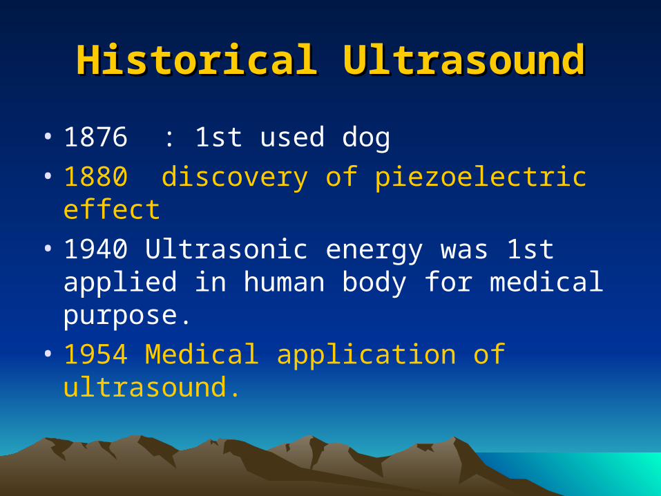
Historical UltrasoundHistorical Ultrasound
• 1876 : 1st used dog
• 1880 discovery of piezoelectric effect
• 1940 Ultrasonic energy was 1st applied in human body for medical purpose.
• 1954 Medical application of ultrasound.
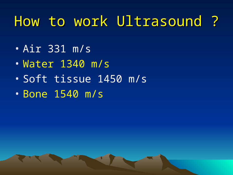
How to work Ultrasound ?How to work Ultrasound ?
• Air 331 m/s
• Water 1340 m/s
• Soft tissue 1450 m/s
• Bone 1540 m/s

How to work Ultrasound ?How to work Ultrasound ?
• Tranceducer
• According to wavelength and speed and density tissue reflect to oscillator
• Display to monitor
• A-Mode, B-Mode , M-Mode
• 2 D , 3D ,4D

Safety UltrasoundSafety Ultrasound
• Ti:up to 1 degree Celsius
• Tis: Soft tissue
• TiC: Cranium
• TiB: Bone
• Routine 2D may be safe
• 3D & 4D unsafe or hasard
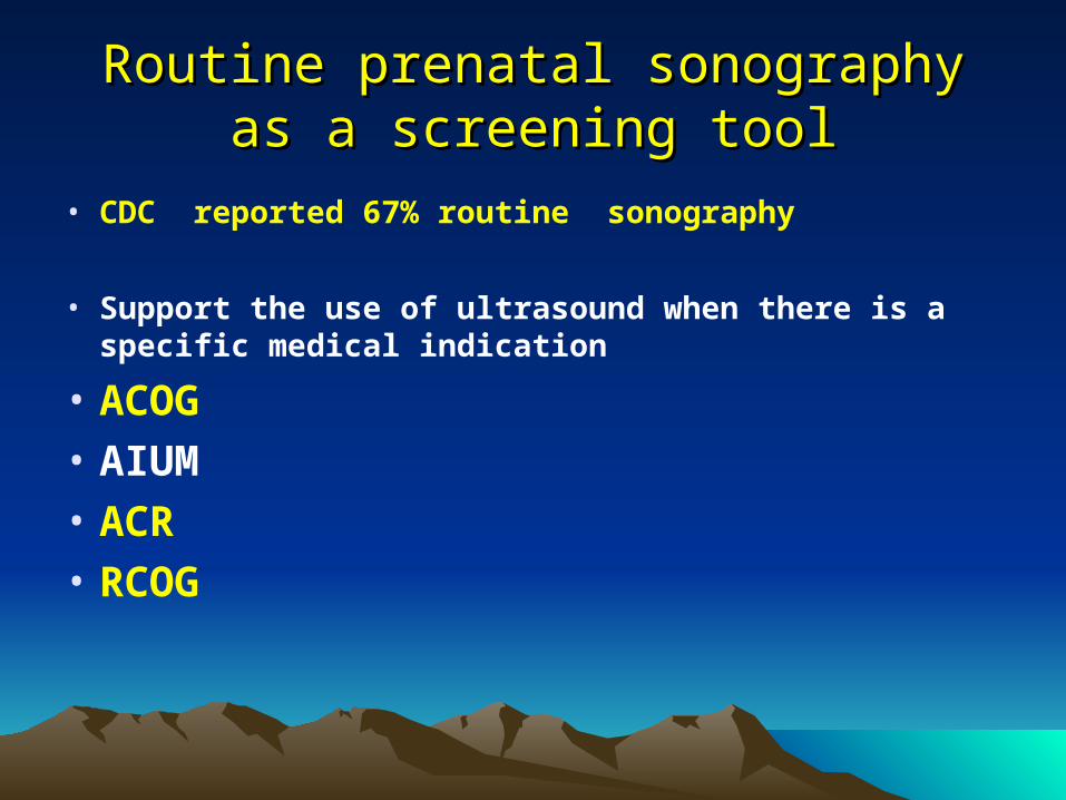
Routine prenatal sonographyRoutine prenatal sonography as a screening tool as a screening tool
• CDC reported 67% routine sonography
• Support the use of ultrasound when there is a specific medical indication
• ACOG• AIUM• ACR• RCOG

Using ultrasound in OBsUsing ultrasound in OBs
• Confirm date of delivery ( EDC)
• Diagnosis of abnormal pregnancy
EP , Blighted ovum(1st Trimester)
• Anomaly
• Reduced diagnose of
IUGR(RGF) ,Postdatisem
Prevent abuse tocolytic (date )
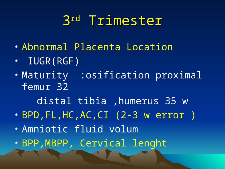
33rdrd Trimester Trimester
• Abnormal Placenta Location
• IUGR(RGF)
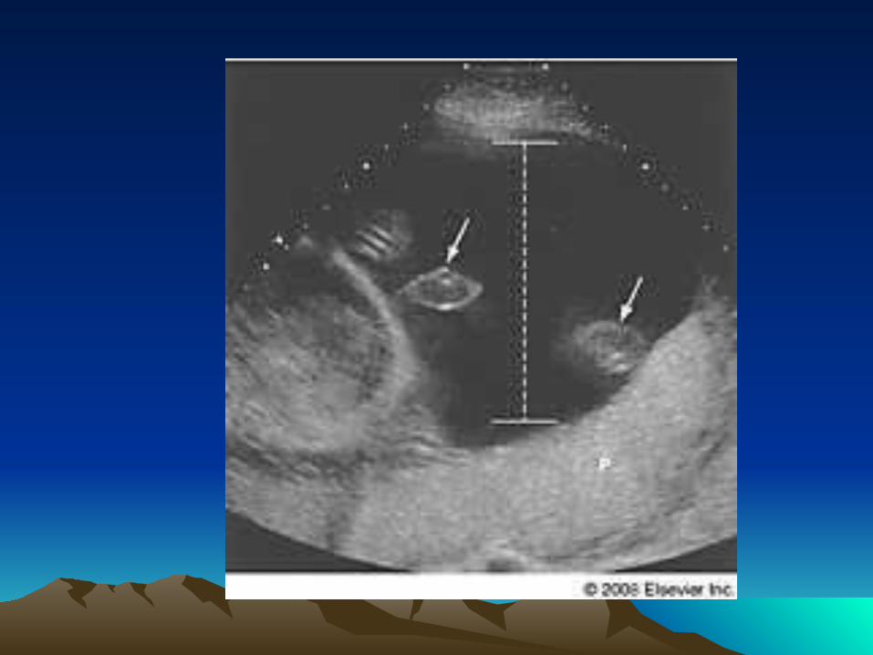
• Maturity :osification proximal femur 32
distal tibia ,humerus 35 w
• BPD,FL,HC,AC,CI (2-3 w error )
• Amniotic fluid volum
• BPP,MBPP, Cervical lenght

33rdrd Trimester TrimesterMultiple MarkersMultiple Markers
• HC,FL equal to PD,FL,AC,HC
• HC/AC
• BPD/FL
• FL/AC
• Others: interaorbital,clavicle length
Foot length….etc.
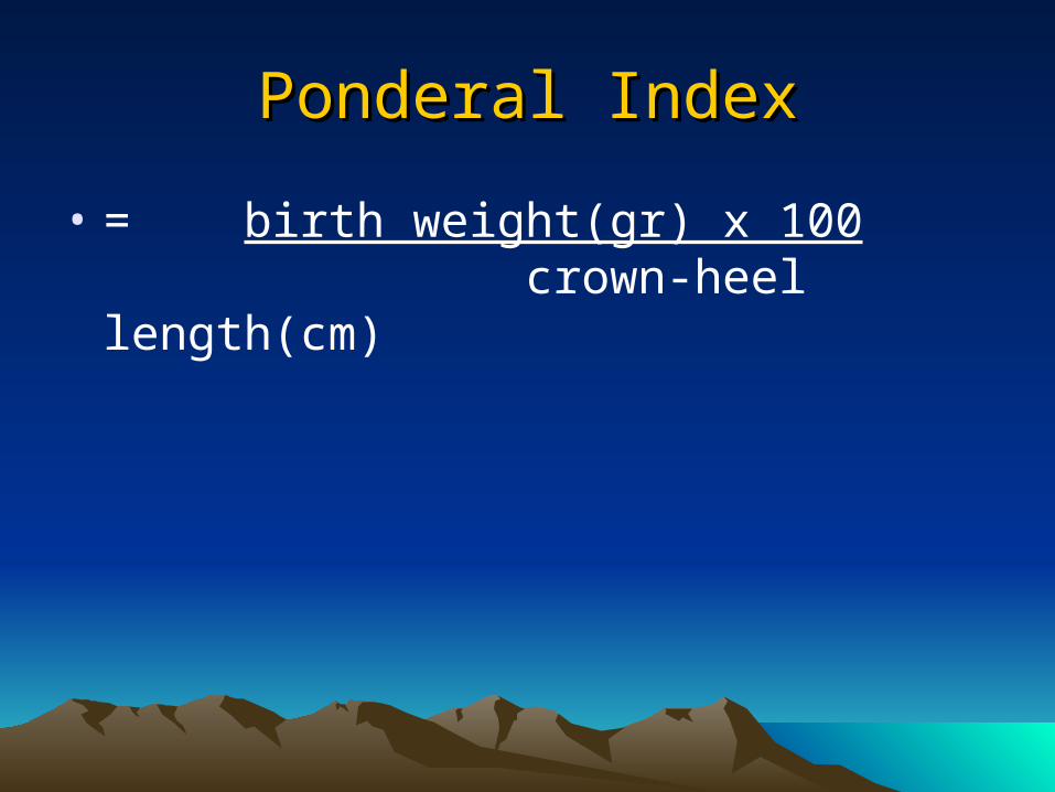
Ponderal IndexPonderal Index
• = birth weight(gr) x 100 crown-heel length(cm)


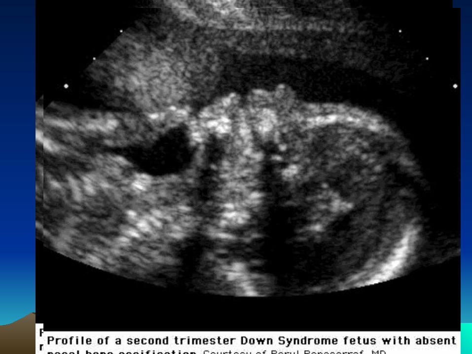
nasal bone Ossification
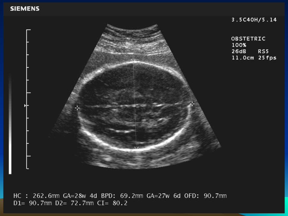

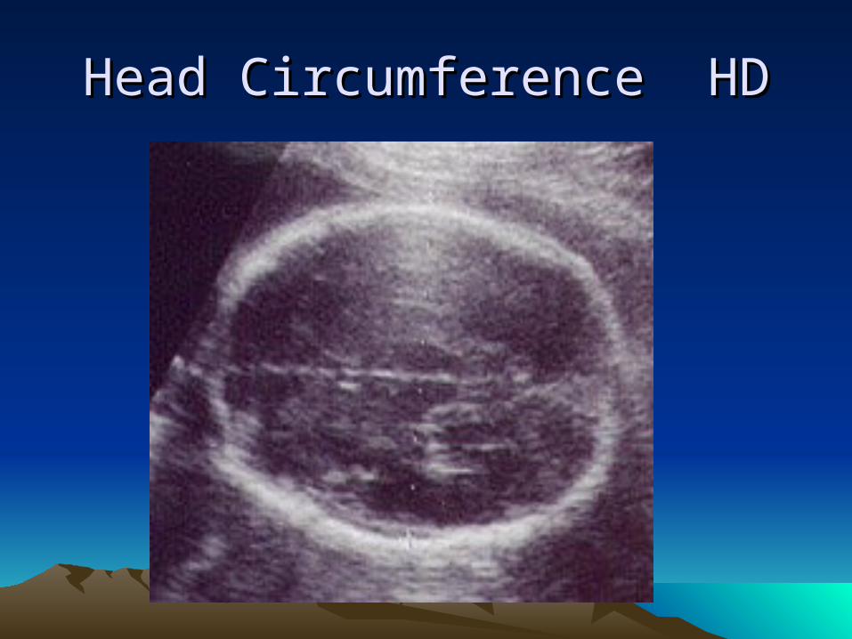
Head Circumference HDHead Circumference HD
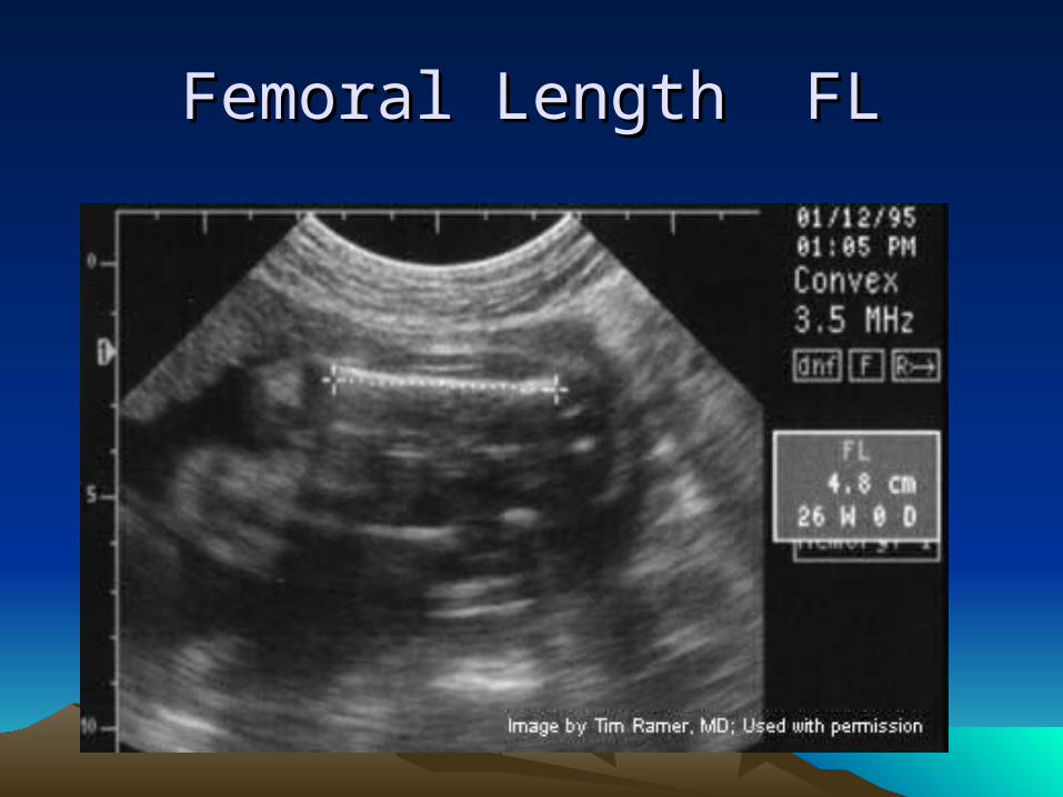
Femoral Length FLFemoral Length FL


CRLCRL

ACAC


جنون ” ای قطره همیشه عشق درهمیشه هم جنون در و هست
عقل ای “قطره( نیچه (فردریک
“Always there is a drop of madness in love, yet always there is a drop of reason in madness” (F. Nietzshe)