in the name of allah. the skull 2 norma verticalis
TRANSCRIPT

IN THE NAME OF ALLAH

The Skull
2

Norma Verticalis
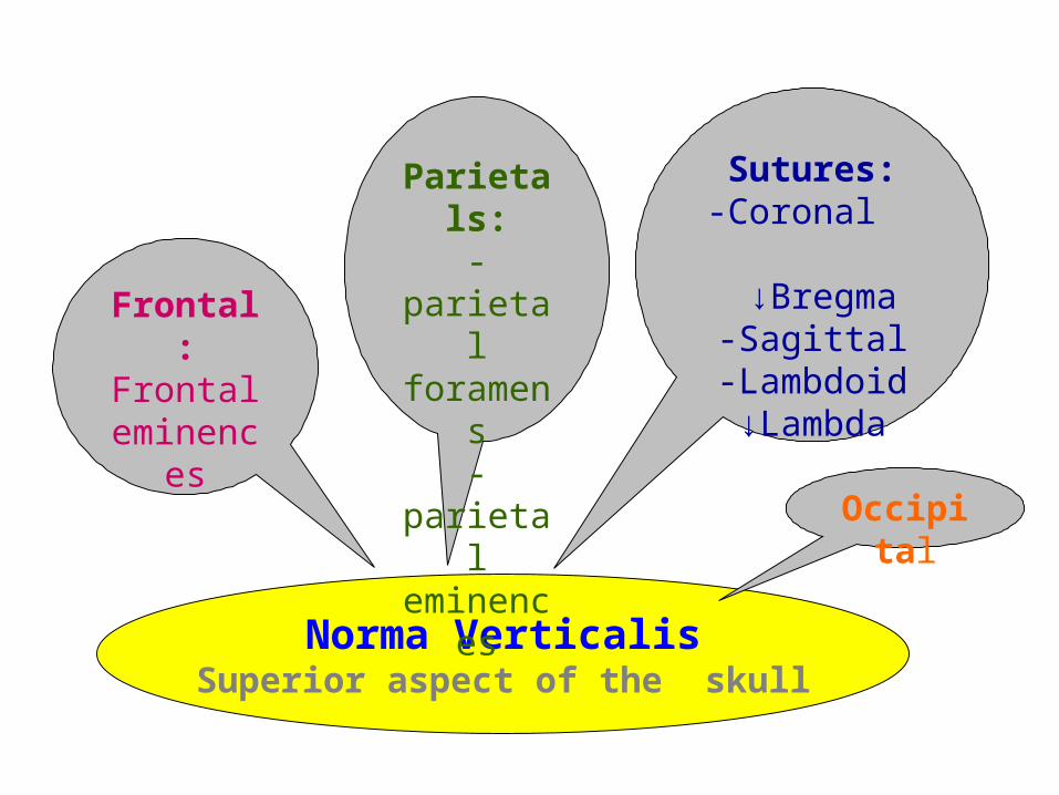
Norma VerticalisSuperior aspect of the skull
Frontal:Frontal
eminences
Parietals:-parietal foramens-parietal
eminences
Sutures:-Coronal
↓Bregma-Sagittal
-Lambdoid↓Lambda
Occipital

Norma Verticalis

Coronalsuture
Sagittalsuture
Emissary foramen
Lambdoidsuture
Calvaria • Emissary foramen- small,
inconsistent (not always present) foramina for passage of emissary veins
• Bregma- is the landmark formed by the intersection of the sagittal & coronal sutures
• Lambda-is the landmark formed by the intersection of the sagittal & lambdoid sutures
Bregma
Lambda

Anterior fontanelle
Posterior fontanelle
Infant Calvaria• Anterior Fontanelle (soft
spot) is the future site of the bregma
• By about 18 months- the surrounding bones fuse together and is no longer palpable
• Posterior Fontanelle is triangular and marks the future site of the lambda
• Fusion of surrounding bones occurs by about 6 months
Fontanelles- membranous gaps (soft spots) in skull that permit growth

Norma Occipitalis

Norma Occipitalis(Post. Aspect)
Bones:-Occipital-Parietals-Mastoid process
Landmarks:-External occipital
protuberance-Inion
-Ext. occipital crest-Sup. & Inf.
(Highest)Nuchal lines
Sutures:-Sagital suture
-Lambdoid suture
*Lambda

Norma Occipitalis


Norma frontalis

Norma Frontalis (Anterior aspect of the skull)
Regions (cavities):-Nasal-Orbit-Oral
Bones:-Frontal
-Zygomatic-Maxillae-Mandible
-Nasal


Norma Frontalis

Frontal bone
Nasion Glabella
• Squamous (flat) portion forms the skeleton of the forehead
• Fontal bone forms the roof of the orbit
• Nasion is an area where the frontal bone intersects with the nasal bones
• Glabella- smooth, slightly depressed area located just superior to the nasion
Frontal BoneFrontal Bone
Squamousportion
Orbital portion
Nasion
Glabella
Frontal Bone

• Supraorbital margin- marks the boundary between the squamous and orbital portions
• Supraorbital notch or foramen is for the passage of the supraorbital nerve and vessels
• A prominent ridge just superior to the supraorbital margin is the superciliary arch (more pronounced in males)
• Zygomatic process of the frontal bone articulates with the zygomatic bone
Supraorbitalmargin
Supraorbitalnotch
Superciliaryarch
Zygomaticprocess
Frontal BoneFrontal Bone

Frontalprocess
Temporalprocess
Zygomaticofacialforamen
• Cheek bones• Forms a portion of the lateral wall
of the orbit• Frontal Process of the zygomatic
bone articulates with the frontal bone
• Temporal process of the zygomatic bone articulates with the temporal bone
• Zygomaticofacial foramen- small foramen for passage of the zygomaticofacial nerve
Zygomatic BonesZygomatic Bones
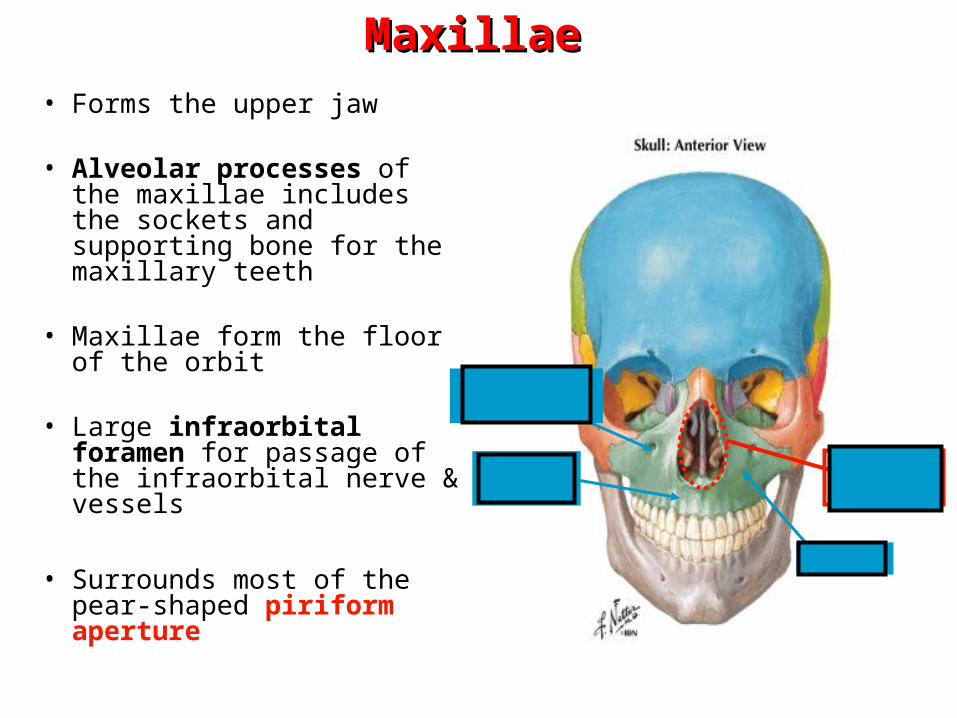
Piriform aperture
Alveolarprocess
Infraorbitalforamen
• Forms the upper jaw
• Alveolar processes of the maxillae includes the sockets and supporting bone for the maxillary teeth
• Maxillae form the floor of the orbit
• Large infraorbital foramen for passage of the infraorbital nerve & vessels
• Surrounds most of the pear-shaped piriform aperture
MaxillaeMaxillae
Maxilla

• Frontal processes of the maxillae articulates with the frontal bone
• Zygomatic processes of the maxillae articulates with the zygomatic bones
• Anterior nasal spine- sharp prominence at inferior aspect of the piriform aperture
• Intermaxillary suture- site where the two maxilla are united in the median plane
MaxillaeMaxillae
frontal process
Anterior nasal spine
Intermaxillarysuture
Zygomatic process

• Seen within the piriform aperture are the scrolled middle nasal conchae (part of the ethmoid bone) & the inferior nasal conchae
• Vomer bone along with the perpendicular plate of the ethmoid bone which together form the bony nasal septum can also be identified with the piriform aperture
Vomer
Middle &Inferiorconcha
Perpendicular plate

1
2
3
457
mediallateral
superior
inferior
Osteology of the Orbit
• Seven bones articulate to make each orbit:
1. Frontal2. Zygomatic3. Maxillary4. Lacrimal5. Ethmoid6. Palatine7. Sphenoid
6

Roof:-Orbital plate of the
frontal bone-Lesser wing of the
sphenoid
Floor:-Maxilla
-Zygomatic(Infraorbital groove & canal)
-Palatine
Lateral wall:-Zygomatic-Sphenoid
Medial wall:-Maxillae-Lacrimal-Ethmoid-Sphenoid
Sup. Orbital fissure
Orbit
Inf. Orbital fissure

Inferiororbitalfissure & groove
Optic canal
Superiororbitalfissure
Ethmoidalforamina
Osteology of the Orbit• Optic canal- transmits the
optic nerve and ophthalmic artery
• Superior orbital fissure- transmits CN III, IV, V1 & VI
• Inferior orbital fissure & groove- transmits the infraorbital vessels & nerve
• Anterior & posterior ethmoidal foramina- transmits vessels & nerves with same name


Nasal cavity
Roof:-Frontonasal-Ethmoidal-Sphenoidal
Medial wall:Nasal septum:
-Perpendicular plate of he ethmoid
-Vomer
Floor:-Palatine process of the
maxilla-Horizontal plate of the
palatine
Lateral wall:Irregular, conchae:
-Superior-Middle-Inferior


Dr Namavar 30

Dr Namavar 31

