in situ isolation and characterization of the
TRANSCRIPT

Advances in Environmental Research, Vol. 9, No. 3 (2020) 215-232
DOI: https://doi.org/10.12989/aer.2020.9.3.215 215
Copyright © 2020 Techno-Press, Ltd. http://www.techno-press.org/?journal=aer&subpage=7 ISSN: 2234-1722 (Print), 2234-1730 (Online)
In situ isolation and characterization of the biosurfactants of B. Subtilis
Wasim S. Akthar, Mohamed Sheik Aadham and Arif S. Nisha*
PG & Research Department of Biotechnology,
Srimad Andavan Arts & Science College (Autonomous), Trichy – 05, India
(Received November 26, 2019, Revised September 11, 2020, Accepted September 24, 2020)
Abstract. Crude oils are essential source of energy. It is majorly found in geographical locations beneath the
earth’s surface and crude oil is the main factor for the economic developments in the world. Natural crude oil
contains unrefined petroleum composed of hydrocarbons of various molecular weights and it contains other
organic materials like aromatic compounds, sulphur compounds, and many other organic compounds. These
hydrocarbons are rapidly getting degraded by biosurfactant producing microorganisms. The present study
deals with the isolation, purification, and characterization of biosurfactant producing microorganism from
oil-contaminated soil. The ability of the microorganism producing biosurfactant was investigated by well
diffusion method, drop collapse test, emulsification test, oil displacement activity, and blue agar plate
method. The isolate obtained from the oil contaminated soil was identified as Bacillus subtilis. The
identification was done by microscopic examinations and further characterization was done by Biochemical
tests and 16SrRNA gene sequencing. Purification of the biosurfactant was performed by simple liquid-liquid
extraction, and characterization of extracted biosurfactants was done using Fourier transform infrared
spectroscopy (FTIR). The degradation of crude oil upon treatment with the partially purified biosurfactant
was analyzed by FTIR spectroscopy and Gas-chromatography mass spectroscopy (GC-MS).
Keywords: biosurfactant; crude oil; well diffusion method; drop collapse test; emulsification test; oil
displacement activity; blue agar plate method; 16SrRNA gene sequencing; FTIR; GC-MS
1. Introduction
Petroleum products are often released either accidentally or intentionally into the environment through the spill, leakage, transport, or other incidents that affect residential, agricultural or
recreational land use and it also affects the water areas. It damages the ecosystems and it
negatively affects the health of plants, animals, and human beings. Therefore, remediation of oil-polluted sites by various chemical and biological methods has become crucial to control oil
pollution. Most of the hydrocarbons that are present in oils are insoluble in water and therefore, the
process of remediation becomes complex and needs several chemicals and reagents, which otherwise causes deleterious effects to the environment. Bioremediation by using microorganisms
Corresponding author, Professor, E-mail: [email protected]

Wasim S. Akthar, Mohamed Sheik Aadham and Arif S. Nisha
is a simple and eco-friendly method to remediate the oil-contaminated sites. In recent years,
researchers have started exploring the potentials of microorganisms for degrading complex
hydrocarbons, thereby reducing the environmental pollution (Zhang et al. 2012). Biosurfactants are synthesized by microorganisms, which helps to degrade the hydrocarbon. It
has different chemical nature and molecular size (Santhini and Parthasarathi 2014). These
biosurfactants are the secondary metabolites of microbes which have both hydrophilic and
hydrophobic moiety in its structure. These moieties were found to play a crucial role in the process of oil emulsification, an important process of biodegradation (Al-Wahaibi et al. 2014). Moreover,
these biosurfactants are more advantageous in the context of biodegradability, biocompatibility,
and digestibility (Parthipan et al. 2017). They are not only used to clean-up the environment by degradation but have been found to possess the ability to detoxify the industrial effluents (Olivera
et al. 2003). This distinctive feature of microbial surfactants is regarded to their surface activity,
tolerance to pH, temperature and ionic strength, biodegradability, low toxicity, emulsifying ability and antimicrobial activity (Chakrabarti et al. 2012).
The chemically synthesized surfactants are usually classified according to their polarity,
whereas, biosurfactants are generally categorized by their microbial origin and chemical
composition, which includes Glycolipids, Rhamnolipids, Trehalolipids, Sophorolipids, Lipopeptides and lipoproteins, Surfactin, Lichenysin, Fatty acids, phospholipids, and neutral
lipids, Polymeric biosurfactants, Particulate biosurfactants (Vijayakumar and Saravanan 2015).
For the past several decades, various microorganisms have been investigated for its bioremediation potentials. Among the different genus analyzed, Bacillus spp. has been reported to
hold enormous potentials in the field of biodegradation. Bacillus spp., are gram-positive, spore-
forming bacteria. It can survive in harsh environments like areas near petroleum reservoirs, where
high salinity, high temperature, and anaerobic conditions prevail. It has also been demonstrated that most of the Bacillus spp produces lipopeptide type of biosurfactants (Al-Wahaibi et al. 2014).
The mechanism of these biosurfactants in degrading oils has been studied extensively by several
researchers and it has been reported that they degrade the oil effectively by enhancing their water solubility and increasing the displacement of oil substances from soil particles. Thus,
biosurfactants increase the apparent solubility of these organic compounds at concentrations above
the Critical Micelle Concentration (CMC) which enhances their availability for microbial uptake (Chang et al. 2008). For these reasons, the use of biosurfactants in bioremediation of an oil-
polluted environment could be an effective approach both in the context of pollution control and
environment protection (Calvo et al. 2009).
The present study aims to isolate and screen for biosurfactant producing microorganisms from oil-contaminated soil. In particular, the biosurfactant producing organisms has been isolated and
screened from the oil-contaminated regions. Those isolates, which shows excellent degradation
potentials has been identified and characterized based on the microscopic, biochemical, and 16SrRNA gene sequencing. In this work, the structural analyses of the biosurfactant were done by
using Fourier transform infrared spectroscopy (FTIR) spectroscopy and Gas-chromatography mass
spectroscopy (GC-MS), respectively.
2. Materials and methods
2.1 Sample collection
Soil samples were collected from oil spilled surfaces of petrol bunk in Thiruvanaikaval near
216

In situ isolation and characterization of the biosurfactants of B. Subtilis
Srirangam, Tiruchirappalli, Tamil Nadu, India. The samples were collected in sterile polythene
bags and were taken to the laboratory for analysis.
2.2 Enrichment and isolation of bacterial isolates
Samples were enriched by inoculating 5 g oil spilled soil in 50 ml of nutrient broth and
incubated at 25oC for 72 hrs. The nutrient broth contained (g/L): Peptone – 5.0 g, Beef extract – 3.0 g, Sodium chloride – 8.0 g, Agar – 15.0 g. After incubation, the medium was serially diluted
from 10-1 to 10-6 in sterile water. From the dilutions (10-1 to 10-6) 1 ml of enriched culture was
transferred to a sterile petri dish and over that 20 ml of nutrient agar was poured. The plates were then inverted and incubated at 26oC for 48 hrs. Controls and replica plates were maintained. After
incubation, the different discrete colonies formed on the plate that had between 25 and 180 colony-
forming units (CFU) were streaked on nutrient agar and incubated at room temperature (37oC) for 24 hrs because of to obtain the pure cultures. These pure cultures, were sub-cultured on nutrient
agar slant, incubated at 37oC for 24 hrs, and stored in the refrigerator for biosurfactants production
and screening.
2.3 Identification of isolates
Cells were observed with Gram staining under a microscope (oil immersion, 100x, 40x). The shape of the cells (cocci, bacilli, cocobacillus) and analyze the isolated microorganism for gram-
positive or gram-negative nature and therefore further characterization was done through
biochemical tests. Different biochemical tests were performed to characterize the isolates and
unknown bacterial isolates were characterized by 16SrRNA gene sequencing-Bacterial identification
2.4 Screening of oil degradation by well diffusion method
The screening to isolate the oil-degrading microbes on nutrient agar well plates was performed.
100 µl of crude oil was spread on nutrient agar plates, wells were prepared and 50 µl of bacterial culture was then incubated at 37oC for overnight and observed results. The presence a clear zone
surrounding the well indicates the oil-degrading capacity of the organism.
2.5 Screening for biosurfactant activity
Cultures were inoculated in the mineral salt medium. Mineral salt contained (g/L): Di-
Potassium phosphate (K2HPO4) – 1.8 g, Ammonium chloride (NH4Cl) – 4.0 g, Magnesium sulfate (MgSO4.7H2O) – 0.2 g, Sodium chloride (NaCl) – 0.1 g, Iron (II) Sulphate Heptahydrate
(FeSO4.7H2O) – 0.01 g, agar –15 g, distilled water – 1 liter.
2.5.1 Drop collapsing test (Youssef et al. 2004) Screening of biosurfactant production was performed using the qualitative drop-collapse test
described by crude oil that was used in this test. Two microliters of oil were smeared all over the
surface of the well plate and it was left to equilibrate for 24 h. Five microliters of the 48 h culture, before and after centrifugation at 12,000 g for 5 min to remove cells, was transferred to the oil-
coated well region, and drop size will be observed after 1 min with the aid of a magnifying glass.
217

Wasim S. Akthar, Mohamed Sheik Aadham and Arif S. Nisha
The result was considered positive for biosurfactant production when the drop was flat and those
cultures that gave round drops were scored as negative, indicative of the lack of biosurfactant.
2.5.2 Emulsification test (Bodour et al. 2004) Several colonies of pure culture were suspended in test tubes containing 2 ml of the mineral salt
medium after 48 h of incubation, 2 ml hydrocarbon (oil) was added to each tube. Then, the mixture
was vortexed at high speed for 1 min and allowed to stand for 24 h. The emulsion index is the height of the emulsion layer (cm) divided by total height (cm), multiplied by 100 Eq. (1).
Emulsification index = Height of the layer / Total height x 100 (1)
2.5.3 Oil spreading method (Saminathan and Rajendran 2016) In this method, 20 ml of distilled water was added to a plastic petri dish followed by the
addition of 20 µl of crude oil to the surface of the water. 10 µl of cell-free culture broth was then added to the oil surface. If biosurfactant is present in the cell-free culture broth, the oil will be
displaced with an oil-free clearing zone, and the diameter of this clearing zone indicates the
surfactant activity, also called oil displacement activity. Negative control was maintained with
distilled water (without surfactant), in which no oil displacement or clear zone was observed. 2.5.4 Hydrocarbon overlay method (Saravanan and Vijayakumar 2012) The hydrocarbon overlay agar method was performed with some modifications. The mineral
agar plate will be coated individually with 100 µl of crude oil. Plates were inoculated with isolates
and incubated at 30oC for 48-72 h. Colonies surrounded by an emulsified halo was considered as
positive for biosurfactant production.
2.5.5 Blue agar plate method (Satpute et al. 2008) Mineral salt agar medium supplemented with glucose as carbon source (2%) and
cetyltrimethylammonium bromide (CTAB: 0.5 mg/ml) and methylene blue (MB: 0.2 mg/ml) was used for the detection of anionic biosurfactant. Thirty microliters of cell-free supernatant were
loaded into each well prepared in methylene blue agar plate using cork borer (4 mm). The plate
was then incubated at 37oC for 48-72 h. A dark blue zone around the culture was considered positive for anionic biosurfactant production.
2.6. Characterization of biosurfactants 2.6.1 Ninhydrin reaction (Zhang et al. 2012) Exactly 1 ml of extracted solution was put into the tube and added 3 drops of 0.5% ninhydrin
solution, and the tube was put into the boiling water bath for few minutes, to observe the change of color of the reaction mixture.
2.6.2 Sulfuric acid – phenol reaction and measurement of biosurfactants by ultraviolet analysis (Zhang et al. 2012)
The culture broth was diluted by 100 times first, and 2 ml of the diluted broth was transferred
into a 15 ml glass test tube, 1 ml phenol, and 5 ml sulphuric acid was added and vortexed. Then
tubes were heated for 15 min in a boiling water bath, cooled to room temperature, after which the optical density at 400-600 nm was measured.
218

In situ isolation and characterization of the biosurfactants of B. Subtilis
2.6.3. Para film-M test (Saminathan and Rajendran 2016) One drop of the Bromophenol blue indicator was added to 2 ml of cell-free supernatant. 10 µl
of the sample solution was carefully added on parafilm-M with a micropipette like a drop, the shape of the drop on the surface was inspected after 1 minute. The diameters of the droplets were
evaluated.
2.6.4. Surface tension (Saminathan and Rajendran 2016) The surface tension of the oil after treatment with biosurfactant was measured using the drop
weight method. Measurements were done in treated oil and untreated oil Eqs. (2) and (3).
Surface tension (ST) = mg/3.8rNm-1 (2)
where m = mass of one drop of the liquid
g = acceleration due to gravity
r = radius of the burette To determine the ST, the mass of the medium has to be calculated by simply weighing the drop
of the medium. Mass of one drop of the oil;
Where m = W2 – W1 / Total droplet
W2- Weight of the sample with the beaker
W1- Weight of the empty beaker
(3)
2.7 Growth study of isolated microorganism with oil as the sole carbon source A growth study was conducted for the isolated microorganism from oil-contaminated soil. The
study was done in a 250 ml flask using 100 ml of nutrient broth kept in the shaker at 30oC
temperature for 18 hours and reading was taken at a regular interval of 2 hours at absorbance 640 nm. A growth study was done to differentiate the growth of organisms in normal medium and
medium with oil.
2.8 Purification of biosurfactant
The biosurfactant was extracted from the culture medium after cell removal by centrifugation at 12,500 rpm for 30 min. The pH of the supernatant was adjusted to 2.0 with 6 M HCL, and an equal
volume of ethyl acetate was added in a separation funnel. The mixture was vigorously shaken for
several times and allowed to set until phase separation. The organic phase was collected by
repeating the above procedures 2 to 3 times and using anhydrous sodium sulfate, the water was removed and concentrated using rotary evaporation. The resulting product was considered as the
crude biosurfactant.
For further purification, the crude biosurfactant was dissolved in 0.05M sodium bicarbonate. After filtration, the pH of this solution was adjusted to 2.0 ml using 6 M HCL, and then the
solution was kept at 4oC to 8oC for 24 h. The precipitate was finally collected by centrifugation at
12,500 rpm for 15 min, freeze-dried, and analyzed by FTIR spectroscopy (Vijayakumar and Saravanan 2012).
219

Wasim S. Akthar, Mohamed Sheik Aadham and Arif S. Nisha
2.9 Structural analysis of biosurfactant 2.9.1 Fourier Transform IR Spectroscopy (FT-IR) Infrared spectroscopy is a simple method for structural analysis. Samples were lyophilized and
milled with KBr to form a uniform capsule and were characterized via FT-IR spectroscopy on a
Perkin Elmer 2000 FTIR spectrometer operated in the absorbance mode at a resolution of 4 cm-1
(Ahmad et al. 2018).
2.9.2 Gas-chromatography mass spectroscopy (GC-MS) Isolates cultured in the nutrient medium were washed twice with 0.85% sodium chloride
solution to remove the remaining carbon source. The washed cells were transferred into 300 ml
Erlenmeyer flasks with stoppers each containing 10 ml of MSM supplemented with 200 µl of
crude oil that had been cultured with shaking incubator at 180 rpm for 7 days. After incubation, the cell density was measured at 600 nm (OD600), and the culture broth was extracted twice with 10ml
of n-hexane each time. All extracts were evaporated to a final volume of 2 ml with a desiccator.
Hydrocarbons were quantified by gas chromatography (GC) system (Nisha and Kinnari 2017).
3. Results
3.1 Identification and Biochemical characterization of isolated microorganism
The soil samples were collected from the oil spilled area. The samples were serially diluted
using sterile distilled water and three different bacterial isolates were observed on nutrient agar plates. The colonies were selected based on the colony morphology and those isolates were
streaked on a nutrient agar plate to obtain pure culture and they were used for further study.
Morphological characterization of these isolates was done by Gram’s Staining and the results show that isolate 1 was Gram-positive, whereas isolates 2 & 3 were found to be Gram-negative Table 1.
From the Gram’s Staining results, it was found that these isolates include both Gram-positive
and Gram-negative organisms and therefore further characterization was done through biochemical tests. Different biochemical tests were performed to characterize the isolates in Table
2.
Table 1 Results of gram staining
Isolates Isolate 1 Isolate 2 Isolate 3
Gram staining results Positive Negative Negative
Table 2 Results of bio-chemical characterization
Tests Isolate 1 Isolate 2 Isolate 3
Methyl red test Negative Negative Negative
Indole test Negative Positive Positive
Vogas-proskeur test Positive Negative Positive
Simmon citrate test Fully color changed Partially color changed No color change
220

In situ isolation and characterization of the biosurfactants of B. Subtilis
Fig. 1 Gram-positive organism (Isolate 1) shows the growth in the presence of oil
Fig. 2 Phylogenetic tree
3.2 Screening of oil degradation by well diffusion method
The presences of a clear zone surrounding the well indicate the oil-degrading capacity of the
organism. Among all these three isolates, only the Gram-positive organism (Isolate 1) showed the
high growth in the presence of oil and it exhibited the zone around the well when compared to other organisms Fig. 1. Therefore, Isolate 1 was taken for 16S rRNA gene sequencing and further
screening and characterization of biosurfactants was done in Isolate 1.
3.3 16SrRNA gene sequencing-Bacterial identifications
The results of 16srRNA sequencing of Isolate 1 suggests that the isolated organism was found
to be Bacillus subtilis. Phylogenetic tree was constructed by the neighbour-joining method, Based on the alignment of
16SrRNA gene sequencing Fig 2.
3.4 Growth curve
Bacterial growth was slightly slowed down in the presence of crude oil when compared to normal medium Fig. 3.
221

Wasim S. Akthar, Mohamed Sheik Aadham and Arif S. Nisha
Fig. 3 Growth of organism in normal medium and medium with oil
Fig. 4 Drop collapsing test (I1-3 refers to the triplicates of the isolates)
3.5 Screening for biosurfactant activity 3.5.1 Drop collapsing test The droplet is expected to collapse when a biosurfactant produced by the microorganisms
inside the droplet weakens the surface tension around the droplet. The biosurfactant weakens the
repulsion forces against the hydrophobic surface and the droplet is unable to hold its structure against the forces of gravity resulting in the collapse. Among all the three isolates, Isolate 1
showed excellent biosurfactant activity by producing collapse in the oil Fig. 4.
3.5.2 Emulsification test Since the Biosurfactants can emulsify various hydrocarbons, the emulsifying property of the
biosurfactant was analyzed using crude oil. The emulsification index on the hydrocarbons was calculated by the standard method. Emulsification tests were done in triplicates and the results
were illustrated in Table 3 and Fig. 5. The emulsification activity was found to be high and the
isolate exhibited an emulsification index of 0.81%.
222
In situ isolation and characterization of the biosurfactants of B. Subtilis
Fig. 5 Graphical representation of the Emulsification test (S1, S2 & S3 represent the triplicates of the assay)
Table 3 Emulsification assay
Isolate 1 (Sample 1) Isolate 1 (Sample 2) Isolate 1 (Sample 3)
Culture supernatant 2 ml 2 ml 2 ml
Crude oil 2 ml 2 ml 2 ml
Emulsified layer 0.8 0.5 0.4
(Sample 1-3 refers to the triplicates of the assay)
(a) (b)
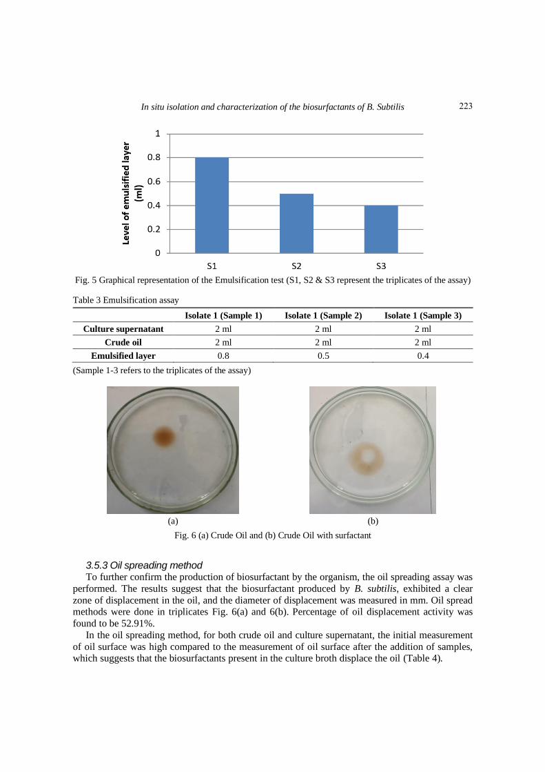
Fig. 6 (a) Crude Oil and (b) Crude Oil with surfactant
3.5.3 Oil spreading method To further confirm the production of biosurfactant by the organism, the oil spreading assay was
performed. The results suggest that the biosurfactant produced by B. subtilis, exhibited a clear
zone of displacement in the oil, and the diameter of displacement was measured in mm. Oil spread methods were done in triplicates Fig. 6(a) and 6(b). Percentage of oil displacement activity was
found to be 52.91%. In the oil spreading method, for both crude oil and culture supernatant, the initial measurement
of oil surface was high compared to the measurement of oil surface after the addition of samples, which suggests that the biosurfactants present in the culture broth displace the oil (Table 4).
223

Wasim S. Akthar, Mohamed Sheik Aadham and Arif S. Nisha
Table 4 Oil displacement activity
Isolate 1 (Sample 1) Isolate 1 (Sample 2) Isolate 1 (Sample 3)
The initial measurement of
oil surface in (mm) 50 45 43
Measurement of oil surface
after addition of samples (mm)
30 28 20
(Sample 1-3 refers to the triplicates of the activity)
Fig. 7 Graphical representation of the results of the oil spreading method. *P<0.05 (S1, S2 & S3
represents the triplicates of the assay)
Fig. 8 Hydrocarbon overlay method
3.5.4 Hydrocarbon overlay method The formation of emulsified halos surrounding the well-containing culture was considered
positive for biosurfactant production. Fig. 8 clearly shows the presence of an emulsified halo with
a radius of 2.7 cm, which suggests that the biosurfactant shows excellent emulsifying activity on
oil.
224

In situ isolation and characterization of the biosurfactants of B. Subtilis
Fig. 9 No clear zone formed around the culture
Fig. 10 Results of Ninhydrin reaction (S1-S3 refers to the triplicates of the assay)
3.5.5 Blue agar plate method The CTAB agar plate method is a semi-quantitative assay for the detection of extracellular
glycolipids or other anionic surfactants. If anionic surfactants are secreted by the microbes growing on the plate they form a dark blue halo zone. Interestingly, no clear zone was formed
around the culture, which suggests that the biosurfactant produced by B. subtilis is not an anionic
biosurfactant Fig. 9.
3.6 Characterization of biosurfactants 3.6.1 Ninhydrin reaction To determine the nature of the biosurfactant that is produced by B. subtilis, the ninhydrin test
was carried out. The Ninhydrin reaction solution will be changed into violet-blue color, indicating
that the strains can produce lipopeptides. Interestingly, the culture supernatant turned violet-blue color, suggesting that the biosurfactant produced by B. subtilis is a lipopeptide Fig. 10.
3.6.2 Sulfuric acid – phenol reaction and measurement of biosurfactants by UV-Visible spectroscopic analysis
The sulphuric acid- phenol reaction test was performed to analyse the nature of the
225

Wasim S. Akthar, Mohamed Sheik Aadham and Arif S. Nisha
Fig. 11 UV-Visible spectroscopic analysis of biosurfactant by sulphuric acid – phenol reaction
Fig. 12 Para film-M test (N-Distilled water, P-SDS, O-Sample)
Table 5 Surface tension
Surface tension
Crude oil 9.172 Nm-1
Isolate 1 (Sample 1) 1.124 Nm-1
Isolate 1 (Sample 2) 0.918 Nm-1
Isolate 1 (Sample 3) 0.9071 Nm-1
(Sample 1-3 refers to the triplicates of the strains)
biosurfactant present in B. subtilis. The results of the study show that the maximum absorbance
was observed at 440 nm, which indicates that the biosurfactant is not of glycolipid in nature, as the glycolipids always exhibit the highest absorbance at 480 nm Fig. 11.
3.6.3 Para film-M test The parafilm drop test was performed to analyze the surface tension reducing capability of the
biosurfactant. The results suggest that after 1 minute, the shape of the drop was widened due to the
presence of biosurfactant in the culture supernatant, which suggests that the former reduces the surface tension Fig. 12.
226

In situ isolation and characterization of the biosurfactants of B. Subtilis
Fig. 13 Graphical representation of the results of surface tension analysis *P<0.0.05 (S1, S2 & S3 represents the triplicates of the assay)
Fig. 14 FTIR spectra analysis of biosurfactant obtained from B. subtilis
3.6.4 Surface tension analysis To further validate the role of biosurfactant in reducing the Surface tension of the oil, this
analysis was performed by the drop weight method. Crude oil after treatment with the
biosurfactant from B. subtilis reduced the surface tension to 0.983±0.122 Nm-1. The surface tension of the crude oil without treatment was 9.172 (Table 5).
3.7 Characterization and Structural analysis of biosurfactant 3.7.1 Fourier Transform IR Spectroscopy (FT-IR) FTIR Spectral analysis was performed to characterize the nature of biosurfactant present in B.
227

Wasim S. Akthar, Mohamed Sheik Aadham and Arif S. Nisha
subtilis. The results suggested the biosurfactant extracted from B. subtilis is a lipopeptide Fig 14.
The spectrum showed a broad absorbance peak cantered around 3363 cm-1, ranging from 3100 cm-
1 to 3600 cm-1. This is a typical feature of compounds containing carbon and amino groups and is caused due to stretching vibrations of C-H and N-H bonds, present in the compound. The
absorbance peak at 2934 cm-1 indicates the presence of alkyl chain (-CH2- and –CH3). The
absorbance peak at 1706 cm-1 is attributed to the stretching vibrations of C=O bonds. The
absorbance peak at 1634 cm-1 implies that peptide groups are present in the sample. A weak absorbance signal at 1448 cm-1 is due to bending vibrations of C-H bonds associated with alkyl
chains. Another absorption peak at 1243 cm-1 is due to C-O stretching vibrations related to esters.
3.7.2 FTIR analysis of oil degradation The FTIR analysis of crude oil was carried out and data been presented in Table 6.
Fig. 15 shows that the peaks of crude oil and degraded oil. The results show that the transmittance of crude oil was less and it was decreased in degraded oil (Table 7). This might be
because of the action of biosurfactants on crude oil. The biosurfactant may break the complex
compound to simple compound in crude oil; hence the transmittance was high in the degraded oil.
Table 6 FTIR analysis of crude oil
Positions (cm-1) Intensity Assigned configuration
743 Medium CH2(bend) aliphatic
1155 Weak C-H(bend)aliphatic
1380 Strong C-H(bend)aliphatic
1467 Strong CH3(bend)aliphatic
1715 Medium C=O(stretch)aliphatic
2852 Strong CH2(bend)aliphatic
2922 Strong CH(bend)aliphatic
Fig. 15 Difference between the peaks of crude oil and degraded oil
228
In situ isolation and characterization of the biosurfactants of B. Subtilis
Table 7 FTIR analysis of degraded oil
Positions (cm-1) Intensity Assigned configuration
743 Weak CH2(bend) aliphatic
1155 Weak C-H(bend)aliphatic
1380 Medium C-H(bend)aliphatic
1467 Medium CH3(bend)aliphatic
1715 Weak C=O(stretch)aliphatic
2852 Medium CH2(bend)aliphatic
2922 Medium CH(bend)aliphatic
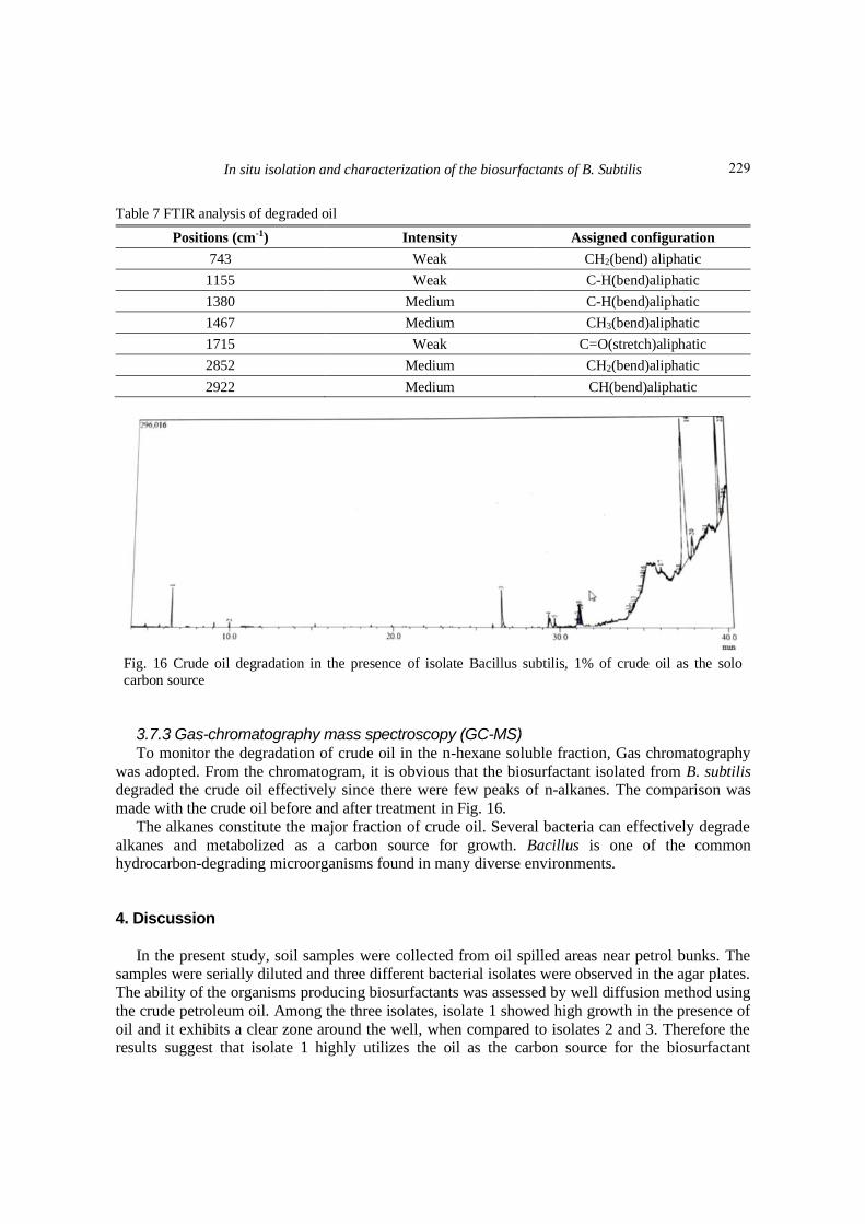
Fig. 16 Crude oil degradation in the presence of isolate Bacillus subtilis, 1% of crude oil as the solo
carbon source
3.7.3 Gas-chromatography mass spectroscopy (GC-MS) To monitor the degradation of crude oil in the n-hexane soluble fraction, Gas chromatography
was adopted. From the chromatogram, it is obvious that the biosurfactant isolated from B. subtilis degraded the crude oil effectively since there were few peaks of n-alkanes. The comparison was
made with the crude oil before and after treatment in Fig. 16.
The alkanes constitute the major fraction of crude oil. Several bacteria can effectively degrade
alkanes and metabolized as a carbon source for growth. Bacillus is one of the common hydrocarbon-degrading microorganisms found in many diverse environments.
4. Discussion
In the present study, soil samples were collected from oil spilled areas near petrol bunks. The samples were serially diluted and three different bacterial isolates were observed in the agar plates.
The ability of the organisms producing biosurfactants was assessed by well diffusion method using
the crude petroleum oil. Among the three isolates, isolate 1 showed high growth in the presence of
oil and it exhibits a clear zone around the well, when compared to isolates 2 and 3. Therefore the results suggest that isolate 1 highly utilizes the oil as the carbon source for the biosurfactant
229

Wasim S. Akthar, Mohamed Sheik Aadham and Arif S. Nisha
production. The isolates were identified through Gram’s staining. The Gram’s staining results
show that isolate 1 was Gram-positive, whereas both isolates 2 & 3 were Gram-negative. The
characterization of the isolate 1 was done by biochemical tests and 16SrRNA gene sequencing. The results suggest that the isolate 1 was Bacillus subtilis and the phylogenetic tree was
constructed using the neighbour-joining method. Bacillus spp., are gram-positive bacteria, it can
survive even in harsh conditions like petroleum reservoirs (Al-Wahaibi et al. 2014). Several
reports suggest that Bacillus subtilis has the capability of degrading hydrocarbons and it proliferates quickly acclimatizing with the harsh environments. Moreover, it takes specific time
duration for their populations to grow in response to the influx of new resources (Parthipan et al.
2017). In the present study, screening of biosurfactant activity was done by various methods including drop collapsing test, emulsification test, oil spreading method, hydrocarbon overlay
method, and blue agar plate method. In the drop collapse method, Bacillus subtilis showed the
positive result by the appearance of a flat drop in the oil, the flat drop is the positive result for the drop collapsing test, which was suggested by Youssef et al. (2004).
The results of emulsification activity of Bacillus subtilis show that the emulsification potential
was found to be 20.81%, as most of the Biosurfactants exhibit high emulsifying activity and
surface activity (Chakrabarti et al. 2012). The oil displacement activity is one of the important is a sign of extra-cellular biosurfactant production in the organisms (Saminathan and Rajendran 2016).
In the present study, the results of oil spreading method suggests that Bacillus subtilis exhibit a
clear zone of displacement which suggests that, the organism produces the biosurfactant extracellularly. The results of the hydrocarbon overlay method showed the formation of emulsified
halos surrounding the well-containing culture, which indicates that Bacillus subtilis has excellent
emulsifying activity on oil. The CTAB agar plate method is a semi-quantitative assay for the
detection of extracellular glycolipids or other anionic surfactants. If anionic surfactants are secreted by the microbes growing on the plate, a dark blue halo zone will be produced (Satpute et
al. 2008). In the present study, Bacillus subtilis showed a negative result, therefore the
biosurfactant produced by Bacillus subtilis is not an anionic biosurfactant. The extracted biosurfactant was characterized by ninhydrin reaction, sulfuric acid – phenol reaction. The
measurement of biosurfactants was done by ultraviolet analysis, Para film-M test, and surface
tension. In ninhydrin reaction the formation of violet-blue color suggests that the biosurfactant produced by Bacillus subtilis is a lipopeptide (Zhang et al. 2012). The results of the sulfuric acid –
phenol reaction showed that the maximum absorbance was observed at 440 nm, which indicates
that the nature of biosurfactant produced by Bacillus subtilis is not a glycolipid, as the glycolipids
always exhibit the highest absorbance at 480 nm (Zhang et al. 2012). The Para film-M test result showed that the drop was widened due to the presence of biosurfactant and it reduces the surface
tension. Surface tension analysis was performed by the drop weight method. The result showed
that the biosurfactant of Bacillus subtilis reduces the surface tension of the oil. The structural analysis of biosurfactant was done by Fourier Transform IR Spectroscopy (FT-IR) and Gas-
chromatography mass spectroscopy (GC-MS). Infrared spectroscopy is a simple method for
structural analysis. The results of the FT-IR analysis suggest that the biosurfactant extracted from Bacillus subtilis is a lipopeptide (Ahmad et al. 2018) and also the results of FT-IR in oil
degradation showed that the biosurfactant may break the complex compounds to simple
compounds in crude oil. In gas-chromatography mass spectroscopy (GC-MS) results showed that
the biosurfactant isolated from Bacillus subtilis can effectively degrade alkanes when compared to the GC-MS chromatogram of the crude oil (Wang et al. 2006) and the bacterium might utilize the
oil as sole carbon source for its growth (Nisha and Kinnari 2017).
230

In situ isolation and characterization of the biosurfactants of B. Subtilis
5. Conclusions
Bacillius subtilis was found to degrade the crude oil efficiently and the organism utilizes the oil
as sole carbon source. The organism was found to possess biosurfactants, which is of lipopeptide
type. Upon further purification and characterization, the biosurfactant could be produced in large scale and could be utilized for the remediation of oil contaminated environment. This eco-friendly
method will be more advantageous and less-toxic when compared to the chemical treatment
methods that are currently being employed for the remediation processes.
Acknowledgements
The authors gratefully acknowledge the Management of Srimad Andavan Arts and Science College, Trichy for their support and able guidance. The authors would also like express their
sincere thanks to staff members of Department of Physics, SAASC for their support to perform the
surface tension analysis.
References Ahmad, I., Sohail, S. M., Khan, H., Khan, R. and Ahmad, W. (2018), “Characterization of petroleum crude
oils by Fourier transform Infrared (FT-IR) and gas chromatography-mass spectrometerys”, Petrol. Petrochem. Eng., 2(2), 1-7.
Al-Wahaibi, Y.M., Al-Bahry, S.N., Elshafie, A.E., Al-Bemani, A.S., Joshi, S.J. and Al-Bahri, A.K. (2014),
“Screening of minimal salt media for biosurfactant production by bacillus spp”, World Acad. Sci. Eng.
Technol. Int. J. Environ. Ecol. Eng., 8(2).
Bodour, A.A., Guerrero-Barajas, C., Jiorle, B.V., Malcomson, M.E., Paull, A.K., Somogyi, A., Trinh, L.N.,
Bates, R.B. and Maier, R.M. (2004), “Structure and characterization of flavolipids, a novel class of
biosurfactants produced by Flavobacterium sp. strain MTN11”, Appl. Environ. Microbiol., 70(1), 114-
120. https://doi.org/10.1128/AEM.70.1.114-120.2004.
Calvo, C., Manzanera, M., Silva-Castro, G.A., Uad, I. and González-López, J. (2009), “Application of
bioemulsifiers in soil oil bioremediation processes. Future prospects”, Sci. Total Environ., 407(12), 3634-
3640. https://doi.org/10.1016/j.scitotenv.2008.07.008. Chakrabarti, S. (2012), “Bacterial biosurfactant: Characterization, antimicrobial and metal remediation
properties”, Ph.D. Dissertation, National Institute of Technology Rourkela, Odisha, India.
Chang, M.W., Holoman, T.P. and Yi, H. (2008), “Molecular characterization of surfactant-driven microbial
community changes in anaerobic phenanthrene-degrading cultures under methanogenic conditions”,
Biotechnol. Lett., 30(9), 1595-1601. https://doi.org/10.1007/s10529-008-9731-4.
Daxini, N. and Mistry, K. (2018), “Biosurfactant assistance in crude oil degradation by halophilic Bacillus
cereusND1”, Niscair csir, 47(8),1640-1647.
Olivera, N.L., Commendatore, M.G., Delgado, O. and Esteves, J.L. (2003), “Microbial characterization and
hydrocarbon biodegradation potential of natural bilge waste microflora”, J. Industr. Microbiol.
Biotechnol., 30(9), 542-548. https://doi.org/10.1007/s10295-003-0078-5.
Parthipan, P., Preetham, E., Machuca, L.L., Rahman, P.K., Murugan, K. and Rajasekar, A. (2017), “Biosurfactant and degradative enzymes mediated crude oil degradation by bacterium Bacillus subtilis
A1”, Front. Microbiol., 8, 193. https://doi.org/10.3389/fmicb.2017.00193.
Saminathan, P. and Rajendran, P. (2016), “Molecular identification and characterization of the biosurfactant
produced by pseudomonas aeruginosa-PSPA15 from the oil contaminated soil”, Int. J. Curr. Microbiol.
231

Wasim S. Akthar, Mohamed Sheik Aadham and Arif S. Nisha
App. Sci., 5, 708. http://doi.org/10.20546/ijcmas.2016.508.080. Santhini, K. and Parthasarathi, R. (2014), “Isolation and screening of biosurfactant producing
microorganisms from hydrocarbon contaminated soils from automobile workshop”, Int. J. Pharmaceut.
Biol. Arch., 5(2), 158-167.
Saravanan, V. and Vijayakumar, S. (2012), “Isolation and screening of biosurfactant producing
microorganisms from oil contaminated soil”, J. Acad. Indus. Res., 1(5), 264-268.
Satpute, S.K., Bhawsar, B.D., Dhakephalkar, P.K. and Chopade, B.A. (2008), “Assessment of different
screening methods for selecting biosurfactant producing marine bacteria”, Csir, 37(3), 243-250.
Vijayakumar, S. and Saravanan, V. (2015), “Biosurfactants-types, sources and applications”, Res. J.
Microbiol., 10(5), 181. http://doi.org/10.3923/jm.2015.181.192.
Whang, L.M., Liu, P.W.G., Ma, C.C. and Cheng, S.S. (2008), “Application of biosurfactants, rhamnolipid,
and surfactin, for enhanced biodegradation of diesel-contaminated water and soil”, J. Hazard. Mater., 151(1), 155-163. https://doi.org/10.1016/j.jhazmat.2007.05.063.
Youssef, N.H., Duncan, K.E., Nagle, D.P., Savage, K.N., Knapp, R.M. and McInerney, M.J. (2004),
“Comparison of methods to detect biosurfactant production by diverse microorganisms”, J. Microbiol.
Meth., 56(3), 339-347. https://doi.org/10.1016/j.mimet.2003.11.001.
Zhang, X., Xu, D., Zhu, C., Lundaa, T. and Scherr, K.E. (2012), “Isolation and identification of
biosurfactant producing and crude oil degrading Pseudomonas aeruginosa strains”, Chem. Eng. J., 209,
138-146. https://doi.org/10.1016/j.cej.2012.07.110.
CC
232