in situ forming reduction-sensitive degradable nanogels for facile loading and triggered...
TRANSCRIPT
In Situ Forming Reduction-Sensitive Degradable Nanogels for FacileLoading and Triggered Intracellular Release of ProteinsWei Chen,†,‡ Meng Zheng,† Fenghua Meng,† Ru Cheng,† Chao Deng,† Jan Feijen,†,‡
and Zhiyuan Zhong*,†
†Biomedical Polymers Laboratory and Jiangsu Key Laboratory of Advanced Functional Polymer Design and Application, Departmentof Polymer Science and Engineering, College of Chemistry, Chemical Engineering and Materials Science, Soochow University,Suzhou, 215123, P. R. China‡Department of Polymer Chemistry and Biomaterials, Faculty of Science and Technology, MIRA Institute for Biomedical Technologyand Technical Medicine, University of Twente, P.O. Box 217, 7500 AE Enschede, The Netherlands
*S Supporting Information
ABSTRACT: In situ forming reduction-sensitive degradable nano-gels were designed and developed based on poly(ethylene glycol)-b-poly(2-(hydroxyethyl) methacrylate-co-acryloyl carbonate) (PEG-P(HEMA-co-AC)) block copolymers for efficient loading as well astriggered intracellular release of proteins. PEG-P(HEMA-co-AC)copolymers were prepared with controlled Mn of 9.1, 9.5, and 9.9kg/mol and varying numbers of AC units per molecule of 7, 9 and11, respectively (denoted as copolymer 1, 2, and 3) by reversibleaddition−fragmentation chain transfer copolymerization. Thesecopolymers were freely soluble in phosphate buffer but formed disulfide-cross-linked nanogels with defined sizes rangingfrom 72.5 to 124.1 nm in the presence of cystamine via ring-opening reaction with cyclic carbonate groups. The sizes of nanogelsdecreased with increasing AC units as a result of increased cross-linking density. Dynamic light scattering studies showed thatthese nanogels though stable at physiological conditions were rapidly dissociated in response to 10 mM dithiothreitol (DTT).Interestingly, FITC-labeled cytochrome C (FITC−CC) could be readily loaded into nanogels with remarkable loadingefficiencies (up to 98.2%) and loading contents (up to 48.2 wt.%). The in vitro release studies showed that release of FITC−CCwas minimal under physiological conditions but significantly enhanced under reductive conditions in the presence of 10 mMDTT with about 96.8% of FITC−CC released in 22 h from nanogel 1. In contrast, protein release from 1,4-butanediamine cross-linked nanogels (reduction-insensitive control) remained low under otherwise the same conditions. MTT assays showed thatthese nanogels were nontoxic to HeLa cells up to a tested concentration of 2 mg/mL. Confocal microscopy results showed thatnanogel 1 delivered and released FITC−CC into the perinuclei region of HeLa cells following 8 h incubation. CC-loadedreductively degradable nanogels demonstrated apparently better apoptotic activity than free CC as well as reduction-insensitivecontrols. These in situ forming, surfactant and oil-free, and reduction-sensitive degradable nanogels are highly promising fortargeted protein therapy.
■ INTRODUCTION
In the past decades, protein drugs have emerged as effectivetreatments for severe human diseases, including cancer.1−3 Ascompared to chemotherapeutics, protein drugs have merits ofhigh specificity, high therapeutic activity, and low cytotoxicity.The clinical applications of protein drugs, however, encounterseveral challenges such as swift degradation in vivo, possibleimmune responses, inferior cellular uptake, and poor intra-cellular trafficking.4 The clinical success of protein therapy,therefore, relies on the development of safe and efficientdelivery systems.In recent years, various types of polymeric nanocarriers
including polyion complex micelles,5−7 polymersomes,8−10
nanocapsules,11−13 and nanogels14−16 have been developedfor protein delivery. Notably, nanogels with advantages ofrelatively uncomplicated fabrication, high protein loadingcapacity, high stability, and controlled protein release profile
have appeared as one of the most ideal protein nano-carriers.17,18 For example, Akiyoshi et al. reported that cationiccholesteryl-modified pullulan nanogels mediated effectiveintracellular delivery of various proteins including β-galactosi-dase,19 cytokines,20 and cancer vaccines.21 De Smedt et al.reported the controlled release of bovine serum albumin (BSA)and lysozyme from degradable dextran nanogels.22 Park et al.found that heparin−pluronic nanogels were able to deliverRNase A into HeLa cells giving significant anticancer effects.23
The codelivery of paclitaxel and DNase using heparin−pluronicnanogels exhibited a dose-dependent synergistic cytotoxicity.24
In particular, bioresponsive nanogels that release cargos inresponse to endosomal pH or cytoplasmic glutathione (GSH)
Received: February 7, 2013Revised: March 9, 2013Published: March 12, 2013
Article
pubs.acs.org/Biomac
© 2013 American Chemical Society 1214 dx.doi.org/10.1021/bm400206m | Biomacromolecules 2013, 14, 1214−1222

have received recent interests.25−29 For instance, Akiyoshi et al.reported that pH-sensitive pullulan nanogels released proteinsmuch faster under acidic conditions than at physiological pH.30
Kataoka and Matyjaszewski developed reduction-sensitivedegradable nanogels by inverse miniemulsion atom transferradical polymerization (ATRP).31 Park et al. reported thatdisulfide-cross-linked heparin-Pluronic nanogels with enhancedcolloidal stability effectively released encapsulated RNase Ainside cells.32 Chen et al. reported one-step preparation ofdisulfide-cross-linked PEG-polypeptide nanogels by NCAcopolymerization using L-cystine N-carboxyanhydride as across-linker.33 Thayumanavan et al. developed reduction-sensitive self-cross-linked nanogels based on a randomcopolymer containing pendant oligoethyleneglycol and pyridyldisulfide side chains.34,35 The disulfide-cross-linked nanogelsare of particular interest for intracellular protein release in thatthey on one hand possess superior colloidal stability and on theother hand are prone to rapid de-cross-linking and dissociationinside cells due to the presence of a high reducing potential inthe cytoplasms and nuclei of cells.36,37 However, most of thereported nanogels require the use of oil, surfactants, and toxicreagents that might lead to protein denaturation/deactivationas well as cytotoxicity concerns. It should further be noted thatpreformed nanogels also often exhibit inferior drug loading andsignificant burst release.In this paper, we report in situ forming reduction-sensitive
degradable nanogels from PEG-P(HEMA-co-AC) block co-polymers using cystamine (Cys) as a cross-linker for facileloading and triggered intracellular release of proteins (Scheme1). To the best of our knowledge, this represents a first reporton in situ forming, surfactant and oil-free, catalyst-free,chemically cross-linked, and stimuli-sensitive degradable nano-gels. Poly((2-hydroxyethyl) methacrylate) (PHEMA) possessesexcellent biocompatibility and is used widely for biomedicalapplications.38 The pendant cyclic carbonate units wouldfacilitate cross-linking of PEG-P(HEMA-co-AC) with Cysunder an aqueous condition via ring-opening reaction. Notably,our cross-linking reaction does not involve any catalysts or yieldany byproducts. These in situ forming degradable nanogelscould efficiently load proteins under mild conditions whilequickly releasing proteins inside cells. Here, the preparation ofin situ forming reduction-responsive degradable nanogels,loading and reduction-triggered release of CC, and intracellularprotein release, as well as antitumor activity of CC-loadednanogels, were investigated.
■ EXPERIMENTAL SECTIONMaterials. Methoxy poly(ethylene glycol) (PEG, Mn = 5.0 kg/mol,
PDI = 1.03) was purchased from Fluka. PEG was dried by dissolutionin anhydrous toluene followed by azeotropic distillation. 5-Methyl-5-prop-2-enoyloxymethyl-1,3-dioxan-2-one (acryloyl carbonate, AC) wassynthesized according to our previous report.39 4-Cyanopentanoic aciddithionaphthalenoate (CPADN) was synthesized according to thedescribed procedure for 4-cyanopentanoic acid dithiobenzoate.40 PEG-CPADN was synthesized by an esterification reaction of PEG andCPADN, similar to the synthesis of CPADN-PCL-CPADN macro-RAFT agent.41 2-Hydroxyethyl methacrylate (HEMA, 97%, Fluka)was purified by passing through a basic alumina column before use.Azobisisobutyronitrile (AIBN, 98%, J&K) was recrystallized frommethanol. Cystamine dihydrochloride (Cys·2HCl, >98%, Alfa Aesar),dithiothreitol (DTT, 99%, Merck), fluorescein isothiocyanate (FITC,95%, Fluka), cytochrome C from equine heart (CC, Sigma), 1,4-butanediamine (BDA, 99%, J&K), and 2,2′-azino-bis(3-ethylbenzo-thiazoline-6-sulfonic acid) diammonium salt (ABTS, Amresco) wereused as received.
Characterization. 1H NMR spectra were recorded on a UnityInova 400 spectrometer operating at 400 MHz using deuterateddimethylsulfoxide (DMSO-d6) as the solvent. The chemical shifts werecalibrated against residual solvent signals of DMSO-d6. The molecularweight and polydispersity of copolymers were determined by a Waters1515 gel permeation chromatograph (GPC) instrument equipped withtwo linear PLgel columns (500 Å and Mixed-C) following a guardcolumn and a differential refractive-index detector. The measurementswere performed using THF as the eluent at a flow rate of 1.0 mL/minat 30 °C and a series of narrow polystyrene standards for thecalibration of the columns. The size distribution of nanogels wasdetermined using dynamic light scattering (DLS). Measurements werecarried out at 25 °C using a Zetasizer Nano-ZS from MalvernInstruments equipped with a 633 nm He−Ne laser using back-scattering detection. Transmission electron microscopy (TEM) wasperformed using a Tecnai G220 TEM operated at an acceleratingvoltage of 200 kV. The samples were prepared by dropping 10 μL ofnanogel dispersion (0.2 mg/mL) on the copper grid followed bystaining with phosphotungstic acid.
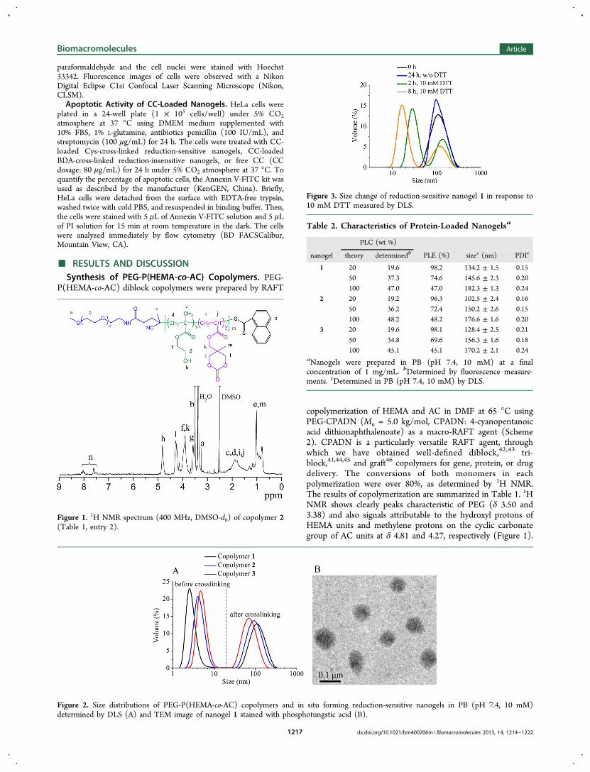
Synthesis of PEG-P(HEMA-co-AC) Copolymers. PEG-P-(HEMA-co-AC) diblock copolymer was synthesized by reversibleaddition−fragmentation chain transfer (RAFT) copolymerization ofHEMA and AC using PEG-CPADN (Mn = 5.0 kg/mol) as a macro-RAFT agent and AIBN as an initiator (Scheme 2). In a typicalexample, under a nitrogen atmosphere, HEMA (64 mg, 490 μmol),AC (36 mg, 180 μmol), PEG-CPADN (100 mg, 20 μmol), AIBN(0.164 mg, 1.0 μmol), and 3 mL of DMF were added into a 10 mLSchlenk flask. The flask was sealed and placed in an oil baththermostatted at 65 °C. The reaction proceeded with magnetic stirringfor 24 h. One sample was taken for the determination of monomer
Scheme 1. Illustration of In Situ Forming Reduction-Sensitive Nanogels for Facile Loading and Triggered Release of Proteins
Biomacromolecules Article
dx.doi.org/10.1021/bm400206m | Biomacromolecules 2013, 14, 1214−12221215

conversions. The resulting copolymers were isolated by precipitationin diethyl ether, filtration, and drying in vacuo at room temperature.Yield: 65−70%. 1H NMR (400 MHz, DMSO-d6) PEG: δ 3.50, 3.38;HEMA moieties: δ 4.81, 3.90, 3.58, 1.89, 1.01; AC moieties: δ 4.27,3.90, 1.89, 0.79.In Situ Formation of Reduction-Sensitive Nanogels. To a
solution of PEG-P(HEMA-co-AC) copolymer (2.0 mg) in phosphatebuffer (PB, pH 7.4, 10 mM) at a concentration of 1.0 mg/mL wasadded Cys in PB (1.0 mg/mL). The molar ratio between aminogroups in Cys and cyclic carbonate groups in PEG-P(HEMA-co-AC)copolymer was set at 1/1. The combined solution following adjustingits pH to 8.0 was stirred at 50 °C for 4 h. The size distribution offormed nanogels was determined by DLS and TEM.Size Change of Nanogels Triggered by DTT. Reduction-
sensitive PEG-P(HEMA-co-AC) nanogels were prepared at aconcentration of 1.0 mg/mL. The size change of nanogels in responseto 10 mM DTT was monitored by DLS. In a typical experiment, to 1.0mL of nanogel dispersion in PB was added 10 μL of 1.0 M DTT (finalDTT concentration is 10 mM). At different time intervals, the sizedistribution of nanogels was determined. BDA-cross-linked nanogelswere used as a reduction-insensitive control.Encapsulation and Triggered Release of FITC−CC. To 200 μL
of PEG-P(HEMA-co-AC) copolymer solution (5.0 mg/mL) wasadded varying volumes of CC or FITC-labeled cytochrome C (FITC−CC) solution in PB (pH 7.4, 10 mM) to obtain protein/polymer ratiosof 20, 50, and 100 wt %, respectively. Cys-cross-linked nanogels wereformed as described above. Free protein was removed by dialysis(MWCO 500 kDa) against PB (pH 7.4, 10 mM) at room temperaturefor 10 h. The dialysis medium was refreshed each hour.To determine protein loading contents (PLC), thus prepared
protein-loaded nanogels were treated for 1 h with 100 mM DTT,diluted 1000 times, and analyzed using fluorescence measurements(FLS920, excitation at 492 nm, emission from 510 to 690 nm). Theamount of loaded protein was obtained by comparing the fluorescenceof the above samples to a calibration curve of known FITC−CC
concentrations. PLC and protein loading efficiency (PLE) werecalculated according to the following formula:
=
×
PLC(wt%) (weight of loaded protein/weight of copolymer)
100%
=
×
PLE(%) (weight of loaded protein/weight of protein in the feed)
100%Triggered protein release from reduction-sensitive PEG-P(HEMA-
co-AC) nanogels was investigated using a dialysis method (MWCO500 kDa) at 37 °C in two different media, that is, PB (100 mM, pH7.4) and PB (100 mM, pH 7.4) with 10 mM DTT. The protein releasefrom BDA-cross-linked nanogels was used as reduction-insensitivecontrol. At desired time intervals, 5 mL of release media was taken outand replenished with an equal volume of corresponding fresh media.The amount of released protein was determined by fluorescencemeasurements (FLS920, excitation at 492 nm, emission from 510 to690 nm). The release experiments were conducted in triplicate.
Activity of Released CC by ABTS Assay. The activity of CCreleased from CC-loaded PEG-P(HEMA-co-AC) nanogels wasexamined using 2,2′-azino-bis(3-ethylbenzothiazoline-6-sulfonic acid)(ABTS) assays. CC was loaded into Cys-cross-linked nanogels asmentioned above and released in PB (pH 7.4, 100 mM) for 2 days.The released CC was collected, quantified by bicinchoninic acid(BCA) method, and diluted to a final concentration of 10 μg/mLusing PB (pH 7.4, 100 mM). To CC solution in quartz cuvettes weresimultaneously added 20 μL of hydrogen peroxide solution (45 mM)and 200 μL of ABTS solution (1.0 mg/mL) in PB. The absorbance at410 nm of the oxidized product was monitored for 10 min. Native CCat a concentration of 10 μg/mL in PB (pH 7.4, 100 mM) was used as acontrol.
MTT Assay. The cytotoxicity of PEG-P(HEMA-co-AC) nanogelswas studied by MTT assays in HeLa cells. The cells were seeded ontoa 96-well plate at a density of 1 × 10⁴ cells per well in 100 μL ofDulbecco’s modified Eagle medium (DMEM) containing 10% FBSand incubated for 24 h (37 °C, 5% CO2). The medium was replacedby 80 μL of fresh DMEM medium containing 10% FBS and 20 μL ofvarious concentrations of nanogel dispersion. The cells were incubatedfor another 24 h, the medium was aspirated and replaced by 100 μL offresh medium, and 20 μL of MTT solution (5.0 mg/mL) was added.The cells were incubated for 4 h, and then 100 μL of DMSO wasadded to dissolve the resulting purple crystals. The optical densities at570 nm were measured using a microplate reader. The relative cellviability (%) was determined by comparing the absorbance at 570 nmwith control wells containing only cell culture medium. Data arepresented as average ± standard deviation (n = 4).
Confocal Microscopy Observation of HeLa Cells Incubatedwith FITC−CC Loaded Nanogels. HeLa cells were plated onmicroscope slides in a 24-well plate (5 × 10⁴ cells/well) using DMEMmedium containing 10% FBS. The cells were incubated withpredetermined amounts of FITC−CC loaded nanogels or freeFITC−CC at 37 °C under 5% CO2 atmosphere. After 4 h incubation,the culture medium was removed and the cells on microscope plateswere washed three times with PBS. The cells were fixed with 4%
Scheme 2. Synthesis of PEG-P(HEMA-co-AC) DiblockCopolymer Using RAFT Polymerization
Table 1. Synthesis of PEG-P(HEMA-co-AC) Diblock Copolymers and Nanogels
HEMA/AC/CPADNa Mn (kg/mol) size (nm)
copolymer design 1H NMR 1H NMRb GPCc PDI GPCc polymerd nanogele
1 24.6/9/1 20.8/7/1 9.1 7.8 1.09 5.2 ± 0.8 124.1 ± 1.32 24.6/12/1 20.8/9/1 9.5 8.3 1.30 8.3 ± 1.1 95.8 ± 2.13 24.6/15/1 20.8/11/1 9.9 9.5 1.10 10.5 ± 0.6 72.5 ± 1.8
aPolymerization was carried out in DMF at 65 °C for 24 h using PEG-CPADN (Mn = 5.0 kg/mol) as a macro-RAFT agent and AIBN as an initiatorat three different HEMA/AC/CPADN molar ratios. bEstimated by 1H NMR end-group analysis. cGPC was performed using THF as an eluent at aflow rate of 1.0 mL/min at 35 °C and a series of narrow polystyrene standards for the calibration of the columns. dHydrodynamic size of copolymerin PB (pH 7.4, 10 mM) determined by DLS. eHydrodynamic size of in situ forming reduction-sensitive nanogels in PB (pH 7.4, 10 mM) determinedby DLS.
Biomacromolecules Article
dx.doi.org/10.1021/bm400206m | Biomacromolecules 2013, 14, 1214−12221216
paraformaldehyde and the cell nuclei were stained with Hoechst33342. Fluorescence images of cells were observed with a NikonDigital Eclipse C1si Confocal Laser Scanning Microscope (Nikon,CLSM).Apoptotic Activity of CC-Loaded Nanogels. HeLa cells were
plated in a 24-well plate (1 × 10⁵ cells/well) under 5% CO2atmosphere at 37 °C using DMEM medium supplemented with10% FBS, 1% L-glutamine, antibiotics penicillin (100 IU/mL), andstreptomycin (100 μg/mL) for 24 h. The cells were treated with CC-loaded Cys-cross-linked reduction-sensitive nanogels, CC-loadedBDA-cross-linked reduction-insensitive nanogels, or free CC (CCdosage: 80 μg/mL) for 24 h under 5% CO2 atmosphere at 37 °C. Toquantify the percentage of apoptotic cells, the Annexin V-FITC kit wasused as described by the manufacturer (KenGEN, China). Briefly,HeLa cells were detached from the surface with EDTA-free trypsin,washed twice with cold PBS, and resuspended in binding buffer. Then,the cells were stained with 5 μL of Annexin V-FITC solution and 5 μLof PI solution for 15 min at room temperature in the dark. The cellswere analyzed immediately by flow cytometry (BD FACSCalibur,Mountain View, CA).
■ RESULTS AND DISCUSSIONSynthesis of PEG-P(HEMA-co-AC) Copolymers. PEG-
P(HEMA-co-AC) diblock copolymers were prepared by RAFT
copolymerization of HEMA and AC in DMF at 65 °C usingPEG-CPADN (Mn = 5.0 kg/mol, CPADN: 4-cyanopentanoicacid dithionaphthalenoate) as a macro-RAFT agent (Scheme2). CPADN is a particularly versatile RAFT agent, throughwhich we have obtained well-defined diblock,42,43 tri-block,41,44,45 and graft46 copolymers for gene, protein, or drugdelivery. The conversions of both monomers in eachpolymerization were over 80%, as determined by 1H NMR.The results of copolymerization are summarized in Table 1. 1HNMR shows clearly peaks characteristic of PEG (δ 3.50 and3.38) and also signals attributable to the hydroxyl protons ofHEMA units and methylene protons on the cyclic carbonategroup of AC units at δ 4.81 and 4.27, respectively (Figure 1).
Figure 1. 1H NMR spectrum (400 MHz, DMSO-d6) of copolymer 2(Table 1, entry 2).
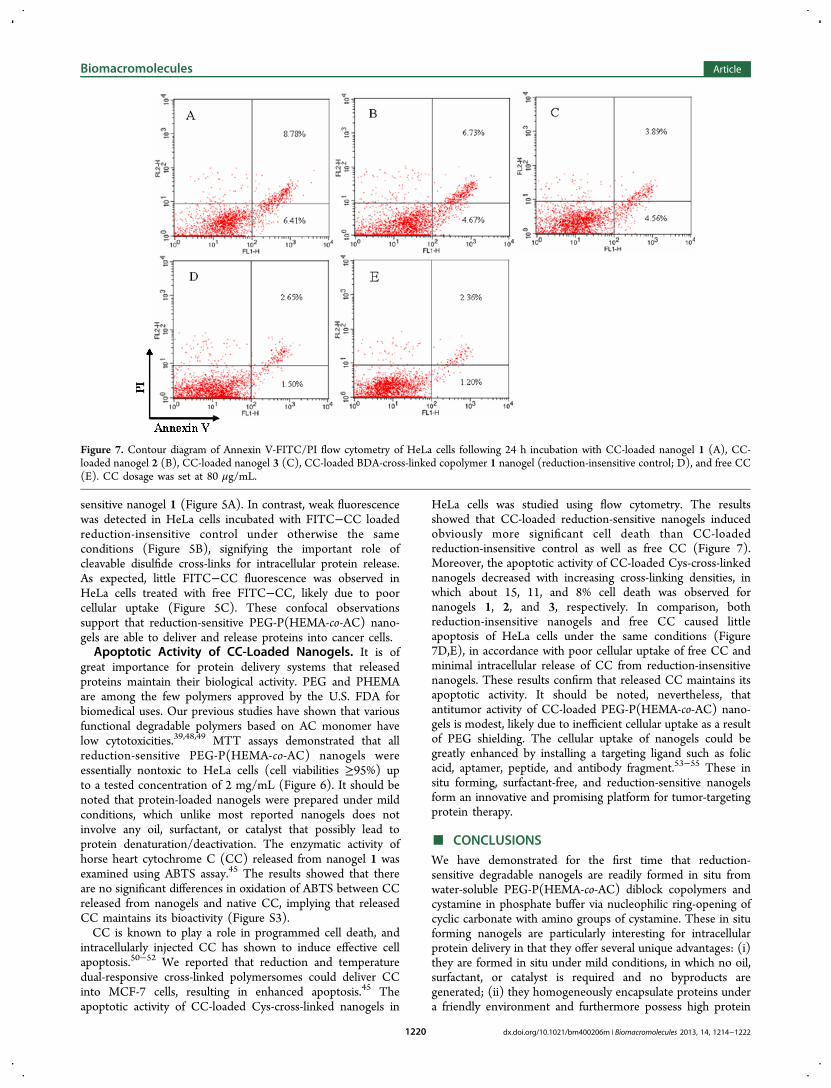
Figure 2. Size distributions of PEG-P(HEMA-co-AC) copolymers and in situ forming reduction-sensitive nanogels in PB (pH 7.4, 10 mM)determined by DLS (A) and TEM image of nanogel 1 stained with phosphotungstic acid (B).
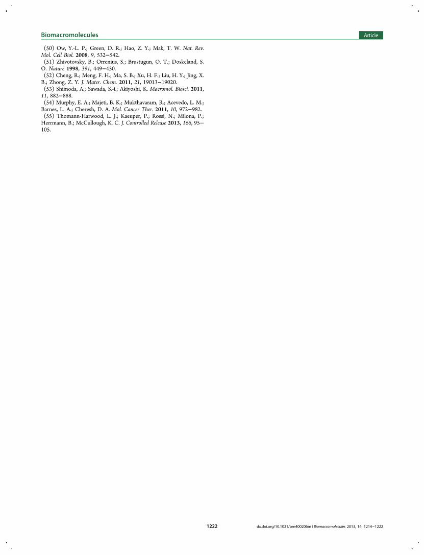
Figure 3. Size change of reduction-sensitive nanogel 1 in response to10 mM DTT measured by DLS.
Table 2. Characteristics of Protein-Loaded Nanogelsa
PLC (wt %)
nanogel theory determinedb PLE (%) sizec (nm) PDIc
1 20 19.6 98.2 134.2 ± 1.5 0.1550 37.3 74.6 145.6 ± 2.3 0.20100 47.0 47.0 182.3 ± 1.3 0.24
2 20 19.2 96.3 102.5 ± 2.4 0.1650 36.2 72.4 150.2 ± 2.6 0.15100 48.2 48.2 176.6 ± 1.6 0.20
3 20 19.6 98.1 128.4 ± 2.5 0.2150 34.8 69.6 156.3 ± 1.6 0.18100 45.1 45.1 170.2 ± 2.1 0.24
aNanogels were prepared in PB (pH 7.4, 10 mM) at a finalconcentration of 1 mg/mL. bDetermined by fluorescence measure-ments. cDetermined in PB (pH 7.4, 10 mM) by DLS.
Biomacromolecules Article
dx.doi.org/10.1021/bm400206m | Biomacromolecules 2013, 14, 1214−12221217

The chemical shift of methylene protons on the cycliccarbonate group remained unchanged, indicating that thecyclic carbonate remained intact during copolymerization andsubsequent workup procedures. The average numbers ofHEMA and AC units per P(HEMA-co-AC) block could bedetermined by comparing the intensities of signals at δ 4.81 andδ 4.27 with 3.38 (methoxyl protons of PEG), respectively.Notably, the results demonstrated that the number of AC unitsper polymer chain increased from 7 and 9 to 11 (denoted ascopolymers 1−3) with increasing the amount of AC monomerin the feed (Table 1). GPC results showed that these diblockcopolymers had unimodal distributions with moderatepolydispersities of 1.09−1.30 (Table 1), further supportingthe successful synthesis of PEG-P(HEMA-co-AC) copolymers.Formation of Reduction-Sensitive Cross-Linked Nano-
gels. PEG-P(HEMA-co-AC) copolymers were well-soluble inPB (pH 7.4, 10 mM). DLS studies showed that PEG-P(HEMA-co-AC) copolymers had average hydrodynamic diametersranging from 5.2 to 10.5 nm at a polymer concentration of1.0 mg/mL (Figure 2A), indicating that they exist as a unimerin PB. Disulfide-cross-linked nanogels were prepared by addingequivalent amounts of Cys relative to the AC units into thecopolymer solution in PB followed by adjusting the pH of thesolution pH to 8.0 and stirring at 50 °C for 4 h. Interestingly,Figure 2A shows that nanogels with average sizes varying from72.5 to 124.1 nm, depending on the amounts of AC units in thecopolymers, and moderate polydispersity indices (PDI) of0.16−0.21 were formed. The sizes of nanogels decreased withincreasing AC contents, likely due to increased cross-linking
densities. TEM micrographs showed that these nanogels had aspherical morphology with particle sizes in accordance withthose determined by DLS (Figure 2B). We hypothesized thatnanogels were formed through nucleophilic ring-openingreaction of cyclic carbonates with the amino groups in Cys.To confirm our hypothesis, a model reaction was carried out bydropwise adding a solution of copolymer 1 in PB into BDAsolution (20-fold excess, to prevent cross-linking) at 50 °C for 4h. The product was isolated and analyzed by 1H NMR. Theresults revealed that signals at δ 4.27 due to methylene protonson the cyclic carbonate of AC units vanished (Figure S2),indicating complete ring-opening of cyclic carbonate groups.This model reaction confirms that the cyclic carbonates inPEG-P(HEMA-co-AC) copolymers undergo rapid ring-openingreaction in the presence of diamines under mild conditions, inline with a previous report by Endo et al. for ring-opening ofsix-membered cyclic carbonates with diamines.47
The reduction sensitivity of Cys-cross-linked PEG-P(HEMA-co-AC) nanogels was studied by monitoring change of nanogelsizes over time in response to 10 mM DTT using DLS. Theresults showed that nanogel 1 was quickly dissociated in thepresence of 10 mM DTT in which a large population withreduced sizes of about 30 and 15 nm was observed in 2 and 8 h,respectively (Figure 3). In contrast, little size change wasdetected for BDA-cross-linked nanogels (reduction-insensitivecontrol) in 24 h under the same reducing conditions (FigureS2). Moreover, no size change was observed for nanogel 1 in 12h in the absence of DTT (Figure 3). It is evident, therefore, thatCys-cross-linked PEG-P(HEMA-co-AC) nanogels can berapidly disrupted under an intracellular-mimicking reducingcondition.
Loading and Triggered Release of Proteins. Proteinscould be readily loaded into PEG-P(HEMA-co-AC) nanogelsby mixing PB solutions of copolymer and protein prior to cross-linking with Cys. The unloaded protein was removed byextensive dialysis. It should be noted that lysine amino groupsin protein are mostly protonated at pH 8.0 due to their highpKa of approximately 10. The much higher pKa of aminogroups in protein than Cys (pKa = 8.2) would render little, ifpresent, involvement of protein amino groups in the cross-linking reaction with cyclic carbonate. To confirm this, weperformed a control experiment on a mixture of CC and PEG-P(HEMA-co-AC) (i.e., without Cys) under otherwise the sameconditions (pH 8.0, 50 °C, 4 h). DLS showed that nanogelswere not formed. Furthermore, trinitrobenzene sulfonic acid(TNBSA) assays revealed that there was essentially no changein the amount of amino groups in protein, supporting absenceof reaction between protein and cyclic carbonates undernanogel preparation conditions. It should further be noted thatthiol groups of cysteine in CC have formed thioether groupswith the heme group and reactions between the disulfide bondsof Cys and thioether groups in CC are not anticipated. Theresults showed that all PEG-P(HEMA-co-AC) nanogels hadhigh loading capacity for FITC−CC, in which AC contentsappeared to have little influences (Table 2). For example, highPLE ranging from 96.3 to 98.2% were obtained at a theoreticalPLC of 20 wt %. PLE decreased with increasing theoreticalPLC. Notably, a high protein loading content of 48.2 wt % wasaccomplished at a theoretical PLC of 100 wt %. The size ofnanogels increased from 134.2 to 182.3 nm with increasingPLC from 19.6 to 47.0 wt %. The high protein loadingcapability of nanogels might be attributed to the hydrophobicand hydrogen bonding interactions between P(HEMA-co-AC)
Figure 4. In vitro release of FITC−CC from in situ forming reduction-sensitive PEG-P(HEMA-co-AC) nanogels in PB (pH 7.4, 100 mM) at37 °C. (A) Release of FITC−CC from nanogel 1 in the presence orabsence of 10 mM DTT. BDA-cross-linked copolymer 1 nanogel wasused as a reduction-insensitive control; (B) Release of FITC−CCfrom nanogels 1−3 in the presence or absence of 10 mM DTT.
Biomacromolecules Article
dx.doi.org/10.1021/bm400206m | Biomacromolecules 2013, 14, 1214−12221218

blocks with CC, as well as the initial charge interaction betweenthe protonated amino groups in Cys and negatively chargedgroups in CC. The hydrophobic and charge interactions would,however, decrease during nanogel formation as amino groupsin Cys react with cyclic carbonate units. Hence, interactionsbetween protein and “core-shell” nanogels following completecross-linking would become weak. The proteins could beeffectively released from nanogels upon de-cross-linking undera reductive condition. It should further be noted that protein-loaded nanogels could also be prepared at 40 °C for 8 h.The in vitro release of proteins from nanogels was
investigated using PB (100 mM, pH 7.4) at 37 °C in the
presence or absence of 10 mM DTT. As shown in Figure 4A,FITC−CC was rapidly released from nanogel 1 in the presenceof 10 mM DTT, in which 50.6 and 96.9% of FITC−CC wasreleased in 5 and 22 h, respectively. This fast protein release islikely due to de-cross-linking and disruption of nanogels undera reductive condition (Figure 3). In contrast, low FITC−CCrelease (∼30%) was observed in 22 h for reduction-insensitiveBDA-cross-linked nanogels under otherwise the same con-ditions as well as for nanogel 1 in the absence of DTT (Figure4A). Nanogels 2 and 3 exhibited similar protein release profiles,in which much faster release of FITC−CC was observed in thepresence of 10 mM DTT (Figure 4B). Notably, the proteinrelease rate decreased with increasing AC contents, wherein96.9, 80.0, and 55.6% of FITC−CC was released in 22 h fromnanogels 1, 2, and 3, respectively (Figure 4B), indicating thatprotein release rate from these nanogels could be tuned bycross-linking densities. Similar results were reported byThayumanavan et al. for release of nile red from disulfide-cross-linked nanogels with different cross-linking densities.35 Itshould be noted that in all cases no burst release of protein wasobserved. These results indicate that protein release fromdisulfide-cross-linked PEG-P(HEMA-co-AC) nanogels pro-ceeds in a controlled manner and can be triggered by areductive environment analogous to that of the intracellularcompartments such as the cytosol and the cell nucleus.
Intracellular Release of Proteins. The cellular uptake andintracellular protein release behavior of protein-loaded nanogelswas studied using CLSM in HeLa cells. Interestingly, strongfluorescence was observed in the perinuclei region of HeLa cellsfollowing 4 h incubation with FITC−CC loaded reduction-
Figure 5. CLSM images of HeLa cells incubated with FITC−CC-loaded nanogels and free FITC−CC (60 μg/mL) for 4 h. For each panel, theimages from left to right showed FITC−CC fluorescence in cells (green), cell nuclei stained by Hoechst 33342 (blue), and overlays of the twoimages. The scale bars correspond to 30 μm in all the images: (A) nanogel 1; (B) BDA-cross-linked copolymer 1 nanogel (reduction-insensitivecontrol); (C) free FITC−CC.
Figure 6. MTT assays of in situ forming reduction-sensitive PEG-P(HEMA-co-AC) nanogels. HeLa cells were incubated with nanogelsfor 24 h. Data are presented as average ± standard deviation (n = 4).
Biomacromolecules Article
dx.doi.org/10.1021/bm400206m | Biomacromolecules 2013, 14, 1214−12221219
sensitive nanogel 1 (Figure 5A). In contrast, weak fluorescencewas detected in HeLa cells incubated with FITC−CC loadedreduction-insensitive control under otherwise the sameconditions (Figure 5B), signifying the important role ofcleavable disulfide cross-links for intracellular protein release.As expected, little FITC−CC fluorescence was observed inHeLa cells treated with free FITC−CC, likely due to poorcellular uptake (Figure 5C). These confocal observationssupport that reduction-sensitive PEG-P(HEMA-co-AC) nano-gels are able to deliver and release proteins into cancer cells.Apoptotic Activity of CC-Loaded Nanogels. It is of
great importance for protein delivery systems that releasedproteins maintain their biological activity. PEG and PHEMAare among the few polymers approved by the U.S. FDA forbiomedical uses. Our previous studies have shown that variousfunctional degradable polymers based on AC monomer havelow cytotoxicities.39,48,49 MTT assays demonstrated that allreduction-sensitive PEG-P(HEMA-co-AC) nanogels wereessentially nontoxic to HeLa cells (cell viabilities ≥95%) upto a tested concentration of 2 mg/mL (Figure 6). It should benoted that protein-loaded nanogels were prepared under mildconditions, which unlike most reported nanogels does notinvolve any oil, surfactant, or catalyst that possibly lead toprotein denaturation/deactivation. The enzymatic activity ofhorse heart cytochrome C (CC) released from nanogel 1 wasexamined using ABTS assay.45 The results showed that thereare no significant differences in oxidation of ABTS between CCreleased from nanogels and native CC, implying that releasedCC maintains its bioactivity (Figure S3).CC is known to play a role in programmed cell death, and
intracellularly injected CC has shown to induce effective cellapoptosis.50−52 We reported that reduction and temperaturedual-responsive cross-linked polymersomes could deliver CCinto MCF-7 cells, resulting in enhanced apoptosis.45 Theapoptotic activity of CC-loaded Cys-cross-linked nanogels in
HeLa cells was studied using flow cytometry. The resultsshowed that CC-loaded reduction-sensitive nanogels inducedobviously more significant cell death than CC-loadedreduction-insensitive control as well as free CC (Figure 7).Moreover, the apoptotic activity of CC-loaded Cys-cross-linkednanogels decreased with increasing cross-linking densities, inwhich about 15, 11, and 8% cell death was observed fornanogels 1, 2, and 3, respectively. In comparison, bothreduction-insensitive nanogels and free CC caused littleapoptosis of HeLa cells under the same conditions (Figure7D,E), in accordance with poor cellular uptake of free CC andminimal intracellular release of CC from reduction-insensitivenanogels. These results confirm that released CC maintains itsapoptotic activity. It should be noted, nevertheless, thatantitumor activity of CC-loaded PEG-P(HEMA-co-AC) nano-gels is modest, likely due to inefficient cellular uptake as a resultof PEG shielding. The cellular uptake of nanogels could begreatly enhanced by installing a targeting ligand such as folicacid, aptamer, peptide, and antibody fragment.53−55 These insitu forming, surfactant-free, and reduction-sensitive nanogelsform an innovative and promising platform for tumor-targetingprotein therapy.
■ CONCLUSIONSWe have demonstrated for the first time that reduction-sensitive degradable nanogels are readily formed in situ fromwater-soluble PEG-P(HEMA-co-AC) diblock copolymers andcystamine in phosphate buffer via nucleophilic ring-opening ofcyclic carbonate with amino groups of cystamine. These in situforming nanogels are particularly interesting for intracellularprotein delivery in that they offer several unique advantages: (i)they are formed in situ under mild conditions, in which no oil,surfactant, or catalyst is required and no byproducts aregenerated; (ii) they homogeneously encapsulate proteins undera friendly environment and furthermore possess high protein
Figure 7. Contour diagram of Annexin V-FITC/PI flow cytometry of HeLa cells following 24 h incubation with CC-loaded nanogel 1 (A), CC-loaded nanogel 2 (B), CC-loaded nanogel 3 (C), CC-loaded BDA-cross-linked copolymer 1 nanogel (reduction-insensitive control; D), and free CC(E). CC dosage was set at 80 μg/mL.
Biomacromolecules Article
dx.doi.org/10.1021/bm400206m | Biomacromolecules 2013, 14, 1214−12221220

loading capacity; (iii) they exhibit excellent stability withlimited protein release under physiological conditions; (iv) theyare prone to rapid de-cross-linking and disassociation underintracellular-mimicking reductive environments, resulting inefficient intracellular protein release; and (v) importantly,released proteins preserve their bioactivity. These in situforming reduction-sensitive degradable nanogels provide anovel and promising approach for targeted intracellular proteindelivery.
■ ASSOCIATED CONTENT*S Supporting Information1H NMR spectrum of the model reaction, size distribution ofBDA-cross-linked copolymer 1 nanogels in the presence of 10mM DTT, oxidation of ABTS catalyzed by CC and CCreleased from nanogel 1, and statistical studies of HeLa cellapoptosis. This material is available free of charge via theInternet at http://pubs.acs.org.
■ AUTHOR INFORMATIONCorresponding Author*Tel./Fax: +86-512-65880098. E-mail: [email protected] authors declare no competing financial interest.
■ ACKNOWLEDGMENTSThis work was supported by the National Natural ScienceFoundation of China (NSFC 51003070, 51103093, 51173126,and 51273139), the National Science Fund for DistinguishedYoung Scholars (51225302), and a Project Funded by thePriority Academic Program Development of Jiangsu HigherEducation Institutions.
■ REFERENCES(1) Leader, B.; Baca, Q. J.; Golan, D. E. Nat. Rev. Drug Discovery2008, 7, 21−39.(2) Friday, B. B.; Adjei, A. A. Clin. Cancer Res. 2008, 14, 342−346.(3) Berndt, N.; Hamilton, A. D.; Sebti, S. M. Nat. Rev. Cancer 2011,11, 775−791.(4) Frokjaer, S.; Otzen, D. E. Nat. Rev. Drug Discovery 2005, 4, 298−306.(5) Lee, Y.; Ishii, T.; Cabral, H.; Kim, H. J.; Seo, J.-H.; Nishiyama, N.;Oshima, H.; Osada, K.; Kataoka, K. Angew. Chem., Int. Ed. 2009, 48,5220−5220.(6) Lee, Y.; Ishii, T.; Kim, H. J.; Nishiyama, N.; Hayakawa, Y.; Itaka,K.; Kataoka, K. Angew. Chem., Int. Ed. 2010, 122, 2606−2609.(7) Gao, G. H.; Park, M. J.; Li, Y.; Im, G. H.; Kim, J.-H.; Kim, H. N.;Lee, J. W.; Jeon, P.; Bang, O. Y.; Lee, J. H.; Lee, D. S. Biomaterials2012, 33, 9157−9164.(8) Meng, F. H.; Zhong, Z. Y. J. Phys. Chem. Lett. 2011, 2, 1533−1539.(9) Meng, F. H.; Zhong, Z. Y.; Feijen, J. Biomacromolecules 2009, 10,197−209.(10) Christian, D. A.; Cai, S.; Bowen, D. M.; Kim, Y.; Pajerowski, J.D.; Discher, D. E. Eur. J. Pharm. Biopharm. 2009, 71, 463−474.(11) Shu, S.; Sun, C.; Zhang, X.; Wu, Z.; Wang, Z.; Li, C. ActaBiomater. 2010, 6, 210−217.(12) Wen, J.; Anderson, S. M.; Du, J.; Yan, M.; Wang, J.; Shen, M.;Lu, Y.; Segura, T. Adv. Mater. 2011, 23, 4549−4553.(13) Zhao, M. X.; Biswas, A.; Hu, B. L.; Joo, K.-I.; Wang, P.; Gu, Z.;Tang, Y. Biomaterials 2011, 32, 5223−5230.(14) Oh, J. K.; Drumright, R.; Siegwart, D. J.; Matyjaszewski, K. Prog.Polym. Sci. 2008, 33, 448−477.(15) Kabanov, A. V.; Vinogradov, S. V. Angew. Chem., Int. Ed. 2009,48, 5418−5429.
(16) Molinos, M.; Carvalho, V.; Silva, D. M.; Gama, F. M.Biomacromolecules 2012, 13, 517−527.(17) Sasaki, Y.; Akiyoshi, K. Chem. Lett. 2012, 41, 202−208.(18) Chacko, R. T.; Ventura, J.; Zhuang, J.; Thayumanavan, S. Adv.Drug Delivery Rev. 2012, 64, 836−851.(19) Ayame, H.; Morimoto, N.; Akiyoshi, K. Bioconjugate Chem.2008, 19, 882−890.(20) Fujioka-Kobayashi, M.; Ota, M. S.; Shimoda, A.; Nakahama, K.-i.; Akiyoshi, K.; Miyamoto, Y.; Iseki, S. Biomaterials 2012, 33, 7613−7620.(21) Nochi, T.; Yuki, Y.; Takahashi, H.; Sawada, S.-i.; Mejima, M.;Kohda, T.; Harada, N.; Kong, I. G.; Sato, A.; Kataoka, N.; Tokuhara,D.; Kurokawa, S.; Takahashi, Y.; Tsukada, H.; Kozaki, S.; Akiyoshi, K.;Kiyono, H. Nat. Mater. 2010, 9, 572−578.(22) Van Thienen, T. G.; Raemdonck, K.; Demeester, J.; De Smedt,S. C. Langmuir 2007, 23, 9794−9801.(23) Choi, J. H.; Jang, J. Y.; Joung, Y. K.; Kwon, M. H.; Park, K. D. J.Controlled Release 2010, 147, 420−427.(24) Joung, Y. K.; Jang, J. Y.; Choi, J. H.; Han, D. K.; Park, K. D. Mol.Pharmaceutics 2013, 10, 685−693.(25) Oishi, M.; Nagasaki, Y. Nanomedicine 2010, 5, 451−468.(26) Lee, Y.; Park, S. Y.; Kim, C.; Park, T. G. J. Controlled Release2009, 135, 89−95.(27) Morinloto, N.; Qiu, X. P.; Winnik, F. M.; Akiyoshi, K.Macromolecules 2008, 41, 5985−5987.(28) Qiao, Z. Y.; Zhang, R.; Du, F. S.; Liang, D. H.; Li, Z. C. J.Controlled Release 2011, 152, 57−66.(29) Zha, L. S.; Banik, B.; Alexis, F. Soft Matter 2011, 7, 5908−5916.(30) Morimoto, N.; Hirano, S.; Takahashi, H.; Loethen, S.;Thompson, D. H.; Akiyoshi, K. Biomacromolecules 2012, 14, 56−63.(31) Oh, J. K.; Siegwart, D. J.; Lee, H.-i.; Sherwood, G.; Peteanu, L.;Hollinger, J. O.; Kataoka, K.; Matyjaszewski, K. J. Am. Chem. Soc. 2007,129, 5939−5945.(32) Nguyen, D. H.; Choi, J. H.; Joung, Y. K.; Park, K. D. J. Bioact.Compat. Polym. 2011, 26, 287−300.(33) Ding, J.; Shi, F.; Xiao, C.; Lin, L.; Chen, L.; He, C.; Zhuang, X.;Chen, X. Polym. Chem. 2011, 2, 2857−2864.(34) Ryu, J. H.; Jiwpanich, S.; Chacko, R.; Bickerton, S.;Thayumanavan, S. J. Am. Chem. Soc. 2010, 132, 8246−8247.(35) Ryu, J. H.; Chacko, R. T.; Jiwpanich, S.; Bickerton, S.; Babu, R.P.; Thayumanavan, S. J. Am. Chem. Soc. 2011, 132, 17227−17235.(36) Meng, F.; Hennink, W. E.; Zhong, Z. Biomaterials 2009, 30,2180−2198.(37) Cheng, R.; Feng, F.; Meng, F. H.; Deng, C.; Feijen, J.; Zhong, Z.Y. J. Controlled Release 2011, 152, 2−12.(38) Chirila, T. V. Biomaterials 2001, 22, 3311−3317.(39) Chen, W.; Yang, H. C.; Wang, R.; Cheng, R.; Meng, F. H.; Wei,W. X.; Zhong, Z. Y. Macromolecules 2010, 43, 201−207.(40) Thang, S. H.; Chong, Y. K.; Mayadunne, R. T. A.; Moad, G.;Rizzardo, E. Tetrahedron Lett. 1999, 40, 2435−2438.(41) Zhu, C.; Zheng, M.; Meng, F.; Mickler, F. M.; Ruthardt, N.;Zhu, X.; Zhong, Z. Biomacromolecules 2012, 13, 769−778.(42) Zhan, F.; Chen, W.; Wang, Z.; Lu, W.; Cheng, R.; Deng, C.;Meng, F.; Liu, H.; Zhong, Z. Biomacromolecules 2011, 12, 3612−3620.(43) Zhang, J.; Wu, L.; Meng, F.; Wang, Z.; Deng, C.; Liu, H.;Zhong, Z. Langmuir 2012, 28, 2056−2065.(44) Zhu, C. H.; Jung, S.; Luo, S. B.; Meng, F. H.; Zhu, X. L.; Park, T.G.; Zhong, Z. Y. Biomaterials 2010, 31, 2408−2416.(45) Liu, G. J.; Ma, S. B.; Li, S. K.; Cheng, R.; Meng, F. H.; Liu, H. Y.;Zhong, Z. Y. Biomaterials 2010, 31, 7575−7585.(46) Cheng, R.; Wang, X.; Chen, W.; Meng, F.; Deng, C.; Liu, H.;Zhong, Z. J. Mater. Chem. 2012, 22, 11730−11738.(47) Tomita, H.; Sanda, F.; Endo, T. J. Polym. Sci., Part A: Polym.Chem. 2001, 39, 860−867.(48) Xiong, J.; Meng, F. H.; Wang, C.; Cheng, R.; Liu, Z.; Zhong, Z.Y. J. Mater. Chem. 2011, 21, 5786−5794.(49) Yang, R.; Meng, F. H.; Ma, S. B.; Huang, F. S.; Liu, H. Y.;Zhong, Z. Y. Biomacromolecules 2011, 12, 3047−3055.
Biomacromolecules Article
dx.doi.org/10.1021/bm400206m | Biomacromolecules 2013, 14, 1214−12221221
(50) Ow, Y.-L. P.; Green, D. R.; Hao, Z. Y.; Mak, T. W. Nat. Rev.Mol. Cell Biol. 2008, 9, 532−542.(51) Zhivotovsky, B.; Orrenius, S.; Brustugun, O. T.; Doskeland, S.O. Nature 1998, 391, 449−450.(52) Cheng, R.; Meng, F. H.; Ma, S. B.; Xu, H. F.; Liu, H. Y.; Jing, X.B.; Zhong, Z. Y. J. Mater. Chem. 2011, 21, 19013−19020.(53) Shimoda, A.; Sawada, S.-i.; Akiyoshi, K. Macromol. Biosci. 2011,11, 882−888.(54) Murphy, E. A.; Majeti, B. K.; Mukthavaram, R.; Acevedo, L. M.;Barnes, L. A.; Cheresh, D. A. Mol. Cancer Ther. 2011, 10, 972−982.(55) Thomann-Harwood, L. J.; Kaeuper, P.; Rossi, N.; Milona, P.;Herrmann, B.; McCullough, K. C. J. Controlled Release 2013, 166, 95−105.
Biomacromolecules Article
dx.doi.org/10.1021/bm400206m | Biomacromolecules 2013, 14, 1214−12221222