in situ cell death detection kit, pod
TRANSCRIPT

In Situ Cell Death Detection Kit, POD
y Version 15Content version: May 2016
Kit for immunohistochemical detection and quantification of apoptosis (pro-grammed cell death) at single cell level, based on labeling of DNA strand breaks (TUNEL technology): Analysis by light microscopy.
Cat. No. 11 684 817 910 1 Kit (50 tests)
Store the kit at �15 to �25°C
For life science research only. Not for use in diagnostic procedures.
sigma-aldrich.com

1. Preface
1.1 Table of contents
1. Preface .............................................................................................................................21.1 Table of contents ..................................................................................................................................... 21.2 Kit contents ................................................................................................................................................ 3
2. Introduction .....................................................................................................................52.1 Product overview ..................................................................................................................................... 52.2 Background information ....................................................................................................................... 8
3. Procedures and required materials ............................................................................93.1 Flow chart .................................................................................................................................................103.2 Preparation of sample material ........................................................................................................10
3.2.1 Adherent cells, cell smears and cytospin preparations ..............................................113.2.2 Tissue sections ...........................................................................................................................11
3.2.2.1 Treatment of paraffin-embedded tissue ............................................................113.2.2.2 Treatment of cryopreserved tissue ......................................................................12
3.3 Labeling protocol ...................................................................................................................................133.3.1 Before you begin .......................................................................................................................133.3.2 Labeling protocol for adherent cells, cell smears, cytospin
preparations and tissues ........................................................................................................143.3.3 Labeling protocol for difficult tissue ..................................................................................15
3.4 Signal conversion ..................................................................................................................................16
4. Appendix ....................................................................................................................... 174.1 Troubleshooting .....................................................................................................................................174.2 References ...............................................................................................................................................204.3 Ordering guide .......................................................................................................................................21
Changes to previous version ..............................................................................................................21Trademarks ...............................................................................................................................................21Disclaimer of License............................................................................................................................21Regulatory Disclaimer ...........................................................................................................................21
2 sigma-aldrich.comIn Situ Cell Death Detection Kit, POD y Version 15

1.2 Kit contents
Caution The Label solution contains cacodylate, toxic by inhalation and swallowed, and cobalt dichloride, which may cause cancer by inhalation. Avoid exposure and obtain special instructions before use.When using do not eat, drink or smoke. After contact with skin, wash immediately with plenty of water. In case of accident or if you feel unwell seek medical advice immediately (show label where possible). Collect the supernatants from the labeling reactions in a tightly closed, non-break-able container and indicate contents. Discard as regulated for toxic waste.Note: In contrast to preceding kits/vials, now the Enzyme Solution does no longer contain potassium cacodylate. Thus vial 1 is not toxic.
Kit contents Please refer to the following table for the contents of the kit.
Vial/Cap
Label Contents
1blue
Enzyme Solution • Terminal deoxynucleotidyl transferase from calf thy-mus (EC 2.7.7.31), recombinant in E. coli, in storage buffer
• 10× conc.• 5 × 50 �l
2violet
Label Solution • Nucleotide mixture in reaction buffer• 1× conc.• 5 × 550 �l
3yellow
Converter-POD • Anti-fluorescein antibody, Fab fragment from sheep, conjugated with horse-radish peroxidase (POD)
• Ready-to-use• 3.5 ml
Additional equipment required
In addition to the reagents listed above, you have to prepare several solutions. In the table you will find an overview about the equipment which is needed for the different procedures.Detailed information is given in front of each procedure.
Procedure Equipment Reagents
Preparation of sample material (section 3.2)
• Adherent cells, cell smears and cytospin preparations (section 3.2.1)
• Cryopreserved tissue (section 3.2.2.2)
• Washing buffer: Phosphate buffered saline (PBS*)
• Blocking solution: 3% H2O2 in methanol• Fixation solution: 4% Paraformalde-
hyde in PBS, pH 7.4, freshly prepared• Permeabilisation solution: 0.1% Triton
X-100 in 0.1% sodium citrate, freshly prepared (6)
sigma-aldrich.com 3In Situ Cell Death Detection Kit, POD y Version 15

1.2 Kit contents, continued
Paraffin-embedded tissue (section 3.2.2.1)
• Xylene and ethanol (absolute, 95%, 90%, 80%, 70%, diluted in double dis-tilled water)
• Washing buffer: PBS*• Proteinase K*, PCR Grade, working
solution: [10 - 20 �g/ml in 10 mM Tris/HCl, pH 7.4-8]
Alternative treatments• Permeabilisation solution: (0.1% Triton
X–100, 0.1% sodium citrate) , freshly prepared
• Pepsin* (0.25% - 0.5% in HCl, pH 2) or trypsin*, 0.01 N HCl
• 0.1 M Citrate buffer, pH 6 for micro-wave irradiation
Labeling protocol (section 3.3)
Positive control (section 3.3.1)
• Micrococcal nuclease or • DNase I, recombinant, grade I *
Adherent cells, cellsmears, cytospinpreparations, and tissues (section 3.3.2)
• Parafilm or covers-lips
• Humidi-fied chamber
Washing buffer: PBS*
Difficult tissue (section 3.3.3)
• Plastic jar• Micro-
wave• Humidi-
fied chamber
• Citrate buffer, 0.1 M, pH 6.0.• Washing buffer: PBS*• Tris-HCl, 0.1 M pH 7.5, containing 3%
BSA* and 20% normal bovine serum
Signal conversion (section 3.4)
• Humidi-fied chamber
• Parafilm or covers-lip
• Washing buffer: PBS*• DAB Metal Enhanced Substrate Set* or
alternative POD substrates • Mounting medium for light microscopy
4 sigma-aldrich.comIn Situ Cell Death Detection Kit, POD y Version 15

2. Introduction
2.1 Product overview
Test principle Cleavage of genomic DNA during apoptosis may yield double-stranded, low molecu-lar weight DNA fragments (mono- and oligonucleosomes) as well as single strand breaks (“nicks”) in high molecular weight DNA.Those DNA strand breaks can be identified by labeling free 3’-OH termini with mod-ified nucleotides in an enzymatic reaction.
Stage Description1 Labeling of DNA strand breaks, by Terminal deoxynucleotidyl transferase
(TdT), which catalyzes polymerization of labeled nucleotides to free 3’-OH DNA ends in a template-independent manner (TUNEL-reaction).
2 Incorporated fluorescein is detected by anti-fluorescein antibody Fab frag-ments from sheep, conjugated with horse-radish peroxidase (POD). After substrate reaction, stained cells can be analyzed under light microscope.
3 After substrate reaction, stained cells can be analyzed under light micro-scope.
Fixed cells withFixed cells withfluorefluorescein-labeledscein-labeledDDNNA strand breaksA strand breaks
Anti-fluoreAnti-fluoresceinsceinantibody conjugatedantibody conjugated
with Pwith POODD
Substrate for PSubstrate for POODD
Fig. 1: Test principle
Application The In Situ Cell Death Detection Kit is designed as a precise, fast and simple, non-radioactive technique to detect and quantify apoptotic cell death at single cell level in cells and tissues. Thus, the In Situ Cell Death Detection Kit can be used in many different assay systems. Examples are:• Detection of individual apoptotic cells in frozen and formalin fixed tissue sections
in basic research and routine pathology.• Determination of sensitivity of malignant cells to drug induced apoptosis in cancer
research.• Typing of cells undergoing cell death in heterogeneous populations by double
staining procedures (6).
sigma-aldrich.com 5In Situ Cell Death Detection Kit, POD y Version 15

2.1 Product overview, continued
Specificity The TUNEL reaction preferentially labels DNA strand breaks generated during apop-tosis. This allows discrimination of apoptosis from necrosis and from primary DNA strand breaks induced by cytostatic drugs or irradiation (3, 4).
Test interference False negative results: DNA cleavage can be absent or incomplete in some forms of apoptotic cell death (37). Sterical hindrance such as extracellular matrix components can prevent access of TdT to DNA strand breaks. In either case false negative results can be obtained.False positive results: Extensive DNA fragmentation may occur in late stages of necrosis (4, 38).DNA strand breaks may also be prominent in cell populations with high proliferative or metabolic activity. In either case false positive results may be obtained. To confirm apoptotic mode of cell death, the morphology of respective cells should be examined very carefully. Morphological changes during apoptosis have a characteristic pattern. Therefore evaluation of cell morphology is an important parameter in situations where there is any ambiguity regarding interpretation of results.
Sample material • Cytospin and cell smear preparations• Adherent cells cultured on chamber slides (31)• Frozen or formalin-fixed, paraffin-embedded tissue sections (1, 25, 26, 29, 30, 32–
34, 36, 39)
Assay time 2–3 hours, excluding culture, fixation and permeabilisation of cells and preparation of tissue sections.
Number of tests The kit is designed for 50 tests.
Kit storage/stability The unopened kit is stable at �15 to �25°C until the expiration date printed on the label.
Reagent Storage and stability
TUNEL reaction mixture The TUNEL reaction mixture should be prepared immedi-ately before use and should not be stored. Keep TUNEL reaction mixture on ice until use.
Converter-POD Once thawed the Converter-POD solution should be stored at +2 to +8°C (maximum stability 6 months).Note: Do not freeze!
6 sigma-aldrich.comIn Situ Cell Death Detection Kit, POD y Version 15

2.1 Product overview, continued
Advantage Please refer to the following table.
Benefit Feature
Sensitive Detection of apoptotic cell death at single cell level at very early stages (1, 2, 6).
Specific Preferential labeling of apoptosis versus necrosis (3, 4).
Fast Short assay time (2-3 h).
Convenient • Reagents are provided in stable, optimized form.• No dilution steps required.
Flexible • Suitable for fixed cells and tissue. This allows accumu-lation, storage and transport of samples (2, 5).
• Double staining enables identification of type and dif-ferentiation state of cells undergoing apoptosis (6).
Function-tested Every lot is function-tested on apoptotic cells in compari-son to a master lot.
sigma-aldrich.com 7In Situ Cell Death Detection Kit, POD y Version 15

2.2 Background information
Cell death Two distinct modes of cell death, apoptosis and necrosis, can be distinguished based on differences in morphological, biochemical and molecular changes of dying cells.Programmed cell death or apoptosis is the most common form of eukaryotic cell death. It is a physiological suicide mechanism that preserves homeostasis, in which cell death naturally occurs during normal tissue turnover (8, 9). In general, cells undergoing apoptosis display a characteristic pattern of structural changes in nucleus and cytoplasm, including rapid blebbing of plasma membrane and nuclear disintegration. The nuclear collapse is associated with extensive damage to chroma-tin and DNA-cleavage into oligonucleosomal length DNA fragments after activation of a calcium-dependent endogenous endonuclease (10, 11). However, very rare exceptions have been described where morphological features of apoptosis are not accompanied with oligonucleosomal DNA cleavage (37).
Apoptosis Apoptosis is essential in many physiological processes, including maturation and effector mechanisms of the immune system (12, 13), embryonic development of tis-sue, organs and limbs (14), development of the nervous system (15, 16) and hor-mone-dependent tissue remodeling (17). Inappropriate regulation of apoptosis may play an important role in many pathological conditions like ischemia, stroke, heart disease, cancer, AIDS, autoimmunity, hepatotoxicity and degenerative diseases of the central nervous system (18–20).In oncology, extensive interest in apoptosis comes from the observation, that this mode of cell death is triggered by a variety of antitumor drugs, radiation and hyper-thermia, and that the intrinsic propensity of tumor cells to respond by apoptosis is modulated by expression of several oncogenes (21).
Identification of apoptosis
Several methods have been described to identify apoptotic cells (22– 24). Endonucle-olysis is considered as the key biochemical event of apoptosis, resulting in cleavage of nuclear DNA into oligonucleosome-sized fragments. Therefore, this process is commonly used for detection of apoptosis by the typical “DNA ladder“ on agarose gels during electrophoresis. This method, however, can not provide information regarding apoptosis in individual cells nor relate cellular apoptosis to histological localization or cell differentiation.This can be done by enzymatic in situ labeling of apoptosis induced DNA strand breaks. DNA polymerase as well as terminal deoxynucleotidyl transferase (TdT) (1-6, 25-36, 41) have been used for the incorporation of labeled nucleotides to DNA strand breaks in situ. The tailing reaction using TdT, which was also described as ISEL (in situ end labeling) (5, 35) or TUNEL (TdT-mediated dUTP nick end labeling) (1, 6, 31, 33) technique, has several advantages in comparison to the in situ nick transla-tion (ISNT) using DNA polymerase:• Label intensity of apoptotic cells is higher with TUNEL compared to ISNT, resulting
in an increased sensitivity (2, 4).• Kinetics of nucleotide incorporation is very rapid with TUNEL compared to the
ISNT (2, 4).• TUNEL preferentially labels apoptosis in comparison to necrosis, thereby discrimi-
nating apoptosis from necrosis and from primary DNA strand breaks induced by antitumor drugs or radiation (3, 4).
8 sigma-aldrich.comIn Situ Cell Death Detection Kit, POD y Version 15

3. Procedures and required materials
The working procedure described below has been developed and published by R. Sgonc and colleagues (6). The main advantage of this simple and rapid procedure is the use of fluorescein-dUTP to label DNA strand breaks. This allows the detection of DNA fragmentation by fluorescence microscopy directly after the TUNEL reaction prior to the addition of the secondary anti-fluorescein-POD-conjugate.
3.1 Flow chart
Assay procedure The assay procedure is explained in the following flow chart.
Adherent cells, cell smears and cytospin
preparations
Cryopreserved tissue sections
Paraffin-embeddedtissue sections
↓ ↓ ↓
Fixation
• Dewaxation • Rehydration• Protease treat-
ment
↓ ↓Permeabilisation of samples
↓Addition of TUNEL reaction mixture
↓OPTIONAL: Analysis of samples by fluorescence microscopy
↓Addition of Converter-POD
↓Addition of Substrate solution
↓Analysis of samples by light microscopy
sigma-aldrich.com 9In Situ Cell Death Detection Kit, POD y Version 15

3.2 Preparation of sample material
3.2.1 Adherent cells, cell smears and cytospin preparations
Additional solutions required
• Washing buffer: Phosphate buffered saline (PBS)• Blocking solution: 3% H2O2 in methanol• Fixation solution: 4% Paraformaldehyde in PBS, pH 7.4, freshly prepared• Permeabilisation solution: 0.1% Triton X-100 in 0.1% sodium citrate, freshly pre-
pared (6)
Procedure In the following table describes the fixation of cells, blocking of endogenous peroxi-dase and cell permeabilisation.Note: Fix and permeabilisate two additional cell samples for the negative and posi-tive labeling controls.
Step Action
1 Fix air dried cell samples with a freshly prepared Fixation solution for 1 h at +15 to +25°C.
2 Rinse slides with PBS.
3 Incubate with Blocking solution for 10 min at +15 to +25°C.
4 Rinse slides with PBS.
5 Incubate in Permeabilisation solution for 2 min on ice (+2 to +8°C).
6 Proceed as described under 3.3.
10 sigma-aldrich.comIn Situ Cell Death Detection Kit, POD y Version 15
3.2.2 Tissue sections
3.2.2.1 Treatment of paraffin-embedded tissue
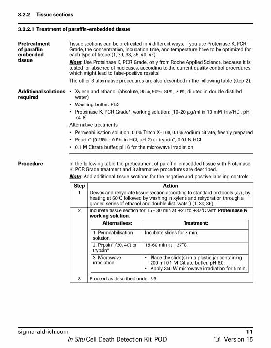
Pretreatment of paraffin embedded tissue
Tissue sections can be pretreated in 4 different ways. If you use Proteinase K, PCR Grade, the concentration, incubation time, and temperature have to be optimized for each type of tissue (1, 29, 33, 36, 40, 42).Note: Use Proteinase K, PCR Grade, only from Roche Applied Science, because it is tested for absence of nucleases, according to the current quality control procedures, which might lead to false-positive results!The other 3 alternative procedures are also described in the following table (step 2).
Additional solutions required
• Xylene and ethanol (absolute, 95%, 90%, 80%, 70%, diluted in double distilled water)
• Washing buffer: PBS• Proteinase K, PCR Grade*, working solution: [10-20 �g/ml in 10 mM Tris/HCl, pH
7.4-8] Alternative treatments• Permeabilisation solution: 0.1% Triton X–100, 0.1% sodium citrate, freshly prepared• Pepsin* (0.25% - 0.5% in HCl, pH 2) or trypsin*, 0.01 N HCl• 0.1 M Citrate buffer, pH 6 for the microwave irradiation
Procedure In the following table the pretreatment of paraffin-embedded tissue with Proteinase K, PCR Grade treatment and 3 alternative procedures are described.Note: Add additional tissue sections for the negative and positive labeling controls.
Step Action1 Dewax and rehydrate tissue section according to standard protocols (e.g., by
heating at 60°C followed by washing in xylene and rehydration through a graded series of ethanol and double dist. water) (1, 33, 36).
2 Incubate tissue section for 15 - 30 min at +21 to +37°C with Proteinase K working solution.
Alternatives: Treatment:
1. Permeabilisation solution
Incubate slides for 8 min.
2. Pepsin* (30, 40) or trypsin*
15- 60 min at +37°C.
3. Microwave irradiation
• Place the slide(s) in a plastic jar containing 200 ml 0.1 M Citrate buffer, pH 6.0.
• Apply 350 W microwave irradiation for 5 min.
3 Proceed as described under 3.3.
sigma-aldrich.com 11In Situ Cell Death Detection Kit, POD y Version 15

3.2.2.2 Treatment of cryopreserved tissue
Additional solutions required
• Fixation solution: 4% Paraformaldehyde in PBS, pH 7.4, freshly prepared• Washing buffer: PBS• Blocking solution: 3% H2O2 in methanol• Permeabilisation solution (0.1% Triton X–100, 0.1% sodium citrate), freshly prepared
Cryopreserved tissue
In the following table the pretreatment of cryopreserved tissue is described.Note: Fix and permeabilisate two additional samples for the negative and positive labeling controls.
Step Action
1 Fix tissue section with Fixation solution for 20 min at +15 to +25°C.
2 Wash 30 min with PBS.Note: For storage, dehydrate fixed tissue sections 2 min in absolute ethanol and store at �15 to �25°C.
3 Incubate with Blocking solution for 10 min at +15 to +25°C.
4 Rinse slides with PBS.
5 Incubate in Permeabilisation solution for 2 min on ice (+2 to +8°C).
6 Proceed as described under 3.3.
12 sigma-aldrich.comIn Situ Cell Death Detection Kit, POD y Version 15

3.3 Labeling protocol
3.3.1 Before you begin
Preparation of TUNEL reaction mixture
One pair of tubes (vial 1: Enzyme Solution, and vial 2: Label Solution) is sufficient for staining 10 samples by using 50 �l TUNEL reaction mixture per sample and 2 nega-tive controls by using 50 �l Label Solution per control.Note: The TUNEL reaction mixture should be prepared immediately before use and should not be stored. Keep TUNEL reaction mixture on ice until use.
Step Action
1 Remove 100 �l Label Solution (vial 2) for two negative controls.
2 Add total volume (50 �l) of Enzyme solution (vial 1) to the remaining 450 �l Label Solution in vial 2 to obtain 500 �l TUNEL reaction mixture.
3 Mix well to equilibrate components.
Additional reagents required
• Micrococcal nuclease or • DNase I recombinant, grade I *
Controls Two negative controls and a positive control should be included in each experimental set up.
Negative control:
Incubate fixed and permeabilized cells in 50 �l/well Label Solu-tion (without terminal transferase) instead of TUNEL reaction mix-ture.
Positive control:
Incubate fixed and permeabilized cells with micrococcal nucle-ase or DNase I recombinant, grade I (3,000 U/ml– 3 U/ml in 50 mM Tris-HCl, pH 7.5, 10 mM MgCl2 1 mg/ml BSA) for 10 min at +15 to +25°C to induce DNA strand breaks, prior to labeling pro-cedures.
sigma-aldrich.com 13In Situ Cell Death Detection Kit, POD y Version 15

3.3.2 Labeling protocol for adherent cells, cell smears, cytospin preparations and tissues
Additional equipment and solutions required
• Washing buffer: PBS• Humidified chamber• Parafilm or coverslip
Procedure Please refer to the following table.
Step Action
1 Rinse slides twice with PBS.
2 Dry area around sample.
3 Add 50 �l TUNEL reaction mixture on sample. Note: For the negative control add 50 �l Label solution each. To ensure a homogeneous spread of TUNEL reaction mixture across cell monolayer and to avoid evaporative loss, samples should be covered with parafilm or cover-slip during incubation.
4 Add lid and incubate for 60 min at +37°C in a humidified atmosphere in the dark.
5 Rinse slide 3 times with PBS.
6 Samples can be analyzed in a drop of PBS under a fluorescence microscope at this state. Use an excitation wavelength in the range of 450 –500 nm and detection in the range of 515 – 565 nm (green).
14 sigma-aldrich.comIn Situ Cell Death Detection Kit, POD y Version 15

3.3.3 Labeling protocol for difficult tissue
Additional equipment and solutions required
• Citrate buffer, 0.1 M, pH 6.0.• Washing buffer: PBS• Tris-HCl, 0.1 M pH 7.5, containing 3% BSA and 20% normal bovine serum• Plastic jar• Microwave• Humidified chamber
Procedure Please refer to the following table.
Step Action
1 Dewax paraformaldehyde- or formalin-fixed tissue sections according to standard procedures.
2 Place the slide(s) in a plastic jar containing 200 ml 0.1 M Citrate buffer, pH 6.0.
3 • Apply 750 W (high) microwave irradiation for 1 min.• Cool rapidly by immediately adding 80 ml double dist. water (+20 to
+25°C).• Transfer the slide(s) into PBS (+20 to +25°C).DO NOT perform a proteinase K treatment!
4 Immerse the slide(s) for 30 min at +15 to +25°C in Tris-HCl, 0.1 M pH 7.5, containing 3% BSA and 20% normal bovine serum.
5 Rinse the slide(s) twice with PBS at +15 to +25°C.Let excess fluid drain off.
6 Add 50 �l of TUNEL reaction mixture on the section and.Note: For the negative control add 50 �l Label solution.
7 Incubate for 60 min at +37°C in a humidified atmosphere in the dark.
8 • Rinse slide(s) three times in PBS for 5 min each.• Samples can be analyzed in a drop of PBS under a fluorescence micro-
scope at this state. Use an excitation wavelength in the range of 450 – 500 nm and detection in the range of 515 – 565 nm (green).
sigma-aldrich.com 15In Situ Cell Death Detection Kit, POD y Version 15

3.4 Signal conversion
Additional equipment and solutions required
Washing buffer: PBS• Humidified chamber• Parafilm or coverslip• DAB Substrate* or alternative POD substrate• Mounting medium for light microscopy
Procedure Please refer to the following table.
Step Action
1 Dry area around sample.
2 Add 50 �l Converter-POD (vial 3) on sample.Note: To ensure a homogeneous spread of Converter-POD across cell monolayer and to avoid evaporative loss, samples should be covered with parafilm or cover slip during incubation.
3 Incubate slide in a humidified chamber for 30 min at 37°C.
4 Rinse slide 3× with PBS.
5 Add 50 – 100 �l DAB Substrate or alternative POD substrates.
6 Incubate slide for 10 min at +15 to +25°C.
7 Rinse slide 3× with PBS.
8 Mount under glass coverslip (e.g., with PBS/glycerol) and analyze under light microscope.Alternative: Samples can be counterstained prior to analysis by light micro-scope.
16 sigma-aldrich.comIn Situ Cell Death Detection Kit, POD y Version 15

sigma-aldrich.com 17
4. Appendix
4.1 Troubleshooting
This table describes various troubleshooting parameters.
Problem Step/Reagent of Pro-cedure
Possible cause Recommendation
Nonspecificlabeling
Embedding of tissue UV-irradiation for polym-erization of embedding material (e.g., methacry-late) leads to DNA strand breaks
Try different embedding material or different polymerization reagent.
Fixation Acidic fixatives (e.g., methacarn, Carnoy’s fixa-tive)
• Try 4% buffered paraformaldehyde.• Try formalin or glutaraldehyde.
TUNEL reaction TdT concentration too high Reduce concentration of TdT by dilut-ing it 1:2 up to 1:10 with TUNEL Dilu-tion Buffer *.
Converter solution Endogenous POD activity Block endogenous POD by immersing for 10 min in 3% H2O2 in methanol prior to cell permeabilisation.
Non-specific binding of anti-fluorescein-POD
• Block with normal anti-sheep serum.
• Block for 20 min with PBS contain-ing 3% BSA.
• Reduce concentration of converter solution to 50%.
Nucleases Some tissues (e.g., smooth muscles) show DNA strand breaks very soon after tissue preparation
• Fix tissue immediately after organ preparation.
• Perfuse fixative through liver vein.
Some enzymes are still active
Block with a solution containing ddUTP and dATP.
High back-ground
Fixation Formalin fixation leads to a yellowish staining of cells containing melanin pre-cursors
Try methanol for fixation but take into account that this might lead to reduced sensitivity.
TUNEL reaction Concentration of labeling mix is too high for mamma carcinoma
Reduce concentration of labeling mix to 50% by diluting with TUNEL Dilu-tion Buffer*.
Converter solution Endogenous POD activity Block endogenous POD by immersing for 10 min in 3% H2O2 in methanol prior to cell permeabilisation.
Non-specific binding of anti-fluorescein-POD
• Block with normal anti-sheep serum.
• Block for 20 min with PBS contain-ing 3% BSA.
• Reduce concentration of converter solution to 50%.
Sample Mycoplasma contamina-tion
Mycoplasma detection Kit*.
In Situ Cell Death Detection Kit, POD y Version 15

18 sigma-aldrich.com
Highly proliferating cells Double staining e.g.,, with Annexin-V-Fluos*. Note: Measuring via microplatereader not possible because of too high background.
Low labeling Fixation Ethanol and methanol can lead to low labeling (nucleosomes are not cross-linked with proteins during fixation and are lost during the procedure steps)
• Try 4% buffered paraformaldehyde.• Try formalin or glutaraldehyde.
Extensive fixation leads to excessive crosslinking of proteins
• Reduce fixation time.• Try 2% buffered paraformaldehyde.
Permeabilisation Permeabilisation too short so that reagents can’t reach their target mole-cules
• Increase incubation time.• Incubate at higher temperature
(e.g., +15 to +25°C).• Try Proteinase K , PCR Grade (con-
centration and time has to be opti-mized for each type of tissue).
• Try 0.1 M sodium citrate at 70°C for 30 min.
Paraffin-embedding Accessibility for reagents is too low
• Treat tissue sections after dewaxing with Proteinase K, PCR Grade (con-centration, time and temperature have to be optimized for each type of tissue).
• Try microwave irradiation at 370 W (low) for 5 min in 200 ml 0.1 M Citrate buffer pH 6.0 (has to be optimized for each type of tissue).
Problem Step/Reagent of Pro-cedure
Possible cause Recommendation
In Situ Cell Death Detection Kit, POD y Version 15

No signal on positive control
DNase treatment Concentration of DNase is too low
• For cryosections apply 3 U/ml DNase I recombinant, grade I.
• For paraffin-embedded tissue sec-tions apply 1500 U/ml DNase I recombinant, grade I.
• In general, use 1 U/ml DNase I recombinant, grade I, dissolved in 10 mM Tris-HCl pH 7.4 containing 10 mM NaCl, 5 mM MnCl2, 0.1 mM CaCl2, 25 mM KCl and incubate 30 min at +37°C.
• Alternative buffer 50 mM Tris- HCl pH 7.5 containing 1 mM MgCl2 and 1 mg/ml BSA.
Weak signals Counterstaining Not suitable dye • Counterstaining with 5% methyl green in 0,1 M veronal acetate, pH 4.0 or Hematoxilin is possible (43).
• Double-staining with propidium iodide is possible but only for detection of morphological cell changes.
Problem Step/Reagent of Pro-cedure
Possible cause Recommendation
sigma-aldrich.com 19In Situ Cell Death Detection Kit, POD y Version 15

4.2 References
1 Gavrieli, Y., Sherman, Y. & Ben-Sasson, S. A. (1992) J. Cell Biol. 119, 493–501.2 Gorczyca, W., Gong, J. & Darzynkiewicz, Z. (1993) Cancer Res. 53, 1945–1951.3 Gorczyca, W. et al. (1993) Leukemi a 7, 659–670.4 Gold, R. et al. (1994) Lab. Invest. 71, 219.5 Gorczyca, W. et al. (1994) Cytometry 15, 169–175.6 Sgonc, R. et al. (1994) Trends Genetics 10, 41–42.7 Schmied, M. et al. (1993) Am. J. Pathol. 143, 446–452.8 Wyllie, A. H. et al. (1980) Int. Rev. Cytol. 68, 251.9 Kerr, J. F. R. et al. (1972) Br. J. Cancer 26, 239–257.
10 Duvall, E. & Wyllie, A. H. (1986) Immunol. To day 7, 115.11 Compton, M. M. (1992) Canc. Metastasis Rev. 11, 105–119.12 Allen, P. D., Bustin, S. A. & Newland, A. C. (1993) Blood Reviews 7, 63–73.13 Cohen, J. J. & Duke, R. C. (1992) Annu. Rev. Immunol. 10, 267–293.14 Clarke, P. G. H. (1990) Anat. Embryol. 181, 195–213.15 Johnson, E. M. & Deckwerth, T. L. (1993) Annu. Rev. Neurosci. 16, 31–46.16 Batistatou, A. & Greene, L. A. (1993) J. Cell Biol. 122, 523–532.17 Strange, R. et al. (1992) Development 115, 49–58.18 Carson, D. A. & Ribeiro, J. M. (1993) Lancet 341, 1251–1254.19 Edgington, S. M. (1993) Biotechnology 11, 787–792.20 Gougeon. M.-L. & Montagnier, L. (1993) Science 260, 1269–1270.21 Hickman, J. A. (1992) Cancer Metastasis Rev. 11, 121–139.22 Afanasyev, V. N. et al. (1993) Cytometry 14, 603–609.23 Bryson, G. J., Harmon, B. V. & Collins, R. J. (1994) Immunology Cell Biology 72, 35–4124 Darzynkiewicz, Z. et al. (1992) Cytometry 13, 795–808.25 Ando, K. et al. (1994) J. Immunol. 152, 3245–3253.26 Berges, R. R. et al. (1993) Proc. Natl. Acad. Sci. USA 90, 8910– 8914.27 Gorczyca, W. et al. (1992) Int. J. Oncol. 1, 639–648.28 Gorczyca, W. et al. (1993) Exp. Cell Res. 207, 202–205.29 Billig, H., Furuta, I. & Hsueh, A. J. W. (1994) Endocrinology 134, 245–252.30 MacManus, J. P. et al. (1993) Neurosci. Lett. 164, 89–92.31 Mochizuki, H. et al. (1994) Neurosci. Lett. 170, 191–194.32 Oberhammer, F. et al. (1993) Hepatology 18, 1238–1246.33 Portera-Cailliau, C. (1994) Proc. Natl. Acad. Sci. USA 91, 974 –978.34 Preston, G. A. et al. (1994) Cancer Res. 54, 4214–4223.35 Weller, M. et al. (1994) Eur. J. Immunol. 24, 1293–1300.36 Zager, R.A. et al. (1994) J. Am. Soc. Nephrol. 4, 1588–1597.37 Cohen, G. M. et al. (1992) Biochem. J. 286, 331–334.38 Collins, R. J. et al. (1992) Int. J. Rad. Biol. 61, 451–453.39 Sei, Y. et al. (1994) Neurosci. Lett. 171, 179–182.40 Ansari, B. et al. (1993) J. Pathol. 170, 1–8.41 Gold, R. et al. (1993) J. Histochem. Cytochem. 41, 1023–1030.42 Negoescu, A. et.al. (1998) Biochemica 3, 34-41.43 Umermura, S. et al. (1996) J. Histochem. Cytochem. 44, 125-132 .
20 sigma-aldrich.comIn Situ Cell Death Detection Kit, POD y Version 15

sigma-aldrich.com 21
4.3 Ordering Guide
*available from Roche Diagnostics
Changes to previ-ous version
Editorial changes
Trademarks All other product names and trademarks are the property of their respective owners.
Disclaimer of License
For patent license limitations for individual products please refer to: List of biochemi-cal reagent products
Regulatory Dis-claimer
For life science research only. Not for use in diagnostic procedures.
Apoptosis-specific phy-siological change Detection mode/Product Pack size Cat. No.
DNA fragmentation Gel ElectrophoresisApoptotic DNA-Ladder Kit 20 tests 11 835 246 001In situ assayIn Situ Cell Death Detection Kit, TMR red (also useable for FACS)
1 kit (50 tests)
12 156 792 910
In Situ Cell Death Detection Kit, Fluorescein (also useable for FACS)
1 kit (50 tests)
11 684 795 910
In Situ Cell Death Detection Kit, AP 1 kit (50 tests)
11 684 809 910
In Situ Cell Death Detection Kit, POD 1 kit (50 tests)
11 684 817 910
Single reagents for TUNEL and supporting reagentsTUNEL AP 70 tests
(3.5 ml)11 772 457 001
TUNEL POD 70 tests (3.5 ml)
11 772 465 001
TUNEL Enzyme 2× 50 �l (20 tests)
11 767 305 001
TUNEL Label 3× 550 �l (30 tests)
11 767 291 910
In Situ Cell Death Detection Kit, POD y Version 15

Contact and Support If you have questions or experience problems with this or any Roche product for Life Science, please contact our Technical Support staff. Our scientists are committed to providing rapid and effective help.Please also contact us if you have suggestions for enhancing Roche product performance or using our products in new or specialized ways. Such customer information has repeatedly proven invaluable to the research community worldwide.
To ask questions, solve problems, suggest enhancements or report new appli-cations, please visit our Online Technical Support Site.
Visit sigma-aldrich.com, to download or request copies of the following mate-rials.
• Instructions for Use• Safety Data Sheets• Certificates of Analysis• Information Material
To call, write, fax, or email us, visit sigma-aldrich.com and select your home country to display country-specific contact information.
Roche Diagnostics GmbH Sandhofer Strasse 116 68305 Mannheim Germany
0516
.116
9728
500
1 5