in search of recollection and familiarity signals in the...
TRANSCRIPT
In Search of Recollection and Familiarity Signalsin the Hippocampus
Peter E. Wais1, Larry R. Squire1,2, and John T. Wixted1
Abstract
& fMRI studies of recognition memory have often been inter-preted to mean that the hippocampus selectively subservesrecollection and that adjacent regions selectively subserve fa-miliarity. Yet, many of these studies have confounded recol-lection and familiarity with strong and weak memories. In asource memory experiment, we compared correct source judg-ments (which reflect recollection) and incorrect source judg-ments (often thought to reflect familiarity) while equating forold–new memory strength by including only high-confidencehits in the analysis. Hippocampal activity associated with both
correct source judgments and incorrect source judgments ex-ceeded the activity associated with forgotten items and did soto a similar extent. Further, hippocampal activity was greaterfor high-confidence old decisions relative to forgotten itemseven when source decisions were at chance. These results iden-tify a recollection signal in the hippocampus and may identifya familiarity signal as well. Similar results were obtained in theparahippocampal gyrus. Unlike in the medial temporal lobe, ac-tivation in prefrontal cortex increased differentially in associa-tion with source recollection. &
INTRODUCTION
Dual-process theories of recognition memory hold thattwo distinct memory processes, recollection and familiar-ity, underlie one’s ability to recognize an item as havingbeen previously encountered (Mandler, 1980). Recollec-tion involves the retrieval of contextual detail associatedwith the test item, whereas familiarity involves simplyknowing that the item was encountered before. Accordingto one view (the high-threshold/signal-detection model),high confidence in a recognition decision strongly im-plies that the decision was based on recollection, where-as lower confidence necessarily implies that the decisionwas based on familiarity (Yonelinas, 1994). A closelyrelated view holds that decisions accompanied by a ‘‘re-member’’ response are based on recollection, where-as decisions accompanied by a ‘‘know’’ response arebased on familiarity. A common finding in the functionalmagnetic resonance imaging (fMRI) literature is that hip-pocampal activity associated with highly confident re-sponses (or remember responses) exceeds the activityassociated with forgotten items, whereas the activity asso-ciated with less confident responses (or know responses)does not (Ranganath et al., 2004; Eldridge, Knowlton,Furmanski, Bookheimer, & Engel, 2000). This pattern offindings has often been interpreted to mean that the hip-pocampus selectively subserves recollection.
An alternative view holds that the level of confidenceassociated with a recognition decision is related to mem-
ory strength and that memory strength reflects varyingdegrees of both recollection and familiarity (Wixted,2007). According to this view, recognition decisionsmade with low confidence reflect small contributionsof recollection and familiarity, whereas decisions madewith high confidence reflect large contributions of rec-ollection and familiarity. On average, recollection willtypically be associated with higher confidence than fa-miliarity. Nevertheless, the confidence distributions as-sociated with recollection and familiarity extend acrossthe full range of memory strength. Accordingly, confi-dence ratings per se cannot be used to disentangle thetwo processes. This view further holds that remember–know judgments are tantamount to confidence ratings(Dunn, 2004, 2008). If so, then know judgments do notreflect strong, familiarity-based memories that are de-void of recollection. Instead, compared to rememberjudgments, they reflect weaker memories that are as-sociated with lesser degrees of confidence and lesserdegrees of recollection (Wais, Mickes, & Wixted, 2008).According to this alternative view, fMRI studies that haverelied on confidence ratings or the remember–know pro-cedure to identify the neural correlates of recollectionand familiarity involve a memory strength confound. Thatis, it is not clear whether the reported effects in thesestudies were due to differences between recollection andfamiliarity or due to differences in memory strength (Wais,2008; Squire, Wixted, & Clark, 2007).
A different approach to separating recollection andfamiliarity involves the source memory procedure. Cor-rectly identifying an item as old and with correct source
1University of California, San Diego, La Jolla, CA, 2Veterans AffairsMedical Center, San Diego, CA
D 2009 Massachusetts Institute of Technology Journal of Cognitive Neuroscience 22:1, pp. 109–123

information is thought to identify a recollection-based de-cision, whereas correctly identifying an item as old butwith incorrect source information is thought to identifya familiarity-based decision (Ranganath et al., 2004). A typ-ical finding from fMRI studies is that hippocampal activ-ity associated with source-correct responses exceeds theactivity associated with forgotten items, whereas the activ-ity associated with source-incorrect responses does not(Kensinger & Schacter, 2006; Weis et al., 2004). This pat-tern has also been interpreted to mean that the hippo-campus selectively subserves recollection (Eichenbaum,Yonelinas, & Ranganath, 2007).
Yet, source memory studies analyzed in this way alsoconfound the strength of memory (as indicated by con-fidence in the old–new decision) with source-correctand source-incorrect decisions. Specifically, confidenceis typically higher for old–new decisions that are subse-quently associated with correct source judgments than forold–new decisions that are subsequently associated withincorrect source judgments (Gold et al., 2006; Slotnick& Dodson, 2005). This difference in old–new confi-dence suggests that comparisons between source-correctand source-incorrect decisions involve relatively strong,recollection-based decisions and relatively weak, familiarity-based decisions. This strength confound is problematicbecause the distinction between recollection and famil-iarity is independent of memory strength. Moreover,the relationship between the BOLD signal in the hippo-campus and the neural activity that underlies memorystrength is unknown and may be nonlinear ( Johnson,Muftuler, & Rugg, 2008; Squire et al., 2007). If so, thendifferences in activity between conditions associated withstrong versus intermediate-strength memories (e.g.,source correct vs. source incorrect, or remember vs.know) may be more detectable in the hippocampusthan differences in activity between conditions associ-ated with intermediate-strength versus very weak memo-ries (e.g., source incorrect vs. misses, or know vs. misses).In light of these considerations, it is important to equatefor memory strength if the objective is to compare activityin the hippocampus associated with recollection and fa-miliarity (Wais, 2008; Squire et al., 2007; Wixted, 2007).
To equate memory strength across conditions, we madeuse of confidence ratings. Evidence that confidence rat-ings provide a valid measure of memory strength isprovided by past work showing that the confidence ex-pressed in an old–new decision is strongly related to theaccuracy of that decision (e.g., Mickes, Wixted, & Wais,2007). In our fMRI study, we equated memory strengthfor source-correct versus source-incorrect judgmentsby using only old decisions that were made with highconfidence, and the question of interest was whetherhippocampal activity measured at retrieval would be dif-ferentially elevated for source-correct judgments evenunder these conditions. If the typical pattern is foundeven after controlling for memory strength (i.e., if dif-ferentially elevated hippocampal activity is detected for
the source-correct condition), then it would clearly weighagainst the suggestion that prior fMRI studies have beencompromised by a memory strength confound (Wais,2008; Squire et al., 2007) and would support the notionthat the hippocampus does not play a role in familiarity-based decisions. However, if hippocampal activity asso-ciated with source-correct and source-incorrect decisionsare both elevated once they are equated for memorystrength, then the strength-confound hypothesis—and theidea that the hippocampus does play a role in familiarity-based decisions—would remain viable. Such an outcomewould not definitively identify a familiarity signal in thehippocampus because one might suppose that strongmemories accompanied by incorrect source decisions donot reflect familiarity but instead reflect the undetectedrecollection of details unrelated to the source question.Nevertheless, if equally strong source-correct and source-incorrect decisions are both associated with elevatedhippocampal activity, it would indicate that prior argu-ments against a familiarity signal in the hippocampus maybe less definitive then they have been taken to be, and itwould underscore the importance of controlling for mem-ory strength in future investigations into the neuroana-tomical basis of recollection and familiarity.
METHODS
Participants
Informed consent was obtained from 18 students (6 wom-en) at the University of California, San Diego. All partici-pants were right-handed. Two participants who did notscore above chance levels for their source memory judg-ments were excluded from further analysis.
Stimuli
Two hundred forty English nouns were selected fromthe MRC Psycholinguistic Database with the followingconstraints: word frequency of 50 to 300, length of 5 to12 letters, and two to four phonemes. The words wererandomly divided into one list of 192 targets and anotherlist of 48 foils.
Behavioral Procedure and Data Analysis
Participants studied a list of words presented on a desk-top computer. Words were presented in six blocks of32 words each. Words in each block were randomlyordered for each participant. During each 2.5-sec trialin the study session, participants responded to one oftwo contextual-cue questions posed for each word, thecommon question or the discuss question (common:does the word describe something you expect to en-counter in a typical week?; or discuss: does the worddescribe something you would discuss with a closefriend?). The contextual-cue question was the same for
110 Journal of Cognitive Neuroscience Volume 22, Number 1

all trials within a block so that participants studied thetarget words as alternating blocks with either the ‘‘com-mon’’ or ‘‘discuss’’ cues. Participants were instructed toread each word, enter a yes or no answer to the cuedquestion, and to remember the word, including its con-textual cue, for a memory test during their subsequentscanning session.
The memory test for each participant was conducted inthe MRI scanner approximately 3 hr after the study ses-sion. Participants saw test items in six blocks of 40 words(5.0 sec per word). Each test block included 32 targetsfrom the study session, plus eight foils. The test itemswere presented in a random order to each participantand intermixed with trials from an odd–even digit taskdescribed below (fMRI Scanning Parameters, Procedure,and Data Analysis). For each word presented in the testphase, participants first gave a confidence judgment as towhether the word was old or new (1 = definitely new,2 = probably new, 3 = maybe new, 4 = maybe old, 5 =probably old, 6 = definitely old) and then gave a confi-dence judgment for their source decision (1 = definitelydiscuss, 2 = probably discuss, 3 = maybe discuss, 4 =maybe common, 5 = probably common, 6 = definitelycommon). For clarity, we will refer to old–new confidenceratings in terms of the 1-through-6 numerical scale, butwe will refer to source confidence ratings in terms of cor-
rect and incorrect decisions that were made with low,medium, or high confidence (e.g., correct source con-fidence ratings of 1 for ‘‘discuss’’ items and 6 for ‘‘com-mon’’ items will both be referred to as correct sourcedecisions made with high confidence).
Because we were concerned that our task instructionsshould be received as logical, participants were instructednot to enter a source judgment for words endorsed asnew. Nevertheless, the task demand remained constantfor the old–new test in each trial, and our analysis of thefMRI results was based on the data collected during theold–new test. The old–new scale and the source decisionscale were each presented for 2.5 sec beneath the testword on each trial (Figure 1). In order to facilitate fMRIanalysis (see below), participants also performed an odd–even classification task (Stark & Squire, 2001) on trialsrandomly intermixed with the memory task. For this base-line task, the digits 1 to 9 were presented for 1.25 seceach in blocks of 2, 4, 6, or 12.
fMRI Scanning Parameters, Procedure,and Data Analysis
Imaging was carried out in a GE Signa Excite 3-T scannerat the Center for Functional MRI (University of California,San Diego). Functional images were acquired using a
Figure 1. Example of test
procedure and behavioralanalysis: The stimulus ‘‘wheel’’
was studied under the
common contextual-cuecondition and endorsed as a
high-confidence hit (5 or 6)
in the old–new recognition
task. For the subsequentsource memory task, a
response of 1 or 2 indicates
false source memory, a
response of 3 or 4 indicatessource guesses, and a response
of 5 or 6 indicates true source
memory (source responses 1,2, and 3 are incorrect and
source responses 4, 5, and 6
are correct).
Wais, Squire, and Wixted 111

gradient-echo, echo-planar, T2*-weighted pulse sequence(TR = 2.5 sec, TE = 30, 908 flip angle, bandwidth =250 MHz, FOV = 22 cm). Forty-two slices coveringthe whole brain were acquired perpendicular to thelong axis of the hippocampus (matrix size = 64 � 64,slice thickness = 5 mm). Following six functional runs,high-resolution structural images were acquired using aT1-weighted, fast spoiled gradient-echo (FSPGR) pulse se-quence (TE = 3.1, 128 flip angle, FOV = 25 cm, 172 slices,1 mm slice thickness, matrix size = 256 � 256).
Between word presentations, participants were given0, 2, 4, 6, or 12 trials of the 1.25-sec baseline task thatserved to jitter the MR signal acquired for subsequentdeconvolution of the hemodynamic response function(hrf ). For each participant, the fMRI data were parti-tioned into 10 categories (see Results for an explanationof the trials in each category). The first seven categorieswere based on the old–new confidence ratings providedon each trial: (a) correct old responses to targets (i.e.,hits) that were rated 6; (b) hits that were rated 5; (c)high-confidence hits (rated 5 or 6) that were subse-quently associated with correct source decisions; (d)high-confidence hits (5s or 6s) that were subsequentlyassociated with incorrect source decisions; (e) misses (tar-gets rated 1, 2, 3, or in some cases, 4); (f ) false alarms(foils rated 4, 5, or 6); and (g) correct rejections (foilsrated 1, 2, or 3). Additionally, the high-confidence hits(those associated with an old–new confidence rating of5 or 6) were subdivided into three additional categoriesbased on the confidence ratings for the source decision:(h) ‘‘true source’’ decisions (correct source decisionsmade with medium or high confidence on the sourceconfidence scale); (i) ‘‘source guesses’’ (correct and in-correct source decisions made with low confidence); and(j) ‘‘false source’’ decisions (incorrect source decisionsmade with medium or high confidence).
For each of the 10 categories, a hemodynamic re-sponse (relative to the baseline condition) was esti-mated for the 25 sec following the presentation of theword by using signal deconvolution with the AFNI suiteof programs (Cox, 1996). Data analysis was then basedon the area under the hrf from 0 to 15 sec followingthe presentation of the word (at about 15 sec, the hrfreturned to baseline). The anatomical scans and the fMRIdata were normalized to the template of the Talairachbrain (Talairach & Tournoux, 1988). Functional data wereresampled to 2 � 2 � 2 mm and blurred with a 4-mmFWHM Gaussian kernel. These data were used for thewhole-brain analysis. For the analysis of medial temporallobe (MTL) activity, the ROI–large deformation diffeomor-phic metric mapping (ROI–LDDMM) alignment method(Miller, Beg, Ceritoglu, & Stark, 2005) was used to improvecross-participant alignment and increase statistical reliabil-ity (Kirwan, Jones, Miller, & Stark, 2007).
Voxel-based t tests (threshold of p < .001, two-tailed)were then carried out as group analyses across all 16 par-ticipants for both the whole brain and MTL analyses
based on the area under the hrf for contrasts of interest(described below). Monte Carlo simulations were thenused to correct for multiple comparisons and to deter-mine how large a cluster of voxels was needed in orderto be statistically significant ( p < .05). The coordinatesof all of the regions of activity we identified that werestatistically significant ( p-corrected < .05) are listed inTable S1 as supplementary information.
RESULTS
Behavioral Results
The participants demonstrated good old–new recogni-tion memory for the target words (d0 = 1.93 ± 0.15; and79 ± 2% correct for the old–new response). Wordsstudied in the ‘‘common’’ cue condition were recog-nized as readily as words studied in the ‘‘discuss’’ cuecondition (d0 = 1.98 ± 0.17 vs. d0 = 1.89 ± 0.16). Thedistribution of responses for hits and false alarms acrossthe six-level old–new confidence scale revealed a biasto respond ‘‘old’’ (Figure 2). As a result, responses totargets given a confidence level 4 (maybe old) were atchance accuracy overall (56 ± 6% correct), whereas re-sponses to foils given a confidence level 3 (maybe new)were much more accurate (80 ± 4% correct). The ac-curacy of responses to targets given a confidence rat-ing of 5 or 6 was also high (71 ± 6% correct and 87 ±3% correct, respectively). The old–new accuracy calcu-lations for each rating included scores of participantswho had at least five observations for that rating (e.g., atleast 5 ratings of 3).
Overall source accuracy was 64 ± 2% correct, andsource d0 was 0.71 ± 0.10. As has been observed in priorstudies, source accuracy varied as a function of confi-dence in the old–new decision for targets. Source ac-curacy was highest for items that received an old–new
Figure 2. Proportion of responses to targets and foils for each
confidence level in the old–new task, n = 16, mean recognitiond0 = 1.93 (0.15).
112 Journal of Cognitive Neuroscience Volume 22, Number 1

confidence rating of 6 (67% correct, significantly abovechance, p < .05), next highest for items that received anold–new confidence rating of 5 (63% correct, significantlyabove chance, p < .05), and lowest for items that receivedan old–new confidence rating of 4 (50% correct).
For target items that were correctly declared to be old(i.e., targets that received a rating of 4, 5, or 6), we com-puted the mean old–new confidence rating separatelydepending on whether the subsequent source decisionwas correct or incorrect. The mean old–new confidenceassociated with source-correct decisions (Table 1) wassignificantly higher than the mean old–new confidenceassociated with source-incorrect decisions ( p < .02).Thus, our results exhibit the typical memory strengthconfound that has been observed in prior source mem-ory studies (Gold et al., 2006; Slotnick & Dodson, 2005).
To eliminate this confound, we combined old–newhits that had been given confidence ratings of 5 or 6 andthen divided those responses into source-correct andsource-incorrect categories (decisions made with a con-fidence rating of 6 yielded too few observations to de-tect MTL activity reliably). A possible difficulty with thisapproach is that it could introduce the strength con-found we sought to avoid (i.e., the old–new confidenceratings for incorrect source judgments could be lower,on average, than the old–new confidence ratings forsource-correct judgments). However, in our study, thisdid not occur. As shown in Table 1, for old–new deci-sions made with a confidence rating of 5 or 6, the meanold–new confidence for source-incorrect decisions (5.82)was virtually identical to the mean old–new confidencefor source-correct decisions (5.84), and the small differ-ence between them did not approach significance. Thus,a comparison of activity associated with these source-correct and source-incorrect decisions is not confoundedwith memory strength (as measured by confidence in theold–new decision).
To allow for a further analysis of neural activity as-sociated with source recollection, we also partitionedcorrect old decisions in a more fine-grained manner.Specifically, instead of separating them into two catego-ries (i.e., source-correct and source-incorrect), we sepa-rated them into three categories: true source judgments(i.e., correct source judgments made with medium orhigh confidence), source guesses (source judgments
made with low confidence, whether correct or incorrect),and false source judgments (incorrect source judgmentsmade with medium or high confidence). Table 1 showsthat there is an old–new strength confound if all of thecorrect old decisions are used, so we included only olddecisions made with a confidence rating of 5 or 6 (whicheliminated the confound). For these high-confidence olddecisions, 44% were followed by true source judgments,37% were followed by source guesses, and 19% were fol-lowed by false source judgments. The source accuracy ofthe guesses was 55 ± 2%, which did not differ significantlyfrom chance ( p = .12). Thus, source information was, infact, absent when items were recognized as old with highconfidence and the source judgment was a guess.
In the fMRI analyses described below, we comparedthe activity associated with either two or three catego-ries of source judgments (equated for old–new confi-dence) against the activity associated with forgottenitems. Typically, a target item is considered to beforgotten if it is incorrectly declared to be new (i.e., ifit receives a confidence rating of 1, 2, or 3). However, asindicated above, many of the participants in our exper-iment exhibited a liberal response bias such that ratingsof 4 were as likely to be given to targets as to foils(Figure 2). In that case, a confidence rating of 4 for atarget indicates a forgotten item as well. In all of theanalyses described below, we considered old–new con-fidence ratings of 1, 2, 3, or 4 to reflect forgotten wordsfor the 11 of 16 participants who exhibited no betterthan chance accuracy when responding 4 (maybe old),and we considered responses of 1, 2, or 3 to denoteforgotten words for the remaining five participantswhose old–new confidence ratings of 4 were associatedwith greater than chance accuracy.
fMRI Results
Our objective was to measure activity associated withcorrect old decisions accompanied by source recollec-tion and correct old decisions that were not accompa-nied by source recollection after eliminating the typicalmemory strength confound. To that end, we first mea-sured activity associated with source-correct and source-incorrect judgments using only those old decisions thatwere made with relatively high confidence (i.e., 5 or 6).
Table 1. Mean Old–New Confidence (SEM) for Different Source Accuracy Conditions and Different Levels of Source ConfidenceWhen All Hits Were Included (Old–New Confidence Ratings of 4, 5, or 6) and When Only Relatively High-confidence Hits WereIncluded (Old–New Confidence Ratings of 5 or 6)
By Two Source Categories By Three Source Categories
Old–New Ratings Partitioned Source Correct Source Incorrect True Source Source Guess False Source
4, 5, 6 5.62 (0.08)* 5.44 (0.09)* 5.73 (0.07)y 5.37 (0.10)y,z 5.58 (0.07)z,y
5, 6 5.84 (0.04) 5.82 (0.04) 5.85 (0.05) 5.81 (0.04) 5.81 (0.04)
Values that share symbols (e.g., *) differ significantly from each other ( p < .05).
Wais, Squire, and Wixted 113

In one voxel-based t test (thresholded at p < .001), wefound that, in the left hippocampus, activity associatedwith source-correct decisions was significantly greaterthan the activity associated with forgotten items (Fig-ure 3). In the same region of the left hippocampus, asecond, independent voxel-based t test revealed that ac-tivity associated with source-incorrect decisions was alsosignificantly greater than activity associated with forgot-ten items (Figure 3). We also directly contrasted source-correct decisions versus source-incorrect decisions, butno statistically significant regions ( p < .001) were iden-tified in the MTL. These results suggest that increasedactivation in the left hippocampus is associated with in-creased strength of memory (i.e., high-confidence hitsversus forgotten items) and that activity does not differwhether or not the decision is accompanied by success-ful source recollection.
The analysis summarized in Figure 3 assumes thatsource-correct decisions were associated with recollec-tion and that source-incorrect decisions were associatedwith the absence of recollection (and may have beenbased on familiarity). However, both of these categoriesincluded source memory judgments made with low, me-dium, and high confidence (i.e., ‘‘maybe,’’ ‘‘probably,’’
or ‘‘definitely’’). Thus, some source-incorrect decisionswere made with high source confidence and may havereflected false recollection. Conceivably, the hippocam-pal activity associated with source-incorrect decisions inthe analysis described above reflects false recollection,as has been reported in other paradigms (Schacter &Slotnick, 2004; Cabeza, Rao, Wagner, Mayer, & Schacter,2001). To address this issue, we used voxel-based t tests(thresholded at p < .001) to contrast activity associatedwith true source judgments, source guesses, and falsesource judgments against the activity associated withforgotten items. Once again, in order to eliminate astrength confound that would otherwise exist, only olddecisions made with high confidence (5 or 6) were in-cluded in the following analyses. For each identified clus-ter ( p-corrected < .05), signal data were also extractedfor the other source conditions.
Regions in the posterior hippocampus, bilaterally, ex-hibited significantly greater activity associated with truesource decisions than for forgotten items (Figure 4A). Insignal data extracted from this cluster, the activity levelsfor source guesses and false source decisions were bothnumerically higher than the level associated with forgot-ten items and did not differ significantly from the levelassociated with true source decisions. In a separate con-trast, activity associated with source guesses was signifi-cantly greater than the activity associated with forgottenitems in the right posterior hippocampus (Figure 4B).The location of this cluster was virtually identical to thelocation of the cluster in the right hippocampus identi-fied by the comparison between true source decisionsand forgotten items (shown in Figure 4A). The activitylevels extracted from this cluster for true source decisionsand false source decisions were numerically higher thanthe level associated with forgotten items and did notdiffer from the level associated with true source decisions.
The findings presented in Figure 4A and B may indi-cate both a recollection and a familiarity signal in thehippocampus when confidence in memory is strong. Evi-dence for a recollection signal comes from the fact thatthe activity associated with true source memories sig-nificantly exceeded the activity associated with forgottenitems (Figure 4A). Such an interpretation is consistentwith much prior evidence, suggesting that the hippocam-pus plays an important role in recollection. For exam-ple, Manns, Hopkins, Reed, Kitchener, and Squire (2003)found that selective hippocampal lesions were associatedwith clear deficits in recall performance, which is gener-ally assumed to be based exclusively on recollection.
Evidence for a possible familiarity signal in the hippo-campus comes from the fact that the activity associatedwith source guesses (using only old decisions made withhigh confidence) significantly exceeded the activity asso-ciated with forgotten items (Figure 4B). This finding hasnot been previously reported, perhaps because whenold decisions made with lower confidence are includedin the analysis, rather than only old decisions made with
Figure 3. Activation identified for separate contrasts of correct
source judgments versus forgotten items and incorrect source
judgments versus forgotten items. To equate for memory strength,the source-correct and source-incorrect data were based on old
decisions made with high confidence. Error bars for the two source
categories represent the SEM of the difference scores for each
comparison, whereas the error bar for the forgotten items representsthe root mean square of the SEM values associated with the two
individual comparisons (* denotes a significant difference relative
to forgotten items, p-corrected < .05).
114 Journal of Cognitive Neuroscience Volume 22, Number 1
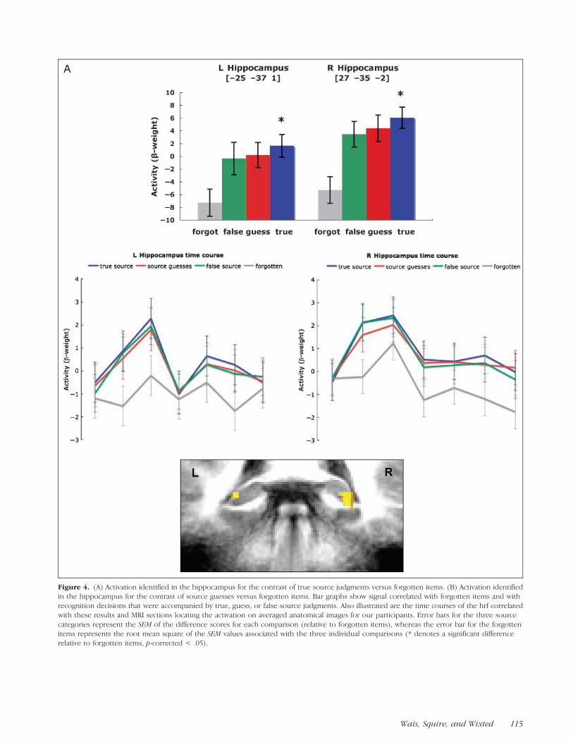
Figure 4. (A) Activation identified in the hippocampus for the contrast of true source judgments versus forgotten items. (B) Activation identifiedin the hippocampus for the contrast of source guesses versus forgotten items. Bar graphs show signal correlated with forgotten items and with
recognition decisions that were accompanied by true, guess, or false source judgments. Also illustrated are the time courses of the hrf correlated
with these results and MRI sections locating the activation on averaged anatomical images for our participants. Error bars for the three source
categories represent the SEM of the difference scores for each comparison (relative to forgotten items), whereas the error bar for the forgottenitems represents the root mean square of the SEM values associated with the three individual comparisons (* denotes a significant difference
relative to forgotten items, p-corrected < .05).
Wais, Squire, and Wixted 115

high confidence, then memories associated with incor-rect source decisions (or source guesses) are too weak,on average, to elicit an fMRI signal in the hippocampus(Wais, 2008; Squire et al., 2007).
In order to test for activity in the hippocampus thatmight be selectively associated with decisions accompa-nied by source recollection, we next contrasted activityassociated with true source decisions against activity asso-
Figure 4. (continued)
116 Journal of Cognitive Neuroscience Volume 22, Number 1
ciated with source guesses (again using only old decisionsmade with high confidence). No statistically significant re-gions ( p < .001) were identified for this contrast in thehippocampus. Taken together, these results suggest thathippocampal activity signals strong memory, whether ornot source recollection is involved.
With the same analysis just described, the voxel-basedt tests also revealed several areas of activity in the para-hippocampal gyrus. Specifically, in left parahippocam-pal cortex, activity associated with true source decisionswas significantly greater than activity associated withforgotten items (Figure 5A), just as was the case in thehippocampus bilaterally (Figure 4A). Also as in the hip-pocampus, in signal data extracted from this cluster,the activity levels for source guesses and false sourcememories were both numerically greater than that as-sociated with forgotten items and did not differ sig-nificantly from the level associated with true sourcedecisions. In left perirhinal cortex, the activity associatedwith source guesses was significantly greater than theactivity associated with forgotten items (Figure 5B), justas was the case in the right hippocampus (Figure 4B).Also as in the right hippocampus, the activity levelsassociated with true source decisions and false sourcedecisions extracted from this cluster were both numer-ically greater than that associated with forgotten itemsand did not differ significantly from the level for sourceguesses. Lastly, a contrast between activity associatedwith true source decisions versus activity associated withsource guesses identified no significant regions withinthe parahippocampal gyrus. Thus, the pattern in theparahippocampal gyrus was similar to that seen in thehippocampus.
Next, because both the dorsolateral and the ventro-lateral regions of prefrontal cortex (DLPFC and VLPFC,respectively) have been associated with source memoryprocesses in prior fMRI studies (Badre & Wagner, 2007;Ranganath & Blumenfeld, 2007), we examined the whole-brain data to determine whether activity in prefrontal cor-tex (PFC) associated with true source judgments, sourceguesses, or false source judgments was greater than ac-tivity associated with forgotten items (again using onlyold decisions made with high confidence). Voxel-basedt tests (thresholded at p < .001) identified one region inleft DLPFC (approximately BA 47/BA 11) and one regionin left VLPFC (approximately BA 44) where the activityassociated with true source judgments was significantlygreater than for forgotten items. To test whether activitywas selectively associated with decisions accompaniedby source recollection, we next performed the contrastof true source decisions versus source guesses in thewhole-brain data (again using only old decisions madewith high confidence). Unlike in the MTL, this contrastidentified two areas that were significantly more activewhen source information was recollected. Activation inleft VLPFC (Figure 6A), approximately BA 45, and rightDLPFC (Figure 6B), approximately BA 46, increased sig-
nificantly during responses correlated with true sourcememory as compared to source guesses. No clusters ofactivity in PFC were identified by the contrast of truesource decisions versus false source decisions. Thus, evenwhen memories were equated for confidence, a signalassociated with source recollection was identified in leftVLPFC and right DLPFC.
All of the preceding analyses were designed to testfor activity correlated with the presence or absence ofsource recollection after memory strength was equated.To determine whether our findings would be similarto those reported in previous studies that did not takesteps to equate for memory strength, we also conductedan analysis that was based on the notion that old–newconfidence ratings of 6 denote recollection-based deci-sions, whereas confidence ratings of less than 6 denotefamiliarity-based decisions (Yonelinas, 1994). This the-ory has often been used to guide fMRI analyses in thepast (Daselaar, Fleck, & Cabeza, 2006; Montaldi, Spencer,Roberts, & Mayes, 2006; Yonelinas, Otten, Shaw, & Rugg,2005; Ranganath et al., 2004) even though considerableevidence suggests that weak memories are associatedwith lower degrees of recollection, not the absenceof recollection (Slotnick & Dodson, 2005). Voxel-basedt tests of the LDDMM data for the MTL (thresholded atp < .005) identified a region in the right hippocampuswhere the activity correlated with hits rated 6 was greaterthan the activity associated with forgotten items (Fig-ure 7). By contrast, no regions were identified in whichthe activity associated with hits rated 5 differed signifi-cantly from the activity associated with forgotten items.This result is similar to the pattern of data that has beeninterpreted previously to indicate that the hippocam-pus selectively serves recollection (Daselaar et al., 2006;Montaldi et al., 2006; Yonelinas et al., 2005; Ranganathet al., 2004). An alternative interpretation suggested by allof the other data reported above is that this result indi-cates instead that hippocampal activity is readily detect-able when confidence in memory is strong.
DISCUSSION
This study attempted to investigate neural activity as-sociated with memory retrieval when memory strengthwas equated for decisions based on recollection and de-cisions based on familiarity. Previous research on thisissue, which has suggested a functional dissociation with-in the MTL, relied on methods that distinguished strongrecollection-based memories from weak memories thatwere thought to be based on familiarity (Appendix; cf.Wais, 2008). These weak memories were associatedwith either low confidence per se (Daselaar et al., 2006;Ranganath et al., 2004), know judgments (Eldridge,Engel, Zeineh, Bookheimer, & Knowlton, 2005), or thefailure to recollect task-relevant source details (Davachi,Mitchell, & Wagner, 2003). We removed the memory
Wais, Squire, and Wixted 117
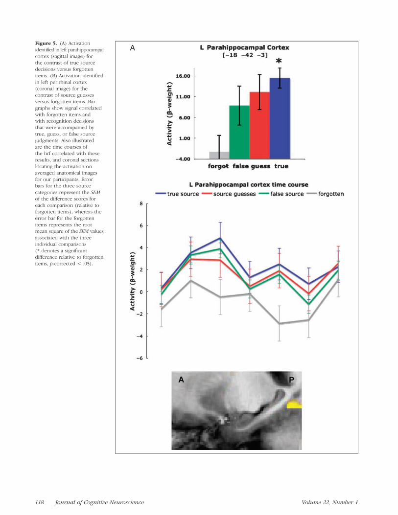
Figure 5. (A) Activation
identified in left parahippocampal
cortex (sagittal image) for
the contrast of true sourcedecisions versus forgotten
items. (B) Activation identified
in left perirhinal cortex(coronal image) for the
contrast of source guesses
versus forgotten items. Bar
graphs show signal correlatedwith forgotten items and
with recognition decisions
that were accompanied by
true, guess, or false sourcejudgments. Also illustrated
are the time courses of
the hrf correlated with theseresults, and coronal sections
locating the activation on
averaged anatomical images
for our participants. Errorbars for the three source
categories represent the SEM
of the difference scores for
each comparison (relative toforgotten items), whereas the
error bar for the forgotten
items represents the rootmean square of the SEM values
associated with the three
individual comparisons
(* denotes a significantdifference relative to forgotten
items, p-corrected < .05).
118 Journal of Cognitive Neuroscience Volume 22, Number 1

Figure 5. (continued)
Wais, Squire, and Wixted 119

strength confound by taking old–new decisions madewith relatively high confidence and then separating theminto categories according to whether source memory wascorrect or incorrect. Under those conditions, hippocam-
pal activity was elevated (and equally so) for both source-correct and source-incorrect decisions relative to forgot-ten items. The same was true for high-confidence old–newdecisions that were followed by source guesses (i.e.,low-confidence source decisions associated with chanceaccuracy). In a conceptually similar study, Kirwan, Wixted,and Squire (2008) investigated activity at encoding andfound that hippocampal activity increased with sub-sequent item memory strength even in the absence ofsource recollection. We next consider what our resultsmean depending on how source recollection failure isinterpreted.
Interpretation 1: Source Recollection FailureImplies Familiarity-based Item Decisions
If the failure to recollect task-relevant source detail fol-lowing a correct old decision is indicative of a familiarity-based decision (as has been assumed in prior studies),then our results suggest that hippocampal activity is as-sociated with both recollection and familiarity. Inter-preted in this fashion, our findings are at odds with theview that accords to the hippocampus a selective rolein recollection and perirhinal cortex a selective role infamiliarity (Eichenbaum et al., 2007; Brown & Aggleton,2001). Moreover, our findings suggest that the failure ofprior fMRI studies to detect increased hippocampal ac-tivity associated with familiarity-based responses mayhave more to do with the failure to detect a weak mem-ory than with the absence of familiarity-related hippo-campal activity (Stark & Squire, 2001).
Figure 6. Activation identified in prefrontal cortex for the contrast of high-confidence hits that were accompanied by true source judgments
versus high-confidence hits that were accompanied by source guesses. (A) Left VLPFC (BA 45) and (B) right DLPFC (BA 46). Error bars represent the
SEM of the difference scores (* denotes a significant difference between true source and source guesses, p-corrected < .05).
Figure 7. Activation identified for the contrast of hits rated as
old–new ‘‘6’’ versus forgotten items. Error bars represent the SEMof the difference scores (* denotes a significant difference relative
to forgotten items, p-corrected < .05).
120 Journal of Cognitive Neuroscience Volume 22, Number 1
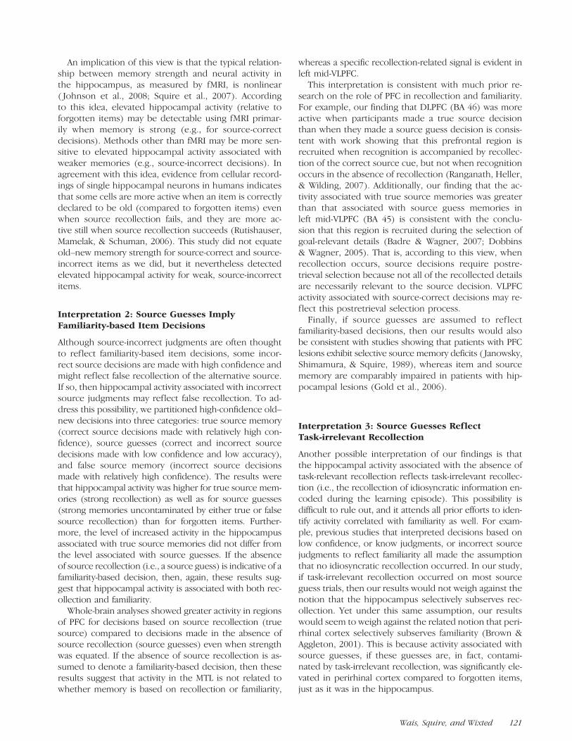
An implication of this view is that the typical relation-ship between memory strength and neural activity inthe hippocampus, as measured by fMRI, is nonlinear( Johnson et al., 2008; Squire et al., 2007). Accordingto this idea, elevated hippocampal activity (relative toforgotten items) may be detectable using fMRI primar-ily when memory is strong (e.g., for source-correctdecisions). Methods other than fMRI may be more sen-sitive to elevated hippocampal activity associated withweaker memories (e.g., source-incorrect decisions). Inagreement with this idea, evidence from cellular record-ings of single hippocampal neurons in humans indicatesthat some cells are more active when an item is correctlydeclared to be old (compared to forgotten items) evenwhen source recollection fails, and they are more ac-tive still when source recollection succeeds (Rutishauser,Mamelak, & Schuman, 2006). This study did not equateold–new memory strength for source-correct and source-incorrect items as we did, but it nevertheless detectedelevated hippocampal activity for weak, source-incorrectitems.
Interpretation 2: Source Guesses ImplyFamiliarity-based Item Decisions
Although source-incorrect judgments are often thoughtto reflect familiarity-based item decisions, some incor-rect source decisions are made with high confidence andmight reflect false recollection of the alternative source.If so, then hippocampal activity associated with incorrectsource judgments may reflect false recollection. To ad-dress this possibility, we partitioned high-confidence old–new decisions into three categories: true source memory(correct source decisions made with relatively high con-fidence), source guesses (correct and incorrect sourcedecisions made with low confidence and low accuracy),and false source memory (incorrect source decisionsmade with relatively high confidence). The results werethat hippocampal activity was higher for true source mem-ories (strong recollection) as well as for source guesses(strong memories uncontaminated by either true or falsesource recollection) than for forgotten items. Further-more, the level of increased activity in the hippocampusassociated with true source memories did not differ fromthe level associated with source guesses. If the absenceof source recollection (i.e., a source guess) is indicative of afamiliarity-based decision, then, again, these results sug-gest that hippocampal activity is associated with both rec-ollection and familiarity.
Whole-brain analyses showed greater activity in regionsof PFC for decisions based on source recollection (truesource) compared to decisions made in the absence ofsource recollection (source guesses) even when strengthwas equated. If the absence of source recollection is as-sumed to denote a familiarity-based decision, then theseresults suggest that activity in the MTL is not related towhether memory is based on recollection or familiarity,
whereas a specific recollection-related signal is evident inleft mid-VLPFC.
This interpretation is consistent with much prior re-search on the role of PFC in recollection and familiarity.For example, our finding that DLPFC (BA 46) was moreactive when participants made a true source decisionthan when they made a source guess decision is consis-tent with work showing that this prefrontal region isrecruited when recognition is accompanied by recollec-tion of the correct source cue, but not when recognitionoccurs in the absence of recollection (Ranganath, Heller,& Wilding, 2007). Additionally, our finding that the ac-tivity associated with true source memories was greaterthan that associated with source guess memories inleft mid-VLPFC (BA 45) is consistent with the conclu-sion that this region is recruited during the selection ofgoal-relevant details (Badre & Wagner, 2007; Dobbins& Wagner, 2005). That is, according to this view, whenrecollection occurs, source decisions require postre-trieval selection because not all of the recollected detailsare necessarily relevant to the source decision. VLPFCactivity associated with source-correct decisions may re-flect this postretrieval selection process.
Finally, if source guesses are assumed to ref lectfamiliarity-based decisions, then our results would alsobe consistent with studies showing that patients with PFClesions exhibit selective source memory deficits ( Janowsky,Shimamura, & Squire, 1989), whereas item and sourcememory are comparably impaired in patients with hip-pocampal lesions (Gold et al., 2006).
Interpretation 3: Source Guesses ReflectTask-irrelevant Recollection
Another possible interpretation of our findings is thatthe hippocampal activity associated with the absence oftask-relevant recollection reflects task-irrelevant recollec-tion (i.e., the recollection of idiosyncratic information en-coded during the learning episode). This possibility isdifficult to rule out, and it attends all prior efforts to iden-tify activity correlated with familiarity as well. For exam-ple, previous studies that interpreted decisions based onlow confidence, or know judgments, or incorrect sourcejudgments to reflect familiarity all made the assumptionthat no idiosyncratic recollection occurred. In our study,if task-irrelevant recollection occurred on most sourceguess trials, then our results would not weigh against thenotion that the hippocampus selectively subserves rec-ollection. Yet under this same assumption, our resultswould seem to weigh against the related notion that peri-rhinal cortex selectively subserves familiarity (Brown &Aggleton, 2001). This is because activity associated withsource guesses, if these guesses are, in fact, contami-nated by task-irrelevant recollection, was significantly ele-vated in perirhinal cortex compared to forgotten items,just as it was in the hippocampus.
Wais, Squire, and Wixted 121

Summary
The present research draws attention to a strength con-found that exists in prior fMRI studies of recollectionand familiarity, and it represents an attempt to comparethe activity associated with high-strength recollection-based decisions with the activity associated with similarlyhigh-strength familiarity-based decisions. Although morethan one interpretation of our results is possible, thesefindings raise the possibility (unaddressed by prior re-search; cf., Wais, 2008) that familiarity-based activity inthe hippocampus can be detected using fMRI whenmemory is strong. More work is needed to definitivelyresolve this issue, but our results should encouragethe search for additional behavioral methods to isolatestrong, familiarity-based decisions when seeking to iden-tify the neuroanatomical basis of recollection and famil-iarity in the MTL.
APPENDIX
Model-based Interpretations
The high-threshold/dual-process model (Yonelinas, 1999)assumes that responses based on recollection and re-sponses based on familiarity can be easily isolated fromeach other because they are independent processes thatcontribute to different recognition decisions. More spe-cifically, the high-threshold model views the strength offamiliarity as being continuous (i.e., ranging from weakconfidence based on familiarity to strong confidencebased on familiarity), whereas it views recollection as acategorical process that, when it occurs, only gives riseto the strongest confidence. Because recollection reliablyyields responses with the highest memory confidence,this model assumes that the occurrence of recollectionpreempts familiarity (which is variable in strength) andthat responses made with the highest confidence aretypically based on recollection. If recollection does notoccur, then the response is exclusively based on familiar-ity instead (Parks & Yonelinas, 2007). Accordingly, recog-nition decisions made with the highest confidence canbe taken to primarily denote recollection because deci-sions based on highest-confidence familiarity are usuallyfew in number, whereas recognition decisions made withconfidence below that threshold can be taken to denotefamiliarity.
Similar considerations apply to experiments that use theRemember/Know/New procedure (Gardiner & Richardson-Klavehn, 2000; Tulving, 1985). Standard instructionsfor this procedure ask subjects to respond ‘‘Remember’’based on recollection whenever it occurs and to other-wise respond ‘‘Know’’ based on a strong sense of familiar-ity. The Remember/Know procedure, as it has typicallybeen applied in neuroimaging studies, assumes that deci-sions are based exclusively on recollection or familiarity.The most common interpretation of the Remember/Knowprocedure also assumes that the recollection process will
preempt the familiarity process, just as the high-thresholdview does.
Like other dual-process models, the aggregated-strength/dual-process model (Wixted, 2007) assumes thatrecollecting the contextual details associated with an itemis a separate process from appreciating the familiarity ofan item. However, according to this view, the strength ofrecollection underlying recognition responses varies fromweak to strong, just as the strength of underlying familiar-ity varies from weak to strong (Slotnick & Dodson, 2005).In addition, it assumes that recollection and familiarity areaggregated to determine the memory strength of a par-ticular item. That is, this model uniquely holds that bothprocesses contribute to individual recognition decisions(Wixted, 2007; Kelley & Wixted, 2001). This aggregatedstrength view of recognition is compatible with standardsignal-detection theory, and it implies that the strength ofmemory per se cannot be interpreted as a sign of under-lying recollection or familiarity.
Acknowledgments
This study was supported by an Innovative Research Grantfrom the Kavli Institute for Brain and Mind at the University ofCalifornia, San Diego, the Medical Research Service of the De-partment of Veteran Affairs, NIMH, and the Metropolitan LifeFoundation.
Reprint requests should be sent to Peter E. Wais, Departmentof Neurology, University of California, San Francisco, 1700 4thStreet Room 102C, San Francisco, CA 94158, or via e-mail: [email protected].
REFERENCES
Badre, D., & Wagner, A. (2007). Left ventrolateral prefrontalcortex and the cognitive control of memory.Neuropsychologia, 45, 2883–2901.
Brown, M., & Aggleton, J. (2001). Recognition memory: Whatare the roles of the perirhinal cortex and hippocampus?Nature Reviews Neuroscience, 2, 51–61.
Cabeza, R., Rao, S., Wagner, A., Mayer, A., & Schacter, D.(2001). Can medial temporal lobe regions distinguishtrue from false? An event-related functional MRI study ofveridical and illusory recognition memory. Proceedingsof the National Academy of Sciences, U.S.A., 98, 4805–4810.
Cox, R. W. (1996). AFNI: Software for analysis and visualizationof functional magnetic resonance neuroimages. Computersand Biomedical Research, 29, 162–173.
Daselaar, S., Fleck, M., & Cabeza, R. (2006). Triple dissociationin the medial temporal lobes: Recollection, familiarity andnovelty. Journal of Neurophysiology, 96, 1902–1911.
Davachi, L., Mitchell, J., & Wagner, A. (2003). Multiple routesto memory: Distinct medial temporal lobe processes builditem and source memories. Proceedings of the NationalAcademy of Sciences, U.S.A., 100, 2157–2162.
Dobbins, I., & Wagner, A. (2005). Domain-general anddomain-sensitive prefrontal mechanisms for recollectingevents and detecting novelty. Cerebral Cortex, 15,1768–1778.
Dunn, J. (2004). Remember–know: A matter of confidence.Psychological Review, 111, 524–542.
122 Journal of Cognitive Neuroscience Volume 22, Number 1

Dunn, J. C. (2008). The dimensionality of the remember–know task: A state–trace analysis. Psychological Review,115, 426–446.
Eichenbaum, H., Yonelinas, A., & Ranganath, C. (2007).The medial temporal lobe and recognition memory.Annual Review of Neuroscience, 30, 123–152.
Eldridge, L., Engel, S., Zeineh, M., Bookheimer, S., &Knowlton, B. (2005). A dissociation of encoding andretrieval processes in the human hippocampus. Journalof Neuroscience, 25, 3280–3286.
Eldridge, L., Knowlton, B., Furmanski, C., Bookheimer, S.,& Engel, S. (2000). Remembering episodes: A selectiverole for the hippocampus during retrieval. NatureNeuroscience, 3, 1149–1152.
Gardiner, J., & Richardson-Klavehn, A. (2000). Rememberingand knowing. The Oxford Handbook of Memory. New York:Oxford University Press.
Gold, J., Smith, C., Bayley, P., Shrager, Y., Brewer, J., Stark, C.,et al. (2006). Item memory, source memory, and themedial temporal lobe: Concordant findings from fMRI andmemory-impaired patients. Proceedings of the NationalAcademy of Sciences, U.S.A., 103, 9351–9356.
Janowsky, J., Shimamura, A., & Squire, L. (1989). Sourcememory impairment in patients with frontal lobe regions.Neuropsychologia, 27, 1043–1056.
Johnson, J., Muftuler, L., & Rugg, M. (2008). Multiplerepetitions reveal functionally- and anatomically-distinctpatterns of hippocampal activity during continuousrecognition memory. Hippocampus, 18, 975–980.
Kelley, R., & Wixted, J. (2001). On the nature of associativeinformation in recognition memory. Journal ofExperimental Psychology: Learning, Memory andCognition, 27, 701–722.
Kensinger, E., & Schacter, D. (2006). Amygdala activity isassociated with the successful encoding of item, but notsource, information for positive and negative stimuli.Journal of Neuroscience, 26, 2564–2570.
Kirwan, C., Jones, C., Miller, M., & Stark, C. (2007).High-resolution investigation of the medial temporallobe. Human Brain Mapping, 10, 959–966.
Kirwan, C., Wixted, J., & Squire, L. (2008). Activity in the medialtemporal lobe predicts memory strength, whereas activity inthe prefrontal cortex predicts recollection. Journal ofNeuroscience, 28, 10541–10548.
Mandler, G. (1980). Recognizing: The judgment of previousoccurrence. Psychological Review, 87, 252–271.
Manns, J., Hopkins, R., Reed, J., Kitchener, E., & Squire, L.(2003). Recognition memory and the human hippocampus.Neuron, 37, 171–180.
Mickes, L., Wixted, J., & Wais, P. (2007). A direct test of theunequal-variance signal-detection model of recognitionmemory. Psychonomic Bulletin & Review, 14,858–865.
Miller, M., Beg, M., Ceritoglu, C., & Stark, C. (2005). Increasingthe power of functional maps of the medial temporallobe by using large deformation diffeomorphic metricmapping. Proceedings of the National Academy ofSciences, U.S.A., 102, 9685–9690.
Montaldi, D., Spencer, T., Roberts, N., & Mayes, A. (2006).The neural system that mediates familiarity memory.Hippocampus, 16, 504–520.
Parks, C., & Yonelinas, A. (2007). Moving beyond puresignal-detection models: Comment on Wixted (2007).Psychological Review, 114, 188–202.
Ranganath, C., & Blumenfeld, R. (2007). Prefrontal cortexand human memory: An integrative review of findingsfrom neuropsychology and neuroimaging. Neuroscientist,13, 280–291.
Ranganath, C., Heller, A., & Wilding, E. (2007). Dissociablecorrelates of two classes of retrieval processing inprefrontal cortex. Neuroimage, 35, 1663–1673.
Ranganath, C., Yonelinas, A., Cohen, M., Dy, C., Tom, S.,& D’Esposito, M. (2004). Dissociable correlates ofrecollection and familiarity within the medial temporallobes. Neuropsychologia, 42, 2–13.
Rutishauser, U., Mamelak, A., & Schuman, E. (2006). Single-triallearning of novel stimuli by individual neurons of the humanhippocampus–amygdala complex. Neuron, 49, 805–813.
Schacter, D., & Slotnick, S. (2004). The cognitive neuroscienceof memory distortion. Neuron, 44, 149–160.
Slotnick, S., & Dodson, C. (2005). Support for a continuous(single-process) model of recognition memory andsource memory. Memory & Cognition, 33, 151–170.
Squire, L., Wixted, J., & Clark, R. (2007). Recognitionmemory and the medial temporal lobe: A new perspective.Nature Reviews Neuroscience, 8, 872–883.
Stark, C., & Squire, L. (2001). When zero is not zero: Theproblem of ambiguous baseline conditions in fMRI.Proceedings of the National Academy of Sciences, U.S.A.,98, 12760–12766.
Talairach, J., & Tournoux, P. (1988). Co-planar stereotaxicatlas of the human brain. 3-dimensional proportionalsystem: An approach to cerebral imaging. New York:Thieme Medical Publishers.
Tulving, E. (1985). Memory and consciousness. CanadianJournal of Psychology, 26, 1–12.
Wais, P. (2008). fMRI signals associated with memory strengthin the medial temporal lobes: A meta-analysis.Neuropsychologia, 46, 3185–3196.
Wais, P., Mickes, L., & Wixted, J. (2008). Remember/Knowjudgments probe degrees of recollection. Journal ofCognitive Neuroscience, 20, 400–405.
Weis, S., Specht, K., Klaver, P., Tendolkar, I., Willmes, K.,Ruhlmann, J., et al. (2004). Process dissociation betweencontextual retrieval and item recognition. NeuroReport,1, 2729–2733.
Wixted, J. (2007). Dual-process theory and signal-detectiontheory of recognition memory. Psychological Review,114, 152–176.
Yonelinas, A. (1994). Receiver operating characteristics inrecognition memory: Evidence for a dual process model.Journal of Experimental Psychology: Learning, Memory,and Cognition, 20, 1341–1354.
Yonelinas, A. (1999). The contribution of recollection andfamiliarity to recognition and source memory: An analysisof receiver operating characteristics and a formal model.Journal of Experimental Psychology: Learning, Memory,and Cognition, 25, 1415–1434.
Yonelinas, A., Otten, L., Shaw, K., & Rugg, M. (2005).Separating the brain regions involved in recollectionand familiarity in recognition memory. Journal ofNeuroscience, 25, 3002–3008.
Wais, Squire, and Wixted 123