in nasopharyngeal carcinoma by inhibiting hsa-mir- lncrna
TRANSCRIPT

Page 1/25
LncRNA ZFAS1 contributed to irradiation resistancein nasopharyngeal carcinoma by inhibiting hsa-miR-7-5p/ENO2Jiaojiao Peng
Sichuan University West China HospitalFeng Liu
Sichuan University West China HospitalHong Zheng
Sichuan University West China HospitalQi Wu
Sichuan University West China HospitalShixi Liu ( [email protected] )
Sichuan University West China Hospital https://orcid.org/0000-0003-0319-080X
Primary research
Keywords: ZFAS1, miR-7-5p, ENO2, nasopharyngeal carcinoma, irradiation resistance
Posted Date: March 10th, 2020
DOI: https://doi.org/10.21203/rs.3.rs-16630/v1
License: This work is licensed under a Creative Commons Attribution 4.0 International License. Read Full License

Page 2/25
AbstractBackground: In our previous research, we found that lncRNA ZFAS1 could promote nasopharyngealcarcinoma by inhibiting its downstream target axis. But we hadn’t shown whether ZFAS1 had associationwith irradiation resistance of NPC. In this study, we aimed to study the role of ZFAS1 in irradiationresistance of nasopharyngeal carcinoma.
Methods: Bioinformatics analysis was �rst conducted to identify the potentially signi�cant gene thatparticipated in radio resistance of nasopharyngeal carcinoma. qRT-PCR was used to detect theexpression of ZFAS1, miR-7-5p and ENO2 mRNA. The location of ZFAS1 was measured in cell fractions.The relationship between ZFAS1, miR-7-5p and ENO2 mRNA was validated by luciferase reporter assayand RNA pull down. Edu assay and �ow cytometric apoptosis assay were conducted to observe howZFAS1, miR-7-5p and ENO2 affected nasopharyngeal carcinoma proliferation and apoptosis underirradiation.
Results ZFAS1 was upregulated in nasopharyngeal carcinoma, which was associated with radioresistance. ZFAS1 strengthened the ability of irradiation resistance in nasopharyngeal carcinoma. ZFAS1acted on miR-7-5p to promote ENO2 upregulation, thereby promoting the irradiation resistance.
Conclusion: ZFAS1 sponged miR-7-5p to further affect ENO2 expression to augmenting the irradiationresistance in nasopharyngeal carcinoma.
BackgroundNasopharyngeal carcinoma (NPC) is a type of malignant tumor which occurs in nasopharyngealepithelial cells with high morbidity and mortality in China. [1] NPC usually induces poor prognosis inpatients due to the strong ability of migration and early distant metastasis [2, 3]. Epstein-Barr virus isoften considered as a risk factor for the development of NPC with other factors containing smokingcigarette, eating nitrosamine dietary, long-term exposure to chemical carcinogens and geneticsusceptibility in consideration [4, 5]. At present, radiotherapy is the �rst choice to be considered forpatients’ treatment [6, 7]. But there are also a number of patients with radio resistance have poorprognosis [7, 8]. Therefore, it is necessary to discover how to reverse radio resistance in NPC patients.
Long noncoding RNA (lncRNA) is a type of RNA that is more than 200 nucleotides in length withregulating gene expression at multiple levels [9]. Recent studies have shown that lncRNA is involved inmany important regulatory processes including X-chromosome silencing, genomic imprinting, chromatinmodi�cation, transcriptional activation and transcriptional interference [10–14]. LncRNA ZFAS1 locatesat the chromosome 20q13.13 with the exon count of 5, and is �rst found in breast cancer [15]. There weresome researches reporting that the expression of ZFAS1 was aberrant in hepatocellular carcinoma,prostate cancer, colorectal cancer and esophageal squamous cell carcinoma [16–19]. In our previousstudy, we also reported that lncRNA ZFAS1 participated in the development of NPC as a tumor promoter[20]. However, we hadn’t demonstrated whether ZFAS1 played a role in radio resistance in NPC.

Page 3/25
MiRNA with a length of 18–25 nt has been proved to play an essential role in the development of manycancers including pancreatic cancer, breast cancer, prostate cancer, colorectal cancer, gastric cancer, lungsquamous cell carcinoma and ovarian cancer [21–27]. The involvement of miR-7-5p in human cancergenesis has been reported during the last six years; however, the participation of miR-7-5p in irradiationresistance of human cancers including nasopharyngeal carcinoma has never been studied. Only thatKazuo’s team reported in 2019 that miR-7-5p was up-regulated in clinically relevant resistance HeLa(cervical cancer cell line) and SAS (oral squamous cell carcinoma) cell lines [28]. In addition, controversialroles of miR-7-5p in chemo-drug resistance have been reported in human hepatocellular carcinoma [29],cervical cancer [30], breast cancer [31], and small cell lung cancer [32]. Whether in nasopharyngeal miR-7-5p promotes radio resistance remains undiscovered.
ENO2 is one of three enolase isoenzymes that were found in mammals [33]. ENO2 was majorly located inmature neurons and cells for neuronal origin and highly expressed in some cancers includingglioblastoma, neuroendocrine prostate cancer and renal cell carcinoma [34–36]. There was a reportcovering that ENO2 was a responsive gene of HIF, which took part in the development of tumor growth[37]. We identi�ed ENO2 to be a potential downstream effector of miR-7-5p, and predicted it tosigni�cantly involve in NPC irradiation resistance. How miR-7-5p-ENO2 axis participates in NPCirradiation resistance was studied in the present study.
In our previous study, we demonstrated that lncRNA ZFAS1 was the promoter of NPC. However, we didn’tverify whether ZFAS1 affected radio resistance. In addition to the last research, we identi�ed a noveldownstream effector axis, miR-7-5p-ENO2. We then studied the effects of ZFAS1-miR-7-5p-ENO2interactome on NPC resistance to radiation.
Materials And MethodsTissues collection
Fifty-�ve patients who were diagnosed with nasopharyngeal carcinoma in West China Hospital ofSichuan University were enrolled in our study. The ethics committee of West China Hospital of SichuanUniversity approved our study protocol. NPC tissues and adjacent healthy tissues were frozen untilexperiments and clinical characteristics were recorded. The collection and the use of tissues followed theethical standards built by the Helsinki Declaration.
Cell culture
The human NPC cell lines SUNE-1, 5-8F, and C666-1 as well as normal nasal mucosal epithelial cell NP-69were all bought from BeNa Culture Collection (Beijing, China). SUNE-1, 5-8F, and C666-1 were cultured inRPMI-1640 with 10% FBS.
RNA was detected by real-time quanti�cation PCR.
Page 4/25
Total RNA was disintegrated by Trizol (DP501, Tiangen Biochemical, China). The RNA was reversetranscribed into cDNA using a cDNA synthesis kit (KR211, Tiangen Biochemical, China) after checking thepurity of RNA. Next, the expression of lncRNA ZFAS1, miR-7-5p and ENO2 mRNA were analyzed by ABI7500 using a SYBR Green PCR kit (FP411, Tiangen Biochemical, China). U6 acted as a reference gene formiR-7-5p while GAPDH acted as a reference gene for lncRNA ZFAS1 and ENO2 mRNA.
Cell transfection
Si-ZFAS1-1, si-ZFAS1-2, miR-7-5p inhibitor, ENO2 siRNA (si-ENO2), and si-NC were designed andsynthesized by Tiangen Biochemical (Beijing, China). Lipofectamine 2000 reagent (ThermoFisher,11668027, USA) was used to transfect plasmids into target cells (SUNE-1 and C666-1 cell lines) on thebasis of the protocol.
Cell fractionation
The Invitrogen PARIS Kit (ThermoFisher, AM1921, USA) was employed for separating and purifyingcytoplasmic and nuclear RNA based on speci�cations. Expression levels of lncRNA ZFAS1, GAPDH(cytoplasmic control) and U6 (nuclear control) in cytoplasm and nucleus were examined using qRT-PCR.
Luciferase reporter assay
We purchased constructed plasmids containing wild type or mutant type of lncRNA ZFAS1 and ENO2mRNA from Tiangen Biochemical (Beijing, China). Next, we transfected these plasmids into SUNE-1 andC666-1 cell lines by Lipofectamine 2000 reagent (ThermoFisher, 11668027, USA). Then, miR-892b mimicswere co-transfected into the same cells by the same method. After 48 hours, we gathered cells to lyse bylysis buffer. The dual-luciferase reporter assay system (GeneCopoeia, LF031, China) was used to analyzethe relative luciferase activity.
RNA pull-down assay
RNA pull-down experiment was performed as the instruction of the kit (ThermoFisher, 20164, USA). First,miR-7-5p, antisense oligo, and miR-7-5p mutant were labeled with biotin, which can bind to thestreptavidin magnetic beads. Cell lysates were incubated with biotin-labeled miR-7-5p, antisense oligo,and miR-7-5p mutant. Only biotin labeled miR-7-5p could bind to the target in a RISC dependent manner.Then, the incubated lysate samples went through the streptavidin magnetic beads. The elusion was doneusing non-denaturing Biotin Elution Buffer or SDS-PAGE Loading Buffer. At last, qRT-PCR was undertakento measure the expression of lncRNA ZFAS1 or miR-7-5p in the elution.
CCK-8 assay
CCK-8 kit was purchased from Dojindo Laboratories (Kumamoto, Japan) to determine the cell viability. Inthe irradiation dose-dependent assay, cell viability of every group was detected at 0Gy, 4Gy, and 8Gydoses of irradiation respectively. In the irradiation time-dependent assay, cell viability of every group was

Page 5/25
detected at 24h, 48h, 72h and 96h, respectively. 10 μL CCK-8 solution was added to every well to incubatewith the cells for 2h. The absorbance of cells in every well was read at 450 nm using an ELISA platereader.
Edu assay
The proliferation of NPC cells under irradiation was measured by Edu assay which was conducted usingEdu Apollo DNA in vitro kit bought from RIBOBIO (C10341-3, Guangzhou, China). All manipulates weredone following the protocol from the kit. Three random �elds under a �uorescence microscope wereselected and the Edu positive cells were counted. The Edu positive rate (=Edu positive cell number/DAPIpositive cell number) represented the proliferation condition of the cells. Representative images of everygroup were given.
Flow cytometric apoptosis assay
Flow cytometry was employed for the apoptosis detection. Brie�y, cell suspension with 104 cells were putin every tube, and went through centrifugation to lose the culture media. The cells were then washed withcold PBS twice. Then, the cold PBS was removed. 100 µl 1× binding buffer was added to the cells and thecells were re-suspended. 5 µl Annexin V and 5 µl PI (Beyotime, Beijing, China) were added to the cells indark and incubated for 15 min. 300 µl 1× binding buffer was added to the cells and the cells were re-suspended again. The cell suspension was moved to 5 ml �ow tubes. Within the next one hour, the cellswent through �ow cytometry analysis.
Statistical analyses
All the data were analyzed using Graphpad Prism 8.0. All results represented three independentexperiments. Values were expressed as mean±SD. Two-tailed t-test was used for analyzing differencesbetween two groups, while one-way ANOVA with Dunnett’s post hoc test was used for analyzingdifferences in multiple groups. P-values less than 0.05 were regarded as statistically signi�cant.
ResultENO2 and miR-7-5p were identi�ed as the potential downstream interactome of ZFAS1 in NPC
WebGestalt algorithm (�gure1.A-B) and Metascape algorithm (�gure1.C-D) were used to enrich thepathway and GO terms of GSE48503 differentially expressed genes (DEGs). The results showed thatglycolysis and HIF-1 pathway were the primary enriched pathways in NPC irradiation resistance.Glycolysis and HIF-1 pathway were proved to be closely associated with irradiation resistance of cancers[38-41]. We thus hypothesized that the genes that participated in glycolysis and/or HIF-1 pathway couldbe a critical gene that participated in NCP irradiation resistance. We found the ENO2 and EGLN3 were thetwo genes with this potential. Thus, we used STRING algorithm to further analyse the interaction strengthbetween the DEGs. We saw that ENO2 showed the most interaction evidence with its neighboring genes

Page 6/25
(�gure1. E). Therefore, we chose ENO2 as our study subject. Subsequently, we sought for the commonmiRNAs that were both downstream targets of ZFAS1 and upstream regulators of ENO2. The downstream target miRNAs of ZFAS1 were obtained from ENCORI database (http://starbase.sysu.edu.cn/),and the upstream regulating miRNAs of ENO2 were obtained from TargetScan Human 7.2(http://targetscan.org/vert_72/). Lastly, the expression patterns of the overlapped six miRNAs wereexplored on dbDEMC database (�gure1. F). They were all shown to be signi�cantly downregulated inNPC; however, we have known that miR-7-5p was a signi�cant tumor suppressor in many human cancersand it has been only limitedly studied in NPC, and its role in NPC under irradiation has not beeninvestigated yet. To �ll this gap, we determined miR-7-5p to be our miRNA of interest.
ZFAS1 was upregulated in nasopharyngeal carcinomas and NPC cell lines, and primarily located incytoplasm.
ZFAS1 had a higher expression level in nasopharyngeal carcinomas compared with adjacent healthytissues (�gure2.A). At the same time, the expression of ZFAS1 was found signi�cantly upregulated inNPC cell lines SUNE-1, 5-8F and C666-1 rather than in normal nasal mucosal epithelial cell NP-69(�gure2.B). Subsequently, total RNA was extracted from SUNE-1 and C666-1 cell lines with nuclear andcytoplasm RNA separated. The result of qRT-PCR showed that ZFAS1 was majorly located in cytoplasm(�gure2.C).What’s more, both SUNE-1 and C666-1 cells went through irradiation treatment. The survivalanalysis result suggested that both cell lines were dead completely at 8Gy and the IC50 was somewherebetween 2Gy and 4Gy (�gure2.D). Before further experiments were conducted, we testi�ed and validatedthe transfection e�ciency of ZFAS1 siRNAs in both cell lines (�gure2.E).
Knockdown of ZFAS1 increased the radiation sensitivity of nasopharyngeal carcinoma in vitro.
We wonder whether knockdown of ZFAS1 could increase the irradiation sensitivity of NPC cells, thus wedetected the survival outcomes of NPC cells under different doses of irradiation treatment and differenttiming using CCK-8 assay. We found that along with the increase of irradiation, the knockdown of ZFAS1showed more decreased cell survival compared with the control group (�gure3. A). We found thatexposure to 8Gy irradiation gave the lowest survival rate in SUNE-1 and C666-1 cell lines throughobserving the minimum survival rate. In addition, under 8Gy irradiation treatment, both cell linesdemonstrated signi�cantly decreased survival rate in a time dependent manner (�gure3. B). Next, weobserved that both si-ZFAS1s decreased the proliferation of SUNE-1 and C666-1 cell lines compared withcontrol group by Edu assay. The Edu positive rate in ZFAS1 knockdown groups decreased byapproximately ½ in SUNE-1 cell line and 1/3 in C666-1 cell line (�gure3. C). Furthermore, knockdown of si-ZFAS1 resulted in more apoptotic cells than control group under 8Gy irradiation treatment. The apoptosisrate in ZFAS1 knockdown groups was more than 2-fold of that in control group (�gure3. D).
ZFAS1 was the upstream target gene of miR-7-5p.
The binding sequences of ZFAS1 on miR-7-5p were obtained from ENCORI and were illustrated in �gure4.A. To determine the relationship between ZFAS1 and miR-7-5p, we carried out both luciferase reporter

Page 7/25
assay and RNA pull-down assay. The luciferase reporter gene assay result suggested that wild typeZFAS1 could bind with miR-7-5p to decrease the �uorescence intensity compared with other groups(�gure4.B). RNA pull-down assay results also showed that ZFAS1 was enriched with the presence of miR-7-5p mimics compared with the presence of antisense oligo or miR-7-5p mutant (�gure4.C). Moreover, theexpression of miR-7-5p was signi�cantly less in NPC tissues than in the adjacent tissues (�gure4.D).Then, we detected the miR-7-5p expression in NPC cell lines and normal control cell NP-69. The resultshowed that miR-7-5p was signi�cantly downregulated in NPC cell lines contrast to NP-69 (�gure 4.E). Itwas also revealed that ZFAS1 had a negative association with miR-7-5p (�gure4.F). Again, before weconducted any further experiments, we examined the transfection e�ciency of certain molecules. To beprecise, after transfection of si-ZFAS1 (si-ZFAS1-1), miR-892b inhibitor, si-NC and co-transfection si-ZFAS1+miR-7-5p inhibitor, we observed that si-ZFAS1 group expressed 1.5-fold more miR-7-5p while miR-7-5p inhibitor group expressed 75% less miR-7-5p compared with control group. In addition, theexpression of miR-7-5p showed no signi�cant difference between the co-transfection of si-ZFAS1 andmiR-7-5p inhibitor group and the control group (�gure4. G).
Knockdown of ZFAS1 promoted the radiation sensitivity of NPC cells by acting on miR-7-5p.
To further discuss how ZFAS1 affects radiation sensitivity of NPC by regulating miR-7-5p, we designed arescue experiment. CCK-8 assay results suggested that si-ZFAS1 weakened the cell viability of SUNE-1cell line at 48h, 72h and 96h, and C666-1 cells at 72h and 96h, which was offset by the introduction ofmiR-7-5p inhibitor at the environment of 8Gy irradiation (�gure5.A). Equally, Edu assay resultsdemonstrated a similar result with CCK-8 assay. The proliferating SUNE-1 and C666-1 cells signi�cantlydecreased in si-ZFAS1 group but increased in miR-7-5p inhibitor group: the Edu positive rate droppedapproximately half in si-ZFAS1 group, and raised by 20% in miR-7-5p inhibitor group. What’s more, whenwe cotransfected miR-7-5p and si-ZFAS1, we found that the proliferating cells increased contrast to si-ZFAS1 group (�gure5.B). Besides, we determined that si-ZFAS1 promoted cell apoptosis while miR-7-5pinhibitor inhibited cell apoptosis in SUNE-1 and C666-1 cell lines. The apoptosis rate in the si-ZFAS1group was more than 2-fold of the control group, whereas that in the miR-7-5p inhibitor group was merelyless than half of the control group. The number of apoptotic cells did not differ from the control groupwhen miR-7-5p inhibitor and si-ZFAS1 were co-transfected (�gure5.C).
MiR-7-5p was the upstream target gene of ENO2 mRNA.
The binding scheme predicted by TargetScan Human 7.2 (http://www.targetscan.org/vert_72/) showingthe targeting relationship between miR-7-5p and ENO2 mRNA was illustrated in �gure6.A. The bindingrelationship was validated using dual luciferase reporter gene assay and RNA pull-down assay.Luciferase reporter assay demonstrated that wide type ENO2 mRNA be targeted by miR-7-5p and led tothe �uorescence intensity decrease compared with other groups (�gure6.B). RNA pull-down assay resultsshowed that ENO2 was signi�cantly enriched with the addition of miR-7-5p mimics contrast to antisenseoligo and miR-7-5p mutant groups in both SUNE-1 cell line and C666-1 cell line (�gure6.C). When wedetected ENO2 mRNA expression in NPC tissues and NPC cell lines, the result revealed that ENO2 mRNA

Page 8/25
was signi�cantly upregulated in NPC tissues and cells (�gure6.D-E). The expression of miR-7-5p andENO2 mRNA was negatively associated (�gure6.F). What’s more, we built cell models stably transfectedwith si-NC, si-ENO2, miR-7-5p inhibitor, and si-ENO2+miR-7-5p inhibitor for further experiments (�gure6.G).
MiR-7-5p inhibition enhanced the radiation resistance of NPC cells by acting on ENO2 mRNA.
To further explore how miR-7-5p took part in radiation resistance in NPC by regulating ENO2 mRNA, weperformed rescue experiments. Cell viability was measured by CCK-8 assay, the results of whichsuggested that at the environment of 8Gy irradiation, si-ENO2 signi�cantly weakened the cell viability ofNPC cells with miR-7-5p inhibitor offsetting it at 72h and 96h in both SUNE-1 and C666-1 cell lines(�gure7.A). Equally, Edu assay demonstrated a similar result with CCK-8 assay. The proliferating cells inSUNE-1 and C666-1 cell lines decreased in si-ENO2 group, but increased in miR-7-5p inhibitor group.What’s more, when we cotransfected miR-7-5p inhibitor with si-ENO2, we found that the proliferating cellsincreased contrast to si-ENO2 group (�gure7.B). Besides, we determined that si-ENO2 promoted cellapoptosis by almost 50%, while miR-7-5p inhibitor inhibited cell apoptosis by almost 50% in SUNE-1 andC666-1 cell lines. The number of apoptosis cell increased when miR-7-5p inhibitor and si-ENO2 were co-transfection compared with si-ENO2 group (�gure7.C). In �gure 8, we simply illustrated the hypothesizedmechanism in NPC radio resistance involving ZFAS1, miR-7-5p and ENO2. Basically, under the irradiation,the high ZFAS1 and ENO2 levels with low miR-7-5p level resulted in more resistance to irradiation ofcancerous cells with less resistance to irradiation, i.e., ZFAS1 promoted NPC cell’s resistance to irradiationby downregulating miR-7-5p, thereafter releasing more ENO2 mRNA.
DiscussionIn our experiments, we observed that ZFAS1 was signi�cantly upregulated in NPC tissues and cells aswell as ENO2 while miR-7-5p was downregulated in NPC tissues and cells. Our results revealed thatZFAS1 knockdown weakened irradiation resistance in NPC in vitro. To further explore the mechanism howZFAS1 affected the irradiation sensitivity, we introduced miR-7-5p inhibitor and ENO2 siRNA to our cellmodels, SUNE-1 and C666-1 cell lines. It was found that the knockdown of ZFAS1 and ENO2 led toincreased sensitivity to irradiation, whereas the inhibition of miR-7-5p led to the opposite.
In the last two years, lncRNA ZFAS1 has been proved to promote cancer genesis of NPC by some teams.For instance, it was reported to promote NPC genesis by activating Wnt/β-actin pathway [42] and byinhibiting PI3K/AKT pathway [43]. In addition, it was reported to regulate miR-135a to promote NPCgenesis [44]. In our previous study, lncRNA ZFAS1 was also found highly expressed in NPC tissues andaugmented NPC by regulating miR-893b-LPAR1 interactome [20]. Since 2017, accumulating studies havebeen conducted to study the mechanism of drug resistance involving ZFAS1 in various human cancers.For instance, ZFAS1 was found to promote resistance to Adriamycin in T-cell acute lymphoblasticleukemia [45]. ZFAS1 knockdown led to signi�cantly impaired resistance to cis-platinum or paclitaxel ingastric cancer [46]. Increased resistance to temozolomide in glioma [47]. Other lncRNAs have been

Page 9/25
reported to be associated with the irradiation resistance of NPC. For instance, lncRNA MINCR wasoverexpressed in NPC with worse prognosis in patients by promoting the radio resistance of NPC. MINCRdecreased NPC cell radiation sensitivity through the competitive binding with miR-892b, increasing ZEB1,and activating AKT/PI3K signaling [5]. In addition, lncPVT1 silence promoted the radio sensitivity in NPCcells through inhibiting H3K9 and TIF1β combination. PVT1 was proposed to act as an oncogenic part inradio resistance through activating HIF-1α [48]. However, lnc ZFAS1 has not been studied in the radiationresistance of human cancers including NPC. To �ll this gap, we herein further discussed whether ZFAS1affected radio resistance in NPC. In our study, we found that the knockdown of lncRNA ZFAS1 was ableto impair the radio resistance in NPC, shown by the impaired cell survival and proliferation, and enhancedcell apoptosis.
During the last one decade, miR-7-5p has been discovered to be a tumor suppressor in various humancancers. It was found signi�cantly downregulated and played a tumor suppressing role in gastric cancer,presented by inhibited colony formation and invasion [49]; in colorectal cancer, presented by impaired cellviability, proliferation, migration, invasion, and EMT [50]; and breast cancer, demonstrated by suppressedcell proliferation, migration and invasion [51]. Also, in human NPC, it was found to be a tumor suppressorand led to signi�cantly suppressed colony formation [52]. Yet, how miR-7-5p regulates irradiationresistance in human cancers has not been thoroughly studied. Herein, we found that the inhibition of miR-7-5p by miR-7-5p inhibitor signi�cantly enhanced the radio resistance of NPC cells, thus we concludedthat miR-7-5p, as a ceRNA of ZFAS1, could enhance the radio sensitivity in NPC.
ENO2 has been reported as a HIF-responsive gene in a xenograft model in renal cell carcinoma [37]. Astudy also reported that silencing ENO2 by siRNA technique selectively inhibited the growth and survivalof ENO1 deleted cells in glioblastoma [34]. In addition, there was a study determining that ENO2 involvedin the glycolytic pathway in colorectal cancer [53]. Plus, ENO2 was upregulated in neuroendocrineprostate cancer, leading to epithelial to mesenchymal transition and cancer stem cell phenotype [54].Combining with our study, we concluded that ENO2 might be an oncogene in NPC, and it wassigni�cantly upregulated in NPC. We also hypothesized that ENO2 might take part in glycolytic pathwayand HIF-1α pathway to regulate tumor radio resistance.
In our study, we identi�ed that ENO2 could be an important player in hypoxia response via HIF-1 signalingby a bioinformatics method. Hypoxic tumor cells are resistant to radiotherapy. HIF-1 signaling has beenregarded as an important signaling in radiotherapy resistance of human cancers [55]. Yet, we haven’tbeen able to design experiments to study the role of ZFAS1 on tumor hypoxia, and we did not researchhow ENO2 regulated HIF-1α pathway by being regulated by ZFAS1 and miR-7-5p. In addition, animalexperiments remain to be conducted to validate the effects of the interactome in vivo.
ConclusionTaken together, our experiment suggested that lncRNA ZFAS1 acted as a radiation resistance enhancer inNPC. Mechanically, ZFAS1 competitively bound with miR-892b, thereby increasing the expression of

Page 10/25
ENO2 to enhance the radiation resistance of NPC.
DeclarationsEthics approval and consent to participate
Ethic Committee of West China Hospital (Sichuan, China) approved the study.
Consent for publication
Informed consent was obtained from all patients.
Availability of Data and Materials
The datasets used during the current study are available from the corresponding author on reasonablerequest.
Funding
This research has received no funds.
Competing interests
There is no con�ict of interest existed among the authors.
Authors' contributions
SXL designed the experiments. JJP and FL conducted the experiments. HZ and QW collected the dataand wrote the manuscript.
Acknowledgements
Not applicable.
AbbreviationsmiRNAs: microRNAs; PBS: phosphate bufered solution; EdU: 5-ethynyl-2′-deoxyuridine; NPC:Nasopharyngeal carcinoma ; qPCR: quantitative real-time PCR.
References1. Lu, Y., et al., EVI1 promotes epithelial-to-mesenchymal transition, cancer stem cell features and
chemo-/radioresistance in nasopharyngeal carcinoma. J Exp Clin Cancer Res, 2019. 38(1): p. 82.
2. Guo, Y., et al., Improved Radiotherapy Sensitivity of Nasopharyngeal Carcinoma Cells by miR-29-3pTargeting COL1A1 3'-UTR. Med Sci Monit, 2019. 25: p. 3161-3169.

Page 11/25
3. Gupta, A.K., et al., Local recurrence in head and neck cancer: relationship to radiation resistance andsignal transduction. Clin Cancer Res, 2002. 8(3): p. 885-92.
4. Hildesheim, A. and C.P. Wang, Genetic predisposition factors and nasopharyngeal carcinoma risk: areview of epidemiological association studies, 2000-2011: Rosetta Stone for NPC: genetics, viralinfection, and other environmental factors. Semin Cancer Biol, 2012. 22(2): p. 107-16.
5. Zhong, Q., Y. Chen, and Z. Chen, LncRNA MINCR regulates irradiation resistance in nasopharyngealcarcinoma cells via the microRNA-223/ZEB1 axis. Cell Cycle, 2020. 19(1): p. 53-66.
�. Liang, Z.G., et al., The role of autophagy in the radiosensitivity of the radioresistant humannasopharyngeal carcinoma cell line CNE-2R. Cancer Manag Res, 2018. 10: p. 4125-4134.
7. Lee, A.W., et al., Management of Nasopharyngeal Carcinoma: Current Practice and FuturePerspective. J Clin Oncol, 2015. 33(29): p. 3356-64.
�. Kong, F., et al., Patterns of local-regional failure after primary intensity modulated radiotherapy fornasopharyngeal carcinoma. Radiat Oncol, 2014. 9: p. 60.
9. Sun, W., et al., Decreased expression of long noncoding RNA AC096655.1-002 in gastric cancer andits clinical signi�cance. Tumour Biol, 2013. 34(5): p. 2697-701.
10. Engreitz, J.M., et al., The Xist lncRNA exploits three-dimensional genome architecture to spreadacross the X chromosome. Science, 2013. 341(6147): p. 1237973.
11. Ard, R., P. Tong, and R.C. Allshire, Long non-coding RNA-mediated transcriptional interference of apermease gene confers drug tolerance in �ssion yeast. Nat Commun, 2014. 5: p. 5576.
12. Kanduri, C., Long noncoding RNAs: Lessons from genomic imprinting. Biochim Biophys Acta, 2016.1859(1): p. 102-11.
13. Li, Y., et al., HBXIP and LSD1 Scaffolded by lncRNA Hotair Mediate Transcriptional Activation by c-Myc. Cancer Res, 2016. 76(2): p. 293-304.
14. Sun, M., et al., LncRNA HOXA11-AS Promotes Proliferation and Invasion of Gastric Cancer byScaffolding the Chromatin Modi�cation Factors PRC2, LSD1, and DNMT1. Cancer Res, 2016. 76(21):p. 6299-6310.
15. Askarian-Amiri, M.E., et al., SNORD-host RNA Zfas1 is a regulator of mammary development and apotential marker for breast cancer. RNA, 2011. 17(5): p. 878-91.
1�. Li, T., et al., Ampli�cation of Long Noncoding RNA ZFAS1 Promotes Metastasis in HepatocellularCarcinoma. Cancer Res, 2015. 75(15): p. 3181-91.
17. Cui, X., et al., ZNFX1 anti-sense RNA 1 promotes the tumorigenesis of prostate cancer by regulating c-Myc expression via a regulatory network of competing endogenous RNAs. Cell Mol Life Sci, 2019.
1�. Xia, L., et al., Circular RNA circ-CBFB promotes proliferation and inhibits apoptosis in chroniclymphocytic leukemia through regulating miR-607/FZD3/Wnt/beta-catenin pathway. BiochemBiophys Res Commun, 2018. 503(1): p. 385-390.
19. Li, Z., et al., Exosomal lncRNA ZFAS1 regulates esophageal squamous cell carcinoma cellproliferation, invasion, migration and apoptosis via microRNA-124/STAT3 axis. J Exp Clin Cancer

Page 12/25
Res, 2019. 38(1): p. 477.
20. Peng, J., et al., Long noncoding RNA ZFAS1 promotes tumorigenesis and metastasis innasopharyngeal carcinoma by sponging miR-892b to up-regulate LPAR1 expression. J Cell Mol Med,2020. 24(2): p. 1437-1450.
21. Ko, J., et al., miRNA Pro�ling of Magnetic Nanopore-Isolated Extracellular Vesicles for the Diagnosisof Pancreatic Cancer. Cancer Res, 2018. 78(13): p. 3688-3697.
22. Telonis, A.G. and I. Rigoutsos, Race Disparities in the Contribution of miRNA Isoforms and tRNA-Derived Fragments to Triple-Negative Breast Cancer. Cancer Res, 2018. 78(5): p. 1140-1154.
23. Nagesh, P.K.B., et al., miRNA-205 Nanoformulation Sensitizes Prostate Cancer Cells to Chemotherapy.Cancers (Basel), 2018. 10(9).
24. Marcuello, M., et al., Analysis of A 6-Mirna Signature in Serum from Colorectal Cancer ScreeningParticipants as Non-Invasive Biomarkers for Advanced Adenoma and Colorectal Cancer Detection.Cancers (Basel), 2019. 11(10).
25. Yu, L., et al., Clinical Utility of a STAT3-Regulated miRNA-200 Family Signature with PrognosticPotential in Early Gastric Cancer. Clin Cancer Res, 2018. 24(6): p. 1459-1472.
2�. Galka-Marciniak, P., et al., Somatic Mutations in miRNA Genes in Lung Cancer-Potential FunctionalConsequences of Non-Coding Sequence Variants. Cancers (Basel), 2019. 11(6).
27. Kanlikilicer, P., et al., Exosomal miRNA confers chemo resistance via targeting Cav1/p-gp/M2-typemacrophage axis in ovarian cancer. EBioMedicine, 2018. 38: p. 100-112.
2�. Tomita, K., et al., MiR-7-5p is a key factor that controls radioresistance via intracellular Fe(2+) contentin clinically relevant radioresistant cells. Biochem Biophys Res Commun, 2019. 518(4): p. 712-718.
29. Kabir, T.D., et al., A microRNA-7/growth arrest speci�c 6/TYRO3 axis regulates the growth andinvasiveness of sorafenib-resistant cells in human hepatocellular carcinoma. Hepatology, 2018.67(1): p. 216-231.
30. Yang, F., et al., MicroRNA-7-5p Promotes Cisplatin Resistance of Cervical Cancer Cells andModulation of Cellular Energy Homeostasis by Regulating the Expression of the PARP-1 and BCL2Genes. Med Sci Monit, 2018. 24: p. 6506-6516.
31. Gao, D., et al., Screening circular RNA related to chemotherapeutic resistance in breast cancer.Epigenomics, 2017. 9(9): p. 1175-1188.
32. Lai, J., et al., MiR-7-5p-mediated downregulation of PARP1 impacts DNA homologous recombinationrepair and resistance to doxorubicin in small cell lung cancer. BMC Cancer, 2019. 19(1): p. 602.
33. Law, M.L. and F.T. Kao, Regional mapping of the gene coding for enolase-2 on human chromosome12. J Cell Sci, 1982. 53: p. 245-54.
34. Muller, F.L., et al., Passenger deletions generate therapeutic vulnerabilities in cancer. Nature, 2012.488(7411): p. 337-42.
35. Kim, J., et al., FOXA1 inhibits prostate cancer neuroendocrine differentiation. Oncogene, 2017.36(28): p. 4072-4080.
Page 13/25
3�. Teng, P.N., et al., Differential proteomic analysis of renal cell carcinoma tissue interstitial �uid. JProteome Res, 2011. 10(3): p. 1333-42.
37. Zhang, T., et al., The contributions of HIF-target genes to tumor growth in RCC. PLoS One, 2013.8(11): p. e80544.
3�. Augoff, K., A. Hryniewicz-Jankowska, and R. Tabola, Lactate dehydrogenase 5: an old friend and anew hope in the war on cancer. Cancer Lett, 2015. 358(1): p. 1-7.
39. Moreno-Sanchez, R., et al., Energy metabolism in tumor cells. FEBS J, 2007. 274(6): p. 1393-418.
40. Bhattarai, D., X. Xu, and K. Lee, Hypoxia-inducible factor-1 (HIF-1) inhibitors from the last decade(2007 to 2016): A "structure-activity relationship" perspective. Med Res Rev, 2018. 38(4): p. 1404-1442.
41. Wigerup, C., S. Pahlman, and D. Bexell, Therapeutic targeting of hypoxia and hypoxia-induciblefactors in cancer. Pharmacol Ther, 2016. 164: p. 152-69.
42. Chen, X., et al., Long non-coding RNA ZFAS1 promotes nasopharyngeal carcinoma through activationof Wnt/beta-catenin pathway. Eur Rev Med Pharmacol Sci, 2018. 22(11): p. 3423-3429.
43. Wang, X., et al., LncRNA ZFAS1 promotes proliferation and migration and inhibits apoptosis innasopharyngeal carcinoma via the PI3K/AKT pathway in vitro. Cancer Biomark, 2019. 26(2): p. 171-182.
44. Wang, M., et al., Knockdown of lncRNA ZFAS1 inhibits progression of nasopharyngeal carcinoma bysponging miR-135a. Neoplasma, 2019. 66(6): p. 939-945.
45. Liu, Q., et al., The regulatory ZFAS1/miR-150/ST6GAL1 crosstalk modulates sialylation of EGFR viaPI3K/Akt pathway in T-cell acute lymphoblastic leukemia. J Exp Clin Cancer Res, 2019. 38(1): p. 199.
4�. Xu, W., et al., Silencing of lncRNA ZFAS1 inhibits malignancies by blocking Wnt/beta-cateninsignaling in gastric cancer cells. Biosci Biotechnol Biochem, 2018. 82(3): p. 456-465.
47. Li, X., et al., The long noncoding RNA ZFAS1 promotes the progression of glioma by regulating themiR-150-5p/PLP2 axis. J Cell Physiol, 2020. 235(3): p. 2937-2946.
4�. Wang, Y., et al., The lncRNA PVT1 regulates nasopharyngeal carcinoma cell proliferation viaactivating the KAT2A acetyltransferase and stabilizing HIF-1alpha. Cell Death Differ, 2019.
49. Xin, L., et al., DNA-methylation-mediated silencing of miR-7-5p promotes gastric cancer stem cellinvasion via increasing Smo and Hes1. J Cell Physiol, 2020. 235(3): p. 2643-2654.
50. Zheng, Y., P. Nie, and S. Xu, Long noncoding RNA CASC21 exerts an oncogenic role in colorectalcancer through regulating miR-7-5p/YAP1 axis. Biomed Pharmacother, 2020. 121: p. 109628.
51. Li, Y.L., et al., Up-regulated lnc-lung cancer associated transcript 1 enhances cell migration andinvasion in breast cancer progression. Biochem Biophys Res Commun, 2020. 521(2): p. 271-278.
52. Zhong, Q., et al., Circular RNA CDR1as sponges miR-7-5p to enhance E2F3 stability and promote thegrowth of nasopharyngeal carcinoma. Cancer Cell Int, 2019. 19: p. 252.
53. Yeh, C.S., et al., Signi�cance of the glycolytic pathway and glycolysis related-genes in tumorigenesisof human colorectal cancers. Oncol Rep, 2008. 19(1): p. 81-91.

Page 14/25
54. Lee, E., et al., Reduction of two histone marks, H3k9me3 and H3k27me3 by epidrug inducesneuroendocrine differentiation in prostate cancer. J Cell Biochem, 2018. 119(4): p. 3697-3705.
55. Moeller, B.J. and M.W. Dewhirst, HIF-1 and tumour radiosensitivity. Br J Cancer, 2006. 95(1): p. 1-5.
TablesTable 1 Clinical parameters of patients with nasopharyngeal carcinoma in this study
Pathological characteristics Case(n)GenderMale 29 52.7%Female 26 47.3%Age≥25 30 54.5%25 25 45.5%
T classificationT1+T2 19 34.5%T3 20 36.4%T4 16 29.1%N classificationN0+N1 20 36.4%N2+N3 35 63.6%Distant metastasisNegative 37 67.3%Positive 18 32.7%
Table 2 The sequences of the primers in this study

Page 15/25
Primer Sequenceslnc ZFAS1Forward sequence 5'-ATTGTCCTGCCCGTTAGAGC-3'Reverse sequence 5'-ACTTCCAACACCCGCATTCA-3'miR-7-5pForward sequence 5'-AAAACTGCTGCCAAAACCAC-3'Reverse sequence 5'-GCTGCATTTTACAGCGACCAA-3'ENO2Forward sequence 5'-TCGCTTTGCCGGACATAACT-3'Reverse sequence 5'-GACACATCGTTCCCCCAAGT-3'GAPDHForward sequence 5'-GTCAAGGCTGAGAACGGGAA-3'Reverse sequence 5'-AAATGAGCCCCAGCCTTCTC-3'U6Forward sequence 5'-TGCGGGTGCTCGCTTCGGCAGC-3'Reverse sequence 5'-CCAGTGCAGGGTCCGAGGT-3'
Figures

Page 16/25
Figure 1
The identi�cation of potential mRNAs and miRNAs that participate in NPC and NPC irradiation resistance.A. KEGG pathway enrichment of GSE48503 DEGs using WebGestalt algorithm(http://www.webgestalt.org/option.php). B. Panther pathway enrichment of GSE48503 DEGs usingWebGestalt algorithm. C. The heatmap of Metascape analysis of GSE48053 DEGs. D. The enrichmentcluster of Metascape analysis of GSE48053 DEGs. E. STRING analysis of the DEGs showing the

Page 17/25
interaction between the DEGs. F. The overlapped miRNAs of the targets of ENO2 predicted by TargetScanHuman 7.2 and those of ZFAS1 predicted by ENCORI, and the expression pattern in NPC in dbDEMC 2.0database.
Figure 2
ZFAS1 was upregulated in nasopharyngeal carcinoma and located in cytoplasm. A. The differentexpression levels of ZFAS1 between nasopharyngeal carcinoma tissues and normal adjacent tissues wasmeasured by qRT-PCR. B. The different expression of ZFAS1 between nasopharyngeal carcinoma celllines containing SUNE-1,5-8F, and C666-1, and normal nasal mucosal epithelial cell NP-69 was detectedby qRT-PCR. **P<0.01, compared with NP-69 cell line. C. The expression of ZFAS1 at nuclear or cytoplasm
Page 18/25
in SUNE-1 and C666-1 cell lines was measured by qRT-PCR. **P<0.01, compared with the correspondingcytoplasm. D. The survival rate of SUNE-1 and C666-1 nasopharyngeal carcinoma cell lines after givingdifferent doses of irradiation. **P<0.01, compared with 0 Gy. E. The transfection e�ciency validation ofsi-ZFAS1-1 and si-ZFAS1-2, which are siRNAs that target ZFAS1 using qRT-PCR. **P<0.01, compared withCON (control) group.
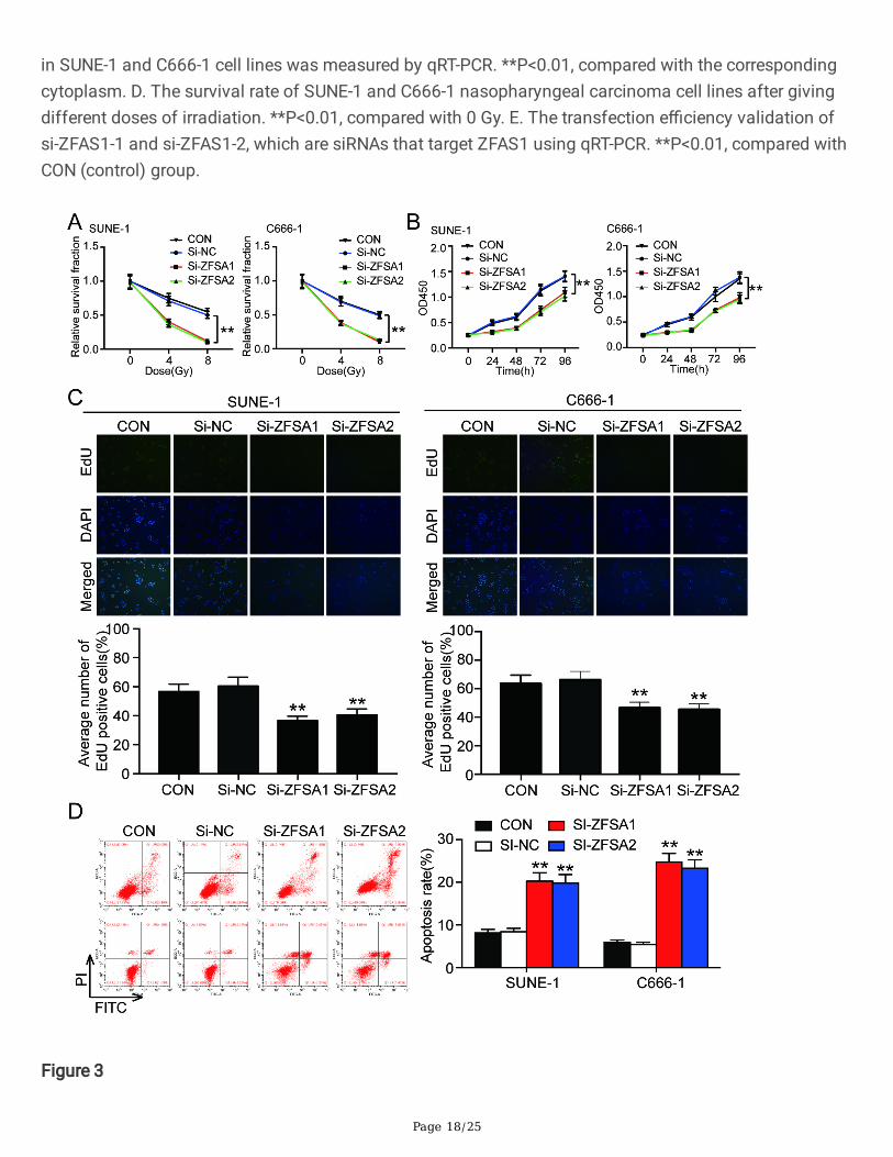
Figure 3

Page 19/25
The knockdown of ZFAS1 weakened the radiation resistance of nasopharyngeal carcinoma cells in vitro.A. The viability changes of nasopharyngeal carcinoma cell lines SUNE-1 and C666-1 after giving differentdoses of irradiation. B. At 8Gy irradiation, the survival rate of cells in every groups was detected at 24h,48h, 72h, and 96h in SUNE-1 and C666-1 cell lines. C. Average numbers of Edu positive cells in SUNE-1and C666-1 cell lines with 8Gy irradiation. D. The cell apoptosis of SUNE-1 and C666-1 cell lines with 8Gyirradiation detected by �ow cytometry. **P<0.01, compared with CON group.
Figure 4

Page 20/25
ZFAS1 was the upstream gene of miR-7-5p. A. The predicted binding sequences between ZFAS1 and miR-7-5p obtained from ENCORI algorithm. B. Luciferase reporter assay was used to determine the targetrelationship between ZFAS1 and miR-7-5p. **P<0.01, compared with miR-NC group. NC: negative control.C. RNA pull down was used to demonstrate the association between ZFAS1 and miR-7-5p. mut: mutant.**P<0.01, compared with antisense oligo group. D. The expression of miR-7-5p in NPC tissues andadjacent tissues. E. The expression of miR-7-5p in NPC cell lines. *P<0.05, **P<0.01, compared with NP-69 cell line. F. ZFAS1 expression showed a negative relationship with miR-7-5p expression. G. Thetransfection e�ciency of miR-7-5p inhibitor. **P<0.01, compared with CON group.

Page 21/25
Figure 5
The knockdown of ZFAS1 suppressed the radiation resistance of nasopharyngeal carcinoma cells byacting on miR-7-5p. A. CCK-8 was used to detect the viability of SUNE-1 and C666-1 cells aftertransfection with si-ZFAS1 or miR-7-5p inhibitor under 8Gy irradiation. B. Edu assay was used to observethe cell proliferation of SUNE-1 and C666-1 cell lines after transfection with si-ZFAS1 or miR-7-5p under8Gy irradiation. C. The cell apoptosis in SUNE-1 and C666-1 cell lines after transfection with si-ZFAS1 or
Page 22/25
miR-7-5p inhibitor with giving 8Gy irradiation by �ow cytometry. *P<0.05, **P<0.01, compared with CONgroup; #P<0.05, ##P<0.01, compared with miR-7-5p inhibitor group.
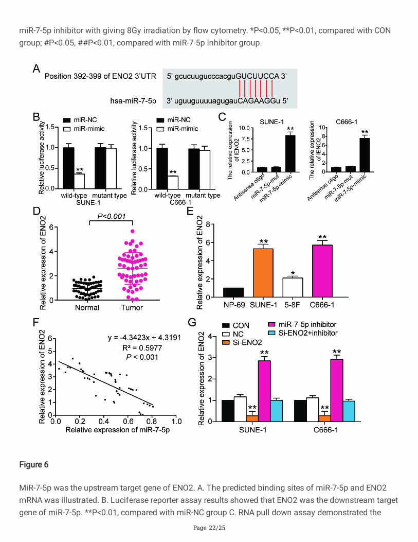
Figure 6
MiR-7-5p was the upstream target gene of ENO2. A. The predicted binding sites of miR-7-5p and ENO2mRNA was illustrated. B. Luciferase reporter assay results showed that ENO2 was the downstream targetgene of miR-7-5p. **P<0.01, compared with miR-NC group C. RNA pull down assay demonstrated the

Page 23/25
regulation relationship between ENO2 and miR-7-5p. **P<0.01, compared with antisense oligo group. D.The expression of ENO2 mRNA in NPC tissues and adjacent healthy tissues. E. The expression of ENO2in NPC cell lines and normal cell NP-69. *P<0.05, **P<0.01, compared with NP-69 cell line. F. ENO2 mRNAexpression had a negative relationship with miR-7-5p expression. G. qRT-PCR was used to observe theexpression of ENO2 mRNA in SUNE-1 and C666-1 cells after transfection with si-ZFAS1 or miR-7-5p.**P<0.01, compared with CON group.
Figure 7

Page 24/25
MiR-7-5p inhibition enhanced the radiation resistance of nasopharyngeal carcinoma cells by acting onENO2. A. CCK-8 assay results showed that si- ENO2 suppressed the cell viability in SUNE-1 and C666-1cell lines while miR-7-5p inhibition promoted the cell viability at 8 Gy irradiation. B. Edu assay showedthat si-ENO2 suppressed the growth of SUNE-1 and C666-1 cell lines while miR-7-5p inhibition enhancedcell viability at 8 Gy irradiation. C. The cell apoptosis in SUNE-1 and C666-1 cell lines after transfectionwith si-ENO2 and miR-7-5p inhibitor under 8 Gy irradiation.
Figure 8
Page 25/25
The illustration of the hypothesized mechanism involving ZFAS1, miR-7-5p and ENO2 in NPC resistanceto irradiation. Basically, high ZFAS1 level, low miR-7-5p level, and high ENO2 mRNA level in NPC results inenhanced resistance to irradiation.