in 288 905 - defense technical information · pdf filein 288 905 armed services ......
TRANSCRIPT

UNCLASSIFIED
in 288 905
ARMED SERVICES TECHNICAL INFORMATION AGENCYARLINGTON HALL STATIONARLINGTON 12, VIRGINIA
UNCLASSIFIED

NOTICE: Vhen governent ox,~ other &r-.Jvinga, speci -fications or other data azo us~aed for amy purposeother than in oonnectioc with a de!4 'Atuly relatedgoverament procurement ifration,. the U. S.Government thereby incurs no responsibility, nor anyobligation whatsoevery and the fat that the G3overn-ment may have fouAmlated, furnishet, or in mnv vaysupplied the said drawings, speoifiations, or otherdata is not to be regarded by impl:4oation or other-vise an in any ma-nr licen~sing the holder or anyother person or corporat. aj or o :nveyizg any rightsor permission to manufaoture, use or sell anypatented Invention that may in any way be relatedthereto.
43-A %
(m RL-TDR-62-98 (1)
TECHNIQUFS OF PHYSIOLOGICAL MONITORING
jVOLUME 1. FUNDAMENTALS
TECHNICAL DOCUMENTARY REPORT NO. AMRL-TDR-62-98 (I)
September 1962
Biomedical Laboratory6570th Aerospace Medical Research Laboratories
Aerospace Medical Division
Air Force Systems Command
Wright-Patterson Air Force Base, Ohio
I A ST IA.NOV261962
Contract Monitor: Mr. Miles A, McLennl '/
Project No. 7222, Task No. 722203 .
[Prepared Under Contract No. AF33(616)-7750by
RCA Service CompanyCherry Hill, Camden 8, New Jersey]
NOTICES
When US Government drawings, specifications, or other data are used forany purpose other than a definitely related government procurement operation,the government thereby incurs no responsibility nor any obligation whatsoever;and the fact that the government may have formulated, furnished, or in any waysupplied the said drawings, specifications, or other data is not to be regardedby implication or otherwise, as in any manner licensing the holder or any otherperson or corporation, or convey.ng any rights or permission to manufacture,use, or sell any patented invention that may in any way be related thereto.
Qualified requesters may obtain copies from ASTIA. Orders will be expeditedif placed through the librarian or other person designated to request documentsfrom ASTIA.
Do not return this copy. Retain or destroy.
Stock quantities available at Office of Technical Services, Department ofCommerce, $2.75.
7-168, 1,000, 10-15-62

C.) Q
cc In
z -00 0
0 -a
g~ 8 0 , In g I A
02J
000 00
0'
~1 U
* .... .0 ~
Ri .-F I t,. L- .15
In ~
I, 'S
I'D 1.1, O
cc u '00 oac "
0.j g i ~ ~~~ Z: .0,-4 "0 1

FOREWORD
This study was initiated by the Biomedical Laboratory of the6570th Aerospace Medical Research Laboratories, Aerospace MedicalDivision, Wright-Patterson Air Force Base, Ohio. The research wasconducted by the RCA Service Company, Camden, New Jersey, underContract AF 33(616) -7750. Mr. Walter L. Becker was principalinvestigator for the RCA Service Co. Mr. Miles A. McLennan of theMedical Electronics Section, Biophysics Branch, was the contractmonitor for the Biomedical Laboratory. The work was performedin support of Project No. 7222, "Biophysics of Flight, " Task No.722203, "Specialized Instrumentation. "
Engaged in the preparation of this volume for the RCA ServiceCompany were Mr. Robert E. Barbiere, Editor, Mr. Raymond C.Baldwin, Technical Writer, and Mr. John A. Letus, TechnicalWriter. Mr. Carl Berkley, Scientific Director of the Foundationfor Medical Technology, participated as a technical consultant.

ABSTRACT
This volume is the first of a three-volume handbook covering theapplications of electronics in monitoring bioelectric physiologicalresponses. The fundamental concepts and methods presented in thisvolume form a foundation for the detailed technical discussions in thesucceeding volumes and, it is hoped, provide a common language andbasis of understanding between the physiclogist and electronic engineerengaged in this field. The data obtained by monitoring physiologicalresponses in varied environments can be used to improve the efficiencyand increase the safety of a human subject in aircraft and spacecraft.
PUBLICATION REVIEW
This technical documentary report has been reviewed and isapproved.
JO M. QUASHNOCKColonel, USAF, MCChief., Biomedical Laboratory
Uli
TABLE OF CONTENTS
Page
SECTION I - INTRODUCTION
G EN ERAL ........................................... 1
MEASUREMENT OBJECTIVE ................................... 2
I Research Information .................................. 2II Functional Information ...... .......................... 2
THE MEASUREMENT PROBLEM ................................. 2
I The Subject ....................................... 4II Input Factors ...................................... 4
A. Sustaining ...................................... 4B. Sensory ................................... 4C. Psychological ............................... 4
III Output Factors ................................. 5
A. Physiological .................................... 5B. Intermediate ................................... 5C. Psychological ................. ................. 5
SECTION II - BASIC PHYSIOLOGICAL SYSTEMS
BEHAVIORAL SYSTEM ....................................... 6
1 Description and Function ................................ 6
A. Nervous System ...................... 6B. Central Nervous System .... . . .............. 7C. Peripheral Nervous System ........... ............ 9
II General Monitoring Methods ............ ................ 10
A. Electroencephalography ...... .................. .... 10R. Electromyography ............................... 12,2. Galvanic Skin Response ..... ...................... 12
iv

TABLE OF CONTENTS (Continued)
Page
Ill. Problem Areas ................................. 13
RESPIRATORY SYSTEM ................................... 13
I Description and Function ..... ........................ .13
A. The Breathing Process .............................. 14B. The Capacity of the Lungs .......................... 14C. Chemical Composition of Inspired and Expired Air 15D. Relationship of Oxygen and Blood ..... ............... 15E. The Control of Respiration .......................... 15F. Minute Volume ............................. 16
II Basic Monitoring Methods ............................. 16
A. Air Flow Measurements ............................. 17B. Chest Movement Measurements ..... ................. 17C. Minute Volume .............................. 17Dl. )xygen and Cr6on Dioxide Partial Pressure ........... .... 17
CIRCULATORY SYSTEM ....................................... 18
I Description and Function ..... .......................... 18
A. Heart ..... ................................... 18B. Blood Vessels ..................................... 21C. Lymphatic System ............................... 22D. Blood Pressure .............................. 22
II General Monitoring Methods ..... ...................... 24
A. Pulse Rate ..................................... 25B. Blood Pressure .............................. 25C. Cardiac Activity ............................ 27D. Blood Flow Rate ................................. 29E. Oxygen Saturation of Blood ..... ................... 29
METABOLIC SYSTEM ................................... 31
I Description and Function ..... ......................... 31II Basic Monitoring Methods ..... ........................ 31III Problem Areas ................................. 32
V
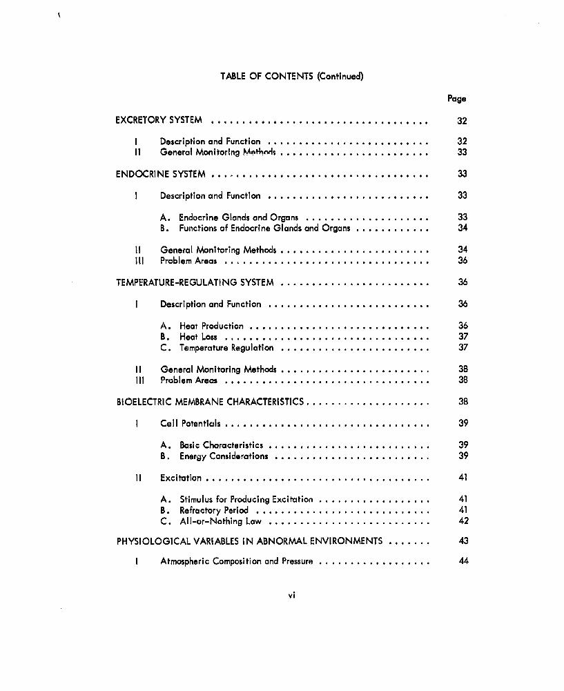
TABLE OF CONTENTS (Continued)
Page
EXCRETORY SYSTEM ................................... 32
I Description and Function ............................. 32II General Monitoring MAtk -Is ........................ 33
ENDOCRINE SYSTEM ....................................... 33
I Description and Function .*......................... 33
A. Endocrine Glands and Organs ....................... 33B. Functions of Endocrine Glands and Organs .............. 34
II General Monitoring Methods ............................ 34III Problem Areas ................................. 36
TEMPERATURE-REGULATING SYSTEM ........................... 36
I Description and Function ..... ........................ 36
A. Heat Production ................................ 36B. Heat Loss ................................. 37C. Temperature Regulation ........................... 37
II General Monitoring Methods ............................ 38
III Problem Areas ................................. 38
BIOELECTRIC MEMBRANE CHARACTERISTICS ....................... 38
I Cell Potentials ................................. 39
A. Basic Characteristics .......................... 39B. Energy Considerations ..... ....................... 39
II Excitation ........................................ 41
A. Stimulus for Producing Excitation .... ................ 41B. Refractory Period ................................ 41C. All-or-Nothing Law ............................. 42
PHYSIOLOGICAL VARIABLES IN ABNORMAL ENVIRONMENTS ......... 43
I Atmospheric Composition and Pressure ..................... 44
vi

TABLE OF CONTENTS (Continued)
Page
A. Respiration .................................... 44B. Circulation .................................... 47
11 Gravitational Forces ................................. 49
A. Acceleration ................................... 50B. Weightlessness ................................. 55
III Temperature ................................... 57IV Radiation .................................... 60
A. Biological Effects ..... .......................... 60B. Measures of Radiation ..... ....................... 60C. Radiation Exposure ..... ......................... 61D. Physiological Effects of Radiation ..................... 63
V Noise and Vibration ................................. 67VI Emotional Stress .................................... 69
A. Artificial Environment .... ........................ 69B. Day-Night Cycle ............................... 70C . Fatigue ................................... 71
SECTION III - BASIC PHYSIOLOGICAL MEASURING SYSTEMS
INTRODUCTION ...................................... 73
GENERAL SYSTEM CONSIDERATIONS ............................ 73
I General System Requirements ........................... 74
A. Frequency Spectra ................................ 74B. Dynamic Range .............................. 74
II Error Considerations ..... ............................ 74
A. Frequency Characteristics ........................... 76B. Impedance Matching ............................. 77C. Nonlinearity .................................. 78D. Interfeicnce and Artifacts .......................... 78
vii

TABLE OF CONTENTS (Continued)
Page
SIGNAL PICKUP DEVICES ................................. 80
I Transducer .................................... 80
A . General .................................. 80, . Basic Considerations .............................. 80
II Electrodes .................................... 81
A. Basic Considerations .............................. 81B. Electrode Arrays ................................ 85
SIGNAL PROCESSING ................................... 85
1 General ................................. ... 85II Amplification ............ ............... ..... 86
A. Requirements ................................... 86B. Basic Amplifier Types for Physiological Applications ...... 86
DI SPLAY ............................................ 90
I Basic Data Display Method ......................... 90II General Principles of Data Readout and Display Devices ...... 90
A. Frequency-Response Grouping of Recording Devices ...... 90B. Direct Writing Versus Photodevelopment .................. 91C. Curvilinear Versus Rectilinear Recording .... ........... 92
III Recorder Impedances ................................. 92
SPECIAL ACCESSORIES .................................. 92
I Telemetry .................................... 93II Magnetic Tape Recording ............................. 93III Computer Processing ................................. 94IV Time Indexing ................................. 94
A . General .................................. 94B. Establishing Correlation ........................... 94
viii

TABLE OF CONTENTS (Continued)
Page
SYSTEM INTEGRATION FACTORS ........................... 95
I Physical Factors .................................... 95
A. Size and Weight Limitations .... .................... 95B. Placement and Attachment of Pickup Devices ............ 96
II Information Handling Capability of Data Transmission Links ..... 96III General Environment ................................ 96
A. Shock and Vibration ............................. 96B. Temperature ................................ .... 96C. O ther Factors ............................... 97
IV Equipment Interaction ................................ 97
V Power Supply Problems ................................ 97
ESTABLISHING VALIDITY OF REMOTE MEASUREMENTS ............ 97
EXPLANATION OF TERMS ................................... 98
BIBLIOGRAPHY ....................................... 114
ix

LIST OF ILLUSTRATIONS
Figure Title Page
I Factors Involved in Physiological Measurements ............... 32 The Cerebral Cortex Showing Areas and Lobes ................ 83 Electroencephalogram Showing Response Produced by Opening
and Closing Eyes ..................................... 124 Capacity of the Lungs ................................. 145 The Chambers of the Heart ............................. 186 Cardiac Pressure Cycle with Corresponding EKG and Heart
Sounds ...................................... 207 Composition of the Electrocardiogram ...................... 288 Light Absorption by Hemoglobin .................... 309 Location of the Endocrine Glands and Organs ............. .... 35
10 Fiber Polarization ................................... 4011 Human Tolerance to Oxygen Partial Pressures ............... 4512 Human Tolerance to Carbon Dioxide Partial Pressures ........ 4813 Standard Terminology for Acceleration .................. .... 5114 Human Tolerance to Temperature ...................... .5815 Heat Production of Nearly Nude Man .................. .... 5916 Basic Permissible Exposure Rates to External Sources of
Ionizing Radlailon ... .. . . ...... ..... . ....... .....17 Human Tolerance to Vibration ........................... 6818 Bansic Elements of a Physiological Measuring System ......... .... 7319 Frequency Distortion of a Square Wave ................. .... 7620 Typical Frequency Response Curve ........................ 7721 Nonlinearity of a Sine Wave ............................. 7922 Potential Source Parallel to the Skin Surface .............. .... 8223 Potential Source Perpendicular to the Skin Surface .......... .... 8324 Relative Impedances in the Use of Electrodes .............. .... 8425 Simplified Single-Ended Amplifier ........................ 8726 Simplified Balanced Amplifier ............................ 8827 Simplified Differential Amplifier ......................... 8928 Physiological Measuring System with Accessories ........... .... 93
LIST OF TABLES
Table Title Page
I Gross Effects of Short-Duration Acceleration Forces ......... .... 52II Clinical Symptoms of Radiation Sickness .................... 63III Frequency Spectrum Requirements for Individual Monitoring
Systems ............................................ 75
x
INTRODUCTION
GENERAL
This volume is the first of a three-volume handbook covering the electronic moni-toring of a physiological subject in an abnormal environment. The handbook describesinstrumentation techniques that have proven useful in determining the vital state ofnormal subjects in varied environments. Aircraft and spacecraft applications are theprinciple considerations, but some contribution may be made towards stabilization ofmeasurement practices.
Although the subject matter of this handbook lies somewhere between the bio-sciences and electronic technology, the material is presented so as to be understandableto doctors, physiologists, engineers, or technicians who are interested in the field ofphysiological monitoring. The handbook provides a common language and terminologyfor electronics and medical personnel. It is hoped it will contribute to the solution ofthe liaison problem between physiologists and engineers. It is not intended, however,to replace basic texts in either feid.
Volume I, introductory in nature, presents basic physiological and electronic fun-damentals Involved in monitoring physiological reactions. The problem is defined,general procedures are outlined, and background material is supplied which should aidboth electronics and medical personnel in gaining an appreciation of the factors in-volved. (Volume II takes up specific equipments and equipment applications, andVolume III describes complex systems, problems, and system calibration procedures.)
Volume I consists of three sections. The first section explains the purpose and de-fines the scope of the handbook, and briefly describes the objectives, problems, andfactors involved in physiological measurements. Section II describes the various bodysystems and physiological phenomena subject to electronic sensing and presentation.The principles and philosophy of physiological measurement are discussed in Section III.
Physiological and psychometric monitoring parallel each other closely. However,this manual is reqtricted principally to techniques applicable to the measurement ofphysiological and related phenomena. Although there is an indefinite area where thephysiological and psychological areas overlap, generally, they can be distinguished asfollows: the physiologicol area is related to the basic or vegetative functions of livingorganisms; the psychological area is related to the higher order functions involvingsensation, perception, memory, and thought.

INTRODUCTION
MEASUREMENT OBJECTIVE
Measurements provide factual, quantitative information that can be used forplanning protective measures which will ensure the safety and functional capability ofpersonnel engaged in hazardous missions. The information is of two types: researchand functional.
I. Research Information
Research information establishes the relationship between some factor of the en-vironment and the subject's reaction. Both input and output factors must be measured(see figure 1), and any measurement involving multiple factors requires the monitoringof the constants. The accepted procedure is to set up a controlled situation in thelaboratory, if possible, in which all input factors are held constant, except the one un-der investigation. One factor then is varied in a controlled manner while observingall possible output factors. The environmental factor may be vibration, zero G,a radi-ation field, or perhaps the isolation of space. However, some of the conditions, i.e.,input factors encountered in aircraft and space flight, are very difficult or impossibleto simulate in the laboratory. Prolonged zero G, cosmic radiation, and psychologicalstress are among them.
The effect upon the vitul btute of the subject must be known quantitatively, ifpossible, to determine if protective measures are necessary or effective. Once the re-search information is obtained and the protective problem solved, this instrumentationis no longer needed, and a functional or historical type of monitoring system should beall that is required.
!1. Functional Information
Functional information is sought concerning deviations from expected or hopefullyestablished conditions, and should provide the answer to what went wrong. Ideally,only the input factors shown in figure 1 need be measured. The information obtainedmay help in tuture design work, but it is primarily historical, i.e., a record of events.Functional information probably will remain a requirement as long as a mission con-tains a large element of risk.
THE MEASUREMENT PROBLEM
Figure 1 indicates the scope of the measurement problem. How may meaningfulrelations be established between such a large number of inputs and outputs? What in-put may logically be related to what output? These are a few of the questions pre-sented. In this handbook, emphasis is placed on the measurements of the output orresponse factors, the items noted on the right-hand side of figure 1 . This is done with-out intent to deemphasize the importance of the factors on the input side. Certainly
2

THE MEASUREMENT PROBLEM
0t It zo 0~ o0
I t 1 - f 0 <- S i1EV 04 C3Z w 0u r n i - n * ItLZ ~
0 CL 0 (
(L zLU
"40101± .1
4L Z
Lu 0 0
L) I x LU Q
w04- z A '
U >
t3t
o Zr
X. w )I :-
W C0L LL >O wL
0 CD ~LL 4cU<>
3
INTRODUCTION
all of the environmental factors must he encompassed if the measurement is to have realsignificance. However, since the techniques for quantifying physical factors of theenvironment are within the scope of the physical sciences, the reader will be referred,when possible, to texts and references in the field of physical measurement.
I. The Subject
The box labeled "subject" in figure 1 rcpiusents a human being, which is beyonddoubt the most complex device in existence. A great deal is known about the device'sphysical structure (anatomy) and something about the functional processes (physiology)that maintain the basic life condition, i.e.,the vegetative state. Very little, however,is known about the system that governs the higher order functions or the psychologicalbehavior.
From the engineer's standpoint, the subject is jammed full of nonlinear devices andinterconnecting subsystems with multiple feedback loops. It also has a self-adaptingprogrammer and tremendous information storage capability. The output from the subjectis often governed by the events of hours, days, or years past.
II. Input Factors
one may affect the output. These factors are grouped into the following three cate-gories:
A. Sustaining
Sustaining factors include food; drink; atmospheric factors such as tempera-ture, pressure, and composition (oxygen, nitrogen, carbon dioxide, water, and residualsor contaminants); the accelerative or G field; infectious agents; and radiation. Ex-cept for temperature and radiant heat (radiation), these inputs are, in general, non-sensory in their steady-state condition, and are important determinants of the lowerorder or basic physiological behavior.
B. Sensory
Light, sound, odor, transient pressure and vibration are sensory phenomena andform the basis of higher order behavior.
C. Psychological
Psychological factors ore the difficult-to-measure items that are regeneratedwithin the subject by input factors reacting with stored post experience, as indicated infigure 1 by the re-entry loop. These are the factors of anxiety, fatigue and stress, thereflex, inherent and learned behavior patterns, motivation, attitude, and other "affect"
4
THE MEASUREMENT PROBLEM
items that compose what the psychologists term the egoautonomy.
III. Output Factors
Some input factors may present measurement problems, but the situation at the out-put is much more difficult. Authorities disagree markedly on the significance of manyevidences of physiological activity, and more so on those of psychological behavior.They do agree, however, that there are two general classes: namely the physiologicaland psychological; and, because the dividing line between these two is so uncertain, athird class, called intermediate, is identified.
A. Physiological
The physiological class includes evidences of activity, indications of respira-tion and metabolism, measures of heart activity, measurements of systolic or diastolicblood pressure, etc.
B. Intermediate
Within the uncertain area dividing the physiological and psychological classeslie behavioral measurements such as skin responses (GSR), biochemical reactions, andbrain or electroencepholographic recordings (EEG).
C. Psychological
Factors that are measured, such as operant behavior, tracking, and problemsolving, are used as indices of the factors of perception, cognition, and performance.These measures all involve complex motor-action (limb movement) response to locallyprogrammed (standardized) stimuli. Treatment of this subject will be minimal, not be-cause it is unimportant, but because of the uncertain state of the measurement art inthis field.
5

92em II
BASIC PHYSIOLOGICALSYSTEMS
BEHAVIORAL SYSTEM
I. Description and Function
Biologically, behavior is regarded as the action and response of a physiologicalsubject to stimulation, and the parts of the body responsive to this stimulation comprisethe behavioral system. Although the behavioral system could be considered in both itsphysiological and psychological aspects, only the physiology of the nervous system andthose emotional and motivational conditions which stimulate the nervous system physi-ologically are discussed here.
Man has physiological needs which must be fulfilled in order to sustain life. Whenphysiological needs remain unsatisfied, stresses are created which drive organisms intoactivity. Organisms, by their own activity, tend to maintain a physiological equilib-rium. Expressed in another way, each disturbing influence induces a compensatoryrrtivity to neutralize its own disturbance. This concept is known as homeostaiss. Alexample of this is sweating to maintain constant body temperature.
A. Nervous System
The nervous system is the communications network of the body. Pulse signalstraveling through the nervous system affect our thoughts and emotions, and regulate ourrespiration, heart activity, and the activity of muscles and glands throughout the body.
The nervous system is divided into two major subsystems: the central nervoussystem and the peripheral nervous system. The central nervous system consists of thebrain and the spinal cord, and the peripheral nervous system consists of the cranial andspinal nerves that extend to all parts of the body from the brain and the spinal cord.
The nervous system is formed of a network of nerve cells or neurons. Theneuron usually is less than 0.1 millimeter in diameter, but sometimes is as long as 3meters. It is composed of three main parts: dendrites, a cell body, and an axon.(Dendrites and axons are referred to as nerve fibers.) Impulses are picked up by thedendrites, passed through the cell body, and conducted by the axon to the junction withother neurons in the network. In this manner, impulses travel from neuron to neuron inone direction only.
Neurons generally are divided into three groups: afferent neurons which
6
BEHAVIORAL SYSTEM
conduct impulses from the 3ensory receptors to the central nervous system, efferent neu-rons which conduct impulses from the central nervous system to the muscles or glands,and association neurons which lie totally within the central nervous system. Afferentneurons have only sense functions and efferent neurons usually have purely motorfunctions. The association neurons transfer incoming pulses to various parts of thecentral nervous system and relay impulses originating in the central nervous system toefferent neurons, other association neurons, or to nerve centers. Neurons are notconnected to each other physically, but transmit impulses by means of synaptic junc-tions. The synapse is a sort of chemical rectifier which allows impulses to pass throughit only in one direction (suggestive of the switching diodes used in the logic circuits ofelectronic computers).
The afferent nerves which convey environmental information (sensation) fromthe peripheral nervous system to the central nervous system are referred to as the sen-sory nerves. The efferent nerves, particularly those which convey nerve impulses fromthe central nervous system to the peripheral nervous system causing response, are re-ferred to as the motor nerves. All impulses carried by nerve fibers, both sensory andmotor, are essentially alike. Fibers from the eyes do not carry impulses that are dif-ferent from those carried by fibers from the ears. The eye sees when it is stimulctedand the ear hears when it is stimulated because the impulses, although alike, go todiff~rpnt rpaions of the brain. Nerve impulses, however, do differ in frequency withoutrespect to ihelir point of origin or point of termination. As the intensity of the stimulusincreases, the frequency of the nerve impulse changes. Strong stimuli also may stimu-late more fibers.
B. Central Nervous System
The central nervous system, composed of the spinal cord and the brain, is con-structed of many thousands of millions of neurons, each connected transsynapticallywith hundreds of others.
1. The Spinal Cord
The spinal cord serves as a pathway for nervous impulses to and from thebrain and as a center for carrying out and coordinating many reflex actions independ-ently of the brain. It is a thick longitudinal cord of nervous tissue securely encased bythe bony vertabrae of the spinal column and, continuous with the hindbrain, extendsvirtually the whole length of the spinal column. At intervals along its lengih, Pii- ofnerves (the spinal nerves) extend from the spinal cord to the various parts of the trunkand limbs.
2. The Brain
The brain is a large mass of nerve tissue contained in the skull or cranialcavity. The brain consists of gray matter made up largely of nerve cells, and white
7
BASIC PHYSIOLOGICAL SYSTEMS
matter made up chiefly of nerve fibers arising from the nerve cells of the brain orreaching it from other parts of the central nervous system.
The brain represents only about 2 percent of the total weight of the body,yet it functions as the master control unit. It analyzes information it receives from thesensory organs and receptors and makes decisions and issues instructions (in the Form ofefferent neuron impulses) to the muscles and glands. The ultimate control of behaviordepends upon the integrating mechanisms of the brain.
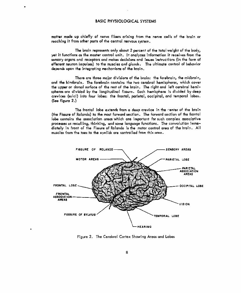
There are three major divisions of the brain: the forebrain, the midbrain,and the hindbrain. The forebrain contains the two cerebral hemispheres, which coverthe upper or dorsal surface of the rest of the brain. The right and left cerebral hemi-spheres are divided by the longitudinal fissure. Each hemisphere is divided by deepcrevices (sulci) into four lobes: the frontal, parietal, occipital, and temporal lobes.(See figure 2.)
The frontal lobe extends from a deep crevice in the -enter of the brain(the Fissure of Rolando) to the most forward section. The forward section of the frontallobe contains the association areas which are important for such complex associativeprocesses as recalling, thinking, and some language functions. The convolution imme-diately in front of the Fissure of Rolando is the motor control area of the brain. Allmuscles from the toes to the eyelids are controlled from this area.
FISSURE OF ROLANDO SENSORY AREAS
MOTOR AREAS f PARIETAL LOBE
PARI ETAL-.--"- / ASSOCIATION
AREAS
FRONTAL LOBE OCCIPITAL LOSE
FRONTALASSOCIA1 ION
AREAS AREAS VISION
FISSURE OF SYLVIUS T_ 'EMPORAL LOBE
HEARING
Figure 2. The Cerebral Cortex Showing Areas and Lobes
8
BEHAVIORAL SYSTEM
The parietal lobe, directly behind the Fissure of Rolando, is the area ofthe brain that is sensitive to bodily sensations (somatesthetie). All impulses originatingin the skin and in the receptors sensitive to muscular senses, such as the perception ofmovement, weight, resistance, and position (kinesthetic), are received here. Theoccipital lobe, the rear section of the brain, receives all impulses originating in thevisual receptors. A deep crevice, the Fissure of Sylvius, which runs longitudinallyalong the side of each hemisphere, separates the temporal lobe from the frontal andparietal lobes. Impulses originating in the auditory receptors are carried to thetemporal lobes.
The outer layer of each hemisphere is gray and is raised in irregularridges called convolutions or gyri. These gray layers are the center of the highest andmost complex nervous functions and are known as the cerebral cortex or gray matter.The cerebral cortex is regarded as the seat of consciousness and is essential to all vol-untary activity. Neurons with sensory functions enter the central nervous system andare directed through the thalamus to appropriate areas of the cortex. Sensory areas ofthe cortex are connected to specific motor areas of the cortex by association neurons,and the motor areas transmit impulses to muscles throughout the body. The primary re-ceiving areas of the cortex are not the only end stations for afferent neurons. Hearing,vision, and touch are each doubly, and possibly trebly, represented in each hemisphereof the brain with separate pathways. The motor areas also have been found to haveduplicates.
The more complex body reflex actions are regulated through the varioussubcortical (beneath the cortex) parts of the brain. The midbrain serves as a correla-tion center for optic and auditory reflexes and contributes to muscle tone. Within thehindbrain, the medulla, which is the connecting point between the spinal cord and thebrain, regulates the autonomic activities, such as respiration, heartbeat, and sugar me-tabolism. The cerebellum, also a part of the hindbrain, maintains bodily equilibriumand coordinates muscular activity.
C. Peripheral Nervous System
1 . Cranial Nerves
The cranial nerves are 12 pairs of nerves that pass through openings(foramens) in the skull in their paths from various parts of the brain to (with certain ex-ceptions) organs and other parts of the head. Some of these nerves are sensory, con-cerned with the sense of smell, sight, hearing, taste, and feeling; others are motornerves, controlling the salivary glands and muscles in the eyes, jaw, tongue, pharynx,and shoulder.
2. Spinal Nerves
The spinal nerves are 31 pairs of nerves that extend from either side of
9
BASI.. PHYSIOLOGICAL SYSTEMS
the spinal cord throughout its length. These nerves, containing both sensory and motorfibers, connect to muscles and skin of the trunk and limbs and to nerves of the sympa-thetic system, a part of the automatic nervous system.
3. Autonomic Nervous System
The autonomic nervous system exerts a regulatory influence over involun-tary muscles, glands, and the viscera (the organs contained within the cranial, thoracic,abdominal, and pelvic cavities). Autonomic activities are under the control of thehypothalamus, a part of the forebrain, and are primarily motor in function. Two func-tionally antagonistic systems make up the autonomic nervous system: the sympathetic(sometimes called orthosympathetic) and parasympathetic. The sympathetic nervoussystem mobilizes the internal environment to combat external stress more effectively,whereas the parasympathetic protects, conserves, and re st o r e s the resources of theorganism. For example, the sympathetic connection with the heart accelerates its ac-tivity, while the parasympathetic connection acts to slow it down. The activities ofthe stomach, conversely, are restricted by the sympathetic system and accelerated bythe parasympathetic. The adrenal gland is the only organ known to be under control ofonly one division of the autonomic nervous system: it is accelerated bythe sympatheticsystem and is not affected by the parasympathetic. An emotionally aroused individual'sheart pounds, his stomach contractions and gastric secretions are checked, his bladderrelaxes, his ii-uwiou and bronchl arc dilated, and his adreo 'in q eretion is acceler-ated - all because his sympathetic nervous system has assumed control.
Under normal circumstances, the parasympathetic nervous system (1) con-stricts the pupils of the eyes, (2) promotes profuse,watery secretions of saliva, (3) slowsthe heart rate, (4) promotes peristalsis in the esophagus, (5) promotes secretion of pan-creatic juices, (6) augments peristalsis and secretory activity in the stomach and theintestines, (7) constricts the bladder, and (8) redistributes blood volume toward theviscera. The sympathetic nervous system (1) dilates the pupils of the eyes, (2) promotesscanty, viscous secretions of saliva, (3) accelerates the heart rate, (4) dilates the tra-chea and bronchi, (5) inhibits peristalsis and secretory activity in the stomach andintestines, (6) relaxes the bladder, and (7) redistributes blood volume to the muscles.
II. General Monitoring Methods
The behavioral system may be monitored by measuring the electrical potentialsdeveloped in the nervous system or muscles, or by rreasuring the resistances or conduct-ances of the skin.
A. Electroencephalography
The neurons of the brain generate electric potentials. This electrical activityof the brain may be detected with electrodes placed on the surface of the head orthrough the skull directly on the cortex. The recordings made with electrodes placed
10

BEHAVIORAL SYSTEM
on the scalp require very high amplification, although in all other ways they are thesame as those made from the surface of the cortex. When measured on the scalp, thepotential difference between electrodes (one electrode is connected to the lobe of theear as a reference) is approximately 30 millionths of a volt (30 x 10-6 volt). Rarely inthe normal man does the potential difference exceed 200 millionths of a volt, which isabout 1/10th or less of the magnitude of electrocardiographic potentials. The rhyth-mically varying potentials of the brain are recorded on oscillographs, producing recordscalled electroencephalograms (EEG).
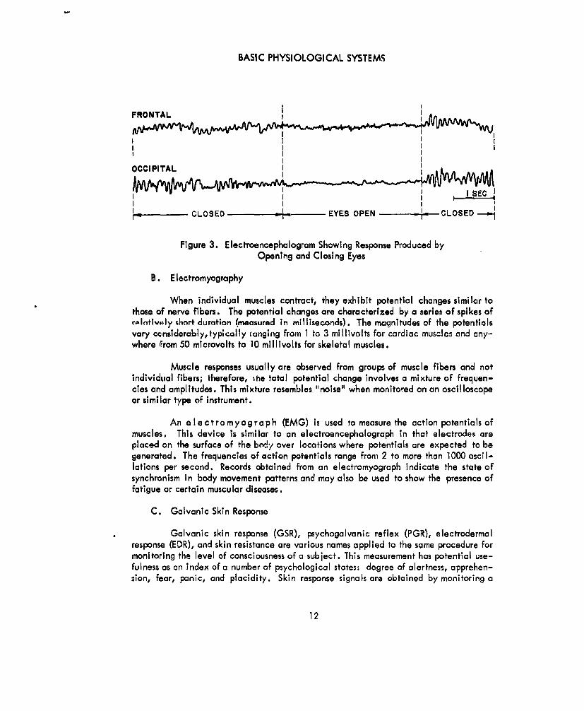
The waveforms recorded by the electroencephalograph are nonperiodic, low-frequency, complex waves of very low power. These waves contain many frequencieswith shifting phase relationships and varying amplitudes within the range of 1 to 60cycles per second. Brain waves have two predominant rhythms: alpha and beta. Themost common is the alpha rhythm, which has a frequency range of approximately 8 to13 oscillations per second. The alpha rhythm is desynchronized or reduced in ampli-tude by visual activity and alert attention, and for this reason, it is often referred to asthe resting rhythm. It is obtained most easily from the parietal and occipital lobes,although it can be detected almost anywhere on the scalp. The beta type of rhythm isdominated by waves of approximately 18 to 30 oscillations per second. It is detectedmost easily in the frontal lobe.
Thc clectroencephalograms of different persons differ w*dely; hw-eer, theelectroencephalogram of an individual normal adult varies little from hour to hour orover pcriods of several months. Electroencephalographic records indicate many thingsabout the state of the subject. Although not c. mpletely reliable as yet, important be-havioral patterns such as a subject's state of alertness and whether his eyes are open orclosed can be monitored. The presence of the dominant rhythms and the frequenciesand amplitudes of the brain waves may be used to determine the subject's state ofalertness. Since closing the eyes raises the alpha rhythm amplitude, monitoring candetermine if the subject's eyes are open or closed (figure 3). Also hypoxia tends toshift the rhythm toward the very low frequencies; therefore, indications are obtainedon certain functions of the respiratory and circulatory systems as well as the behavioralsystem.
Artifacts make the interpretation of electroencephalograms difficult. Themuscles of the scalp, neck, and jaws are stimulated continuously; consequently, elec-tromyographic (muscle) potentials may appear in electroencephalograms. Electromyo-graphic potentials may be eliminated to a iarge extent by filtering out frequencieshigher than 50 oscillations per second. Muscle potentials not removed by filters arerecognizable by their spiky appearance, relatively high frequency, and short duration.
Voltages generated by the neurons in the brain are suppressed greatly and therhythms reduced considerably by narcotics, anesthetics, and alcohol.
11
BASIC PHYSIOLOGICAL SYSTEMS
FRONTAL
OCCI PITAL
I I SEC
r----CLOSED - -- EYES OPEN -CLOSED--'l
Figure 3. Electroencephalogram Showing Response Produced by
Opening and Closing Eyes
B. Electromyography
When individual muscles contract, they exhibit potential changes similar tothose of nerve fibers. The potential changes are characterized by a series of spikes ofroIntively %hort duration (measured in milliseconds). The magnitudes of the potentialsvary ccnsiderably, typically ranging from 1 to 3 millivolts for cardiac musclc: and any-where from 50 microvolts to 10 millivolts for skeletal muscles.
Muscle responses usually are observed from groups of muscle fibers and notindividual fibers; therefore, ihe total potential change involves a mixture of frequen-cies and amplitudes. This mixture resembles "noise" when monitored on an oscilloscopeor similar type of instrument.
An e lectromyograph (EMG) is used to measure the action potentials ofmuscles. This device is similar to an electroencephalograph in that electrodes areplaced on the surface of the bcdy over locations where potentials are expected to begenerated. The frequencies of action potentials range from 2 to more than 1000 oscil-lations per second. Records obtained from an electromyograph indicate the state ofsynchronism in body movement patterns and may also be used to show the presence offatigue or certain muscular diseases.
C. Galvanic Skin Response
Galvanic skin response (GSR), psychogalvanic reflex (PGR), electrodermalresponse (EDR), and skin resistance are various names applied to the same procedure formonitoring the level of consciousness of a subject. This measurement has potential use-fulness as an index of a number of psychological states: degree of alertness, apprehen-sion, fear, panic, and placidity. Skin response signals are obtained by monitoring a
12

BEHAVIORAL SYSTEM
small electric current between two electrodes attached to the skin of a subject to de-termine the resistance or conductance between the electrodes.
Physical reactions, such as changes in skin resistance, result from the reactionof the autonomic nervous system to internal or external stimuli. Autonomic nerveschange the vascular bed tone by constricting near-surface capillaries and controllingthe activity of the sweat glands. Both these factors are important causes of changingskin response.
Temperature-control mechanisms of the body also employ the sweat glands;consequently, skin resistance measurements of the behavioral system must not be con-fused with tho,; of the temperature-regulatirg system. Two areas of the body withheavy concentrations of sweat glands that do not actively participate in temperatureregulating, except in temperature extremes, are the palmar areas (the palms of thehands) and the plantar surfaces (the soles of the feet). When the subject is adjusted tothe normal range of temperatures, changes in the electrical conduction of the palmarand plantar surfaces reflect the level of activation of the subject. Psychologically,activation is a dimension of more or less attention, more or less arousal, or more or lesswakefulness.
Ill. Problem Areas
In general, bioelectric signals are distorted by the instability of amplifiers andrecording instruments, electrode artifacts, and signal overlapping. Amplifiers and re-cording instruments are discussed elsewhere in this manual. The electrode artifactsresult from variations in electrical resistance at the point of contact with the skincaused by changes in mechanical pressure or the evaporation of the electrode jelly.Spurious potentials can be generated at bimetallic junctions or between the electrodesand saline body secretions. Bioelectric signals overlap (For example, electroenceph-alographic and electromyographic potentials) because the various separated bodytissuesare not insulated from each other. The reliability of signal responses may degradewhen monitoring occurs for long periods of time because of maceration of the skin byelectrode pastes.
RESPIRATORY SYSTEM
I. Description and Function
The respiratory system provides the body with oxygen from the atmosphere to oxi-dize carbonaceous food, thereby generating heat and supplying the body with energyfor performing work. The system also returns to the atmosphere the carbon dioxide andsome water that are formed by chemical reactions occurring in the cells and bodytissues. Respiration processes involve the exchange of gases at the body cells and inthe lungs. The exchange between the respiratory membrane or alveoli (air cells of thelungs) and the blood in the capillaries of pulmonary circulation is known as external
13
BASIC PHYSIOLOGICAL SYSTEMS
respiration; the diffusion of gases between the blood in the systematic capillaries andthe body cells is known as internal respiration. The exchange of gases is accelerated
by the respiratory movements of inspiration and expiration.
A. The Breathing Process
During inspiration (inhalation), the chest increases in volume and the pressurein the chest cavity (thorax) falls, permitting the higher pressure of the atmosphere toforce air into the lungs. When the intrapulmonic pressure reaches that of the outsideair, inspiration ceases. As the muscles that create the inspiratory movement relinx, thethoracic volume decreases and the pressure within the lungs increases, expelling air tothe atmosphere.
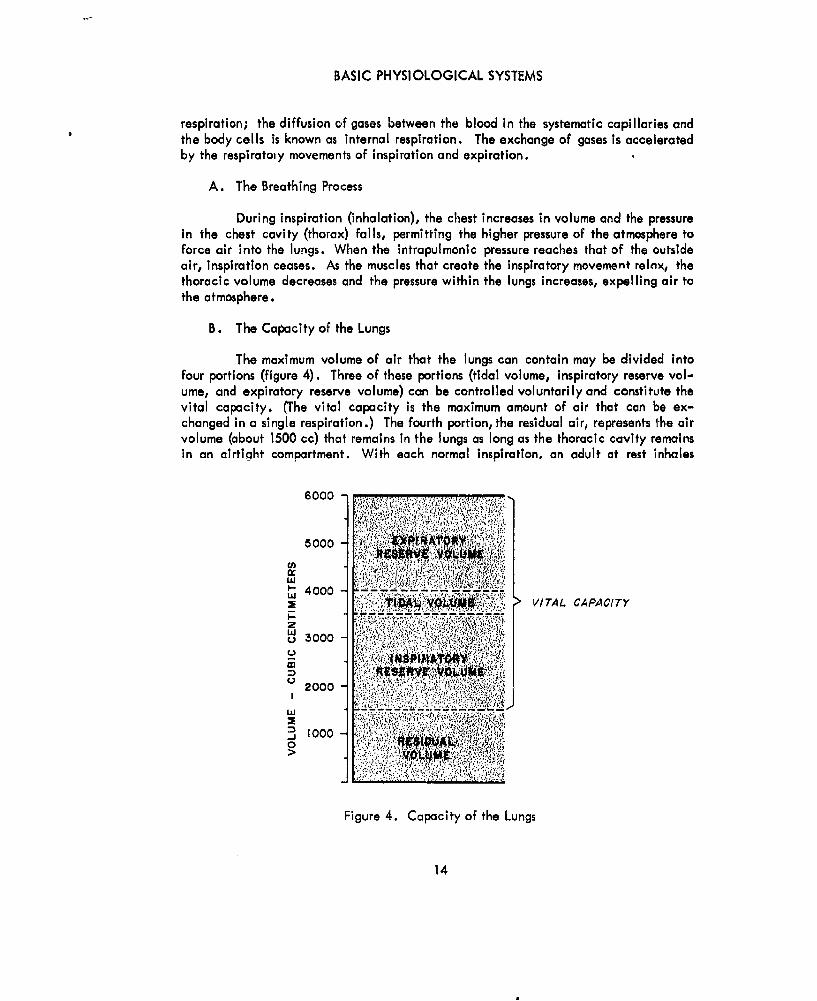
B. The Capacity of the Lungs
The maximum volume of air that the lungs can contain may be divided intofour portions (figure 4). Three of these portions (tidal volume, inspiratory reserve vol-ume, and expiratory reserve volume) can be controlled voluntarily and constitute thevital capacity. (The vital capacity is the maximum amount of air that can be ex-changed in a single respiration.) The fourth portion, the residual air, represents the airvolume (about 1500 cc) that remains in the lungs as long as the thoracic cavity remainsin an airtight compartment. With each normal inspiration, an adult at rest inhales
6 0 0 0 ....... .... . . . . .. .. .,. .6000
LI
' 4000VITAL CAPACITY
,,, .':- ,.,. , : .;£:;.. ..... ..; .......... -
2P
Lz
2000
I 9. 0
S1000
Figure 4. Capacity of the Lungs
14
RESPIRATORY SYSTEM
about 500 cc of air and exhales the same amount. This volume of air is called tidalair. The inspiratory reserve volume (about 2000 cc) is the maximal volume that can beinspired in addition to the tidal air. The air that may be forcibly exhaled after a nor-mal expiration (about 2000 cc) is the expiratory reserve volume. The air remaining inthe lungs after maximal expiration is the residual air.
Tidal inspiration and expiration normally exchange about 500 cc of air, whilemore than 3500 cc of air remain in the lungs. In addition, about one third of the tidalair remains in the air passageways to the lungs; therefore, less than 10 percent of theair in the lungs is renewed during a quiet respiratory cycle. Were it not for the largevolume of alveolar air (sac-air) in the lungs, the blood flowing in the lungs would besubject to alternate large surges of oxygen and carbon dioxide during intermittent in-spiration and expiration. The exchange of alveolar air is dependent on the volume oftidal air, the blood flow rate, and the respiratory rate.
C. Chemical Composition of Inspired and Expired Air
The chemical composition of inspired air is approximately 21 percent oxygenand 0.03 percent carbon dioxide (the remainder is nitrogen). The composition of ex-pired air is approximately 16 percent oxygen and 4.5 percent carbon dioxide. Withinthe alveoli, the oxygen content of the air is 14 percent and the carbon dioxide content
D. Relationship of Oxygen and Blood
An interchange of gases takes place between the alveoli and the blood in thelung capillaries. The blood extracts oxygen from the alveoli and returns carbon di-oxide. The partial pressure of the oxygen in the alveoli, being higher than that of theblood entering the lung capillaries, forces oxygen into the blood by physical diffusion.In the fraction of a second that the blood is present in the blood capillary, enough dif.-fusion takes place to raise theoxygen partial pressure of the blood to almost that of thealveoli. As the oxygen is passing into the blood stream, carbon dioxide is being dif-fused in the opposite direction: from the capillary blood to the alveoli. This occursbecause the partial pressure of the carbon dioxide in the capillary blood is higher thanthe partial pressure of the carbon dioxide in the alveolar air.
The gaseous exchange between the blood and the tissues is the reverse of thattaking place in the lungs, since the oxygen concentration is lower and the carbon di-oxide concentration is higher in the tissues than in the arteries.
E. The Control of Respiration
Although some control can be exercised over breathing, it is essentially invol-untary, controlled automatically by the rhythmical discharge of impulses from nervecells in the medulla of the brain. This involuntary control of respiration is influenced
15

BASIC PHYSIOLOGICAL SYSTEMS
principally by changes of carbon dioxide concentration in the blood. (Changes inoxygen concentration can also influence the breathing rate, but this is an indirect in-fluence through chemoreceptors in the walls of the aorta and the carotid arteries.)Whenever the carbon dioxide concentration of the blood is increased, the excess di-rectly stimulates the respiratory system to increased activity, and deeper, faster breath-ing follows.
As bodily activity increases, more carbon dioxide is produced, which increasesthe concentration of carbon dioxide in the venous blood. This, in turn, raises the car-bon dioxide content in the alveolar air, which reduces the carbon dioxide partial pres-sure gradient between the blood and the alveoli. With a lower pressure gradient, lesscarbon dioxide is passed out of the blood during pulmonary circulation. The carbon-dioxide-laden blood stimulates the chemoreceptors and the respiratory center, increas-ing the rate of stimulation from the respiratory center. This acts to increase the activityof the muscles concerned with respiration through excitation of their motor nerves. Theresulting Increase in ventilation augments the renewal of alveolar air and decreases itscarbon dioxide content. The decrease in carbon dioxide concentration in the alveoliincreases the passage of carbon dioxide from the blood and permits the breathing rateand depth to return to normal.
A lack of oxygen in the blood causes the chemoreceptors to increase respira-ftnn. hnwever, fhkq nnly oLcurs when the nwyeen cnnt'ent nf the lnnrl k rAwlhrAd -tin-
nificantly. The only effect of decreased oxygen on the respiratory center in the brainis a depression, which is characteristic of a lack of oxygen on nerve tissues in general.
F. Minute Volume
Although respiratory depth and rate are important, the product of these two,minute volume, is a more significant index of pulmonary ventilation. The minute vol-ume is the volume of air taken in during each breath times the number of breaths perminute; i.e., the total volume of air breathed per minute. The initial adjustment toincreased bodily activity is an increase in the depth of respiration, but as bodily activ-ity reaches a high level and there is a demand for further increases in ventilation, therespiratory rate is increased. In adults at rest, the normal range of respiratory rate isbetween 12 and 20 complete cycles per minute, although rates of 2 to 30 per minutehave been observed in normal persons. The pulmonary ventilation rate for an adult atrest is between 5.8 and 10.3 liters per minute.
II. Basic Monitoring Methods
There are two basic methods for obtaining information concerning the respiratorysystem: by monitoring the flowof inspired and expired air and by monitoring the move-ments of the chest caused by breathing.
16

RESPIRATORY SYSTEM
A. Air Flow Measurements
A temperature-sensitive device, such as a thermistor, may be used to monitorthe respiratory rate by converting the difference in temperature beheen the inspiredair and the expired air to variations of electrical resistance. The sensing device usu-ally is located in the subject's mask or head attachment.
The depth of respiration is a measure of the volume of air inspired during eachrespiration. This volume can be measured by inserting a temperature-sensitive devicein the oxygen line and by monitoring the changes in temperature in the line caused bythe mass flow of oxygen.
The best method of measuring respiratory rate, monitoring air flow, requiresthat the subject wear a mask or other type of head attachment. If the subject cannotbe encumbered with this equipment, measurement is difficult.
B. Chest Movement Measurements
Respiratory rate and depth may be measured by encircling the chest of thesubject with a strap and measuring the change of chest circuniference. The ends of thestrap (nonelastic) are connected to a strain gage which provides electrical indicationsof chest expansion und curwliu.m.
Chest bands constrict the subject, affect his breathing, and often create ab-dominal expii,;-n which does not register on the strain gage. Artifacts, as a result ofmovements not associated with respiration, may be introduced. In addition, the cheststrap is difficult to calibrate.
C. Minute Volume
Any combination of transducers which records respiration rate and depth maybe used to measure minute volume by Feeding the results of the two readings into anintegrating circuit. The majority of the transducers used in measuring minute volumedescribed thus far require that an oxygen mask be used. When a full pressure suit isused in lieu of a mask, the chest-strap strain-gage transducer must be relied upon tomeasure respiration rate, depth, and their product: minute volume.
D. Oxygen and Carbon Dioxide Partial Pressure
The oxygen and carbon dioxide content of expired air may be determined bypolarographic means. In this method, the current from a special voltaic cell is depend-ent on the presence of either oxygen or carbon dioxide, depending on the design of thecell. Current output is proportional to the amount of gas present. Two cells, one forcach of the gases, are required.
17
BASIC PHYSIOLOGICAL SYSTEMS
This technique is more practical for monitoring the environmental air of acockpit or space vehicle than for monitoring expiratory air directly, since the responsetime is rather long. The response time for the oxygen sensor is approximately 1 minute,while the response time for the carbon dioxide sensor is about 3 minutes. These are notexcessive if the partial pressures are not expected to vary at rapid rates.
The cell electrodes are pressure sensitive; consequently, the pressure musteither be controlled or it must be recorded so that correction factors may be applied.
Other more commonly used systems for measuring the partial pressures of oxy-gen and carbon dioxide are not suitable for space vehicles or high-altiuje aircraftbecause of the weight and space limitations of these craft.
CIRCULATORY SYSTEM
I. Description and Function
The circulatory system consists of the heart and vessels, which circulate the bloodto all parts of the body, and the lymphatics, which return excess tissue fluid to theblood stream. This system is the means by which nutrients and oxygen are distributedto the body cells and their wastes and carbon dioxide are carried away.
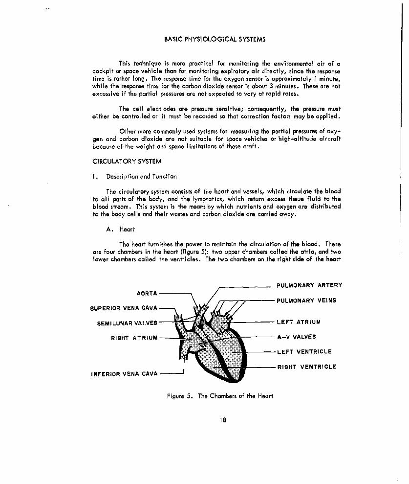
A. Heart
The heart furnishes the power to maintain the circulation of the blood. Thereare four chambers in the heart (figure 5): two upper chambers called the atria, and twolower chambers called the ventricles. The two chambers on the right side of the heart
PULMONARY ARTERYAORTA
PULMONARY VEINSSUPERIOR VENA CAVA
SEMILUNAR VAVES LEFT ATRIUM
RIGHT ATRIUM A-V VALVESl:!i !LEFT VENTRICLE
.. RIGHT VENTRICLE
INFERIOR VENA CAVA
Figure 5. The Chambers of the Heart
18

CIRCULATORY SYSTEM
are associated with pulmonary circulation; that is, the venous blood returning to theheart enters the right atrium and passes through the right ventricle into the lungs, whereit gives up carbon dioxide and absorbs oxygen. Oxygenated blood returning from thelungs enters the left atrium and passes through the left ventricle into the systemic cir-culation system which serves the body. Between each atrium and ventricle there is avalve (A-V valve) which permits blood to pass only from the atrium to the ventricleand not in the reverse direction. Betwoen each ventricle aid its artery, there is aone-way semilunar valve which allows blood to pass into the arteries, but blocks its re-entry to the ventricle. The pumping action of the heart results from pressure changeswithin various sections of the heart because of the contracting of heart muscles. Therhythmic contraction of the heart muscle constitutes the heart beat, and the sequenceof events occurring during one beat of the heart is called the cardiac cycle.
1. Cardiac Cycle
The cardiac cycle begins with the contraction (systole) of the rightatrium, followed closely by the contraction of the left atrium. After a short pause,both ventricles contract. The contraction of each chamber is followed by its relaxa-tion (diastole) and then by a brief period of inactivity. During these contractions,the valves between the chambers of the heart and between the heart and the arteriesrmqond to the chanaes of pressure to determine the direction of flow through the heart.Figure 6 illushlites the pressure changes occurring during tho cardiac cyclc. Venousblood, returning from the lungs and from the systemic circulation, flows into the leftand right atria, respectively, during the period of atrial diastole (relaxation). As theatria fill with blood, the pressures within the citria rise. When the pressures within theatria exceed the pressures within the ventricles, the A-V valves between the chambersopen and blood flows into the ventricles. This accomplishes the greater part of ven-tricular filling. Atrial systole begins, completing ventricular filling, but although thisaids, it is not the chief factor in moving blood into the ventricles.
During the latter part of atrial systole, ventricular systole is initiated.The pressures mount rapidly, and, when they exceed the pressures in the atria, the A-Vvalves between the atria and the ventricles close, preventing the return of blood to theatria. As the pressures in the ventricles continue to rise, they finally exceed the pres-sures in the arteries and cause the semilunar valves to open. The sudden ejection ofblood into the arteries increases arterial pressures and decreases ventricular pressures.When the pressures in the ventricles fall sufficiently (below arterial pressures), thesemilunar valves snap shut.
About the time the semilunar valves open and the ventricles begin toempty, atrial diastole begins. As the ventricle muscles relax, the intraventricular pres-sures continue to fall until the pressures in the atria open the A-V valves and thecardiac cycle is repeated.
19

BASIC PHYSIOLOGICAL SYSTEMS
VENTRICULAR VENTRICULAR PRESSURESYSTOLE DIASTOLE MM
___________________ His
SEMILUNAR SEMILUNARVALVES VALVES
OPEN CLOSE13120
110100
AORTIC (ARTERIAL BLOOD) * a0PRESSURE --
70
--CLOSE60VENTRICULAR PRESSURE 40
OPEN 20
ATRIAL PRESSURE 10
ELECTROCARDIOG3RAM
HEART SOUNDS
FIRST SECOND
Figure 6. Cardiac Pressure Cycle with Corresponding EKG and Heart Sounds
20

CIRCULATORY SYSTEM
2. Electrical Activity
Electrical changes precede each contraction of the heart and are partlyresponsible for the onset of contractions. Because of the chemical and physical proc-esses at work, an active, contracting region becomes electrically negative to a resting,relaxed region. (The electrical phenomena of activity are described under BIOELEC-TRIC MEMBRANE CHARACTERISTICS, page 38.) The electrical activity of the cardiacmuscles is of sufficient intensity to be transmitted to the surface of the body, and maybe observed at almost any spot on the body. Figure 6 shows the relationship of cardiacpotentials to the cardiac cycle.
3. Heart Sounds
During each cardiac cycle, two sounds are produced by the heart. Thefirst sound, which is longer lasting, lower pitched, and softer than the second, resultsfrom the noise of the A-V valves closing and the sound of muscle contraction in theventricles (figure 6). The snapping shut of the semilunar valves at the start of ventriclediastole produces the second sound.
B. Blood Vessels
Ab ,1,lliwuid pevi usly, 'wludJ lIuv;,i iiw e left vanhcle s Ji tr b u tc jthrough arterial branches which ramify to all parts of the body. Blood travels fromthese branches into capillaries which supply all of the tissues. The capillaries unite toform veins; the blood In the veins returns to the heart where it enters on the right side.Blood leaving the right side of the heart divides into two branches and carries blood tothe lungs, where it posses through a network of capillaries lying close to the air sacs(alveoli) of the lungs. Here the blood exchanges carbon dioxide for oxygen. Thecarillaries then coalesce into veins, which return oxygenated blood to the left side ofthe heart.
The circulatory system not only serves to carry oxygen to body cells and toremove carbon dioxide, but it also is responsible for a number of functions which maybe considered nutritive, excretory, regulatory, and protective. The blood vascular sys-tem accomplishes these functions maintaining a constant cellular environment. Asblood flows through the body, it carries metabolic products such as glucose, acids, fats,minerals, etc, from the digestive tract to the body cells. It removes the waste productsof metabolism and transports them to the excretory organs. It distributes hormones fromendocrine glands to cells. Water is transported in the blood to excretory organs to aidin maintaining a constant water level in the body, and peripheral circulation aids inmaintaining constant body temperature. Antibodies and white corpuscles also are trans-ported by the blood to aid in fighting injurious agents.
The blood supply in any part of the body maybe varied greatly by the nervoussystem and other systems of the body acting upon the heart and the blood vessels. The
21

BASIC PHYSIOLOGICAL SYSTEMS
heart can be made to pump more blood, or the same amount at a higher pressure. Theblood supply to a part of the body can be altered by an increase or decrease in thecaliber of blood vessels. These changes occur as a result of nervous control or throughchemical means.
The nervous control of circulation is exerted chiefly by reflexes originating inthe atria, aortic arch, carotid sinus, and mesentery. These reflexes are integrated inthe medulla, hypothalamus, and higher centers in the central nervous system (seeBEHAVIORAL SYSTEM, page 6), and exert their control over the circulation byway ofthe vagus and sympathetic nerves to the heart and the vasomotor nerves to the bloodvessels. In certain circumstances, a chemical control may be exerted directly by secre-tions of the adrenals and kidney, and indirectly by the chemoreceptors in the aorticarch and the carotid body.
C. Lymphatic System
The lymphatic system is an accessory' rart of the circulatory system. It gathersthe excess tissue fluids from the body cells, strains foreign particles and bacteria fromthe fluid, and empties the fluid into the veins.
The blood in the circulatory system flows from the heart through the arteriesinto smaller and smaller blood vessels. The smallest of these vessels, the capillaries,feed the nultietil Luiluinad in [lhe blood to the tir.uc culls of the body by rl, u,through the capillary walls. These nutrients are in the form of a watery solution calledtissue fluid. When the cells have assimilated the nutrients in the fluid, some of thefluid is returned to the capillaries from which the blood flows on to the veins andthence back to the heart.
The excess tissue fluid remaining in the body cells filters into other smalltubes, the lymph capillaries. These capillaries merge with larger and larger lymphvessels until the fluid, now known as the lymph, is returned to the veins.
The flow of lymph is maintained by the pumping action of muscles and fluctu-ation in intrapleural pressure. However, the rate of flow is slow in comparison to theflow of blood in the circulatory system.
D. Blood Pressure
Blood pressure is the result of the pumping action of the heart which emptiesblood into a closed system of elastic blood vessels. The volume capacity of this systemchanges as a result of the stretching of the vessels and by caliber changes in responseto nervous and chemical stimuli.
During ventricular systole, blood is forced into the highly elastic arterial sys-tem faster than it can escape into the arterioles, capillaries, and veins. The arteries
22
( RCULATORY SYSTEM
therefore are stretched to greater capacity. Since blood is leaving the arterial systemconstantly and passing into the capillaries, the pressure falls when the flow of bloodinto the artery is stopped at the end of ventricular systole. The elastic recoil of thearterial walls forces the blood onward through the capillaries at a constantly decreasingpressure until the arteries regain their presystolic caliber (see figure 6). The energythat was stored ;n the arterial walls during the stretching of the vessels is expended be-tween heart beats, ensuring a relatively continuous flow of blood through the arteriolesand capillaries. The pulsations of the arteries are convenient indications of heart rate.
The blood pressure is maintained at a normal level by a combination of fivefactors: cardiac output, peripheral resistance, blood volume, elasticity of the arterialwalls, and blood viscosity.
1. Cardiac Output
The heart controls the amount of blood that is ejected into the arteriesduring each cycle; therefore, anything that increases cardiac output (if other factorsare kept constant) increases arterial pressure. Variations in cardiac output in responseto bodily activity are determined by changes in venous return (the rate at which bloodis returned to the heart), the force of the heart beat, and the frequency of the heartbeat.
The venous return to the heart is (1) increased bythe contraction of skel-etal muscles (increased activity), (2) increased by deep respirations, (3) decreased bythe dilation of blood vessels over a wide area, and (4) affected by the force of gravity(dependent on the position of the subject).
The heart rate is determined by nervous, chemical, and thermal control.The heart is innervated by the vagus nerves, which are part of the parasympatheticdivision of the autonomic nervous system, and the accelerator nerves, which are part ofthe sympathetic division. These nerves are mutually antagonistic in that the vagusnerves decelerate the heart rate while the accelerator nerves accelerate the heartbeat. Besides these nervous controls, the heart rate may be increased or decreased bychemical action. Hormones released bythe vagus nerves and hormones of the endocrinesystem, especially those released by the thyroid and adrenal glands, may increase ordecrease the heart rate. A great excess of carbon dioxide or a marked lack of oxygendepresses the excitability of both the heart muscles and the cardiac centers in the me-dulla. A rise in body temperature also increases the heart rate.
2. Peripheral Resistance
Variations in the caliber of blood vessels change the resistance to theflow of blood, and this resistance is referred to as peripheral resistance. Stimulationof almost any somatic sensory nerve can change the caliber of blood vessels. An ex-cess of carbon dioxide or lack of oxygen acts directly to depress the vasomotor centers,
23

BASIC PHYSIOLOGICAL SYSTEMS
which control the constriction or dilation of blood vessels. Moderate increases in thecarbon dioxide content of the blood or a lack of oxygen stimulates the chemoreceptorsin the aortic and carotid bodies (major arteries leaving the heart) and causes vasocon-striction, which increases the arterial pressure. The temperature of the tissues throughwhich the vessels pass also affects the caliber of blood vessels. A rise or a moderatedrop in temperature dilates the vessels, while a marked drop in temperature constrictsthe vessels.
3. Blood Volume
The circulatory system is a closed system which normally is filled withblood under pressure. Because of the elasticity of the vessels, additional blood can beadded to the system after it is filled, which increases the pressure. Conversely, with-drawal of fluid (as in a hemorrhage) decreases the arterial pressure. Blood volume mustbe kept above a minimum level or circulation ceases.
4. Elasticity of the Arterial Walls
The elasticity of the arterial walls maintains a relatively constant flowof blood to the capillaries by storing the excAq. energy of the ventricular contraction(systole) and releasing this energy during ventricular diastole. The pressure maintainedby the recoil of the arterial walls is known as the diastolic pressure. (This is also theminimum pressure which occurs during ventricular diastolu.) Th1 Jju6tolic pressure isaffected far less than systolic pressure (the maximum pressure occurring during ventric-ular systole) by changes in most of the factors maintaining blood pressure.
5. Blood Viscosity
The more viscous the blood, the greater is the force required to set it inmotion because of its greater resistance to flow. The viscosity of blood is determinedlargely by the number of formed elements present and, to a lesser degree, by the con-centration of plasma proteins. If either of these factors decreases, reistance drops andblood pressure falls. However, since blood viscosity varies only slightly, its effects arenegligible.
II. General Monitoring Methods
The obvious parameters to be measured for monitoring the circulatory system arethe pulse rate and arterial pressure. However, other measurements, such as the varia-tion and rate of change of pulse rate, cardiac cycle, flow rate of blood through specificarteries or body tissues, and the partial pressure of oxygen carried by the blood, canprovide important information.
24

CIRCULATORY SYSTEM
A. Pulse Rate
As oxygen-rich blood leaves the heart under high pressure, some of its flowenergy is expended in distending the elastic walls of the large arteries. The recoil ofthese arteries t en forces the blood through the system after ventricular systole. Thisdistension and recoil is responsible for the pulse wave which can be felt in any artery.A pulso occurs with every systole; therefore, the pulse rate can be measured to deter-mine heart rate.
A transducer may be placed over any artery to pick up the pulse. The mostcommon position is the wrist where the radial artery runs just below the skin. Pulserate or heart rate also may be measured by using electrocardiographic leads to pick upcardiac muscle potential. In this case, the potential picked up by the leads is passedthrough a filter to eliminate high frequency electromyographic potentials. The result-ing wave is clipped so that only the peak of the ventricular systole potential (QRScomplex of the electrocardiogram) remains, and this information is amplified and fedinto a counter (cardiotachometer) to record the heart rate. (The leads used in electro-cardiography and the QRS complex are described under Cardiac Activity,page 27.)
Gross body movement and muscular activity create random signals that inter-fere with pulse rate measurements at the wrist or at the chest. All types of instrumen-tation for measuring pulse ratc r'-+irc filtcrs to climinato thosc random signals. Inaddition, there are other problems resulting from the use of electrocardiographic leadsto record pl se rate similar to those described for monitoring the cardiac cycle.
B. Blood Pressure
Blood pressure is a ma or parameter in monitoring the circulatory system.However, the pressures recorded at various points on the body are different, since bloodwould not flow if there were not a pressure gradient (a progressive drop in pressurefrom the large arteries to the smallrjr arteries, the arterioles, the capillaries, and theveins). Therefore, to obtain comparative blood pressure information, blood pressuremust be recorded at the same place. Almost al! blood pressure measurements are madeon the forearm at the present time. At this location, it is quite easy to measure bothsystolic pressure (the maximum pressure which occurs during ventricular systole) anddiastolic pressure (the minimum pressure which occurs as a result of arterial recoil dur-ing ventricular diastole) by auscultation. The difference between the systolic anddiastolic pressures is called the pulse pressure.
A physician normally measures arterial pressure by encircling the arm with aninflatable cuff, and placing a stethoscope, microphon,', or other listening device overthe brachial artery below the cuff in a position where the pumping sounds of the heartare clearly audible. The cuff is inflated until the pressure in the cuff is greater thanthe arteri at pressure; at this point, no sounds are heard, since arterial pressure is
25
BASIC PHYSIOLOGICAL SYSTEMS
insufficient to force blood past the cuff. The pressure in the cuff is released slowlyuntil the lumen in the compressed artery is opened enough to allow the passage of a jetof blood with each heart beat (ventricular systole). The pressure in the cuff at thispoint, recognized by the sound of the blood in the artery, represents the systolic pres-sure. As the cuff pressure is reduced further, the sound changes in quality and dis-appears completely when the blood flows continuously through the artery. The pressurein the cuff at this point is the diastolic pressure.
Blood pressure maybe monitored by partially automating the above procedure.The cuff is made to inflate rapidly to a pressure above any anticipated systolic pres-sures, after which the gas is bled off slowly, reducing the pressure below any antici-pated diastolic pressures, and the remaining pressure is dumped. This procedure maybe sequenced to repeat at specific intervals. A transducer in the cuff provides anelectrical indication of the pressure in the cuff. A microphone or other listening de-vice placed over the brachial artery, immediately below the cuff, supplies the arterialsounds. The output from the listening device may be combined with the pressure trans-ducer output so that only one signal need be transmitted. The pressure at which thesound resumes after being stopped by the peak cuff pressure indicates systolic pressure,and the pressure at which the sound ends represents the diastolic pressure. If it is in-convenient to monitor blood pressure by auscultation, the microphone may be elimi-nated and a transducer inserted under the occluding cuff, directly over the brachialnrtery. This pulse-sensing transducer may be a flexible bladder containing fluid andconnect hydruulkcullyto a pressure transducer. A strain gage could be mounted directlyover the brachial artery and under the occludl6g cuff, but the signal from the straingage would have, to be amplified before -he data could be telemetered.
The cuff method of monitoring blood pressure gives data comparable to thatobtained by most clinicians; therefore, values of blood pressure taken of a subject inan abnormal environment may be compared with normally accepted values. This is onlytrue, however, if the pressures in the abnormal environment are the same as atmos-pheric. Blood pressure is recorded In terms of gage pressure, which is the differencebetween the pressure in the cuff and atmospheric pressure. In an abnormal environment,if the environmental pressure is not equivalent to atmospheric pressure, all pressurereadings must be converted to some common denominator so they can be compared. Thepressure to which the cuff is inflated will vary, since the gas is admitted to the cuffuntil it reaches a constant gage pressure or constant volume, both of which vary withenvironmental pressure and temperature. Provisions must be made so the pressure ex-ceeds systolic pressure under every condition.
If an automatic inflating-cuff device is used to monitor blood pressure, sev-eral factors must be considered. The test must be done quickly to provide a minimumof discomfort to the subject and to avoid impeding his activities. A tank of air mustbe provided for the remotely located subject to inflate the cuff for the test, therebycreating a space and weight problem. Serious errors may develop if the subject is do-ing more than light arm or wrist movements while being seated. Environmental noise
26

CIRCULATORY SYSTEM
and vibration can create erroneous signals in automated monitoring systems.
C. Cardiac Activity
The cardiac cycle may be monitored by the (1) electrical potentials gener-ated by the contracting cardiac muscles (electrocardiography), (2) small movementsproduced in various parts of the body under the influence of displacements of the heartand blood occurring in connection with cardiac activity (hallistocardiography), or(3) movements of the heart by radiating ultrasonic waves from the chest wall to theheart and using the Doppler effect to detect motion (phonocardigraphy). Only thefirst of these three methods is discussed here, since the artifacts in the other two tech-niques are sufficient to restrict their use to the laboratory at the present time. In addi-tion, considerably more data have been obtained on cardiac cycles through electro-cardiography, and norms have been established from millions of tests.
1. Electrocardiographic Potentials
If the heart were a homogeneous sphere of muscle, the potentials gener-ated would be the same everywhere. This, however, is not the case for the muscles ofthe left ventricle are considerably thicker than those of the right ventricle, and theanatomically nonsymmetrical ventricles form a U-shaped shell with the open portion in'110. rogtion of "- " ... hc. . .. I..
2. Standard Limb Leads
The activities of the cardiac muscles, as previously noted, are accom-panied by electrical changes. These potential changes are transmitted to the surfaceof the body where they may be recorded and correlated with heart action. Since it isimportant for standardization purposes that potentials be read from the same placeseach time, and since the limbs are the only locations that are equipotential over alarge area, three standard combinations of instrument lead connections have beenadopted universally. Standard limb lead I measures the potential difference betweenthe right and left arms; lead II measures the potential between the right arm and leftleg; and lead III measures the potential between the left arm and left leg.
3. Electrocardiogram
Figures 6 and 7 show typical electrocardiograms using any of the threestandard limb leads. The letters on the various parts of the curve shown in figure 7 in-dicate the correlation with heart action. The P wave, the first upward deflection inthe cycle, represents the period of atrium excitation (depolarization of atrial muscula-ture). The PR interval, from the beginning of the P wave to the beginning of the QRScomplex, represents the time required for excitation to travel through the atrial to theventricular muscle. The QRS complex (beginning of Q to end of S) is the time requiredfor the excitation waves to spread through the ventricle walls (usually from about 0.05
27

BASIC PHYSIOLOGICAL SYSTEMS
to 0. 10 second). The beginning of the complex represents the boginning of ventriculardepolarization, while the end of the complex indicates the end of ventricular depolar-ization. Ventricular systole occurs during the QT interval. The duration of thedepolarized state is measured from the end of the QRS complex to the beginning of theT wave. The smooth hump following the QRS complex is the T wave, which representsthe wave of ventricular repolarization.
Electrocardiography has well-established standard values that are avail-able in numerous handbooks. Typical va I ues, therefore, are not presented in thishandbook, except to illustrate relative time intervals in an electrocardiogram.
Primarily, the electrocardiogram permits the moritoring of the circulatoryFactors of heart rate and rhythm. The frequency of ventricular systole indicates theheart rate. Also, the presence of fibrillation (and premature atrial and nodular con-tractions), tachycardla, heart block, and marginal anoxia can be detected.
Since the peak voltage appearing in an electrocardiograph signal usingstandard electrode arrangements is approximately 2 millivolts, the signal must be ampli-
R
P T
S
- P- R INTERVAL, S- T INTERVAL
Q-T INTERVAL-,
Figure 7. Composition of the Electrocardiogram
28

CIRCULATORY SYSTEM
ried considerably, and the amplifiers and recording equipment must be selected care-fully to avoid introducing excessive artifacts. The electrodes should be nonpolarizedand must not be placed over large muscle masses where they can detect electromyo-graphic; potentials. In addition, they must be attached to the body firmly to preventthe skin from moving beneath the electrodes. Artifacts are introduced through imped-ance changes resulting from changes in galvanic skin reflexes, but little can be doneabout these except to try to avoid locations where there are numerous sweat glands, andto recognize the artifacts when they appear in the record.
D. Blood Flow Rate
Measurement of blood flow rate is important for detecting changes in the in-ternal resisiance of blood vessels as well as for diagnosing circulatory ailments. Theflowof blood may be monitored directly by flowmeters or indirectly by volume plethys-mographs. There are two types of flowmeters, electromagnetic and sonic. The elec-tromagnetic flowmeters employ the use of an electromagnetic field in which a bloodvessel is placed. Two electrodes are connected to the vessel and the potential differ-ence between the electrodes is measured. The son i c flowmeter measures the timerequired for a sound wave to travel a measured distance in a vessel. This time factoris a function of blood velocity.
Plethysmography offers the advantage that the veins or vessels need not beexposed to be monitored. With impedance plethysmographic techniques, the changein the volume of body tissues is used to indicate the quantity of blood they contain. Alow-voltage, radio-frequency signal is passed through the subject's finger or other por-tion of the body. The received signal indicates the rhythmical variations in electricalimpedance resulting from pulsatile volume changes (and consequently heart beat). Thisimpedance curve may be correlated with cardiac output, or the information may be fedinto a differentiating network to yield the relative change in the velocity throughoutthe cardiac cycle in a given pulse region. This technique generally is considered"delicate" and difficult to standardize.
Blood flow rate also may be monitored by plethysmographic methods usingstrain-type gages. A strain gage is connected to a strop encircling the subject's finger,arm, or calf to measure the girth of the limb. An occluding cuff is provided below thegage to occlude arterial flow (except on the finger), while another cuff above (on theheart side) the gage occludes venous flow. The change in girth may be calibrated torecord flow rate. This type of device could be integrated with the occluding cuff usedfor determining blood pressure, thereby reducing the discomfort to the subject beingmonitored.
E. Oxygen Saturation of Blood
The oxygen saturation of the b I ood may be determined by measuring thepercentage of hemoglobin in the blood in oxygenated form with a photoelectric
29
BASIC PHYSIOLOGICAL SYSTEMS
photometer. The absorption qualities of oxygenated hemoglobin and reduced hemo-globin differ considerably. Figure 8 shows two curves illustrating the transmissionqualities (or absorptive qualities) of oxygenated hemoglobin and reduced hemoglobin asfunctions of wavelength of transmitted light. Obviously, oxygenated and reducedhemoglobin absorb the light of wavelengths in the region of 640 millimicrons (red)quitedifferently, while light of 800 millimicrons wavelength is absorbed equally by bothforis of hemoglobin. A device can be constructed, therefore, using a photocell foreach wavelength (640 and 800 millimicrons). The light of each wavelength is trans-mitted through a thin section of the body, such as the projecting part of the externalear, and is detected by the photocells. The difference between the readings at eachwavelength is a function of the oxygen saturation of the blood.
100
90 -
8 REDUCED HEMOGLOBINWV!70 -,"-".
Z 60 -
50 -
z IS40 -
30 ->
OXYGENATED_J 20 - iHEMOGLOBIN
10 I
0
600 700 800 900 1000WAVELENGTH (MILLICRONS)
Figure 8. Light Absorption by Hemoglobin
30
CIRCULATORY SYSTEM
To correct for the ear tissue interposed in the optical path of the device, apressure device (mechanical or pneumatic) can be used to force blood from the ear sothat the light absorption of the bloodless tissue can be measured. The light absorptionof the blood alone wouid be the difference between the readings with the ear occludedand with the ear open to circulation. This technique is subject to artifacts from Gforces and vibration that affect equipment performance.
METABOLIC SYSTEM
I. Description and Function
The metabolic system includes those processes and chemical reactions which occurin the body that convert food into simpler substances and supply the energy for vitalfunctions such as muscular activity. The building up or assimilative processes (forma-tion of protoplasm) that make possible the growth and repair of tissues is referred to asanabolism. Catabolism is the breaking down or destructive process which releasesenergy needed for muscular activity. The source of the energy is the chemical energyof the food materials which is transformed by oxidation to heat and mechanical energy(or work).
The elements needed for the metabolic process are taken into the body through thelungs (oxygen) -nr thi nlimentary canal (food and water). As the foodstuffs in thebody are oxidized by the oxygen carried in tho b lo od, they form carbon dioxide,nitrogen-containing products, water, and heat. The total heat produced by cell metab-olism is an index of total metabolism.
Since the heat produced by the oxidation of foodstuffs in the body varies at dif-ferent times due to different circumstances, a standard for each individual must beestablished. This standard is the Basal Metabolic Rate (BMR), which is the heat pro-duced under standard conditions with activity reduced to a minimum. Basal metabolismis the energy expenditure of a subject who is lying down at complete muscular andmental rest, some 12 to 14 hours after taking food. The environmental temperatureshould be maintained at about 200C. Under these basal conditions, the three factorsthat are prominent in stimulating metabolism (physical exercise, ingestion of food, andenvironmental temperature) are controlled. The generated heat results mainly from theactivity of the heart, glands, and respiratory movements, and indicates the energynecessary to maintain vital processes. Basal metabolism is proportional to the surfacearea of the individual, and for normal healthy men between 20 and 50 years old, it isapproximately 38 to 40 calories per square meter per hour.
II. Basic Monitoring Methods
Since man is a warm-blooded animal and maintains within his body a surprisinglyuniform temperature, the study of heat production is used as the principal measure ofmetabolism. Heat production results from oxidizing foodstuffs within the body; there-
31
BASIC PHYSIOLOGICAL SYSTEMS
fore, measuring oxygen consumption is an indirect way of measuring heat production.
At one time, the procedure consisted of measuring the oxygen inhaled, the oxygenand carbon dioxide exhaled, and the nitrogen content of the urine excreted during thetesting period. The inhalations and exhalations indicated how much of each of thethree basic foodstuffs was oxidized and how much heat was produced. The nitrogencontent of urine was indicative of the amount of protein oxidized. It has been foundsince that there is a constant relationship between the amount of oxygen consumed andthe amount of heat produced. Data on oxygen consumption are converted easily toindications of heat production.
Ill. Problem Areas
Except where there is arterial oxygen unsaturation, the rate of oxygen absorptionwithin the lungs is a close measure of the rate of intracellular oxidation and of heatproduction. The rate of oxygen absorption in the lungs is relatively unaffected bymoderate changes in lung ventilation. If carbon dioxide production is measured, how-ever, it should be remembered that carbon dioxide elimination is particularly sensitiveto changes in character and degree of lung ventilation. Blood and other body fluidscontain a large amount of carbon dioxide in loose combination, which is readilywashed out by overventilation of the lungs. While this is of little concern with anapparatus monitoring oxygen consumption alone, determinations of carbon dioxide pro-duction would be invalidated, and next to leaky apparafus, this sensitivity to lungventilation has been the major problem.
EXCRETORY SYSTEM
I. Description and Function
The primary organs of the excretory system are the kidneys, although the lungs,skin, and large intestines also have excretory functions. The lungs excrete carbon di-oxide and some water, and the skin, while regulating body temperaturn, excretes waterand salts. The lining of the large intestine excretes calcium and iron into the lumenwhere they are eliminated with the Feces. (Most constituents of the feces are notmetabolic wastes and so they are not considered as excretory products.) The majorityof metabolic wastes found in the blood are absorbed by the kidneys where they formurine, which is excreted from the body by the urinary system.
The kidneys, ureters, bladder, and urethra constitute the urinary system which ridsthe body of liquid and soluble waste products. Through the production and eliminationof urine, the urinary system maintains a constant blood volume which helps maintain aconstant blood pressure.
Urine formation is the result of two processes: filtration which takes place in therenal capsule, and reabsorption which occurs in the tubules of the kidneys. The rate of
32

EXCRETORY SYSTEM
reabsorption varies to maintain a constant fluid level within the body. The formationand excretion of urine also acts to maintain the electrolytic and hydrogen ion balanceof the blood. Acidily is regulated in the blood itself; however, the kidneys aid byproducing ammonia from urea (a waste product of proteen metabolism) and amino acids.This ammonia combines with water in the kidneys to form a base (ammonium hydroxide),which may be absorbed in the blood to neutralize the acid in the blood. The urineformed in the kidneys is transported to the bladder through the uretrs. Micturition isinitiated by the stimulation of afferent neurons in the bladder wall. When the pressurein the bladder is sufficient to stimulate the nerves (approximately 150 mm of water isadequate), the bladder contracts, while the sphincter (normally constricted to seal thebladder) relaxes, permitting the urine to be expelled through the urethra uponcommand.
II. General Monitoring Methods
Any attempt to monitor the excretory system in an abnormal environment such as aspace vehicle would be extremely complex. Monitoring systems would almost invari-ably require the collection of urine. On short-duration, abnormal environmental tests,it would, therefore, be impractical to monitor the excretory system.
Pressures and temperatures within the alimentary canal can be monitored if thesubject swallows a radio pill with pressure or temperature transducers. The monitoringof the pill's progress through the bodywould provide some useful information, but wouldgive no indication of the body's fluid level or kidney and bladder functions.
ENDOCRINE SYSTEM
I. Description and Function
The endocrine system is composed of glands which resemble ordinary glands in thatthey produce complex compounds from materials derived from the blood or lymph, butthey differ in that they have no ducts for the discharge of their secretions. Thesesecretions, called hormones, are distributed through the blood to regions scatteredthroughout the body. The hormones exert a specific influence on some part or activityof the body.
The endocrine system and the nervous system share in the contrul of all the activi-ties of the body tissues. For the most part, the nervous system controls the more rapidresponses of the body which require immediate response if the organism is to survive,while the endocrine system frequently controls the slower changes or adaptations of thebody to its environment.
A. Endocrine Glands and Organs
The glands with definite endocrine functions (i.e., contain specific chemical
33
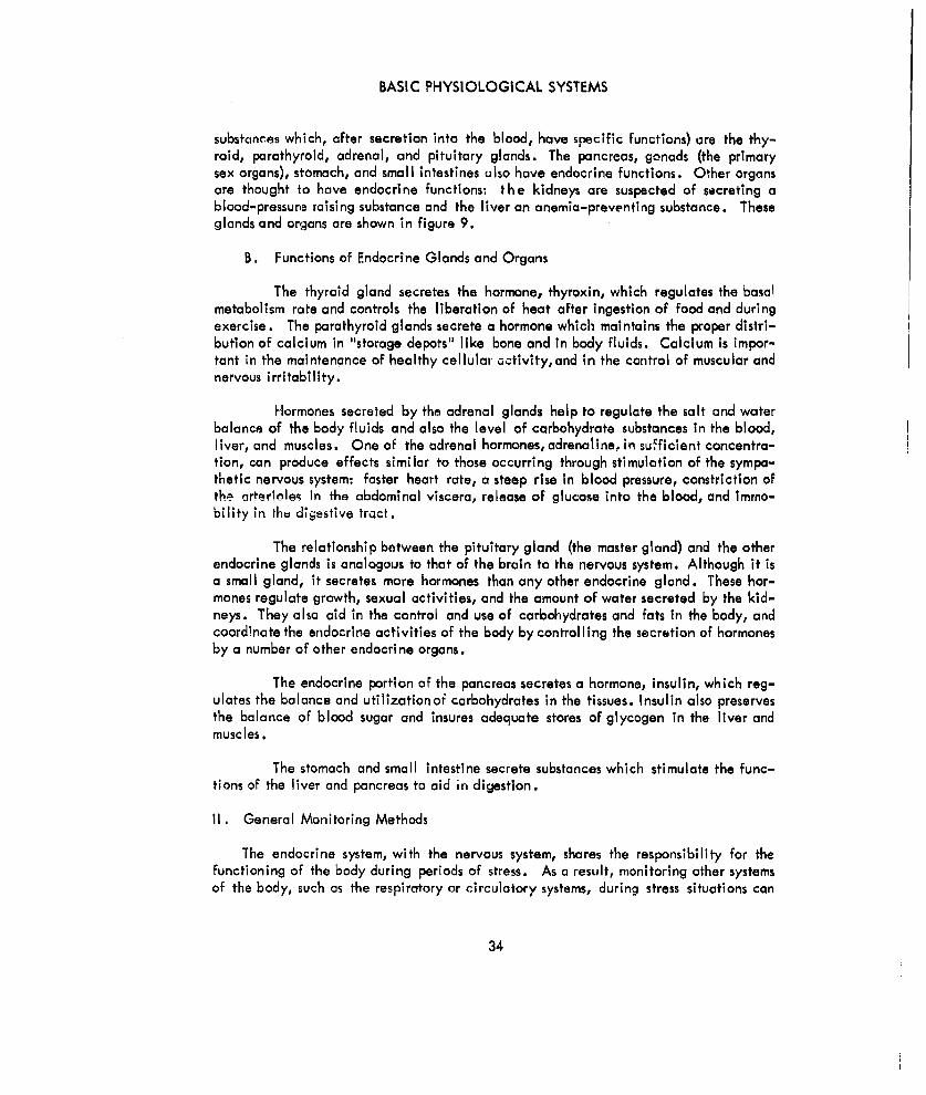
BASIC PHYSIOLOGICAL SYSTEMS
substanees which, after secretion into the blood, have specific functions) are the thy-roid, parathyroid, adrenal, and pituitary glands. The pancreas, gonads (the primarysex organs), stomach, and small intestines also have endocrine functions. Other organsare thought to have endocrine functions: the kidneys are suspected of secreting ablood-pressure raising substance and the liver an anemia-preventing substance. Theseglands and organs are shown in figure 9.
B. Functions of Endocrine Glands and Organs
The thyroid gland secretes the hormone, thyroxin, which regulates the basalmetabolism rate and controls the liberation of heat after ingestion of food and duringexercise. The parathyroid glands secrete a hormone which maintains the proper distri-bution of calcium in "storage depots" like bone and in body fluids. Calcium is impor-tant in the maintenance of healthy cellular activity, and in the control of muscular andnervous irritability.
Hormones secreted by the adrenal glands help to regulate the salt and waterbalance of the body fluids and also the level of carbohydrate substances in the blood,liver, and muscles. One of the adrenal hormones, adrenaline. in sufficient concentra-tion, can produce effects similar to those occurring through stimulation of the sympa-thetic nervous system: faster heart rate, a steep rise in blood pressure, constriction ofthe arteriole in the abdominal viscera, release of glucose into the blood, and imrno-bility in thw digestive tract.
The relationship between the pituitary gland (the master gland) and the otherendocrine glands is analogous to that of the brain to the nervous system. Although it isa small gland, it secretes more hormones than any other endocrine gland. These hor-mones regulate growth, sexual activities, and the amount of water secreted by the kid-neys. They also aid in the control and use of carbohydrates and fats in the body, andcoordinate the endocrine activities of the body by controlling the secretion of hormonesby a number of other endocrine organs.
The endocrine portion of the pancreas secretes a hormone, insulin, which reg-ulates the balance and utilizationof carbohydrates in the tissues. Insulin also preservesthe balance of blood sugar and insures adequate stores of glycogen in the liver andmuscles.
The stomach and small intestine secrete substances which stimulate the func-tions of the liver and pancreas to aid in digestion.
II. General Monitoring Methods
The endocrine system, with the nervous system, shares the responsibility for thefunctioning of the body during periods of stress. As a result, monitoring other systemsof the body, such as the respiratory or circulatory systems, during stress situations can
34
ENDOCRINE SYSTEM
01 PITUITARY GLAND
PARATHYROID GLANDTHYROID GLAND
THYMUS GLAND
LIVER STOMACH
DUODENUM PANCREAS
KIDNE ..... ADRENAL GLAND
Figure 9. Location of the Endocrine Glands and Organs
35

BASIC PHYSIOLOGICAL SYSTEMS
indicate that the endocrine system is functioning properly. Howeverdirect monitoringof the endocrine system is extremelydifficult, even if the monitoring took place in alaboratory.
Radioactive iodine may be injected into the circulatory system and its effect onthe thyroid gland monitored to determine thyroid activity, but this test is done in alaboratory. The level of circulating thyroid hormone may be determined with serumprotein-bound iodine, but this also is a highly sophisticated technique. Similarly, theadrenal gland is monitored in laboratories by both biological assay and chemical deter-mination of an adrenal hormone in the urine, but this, again, is not the type of test thatcould be performed in an abnormal environment such as a spacecraft.
III. Problem Areas
As previously stated, there is no suitable direct means of monitoring the endocrinesystem. Indirect methods, such as monitoring other systems in periods of stress, may in-dicate that the endocrine glands and organs are functioning properly, but since thenervous system shares control of many stress functions, it would be difficult to determinewhether the endocrine system was functioning or whether the nervous system was re-sponsible for the observed responses.
TEMPERATURE-REcULATI NG SYSTEM
I. Description and Function
The extent and rate of activity of "cold-blooded" animals are determined by thetemperature of their environment. If the temperature is cold, their activity is sluggish;if hot, their activity is accelerated. They have no control over their body tempera-ture. Mjn, however, is a "warm-blooded" animal and, therefore, has a temperature-regulating mechanism which maintains a relatively constant temperature within thebody, regardless if the environmental temperature. This consistency of body tempera-ture makes man's activity relatively independent of environmental temperature.
Body temperature is regulated by the interplay of antagonistic processes. Heat isbeing produced and lost by the body continually. Body temperature represents thebalance struck between heat production and heat loss.
A. Heat Production
Heat is produced by the oxidation processes (metabolism) occurring in thetissues and cells. Any factor which affects cell metabolism thus influences heat pro-duction. Man is unable to control the metabolism of cells; his only voluntary mecha-nism for the control of heat production is a change in the activity of the skeletalmuscles. The skeleta! muscles are the greatest source of body heat and their activitycan quickly be adapted to changing temperature requirements by reflex or voluntarycontrol.
36

TEMPERATURE-REGULATING SYSTEM
B. Heat Loss
The loss of body heat is the result of a number of physical processes. Smallamounts of heat are lost by warming the air we breathe and in the urine and feces ex-creted by the body, but the greatest amount of heat loss occurs through the skin. Heatloss from the skin occurs by the three processes of heat transfer: conduction, convec-tion, and radiation. Considerable heat is also given up by evaporation. At high envi-ronmental temperatures, evaporation is the only effective means by which the body cangive up heat, which is the reason humidity is so important to comfort in warm environ-ments. A hot humid atmosphere (1 200 F) cannot be endured for more than a few min-utes, while a dry atmosphere with temperatures as high as 2000 F can be tolerated withno increase in body temperature.
C. Temperature Regulation
The temperature-regulatng center Is in the hypothalamus. It is influencedreflexly as a result of afferent impulses from the skin (cutaneous receptors) and locallyin the center Itself as a result of the temperature of the blood supplying the brain.
When the body is exposed to a cold environment (I.e., the environmentaltemperature is lower than the temperature of the skin), the body tends to lose heatfaster than it can produce heat, The cutaneous receptors send impulses to the controlcenter (hypothalamus), which relays Impulses to the body to conserve body heat.These impulses cause the smooth muscles of the arterioles to contract, reducing the flowof blood in peripheral circulation, thereby reducing conduction and radiation lossesfrom the skin. Sweat gland activity also is reduced to eliminate heat loss throughevaporation. If the environmental temperature is not too low, these actions adequatelypreserve the constant body temperature which is approximately 98.6 0 F (orally) in man.However, if the external temperature continues to drop, a point is reached where re-ductions in heat loss are insufficient to maintain body temperature, and heat productionmust be accelerated. The control center increases the tone of the skeletal muscles,and if this is insufficient, involuntary contractions (shivering and chattering of teeth)result. Experiments have shown more than 400 calories of heat may be produced eachhour by shivering alone.
When the environmental temperature rises above skin temperature, an oppo-site series of events occurs. Stimulation of the hot cutaneous receptors causes thehypothalamus to send out impulses to inhibit the contraction of the smooth muscles ofthe arterioles. Thus the arterioles dilate and more blood flows through the peripheralcirculatory system, permitting more heat to be dissipated by convection and radiation.If the temperature continues to rise, a point is reached where sufficient heat is notgiven up by this process and sweat gland activity is accelerated, permitting greaterevaporation.
37

BASIC PHYSIOLOGICAL SYSTEMS
The regulatory functions described above are stimulated not only by cutaneousreceptors as illustrated in the discussion, but also by the temperature of the blood flow-ing near the hypothalamus in the brain. If the environmental temperature were keptconstant, but the temperature of the blood were altered, the results would be similar tothose resulting from changing environmental temperature.
I. General Monitoring Methods
Body, bloodand skin temperatures are measured commonly in laboratories, doctors'offices, and in homes. However, for the purpose of monitoring the body temperature ofa man in an abnormal environment, the devices used may be limited to two commonforms. One uses the electromotive force generated at the junction of two dissimilarmetals (thermocouples) to indicate temperature, and the other uses the change in elec-trical resistivity in a semiconductor or other material having a temperature coefficientof resistance to indicate temperature (thermistors and resistance thermometers).
The selection of a monitoring device, its method of attachment to the body, andthe location to which it is attached are extremely important if reproducibility is to beexpected. A rectal thermometer, which may consist of any of the devices mentioned,probably will give the most reproducible data. If the devices are attached to the skintoo loosely, they will record the temperature of the surrounding air rather than that ofthe skin, and if attached with sufficient pressure to imbed them partially into the sur-face of the skin, the reading- may be affected by resulting local irritation. Rectalthermometers can be inserted a specific distance in each case to insure consistency inprocedure, and may be taped in place to cause a minimum of discomfort to the subject.
III. Problem Areas
Thermocouples, thermistors, and resistance thermometers are subject to artifactsresulting from the widely different pressures of attaching these devices, as previouslyindicated. In addition, resistance thermometers are subject to artifacts caused by thebending or stretching of the resistance wires. However, they do offer the advantage ofmonitoring the temperature of a larger area of the body surface, while thermocouplesand thermistors merely record the temperature of a particular point. Thermistors maybe made larger to monitor areas rather than points, but there is a time delay in ther-mistors which is a direct function of the heat capacity of the thermistor. The signals(voltages) produced by temperature changes in thermistors are considerably larger thanthose of thermocouples and resistance thermometers, making thermistors more suitablefor telemetering circuits.
BIOELECTRIC MEMBRANE CHARACTERISTICS
Electric potentials can be detected almost anywhere on the surface of the humanbody. These potentials can be amplified and displayed on oscilloscopes or recorded on
38
BI OELECTRIC MEMBRANE CHARACTERISTICS
oscillographs. For example, electroencephalograms are a record of brain wave poten-tials, electromyograms of muscle potentials, electrocardiograms of heart potentials, andgalvanic skin response of cutaneous potentials.
I. Cell Potentials
A. Basic Characteristics
In the quiescent state, a polarized cell or nerve fiber is surrounded normallywith positive ions while the insides of the walls of the fiber are lined with negativeions as illustrated in figure IOA. When the cell or fiber is stimulated, ions flow throughthe walls (figure 10B) and the nerve or cell is depolarized, resulting in a potential dif-ference between the neutral (depolarized) stimulated area and the unstimulated, stillpolarized area (figure 10C).
Many theories have been advanced to explain bioelectrical phenomena. Onetheory assumes that the smallest particles of organic tissues are bipolar molecules (posi-tive on one end and negative on the other end). This theory presupposes a spatialarrangement and separation of molecules. The aligning of charged molecules accountsfor the polarization of cells.
Anothcr thcary, that of sccctivc permcabllity of !ons (soeie '!!ed hemembrane theory), explains that nerve fiber is surrounded by semipermeable cell mem-brane. Because certain ions cannot pass through the membrane when the fiber is atrest, the fiber Is polarized. When the nerve fiber is activated, the active region be-comes more permeable and depolarization occurs as anions (positive ions) pass throughthe membrane and neutralize the cations (negative ions) inside the cell. This theorylikens cell polarization to the charging of a capacitor.
A third theory contends that electrical phenomena within the body are pro-duced in much the same way as potentials are produced in a battery (electrochemistry).The electromotive force in the lissue results from the numerous potential differencesthat exist wherever dissimilar substances contact each other.
B. Energy Considerations
The method by which an impulse is propagated along a nerve is still open todispute; however, the energy for transmission of the impulse is derived undoubtedlyfrom the nerve and not from the stimulus. A resting nerve or muscle cell is in acharged state (polarized) with positive ions on the outside separated by a membranefrom cations on the inside. The potential difference between the inside and outsidesurfaces of unstimulated muscle cells and nerves is called the resting potential. Allexternal points on the cell or fiber are isopotential; therefore, no current flows untilthe cell is stimulated (figure 10).
39
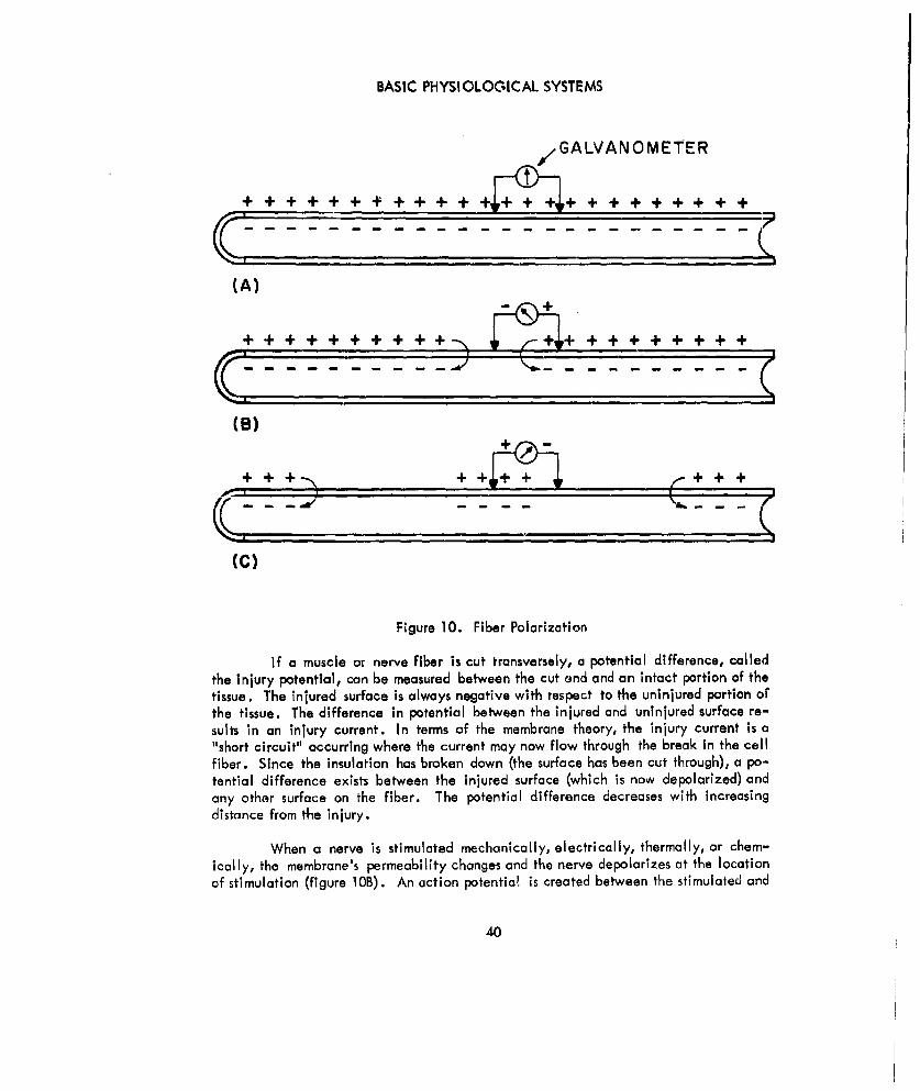
BASIC PHYSIOLOGICAL SYSTEMS
GALVANOMET ER
(A)
+ + + + + + + + + + . . . ..
(B)
~~+ + + +++
(C)
Figure 10. Fiber Polarization
If a muscle or nerve fiber is cut transversely, a potential difference, calledthe injury potential, can be measured between the cut end and an intact portion of thetissue. The injured surface is always negative with respect to the uninjured portion orthe tissue. The difference in potential between the injured and uninjured surface re-suIts in an injury current. In terms of the membrane theory, the injury current is a"short circuit" occurring where the current may now flow through the break in the cellfiber. Since the insulation has broken down (the surface has been cut through), a po-tential difference exists between the injured surface (which is now depolarized) andany other surface on the fiber. The potential difference decreases with increasingdistance from the injury.
When a nerve is stimulated mechanically, electrically, thermally, or chem-ically, the membrane's permeability changes and the nerve depolarizes at the locationof stimulation (figure lOB). An action potentia! is created between the stimulated and
40

BI OELECTRI C MEMBRANE CHARACTERI STI CS
unstimulated areas of the same fiber (similar to an injury potential). The action poten-tiol acts as a stimulus to adjacent areas of the fiber and, in the form of a wave, travelsalong the nerve fiber as a nerve impulse.
Soon after depolarization, constructive forces within the body (homeostasis)restore the membrane to its original state of polarization and irritability. The energyfor the recovery process and for maintaining polarization is furnished by metabolicprocesses.
The stimulation of a muscle results In an action potential similar to that of anerve, except that muscle impulses travel at a slower rate than do nerve impulses.
II. Excitation
A. Stimulus for Producing Excitation
The stimuli to which muscles and nerves respond may be caused by any changein the environment. The nature of the stimuli may be electrical, mechanical (pinch-ing), thermal (contact with something hot), chemical (placing salt on a muscle), optical,sonic, etc. However, every stimulus does not activate tissue. For the stimulus to beeffective (1) it must be of sufficient Intensity, (2) it must last for a sufficient length oftime, and (3) it must have a sufficient rate of change of intensity. The minimum ettec-tive values of these factors are interrelated,
If small electric currents are used to stimulate a muscle, no reaction occursuntil they are of sufficient intensity. If the duration of the stimulating current is ex-tended, the current fails to produce any additional stimulation as long as it Flows con-tinuously, because the third requirement, sufficient rate of change of intensity, is notsatisfied. When the current is interrupted, another stimulation, termed the break shock,occurs because again all three requirements are satisfied. The minimal duration ofcurrent necessary for stimulation decreases with the intensity of the current.
A subminimal stimulus, by definition, does not depolarize nerve fiber mem-brane sufficiently to cause a reaction, but since ions do not return to the partially de-polarized area immediately, a series of subminimal stimuli may cause a reaction if theyoccur in rapid succession.
B. Refractory Period
After a neuron or muscle cell has responded to stimulation, there is a briefinterval during which it cannot again be activated, no matter how intense the stimulus.This interval is known as the absolute refractory period. During this time, the depolar-ized area becomes polarized sufficiently to respond again to stimulation. The repolar-ization rate is more rapid in nervo cells than in muscle cells.
41

BASIC PHYSIOLOGICAL SYSTEMS
A relative refractory period follows the brief absolute refractory period. Dur-ing this period, the narve fiber or muscle may be activated, but only by a stronger thannormal stimulus. A stronger stimulus, therefore, activates a fiber at shorter intervalsthan a weaker one, which, essentially, increases the frequency response. Increasing thestimulus intensity also may stiriiulate more fibers.
The Intensity of the response in a nerve is independent of the intensity of thestimulus. As a result, some other characteristic must be responsible for differentiatingbetween weak and strong stimuli. This other factor is the frequency of the response.A single impulse may cause a muscle twitch, but a stream of impulses through a nervefiber holds a muscle in contraction.
The velocity of the impulse through +he nerve fiber also is independent of thestrength of the stimulus. The velocity of conduction is no faster for strong stimuli thanfor weak stimuli, being solely a function of the size and type of fiber.
Fibers are classified on the basis of function, and physicai properties such asaxon diameter and the presence or absence of myelin (a fatty sheath which acts as anInsulator). Large fibers are more heavily myellnated, conduct faster, and yield largerresponses. Large fibers, called A fibers, subserve the somatic (sensory and motor) sys-tem. Medium sized fibers (B fibers) subserve visceral sensation and muscle. C fibers,smIa!ett ef the three, Me cnce.rned wth the arception , oft'ir mn! wa'rmth. $Iwclassifications overlap considerably, but, generally, A fibers are approximately I to 20microns in diameter, B fibers are less than 3 microns in diameter, and C fibers are lessthan 1 micron in diameter. All three may run together in a single bundle or nerve.
Since the velocity of conduction is dependent on the diameter of the axon ofthe neuron, a nerve impulse travels fastest in an A fiber, slower in a B fiber, and slow-est in a C fiber; the thicker the axon the faster the conduction. The velocity of con-duction in an A fiber varies from 5 to 100 meters per second while the C fibers conductat less than 2 meters per second.
C. All-or-Nothing Law
The all-or-nothing law states that provided a stimulus is strong enough topropagate an impulse and cause a response, the speed of conduction of the impulse andthe magnitude of the response are independent of the magnitude of the stimulus. Thislaw can be clarified with an analogy. Consider a number of thyratron electron tubesconnected in cascade so that the output pulse of each thyratron triggers the next thyra-tron. An impulse (input pulse) must be greater than a minimal amplitude to fire thetube, but no matter how much the input pulse exceeds this minimal amplitude, the tubewill not produce more plate current and the current will not travel faster.
This all-or-nothing law applies to every nerve fiber, but not to the nerve.The nerve is made up of many nerve fibers with different thresholds of excitability.
42

BI OELECTRIC MEMBRANE CHARACTERISTICS
Ther'fore, stimulating a nerve with electric shocks of increasing intensity provokeslarger and larger action potentials until all the nerve fibers are activated. At thispoint, the responses cannot increase.
Within neurons, the all-or-nothing law applies only to the main conductingportion of the axon. Graded response mechanisms mediate excitation at each end ofthe conducting fibers, providing greater flexibility to the nervous system than would beachieved by the all-or-nothing type of response.
As mentioned previously, the energy for the propagation of an impulse origin-ates in the nerve and not in the stimulus. Within limits, when the nerve has a normalenergy supply, the stimulated impulse is normal, and when the transmission is impairedlocally, the response is impaired locally. For example, when a nerve Is impaired bynarcotics or lack of oxygen, its excitability is reduced. If only a section of the nerveis impaired, the response from this section only is subnormal, and areas of nerve fiberon either side give full response. There is no progressive decrement as on impulsepasses an impaired section of nerve fiber; the size of the impulse at any location isdependent only on the state of the fiber at that location.
Referring again to the analogy of the cascaded thyratrons, the amplitude ofthe output pulse of the last thyratron is the same as the output pulse of the first tube inthe series. If one thyratron in the chain were defective so that its output pulse werereduced, the output would either be sufficient or insufficient to fire the next thyratron.If the pulse were too small, the impulse would stop; however, if the output pulse of thedefective tube is sufficient to fire the next tube in the series, the output of the finaltube would be identical to the output pulse of the first tube without any accumulateddecrement, just as occurs in the propagation of nerve impulses in the body.
PHYSIOLOGICAL VARIABLES IN ABNORMAL ENVIRONMENTS
In a steady-state environment, the normal individual is adjusted physiologically tohis surroundings. In a new environment, physiological adjustments must occur. If theenvironmental change is extreme, death could result; if the change is less drastic, var-ious physiological changes adapt the body to survive.
Of the many abnormal environmental factors which may confront man in space, themost critical have been selected for discussion: atmospheric composition and pressure,gravitational f orce s (acceleration and weightlessness), temperatures, radiation, andnoise and vibration. Although not an environment, emotional stress has been includedbecause it is an important condition to which the physiological systems of the bodymust adjust.
Although each environmental factor is discussed separately, special emphasis isgiven to respiratk , heart activity, body temperature, blood pressure, and skin resist-ance, since these physiological responses must be monitored to determine the adaptationof man to the environment.
43

BASIC PHYSIOLOGICAL SYSTEMS
I. Atmospheric Composition and Pressure
The physical environment of man on earth, to a large extent, is the result of thecomposition and pressure of the atmosphere. Man, therefore, is greatly affected by anychanges that occur in the composition and pressure of the air he breathes. In anyexposure to abnormal pressures and air compositions, the most important physiologicalchanges are likely to occur in the respiratory and circulatory systems.
A. Respiration
The humcn respirarory ystem is affected by the partial pressure of the oxygenwhich is inhaled, the barometric pressure, and the partial pressure of the corbon dioxidein the environment.
1. Effects of Oxygen Partial Pressure
With increasing altitude, the barometric pressure decreases logarithmic-ally, while the composition of the atmosphere remains relatively constant. For example,at 18,000 feet the barometric pressure is approximately half that at sea level,while theair still contains approximately 20 percont oxygen. The partial pressure of oxygen atthat altitude, therefore, is only one half its value at sea level. The normal sea-levelpartial przuc f -;-)-cn T19lrn r of mzrcury. 1'
gradually, man encounters symptoms of hypoxia (inadequate supply of oxygen) such assleepiness, headache, lassitude, altered respiratory activity, increased heart rate, im-pairment of thought processes, inability to perform simple tasks, and eventual loss ofconsciousness. To forestall the onset of hypoxia, the respiratory system automaticallymakes a partial but important compensation by increasing pulmonary ventilation. Thisincrease in the depth of inspiration (hyperpnea) varies inversely with the partial pres-sure of oxygen. If the oxygen pressure continues to decrease, the rate and depth ofbreathing increase, causing a state of hyperventilation. The partial pressure of oxygenin the arterial blood, therefore, increases far above the level it would have reached ifhyperventilation were not present. During prolonged exposure to slight oxygen defi-ciencies, the number of red cells in the blood and the blood's hemoglobin content in-crease. These increases result in a greater oxygen-transporting capacity which helpsto reduce the effect of the drop of oxygen pIessure. The affinity of hemoglobin foroxygen also is reduced by low partial pressures of oxygen, thereby favoring the un-loading of oxygen to the tissues during hypoxia.
Despite the attempts of the body to acclimate to low oxygen partial pres-sures, there are limits to the adjusting capacity of the respiratory system. Figure 11indicates the region of man's tolernnc- to variations in the partial pressurc of oxygenin the air he breathes.* The lower band represents the minimum partial pressure of
*Reference 57
44
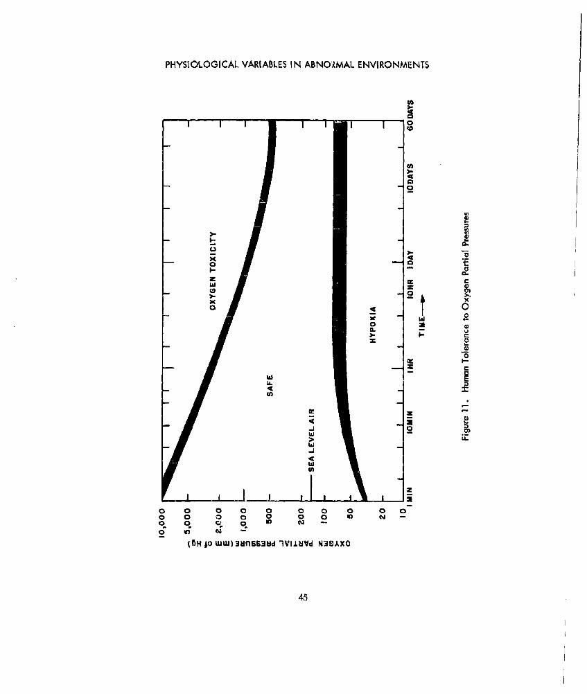
PHYSIOLOGICAL VARIABLES IN ABNO,MAL ENVIRONMENTS
0
0- t
z
wa
0 0z 0
00
C--
0
In-
w --
4LL
fa-
IAJ
o 45
0 00
o 0
455

BASIC PHYSIOLOGICAL SYSTEMS
oxygen that can be tolerated in the air entering the lungs. Any physiological reactionwould depend, of course, both upon the pressure of the oxygen and the rate of penetra-tion into the intolerable region.
A continuous lack of oxygen results in hypoxia and eventually uncon-sciousness or death. The onset of hypoxia may be detected by electroencephalographictechniques. Test, have shown that a lack of oxygen increases the amplitude and de-creases the frequency of brain waves prior to the time the subject loses consciousness.An electrical wavelength-measuring network can be used to determine when a train offour or more 5- to 7-cycle-per-second waves occur. The detection of these waves is asignal of possible oncoming hypoxia. Psychomotor changes in the early stages of hy-poxia would be difficult to detect, since behavior patterns do not change uniformly;periods of lethargy alternate with periods of attention. The major deterioration oftenis in personality rather than in performance.
2. Effects of Barometric Pressure
Man can survive in an abnormal environment in which the pressure is re-duced if the partial pressure of oxygen is maintained within his tolerable limits.Therefore, a man may breathe environmental air to an altitude of approximately 10,000feet. By increasing the concentration of oxygen in the inspired air to compensate forthe reduced barometric pressure (and consequently oxygen partial pressure), man may6rearte comfortably up to altitudes of 33,000 feet. However, even breathing 100 per-cent oxygen is inadequate above 40,000 feet unless the oxygen being breathed is pres-surized. Under these low barometric pressures, oxygen under pressure must be breathedand the body must be placed under pressure to counteract the internal pressure of thelungs. Above 63,000 feet, a full pressure suit or hermetically sealed chamber must beused since, at this altitude, the barometric pressure is approximately 47 millimeters ofmercury. At this pressure, the body fluids boil at body temperature. This boiling is notthe type associated with a pot of water on a kitchen stove, but rather a physical proc-ess involving bubble formations in the body.
3. Effects of Decompression
Hermetically sealed capsules or full pressure suits permit the respiratorysystem to function properly when ambient barometric pressures are reduced greatly, butanother problem is introduced. If transition into excessively low partial pressures ofoxygen occurs rapidly, the intermediate symptoms of hypoxia (sleepiness, headache,psychological impairment, etc) may be bypassed and the subject may lose consciousnessrapidly and undergo spasms or convulsions. If the environmental pressure is reducedrapidly along with the oxygen pressure (as would happen if a meteorite punctured aspace capsule), the subject would suffer sudden decompression. The severity of rapiddecompression and its effects upon the body are dependent upon the change in pressurewhich occurs and the time in which this change occurs. Within the body, solids andliquids are affected only slightly, but gases attempt to expand as rapidly as the pressure
46
PHYSIOLOGICAL VARIABLES IN ABNORMAL ENVIRONMENTS
falls (Boyle's law). Wherever internal gases cannot escape, the tendency to expandresults in an increase in volume, depending on the elastic resistance of the surroundingorgans and tissue. The expansion of gases in the intestinal tract (normally between 1and 1-1/2 liters at sea level) may not only induce intense discomfort and pain, but alsocirculatory and respiratory impairment because of the displacement of the diaphragmand disturbance of blood flow in the abdomen. The fragility of pulmonary tissue couldresult in damage to the lungs if they are full of air at the time of decompression. Thedecompression rate of the lungs is limited inherently by the anatomical structure of therespiratory air passageways, which limits the rate of decompression that can be toler-ated without injury.
4. Effects of Excessive Oxygen
Not only is it dangerous to be exposed to o lack of oxygen for any periodof time, but excessive oxygen also can be harmful. Prolonged exposure to one atmos-phere of pure oxygen, for example, results in inflammation of the lungs, coughing, gasp-ing, pulmonary congestion, numbness of the fingers and toes, and nausea. Exposure tostill higher partial pressures of oxygen produce nervousness, discomfort, irritations of theeyes, nausea, loss of consciousness, and convulsions. Oxygen partial pressures shouldbe kept between 100 and 350 millimeters of mercury for comfort and efficiency. Thesituation becomes critical when the oxygen pressure falls below 60 millimeters of her-r,,ry. When the partial pressure of oxygen exceeds 500 millimeters of mercury forextendeJ periods of time, the oxygen concentration is toxic. This upper iimit is indi-cated by the upper curve in figure 11.
5. Effects of Carbon Dioxide
Man during a normal breathing cycle produces carbon dioxide at thesame rate as he consumes oxygen. Partial pressures of carbon dioxide (in the inspiredair) above 15 to 20 millimeters of mercury increase the breathing rate, distend the airsacs of the lungs, and impair the normal gas exchange between the blood and the airsacs in the lungs. These carbon dioxide partial pressures are equivalent to a concen-tration of 2 to 3 percent at standard atmospheric pressure. Partial pressures above 35millimeters of mercury (equivalent to 5 percent carbon dioxide at one atmosphere) canbe tolerated for only a few minutes, after which the subject experiences heavy panting,marked respiratory distress, fatigue, narcotic effects, unconsciousness, and eventualdeath. The partial pressure of carbon dioxide should be kept below 7 millimeters ofmercury if the subject is to be exposed for long periods of time. Figure 12 illustratesthe safe exposure time for various partial pressures of carbon dioxide.*
B. Circulation
The circulatory system also is affected by environmental pressure and the
*Reference 57
47
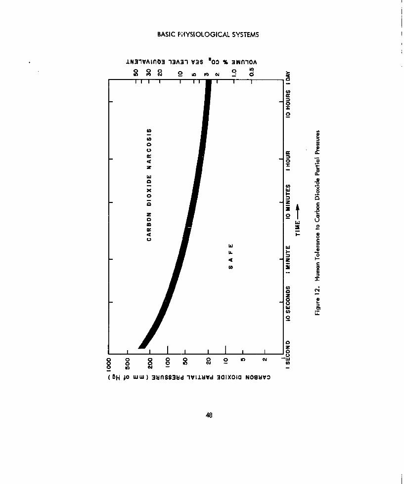
BASIC FHYSIOLOGICAL SYSTEMS
.LN31VAiAO3 13A31 V3S '00 % 3wiA
2 0- 0
2
goV
00
00
0oc
x I-
00
0 0 0
zz
0~
ot2
z
0
48Z

PHYSIOLOGICAL VARIABLES IN ABNORMAL ENVIRONMENTS
composition of inspired air. In fact, the change in oxygen and carbon dioxide contentof the circulating blood causes the respiratory changes.
During hypoxia, the oxygen saturation of the arterial blood falls. For ex-ample, if the subject were exposed to oxygen partial pressures equivalent to breathingenvironmental air at 20,000 feet for 10 minutes, the oxygen' saturation of arterial bloodwould fall to about 70 or 75 percent of normal. However, this moderate lack of oxygenwould cause the heart rate to increase by4O percent to speed the flow of oxygen to thecells. If pure oxygen were breathed at this point, the oxygen saturation of the arterialblood and the heart rate would return to normal within 1 minute.
Low oxygen partial pressures or excesses of carbon dioxide in the inspired airfor prolonged periods of time result in deeper, faster breathing (hyperventilation), whichattempts to increase the partial pressure of oxygen In the arterial blood. The numberof red cells in the blood and the hemoglobin content of the blood increase to improvethe oxygen-carrying capacity of the blood. If the oxygen saturation of the blood con-tinues to drop or carbon dioxide content to increase, some pulmonary vasoconstrictionoccurs so that the flow of blood is directed in the lungs to the alveoli where the oxygentension is greater than the oxygen tension of the lungs as a whole. The excitability ofthe heart muscles and the cardiac center of the medulla become depressed and the rateand force of the heart beat are decreased.
A marked oxygen lack also affects the nervous system and causes vasodilationof the peripheral circulatory system. A moderate excess of carbon dioxide in the cir-culating blood acts directly on the vasomotor center and causes vasoconstriction, whilea great excess of carbon dioxide in the blood causes vasodilatlon of the peripheral cir-culatory system.
II . Gravitational Forces
The gravitational pull of earth is a force that makes a Free-falling bodyfall towardearth at a constant accelerating rate of 32.16 feet per second per second. This forceis expressed as 1 G. Any body that is accelerating at a rate of 32.16 feet per secondper second, regardless of the propelling force, is described as being subjected to a forceof 1 G. The greater the rate of acceleration, the greater the G forces. For example,a body traveling at 64.32 feet per second per second is exposed to a force of 2 G's.
On earth, man is accustomed to mild accelerations, such as increasing speed in anautomobile or riding in an elevator, and to mild sensations of weightlessness, such asthose experienced when submerged in water or in simulated free fall in amusementrides. However, man in the cbnormal environments of space vehicles or high-altitudeaircraft is faced with short-duration exposures to high acceleration forces and longterm exposures (in the order of days, weeks, and possibly months) to zero-G forces(weightlessness).
49
BASIC PHYSIOLOGICAL SYSTEMS
A. Acceleration
Accelerations of 2 G's make it difficult for man to walk and his h-Inds andfeet become heavy. The act of crawling becomes difficult, and walking and climbingbecome impossible under forces caused by 3 G's acceleration. At 4 G's, man is ableto move only with great difficulty, and when the forces increase because of accelera-tions of 5 G's, only slight movements of the head and arms are possible.
Prolonged exposure to excessive acceleration forces could result in uncon-sciousness and eventual death. The tolerable levels of acceleration to which man maybe subjected are dependent on his position relative to the direction of acceleration.(Refer to figure 13 for the terminology of acceleration..) Acceleration forces directedalong the major axis of the body (i.e., from head to foot or vice versa) are calledlongitudinal accelerations. Longitudinal accelerations cause the blood to pool in theextremities, either head or feet. Transverse accelerations are perpendicular to thespinal column. Positive transverse acceleration (forward acceleration), or, as pilotsrefer to it, "eyeballs-in" acceleration, pushes the subject back into his seat. Negativetransverse acceleration (backward acceleration), or "eyeballs-out" acceleration, actsto push a pilot out of his seat. Positive transverse accelerations are better toleratedthan negative transverse accelerations, since it Is much easier to contour a form-fittingposterior restraint away from the functional ventral side of the body.
The three body systems which limit man's tolerance to acceleration are thevisual, cardlovwscular, and respiratory systems. Visual disturbances are encounteredprimarily in negailve transverse accelerations. These disturbances are not Incapaci-tating, but they do affect visual acuity, probably as a result of the pressure of the eye-lids on the cornea distorting the corneal surface. Tears, which sometimes becomeexcessive, also are a problem. Longitudinal accelerations grossly affect the cardio-vascular system by pooling blood in the extremities. Seatward accelerations whereblood is pooled in the lower extremities and circulation to the head is impaired areespecially objectionable. Transverse accelerations are directed perpendicular to themajor arteries and veins of the body, so they do not affect the circulatory system asmuch as the respiratory system. The forces caused by transverse accelerations make itdifficult to expand the chest to inhale. Most people cannot inhale environmental airwhen subjected to transverse accelerations in excess of 6 G's. Table I lists the rangeof short-duration accelerations at which various adverse effects are noticed.
Tolerance to acceleration varies not only From individual to individual, but itvaries within the same person from day to day. Emotional itates have considerableeffect on an individual's G tolerance. High accelerations increase the speed of vas-cular responses and the level of central nervous system urousal If the level of centralnervous system arousal and the speed of vascular response already have been increasedby psychological factors prior to the onset of acceleration forces, the subject's toler-ance to acceleration obviously is lowered.
50
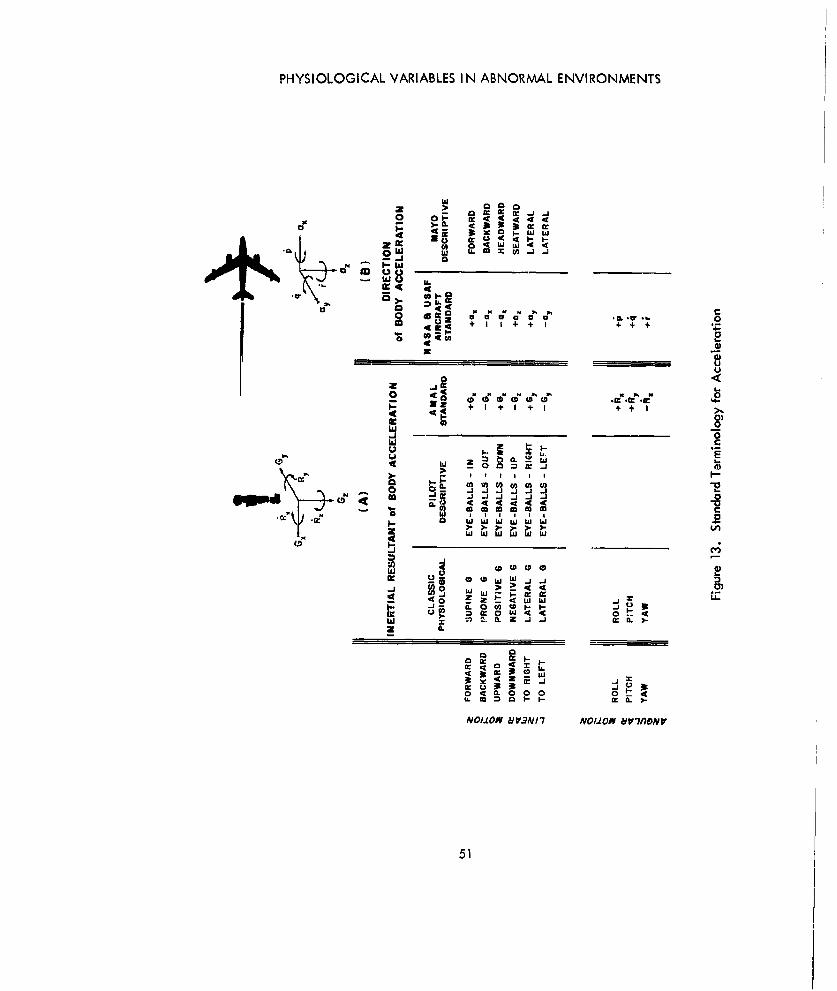
PHYSIOLOGICAL VARIABLES IN ABNORMAL ENVIRONMENTS
0 0Fw w
w~ w
5z. + + T
a a ino
_j0-i 'aeeeee
2
.=w;rN~DNN _ _ _
41I
j _j j j,
j.
w
)- > -- - > .LOqA.000w
PS/.0 w VNI wOII -7f jI
51 L
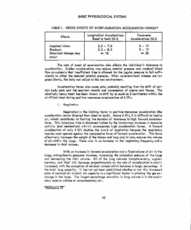
BASIC PHYSIOLOGICAL SYSTEMS
TABLE I. GROSS EFFECTS OF SHORT-DURATION ACCELERATION FORCES*
Effects Longitudinal Accelerations Transverse(head to foot) (G's) Accelerations (G's)
Impaired vision 2.5 - 7.0 0 - 17Blackout 3.5 - 8.0 0- 17Structural damage may - 18 " 30occur
The rate of onset of acceleration also affects the individual's tolerance toacceleration. Sudden accelerations may reduce arterial pressure and cerebral bloodflow so suddenly that insufficient time is allowed for the jugular pressure to fall suffi-ciently to offset the reduced arterial pressure. When accelerational stresses are Im-posed slowly, the body can adjust to the new environment.
Acceleration forces also cause pain, probably resulting from the shift of cer-tain body parts and the resultant stretch and compression of organs and tissues. Therelatively heavy heart has been known to shift by as much as 2 centimeters within theair-filled chest during positive transverse accelerations of 6 G's.
1 Reanir finn
Respiration is the limiting factor in positive transverse acceleration (theacceleration vector directed from chest to back). Above 6 G's, it is difficult to inspireair, which contributes to limiting the duration of tolerance to high forward accelera-tions. This tolerance time is shortened further by the involuntary increase in muscularactivity (and metabolism) w hich accompanies high acceleration forces. A Forwardacceleration of only 4 G's doubles the work of respiration because the respiratorymuscles must operate against the compressive force of forward acceleration. This forceeffectively increases the weight of the thorax and lung and, in turn, reduces the volumeof air within the lungs. There also is an increase in the respiratory frequency and adecrease in tidal volume.
With an increase in forward acceleration and a fixedvolume of air in thelungs, intrapulmonic pressures increase, increasing the relaxation pressure of the lungsand decreasing the tidal volume. All of the lung volumes (complementary, supple-mentary, and tidal air) decrease proportionately as thc rate of acceleration is slowlyincreased, with the exception of residual volume which becomes a larger percentage fthe total lung capacity. It has not yet been established whether or not this increasedratio of residual air to total air capacity is a significant factor in altering the gas ex-change in the lungs. The largest percentage reduction in lung volume is in the expir-atory reserve volume or complementary air.
*Reference "
52

PHYSIOLOGICAL VARIABLES IN ABNORMAL ENVIRONMENTS
Tests have shown that during forward accelerations exceeding 4 G's, therelaxation pressure even at zero lung volume is positive. This implies that completerelaxation of the inspiratory muscles at 4 Gis does not occur. A condition somewhatanalogous to this is negative pressure breathing. An example of negative pressurebreathing is breathing through a rigid tube while submerged in water. Few men havesufficient chest and respiratory muscles to breathe ambient air from the surface of thewater when submerged in deep water. Since forward acceleration exerts a conditionsimilar to negative pressure breathing, the tolerance to forward accelerations may beincreased by poitive presmure breathing.
Backward accelerations are better tolerated bythe respiratorysystem thanforward accelerations because the inertial forces of acceleration assist in increasingthe diameter of the chest during inspiration. Expiration is hindered by backwardaccelerations, but this reduced expiration is an advantage in that the chest is left ex-pandedwith a larger functional residual capacity. Maintaining the lungs in an inflatedcondition, typical of athletes during muscular exercise, and breathing "off the top" ofthe larger lung volume results in more efficient oxygenation of the blood in the lungs.However, despite this advantage, backward accelerations are more difficult to copewith because, although pressure breathing is needed, it is so much easier to contour fita posterior restraint for forward accelerations.
Longitudinal accelerations have not been discussed, since they arelimited by the circulatory system rather than by the respiratory system.
2. Circulation
The most favorable positions for resisting the effocts of abnormal accel-erations on the circulatory system are those in which the acceleration vector is directedacross the major axis of the great blood vessels, i.e., transverse accelerations. How-ever, longitudinal accelerations (accelerations parallel to the great vessels) must bewithstood for short periods and, consequently, the effects of both types of accelerationon the circulatory system must be considered.
Generally, during exposure to acceleration forces, diastolic pressureschange considerably mare than systolic pressures. Diastolic blood pressures decreaseduring subgravity states and increase during periods of acceleration, probably as a resultof resistance changes to the outflow of blood from the peripheral vascular beds andcardiac output changes in response to the demands made on the cardiovascular systemby changes in hydrostatic pressures. Pulse pressures, conversely, increase during sub-gravity exposures and decrease during exposure to high acceleration forces.
During exposure to acceleration forces, the venous pressure is increasedin the thorax and in the abdomen. This increased pressure is not transmitted to theperipheral veins, since the valves in the veins leading into these body cavities appar-ently are closed. The venous pressure increase, therefore, must result from changes
53
BASIC PHYSIOLOGICAL SYSTEMS
within the extremities. When acceleration forces obstruct the return flow of blood fromthe extremities, the continued flow of blood frnm the arteries and the increase in intra-muscular pressure probably combine to increase the venous pressure. The speed withwhich the venous pressure rises depends on the rapidity and effectiveness of peripheralvasoconstriction,
Immediately following exposure to high accelerations, blood pressure de-creases because of the abrupt withdrawal of external pressures on the body. The venousblood in the systemic circulatory system is under higher than normal pressure after theacceleration ceases and the return of venous blood aids in the restoration of preaccel-eration blood pressure levels.
The heart rate decreases during subgravity exposures and increases duringexposures to acceleration; the higher the acceleration level, the faster the heart rate.
The tolerance of circulatory systems to acceleration differs widely amongindividuals. There is no correlation between a man's acceleration tolerances and hisblood pressure or blood sugar level, or any other measurable characteristic. Trainingapparently does little to increase man's tolerance to high accelerations. Tests haveshown that while an inexperienced subject responds much sooner and more vigorously toinitial acceleration stimuli than experienced subjects, the response differences dis-appear during exposure to high accelerations as both types of subjects approach maxi-murm response.
Headward accelerations tend to pool the blood in the extremities. Thisloss of blood to systemic circulation is offset partially by an increase in intro-abdominalpressure, which reduces the total capacity of the blood vessels supplying the viscera.With less blood available for circulation, blood pressure in the upper extremities isreduced. If the arterial pressure Falls below the value necessary to force the blood tothe cerebral cortical level and keep the carotid vessels and the cerebral vascular bedopen, or if the blood flow is not sufficient to oxygenate the retina and the cerebrum,blackout and unconsciousness result.
A blood pressure of 20 millimeters of mercury at eye level is necessary toprevent collapse of the retinal vessels as a result of intraocular pressure. When ex-posed to an accelerating force of 1 G (typical of standing still on the earth's surface),a systolic pressure below 65 millimeters of mercury measured in the arm at the level ofthe heart is not compatible with normal visual functions, and unconsciousness occurs atsystolic pressures below 40 millimeters of mercury. (There is a high correlation betweenpulse rate and blood pressure at eye level, which may provide a means for predictingblackout.) Blood pressure increases during longitudinal accelerations, but it requiresmore pressure to supply the head with sufficient blood to maintain visual functions. Forexample, a mean pressure of approximately 112 mil limeters of mercury at heart level isnecessary to prevent blackout during exposure to 4 G's directed headward. Besidesvisual disturbances, cardiovascular abnormalities, such as cardiac arrhythmias (absence
54

PHYSIOLOGICAL VARIABLES IN ABNORMAL ENVIRONMENTS
of rhythm) and petechial hemorrhages, occur during longitudinal accelerations. Thesedisturbances or abnormalities do not cause any serious loss of ability to operate controlsand react to stimuli, but considerable discomfort may be present.
The rapid onset of longitudinal acceleration, particularly above the 3-Glevel, results in temporary insufficient circulation to the head, which causes dramaticcardiovascular reactions. These reactions include changes in blood flow and decreasesin right atrial pressure, intrathoracic pressure, and oxygen saturation of arterial bloodproportional to the magnitude of the acceleration. These compensatory reactions arecompleted largely within the first 15 seconds of exposure. Vision also may be affected,but experiments have shown that clear vision is regained rapidly; usually within 15seconds after a constant acceleration level is reached.
Cardiac output increases by as much as 20 percent during short exposuresto longitudinal accelerations of less than 4 G's; however, increasing the exposure fromI minute to 10 minutes does not increase the cardiac output above the 1 minute expo-sure level, although the heart rate continues to increase during the prolonged txposureto longitudinal acceleration.
Transverse accelerations arc more tolerable to the circulatory system.Pooling of blood in the extremities is not as common in transverse accelerations as it isin longitudinal acceleration, but some pooling does occur. A general increase in vas-cular bed capacity and the slight pooling of blood are sufficient to drop the bloodpressure below preacceleration levels for a short time after the termination of exposure.Cardiac output is unaffected by transverse accelerations, even d u r i n g exposure toacceleration forces for as long as 10 minutes. Heart rate continues to increase through-out exposure; for example, during 5-G positive transverse acceleration, the heart rateincreases by 80 percent in 10 minutes. Oxygen saturati3n of the arterial blood de-creases, but much of this decrease can be avoided by breathing pure oxygen.
B. Weightlessness
Weightlessness occurs during the orbiting maneuver of a space vehicle whenthe vehicle's acceleration produces a centrifugal force which balances the gravita-tional force. This abnormal environment cannot be simulated in the earth's atmospherefor more than a few minutes. Until man is exposed to weightlessness (zero G) for asubstantial length of time, it is uncertain how the physiological systems of the bodywilladapt to this environment.
There are three physiological factors which are of concern: (1) the capabilityof the subject to function and fulfill a mission while weightless, (2) the temporaryphysiological changes occurring during and following the transition periods of entry toand departure from the zero-G state, and (3) the long-term physiological changes re-sulting from the departure from prolonged periods of zero G. Recent data collectedfrom animals and man in orbiting vehicles indicate that weightlessness can be adapted
55
BASIC PHYSIOLOGICAL SYSTEMS
to for relatively short periods of time without pathological aftereffects.
A mnjor problem which confronts a weightless man appears to be disorienta-tion. The human body is equipped with several sense organs which serve as spatialorientation mechanisms. The most commonly recognized sense organs providing thebody with knowledge of its position in its envircnment are located in the inner ear.Other sense organs also serve exteroceptive functions (react to external forces such asgravity), although their dominant function is to provide sensory information on the ten-sion conditions of the skin, muscles, and ;onnective tissues (proprioceptive). Thesekinesthetic receptors are the nerve endings in muscles (muscle spindles), especiallyantigravity (postural) muscles and the nerve endings in connective tissue (Paciniancorpuscles). When the body is exposed to zero-G accelerations, the proprioceptivefunction of the sense organs is not disturbed, but the exteroceptive function of themechanoreceptors Is eliminated. To compensate for this loss of sensory information,the body must rely on vision to provide positional information. If sufficient visual cucsare provided, disorientation apparently does not occur in the weightless state.
Many of the mechanoreceptors have strong reflex connections with the auto-nomic nervous system, which controls circulation, motion of the stomach, intestines, andother body functions. During abnormal motion conditions, as in an airplane or on aship during rough weather, these reflex connections manifest themselves In the form ofnausea or motion sicknesq. HowvAr, dring 7,arn-C n.r.ol~rntions, uwh nhnormnl ex-c;tation of these receptors does not occur and few subjects experience nausea. Testswith animals in rocket flights and orbiting satellites indicate that the gravireceptors,after a short conditioning period, reach a new equilibrium. These tests also indicatethat although no disturbance occurs to the respiratory system, significant changes mayoccur to the cardiovascular system.
Short-duration tests of man in the weightless condition under water and inparabolic flights unfortunately have been too short for the body to acclimate totally toits new environment, but tests have shown that man can operate controls, drink water,and micturate with little difficulty. Regurgitation tendencies noted during human ex-periments indicate the need for dietary control with respect to fluid volume per Feed-ing. The dispersion of liquids during weightless feedings may require sucking liquidfrom a bottle to avoid aspiration in the sinuses and respiratory tract. Just as the gas-trointestinal system functions without the benefit of gravitational forces, the excretorysystem also functions during weightlessness. The elimination of body waste solids andliquids is a function of the elasticity and tensional tone of visceral tissues, voluntarymuscular function, sphincter action and, to a slight extent, gravitational forces. How-ever, provision must be made for the elimination of body waste products with ease andcomfort to the subject.
The limited exposure to weightlessness which man has experienced and themuch longer exposures of primates indicated no significant alterations in heart rate or
56

PHYSIOLOGICAL VARIABLES IN ABNORMAL ENVIRONMENTS
in eluctrocardiographic pattern. Since exposure to weightlessness can only follow ex-posure to high acceleration forces, a slight log can be expected in the return to normalheart rate from tachycardia induced by the acceleration forces because of the "con-trast effect" of suddenly changing from one abnormal environment to another. Somedegree of overfilling of the right side of the heart, shifts in the venous pooled blood,and minor changes in the electrical and anatomic cardiac axes also may occur, butthese abnormalities are transitory, and normalcy should return during the first few min-utes of weightlessness.
The dog Laika in the Russian satellite, Sputnik II, was exposed to weightless-ness For several days. The only effect noticed (or at least reported by the Russians) wasa slight tachycardia during the acceleration phase of the flight, with no significantphysiological changes occurring during weightlessness.
Ill. Temperature
Man's tolerance to temperatures should be easy to establish, since it is relativelysimple to simulate abnormal temperature environments; however, temperature is a com-plicated variable. Man's tolerance to high temperatures also is a function of humidity,and his tolerance to low tempirature is dependent on wind velocity.
Figure 14 graphically illustrates man's time tolerance to Anvironmn!'nl fomrnr-
turts.* The transition regions , I low for uccliaululioon, W rid velocity, physicalactivity, and protective clothing, but, because of large individual differences (primar-ily due to acclimatization), some men greatly exceed these tolerances. In plotting thehigh-temperature curves, adequate drinking water was assumed to permit man to main-tain the necessary fluid level to support the body-cooling process of perspiring. With-out adequate drinking water, the limits set by the curves will not be reached.
Exposure to high temperatures may cause lassitude, loss of efficiency, weakness,headache, inability to concentrate, increased cardiac output and oxygen consumption,and apathy. If the temperatures exceed man's physiological limitations, he is subjectto nausea, visual disturbances, hyperthermia, heat stroke, and convulsions. The subjectalso will become dehydrated if deprived of adequate drinking water during exposure tohigh temperatures.
Man's physiological tolerance to cold temperatures is affected by the velocity ofthe wind. During exposure to cold temperatures without wind, the body surrounds it-self with a Film of static insulating air. Wind disperses this protective layer of warmair and helps to decrease the skin temperature. Protective clothing acts to trap an in-sulating layer of air around the body and increase man's tolerance to cold temperatures
*Reference=
57
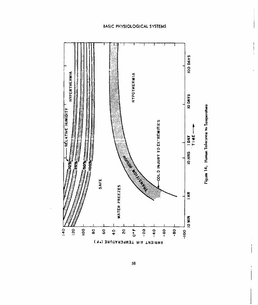
BASIC PHYSIOLOGICAL SYSTEMS
L 1
4 0
V",,"CC.
m i C
w 2. ~ ~ ~0 . ...
Iw A
wJ
(zo 381UdNI NIV IN31INV
58

PHYSIOLOGICAL VARIABLES IN ABNORMAL ENVIRONMENTS
despite the wind. The low-temporature curve shown in figure 14 illustrates man's tol-erance to cold when wearing the warmest clothing that can be worn without relinquish-ing the ability to move about freely clothlng about 1 inch thick).
When the body is unable to produce sufficient heat to maintain normal body tem-perature by metabolic processes, the heat production of the body is augmented byshivering. Tests have shown that a nearly nude sedentary man can produce up to 425calories of heat per hour through shivering. These tests also have shown that increasingthe velocity of the wind has a relatively greater effect on the body's heat productionthan decreasing the dry bulb temperature. Figure 15 shows the heat produced by nearlynude sedentary men while exposed to low temperatures and wind velocilies from lessthan 1 mile per hour to 10 miles per hour. During heat productions of about 400 calo-ries per hour, the subjects are shivering violently and uncontrollably. Any increase inactivity aids in increasing the heat production of the body and, therefore, man's toler-ance to cold.*
*Reference 36
450 ,.,, .
400 WINOn VPiOCITY
350
300
z*o 10 MPH1- 2500
a. -0 MPH '
kiS150
100
0
I0 20 30 40 50 so 70 so 90 100AMBIENT TEMPERATURE (OF)
Figure 15. Heat Production of Nearly Nude M~an
59

BASIC PHYSIOLOGICAL SYSTEMS
When exposed to cold temperatures, man may suffer from exhaustion, local freez-ing, depression of respiration and circulatory functions, hypothermia, paralysis, anddeath.
IV. Radiation
The injurious effects of ionizing radiation have been recognizeJ since the discov-ery of X-rays. Radiation alters the structure of the atoms of the irradiated body cellsand causes symptoms which are no different than those of a variety of injuries. How-ever, unlike most Injuries, there is no immediate pain associated with overexposure toionizing radiation and weeks, months, or even years may elapse between the exposureto radiation and the manifestation of physiological changes.
A. Biological Effects
The biological effects of radiation may be direct or indirect; i.e., the effectsmay be localized within the Irradiated cells or they may be transmitted to body cellsremote from the point of Injury. These effects may be immediate or delayed. In theproduction of tumors and cataracts, for example, there is a latent period during whichno effects are observable.
Radiation can produce both strucoural and functional changes in cells andtls;ucs and, depending on the amount of ionization, the effects may he temporary orpermanent.
As the cells that make up the different tissues and organs of the body differ innature and appearance, so also do they differ In their response to radiation. The Lawof Bergonie and Tribondeau states that "the radlosensitivity of a tissue is directly pro-portional to its degree of differentiation." In other words, cells most active in repro-ducing themselves and cells not fully mature will be most harmed by radiation. Thefollowing is a partial list of cells, in the diminishing order of their radlosensitivity:white blood cells, alveoli (lung) cells, bile duct cells, cells of the tubules of the kid-neys, endothelial cells, structural cells that support organs, muscle cells, bone cells,and nerve cells.
B. Measures of Radiation
A quantity of radiation cannot be measured directly. Instead, it is measuredby the ionization produced by the passage of the radiation through a medium. Theunits of widest acceptance in physiological measurements are the roentgen and the rem.The roentgen is a measure of ionization in air due to X- or gamma-radiation, and therem relates the effectiveness of the different radiations in producing biological damageto the quantity of radiation.
60

PHYSIOLOGICAL VARIABLES IN ABNORMAL ENVIRONMENTS
1. The Roentgen
The roentgen is the standard unit for expressing quantities of X- andgamma-radiation. It is defined as the quantity of X- or gamma-radiation that willproduce 1 electrostatic unit (esu) of charge, either positive or negative, in one cubiccentimeter of standard air (0°C and 760 millimeters of mercury). It is also equivalentto the production of 2.083 x 10⁹ ion pairs per cubic centimeter of standard air.
2. The Rem (Roentgen Equivalent, Man)
The rem was established to compensate for the differences in Ionizationof the wide variety of radiations encountered in atomic energy research and space. Itis the quantityof radiation (of any type)which will produce the same biological effectsin man as those resulting from the absorption of 1 roentgen of X- or gamma-radiation.
The biological effectiveness of ionizing radiation cannot be measuredexactly, since it varies with the part of the body irradiated, the type of radiation, andmany other factors such as the age and sex of the Individual. However, by use of arelative biological effectiveness factor (RBE),a close approximation is possible. Tablesof RBE factors are readily available in books devoted solely to radioblology and there-fore are not presented in this handbook.*
C. Radiation Exposure
The tenure of radiation exposure is either chronic or acute. Chronic expo-sure is the absorption of relatively small amounts of radiation (up to 5 roentgens) dailyover a long period of time. A single exposure to radiation of 25 roentgens or moreconstitutes an acute exposure. For the prevention of radiation i n j u r y, permissiblelevels of radiation exposure have been set for chronic exposures. These permissiblelevels are based upon the effectiveness of the repair processes in the body after irradi-ation. if perissible levels are exceeded, the damage may be irreparable. Figure 16illustrates the basic permissible exposure rates of ionizing radiation to various parts ofthe body.
No permissible level for acute exposure has been established. Present knowl-edge indicates that 25 roentgens causes no observable reactions, 50 roentgens producesnausea and vomiting, 400 to 500 roentgens gives the individual a 50-50 chance of sur-vival without medical care, and 650 roentgens is lethal.
The reaction to radiation injury depends upon the radiosensitivity of the or-gans and tissues irradiated and the function of these organs and tissues in the body.
"Reference5
61
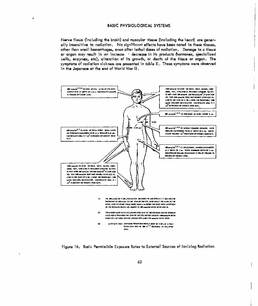
BASIC PHYSIOLOGICAL SYSTEMS
Nerve tissue (including the brain) and muscular tissue (Including the heart) are gener-ally insensitive to radiation. No significaint effects have been noted in these tissues,other than small hemorrhages, even after lethal doses of radiation. Damage to a tissueor organ may result in an increase, decrease in its products (hormones, specializedcells, enzymes, etc), alteration of its growth, or death of the tissue or organ. Thesymptoms of radiation sickness are presented in table 1I. These symptoms were observedin the Japanese at the end of World War 11I.
41). INK1 111In/ OM( NOT OVtKiLt AlitO Aol4,AND IN4 IVi CAAR Of X 004 rI DOW~k tIW11bsl s
-/.k IMLLUMI IACISCAtitt. SIONtiKAI AREA litII~mIINt 111O OFcttt 111014t U RAI
lOSE, AND 10 0400t: .t-SALLMOM tKARatMI ititOVMttNttCIOIOt~l~IMISONS1011 tot 00140 WITi AA #KANTli toLIA 1./4 -3i AMUGKX OFl 41.STDIA
SIlitAIAL IOSA IOC .. cogGO 00i Hill ~ tN 00%~ t i op~g~ i
UDL~tI~tiAT A10 MIItTI 01O1 .. II4ANDI'At...W'.AtItAOfD2lt.
lit ~ ~ ~ ~ SGNFCN AVtEPLUt tFOOt:D ISDML 10%llLtitL OFl OSVAI IN t l~A
(AOL(TOEGIO OFY UINS 901A4.DtE CL~L
150 0 SKN OFHEAD NIC, RADSto$.
Figre16. Bil ADasic PROIErmsil Exposure Rate toEtrnlSuce IoiigRaito
it NT OIA 0 -A (4 4 -/&" OW62D

PHYSIOLOGICAL VARIABLES IN ABNORMAL ENVIRONMENIS
TABLE II. CLINICAL SYMPTOMS OF RADIATION SICKNESS*
Time After Lethal Dose Median Lethal Sublethal DoseExposure (650 r) Dose (400 r) (250-100 r)
First week. Nausea, vomiting Nausea, vomiting Possible nausea, vomit-within 2 hours, after 2 hours. ing.
Diarrhea. No definite symptoms. No definite symptoms.Vomiting.Inflammation ofmouth and throat.
Second week. Fever. Loss of hair. No definite symptoms.Rapid loss in Loss of appetite, gen-weight. eral discomfort.Death.
Third week. Fever. Loss of hair.Severe reddening of Loss of appetite, generalmouth and throat, discomfort.
Sore throat.Pullor.Bleeding.Diarrhea.
Fourth week. Pallor. Recovery likely.Bleeding, diarrhea.Rapid loss in weight.Death (Mortality rateup to 50 percent).
D. Physiological Effects of Radiation
The effects of radiation on the various physiological systems of the body thatare most affected by radiation are described briefly in the following paragraphs. Nu-merous publications are available which present detailed biological effects of radi-ation.
1. The Skin and Hair
The skin is injured easily, but is remarkable for its capacity for local re-pair. Fortunately, most injured skin areas are relatively small compared to the whole
*Reference
63

BASIC PHYSIOLOGICAL SYSTEMS
area of the skin, leaving plenty of normal tissue to promote healing. Skin reaction,observed microscopically, is one of the first clinical indications of irradiation. Cellu-lar changes are recognizable within an hour after even small local exposures. Anexposure of about 140 rem produces an erythema (reddening) of the skin. This reactionindicates partially damaged skin with normal cells among the damaged. The normalcells immediately begin repair, and recovery is fairly rapid. Large dose (chronic oracute) irradiation of the skin may result in the formation of cancerous cells.
Radiation burns develop from exposure to large amounts of radiation inany form, although the most likely causes are alpha and beta radiation. They may bepreceded by symptoms of itching or tingling. There is no biological warning of radia-tion burns; a person may be exposed for hours unknowingly and may suffer permanentskin damage. Severe radiation burns may not heal for months. Radiation burns rarelydevelop from doses of 300 rem or less. The threshold single dose for erythema is about140 rem. Erythema is a danger signal. If it appears, the subject should not be exposedto further radiation.
Irradiation of the cells lining the hair follicles can stop the growth ofhair or cause temporary baldness (epilation). After a large dose (300 to 500 rem) ofbeta and alpha radiation, the hair begins to fall out in bunches.
2. The Circulatory System
The differences in the lifespan and radiosensitivity of the blood cells areimportant factors in radiation injury. When the body is irradiated, the first cells to bereduced in number are the white cells. (A deficiency of white cells is called leuko-penia.) Within a week after severe irradiation, the platelets (blood cells Important inbloodclotting) are reduced in supply, both because of damage to the cells which pro-duce them and because of injury to the platelets themselves. About 7 weeks afterirradiation, red cells of the blood are reduced. (A deficiency of red cells (erythro-cytes) is known as anemia.) Injury from acute exposure probably will heal withoutfurther exposure; injury from chronic exposure is likely to be Formanent.
The effects of reduced supplies of blood cells are manifold. Leukopeniareduces resistance to infection, and the body is susceptible to ordinarily harmless num-bers of bacteria. The loss of platelets results in the blood not clotting and open woundsnot healing. The effects of anemia are pallor, shortness of breath, fluttering of theheart, and general weakness.
The lymphatic system is affected by radiation. Lymph nodes, which nor-really filter out and destroy bacteria, show the first stages of hemorrhaging and infec-tion after acute irradiation. Blood passing into the lymphatic system from damagedblood vessels is collected at the nodes and the reduced number of white cells in thenodes permits bacteria to multiply and produce infection. The spleen which stores lym-phocytes (lymph cells) is affected in a similar manner.
64

PHYSIOLOGICAL VARIABLES IN ABNORMAL ENVIRONMENTS
Bone cells are relatively insensitive to external radiation; however,strontium 89 and 90 as well as plutonium 239 (ptxpularly called "bone-seekers") andother radioisotopes circulated by the blood can damage the bone cells as well as themarrow cells.
3. The Respiratory System
Radiation affects the lungs by damaging the alveoli (air sacs). The dam-age can result from penetrating external radiation, but the greatest hazard is radiationfrom inhaled dusts and vapors. Although only a small proportion of the material takeninto the lungs in one breath remains there when the breath is exhaled, an accumulationof these small amounts may cause severe injury.
Undissolved radioactive particles in the lung can result in the formationof tumors. Soluble materials may pass through the alveoli membranes into the blood tocause damage elsewhere in the body. If radiation creates toxic byproducts in remoteparts of the body, these substances may be carried to the lungs by the circulatorysystem.
4. The Endocrine System
.u.iutlon affcct: the adrcnal c!nd fl ,r!±rn ern vA nrann% nnd: toa lesser extent, the thyroid gland, liver, and gall bladder. When the cells of theadrenal glands are damaged by radiation, the body functions less efficiently while thetissues are repaired and replaced. The individual may be more susceptible to heat,cold, injury, and infection; his blood pressure may drop; and the balance of salts in hisblood plasma may be upset.
Exposure of the reproductive organs to large doses of radiation (about 400rem) may cause sterility. Exposures of about 100 rem severely damage the cells of thesex organs, and thereby slow the production of sperm. The sperm, however, is radiation-resistant, and irradiation of the sex organs does not destroy the sperm. The slowingdown of sperm production, because of injury both to irradiated cells and to the repairfunctions of nonirradiated cells, results in only partial sterility.
The thyroid is not considered sensitive to external radiation; however, itwill concentrate internally absorbed radioactive iodine. Radiation damage to the thy-roid decreases thyroxine production, which reduces the basal metabolism rate and theability of muscle tissue to absorb needed oxygen, severely impairing the health of theindividual.
Radiation damage to the liver and the gall bladder impairs digestion.Small doses of external radiation apparently do not injure the gall bladder, but theymay alter the production of fatty acids in the liver, thereby changing one phase of the
65
BASIC PHYSIOLOGICAL SYSTEMS
body's metabolism. Large doses of external radiation produce small areas of dead tis-sue and hemorrhaging in the liver, gall bladder, and interface between the two organs.The cells of the gall bladder also may show tissue death and peeling, but this has notbeen established to be a direct effect of radiation. Internal radiation is a great hazardbecause the liver concentrates many radioisotopes. Damage is similar to that causedby comparable amounts of external radiation.
5. The Excretory System
Both external and internal radiation may damage the urinary tract. Ex-ternal radiation breaks down the tissues and reduces kidney functions. Internal emitterscan concentrate in the kidneys; for example, the rate of excretion of uranium from thekidneys is slow, so that the chances of extensive damage are great.
Blood in the urine following irradiation indicates that the kidneys havebeen injured. The cells lining the kidneys that filter wastes out of the blood are dam-aged and there is no barrier to prevent the blood from passing directly into the kidneys.Lesser injury to the kidneys is indicated by an increase of amino acids In the urine.
The first effect of irradiation of the alimentary canal is impaired secre-tion and discontinued cell production. The symptoms are nausea and vomiting. When,-ell hrAn n wn fnllnwt, Inrg9a numbers of calls are released from the walls of the canal,and the folds of the intestines become cluttered with debris. Continued vomiting pro-duces thirst; irritation of exposed cells by digestive juices produces convulsions of theintestines and diarrhea.
If cell destruction is widespread and includes the shedding of largemasses of c e I I s, the extensive exposure of tissues under the surface layer producesulcers. Ulcer formation, seldom limited to one area, further Irritates the intestines tocause cell damage elsewhere. Diarrhea, the evidence of intestinal injury, is accom-panied in severe cases by loss of blood through the bowels.
6. The Eyes
Unlike other cells of the body, the transparent lens cells of the eye can-not be replaced by regrowth. When the cells making up the lens become damaged ordie, a cataract forms. The cells lose their transparency slowly and, as a result, theindividual loses his sight. Apparently, only one or two damaged lens cells are neces-sary to initiate cataract formation. These act either by releasing toxic substances orby preventing the transfer of food to the other lens cells. The opacity resulting fromexposure of the eyes to radiation may not occur until 3 to 5 years after exposure.
Radiation is more likely to cause cataracts in younger persons, becauseof the continuing growth of the lens at that age (growing cells are more radiation-sensitive).
66
PHYSIOLOGICAL VARIABLES IN ABNORMAL ENVIRONMENTS
V. Noise and Vibration
Noise and vibration problems in aircraft are not new. Mainly, however, the con-cern has been the breakdown of equipment when exposed to these environments; now,with spacecraft, the effects of noise and vibration on the physiological systems of thebody must be considered. Not only can physical damage result directly from exposureto loud noises or severe vibrations, but this exposure can deteriorate human behaviorand porformance to the extent that man's overall tolerance to physical and psychologi-cal stresses are reduced significantly. For example, the heart rate and respiration rateof the Russian dog in Sputnik II took three times as long to return to normal followingthe actual launching than was required in the simulated ascent in the laboratory. Oneof the suspected causes was noise and vibration; these environments were not simulatedin ground experiments, since they were not anticipated to be severe enough to cause aphysiological response.
High noise levels (i.e., 120 to 130 decibels or more) can damage the hearingmechanism of the bodyand increase psychological stress because of difficulties in com-munication. In addition, noise levels above 120 decibels in the frequency range of 200to 500 cycles per second can cause dizziness and vestibular disturbances. A well de-signed, properly fitted helmet attenuates frequencies above 500 cycles per second by20 to 30 decibels, which ensures that communications can be maintained. Below 300cycles Per sacumd. ..the~ helm pr-vie iteatru.o an.. .. .,. :.a ear oxim tr .. seto measure the oxygen saturation of the blood, one ear is exposed, so another means ofprotection is necessary. Whether or not the ear can tolerate noise levels continuouslyfor long periods of time (days or weeks) without periodic quiet rest periods is yet to bedetermined.
Vibrations may interfere directly with physical activity and performance, maycause various psychological and physiological responses, or may cause physiologicaldamage. Some body displacements caused by vibration, if of sufficient magnitude, canbe disturbing to the sensory and neuromuscular activities, such as reading instruments oradjusting controls. Speech communication may be rendered difficult or visual acuitymay be disturbed.
Vibrations maystimulate receptor organs or may excite parts of the nervous system,resulting in reflex activity. The stimuli also initiate nervous processes relating to foodassimilation and muscular activity. Exposure to vibration (mechanical forces) of suffi-cient strength and duration can cause fatigue and reduce work capacity and alertness.Excitation of brain centers may produce emotional reactions, such as fear or unpleasant-ness, and result in automatic or deliberate compensatory (protective) behavior.Changes in respiration, heart activity, and peripheral circulation are common duringexposure to moderate vibrations. Certain postural reflexes appear to be inhibited byvibratory motion.
Man's tolerance of vibrations is by necessity determined subjectively. A series of
67

BASIC PHYSIOLOGICAL SYSTEMS
curves representing the average vibration tolerance for standing, sitting, and lying po-sitions, as determined by a number of investigators, is presented in figure 17.* Thesecurves are based on exposure of 5 to 20 minutes and represent the three thresholdswhich can be determined most accurately: perception, unpleasantness, and tolerance.The threshold of tolerance depicts the limit of vibrations which the subjects would tol-erate. Long exposure to vibrations above the threshold of perception causes irritationand fatigue. Larger accelerations than those indicated by the maximum-tolerance-to-vibration curve may be tolerated for only a short time without harmful effects. Theupper curve of figure 17 illustrates the higher acce.erations which may be toleruted forless than 1 minute (less than one-half minute for frequencies between 5 and 15 cyclesper second). This curve must be considered the borderline beyond which physical tissuedamage occurs in a relatively short time. The shaded areas on the curves in figure 17represent one standard deviation above and below the mean.
"Reference 30
10'/:SHO RT
T I ME101' ToLERANCE
1.0 MAXTOLERABLE .VIB......."RATION
z0 ... UNPLEASANT 4
0.1 Vi A I
.:IIi
" 0.01
P ER1E PT lBL E
VIBRATION
0,001 t I I I
1 2 5 10 20 50 100FREQUENCY (CPS)
Figure 17. Human Tolerance to Vibration
68
PHYSIOLOGICAL VARIABLES IN ABNORMAL ENVIRONMENTS
Severe vibrations may damage the body. The heart and lungs may be injured fromtheir beating against each other and against the rib cage, or the brain may be injuredby superficial hemorrhage due to the motion of the brain within the skull. Soft tissuesin the intestines and rectum may be torn or crushed, and tendons and joints may bestrained. If the vibration accelerations exceed 3 G's, severe chest pain results andpossible vestibular damage in the ear may occur. A rise in body temperature, probablyresulting from mechanical friction inside the body, and dyspnea (difficult or laboredbreathing) accompany most exposures to vibration.
Repeated abuse to the tissues of the body, even by relatively minor vibrations,gradually affects the capillaries and their nerve supply. For example, after severalmonths of using a pneumatic hammer or hand-held gfinder, the operator experiencespain and numbing of the fingers on exposure to cold. Similar symptoms may occur if asubiect is exposed to a vibrating environment for a considerable length of time.
In designing a space vehicle or other enclosure, certain resonant frequenciesshould be avoided because of physiological and motor performance disturbances. Thereare considerable individual differences depending in part on body build, but, In mostcases, resonant Frequencies between 15 and 20 cycles per second should be avoided toprevent damage to the spine. The head is bothered most by resonant frequencies be-tween 5 and 15 cycles per second and the brain and the eyeballs are believed to beaffected most by frequencies of appiuximululy 50 palus por 3ccond.
VI. Emotional Stress
All loads or influences that displace man's structure, function, and behavior fromtheir accustomed norms may be considered stresses. Exposure to the abnormal environ-ments of space vehicles will doubtless bring Into being a host of new influences, forces,and conditions which are potentially stressful. The three primary influences which willaffect man psychologically are his artificial environment, disruption of his day-nightcycle, and fatigue.
A. Artificial Environment
When man is encapsulated in a space vehicle, he will be subjected to threeinterrelated, yet unique, problems: sensory deprivation, detachment or isolation, andconfinement.
Sensory deprivation may occur in several functionally different situations.The subject may be placed in a contrived sensory environment where the compositepatterns of stimuli to which he is accustomed ore reduced drastically. The effects ofsuch an environment on the subject are equally drastic. These effects include deteri-orative changes in cognitive and emotional processes, as e vi den ced by increasedirritability. Extreme intellectual aberrations in the form of hallucinator' experiencesare possible.
69

BASIC PHYSIOLOGICAL SYSTEMS
Sensory deprivation also occurs in completely normal sensory environments, ifthe subject is committed for a prolonged period of time to a rigid and extremely con-fining pattern of stimuli: typically, a complex work situation. To maintain proficiencyon such tasks, complete attention must be confined to a small perceptual field (thefield of work). This confines the subject to a small, rigid pattern of highly repetitiousstimuli which, in effect, deprives the subject of the ambient sensory eventswhich other-wise would maintain sensory input at normal levels. Within the confines of a spacevehicle, man will be subjected to both of the situations which lead to sensory depriva-tion: extreme reduction of sensory levels normally experlenced, and commitment to theconfining stimuli structure of work. After prolonged exposure to sensory deprivation,physiological abnormalities are observable for a considerable length of time. Behaviorand cognitive functions may be disrupted for several days following exposure to suchan environment.
Detachment is that unusual state of isolation in which man is separated fromhis accustomed behavioral environment by inordinate physical or physiological dis-tances. An extreme state of detachment cannot be produced experimentally, but con-siderable information (subjective) is available in reports of Admiral Byrd, who remainedalone In an antarctic advance base for six months, and Dr. Bombard, who sailed alonefor 65 days across the Atlantic Ocean. Their reports Indicate that the initial shock ofbeing separated from society causes the newly isolated individual to feel apathy, anx-iety, and fear. Since isolation is a;sociated frequently with sensory deprivation, thepcr-.n- l:olatcd :uffcr rcductlon: in overc!H mental efficienry, henlhr'intnry Pyrri-ences, and a feeling of depersonalization and aloneness. Physical separation of menfrom fellow human beings can be made more tolerable by maintaining communicationduring exposure to the abnormal environment. It must be remembered, however, thatseparation from Earth itself may have unique implications, and its effects must be pre-dicted with caution.
Confinement may result from encapsulation or from such minor things as wear-ing a space suit or observing military or social restrictions. Essentially, confinementmeans restriction of movement. Unlike sensory deprivation and isolation, confinementis possibly a greater problem when two or more persons are confined to an environment.Tests indicate that confinement typically engenders substantial levels of irritabilitywhich, for some subjects, develop into frank hostility. This may not alter the reliabilityof performance for a single human operator, but, when other operators are placed in thesame environment, their reliability is affected adversely in direct proportion to theexisting degree of their functional interdependence.
B. Day-Night Cycle
Space travel will divorce man from the normal light-dark, day-night cycleassociated with terrestrial existence. Diurnal curves of body temperature show that abasic biological cycle is tied to the night-sleep and daytime-work schedule, but, withinbroad limits, variations may be introduced without harmful effects. Man does, however,
70

PHYSIOLOGICAL VARIABLES IN ABNORMAL ENV IRONMENTS
require several days for complete psychophysiological adaptation to a new schedule.
The most important feature of a satisfactory long-term regimen is that 7 to 8hours of unbroken rest be incorporated in the schedulA. The long-established maritimeduty shifts of 4 hours on duty and 8 hours off is a workable split-shift arrangement Forthe continuous manning of duty stations with a minimum crew, but emergency shifts (4hours on and 4 hours off) have been shown to be feasible for 3 to 5 days. A test ofconfinement for 7 days in a capsule using a 4-hours-on, 4-hours-off schedule showed ageneral lessening of proficiency throughout the 24-hour day after thA first three days.The subject could not adjust to the extreme revision ot his normal day-night cycle, andhe gradually became more and more fatigued, which lowered his proficiency to thepoint of being inadequate during the greater portion of the 7-day test.
C. Fatigue
Fatigue resulting from sleep deprivation or prolonged commitment to a skilledtask is the greatest problem associated with emotional stress. The other factors con-cerned with emotional stress (artificial environment and day-night cycling) conti 4'Quteto fatigue and tend to emphasize its effects.
Tha nedvrqA affects of sleep deprivation are impaired judgment, slower deci-sion time, decline in vigilance, ;ncieosed variability of raliability, and degradantion otattitudes and feelings. Although electroencephalographic records can show the resultsof sleep deprivation through the loss of the alpha rhythm, monitoring the psychophysio-logical performance of an individual is best performed by galvanic skin response. Itshould be remembered, however, that galvanic skin response measures only the state ofarousal of the individual and not fatigue or stress directly. During isolation studies,the experimenter is able to see the pattern of response of a subject at a glance and de-termine if he is comfortable or not (the resistance level is higher in a comfortable sub-ject; the uncomfortable subject not only has a lower skin resistance, but a sawtoothpattern is recorded) and his state of arousal due to the stresses involved.
Tasks which require perception of minute cues, such as mon ito r i n g slow-continuous indicator movements, should not be undertaken for more than 1 hour at atime since these tasks have the greatest susceptibility to fatigue. The proficientexecution of tasks of this type (observing minute cues) are dependent on the subject'sstate of vigilance or alertness. More proficient performance can be obtained by pro-viding the subject with discrete gross cues or events, such as the flashing on of a light.
Decrements in performance are difficult to define in basic physiological termsand the psychological criteria are subtle and variable, but methods of allaying fatigueare kr.own. High motivation and confidence in the task to be accomplished as well aslimiting the forces and skill required bythe task are prime methodsof reducing fatigue.Physiological stresses such as exposure to heat, cold, hypoxia, and acceleration forcesproduce impairments w hi c h accelerate fatigue and therefore should be minimized.
71

BASIC PHYSIOLOGICAL SYSTEMS
Tests indicate that a certain amount of routine Is desirable when under stress, but toomuch routine can become monotonous, and monotony promotes fatigue.
72

BASIC PHYSIOLOGICALMEASURING SYSTEMS
I NTRODUCTI ON
This section presents the principles of operation and the characteristics of instru-mentation used in "dynamic" measurements of physiological systems. General systemparameters and functions are emphasized rather than the details of specific hardwareand electrical chcuits.
GENERAL SYSTEM CONSIDERATIONS
Measuring systems used in physiological applications are almost entirely electricalin nature. A block diagram of a basic physiological measuring system is shown in fig-ure 18. The signal pickup or detection device normally consists of either a transducer,which converts the phenomena being measured into some form of electrical energy, oran array of electrodes, which detects various biopotentials. Signal modifiers amplifyor othcrwkic modify the :ignul to any dc-ircd form bcforc it i: transfcrrcd to the dltrIetyunit.
The display unit or output indicator, which may provide a one-timevisual observa-tion or a permanent record, must consist of a scale and a pointer. The scale may bedivided on the face of a meter or on the edge of a ruler, or it may be a set of gridlines on a length of recorder chart paper. The pointer may be a meter needle, theedge of a geometric plane or solid, or a line inscribed on a chart. Even the cathode-ray oscilloscope has a scale and a pointer, since the light spot moves with reference to
PHY$IOLOGICALOUANTITY
orPHENOMENA
SIGNAL,. SIGNAL DISPLAYPEICUP MODIFIER UNIT
Figure 18. Basic Elements of a Plhysiological Measuring System
73
BASIC PHYSIOLOGICAL MEASURING SYSTEMS
a fixed set of grid lines. Data can be reduced and analyzed manually or automaticallywith computers and other electronic devices.
I. General System Requirements
An effective physiological measuring system must meet two general requirements:(1) it must have minimal effect or reaction on the phenomenon being measured, and(2) it must respond to only one phenomenon at a time. In addition to these two generalrequirements, a physiological measuring sy erm must have certain dynamic capabilitiesthat correspond to the range of the phenomena being monitored, and the individualcomponents of a measuring system must be compatible with each other as well as withthe physiological system being monitored.
A. Frequency Spectra
The frequency range that must be covered is perhaps the most important factorto consider when selecting a system, a system component, or an instrument. Properfrequency response is especially important in physiological monitoring applications.Even a slight amount of frequency or phase distortion, which would be undetectable inmany other applications, can cause serious inaccuracies in physiological measurements.if the frequency-response capabilities of a measuring system are Inadequate, some com-Potrcnt , of th-_ dcstrcd ziqnal rno La ul.juld v,;muiie..Ily piesefli~~. A-flVE'rrfly,
if the measuring system accepts all frequencies indiscriminately, unwanted signals con-taining frequencies outside the range of the desired physiological signal may createsevere interference.
An all-purpose physiological measuring system should be capable of handlingfrequencies ranging from 0 to greater than 5000 cps and have some means of attenu-ating unwanted or interfering frequencies within this range. Table III presents the fre-quency response requirements for various physiological measurement systems.
B. Dynamic Range
All components of a physiological measuring system must be compatible withthe physiological phenomena being monitored and with each other. For instance, if thenormal range of a pressure variation is 0 to 75 pounds per square inch (psi), a trans-ducer with an operating range of 0 to 50 psi obviously could not be used. On the otherhand, a transducer with an operating range of 0 to 5000 psi would not be a properselection, since all measurements would be at the extreme low end of the transducer'srange, reducing the accuracy of the readings.
II. Error Considerations
If physiological measurements are to be interpreted correctly, the degree of accu-racy of the measurement must be known. The major factors affecting the accuracy of ameasuring system are: (1) frequency response and phase characteristics, (2) impedance
74
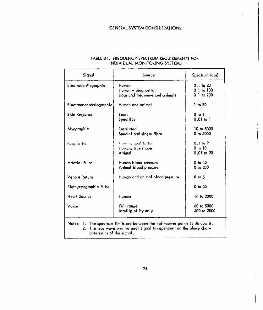
GENERAL SYSTEM CONSIDERATIONS
TABLE Ill. FREQUENCY SPECTRUM REQUIREMENTS FORINDIVIDUAL MONITORING SYSTEMS
Signal Source Spectrum (cps)
Electrocard'ographic Human 0.1 to 20Human - diagnostic 0.1 to 100Dogs and medium-sized animals 0.1 to 200
Electroencephalographic Human and animal 1 to 80
Skin Response Basal 0 to 1Specifics 0.01 to ,
Myographic Restricted 10 to 5000Special and single fibre 0 to 5000
Rc~pirct: on lIBI,.,, . 1i,,, ib I hti 0.1 to 2Human, true shape 0 to 10Animal 0.01 to 20
Arterial Pulse Human blood pressure 0 to 30
Animal blood pressure 0 to 200
Venous Return Human and animal blood pressure 0 to 5
Plethysmographic Pulse 0 to 30
Heart Sounds Human 16 to 2000
Voice Full range 60 to 5000Intelligibility only 400 to 2000
Notes: 1. The spectrum limits are between the half-power points (3 db down).2. The true waveform for each signal is dependent on the phase char-
acteristics of the signal.
75

BASIC PHYSIOLOGICAL MEASURING SYSTEMS
matching, (3) nonlinearity, and (4) Interference and artifacts. The degree of impor-tance of each of these considerations is determined by the deg,:ee of error that can betolerated in the measurement.
A. Frequency Characteristics
Pure waveforms of a single sinusoidal frequency are encountered rarely inphysiological measurements. Instead, physiological signals normally contain a mixtureof sinusoidal waves of different frequencies, varying in both phase and amplitude.Therefore, if a measuring system is to process the signals without distortion, it must becapable of handling all of the frequency, phase, and amplitude components of the sig-nals. Shorp changes in slope, such as those encountered in square waves and stepfunctions, contain high-frequency components in addition to the basic repetition fre-quency. Measuring systems, therefore, must be designed to process such signals. If themeasuring system Is incapable of handling the higher frequency components of a squarewave, for example, distortion occurs, resulting in rounded corners as shown in figure19A. If the system has adequate high-frequency response but lacks low-frequencyresponse, the wave may be distorted as noted in figure 19B.
SIGNAL INPUT SIGNAL INPUT-\
RESPONSE-, RESPONSE
(A) (B)POOR HIGH-FREQUENCY RESPONSE POOR LOW-FREQUENCY RESPONSE
Figure 19. Frequency Distortion of a Square Wave
Consequently, the frequency-response curve of a physiological system, systemcomponent, or instrument is of primary interest. A frequency-response curve plots therelative ou'tput level of the system against the input frequency, with the input ampli-tude held constant at all frequencies. A typical frequency-response curve is shown infigure 20. A general rule to adhere to in order to ensure an output signal with minimal1;;tortion is that the output levels of the highest and the lowest frequency contained in
thr, .neasured signal should be at least 95 percent of the output level of the midrangefreqiency. In some instances, this rule may be modified, depending upon the impor-tance of phase distortion in the measurement. The subject of phase distortion will betreated later in more detail.
76

GENERAL SYSTEM CONSIDERATIONS
1.0
lLI
Iz0.5
01I-
,-JL&J
0
FREQUENCY
Figure 20. Typical Frequency-Response Curve
B. Impedance Matching
There are two major considerationswhin evaluating the impedance character-istics of a measuring system: (1) the input and output impedances of the individualcomponents of the system, and (2) the input impedance of the measuring system withrespect to the impedance of the physiological system being monitored.
When selecting components for a complete measuring system, adjacent com-ponents must be matched properly if the maximum amount of signal is to be transferredwith minimum distortion. The transfer of a physiological signal between componentsmay involve either (1) a transfer of voltage only (disregarding current) or (2) a transferof power (voltage and current). If the signal transfer involves voltage only, the inputcircuit of the driven component may load the output circuit of the driving componentto the point where the signal is distorted. To avoid this, the input impedance of thedriven component should be as high as possible with respect to the output impedance ofthe driving component, consistent with system operation.
If power is to be transferred, the problem is considerably more complex. Themaximum transfer of power occurs only when the impedances of the two adjacent com-ponents are equal. If a particular system component (an amplifier, for example) has acharacteristic low input impedance and a high output impedance, the component pre-ceding it must possess a low output impedance (to match the low input impedance ofthe amplifier), and the component following it must have a high input impedance (to
77

BASIC PHYSIOLOGICAL MEASURING SYSTEMS
match the high output impedance of the amplifier). When these conditions are satis-fied, maximum undistorted transfer of power Will result. In many instances, an imped-ance-matching device, such as a transformer, may be necessary to transfer powerefficiently.
The input impedance of the measuring system must be compatible with thephysiological phenomena being monitored, since the electrical quantities (body resist-ance, potentials, etc.) of the human body normally are especially susceptible to influ-ences from external measuring systems. High-impedance physiological systems, forexample, cannot be monitored by a low-impedance measuring system. The low imped-ance of the measuring system would effectively "shunt" the physiological impedanceand result in erroneous measurements. The input impedance of the measuring systemnormally should be several times greater than the internal impedance of the bodysystem for proper results.
C. Nonlinearity
Nonlinearity is distortion in t+e output indication of a measuring system rela-tive to the input signal. Nonlinearity can result from many factors. For example, amechanical system may have play in a lever system or backlash in a gear train. Themost common cause of nonlinearlty in physiological electronic measuring systems, how-ever, is improper operation of vaCuum tube or transistor circuits. Improper se!ectfln ofcircuit values and/or supply voltages may cause an amplifier to operate in the non-linear portion of its operating range. When this occurs, different levels of signal volt-age result in different factors of amplification. For example, a signal with an amplitudeof 0.5 volt may be amplified by a factor of 10, while a signal with an amplitude of 1volt may be amplified only by a factor of 7. Typical results and causes of nonlinearoperation are shown in figure 21. In some instances, transducers also may exhibit non-linear operation over certain portions of their operating range. As a result, it is ofprime importance that transducers, amplifiers, and all other system components beselected so that the linear portions of their operating ranges coincide with the signallevels to be handled.
D. Interference and Artifacts
Interference and artifacts are sources of error that may originate within themeasuring system itself or external to the system. Interference can be any form of noiseor erroneous signal that tends to mask the desired measurement. There are two generalmethods by which external interference enters the measuring system: electrostatic in-duction and electromagnetic induction. Electrostatic interference results from thecapacitive coupling effect between two conductors. The most common Form of electro-static interference is 60-cycle hum originating from the power lines of the building inwhich the measuring system is located. Electromagnetic interference results from theelectromagnetic field produced by a varying current flow through a conductor. Whenanother conductor is placed within the magnetic field, a current is induced in it.
78

GENERAL SYSTEM CONSIDERATIONS
SIGNALINPUT
RESPONSE
OUTPUTCURVEOU UT
AMPLITUDE
AMPLITUDE de -- - -
LIMITING to
I I INPU TIAMPLITUDE
RESPONSEOUTPUT CURVE
AMPLITUDE
PEAKING dueto
INPUT0 I IAMPLITUDE
Figure 21. Nonlinearity of a Sine Wave
79
BASIC PHYSIOLOGICAL MEASURING SYSTEMS
Erroneous signals resulting from artifacts are those which originate at the sig-nal source itself. Artifacts usually occur as the result of interaction between themeasuring system and the physiological system being monitored.
SIGNAL PICKUP DEVICES
The signal pickup device is probably the most critical item in the measurementchain. The sensing device must be activated by the phenomena being measured anddeliver, as an output, an electrical signal which quantitatively represents the phenom-ena. Furthermore, it must do this with a minimum effect or reaction on the source.
Two general classes of pickup devices are used in biomeasurements: transducersand electrodes. Transducers are used for measuring most of the environmental factorslisted on the input side of figure 1. Electrode systems are required for many of themeasurements on the output side. As mentioned in the introduction, the field of physi-cal measuring devices will not be given detailed treatment. To do so means coveringpractically the whole field of physical measurement. Signal pickup devices concernedonly and uniquely with physiological measurement are discussed.
I. Transducers
A. General
Generally defined, a transducer converts one type of energy into another.However, In physiological instrumentation, the term transducer usually is limited to in-clude only the part of a monitoring system that Is first exposed to the quantity beingmeasured and that converts the quantity into an electrical form (voltage, current, orimpedance).
Several types of transducers are available for monitoring the afferent (input)factors which affect physiological behavior. For example, abnormally high atmosphericpressure may be measured with a piezoelectric prossure transducer, consisting of apiezoelectric crystal which, when exposed to pressure from all sides (hydrostatic pres-sure), produces a corresponding voltage, Environmental temperature variations may bemonitored with o temperature transducer consisting of a resistive elemen (resistancethermometer). Tihe resistance of the element varies with temperature, and any changein temperature is detected as a change in the element's resistance.
B. Basic Considerations
Several basic operating characteristics must be considered when selecting atransducer for a specific application. These include input sensitivity and output im-pedance, which must be considered in all applications, and frequency response whichbecomes an important factor in certain physiological applications such as the monitor-ing of heart sounds.
80

SIGNAL PICKUP DEVICES
I. Input Signal Level Response
The ability of a transducer to respond to input signals of varying ampli-tudes is one of its most important characteristics. The lower limit of the useful inputrange is governed generally by the amount of noise originating within the transducer.The upper limit of the useful input level usually is determined by excessive distortionof the signal or by possible damage to the pickup element.
2. Output Impedance
The output impedance of a transducer and the input impedance of thefollowing stage (the signal modifier in figure 18) determine the amount of power thatcan be transferred from the pickup device at a given output-signal level. Maximumpower transfer occurs only whan the impedances are matched.
3. Frequency Response
The useful frequency ranges of different transducer types vary consider-ably, so the frequency range of the quantity being measured must be known and com-pared with the responses of the available transducers. For monitoring heart sounds, forexample, a microphone (n transducer which converts sound energy into electrical011019y) wwlh u requency response ranging from approximately 30 cps to several thou-sand cps is required.
II. Electrodes
An electrode is a conductor that serves as a current transfer path from one mediumto another. In physiological monitoring, electrodes form the connecting link betweenthe source of biological potentials in the body tissue and the instrumentation. Elec-trodes usually are placed on the surface of the skin and respond to electrical impulsesoriginating at various sources within the body tissue. The placement of the electrodesin relation to the source of potentials and the attenuation of signals by the resistanceof the body tissue and skin are important reliability factors.
A. Basic Considerations
1. Electrode Placement
Electrodes must be positioned cn the skin where they can obtain an ade-quate signal from the potential source. Simply placing the electrodes as close as pos-sible to the source is not adequate. A source of biological potentials is a dipole; thatis, it has a positive and a negative terminal. Potential sources are located in bodytissue, a homogeneous medium that forms a conducting path in all directions from thesource. As the distance from the source increases, the amplitude of the potentials isreduced by the resistance of the body tissue. In figure 22, the poles of the p~tential
81
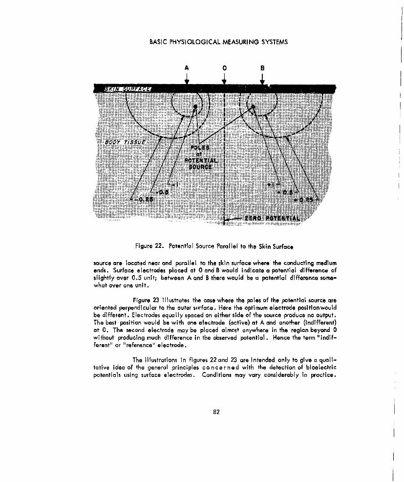
BASIC PHYSIOLOGICAL MEASURING SYSTEMS
A 0 B
AJMS K~lN O FAC
Figure 22. Potential Source Parallel to the Skin Surface
source are located near and parallel to the skin surface whore the conducting mediumends. Surface electrodes placed at 0 and B would indicate a potential difference ofslightly over 0.5 unit; between A and B there would be a ptential difference some-what over one unit.
Figure 23 illustrates the case where the poles of the potential source areoriented perpendicular to the outer surface. Here the optimum electrode psitlonwouldbe different. Electrodes equally spaced on either side of the source produce no output.The best position would be with one electrode (active) at A and another (indifferent)at 0. The second electrode may be placed almost anywhere in the region beyond 0without producing much difference in the observed polential. Hence the term "indif-ferent" or "reference" electrode.
The illustrations in figures 22 and 23 are intended only to give a qluali-tative idea of the gceneral porinciples concerned with the detection of bloelectricpotentials using surface electrodes. Conditions may vary considerably in practice.
82

SIGNAL PICKUP DEVICES
0 A 0
..b .TISSUE
Figure 23. Potential Source Perpendicular to the Skin Surface
2. Skin and Tissue Resistance
The contact made by the electrode and skin surface is almost as impor-tant as electrode placement. The deep body tissues generally present very low resist-ance to the flow of electrical currents, being somewhere in the order of 100 ohms perunit cube. Skin resistance is mostly in the outer layer and can be extremely high,running as high as 2 kilohms per square centimeter for dry skin. The objective (theGSR is an exception) is to have resistance as low and as constant as possible, usingelectrodes of a practicable size.
The relationship of electrode resistance or impedance to the other inputcircuit factors is illustrated in figure 24. Figure 24A represents the circuit pictorially;figure 24B shows the equivalent electrical circuit. From figure 24B, it is evident thatthis is effectively a series circuit, and the same current flows throughout. Therefore,the voltage drop due to the high skin resistance is lost to the load circuit, which canbe a serious factor, For example, in the extrame case of a transistor amplifier havingan input resistance of less than 1000 ohms and a combined electrode resistance of100,000 ohms, the voltage loss or attenuation will be more than 100 to 1.
83

BASIC PHYSIOLOGICAL MEASURING SYSTEMS
w0
z A
4 ro
w-~AIZ
0-0
LLJ 4
ac
-I 4t
4A4
84)

SIGNAL PICKUP DEVICES
B. Electrode Arrays
1. Single-Ended Arrays
'The single-ended or unipolar array is the simplest type of electrodearray. It consists basically of two electrodes, each located at a different part of thebody. One of the electrodes (normally the one nearest the bloelectric source beingmonitored) is called the "active" electrode, while the other one is referred to as the"reference" or "indifferent" electrode. The potential at the active electrode is meas-ured with respect to the potential at the reference electrode.
Three or more electrodes can form a single-ended array by connectingall the electrodes but one to a central terminal as a reference and using the remainingelectrode as the active electrode. When this configuration is used, the average poten-tial at the reference electrodes (potential of the central terminal) is compared with thepotential of the active electrode. Thus, the array remains single-ended; although anumber of electrodes may be used, only one potential difference is available.
2. Balanced Arrays
A basic balanced or double-ended electrode system may consist of onereference or indifferent electrode and two uctivO oloctrodo,. Th p-t~n! I Affrpnrpbetween the reference electrode and the two active electrodes is compared, resultingin an output that is a function of the difference in potential between the two activeelements. In some cases, the reference electrode may be omitted.
SIGNAL PROCESSING
I. General
In a physiological measuring system, the portion of the system located between thesignal pickup device and the display unit generally is known as the signal-processingor modifying section. Signal processing may consist of any or all of several functions,including amplification, attenuation, modulation, froquency conversion, rectifying,filtering, or any other that might be required to prepare the signal for recording, trans-mission, or display.
The operation performed most commonly is amplification. The signal from thepickup device usually is weak and must be raised to a power level that permits thetransmission and operation of recording or display devices. Because of its importancein physiological work, some discussion is provided here an the special features of am-plification systems. Other signal processing functions will be covered in detail in thesucceeding volumes of this handbook.
85
BASIC PHYSIOLOGICAL MEASURING SYSTEMS
II. Amplification
Since most physiological phenomena are evidenced by signals of extremely smallamplitude, amplification is an important phase of signal processing. Physiologicalamplifiers may amplify either voltage, current, or power, and they may use eithervacuum tubes or transistors. A voltage amplifier increases the voltage amplitude of itsinput signal without attempting to increase the power appreciably. A current amplifierincreases signal current considerably, some current amplifiers have no voltage gain,and others provide a voltage output that is less than the input. Power amplifiers in-crease both voltage and current to develop enough power to drive a large device (forexampla, a transducer output is amplified to drive a power-driven recording device).Whether vacuum tubes or transistors are used in the amplifier's circuitry is dependentupon several factors, such as operational requirements, power availability, and envi-ronmental conditions.
A. Requirements
Although moderate signal distortion may be tolerable in some applications,physiological measurements generally require that low-distortion amplifiers be used.Signal distortion can be caused by inadequate frequency response, nonlinear operation,instability, Insufficient or excessive signal Input, and several other factors.
R , lnr. Ampllfirnr Types for Physitlmrtil Applications
Amplifiers can be classed in many ways. For example, they may be groupedby the type of coupling used between stages, by their operating frequency range, or bytheir class of operation. A convenient method of classifying amplifiers, especiallythose used In physiological applications, Is to divide them Into two general groups:single-ended and double-ended (balanced or differential) amplifiers. Each group ofamplifiers may then be subdivided into various types,such as direct-coupled amplifiers,pulse amplifiers, and carrier amplifiers.
1. Single-Ended Amplifiers
A single-ended amplifier consists basically of a single vacuum tube ortransistor circuit with single input and output signals. A simplified single-ended ampli-fier is shown in figure 25. Several single-ended amplifiers can be connected in series(cascaded) to form a multistage amplifier with a higher gain. In practice, single-ended amplifiers present several problems, some of which are great enough to renderthe amplifier unsuitable for many physiological applications. They are especiallyprone to self-oscillation and unstable operation when slight changes in circuit poram-eters (supply voltages, cathode temperature, etc) occur.
86
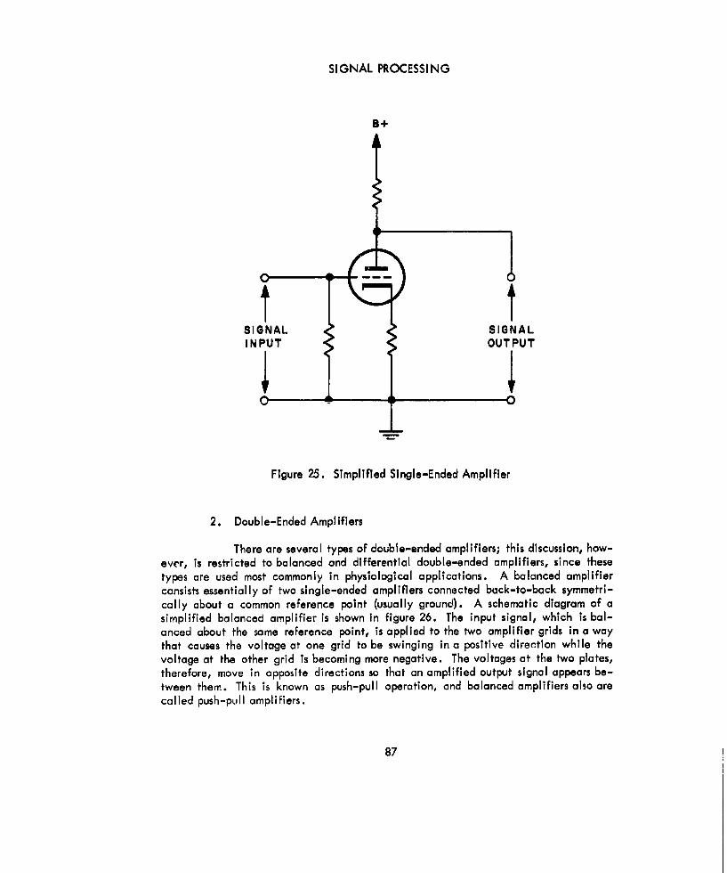
SIGNAL PROCESSING
B+
SIGNAL SIGNALINPUT OUTPUT
Figure 25. Simplified Single-Ended Amplifier
2. Double-Ended Amplifiers
There are several types of double-ended amplifiers; this discussion, how-ever, is restricted to balanced and differential double-ended amplifiers, since thesetypes are used most commonly in physiological applications. A balanced amplifierconsists essentially of two single-ended amplifiers connected back-to-back symmetri-cally about a common reference point (usually grounc0l. A schematic diagram of asimplified balanced amplifier is shown in figure 26. The input signal, which is bal-anced about the same reference point, is applied to the two amplifier grids in a waythat causes the voltage at one grid to be swinging in a positive direction while thevoltage at the other grid is becoming more negative. The voltages at the two plates,therefore, move in opposite directions so that an amplified output signal appears be-tween them. This is known as push-pull operation, and balanced amplifiers also arecalled push-pull amplifiers.
87
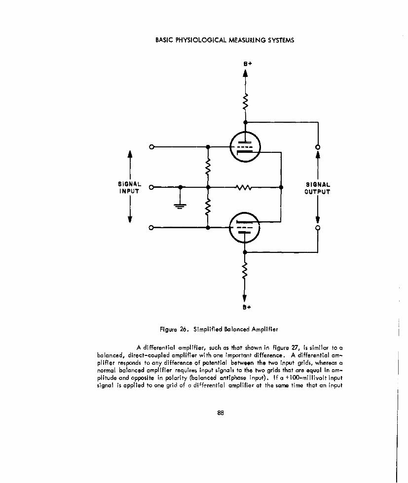
BASIC PHYSIOLOGICAL MEASURING SYSTEMS
9+
SIGNAL __ __ __SIGNAL
INPUT 'YOUTPUT
8+
Figure 26. Simplified Balanced Amplifier
A differential amplifier, such as that shown in figure 27, is similar to abalanced, direct-coupled amplifier with one important difference. A differential am-plifier responds to any difference of potential between the two input grids, whereas anormal balanced amplifier requires input signals to the two grids that are equal in am-plitude and opposite in polarity (balanced antiphase input). If a +100-millivolt inputsignal is applied to one grid of a differential amplifier at the same time that an input
88

SIGNAL PROCESSING
B+
SIGNAL B SIGNALIN PUT OUTPUT
0-1
B+
Figure 27. Simplified Differential Amplifier
of +110 millivolts is applied to the other grid, the effective input signal is 10 milli-volts. The input signals to the two grids of a differential amplifier may be of the sameor opposite polarity (e.g., an input of +40 millivolts applied simultaneously with aninput of -50 millivolts produces an effective input of 90 millivolts). A differentialamplifier also responds to an input applied to one grid only.
Double-ended operation offers several advantages. In a single-ended
89
BASIC PHYSIOLOGICAL MEASURING SYSTEMS
amplifier, for example, a slight change in the supply voltage produces a correspondingchange in the amplifier tube's plate voltage, which is indistinguishable from the outputsignal, resulting in a considerable signal distortion. In a balanced amplifier, however,slight changes in the supply voltage result in plate voltage changes on both amplifiertubes. Since the output of a balanced amplifier is the instantaneous voltage differencebetween the two amplifier plates, no change in the output signal results from slightpower supply variations. The same principle holds true for slight variations in othercircuit parameters. For any given set of circuit parameters, balanced amplifiers alwaysoffer better performance than can be obtained from a single-ended amplifier.
DISPLAY
I. Basic Data Display Method
Data usually are displayed as a function of time. In other words, most displaydevices use time as one of the coordinates of the reading system. For example, a stripof recording paper or film passes a recording point at a known rate and a pen or otherrecording device records the signal. In cathode-ray-tube displays, the trace usuallytravels horizontally as a linear function of time, and vertical displacement of the traceis an Indication of the magnitude of the phenomena. All display devices that presentdata as a function of time can be classified as time-axis recorders.
11 ~n~n~rol PrincriplA. cf Dmoi~ Rndrout cind Display Devices
A. Frequency-Response Grouping of Recording Devices
Recording devices may be classified according to their frequency response;there are low-frequency recorders, intermediate-frequency recorders, high-frequencyrecorders, and ultrahigh-frequency recorders. (These frequency classifications applyonly to recording devices, and should not be confused with the frequency bands in theradio-frequency spectrum.) In all four frequency categories, the response of the re-corders normally extends from zero to some specified limit. The frequency classifica-tions, therefore, mostly apply to the upper frequency-response I Imitations of therecorders, and a high-frequency recorder may be used in place of a low- or interme-diate-frequency recorder. If the low-frequency response limit of the recorder does notextend to zero, such as is the case with a cathode-ray oscilloscope employing a capac-itively coupled input circuit, this generalization does not hold true, since the low-frequency response also must be considered.
1. Low-Frequency Recorders
Low-frequency recorders generally are used in that frequency range from0 to 2 cps. Ink-writing recorders that respond to input signals in the order of a fewmillivolts or to currents of 1 milliampere or less are examples of low-frequency or slow
90
DISPLAY
recorders. These devices are slow because the mechanical arrangements used for con-verting electrical energy into a mechanical displacement of a pen or scribing pointcontain a considerable amount of inertia.
2. Intermediate-Frequency Recorders
Intermediate-frequency recorders have a frequency range extending from0 to approximately 60 cps. The increased frequency range is obtained with the use ofa pen motor, a device using a large quantity of electrical energy to produce a dis-placement against a stiff spring, so that the return from displacement is rapid in spiteof the considerable mosses being moved. The power requirement and the circuitrynecessary to furnish it make the intermedlate-frequency recorder much more expensiveand more difficult to operate and maintain than the low-frequency recorders.
3. High-Frequency Recorders
The frequency response of high-frequency recorders is from 0 cps toapproximately 5000 cps. The inertia and, therefore, the reaction time of high-frequency recorders is reduced by eliminating mass from the system. This is accom-plished by using light beams and mirrors. The mirrors are mounted so that they turnwith the coil movement of a galvanometer. A light beam falling on the mirror is re-tiected to a photosensitive surface and "writes" on the surface a5 ii ; muvul 'Uy ,Ih0mirror displacement. High-frequency recorders require optical sybtems, camera drives,and ga I vanometers. As a result, they are quite expensive compared to low- andintermediate-frequency recorders.
4. Ultrahigh-Frequency Recorders
The cathode-rayoscilloscope is an excellent ultrahigh-frequency recorderin that it is capable of operating with input frequencies ranging from 0 cps to beyondseveral hundred kc. Although the oscilloscope by itself does not provide a permanentrecord of displayed data, it is adaptable to both still and motion picture recording.Thus a record of the monitored data may be obtained and examined thoroughly at alater time.
B. Direct Writing Versus Photodevelopment
There are both direct-writing and photographic type recorders. With direct-writing recorders, the results are available immediately, while the photographic typerequires a development process which results in some delay. Whether the applicationrequires that results be immediate may be a factor in choosing a recording instrument.
Most direct-writing instruments use ink, and the displaced writing point is partof a pen. However, some recorders use heat-sensitive paper, and a heated stylus does
91

BASIC PHYSIOLOGICAL MEASURING SYSTEMS
the writing. Electrosensitive papers also are available, on which the writing is accom-plished by using the moving stylus as one electrode and passing a current through thepaper.
Photographic recorders may use either film or paper. Both are available inmany sizes, speeds, and emulsion types. In general, film is used only when the phe-nomenon occurs so rapidly that It cannot be recorded conveniently on the relativelyslow paper. Recording devices are available in which the recording Is done optically,and the development occurs right In the recorder. Theoretically, this combines theadvantages of direct-writing and light-beam recording; however, some time still isneeded for development.
C. Curvilinear Versus Rectilinear Recording
The scribing point of many recording devices Is located at the end of an armthat is pivoted at its other end; when the pivot is turned, the scribing point describesan arc. If the arm Is parallel to the time axis In the middle of the chart, the scribingpoint is displaced along the time axis either above or below this midpoint. The result-ing curvilinear recording does not provide a true displacement-versus-time record.This Is less a disadvantage If chart papercontaining curved time lines is used; however,it Is still difficult to produce a perfectly accurate result.
The Ideal type of recording is rectilinear recording, in which the vertical dis-placement of the scribing point Is along a true straight line perpendicular to the timeaxis. Mechanical yokes are designed to convert a curvilinear or angular displacementinto a rectilinear displacement; however, these yokes add inertia and mass to the sys-tem and therefore limit the upper frequency of the recorders.
Ill. Recorder Impedances
Impedance must be considered when selecting recorders. Many low-frequencyrecorders used for millivolt measurements actually are recording potentiometers. As aresult, they draw no current when balanced and therefore exhibit Infinite impedance.Most recorders, however, use a galvanometer with a basic D'Arsonval movement or apenmotor coil. Both are current-actuated devices that must draw current in order tooperate. Consequently, they are low-impedance devices and must be matched to low-impedance outputs in the signal-processing units.
SPECIAL ACCESSORIES
The three functional classes of components noted in figure 18 (signal pickupdevices, signal modifiers, and display devices) are common to practically all dynamicmeasuring systems. However, many situations encountered in practical usage requirethe addition of other items to the basic chain, as shown in figure 28.
92
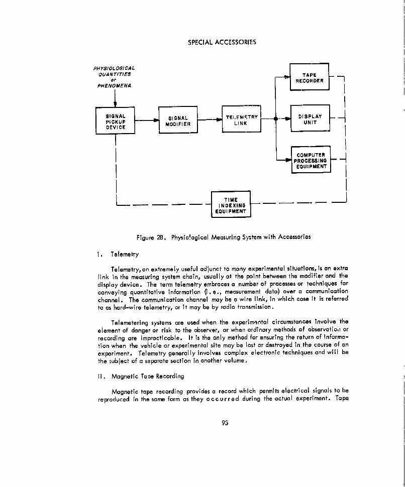
SPECIAL ACCESSORIES
PHYSIOLOGICALQUANTITiES TAPE
or RECORDER
PHENOMENA
,1 riSIGNAL SIGNAL. TELFNIFTRY DISPLAYPICKUP LIKMODIFIER LUNITDEVICE MDIE
COMPUTERPROCESSING
EQUIPMENT
TME1INDEXING
EQUIPMENT
Figure 28. Physio!ogical Measuring System with Accessories
I. Telemetry
Telemetry, an extremely useful adjunct to many experimental situations, is an extralink in the measuring system chain, usually at the point between the modifier and thedisplay device. The term telemetry embraces a number of processes or techniques forconveying quantitative information (i. e., measurement data) over a communicationchannel. The communication channel may be a wire link, in which case it is referredto as hard-wire telemetry, or it may be by radio transmission.
Telemetering systems are used when the experimental circumstances involve theelement of danger or risk to the observer, or when ordinary methods of observationl orrecording are impracticable. It is the only method for ensuring the return of informa-tion when the vehicle or experimental site may be lost or destroyed in the course of anexperiment. Telemetry generally involves complex electronic techniques and will bethe subject of a separate section in another volume.
II. Magnetic Tope Recording
Magnetic tape recording provides a record which permits electrical signals to bereproduced in the same form as they occurred during the actual experiment. Tape
93
BASIC PHYSIOLOGICAL MEASURING SYSTEMS
recording performs a function similar to telemetry, except that tape-recording systemscan provide a time lapse or delay between the experimental events and the subsequentobservation or display.
Tape recording also helps data processing and interpretation. The electrical sig-nals from the tape may be fed repeatedly to electronic computers for processing indifferent ways. Time axes also may be expanded or compressed easily by playing tapeback at speeds different from that at which the recording took place. The general sub-ject of tape recording systems w i I I be treated at greater length In the succeedingvolumes of this handbook.
III. Computer Processing
Computer processing is a general term applied to special operations performed oncertain signals. It Is difficult to draw a definite line between operations classed assignal modification and those classed as computer processing. In many cases, the deci-sion Is arbitrary. However, In general the term computer processing applies to themore complex operations, particularly those Involving accurate and rapid Interpreta-tion of several channels of data. For this purpose, automatic data-processing equip-ment, of which a computer is the central unit, is required. This equipment not onlyrecords and stores data, but It performs arithmetic operations as well as "logic" and"decision" functions.
IV. Time Indexing
A. General
Data obtained by monitoring the physiological reactions of a human in an ab-normal environment are of limited value unless the data can be correlated with vari-ations in the environment. For example, If the subject is placed in an environmenthaving a variable atmospheric pressure, the monitored data are significant only if somemethod is provided to relate the data with the variations in pressure. S I n ce mostphysiological monitoring is accomplished remotely, automatic time correlation is themethod that is used.
A convenient method of displaying data as a function of time is to providestrategically spaced time marks on the recorded data. The displayed data then can becompared with the phenomena occurring in the abnormal environment because theyboth are related in time. In some instances, some form of significant event markersmay be desirable; i.e., markers which represent the occurrence of a particular changeor event in the abnormal environment.
B. Establishing Correlation
One available reference for correlating data with time is the 60-cps line
94

SPECIAL ACCESSORIES
voltagn. When low-speed devices are used to record the monitored data, a synchronouselectric clock (operating from the 60-cps line voltage) can be used to drive a bank ofcams possessing a number of striking arms. These strikers can operate electrical con-tacts so that, by proper external connections, an electrical circuit is closed at intervalsranging from 1 second to 1 minute. The circuit can be completed by a battery and anelectromagnetic event marker, with the marker producing a time mark on the recordingpaper. The accuracy of the time marks depends upon how closely the line voltageapproximates 60 cps. Over an extended period, the cumulative error normally is zero;however, at any instant, an error as great as 2 percent is possible. For greater accu-racy, the synchronous clock may be replaced by an electrically maintained pendulum,rotating a similar cam system.
The 60-cps linevoltage also can be used for producing time markers on a sep-arate time trace for high-speed display devices, such as the cathode-ray tube. How-ever, this method is limited to displays with a time base that is longer than the durationof one complete cycle of the 60-cps line voltage (approximately 17 milliseconds).Although the 60-cps, sine-wave voltage can be used directly for time measurementmarks, its accuracy is limited, since the peaks of the wave are so ill-defined. A moresatisfactory timing waveform consists of a train of pips or triggers. The 60-cps linevoltage can be converted into a train of pips by means of rectifying and differentiatingcircuits.
SYSTEM INTEGRATION FACTORS
The basic physiological measuring systems discussed previously can be used underideal conditions and for simple measurements. In practice, however, simple experi-mental situations seldom are encountered. The experimental environment may be soextreme as to affect system accuracy or even damage components and inactivate thesystem; simultaneous measurements on the same subject may be necessary which maycause interference or interaction problems; the experimental site may be isolated andunavailable for instrument adjustment or calibration; or the transmission link or chan-nel may permit only a limited flow of information because of bandwidth requirementsor transmission path characteristics. All these considerations affect the design of theinstrumentation system. Although system integration factors will be treated in detailin the succeeding volumes cs" 1i s handbook, the most critical factors are summarizedhere to present an overall picture.
I. Physical Factors
A. Size and Weight Limitations
The size and weight of components of a monitoring system become critical inapplications where the subject must carry portions of the system and in airborne andspaceborne applications. Large and bulky amplifiers, power supplies, etc, obviously
95

BASIC PHYSIOLOGICAL MEASURING SYSTEMS
cannot be tolerated in such instances. Miniature components that satisfy the opera-tional requirements of the task at hand must be used.
B. Placement and Attachment of Pickup Devices
The placement and attachment of physiological pickup devices often presenta major problem. If the subject is to perform certain tasks and remain reasonably com-fortable during the monitoring procedure, the electrodes or transducers must be attachedexternally. This may result In a loss of signal accuracy in applications which normallyrequire internal or surgically implanted electrodes for maximum accuracy and relia-bility. When selecting body locations for the attachment of pickup devices, both thephysical restrictions imposed on the subject by the devices and the signal-to-Inter-ference ratio obtained from various locations during body movements must be consideredcarefully. A satisfactory compromise between these two extreme situations must beeffected if propbr system/subject compatibility is to be realized.
II. Information Handling Capability of Data Transmission Links
The data transmission link or channel becomes a critical factor in applicationswhere a telemetering system must be used to relay information from the experimentalsite to a remote display area. This is especially true in circumstances requiring a radiotransmission link. ihe ink mubi we iwi l u ... ...... . .
Ing monitored, the length of time available for transmitting information, transmissionpath characteristics, natural anl manmade interference, etc.
Ill. General Environment
Since environmental factors mayaffect the operation of physiological instrumenta-tion, the design nnd integration of equipment to be used in abnormal surroundings mustbe considered carefully. General environmental factors and their possible effects oninstrumentation are discussed in detail in volumes 2 and 3 of this handbook; a briefsummary of the problem follows:
A. Shock and Vibration
Shock and vibration may change electrical parameters in electronic circuitryor cause complete equipment failure.
B. Temperature
When the environment of temperature is considered, it usually is thought of incombination with other environments. For example, most materials become brittle atlow temperatures; conversely, at high temperatures the stiffness of materials decreases.Therefore, the reaction of equipment to shock and vibration forces will vary dependingupon the temperature.
96
SYSTEM INTEGRATION FACTORS
C. Other Factors
X-ray, gamma, and nuclear radiation, corrosion resulting from gasozone, andother agents, and pressure variations may change physical and electrical parameters,and may result in erratic equipment operation or interrupted operation.
IV. Equipment Interaction
Interaction between equipments often Is a critical factor in system integration.Interaction (the pickup of carriers and pulses radiated from adjacent equipment) canresult from ground currents or from electromagnetic coupling between circuits. Toavoid ground current interaction, all components in a system should be groundedproperly at one common point. Interaction caused by electromagnetic coupling can beminimized by shielding transformer circuits, which are the most potent sources of thistype of interaction.
V. Power Supply Problems
Various problems are encountered in conjunction with the source of power of ameasuring system. In a conventional power supply operating from a 115-volt, a-c Ine
'-.~r, f, fr -mwmn!,, rinniA rind hum may be present, and, when a single power supplyis used to power two or more system components, intercoupling is possibie, aithough Uhigh-quality, well-regulated supply normally does not cause major interaction prob-lems. Artifacts also may be introduced through unstable regulators and diodes.
When portable power sources are necessary, operating efficiency in severe envi-ronments and for extended periods of time becomes a critical factor. Batteries oftenare sensitive to ambient temperatures (they do not operate well under heavy loads atlow temperatures), have a relatively short life expectancy, and are affected by low-pressure environments.
ESTABLISHING VALIDITY OF REMOTE MEASUREMENTS
Test and "dry" runs that simulate actual mission conditions should be a plannedpart of every developmental program. Also, a so-called "blind" test of the data sys-tem should be made to determine the capabilties of the system and establish thevalidity of remote measurements. In a blind test an analytical group is supplied with"display" data from an experimental run in which various parameters are varied pur-posely. The analytical group, without knowledge of the programmed variables, theninterprets or reconstructs the experiment from the accumulated data.
97
EXPLANATION OF TERMS
"A" suppI7Power used to heat the filaments of vacuum tubes in electronic equipment.
AcetylcholineSubstance released from nerves to activate the muscles.
Action potential
The alteration in electrical potential which accompanies the passage of an Impulsealong a nerve fiber.
Adrenal glandEnd e gland which secretes hormones for the regulation of the salt and waterbalance of body fluids, and the regulation of the level of carbohydrate substancesin the blood, liver, and muscles.
Afferentgearing or conducting inward.
A tubular, Tood-carrying passage that extends from the mouth to the anus.
AlveoliAr cells of the lungs.
Amino acidsA group of acids from which new protein molecules are formed.
AmplifierAn electrical device, usually containing vacuum tubes or transistors, whose outputwaveform is an enlarged reproduction of the essential characteristics of the inputwaveform.
Anabol ismThe assimilative processes that make possible the growth and repair of tissues.
Anemia" condition resulting from a deficiency of red cells or hemoglobin in the blood.
AntennaTe apparatus used to radiate the electromagnetic waves produced by a radiotransmitter or to intercept or pick up electromagnetic waves for a radio receiver.
98
Aortic archThe large vessel (curved portion) arising from the left ventricle of the heart anddistributing, by its branches, arterial blood to all parts of the body.
ArteriolesA very small artery.
Artifact- -spurious signal appearing in the display of a biological phenomenon that is
caused by external means.
Atria-- he two upper chambers of the heart which receive the blood from the veins.
AuriclesTh two upper chamberz of the heart which receive the blood from the veins.
Auscultation- e cton and study of sounds arising from various organs, particularly theheart and lungs.
AutonomicIndependent In origin, action, or function.
Axon" rocess of a nerve cell which conveys impulses away from the cell body.
"B" supplyPower used to operate the plate and/or screen grid circuits of vacuum tubes inelectronic equipment.
BallstocardigraphyRecording of the movements of the body caused by the impact of the circulatingblood and recoil of the body.
Basal metabolismThe minimum amount of energy expenditure necessary to maintain body activitywith the body at complete rest.
Basal metabolism rate (BMR)The amount of heat produced by the oxidation of foodstuffs in the bdy per 24hours under standard conditions with activity reduced to a minimum.
99
Behavioral systemBody system consisting of the parts of the body which are responsive to physio-logical stimulation.
BioelectricThe electrical phenomena that occurs in living tissue; the effects of electric cur-rents upon living tissues.
Bradl ycarda... ]imal slowness of hetc0rt action.
Bronchi" 'Me two primary branches of the windpipe within the lungs.
lic SU- J
Power used to supply grid-bias voltage in vacuum tube circuits.
CapacitorAn electrical device consisting of 2 conducting surfaces separated byan Insulatingmaterial. The capacitor blocks the flow of direct current, stores electrical energy,and permits the flow of alternating current. Capacitors are rated in microfarads ormicromicrofarads.
CapillaryA minute blood vs l, one of u nelwoik .Vill~t ih l he b1(uallI ulab i# Undveins.
CardIac cycleThe sequence of events occurring during one beat of the heart.
Carotid sinusThe principal large artery on each side of the neck.
Carrier-current propagationThe method by which radio communication is conducted with the use of conven-tional power lines as the propagation medium.
CatabolismThe process which releases energy for muscular activity by using up energy storedchemically in the body.
Cerebral cortexThe external gray layer of the brain.
100
Cerebral hemispheresThe two main portions of the brain, occupying the entire upper portion of thecranial cavity.
Chemoreceptoroneof the receptors in a li,,ing cell capable of controlling chemical substances.
Circulatory systemBody system, consisting of the heart, blood vessels, and lymphatic system.
ComputerA device that is capable of accepting information, applying definite reasonable
processes to the information, and supplying the results of these processes.
CounterAn .-electrical circuit that receives uniform ptilses, representing units to be counted,and produces a voltage proportional to their frequency.
Cranial cavityThe part of the skull that contains the brain and Its membranes and vessels.
DendriteA oro'r ss nf the ne,,rmn whiekh emrrlp thA nArvA impulge to the cell body.
Detection (demodulation)The process of extracting the signal intelligence from a modulated carrier wave.
DiastoleTh rhythmic period of relaxation and dilatation of a chamber of the heart as Itfills with blood.
Diastolic pressureThe minimum arterial pressure occurring during ventricular diastole.
Differentiating circuitA circuit in which the amplitude of the output waveform is proportional to the rateof change of amplitude of the input waveform.
DistortionAn desired change in the waveform of an electrical quantity.
Doppler effectT'he apparent change in the frequency of an electromagnetic or sound wave as thedistance between the source and the observer increases or decreases.
101

EdemaThe presence of abnormally large amounts of fluid in tissues.
EfferentCa-rrylng away From a center.
ElectrocardiogramThe graphic record obtained with an electrocardiograph.
ElectrocardiographAn instrument that records the changes in voltage produced by the contraction andrelaxation of the heart muscle occurring in a physiological subject in synchronismwith heart beats.
ElectrodeA terminal at which electricity passes from one medium into another.
Electrodermal resnse (EDR)alvanic skin response.
ElectraencephalogramThe graphic record obtained with an electroencephalograph.
F ctrrencephaloraphAn Instrumen t that records the electrical activity of the cerebral cortex.
Electromagnetic couplingCoupling that exists between circuits when they are affected by the same electro-magnetic field.
Electromagnetic fieldField of i nfluence (combined electrical and magnetic Fields) which is produced bythe flow of current through a conductor.
E lectromygramThe grapic record obtained with an electromyograph.
ElectromyographAn instrument that records tho changes of electric potential of muscle.
Electrophysiology
The branch of physiology dealing with the production of electricity by living or-ganisms and the effects of electric currents on living organisms.
102
Endocrine systemBody system composed of glands which produce complex compounds (hormones) that
pass directly into the blood.
EpilationRemoval of hair by destruction of its roots (through the use of forceps, chemicalmeans, or roentgen therapy).
ErythemaAn increased content of blood near the surface of the skin, resulting in red
blotches of variable size and shape.
ErythocyteA blood corpuscle.
Excretory systemBody system, consisting primarily of the kidneys, which eliminates waste products.
Exteroceptor"'An end organ which receives stimuli from the external world.
Frontal lobeAr of the brain tha -ill , i- hod, " mtmIW- ind4 1- renonsible for complexassociation processos, such as fecalling and thinking.
GSymbol for the acceleration due to the gravity at the surface of Earth (approxi-mately 32 feet per second each second).
Galvan'c skin response (GSR)The change in the electrical resistance of the skin that occurs when the subject isexposed to psychosensory stimuli.
GalvanometerAn instrument for indlcathg or measuring an electrical current or a function of acurrent.
Gastrointestinal systemBoady system consisting of the stomach and the intestines.
GravireceptorOne ofthe receptors in a living cell that responds to gravitational forces.
GyrusA convolution on the surface of the cerebral hemisphere.
103

HemoglobinThe respiratory pigment of the red blood cells.
High-vacuum rectifierVacuum-tube rectifier in which conduction occurs entirely by electrons emittedfrom the cathode.
HomeostasisMaintenance of steady states in an organism by coordinated physiologicalprocesses.
HomogeneousOf uniform character in all parts.
Hy'perpneaAn increase in the depth of Inspiration resulting from a reduction in partial pres-
sure of oxygen.
Hyperventi lotionAn Increase in the quantity of air breathed (minute volume) particularly beyondthat required physiologically resulting from an increase In the rate and/or depthof respiration.
A part o the forebrain involved in the regulation of body temperature, sleep,water balance, carbohydrate and fat metabolism, and other functions.
HypoxiaCoditon resulting from an inadequate supply of oxygen.
ImpedanceThe opposition to the flow of alternating current in a circuit that contains resist-ance, inductance, and/or capacitance. The unit of impedance is the ohm.
InductanceThe inherent property of an electric device to set up a self-induced electromotiveforce that opposes any change of current in the circuit, The unit of inductance isthe henry.
InnervateMtribution of nerves to a part.
Integrating circuitA circuit in which the output signal is proportional to the frequency and amplitudeof the input signal.
104

InterferenceElectrical disturbance which causes undesirable responses in electronic equipment.
IonAn atom or group of atoms which has lost or gained one or more orbital electronsand has thus become capable of conducting electricity.
IschemiaLocal or temporary deficiency of blood.
Kelvin scaleAn a ute scale of temperature which has its zero at -273 0C.
KinestheticPertaInIng to the perception of movement, weight, resistance, and position bymuscular receptors.
Korotoff soundsSoundsheard during blood pressure determination.
LeukopenlaA decrease below the normal number of certain whlte blood cells (leukocytes) Inthe .hrI+APrml bload.
Lumen79e space inside a tube such as a blood vessel.
Lymphatic systemBody system which circulates tissue fluids from the blood to the cells and back tothe blood.
MedullaThi lowest portion of the brain stem. It is connected to the spinal cord and is thecenter of many reflex activities.
Mercury-vapor rectifierA rectifier tube containing mercury vapor which is ionized by the application of aplate voltage.
MesenteryT7emembrane that anchors the small intestine to the abdominal wall.
Metabolic systemBody system responsible for those processes and chemical reactions that convertfood into simpler substances and supply the energy for vital functions.
105

MetabolismThe process of synthesizing foodstuffs to provide the energy for vital processes andactivities.
MicturitionThe act of passing urine.
Minute volumeThe total volume of air breathed per minute.
ModulationThe pocess in which the amplitude, frequency, or phase of a wave is varied withtime in accordance with the waveform of superimposed intelligence.
MotorConcerned with or pertaining to motion.
MultiplexingThe process of transmitting a number of messages simultaneously on the same com-munication link.
Frequency division - The use of a different frequency band for each signal In amultiplex circuit.
Time division - The use of different time intervals for each signal in a multiplexcircuit.
MultivibratorA two-stage oscillator in which the output of each stage Is coupled to the input ofthe other. The output waveform is essentially a square wave.
Mutual inductanceom' n property of two associated circuits which determines, for a given rate of
current in one of the circuits, the electromotive force induced in the other.
Myelin"-atty substance encasing the larger nerve fibers. Considered a poor electrical
conductor.
NBSGBritish standard wire gauge.
Neuron-Cmplete nerve cell, including the cell body, axon, and dendrites.
106
OcclusionA osure or a condition of 6inn thut off.
OcularPertaining to the eye.
OscillatorAn electronic device which generates alternating current power at a frequencydetermined by the values of certain circuit elements.
OscillogramThe recorder trace produced by an oscillograph.
Osci I lographAn instrument that records the waveform of one or more varying electricalquantities.
OscilloscopeA de which makes the shape of a voltage or current wave visible on the screenof a cathode-ray tube.
OxygenateIt nmturntc rt sub'ance with oyne.
Oxyhemog lobi nOxidized hemoglobin found mainly in arterial blood.
Parat,,roid glandEndocrine gland which secretes the hormone which maintains the proper distribu-tion of calcium in bones and in body fluids.
Parietal lobeAre fthe brain that is sensitive to bodily sensations.
Peripheral resistanceResistance of peripheral blood vessels to the flow of blood.
PeristalsisThe progressive wave of muscular contraction by which the contents of a tube areforced toward ah end.
Permeability (electrical)A measure of the relative ability of a substance to conduct magnetic lines of forceas compared with air, or magnetic conductivity.
107
Pormeability (physioloical)The property of plasma membranes which permits transit of molecules and ions bysolution in membranes, and by mechanical passage through fine pores in themembranes.
PhonocardlographyGrapic recording of heart sounds and murmurs by electric reproduction.
PhotoconductorA-ugiaji c whose electrical conductivity varies with illumination, as In gases,selenium, and some non-metallic crystals.
PhotoelectricConcerintng the electrical effects on a substance when exposed to light or otherradiation.
PhotometerAn Instrument that measures the intensity or quantity of light.
Phy ologThe science that deals with the functions of living organisms or their ports.
Pituitary glandMaster endocrine gland which secretes hormones which regulate growth, sexualactivities, and the amount of water secreted by the kidney5.
PlateletsBlood cells important in blood clotting.
PlethysmographyTle graphcal display of the change in volume of an organ resulting from anIncrease in the quantity of blood therein.
PleuraTe delicate serous membrane enveloping the lung and lining the internal surfaceof the thoracic cavity.
PotentialA measure of the concentration of an electrical charge at a given point.
Potential differenceThe algebraic difference between the voltages at two given points.
PotentiometerA threeterminal rheostat which functions as an adjustable voltage divider.
108
Precordial leadsLeadsattached to the torso of the subject.
ProprioceptionAppreciation of position, balrince, and changes in equilibrium on the part of themuscular system.
ProprioceptorA receptor or sense organ whose reflex function is concerned with locomotion orposture.
PSchgalvanic reflex (PGR)Elvan|c skin response.
PsychologicalPerining to the functions of the mind, such as sei.satlon, perception, memory,and thought.
Psychomotor
PertaiiTng to muscular action ensuing directly from a mental process.
Pulmonary circulationCirculation ofair In the lungs.
QRS complexThepo~rton of the electrocardiogram representing the electrical activity causedby excitation of the ventricles.
Recorder"'-,,,,,evice that prepares a record, usually graphic or numerical, of the value of a
quantity as a function of time. The recorder is used to store information untilneeded.
Rectifier circuitA circuit witch converts alternating current into unidirectional current.
RectificationTFe process of converting alternating current into unidirectional current.
Relaxation oscillatorAn oscillator wh ch generates a nonsinusoidal periodic vo l tage by graduallystoring electric energy and then quickly releasing that energy or vice versa.
109

RemRoentgen equivalent, man (the quantity of any type of radiation which will pro-duce the some biological effects in man as one roentgen of X or gamma radiation).
Resistance (electrical)The property of an electric circuit that opposes the flow of current through it. Theunit of resistance is the ohm.
Respiration depthA rmeasure nf the volume of air inspired and expired during each respiration.
Respiration rateRate o breathing, usually measured in breaths per minute.
Respiratori membranesThe membranes Tning the respiratory tract.
Respiratory systemBody system that provides the body with oxygen to oxidize carbonaceous food,thereby generating heat and supplying the body with energy for performing work.
ResponseA quantitative expression ot the output of a device or system as a function of theinput under a given set of conditions.
Rheostat" resistor whose value can be varied; often used to adjust the current flow in acircuit.
RoentgenStandard unit for expressing quantities of X and gamma radiation (the quantity ofX or gamma radiation required to produce one electrostatic unit of charge in onecubic centimeter of standard air).
Sei ynsingle-phase, self-synchronous device which converts mechanical position into
electrical signal, or vice versa. A trade name applied to this type of synchro.
SemiconductorA material, such as germanium or silicon, whose electrical resistivity is betweenthat of a good insulator and that of a good conductor such as a metal.
Sine WaveAn electrical wave of a periodically varying quantity with tie form of a sinecurve; the waveform of a normal alternating current.
110
SinusoldalSee sine wave.
Square waveA symmetrical periodic wave that varies, in time and amplitude, between two fixedvalues. Plotted graphically against time, it is square or rectangular in shape.
StimulusSomething that excites an organism or part to functionable activity.
SubcutaneousBeneath the skin.
SynapseTi point at which an impulse passes from an axon of one neuron to a dendrite or
to the cell body of another neuron.
SynchroKiype of wound-rotor a-c motor used for repeating angular motion both as tospeed and total angle.
s ystoleT 'e contraction phase of the cardiac cycle.
Systolic pressureThe maximum arterial pressure occurring during ventricular systole.
TachyardiaAbnormally rapid heart action.
Telemetr,The science of accumulating and transmitting data to a remote receiving station.
ThalamusA mass of gray matter at the base of the brain which relays sensory signals to otherbrain parts and the spirmal cord.
ThermistorA type of resi'stor in which the resistive element is a semiconductor whose resis-tivity varies with temperature. Used in temperature transducers.
ThermocoupleA device consisting of two dissimilar metallic substances across which a voltage isdeveloped when the junction of the two metals is heated.
111
Thermoelectric effectThe electromotie force that results from a difference in temperature between twodissimilar metals that are joined.
Thoraxn- e chest cavity or that portion of the trunk above the diaphragm and below theneck.
ThyratronA hot-cahode gas-discharge electron tube used to control re l ati v el y largeamounts of electric current with a small control voltage.
Thyroid glandEndce gland which secretes the hormone, thyroxin, which regulates the basalmetabolism rate and controls the liberation of heat after Ingestion of food andduring oercise.
Time indexingThe method by which correlation is made between measured data and variations inenvironmental conditions.
TissueA-n aggregate of similar cells and their Intercellular substance Forming one of thestructural materials of the body.
Trachea7 windpipe, a cartilaginous and membranous tube extending from the lower endof the larynx to Its division into the two bronchi.
TransducerA device that transmits energy from one system to another and may be used toconvert the energy from one form to another (mechanical to electrical or viceversa.
TransformerAn lctrical device, without moving parts which by electromagnetic couplingtransfers electrical energy from one circuit to another without change in frequencybut usually with changed values of voltage and current.
TransistorA semiconducting device that transfers en a r g y from one system to another andexercises control over this transfer. Its function is comparable to that of a vacuumtube.
112

Vagus'rhe tenth cranial nerve.
VascularPertaining to a vessel for the conveyan.e of a fluid.
Vascular bedThe total blood supply of the arteries, veins, and capil c" n .r. a ..
Vasoconstri ctionThe constriction of blood vessels.
VasodliationThe diton of blood vessels.
VasomotorRegulating the size of the blood vessels; pertaining to those nerves concerned withthe muscle fibers in the walls of the blood vessels.
Vasomotor centersNerve centers which control the size of the blood vessels.
Venti lationOxygenation of the blood in the capillaries of the lungs.
VentriclesThe two lower chambers of the heart which supply blood to the arteries.
Viscus (pl. viscera)An organ enclosed within the cranial, thoracic, abdominal, or pelvic cavities;particularly the abdominal cavity.
WeightlessnessA contn characterized by the absence of gravitational force.
113
BIBLIOGRAPHY
1. A' nan, Philip L. (Analyst), et al., Handbook of Circulation, WADC TechnicalReport 59-593, Wright Air Development Center, Wright-Patterson AFB, Ohio,1959.
2. Armington, John C. and Leonard L.. Mitnick, Electroencephalogram and SleepDeprivation, Walter Reed Army Institute of Research, Waltor Reed Army MVedicalCenter, Washington, D.C., ASTIA Document No. AD 219707.
3. Astronautics Communications-Electronics, Air Force Manual No. 100-25, Depart-ment of the Air Force, Washington, D.C., 1961.
4. Banner, E. H. W., Electronic Measuring Instruments, Chapman and Hall, Ltd.,London, England, 1954.
5. Barbiere, Robert E., et al., A Radiobiology Guide, RCA Service Company, Inc.,Camden 8, New Jersey, WADC Technical Report 57-118, Wright Air Develop-ment Center, Wright-Patterson AFB, O h I o, 1957, A ST I A Document No. AD118082.
6. Basic Electronics, Navy Trainfran rnior,e NlA\VPFRS 1OOP7, U.. Covirr..-+,Printing Office, Washington, D.C., 1955.
7. Beckman, Edw. L., LCDR, MC, USN, et al., Human Tolerance to High PositiveG Applied at a Rate of 5 to 10 G per Secondl iieporf No. NIX;-MA- Z,Aviation Medical Acceleration Laboratory, U.S. Naval Air Development Center,Johnsville, Pa., 1953, ASTIA Document No. 20520.
8. Begun, S. J., Magnetic Recordn ng Technical Division, Rinehart and Company,Inc., New York, N. Y., 1V58.
9. Benson, Otis 0., Jr., and Hubertus Strughold (Editors), Physics and Medicine ofthe Atmosphere and Space, John Wiley and Sons, Inc., 1960.
10. Black, Harold S.,Modulation Theory, D. VanNostrand Company, Inc., Princeton,N. J., 1953.
11. Boothby, Walter M., M.D. (Editor), Handbook of Respiratory Phsiolo, AirUniversity, USAF School of Aviation Medicine, Randolph Field, Texas, 954,ASTIA Document No. AD 4917.
114
12. Borden, Perry A. and Wilfred J. Mayo-Wells, Telemetering Systems, ReinholdPublishinq Corporation, New York, N. Y., 1959.
13. Braestrl.p, Cail B. and Harold 0. Wyckoff, Radiation Protection, published byCharles C. Thomas, Springfield, III., 1958.
14. Brazier, Mary A. B., B.Sc., Ph.D., The Electrical Activity of the Nervous Sys-tem, The Macmillan Company, New 'or, NY.,1960.
15. Chance, Britton, et al., (Editors), Electronic Time Measurements, McGraw-HillBook Company, Inc., New York, N. Y., 1949.
16. Closed Circuit Respiratory Systems Symposium, WADD Technical Report 60-574,Wright Air Development Division, Wright-Patterson AFB, Ohio, 1960, ASTIADocument No. AD 242587.
17. Communications-Electronics Terminology, Definitions, and Abbreviations, AirForce Manual No. 10"'4,Department of the Air Force, Washington, D.C., 6O.
18. Dantzig, George B., et al., A Mathematical Model of the Human External Res-piratory System, Research MemTandum RM-2519, The Rana Corporation, SantaMonica, Calif. prepared for the Directorate of Development Planning, Head-quarters USAF, 1959, ASTIA Document No. AD 246199.
19. Donaldson, P. E. K., "A Multiple-Channel Oscilloscope for Electrophysiology,"Electronic Engineering, Volume 29, February 1957.
20. Donaldson, P. E. K., et al., Electronic Apparatus for Biological Research,Butterworths Scientific Publications, London, England, 1958.
21. Eagan, C. J., The Mercury Gauge Method of Digital Plethysmography, TechnicalNote AAL-TN-60-15, Arctic Aeromedical Laboratory, Alaskan Air Command,Fort Wainwright, Alaska, 1961, ASTIA Document No. AD 253711.
22. Electronics Technician 3, Navy Training Course, NAVPERS 10188, U.S. Govern-ment Printing Office, Washington, D.C., 1954.
23. Ellinger, Fredrich, M.D., Medical Radiation Biology, published by Charles C.Thomas, Springfield, III., 1957.
24. Flaherty, Bernard E., Lt. Col., USAF (MC), et al., Psychiatry and SpE Flight,School of Aviation Medicine, USAF Aerospace Medical Center (ATC), BrooksAFB, Texas, 1960, ASTIA Document No. AD 245416.
115
25. Frederick, Carl L., at al., Design Techniques for Itrenc-e O rai ofAirborne Electronic Equipment, Wright Air Development Center, WrgtFatro
'B ho,15.
26. Giassner, Harvey F. and George A. Peters, Blo-Electronic Analysis of Perform-ance, DAC Engineering Paper No. 897, Douglas Aircraft Company, Inc., ElSegundo, Calif., pveprared for the Office of Novel Research, 1960, ASTIA Docu-ment No. 235969.
27. Goayof Computer, Missile and Electronic Terms, Field Support Engineering,~7eijtervics, RA Service Co., Camden 8,N. J., 1960.
28. Goldberg, M. N., et alI., I nstrumentation P ac kag 9 for I nfl ight Physiologi caIStudies, North American Aviation, Inc., Los Angeles, Calif., WAD TechnicalReport 60-83, Wright Air Development Division, Wright Patterson AFB, Ohio,1960.
29. Goldman, David E., Commander, MSC, USN, Short Wave Electromagnetic Radi-ation as a Hazard to Personnel, Lecture and Review Series No. 60-6, NavalMedical Research Institute, Be-thesda, Maryland, 1960, ASTIA Document No.AD 252582.
30. Goldman, David E., Commander, MSC, USN and Henning E , Von Glerke, TheFecb.t u' S~nuck arid Vibratian an Man, Le,;lurw and Review Series No. 60:7
Naval Medical Research Institute, Bethesda, Md., 1960, ASTIA Document No.AD 241621.
31. Hammel, Harold T., Thermal and Metabolic Responses of the Alacaluf Indians toMoeaeCold Ex sure eprtment of Physiology, University of Pennsylvania,
WADD fachn aort60-633, Wright Air D evelIop men t Divlslon,Wright-Patterson AFB, Ohio, 1960, ASTIA Document No. 252095.
32. Headley, Robert N., Captain, USAF, MC, et al., Human Factors Responses Dur-ing Ground Impact, WADD Technical Report 60-590, Wright Air DevelopmenDivision, WrightF-Patterson AFB, Ohio, 1960, ASTIA Document No. 252444.
33. Hodgemon, Charles D. (Editor), et al., Handbook of Chemistry and Physics, TheChemical Rubber Publishing Company, Cre-veland, Ohio, 1960.
34. Hoerr, Normand L., M.D. and Arthur Osol, P!i. D. (Editors), IlIlustrated PocketMedical Dictionary, Blakiston Division of McGraw-Hill Book Company, Inc.,
York, N. Y., 19V52.
116

35. Hoffman, Rudolf A., Radiation Hazards of Primary Cosmic Particles, Air ForceMissile Development Center Technical Report 59-32, Holloman AFB, NewMexico, 1959, ASTIA Document No. 226633.
36. lampietro, P. F., et al., Heat Production from Shivering, Technical Report EP-145, U.S. Army Quartermaster Research and Engineering Center, Natick, Mass.,1961, ASTIA Document No. AD 252092.
37. Johnson, Gilbert E., 1/Lt, USAF, etal., Application of Skin Resistance inPschophsiological Studies, WADC Technical Report 59-688, Wright Air Deve!-opment Center, Wright-Patrson AFB, Ohio, 1959, ASTIA Document No. AD243613.
38. Kaehler, Richard C. and John P. Meehan, M.D., Human Pschomotor Perform-ance Under Varied Transverse Accelerations, School of Medicine, Unlveasity ofSoutiern California, WADD Technical Report 60-621, Wright Air DevelopmentDiv!sion, Wright-Patterson AFB, Ohio, 1960, ASTIA Document No. AD 247169.
39. Kolln, Alexander, "Improved Apparatus and Technique for Electromagnetic De-termination of Blood Flow," Review of Scientific Instruments, Volume 23, May1952.
40. Langford-Smlth, F. (F.dltor), Radiovron Designer's Handbook, Amalgamated Wire-less Valve Company Pty, Ltd., Sydney, Australia, reproduced and distributed bythe Tube Division, Radio Corporation of America, Harrison, N.J., 1953.
41. Lion, KurtS., "Electrical Input Transducers," Instrumentation in ScientificResearch, McGraw-Hill Book Company, Inc., New York, N. Y., 1959.
42. Murray, Edward J., Conflict and Repression During Sleep Deprivation, WalterReed Army Institute oTeseaorch, Walter Reed Army Medical Center, Washington,D.C., 1958, ASTIA Document No. AD 226482.
43. Nichols, Myron H. and Lawrence L. Rauch, Radio Telemetry, John Wiley andSons, Inc., New York, N. Y., 1956.
44. Nisbet, W., "Instrument for Measuring Respiratory Rates and Volumes," Journalof Scientific Instruments, Volume 33, April, 1956.
45. O'Kelley, L. F., et al., Telemetry Transducer Handbook, Volume I, ResearchDivision, Radiation Incorporated, Orlando, Florida, WADD Technical Report61-67, Wright Air Development Division, Wright-Patterson AFB, Ohio, 1961.
117

46. Pettitt, Julia A., M.T., et al., MulItie~ I ePscosiologic M~easures DuringGradual Onset Acceleration, WAD Techia Noe5-3,Wih Air D5evel-opment Center, Wright-Patterson AFB, Ohio, 1958, ASTIA Document No. AD130796.
47. Physiology of Flight, Air Force Pamphlet No. 160-10-4, Department of the Air
48. Proceedings of the IRE, Volume 47, No. 11, Bio-Medlcal Electronics Issue,I nstl tute of Rai nieers, Inc., November, 1959.
49. Randall, James, "Electromagnetic Blood Flowmeter," Review of Scientific Instru-ments, Volume 20, December 1949.
50. Rushmer, Robert F., Captain, MC, Physiology of Increased Interthoracic Pressurein Relation to the Effect of CenifuaFrces: (2 cira Pressure,VenousPressure, and Finger Volume During Increae Intrapulmonic Pressure, rep~ort othe School of Aviation Medicine, Randoph FieTxs,73TDocumentNo. 132985.
51. Rushmer, Robert F., Captain, MAC, Physiology of Increased Interthoracic Pressurein Relation to the Effect of Centrifua lForces () Comparison of PhysiolcalM
Rsossto Increased lntrapulimonlc Pressure and to Applied[ Centrifugal Forces,repioo the School of Aviation Medicine, Randolph Field, Texas, I1944, ASTIADocument No. 132986.
52. Schock, Grover J. D., Capt., USAF, Airborne Galvanic Skin Response Studies,AFMDC-TN-59-14, Air Force Missile Deelopment Cnter Hli1AFBNewMexico, 1959, ASTIA Document No. AD 215465.
53. Schwan, Herman P., Determination of Biological Impedances, ElectromedicalDivision, Moore School of Eectrical Engineering, University of Pennsylvania,prepared under grants by the United States Public Health Services and the Officeof Naval Research, 1961, ASTIA Document No. 246604.
54. Smedal, Harold A., Captain, USN (MC), et al.$ lih Evaluation of an AirbornePhysiological Instrumentation System, Including PrlinayResults Under Con-
tins-o Aeleratieratons, Technical Note D-3511 National Aaronatcan Space Aministration, Ames Research Center, Moffett Field, Calif., ASTIADocument No. AD 247141.
55i. Smedal, Harold A., Capt., USN (MC), et ai., Physiological Effects of Accelera-tion Observed During a Centrifuge Study of Pilot Performance, NASA TechnicalNote D-345,Ames Res ~orlcenter, Moffett Field, Calif., 196, ASTIA DocumentNo. AD 247140.
118

56. Sosnow, W. and E. Ross, Electrodes for Recording Primary Bioelectric Signals,Litton Industries, Beverly HiIs,California, prepared for Aerospace Medical Divi-sion, Wright Air Development Division, Wright-Patterson AFB, Ohio, 1960.
57. Space Handbook: Astronautics and Its Applications, staff report of the SelectCommittee on Astronautics and Space Exploration, 85th Congress, 1959.
58. Space Technology Lecture Series, WADC Technical Report 59-732, Wright AirDevelopment Center, Wright-Patterson AFB, Ohio, 1959, ASTIA Document No.AD 235424.
59. Stacy, Ralph W., Ph.D., Biological and Medical Electronics, McGraw-Hill BookCompany, Inc., New York, N. Y., 1960.
60. Stall, Alice M., M.S., Human Tolerance to Positive Gas Determined by Physi-ological End Points, Report No. NADC-MA-5508, U.S. Naval Air DevelopmentCenter, Johnsville, Pa., 1955, ASTIA Documeni No. AD 75326.
61. Tall, Joel, Techniques of Magnetic Recording, The Macmillan Company, NewYork, N. Yf' 1958.
62. fhorner, Melvin W., Major, MC, The Effect of Carbon Dioxide on the Electro-encephalogram at Ground Level, report of the School of Aviatio Medicine,Randolph Field, Texas, 19447T'A Document No. 135571.
63. Tokay, Elbert, Ph. D., Fundamentals of Physiology" Barnes and Noble, Inc.,New York, N. Y., 1959.
64. Tolles, Walter E. and William J. Carbery, A System for Monitoring the Electro-cardiogran, During Body Movement, Airborne instruments Laboratory Division of.utTer_-ammer, Inc., WADC Technical Report 58-453, Wright Air DevelopmentCenter, Wright-Patterson AFB, Ohio, 1959, ASTIA Document No. AD 215538.
65. Traite, M., et al., "Environmental Testing of Future Spacemen," Electro I cs,Volume 32, 9 October 1959.
66. Watson, Johr !., Capt., USAF, MC, et al., RespiratorX Mechanics During For-ward Acceleration, WADD Technical Report 6IJ-59, Wright Air DevelopmentDivision, Wright-Patter-on AFB, Ohio, 1960, ASTIA Document No. AD 252621.
67. Webb, Paul, "Measurement of Respiratory Air Temperature," Review of ScientificInstruments, Volume 23, May 1952.
68. Whitfield, I.C., D.Sc.,An Introduction. :o Elecrronk's for Physi ,ogical Workers,St. Martin's Press, Inc., new 70X1 KT Y., Md.
119

69. Wood, Earl H., M.D. et al., Effect of Headward and Forward Accelerations onthe Cardiovascular System, Mayo Foundation, Mayo Clinic, Rochester, Minn.,WADD Technical Report 60-634, Wright Air Development Division, Wright-Patterson AFB, Ohio, 1961, ASTIA Document No. AD 255298.
120

ri 4
k ~ o
0,8.2
C.) ,
Ci)z
w-c
Cu
~ ~ Cu
401
IJ Q
-d ms. 0
CLi ci 0z
0- -D - - - .
lzow umG -L