improved mr imaging of the brain by using compensating ... · routine single-echo mr, 24 patients...
TRANSCRIPT

Robert M. Quencer' R. Scott Hinks, ,2
Pradip H. Pattanl Michael Horen'
M. Judith Donovan Post'
This article appears in the May/June 1988 issue of AJNR and the July 1988 issue of AJR.
Received July 9, 1987; accepted after revision December 15,1987.
Presented at the annual meeting of the American Society of Neuroradiology, New York City, May 1987.
, Department of Radiology, R-308, University of Miami School of Medicine Magnetic Resonance Imaging Center, P.O. Box 016308, Miami, FL 33101. Address reprint requests to R. M. Quencer.
2 Clinical Science Research Center, Picker International, Highland Heights, OH 44143.
AJNR 9:431-438, May/June 1988 0195-6108/88/0903-0431 (0 American Society of Neuroradiology
431
Improved MR Imaging of the Brain by Using Compensating Gradients to Suppress Motion-Induced Artifacts
Sixty patients were examined with and without extra gradient pulses, which compensate for motion-induced phase errors, in order to determine the effect those gradients had in suppressing the motion artifacts frequently present in the brainstem, temporal lobes, and basal ganglionic regions on routine T2-weighted brain MR imaging. Two comparative studies were performed: (1) in 50 patients the motion-artifact suppression technique (MAST) was compared with a single-echo MR examination, and (2) in 10 patients the MAST technique was compared with the second echo of a symmetric dualecho sequence. In the first study 39 patients were examined at 1.5 T and 11 patients were examined at 0.5 T with the same pulse sequences. We found that MAST resulted in a significant improvement of image quality in 24 of 39 patients on the high-fieldstrength system and in two of 11 patients on the mid-field-strength system. In the second study, we found that in four of the 10 patients, MAST resulted in a suppression of artifacts greater than that achieved by even-echo rephasing . alone. With MAST, artifacts were eliminated that not only obscured normal structures but that could have left doubt about the presence of a true signal abnormality. There was, however, marked suppression of the CSF flow-void phenomenon and increased Signal from flowing blood, particularly in the cortical veins and dural sinuses. Because of this, the use of additional pulse sequences in which these motion-compensating gradients were not used was necessary under certain clinical circumstances.
We conclude that, with these motion-compensating gradients, artifacts are reduced or eliminated, and a marked improvement in image quality can be obtained without the need for cardiac gating.
A recently published report [1 ) describes the use of cardiac-gated brain MR to diminish the ghosting effects caused by pulsating CSF and flowing blood within the carotid and/or vertebrobasilar systems. These artifacts , which are frequently noted along the phase-encoding axis at the base of the brain , involve predominantly the brainstem, inferior portions of the temporal lobes, and basal ganglionic areas. Such artifacts result largely from phase discrepancies in the MR signal between stationary and moving protons. These phase errors are caused by the extra phase rotation experienced by protons moving through the routinely applied magnetic field gradients. This extra phase rotation depends on the strength of the gradients and the order of the motion (velocity, acceleration, pulsatility, and high orders of motion). It is possible to apply extra gradients that can compensate for these phase errors and effectively reduce or eliminate them. Since moving and nonmoving spins alike are brought back to zero phase at the echo peak, the artifacts caused by the phase errors are reduced or eliminated . To study the clinical usefulness of this motion-artifact suppression technique (MAST), we performed a two-part study with T2-weighted brain images in which we compared (1) single-echo vs MAST images (50 patients) and (2) even-echo rephasing vs MAST images (10 patients). Our objective was to determine the preferential means for acquiring T2-weighted brain images and to determine any possible limitations of MAST.
432 QUENCER ET AL. AJNR:9, May/June 1988
Slice Select ion Gradient
Read Gradient
Phase Encoding Gradient
__ ~nL--_-------'[J
RF and Data Sampling
90· Pulse
180· Pulse
Data Sampling
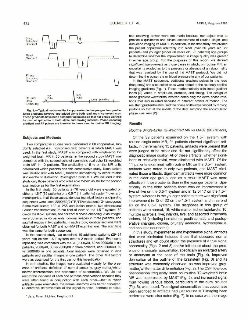
Fig. 1.-Typical motion-artifact suppression technique gradient profile. Extra gradients (arrows) are added along both read and slice-select axes. These gradients have been computer optimized so that net phase shift will be zero at spin echo of both static and moving material. Phase-encoding gradient and RF pulses are identical to those used in routine MR imaging.
Subjects and Methods
Two comparative studies were performed in 60 cooperative, randomly selected (i.e., nonconsecutive) patients in which MAST was used. In the first study, MAST was compared with single-echo T2-weighted brain MR in 50 patients; in the second study MAST was compared with the second echo of symmetric dual-echo T2-weighted brain MR in 10 patients. The availability of time on the MR units determined which patients had this comparative study. Each patient was studied first with MAST, followed immediately by either routine single-echo or dual-echo T2-weighted brain MR. We included in this study only those patients who were able to hold as still for the second examination as for the first examination.
In the first study, 50 patients 2-70 years old were evaluated on either a 1.5-T (39 patients) or a O.5-T (11 patients) system- over a 5-month period. In all these cases, identical technical factors and pulsing sequences were used: 2200/60/2 (TRfTE/excitations); 24 contiguous 5-mm-thick slices; 192 x 256 acquisition matrix; two-dimensional Fourier transformation; 25-cm field of view on the 1.5-T system, 30 cm on the 0.5-T system; and horizontal phase-encoding. Axial images were obtained in 45 patients, coronal images in three patients, and sagittal images in two patients. In each study, identical sections were obtained for both MAST and non-MAST examinations. The scan time was the same for both sequences.
In the second study, we examined 10 additional patients (29-84 years old) on the 1.5-T system over a 2-month period. Even-echo rephasing was compared with MAST (2000/30, 60 vs 2000/60 in six patients, 2000/40, 80 vs 2000/80 in three patients, and 2200/40, 80 vs 2000/80 in one patient). Axial images were obtained in nine patients and sagittal images in one patient. The other MR factors were as described for the first part of this investigation.
In both studies, the images were evaluated visually for the presence of artifacts, definition of normal anatomy, gray-matter/whitematter differentiation, and delineation of abnormalities. We did not record the incidence of each one of these observations because they were often found in conjunction with each other-that is, when artifacts were eliminated, the normal anatomy was better displayed. Quantitative determination of the signal-to-noise, contrast-to-noise,
- Vista, Picker, Highland Heights, OH.
and resolving power were not made because our object was to provide a qualitative and clinical assessment of routine single- and dual-echo imaging vs MAST. In addition , in the first study, we divided the patient population arbitrarily into older (over 50 years old, 22 patients) and younger (under 50 years old, 28 patients) age groups to determine whether the improvement in image quality was greater in either age group. For the purposes of this report, we defined significant improvement as those cases in which, on routine MR, an uncertainty existed as to the presence or absence of an abnormality that was resolved by the use of the MAST protocol. We did not determine the pulse rate or blood pressure in any of our patients.
In the MAST sequence, additional gradient pulses in the read (frequency) and slice-select axes were added to the routinely applied imaging gradients (Fig . 1). These mathematically calculated gradient lobes [2] varied in amplitude, duration, and timing . The design of these gradient waveforms involved computing the extra phase rotations that accumulated because of different orders of motion. The resultant gradients refocused the phase shifts experienced by moving protons so that at the middle of the data sampling the accumulated phase was zero [2].
Results
Routine Single-Echo T2-Weighted MR vs MAST (50 Patients)
Of the 39 patients examined on the 1.5-T system with routine single-echo MR, 24 patients showed significant artifacts; in the remaining 15 patients, artifacts were present that were judged to be minor and did not significantly affect the diagnostic image quality. All of these artifacts, whether significant or relatively trivial, were eliminated with MAST. Of the 11 patients examined with routine MR on the O.5-T system, artifacts were noted in only two patients, and MAST eliminated those artifacts. Significant artifacts were more common in the older age group, and as a result MAST was more effective in those patients than in the younger patients. Specifically, in the older patients there was an improvement in two of five on the O.5-T system and in 12 of 17 on the 1.5-T system, whereas in the younger patients there was significant improvement in 12 of 22 on the 1 .5-T system and in zero of six on the 0.5-T system. The diagnoses in this group of patients were normal, 16; white-matter ischemia/infarcts, 10; multiple sclerosis, five; infarcts, five; and assorted intracranial lesions, 14 (including hematoma, posttraumatic and postoperative changes, glioma, pituitary adenoma, hydrocephalus, and acoustic neurinoma).
In this study, hyperintense and hypointense signal artifacts that were eliminated included those that obscured normal structures and left doubt about the presence of a true signal abnormality (Figs. 2 and 3) and/or left doubt about the presence of a vascular abnormality, specifically an enlarged artery or aneurysm at the base of the brain (Fig. 4). Improved delineation of the outline of the brainstem (Fig. 3) and its structure was commonly observed, as was improved graymatter/white-matter differentiation (Fig. 2). The CSF flow-void phenomenon frequently seen on routine T2-weighted brain MR was suppressed by MAST (Fig . 5), and increased signal from flowing venous blood, particularly in the dural sinuses (Fig. 6), was noted. True signal abnormalities that could have been ascribed to artifacts had just routine MR imaging been performed were also noted (Fig. 7). In no case was the image

AJNR:9, May/June 1988 SUPPRESSION OF MR MOTION ARTIFACTS 433
Fig. 2.-1.5 T. A, Routine single-echo MR image
shows multiple artifacts along horizontal phase-encoding axis in temporal lobes. Hyperintensity in medial portion of left temporal lobe could be mistaken for a lesion. While reciprocal hypointensity in right temporal lobe should be recognized as an artifact, it is conceivable that such an area of decreased signal could obscure a true abnormality.
B, Motion-artifact suppression image clearly shows that those areas are artifactual in nature. Note also improved gray-matter /white-matter differentiation, particularly in temporal lobes.
Fig. 3.-1.5 T. A, Routine single-echo MR image.
High signal is identified in left side of pons. Despite reciprocal hypo intensity on opposite side of pons, it is unclear whether increased signal is a real abnormality.
B, Motion-artifact suppression technique. Pons is seen to be normal. Note also improved visualization of outline of pons by CSF, a finding not present on routine single-echo MR image.
A
quality diminished as a result of application of these extra gradients.
Even-Echo Rephasing vs MAST (10 Patients)
Of the 10 patients studied on a 1.5-T system in which the second echo of a symmetric dual-echo sequence was compared with the MAST sequence, we found a difference in the
B
B
images in four patients . The diagnoses in these patients were normal, three; white-matter ischemia/infarcts, five; lacunar infarct, one; and postoperative changes, one. Although motion-induced artifacts did not occur as frequently with the second-echo as with the single-echo sequence and the artifacts were not as severe nor as prominent, we found that MAST still improved image quality. An increase in signal
434 QUENCER ET AL. AJNR:9, May/June 1988
A B
A B
intensity from the CSF in the basal cisterns, resulting in improved definition of the brainstem (Fig. 8) and suprasellar cisterns (Fig. 9), was noted in cases in which a difference in image quality was seen. Refocusing on the CSF void flow in the aqueduct of Sylvius (Fig. 10) also resulted when the MAST sequence was used.
Discussion
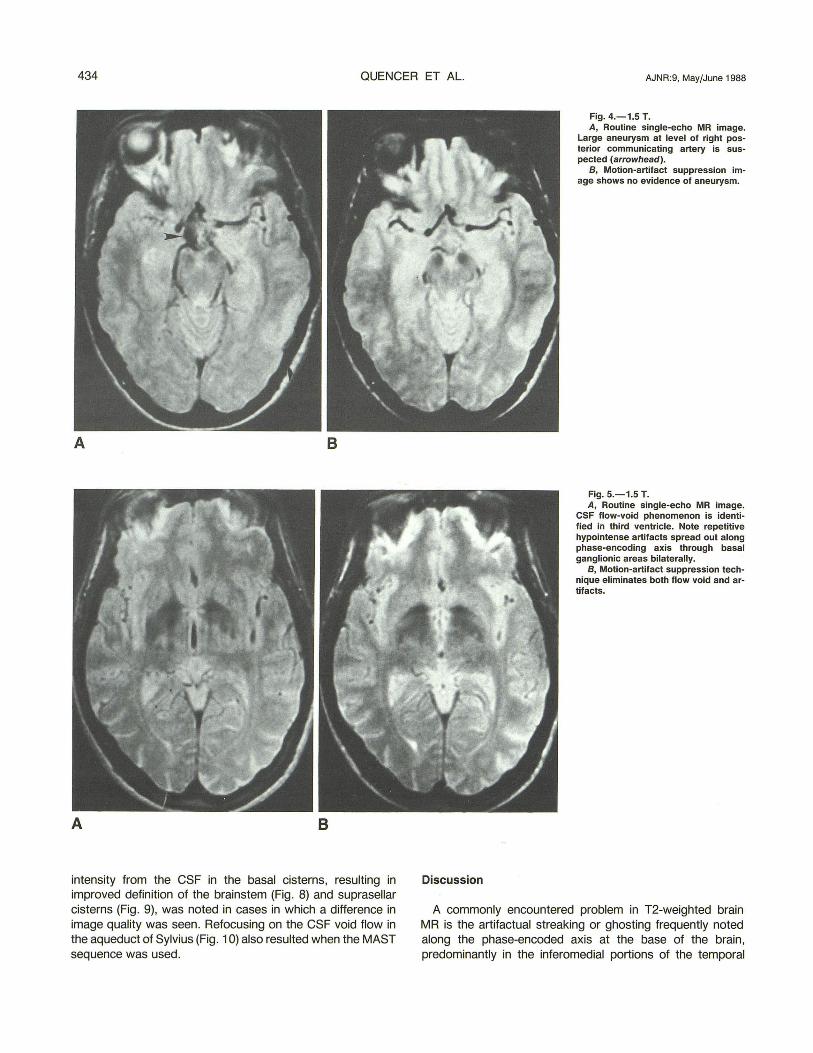
Fig. 4,- 1,5 T. A, Routine single-echo MR image.
Large aneurysm at level of right posterior communicating artery is suspected (arrowhead).
B, Motion-artifact suppression image shows no evidence of aneurysm.
Fig. 5.-1.5 T. A, Routine single-echo MR image.
CSF flow-void phenomenon is identified in third ventricle, Note repetitive hypointense artifacts spread out along phase-encoding axis through basal ganglionic areas bilaterally,
B, Motion-artifact suppression technique eliminates both flow void and artifacts.
A commonly encountered problem in T2-weighted brain MR is the artifactual streaking or ghosting frequently noted along the phase-encoded axis at the base of the brain, predominantly in the inferomedial portions of the temporal

AJNR:9, May/June 1988 SUPPRESSION OF MR MOTION ARTIFACTS 435
Fig. 6.-0.5 T. Little improvement in image quality of temporal lobes or brainstem is seen when routine singleecho MR (A) is compared with motionartifact suppression image (8). However, hyperintense signal from both transverse sinuses (arrowheads) in 8 is not present in A. This must not be mistaken for a venous thrombosis (see text). With motion-artifact suppression, there is an increase in signal intensity in interpeduncular cistern (straight ar· row) and in suprasellar area, particularly on right (curved arrow).
Fig. 7.-1.5 T. Not only does motionartifact suppression (8) eliminate temporaHobe artifacts seen on routine single-echo MR (A), but a small hyperintense pontine signal abnormality is convincingly shown (arrow). With routine MR, the assumption would have been that only artifacts were present in the brainstem, and this abnormality (presumed infarct) might have been missed. 8 better shows basilar artery and prepontine cistern, loss of marked hypointensity of ambient cisterns, and gray-matter/white-matter differentiation.
A
A
lobes (Figs. 2A and 7 A), the brainstem (Fig. 3A), and the basal ganglia (Fig. 5A). The artifacts are present because moving protons, as in pulsating CSF or flowing blood, acquire different phases as they move through the normally applied magnetic
B
B
gradients. While the routine single-echo MR pulse sequence rephases the phase shifts owing to field inhomogeneities and chemical shift, it cannot refocus motion-induced phase shifts. The result is not only a decrease in the signal intensity of

436 QUENCER ET AL. AJNR:9, May/June 1988
A 8
A 8
pulsatile CSF but also a spatial mismapping of the signal beyond its normal confines; that is, a phase-shift effect [3, 4]. Such artifacts can mimic or obscure a disease process and result in a false-positive or false-negative interpretation. Sequences that use long TEs are more susceptible to this type of artifact because there is more time for this motion to occur and because the motion-induced phase shifts are pro-
Fig. 8.-1.5 T. A, Second echo of dual-echo spin
echo sequence (2000/30, 60). Outlines of medulla are not well seen.
B, Motion-artifact suppression sequence (2000/60). Signal from CSF in basal cisterns is more intense, allowing better evaluation of size and contour of brainstem.
Fig. 9.-1.5 T. A, Second echo of dual-echo spin
echo sequence (2000/30, 60). Area of relative hypointensity (arrow) just to left of basilar artery could be mistaken for an abnormality.
B, Motion-artifact suppression sequence (2000/60). Cistern is seen to be normal. Complete refocusing of moving CSF in suprasellar cistern is not achieved in A, but with the extra gradients used in motion-artifact suppression, high orders of motion are compensated for, resulting in more uniform and higher signal intensity of CSF.
portional to time squared and cubed for velocity and acceleration, respectively [2].
The artifacts occur because of the movement of protons either between phase-encoding steps-that is, after sampling the echo but before the next 90° pulse-or during the time when the routinely applied magnetic gradients are on-that is, between the initial 90° pulse and sampling of the echo.
AJNR:9, May/June 1988 SUPPRESSION OF MR MOTION ARTIFACTS 437
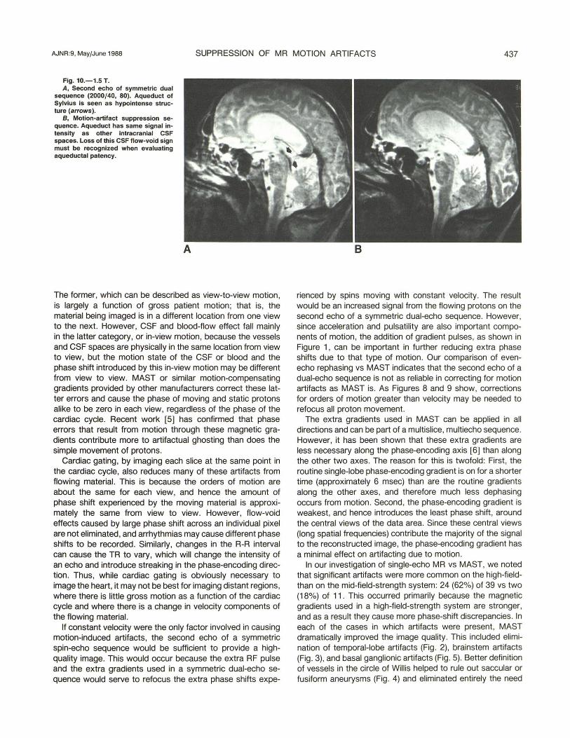
Fig. 10.-1.5 T. A, Second echo of symmetric dual
sequence (2000/40, 80). Aqueduct of Sylvius is seen as hypo intense structure (arrows).
8 , Motion-artifact suppression sequence. Aqueduct has same signal intensity as other intracranial CSF spaces. Loss of this CSF flow-void sign must be recognized when evaluating aqueductal patency.
A
The former, which can be described as view-to-view motion, is largely a function of gross patient motion; that is, the material being imaged is in a different location from one view to the next. However, CSF and blood-flow effect fall mainly in the latter category, or in-view motion, because the vessels and CSF spaces are physically in the same location from view to view, but the motion state of the CSF or blood and the phase shift introduced by this in-view motion may be different from view to view. MAST or similar motion-compensating gradients provided by other manufacturers correct these latter errors and cause the phase of moving and static protons alike to be zero in each view, regardless of the phase of the cardiac cycle. Recent work [5] has confirmed that phase errors that result from motion through these magnetic gradients contribute more to artifactual ghosting than does the simple movement of protons.
Cardiac gating, by imaging each slice at the same point in the cardiac cycle, also reduces many of these artifacts from flowing material. This is because the orders of motion are about the same for each view, and hence the amount of phase shift experienced by the moving material is approximately the same from view to view. However, flow-void effects caused by large phase shift across an individual pixel are not eliminated, and arrhythmias may cause different phase shifts to be recorded. Similarly, changes in the R-R interval can cause the TR to vary, which will change the intensity of an echo and introduce streaking in the phase-encoding direction. Thus, while cardiac gating is obviously necessary to image the heart, it may not be best for imaging distant regions, where there is little gross motion as a function of the cardiac cycle and where there is a change in velocity components of the flowing material.
If constant velocity were the only factor involved in causing motion-induced artifacts, the second echo of a symmetric spin-echo sequence would be sufficient to provide a highquality image. This would occur because the extra RF pulse and the extra gradients used in a symmetric dual-echo sequence would serve to refocus the extra phase shifts expe-
B
rienced by spins moving with constant velocity . The result would be an increased signal from the flowing protons on the second echo of a symmetriC dual-echo sequence. However, since acceleration and pulsatility are also important components of motion, the addition of gradient pulses, as shown in Figure 1, can be important in further reducing extra phase shifts due to that type of motion. Our comparison of evenecho rephasing vs MAST indicates that the second echo of a dual-echo sequence is not as reliable in correcting for motion artifacts as MAST is. As Figures 8 and 9 show, corrections for orders of motion greater than velocity may be needed to refocus all proton movement.
The extra gradients used in MAST can be applied in all directions and can be part of a multislice, multiecho sequence. However, it has been shown that these extra gradients are less necessary along the phase-encoding axis [6] than along the other two axes. The reason for this is twofold: First, the routine single-lobe phase-encoding gradient is on for a shorter time (approximately 6 msec) than are the routine gradients along the other axes, and therefore much less dephasing occurs from motion. Second, the phase-encoding gradient is weakest, and hence introduces the least phase shift , around the central views of the data area. Since these central views (long spatial frequencies) contribute the majority of the signal to the reconstructed image, the phase-encoding gradient has a minimal effect on artifacting due to motion.
In our investigation of single-echo MR vs MAST, we noted that significant artifacts were more common on the high-fieldthan on the mid-field-strength system: 24 (62%) of 39 vs two (18%) of 11 . This occurred primarily because the magnetic gradients used in a high-field-strength system are stronger, and as a result they cause more phase-shift discrepancies. In each of the cases in which artifacts were present, MAST dramatically improved the image quality. This included elimination of temporal-lobe artifacts (Fig. 2) , brainstem artifacts (Fig. 3), and basal ganglionic artifacts (Fig. 5) . Better definition of vessels in the circle of Willis helped to rule out saccular or fusiform aneurysms (Fig. 4) and eliminated entirely the need

438 QUENCER ET AL. AJNR:9, May/June 1988
to perform contrast-enhanced CT in questionable cases, as recommended by some authors [7]. Improved definition of the outline of the brainstem was common (Fig. 3). When thin sections are used to evaluate these areas of the brain, the use of motion-compensating gradients as described here is even more crucial because phase discrepancies are more pronounced with the larger gradient strength used to produce thin sections. Gray-matter/white-matter differentiation is also improved, particularly in the temporal lobes (Figs. 2 and 7), because the effects of transmitted pulsations to the brain were diminished. Not only will MAST suppress artifacts but it may also show true abnormalities where only artifacts would have been suspected on routine MR imaging. Particularly vulnerable to this type of misinterpretation are medial temporal-lobe or brainstem abnormalities (Fig. 7), in which phase artifacts can obscure a true abnormality or in which the diagnosis of an abnormality cannot be made with confidence.
There are a few potential drawbacks to the use of these motion-compensating gradients. Because of the time required to apply these gradients, a TE of no less than 30 msec in spin-echo MR is used currently. This, however, is a minor problem, because with this technique we are concerned mainly with improving the quality of long TR/long TE images. Echo times as short as 18 msec are available when MAST is used with field-echo imaging. Some limitations on the field of view and number of slices are necessary because of the high gradient-current requirements, but we have not found this to be a limitation in routine clinical scanning. When these motioncompensating gradients are used, there can be a difference in the appearance of both flowing venous blood and moving CSF when compared with routine MR. Specifically, there is increased signal from flowing blood in venous structures (e.g, see Fig. 6B). This occurs because the extra gradients that suppress the phase-shift effects serve to refocus the moving protons in vascular structures, resulting in an increased signal. When the question is raised about the possibility of a venous or dural thrombus, comparison with a T1 image will confirm the presence of either normal flow (low signal) or a thrombus (high signal). In arterial structures, the same effect is not noted because the high velocity and consequent time-of-flight effect are the dominant mechanisms of signal loss. In addition, because MAST suppresses the hypointensity of flowing CSF (Figs. 5B and 10B) the presence of a CSF flow void cannot be used to determine the patency of certain CSF pathways; for example, third ventricle, fourth ventricle, and aqueduct. These observations concerning venous flow and CSF flow must be recognized when these extra gradients are used. For example, if the hope is to extract certain physiologic information concerning CSF flow on the basis of the MR images in syringomyelia or in obstruction of the CSF pathways, particularly in the aqueduct, additional MR images would have to be obtained in which these motion-compen-
sating gradients are not used. The presence of a CSF flow void on a routine spin-echo sequence will assure patency of the CSF pathways.
Two obvious questions that could be raised are (1) why were the phase-shift artifacts present in some patients and not in others and (2) why was there a variability in both the intensity and position of the artifacts when they were present. Both physiological and imaging parameters, such as heart rate, CSF pulsation amplitude, and TR, playa role in this phenomenon [3]. Since we had measured neither the blood pressure nor the heart rate of our patients, we decided to look at the one recorded item that may have been a factor: the patient's age. In the older age group (>50), two (40%) of five studied on the 0.5-T unit had these artifacts and 12 (71 %) of 17 studied on the 1.5-T system had such artifacts. This is compared with zero of six on the 0.5-T unit and 12 (55%) of 22 on the 1.5-T unit in the younger age group «50). While we speculate that perhaps a combination of increased pulse pressure and larger cisternal and ventricular spaces in the older age group, together with decreased compliance of the brain, could possibly explain these differences, no data were collected to confirm this.
We believe the use of the extra gradients as described in this article is preferable to cardiac gating to suppress motion artifacts due to flowing blood and CSF movement. This method is simple to use, requires no ECG or plethysmographic monitoring, and can be used effectively in patients with an irregular heart rate. There is even more suppression of motion-induced artifacts with these extra gradients than is seen with second-echo rephasing. The disadvantages of this technique are minimal , easily recognized, and managed in clinical scanning.
REFERENCES
1. Enzmann DR, Rubin JB, O'Donohue J, Griffin C, Drace J, Wright A. Use of cerebrospinal fluid gating to improve T2 weighted images. Part II : Temporal lobes, basal ganglia and brain stem. Radiology 1987;162 :768-773
2. Pattany PM, Phillips JJ , Chiu LC, et al. Motion artifact suppression technique (MAST) for MR imaging. J Comput Assist Tomogr 1987;11 :369-377
3. Rubin JB, Enzmann DR. Imaging of spinal CSF pulsation by 20FT MR: significance during clinical imaging. AJNR 1987;8 :297-306
4. Rubin JB, Enzmann DR. Harmonic modulation of proton MR precessional phase by pulsatile motion: origin of spinal CSF flow phenomena. AJNR 1987;8 :307-318
5. Haacke ZM, Lenz GW. Effects of rephasing gradients on motion artifacts. Presented at the annual meeting of the Radiological Society of North America, Chicago, December 1986
6. Duerk JL, Pattany JM. Analysis of imaging axes significant in motion artifact suppression technique (MAST) refocusing of moving spins. Magn Reson Imaging 1987;5 [SuppI1]:108-109
7. Burt TB. MR of CSF flow phenomenon mimicking basilar artery aneurysm. AJNR 1987;8:55-58