imported cutaneous leishmaniasis in hungary - zobodat · imported cutaneous leishmaniasis in...
TRANSCRIPT

Mitt. Österr. Ges. Tropenmed. Parasitol. 8 (1986) 67-72
Hungarian Tropical Health Institute (Head: Prof. F. Värnai MD)
Imported Cutaneous Leishmaniasis in Hungary
F. Värnai, D. Bänhegyi, Eva Fülöp
Introduction
The number of imported exotic diseases - among them cutaneous leishmaniasis - israpidly increasing due to the development of manifold connections with the tropicaland subtropical countries, as well as to the growing rate of tourism. More and moreHungarian citizens travel year by year also to the developing countries. 31 cutaneousleishmania cases have been diagnosed in Hungarian subjects returning from endemicareas during the past three years.
This paper wants to call attention to the diagnostic and therapeutic problems of theimported disease which is non-endemic neither in Hungary nor in Central Europe. Withreference to our cases I would like to report our new therapeutic results.
This well-known disease belongs to the zooanthroponoses, the pathogen is a proto-zoon of the family Trypanosomatidae. Leishmania donovani, Leishmania tropica majorand minor, and Leishmania braziliensis are the most important pathogenes forhumans. The infection is transmitted to human beings from animal hosts — primarilyfrom dogs and small rodents - by small haematophagous insects, the Phlebotominae.The pathomechanism is different according to the leishmania species, causing thevisceral (or kala-azar), the cutaneous and muco-cutaneous clinical forms.
Leishmaniasis cutanea is called Aleppean sore (first described by RÜSSEL in 1756),Bagdadian, Moroccan or Oriental sore, referring to its place of origin.
From the clinical point of view it is a chronic, granulomatose, ulcerative skin-disease.The pathogenic agent is Leishmania tropica discovered by BOROWSKIJ in 1898. Thevectors of the infection are the Phlebotomus species, the so-called "sandflies" thatbite at night and the flagellates of the parasites are transplanted into the skin duringthe bite. The infection spreads from man to man very rarely and only in poor hygienicand social conditions (6).
Cutaneous leishmaniasis is endemic in mediterranean and subtropical areas, wherethe exposition is seasonal. In Europe it is endemic in some parts of Greece, butautochthonous infection occurs in Switzerland and South-France too (2, 3, 4, 5).
Patients and methods
31 cutaneous leishmaniasis cases were diagnosed and treated between 1981 -1983.The majority of the patients (19) came from our outpatient clinic, where compulsoryscreening examinations are carried out with Hungarian subjects returning home duringand after long-term service abroad.
67
©Österr. Ges. f. Tropenmedizin u. Parasitologie, download unter www.biologiezentrum.at

Diagnosis
Specific tests ot the skin lesions were performed besides routine laboratory tests.Mucus was taken from the sore base of the skin lesions for smear tests. Having beendried, the smears were fixed in methanol or stained with 10% Giemsa solution for10 minutes. Pathogens were found in stained smears under microscope with 400 -1000fold enlargement. Leishmania tropica is a 3-5 j.i size ovoid shaped protozoon, itoccurs most frequently intracellularly, sometimes also extracellularly. In chronic casesthe number of pathogens were significantly reduced, they could only be found intra-cellularly (Fig. 1).
Fig.1
Therapy
Considering the therapy of cutaneous leishmaniasis there are different opinions. Outof several literary data we would like to support HENRIKSON's and LENDE's resultswith the local application of chlorpromazine in three cases in 1983 (1).
Patients were treated locally or both locally and systemically according to the numberand size of the skin lesions and the quantity of pathogens. Sores on the surface werecleared with desinficient ointment or antibiotics. Skin lesions were heated 3 -4 timesa day for 10 minutes each with infrared lamps. In some cases, when the first treatmentseemed to be insufficient, the patient was treated with another method.
The following drugs were used systemically:
1. 300 mg chloroquine (Delagil®) was given to 6 patients daily for a week, it wascompleted with the local heating and chloroquine administration of the lesions.
2. Rifampicin (Tubocin®) was given to 6 patients 900 mg/day for 4 weeks, which wascompleted with local treatment.
3. Pentamidin (Lomidine*) was given to 3 patients 24 mg/day for 2-4 weeks.
4. 1 g/day metronidazole (Klion®) was applied in 2 cases for 2 weeks completed withlocal heating.
68
©Österr. Ges. f. Tropenmedizin u. Parasitologie, download unter www.biologiezentrum.at
5. Pentavalent antimon compounds (Stibophen® or Glucanthime®) were used in7 cases with total doses of 8 g.
6. 7 patients were given 150 mg/day chlorpromazine (Hibernal®) for 2 -4 weeks whichwas completed with local treatment.
Chlorpromazine applied locally:
Cutaneous leishmaniasis skin-lesions were treated either with 0,2% chlorpromazine(25% salycil vaselin ointment) or 5% chlorpromazine given directly to the lesions. Thiskind of treatment was applied in 16 cases. In about 50% of the total cases the localtreatment was completed with chlorpromazine given systemically according to thespread and great number of skin lesions as well as to the quantity of protozoa.
Results
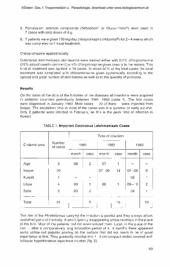
On the basis of the data of the histories of the diseases all infections were acquiredin endemic countries periodically between 1981-1983 (table 1). The first caseswere diagnosed in January 1982. Most cases - 20 of them - were imported fromIraque. The exposition time in most of the cases was late summer or early autumn.Only 2 patients were infected in February, as this is the peak time of infection inKuwait.
TABLE 1: Imported Cutaneous Leishmaniasis Cases
Endemic area
Algir
Iraque
Kuwait
Libya
Syria
Total
Numberof cases
3
20
1
4
3
31
Time of infection
1981
month
08
09
09
case
2
1
2
5
1982
month
07
07-09
08
case
1
14
1
16
1983
month
02-09
02
09-10
08
case
6
1
2
1
10
The bite of the Phlebotomus carrying the infection is painful and they always attackunclothed parts of the body. A small, quickly disappearing urtica develops in the placeof the bite. Most of the patients had not even noticed them. Later, in the place of thebite - after a comparatively long incubation period of 3 -4 months there appearedsome yellow-red papulas peeling on the surface that did not seem to be of greatimportance at first. They gradually develop into 1 - 3 cm compact nodes covered withfollicular hyperkeratous squamous crustae (fig. 2).
69
©Österr. Ges. f. Tropenmedizin u. Parasitologie, download unter www.biologiezentrum.at
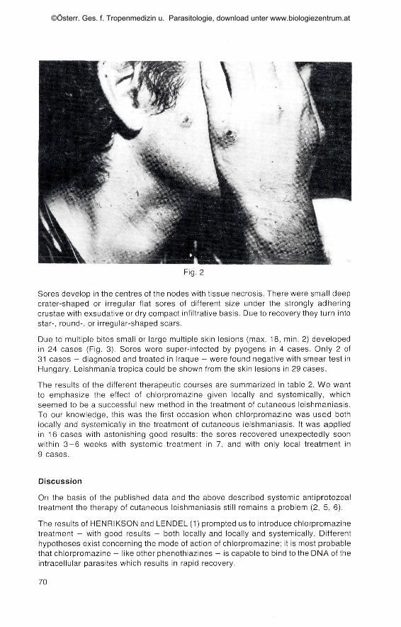
Fig. 2
Sores develop in the centres of the nodes with tissue necrosis. There were small deepcrater-shaped or irregular flat sores of different size under the strongly adheringcrustae with exsudative or dry compact infiltrative basis. Due to recovery they turn intostar-, round-, or irregular-shaped scars.
Due to multiple bites small or large multiple skin lesions (max. 18, min. 2) developedin 24 cases (Fig. 3). Sores were super-infected by pyogens in 4 cases. Only 2 of31 cases - diagnosed and treated in Iraque - were found negative with smear test inHungary. Leishmania tropica could be shown from the skin lesions in 29 cases.
The results of the different therapeutic courses are summarized in table 2. We wantto emphasize the effect of chlorpromazine given locally and systemically, whichseemed to be a successful new method in the treatment of cutaneous leishmaniasis.To our knowledge, this was the first occasion when chlorpromazine was used bothlocally and systemically in the treatment of cutaneous leishmaniasis. It was appliedin 16 cases with astonishing good results: the sores recovered unexpectedly soonwithin 3-6 weeks with systemic treatment in 7, and with only local treatment in9 cases.
Discussion
On the basis of the published data and the above described systemic antiprotozoaltreatment the therapy of cutaneous leishmaniasis still remains a problem (2, 5, 6).
The results of HENRIKSON and LENDEL (1) prompted us to introduce chlorpromazinetreatment - with good results - both locally and locally and systemically. Differenthypotheses exist concerning the mode of action of chlorpromazine; it is most probablethat chlorpromazine - like other phenothiazines - is capable to bind to the DNA of theintracellular parasites which results in rapid recovery.
70
©Österr. Ges. f. Tropenmedizin u. Parasitologie, download unter www.biologiezentrum.at

Fig. 3
TABLE 2: Effectivity of Therapy of Cutaneous
DRUG
Chloroquine(Delagil®)
Rifampicine (Tubocin®)
Pentamidine (Lomidine®)
Metronidazole (Klion®)
Pentavalent antimon compounds(Glucantime® or Stibophen®)
Chlorpromazine (Hibernal®)systemically and locally
Chlorpromazine (Hibernal®)only locally
Cases
6
6
3
2
7
7
9
Recovered
3
2
1
-
3
7
9
Leishmaniasis
Improved
-
4
1
4
-
-
Unchanged
-
-
1
2
-
-
-
71
©Österr. Ges. f. Tropenmedizin u. Parasitologie, download unter www.biologiezentrum.at

Summary
Thirtyone cutaneous leishmaniasis cases imported by Hungarian subjects are repor-ted and the diagnostic possibilities and therapeutical experiences discussed. Theauthors describe the results obtained with chlorpromazine which was applied locallyas well as locally and systemically in 16 cases. The cutaneous lesions healed within3-6 weeks and during the follow-up no relapse was observed.
Zusammenfassung
Importierte kutane Leishmaniose in Ungarn
Die Autoren machen 31 Fälle von kutaner Leihmaniose, welche durch ungarischeStaatsangehörige importiert wurde, und die damit zusammenhängenden diagnosti-schen und therapeutischen Erfahrungen bekannt. In 16 Fällen referieren sie ausErgebnissen mit lokaler und systemischer Chlorpromazin-Therapie.
Wir stellten fest, daß die Hautschädigungen in 3-6 Wochen heilen und kein Rückfallzu registrieren war.
References
1. HENRIKSON, Th. H., LENDE, S. (1983): Treatment of diffuse cutaneous leishmaniasis with Chlorpromazine
ointment. Lancet, Vol. I., 126.
2. KURBAN, A. K. (1973): Behandlung der Haut-Leishmaniose. Hautarzt, 24, 369-372.
3. MAZZI, R. (1976): Kutane Leishmaniose: autochthoner Fall in der Schweiz? Dermatologica, 153, 104-105.
4. STRATIGOS, J. D. et al. (1980): Epidemiology of cutaneous leishmaniasis in Greece. Int. J. Dermatol. 19,
86-88.
5. SCHUPPLI, R. (1982): Leishmaniasis Review. Dermatologica, 165, 1 -6 .
6. VÄRNAI, F.: Tröpusi betegsegek. Medicina Kiadö, Budapest 1973, 185. (Tropical Diseases, University Text-book)
ADDRESS FOR CORRESPONDANCE:
Prof. Ferenc Värnai MDHungarian Tropical Health Institute
Gyäli ut 5-7H-1097 Budapest
72
©Österr. Ges. f. Tropenmedizin u. Parasitologie, download unter www.biologiezentrum.at