importance of isolation to the evolution of …
TRANSCRIPT

Biogeography of thermophilic cyanobacteria and the importance of isolation to the evolution ofmicroorganismsby Robertson Thane Papke
A dissertation submitted in partial fulfillment of the requirements for the degree of Doctor ofPhilosophy m MicrobiologyMontana State University© Copyright by Robertson Thane Papke (2002)
Abstract:Evolutionary theory predicts the divergence of populations when they become geographically isolated.However, Baas Becking's theory that "everything is everywhere and the environment selects" excludesgeographic isolation for microorganisms. In previous diversity and distribution studies, the sequencingof 16S rRNA genes acquired from natural Synechococcus populations residing in hot spring mats fromYellowstone National Park revealed that a single morphology concealed a rich 16S rRNA genotypicdiversity. Predominating within that diversity is a group of closely related 16S rRNA genotypes (theA/B cluster) that are uniquely distributed along thermal and light gradients. Curiously, the uppertemperature limit for cyanobacterial mat formation is different in globally disparate sites suggestingbarriers to dispersal for some populations. I hypothesized that either members of the A/B cluster aredistributed globally, but the highest temperature adapted forms (A types) are limited in their dispersalcapabilities, or alternatively, globally disparate hot springs are dominated by unrelated Synechococcusgenotypes. To test these hypotheses, I performed phylogenetic analysis on PCR-amplified, cloned, 16SrDNA genes recovered from Synechococcus populations residing in hot spring mats in Italy, NewZealand, Japan and the northwest U.S.A. The abundance of detected lineages was determined usinglineage-specific oligonuleotide probes; low-abundance genotypes were sought using the same probes asPCR primers. I also assessed 20 different hot spring physical/chemical properties to determine whetheradaptation was important to the local and global distributions of Synechococcus populations. Resultsrevealed that: (1) A/B cluster 16S rDNA sequences were not detected outside of the U.S., (2) eachcountry had unique dominating Synechococcus genotypes, (3) within the U.S. and Japan there existlocal geographic clades for A/B and Cl lineages, respectively, at the 16S rRNA and internal transcribedspacer region loci, (4) Oscillatoria amphigranulata, a filamentous thermophilic cyanobacterial speciesalso demonstrated unique geographical distributions, and (5) genetic variation did not correlate withtested hot spring physical/chemical parameters. The results revealed that all cyanobacterial lineageshad a different dispersal capability, but even the most widely dispersed exhibited substantial evidenceof geographic isolation. Additional evidence for isolated prokaryotic populations is reviewed and thegeneral importance of isolation in microbial evolution is emphasized.
BIOGEOGRAPHY OF THERMOPHILIC CYANOBACTERIA AND THE
IMPORTANCE OF ISOLATION TO THE EVOLUTION
OF MICROORGANISMS
by
Robertson Thane Papke
A dissertation submitted in partial fulfillment o f the requirements for the degree
Of
Doctor o f Philosophy
m
Microbiology
MONTANA STATE UNIVERSITY Bozeman, Montana
February 2002

M l 4APPROVAL
of a thesis submitted by
Robertson Thane Papke
This dissertation has been read by each member of the dissertation committee and has been found to be satisfactory regarding content, English usage, format, citations, bibliographic style, and consistency, and is ready for submission to the College of Graduate Studies.
Dr. David M Ward(Signature)
6^ Y i. V,v ^Date
Dr. CliffBond
Approved for e Department of Microbiology
(Signature) Date \
Approved for the College of Graduate Studies
(Signature) /e P - e P 7 - 0
DateDr. Bruce McLeod

iii
STATEMENT OF PERMISSION TO USE
In presenting this thesis in partial fulfillment o f the requirements for a doctoral
degree at Montana State University-Bozeman, I agree that the Library shall make it
available to borrowers under the rules o f the Library. I further agree that copying o f this
thesis is allowable only for scholarly purposes, consistent with “fair use” as prescribed in
the U.S. Copyright Law. Requests for extensive copying or reproduction of this thesis
should be referred to University Microfilms International, 300 North Zeeb Road, Ann
Arbor, Michigan 48106, to whom I have granted “ the exclusive right to reproduce and
distribute my dissertation in and from microform along with the non-exclusive right to
reproduce and distribute my abstract in any format in whole or in part.”
Signature
Date O -Z -K t U x

iv
ACKNOWLEDGEMENTS
I would like to thank Dr. David Ward for guiding me through the long process o f
earning a Ph.D. degree and for teaching me the importance o f raising the bar o f my own
expectations and efforts. Thank you Mary Bateson, your kindness and friendship has
made my journey through Bozeman a very happy one. Thank you Kenji Kato for sharing
your house, hospitality and friendship and for your supreme efforts in helping me arrange
my entire my Japanese hot spring collections. I thank the members o f my committee,
especially Adam Richman, for their time and effort in making this a better thesis. And
thanks to all o f the students and fellow scientists at MSU who have enriched my life both
personally and scientifically with special thanks to Greg Colores, Mike Franklin, Myke
Ferris, Uli Nubel and Marcel van der Meer. I could not have done this without any o f
you and I am eternally grateful to each and every one o f you.
This research was funded by grants from the National Science Foundation, NASA,
the Summer Institute in Japan and the Thermal Biology Institute.
1. INTRODUCTION ............................................................................ ...................... I
Development o f Evolutionary Theory in Microbiology....................................... IMicrobial B iogeography....................... ; ............................................................. 4Goals o f the T h esis ...................................................................................................9References C ite d ............................. 12
2. GEOGRAPHIC ISOLATION AND THE EVOLUTIONOF HOT SPRING CYANOBACTERIA................................................................ 16
Introduction................................ 16Hot Spring Mats as Island-like C om m unities.....................................................18Geographic Patterning o f Diversity........................................................................22Lineage-Specific 16S rRNA Probing...................................................................... 27Geochemical P a ttern s ................ 29Importance o f Geographic Isolation........................................................................32C on clu sion ............................................................................................................... 34Methods . ................................................................................................................... 35Sample Collection and Microscopy........................................................................ 35Sequence Acquisition and Analysis........................................................................35
Minimizing PCR and Cloning Artifacts . . . ........................................36rRNA Dot Blot Hybridization....................................... 36Lineage-Specific P C R ................................................................. .37Chemical Analysis............................. .38
References C it e d ......................................................................................................39
THE IMPORTANCE OF ISOLATION INMICROBIAL EVOLUTION. ............................................................................... 44
Introduction............................................................................................................... 44Isolation in Sexual S p ecies..................................................................................... 45Isolation in Prokaryotes................................ 46Physical Isolation o f Bacterial Populations........................................................... 48
Host-symbiont Population Isolation........................................................48Geographic Isolation........................................... . ' ................................... 52
The Ramifications o f Population Isolation ............................................................56References C ite d ................................................................................. 57
4. SUMMARY..................................................................................................... .64
V
TABLE OF CONTENTS
References Cited 66

vi
APPENDIX A: Supplemental Tables.................................................................................. 67
vii
Table Page
1. Physical, chemical and biological data for hot springssampled in different geographic regions o f all countries..................................23
2. Relative abundance o f Synechocooccus 16S rRNA lineagesin mats from each country.....................................................................................28
3. Physical, chemical and biological data for all hotsprings sampled............................................ .......................................................... 68
4. Chemical measurements for all hot springs sampled.........................................74
LIST OF TABLES
, viii
LIST OF FIGURES
Figure Page
1. 16S rKNA gene tree demonstrating the relationships o f clones retrieved from all countries to othercyanobacterial 16S rRNA sequences..................................................................20
2. Phylogenies for ITS variants detected in Yellowstone or Japan relative to springs and subregions from whichthey were retrieved................................................................................................. 25
3. Lineage-specific PCR o f Synechococcus indifferent geographic regions.......................... ......................................... .28
4. Hierarchical cluster analysis o f hot spring chemical parameters compared to 16S rRNA lineages and specific 16S rRNA and ITS genotypes found in eachhot spring.......................................................: . . ... .............................................30
5. Maximum likelihood phytogenies for nine specieso f vesicomyid clams and their associated endosymbionts...............................49
6. Red algal species (Prionitis) phylogenetically comparedto the pathogens found in galls o f each host............................. ......................52
7. REP-PCR band pattern similarity dendrogram demonstrating the relationship between the genetic diversity o f fluorescent Pseudomonas cultivated fromsoils and the geographical origins o f the isolates............................................54

ix
ABSTRACT
Evolutionary theory predicts the divergence o f populations when they become geographically isolated. However, Baas Seeking's theory that "eveiything is everywhere and the environment selects" excludes geographic isolation for microorganisms. In previous diversity and distribution studies, the sequencing o f 16S rRNA genes acquired from natural Synechococcus populations residing in hot spring mats from Yellowstone National Park revealed that a single morphology concealed a rich 16S rRNA genotypic diversity. Predominating within that diversity is a group o f closely related 16S rRNA genotypes (the A/B cluster) that are uniquely distributed along thermal and light gradients. Curiously, the upper temperature limit for cyanobacterial mat formation is different in globally disparate sites suggesting barriers to dispersal for some populations. I hypothesized that either members o f the A/B cluster are distributed globally, but the highest temperature adapted forms (A types) are limited in their dispersal capabilities, or alternatively, globally disparate hot springs are dominated by unrelated Synechococcus genotypes. To test these hypotheses, I performed phylogenetic analysis on PCR- amplified, cloned, 16S rDNA genes recovered from Synechococcus populations residing in hot spring mats in Italy, New Zealand, Japan and the northwest U.S.A. The abundance o f detected lineages was determined using lineage-specific oligonuleotide probes; low- abundance genotypes were sought using the same probes as PCR primers. I also assessed 20 different hot spring physical/chemical properties to determine whether adaptation was important to the local and global distributions o f Synechococcus populations. Results revealed that: (I) A/B cluster 16S rDNA sequences were not detected outside o f the U.S., (2) each country had unique dominating Synechococcus genotypes, (3) within the U.S. and Japan there exist local geographic clades for A/B and Cl lineages, respectively, at the 16S rRNA and internal transcribed spacer region loci, (4) Oscillatoria amphigranulata, a filamentous thermophilic cyanobacterial species also demonstrated unique geographical distributions, and (5) genetic variation did not correlate with tested hot spring physical/chemical parameters. The results revealed that all cyanobacterial lineages had a different dispersal capability, but even the most widely dispersed exhibited substantial evidence of geographic isolation. Additional evidence for isolated prokaryotic populations is reviewed and the general importance o f isolation in microbial evolution is emphasized.

I
CHAPTER I
INTRODUCTION
Development of Evolutionary Theory in Microbiology
Great inroads toward comprehending evolution and the formation of species were
made after naturalists and scientists visited locations around the globe, collected plants,
animals and fossils and charted the organisms’ relatedness against local and/or global
distributions and ecological gradients. The independent formation by Darwin and
Wallace o f the theory of descent with modification via natural selection was completely
dependent upon their observations that different yet related species lived in different
regions o f the world or on separate islands within archipelagos. As biologists searched
for and catalogued the diversity o f organisms on Earth, the disciplines o f biogeography,
and more recently phylogeography revealed many more corresponding patterns of
organismal relatedness with geography1. As a mechanism for speciation geographic
isolation is fundamentally different from natural selection, since population differences
are driven by neutral genetic drift, not adaptation. Rosenzweig2 expressed the importance
of geographic isolation to the development of species when he articulated that
“geographical speciation is the most common mode among most taxa in most places at
most times.” Indeed, the familiar terms used to describe speciation events, allopatric,
parapatrie and sympatric speciation all refer to the relative distances (distant, near or
2
together, respectively) that separate two sister Species. Today, it is recognized that
populations diverge whenever any kind o f barriers to mating success are formed (e.g.
different habitats, differential mating periods [day, season or year], anatomical
incompatibility, different mating rituals, hybrid death or sterility). In the time since
Darwin and Wallace published their great contributions to the science o f biology, much
has been learned about organismal diversity and mechanisms for speciation.
Unfortunately, evolutionary theory did not have a major impact on the field of
microbiology. In 1963, nearly 300 years after van Leeuwenhoek first discovered
microorganisms in his microscope and more than 100 years after Darwin and Wallace
published, Stanier et al.3 concluded that, “...any systematic attempt to construct a detailed
scheme o f natural relationships becomes the purest speculation....” The reasons may be
obvious. Macroorganisms can be visualized and collected and morphologically,
physiologically, ecologically and genetically described with relative ease.
Microorganisms on the other hand are invisible to the naked eye, collections involve
cultivation methods that allow recovery of only those that can grow under the conditions
presented, their morphologies are exceedingly simple and relatively unvaried, and their
diverse phenotypic properties are relatively useless for understanding evolutionary
relationships. Because of these limitations, microbiology as a discipline was relegated to
the applied side o f science (i.e. tools to help the human condition) resulting in countless
applications for food science, disease and medicine, genetics, physiology and cellular
biology.
I
Years after the Stanier lament, Woese4 changed the paradigm of microbiology by
describing the three-domain “tree o f life” based on sequencing the 16S rKNA molecule
of prokaryotes (18S rRNA of eukaryotes). For the first time, the full scope of prokaryotic
diversity was placed within the confines o f phylogenetic relatedness. Classification
based upon evolutionary relationship, once thought impossible, is now possible. The new
classification scheme inspired Norman Pace and others5 to recognize that microorganisms
could be identified in situ (without cultivation) by comparing “naturally” occurring 16S
rRNA molecule sequences (obtained via molecular techniques) to sequences o f cultivated
strains in the three-domain tree. Free from the confines o f cultivation, microbial
ecologists began natural history surveys that further demonstrated the great diversity of
microorganisms and stimulated interesting questions about the causes o f such diversity.
For instance, 16S rRNA analysis o f cyanobacterial mats residing in Octopus and
Mushroom hot springs in Yellowstone National Park demonstrated that the in situ 16S
rRNA gene sequences were different from those o f cultivated isolates6 and that closely
related Synechococcus (unicellular cyanobacteria) were uniquely distributed across
temperature and light gradients7,8. It was suggested9 that the evolutionary/ecological
theory, adaptive radiation (i.e. differential adaptation to various environments) couldI
explain the observed relationship of the genotypes to their unique niches, a theory
modeled after the adaptation o f “Darwin’s finches” to different niches on the Galapagos
Islands. However, without further distribution analysis (e.g. global sampling) it cannot
be determined if the Yellowstone Synechococcus radiated within Yellowstone’s borders
or if they have a wider distribution.
3

4
Microbial Biogeographv
It is interesting to note that with the new microbial paradigm, lots o f problems
have been solved, but new problems have arisen. Perhaps the biggest obstacle in the field
of microbial biogeography is the question o f identity (i.e. how do we know if two
populations belong to the same species or if two organisms belong to the same
population?). This is of extreme importance when trying to determine the geographic
range o f a specific species or population. To differentiate species or populations, it is
critical to have an established set of criteria, which can be applied to and measured on
individuals. In some cases this can be relatively easy. While in New Guinea, Emst
Mayr10 collected and identified 138 species o f birds o f which the island’s indigenous
people identified 137, suggesting that species are not arbitrarily defined but universally
accepted regardless o f who is counting. However, the myriad o f species definitions or
concepts, contradicts this notion11. Furthermore, it is difficult to identify a single species
concept that can be applied to all groups o f organisms, extant and extinct, haploid,
diploid and polyploid, sexual and asexual or macroorganism and microorganism.
Perhaps Darwin12 expressed the problem best when he wrote “there is no possible test but
individual opinion to determine which.. .shall be considered as species and which as
varieties.” If species are so difficult to define, then perhaps that unit o f identity should
not be used, especially with respect to microorganisms where separate species have been
arbitrarily defined as organisms with less than 70% similarity in DNA-DNA
hybridization,13 which roughly correlates to 97% similarity at the 16S rRNA locus14.

Lately, molecular markers (e.g. gene sequence variation) have been extremely successful
for linking relatedness with the distribution o f organisms1. Genetic relatedness can thus
be used to define identity. This is appropriate as divergence is really the issue, not what
species are. In attempts to determine the biogeography of microorganisms, molecular
markers, especially the 16S rRNA gene, have been used in addition to more classical
methods o f identification (e.g. phenotypic properties). However, there has been little
conformity in which measurements should be used to determine the geographic ranges of
the studied organisms. The use o f conserved genes, like 16S rRNA, to identify and/or
define populations may be particularly problematic as they may underestimate the actual
diversity and thereby artificially expand our impressions of territorial range. The pitfalls
in choosing a wrong level o f analysis for determining identity (e.g. morphology, gene
restriction enzyme fragment patterns or conserved vs. variable gene sequences) will be
considered below.
In a study using microscopy to determine the species diversity o f ciliated protozoa
(large unicellular eukaryotes whose species are morphologically defined), Fenchel et al.15
reported (from their study plus others) 181 and 146 species recovered from two
ecologically different sediments occurring in a pond (Priest Pot, UK) and shallow bay
(Niva Bay, Helsingor, Denmark). They determined that the diversity discovered was
approximately 11% of the total number o f free-living ciliate species. Furthermore, they
reasoned that similar results would have been found if additional nearby ciliate habitats
had been sampled (i.e. 10-20 ecologically different sites). The authors were confident
that if the more comprehensive sampling regime had been performed “a very substantial
6
fraction o f all known ciliates” would have been recovered from a relatively small
geographical range. From their interpretation of the data, they concluded, “everything is
(almost) everywhere”. However, the conclusion may be oversimplified. Organisms that
live in similar habitats can often have similar morphologies via convergent or parallel
evolution thereby concealing genetic diversity within a moiphotypically-defined species.
Indeed, many planktonic foraminifera species (morphotypically-defined) are comprised
of more than one genotype and these geontypes have been considered to be cryptic
sibling species16"18. In prokaryotes, all unicellular coccoid to rod-shaped cyanobacteria
fall within the genus Synechococcus 19. However, this genus is not monophyletic, as the
morphology has independently evolved many times20. As both examples clearly
demonstrate, it is risky to make conclusions about the distribution of microorganisms
when identity is based solely upon morphological criteria.
As expressed above, diversity and distribution studies o f microorganisms are
often performed using the 16S rRNA molecule either by restriction enzyme analysis or
by direct sequencing of the molecule. In an attempt to survey the archaeal diversity
present in the world’s oceans (North Atlantic, Cantabrian Sea [Atlantic Ocean], the
Mediterranean Sea, the Santa Barbara Channel [Pacific Ocean], and the Drake Passage
[Southern Ocean]) Massana et al.21 generated 16S rRNA gene libraries from natural
samples. They used two restriction enzymes to construct restriction fragment length
polymorphism (RFLP) patterns from their clone libraries and interpreted any RFLP
patterns that were identical as a single operational taxonomic unit (OTU). The analyses
of Massana et al.,21 revealed that 5 of the 36 OTU’s (representing 87% of the analyzed
a
clones) were “cosmopolitan”. The RFLP method is insensitive, as restriction enzymes
recognize a very small proportion (e.g. 4-8 nucleotides) o f the molecule analyzed. In a
computer simulation using prokaryotic 16S rRNA gene sequences, Moyer et ah,22 tested
the efficacy o f restriction enzymes in determining the diversity o f microorganisms. They
found that RFLP could only differentiate among sequences that were at least 3.9%
different. This clearly leaves a lot of diversity undetected, especially considering the
extremely conserved nature o f the 16S rRNA locus. In a study using Pseudomonas
strains isolated from soil samples collected around the world, Cho and Tiedje 23Compared
the effectiveness o f 16S rRNA RFLP patterns with repetitive extragenic palindrOmic-
PCR (REP-PCR, a very sensitive method that takes advantage of the entire genomic
diversity) for detecting endemic genotypes. In the case o f 16S rRNA RFLP pattern
analysis, only 4 OTU’s were found among 248 isolates and all 4 appeared cosmopolitan
in distribution. However, when REP-PCR was applied to each o f the strains, 85
genotypes were recovered and identical genotypes were only found in samples from the
same geographic sites, indicating high levels of endemism among the strains. Mehta et
ah,24 found similar results when they analyzed Zylella fastidiosa isolated from citrus trees
in Brazil. It would seem that 16S rRNA RFLP patterns completely underestimate the
true diversity o f microorganisms and any conclusions as to “cosmopolitan phylotypes”
should be avoided when using this technique.
Similar or identical 16S rRNA gene sequences have been used to declare that
some organisms have a worldwide distribution. Indeed, Garcia-Pichel et ah,25 found
identical or nearly identical 16S rRNA gene sequences from hypersaline-adapted
8
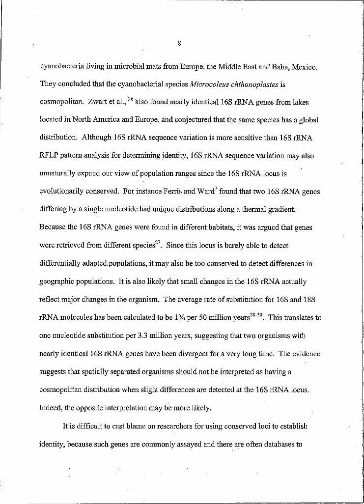
cyanobacteria living in microbial mats from Europe, the Middle East and Baha, Mexico.
They concluded that the cyanobacterial species Microcoleus chthonoplastes is
cosmopolitan. Zwart et al., 26 also found nearly identical 16S rRNA genes from lakes
located in North America and Europe, and conjectured that the same species has a global
distribution. Although 16S rRNA sequence variation is more sensitive than 16S rRNA
RFLP pattern analysis for determining identity, 16S rRNA sequence variation may also
unnaturally expand our view of population ranges since the 16S rRNA locus is
evolutionarily conserved. For instance Ferris and Ward7 found that two 16S rRNA genes
differing by a single nucleotide had unique distributions along a thermal gradient.
Because the 16S rRNA genes were found in different habitats, it was argued that genes
were retrieved from different species27. Since this locus is barely able to detect
differentially adapted populations, it may also be too conserved to detect differences in
geographic populations. It is also likely that small changes in the 16S rRNA actually
reflect major changes in the organism. The average rate o f substitution for 16S and 18S
rRNA molecules has been calculated to be 1% per 50 million years28"30. This translates to
one nucleotide substitution per 3.3 million years, suggesting that two organisms with
nearly identical 16S rRNA genes have been divergent for a very long time. The evidence
suggests that spatially separated organisms should not be interpreted as having a
cosmopolitan distribution when slight differences are detected at the 16S rRNA locus.
Indeed, the opposite interpretation may be more likely.
It is difficult to cast blame on researchers for using conserved loci to establish
identity, because such genes are commonly assayed and there are often databases to

9
which results can be compared. However, researchers should recognize the limits o f the
methods before drawing conclusions. If progress is to be made in microbial
biogeography, it is likely that more informative molecular markers with greater resolving
power will have to be used. For instance, the DNA-dependent RNA polymerase gene
OpoCI) evolves much faster than the 16S rKNA gene. Synechococcus sp. strains
WH7805 and WH8103 differ by 1.4% at the 16S rKNA locus, but differ by 17% at the
rpoCl locus31. The intemal/intervening/intergenic transcribed spacer (ITS) region
located on the rKNA operon between the 16S and 23 S rKNA genes also has a much
higher resolving power32'34. However, for in situ analysis, the ITS region has additional
benefits. Because the ITS is adjacent to the 16S rKNA gene, it is possible to PCR
amplify both loci simultaneously using the 16S rKNA gene to relate the sequence
phylogenetically to other known organisms while using the ITS to discriminate between
closely related genetic variants with identical 16S rKNA sequences.
Goals o f the Thesis
IfRosenzweig and other evolutionary biologists2,10,35"37 are correct in thinking that
geographic isolation is one of the major causes o f speciation, then perhaps it is time for
microbiologists to understand this biological paradigm and apply it to investigations
concerning microbial diversification and distribution, especially since most o f the
putative evidence (and dogma) that supports the “cosmopolitan” hypothesis is based on
observations that can easily be challenged. With this admonishment in mind, it is the

10
goal o f this thesis to provide convincing evidence that microorganisms can become
geographically isolated, that isolation can lead to diverging populations and consequently
that genetic drift may play an active role in the evolution of microorganism
independently o f adaptation (via mutation and lateral gene transfer) and natural selection.
My approach to microbial biogeography was to take advantage o f the island-like
nature of hot springs and previous observations concerning the diversity and distribution
of thermophilic cyanobacterial populations from around the globe. Anomalous
distributions such as the lack o f high-temperature adapted cyanobacteria in regions
outside o f the U.S.A.38,39 led to the main hypotheses:
Synechococcus mats in globally separated hot springs are dominated by A/B
genotypes, but there is a barrier to the dispersal o f higher temperature-adapted A-Iike
genotypes.
Or, alternatively, mats in globally separated hot springs are dominated by
Synechococcus unrelated to A/B genotypes.
The first hypothesis supports the idea that everything is everywhere, but nature
selects. The hypothesis predicts that both B and A-type.Synechococcus are ubiquitously
dispersed; the inability o f Iype-A Synechococcus to live above 63 C in some hot springs
is explained by environmental selection (e.g., sulfide in combination with high
temperature is known to prevent the growth o f cyanobacteria40'41). The alternative
hypothesis is consistent with geographic isolation. A test o f either hypothesis must also

11
address the possibility that distribution is patterned according to adaptation to specific
physical/chemical parameters.
To test these hypotheses, I made extensive collections from hot springs in Italy,
New Zealand, Japan and the northwest United States and analyzed samples by molecular
methods of suitable resolution. I developed a 16S rRNA method that allows genetic
comparisons to previous 16S rRNA studies while simultaneously sampling a higher
resolution genetic marker (ITS region) for detecting sequence variation between identical
or nearly identical 16S-rRNA defined genotypes. This is important because 16S rRNA
gene sequences are likely to conceal geographical isolation given their conserved nature.
I also generated group-specific 16S rRNA probes to quantify populations in their various
locations and, using PCR, to detect rare genotypes that may be present but difficult to
detect given the limitations o f detection methods. Furthermore, to convincingly
demonstrate the role o f adaptation or niche specialization in determining the distribution
of the thermophilic cyanobacteria, in-depth analysis o f the physical/chemical parameters
of sampled hot springs was performed. Because the results of this work could potentially
shift theoretical paradigms in microbiology, chapter 2 was prepared as a research article
for the journal Nature and the experimental results are thus presented in a condensed
style. Furthermore, much additional literature detail is placed intentionally in a
minireview (chapter 3) designed to add my results to a growing body o f evidence on
physical isolation in microbial evolution, an issue that needs to be emphasized to
microbiologists.

12
REFERENCES CITED
1 A vise, J. 2000. Phylogeography: The History and Formation o f Species.Harvard University Press, Cambridge, MA.
2 Rosenzweig, M. 1996. SpeciesDiversityinSpaceandTim e. Cambridge University Press, UK.
3 Stanier, R.Y., Doudoroff, M. and Adelberg, E.A., 1963. The Microbial World,2nd ed., Prentice-Hall, Inc., Englewood Cliffs, NI.
4 Woese C. 1987. Bacterial evolution. Microbiol. Rev. 51:221-271.
5 Olsen, G.L., Lane, DJ., Giovannoni, S.J., and Pace, N.R. 1986. Microbial ecology and evolution: a ribosomal RNA approach. Ann. Rev. Microbiol. 40:337-365.
6 Ward, D.M., Weller, R. and Bateson, M.M. 1990; 16S rRNA sequences reveal numerous uncultured microorganisms in a natural community. Nature 345: 63- 65.
7 Ferris, M., and Ward, D. 1997. Seasonal distributions o f dominant 16S rRNA- defined populations in a hot spring microbial mat examined by denaturing gradient gel electrophoresis. Appl Environ. Microbiol. 63:1375-1381.
8 Ramsing, N., Ferris, M., and Ward, D. 2000. Highly ordered vertical structure of Synechococcus populations within the one-millimeter-thick photic zone of a hot spring cyanobactrial mat. Appl Environ. Microbiol. 66:1038-1049.
9 Ward, D.M., Ferris, MJ., Nold, S.C. and Bateson, M.M. 1998. A natural view of microbial biodiversity within hot spring cyanobacterial mat communities. Microbiol. Mol. Biol. Rev. 62: 1353-1370.
PO Mayr, E. 1991. One Long Argument: Charles Darwin and the Genesis o f Modern Evolutionary Thought. Harvard University Press, Cambridge, MA.
11 Claridge, M., Dawah, H., and Wilson, M. (eds.) 1997. Species: the Units o f Biodiversity. Chapman & Hall, London.
Darwin, C. 1859. Origin o f Species, by Means o f Natural Selection o f the Preservation o f Favoured Races in the Struggle fo r Life. Mentor ed., Penguin Books Ltd., Harmondsworth, UK.
12

13
13 Wayne, L.G., Brenner, D.J., Colwell, R.R., Grimont, P.A.D., Kandler, O., Krichevsky, M.L, Moore, L.H. Moore, W.E.C. Murray, R.G.E., Stackebrandt, E., Starr, M.P., and Triiper, H.G. 1987. Report o f the ad hoc committee on reconciliation o f approaches to bacterial systematics. Int J. Syst BacterioL 37:463-464.
14 Stackebrandt, E., and Goebel B.M. 1994. Taxonomic note: a place for DNA- DNA reassociation and 16S rRNA sequence analysis in the present species definition in bacteriology. Int J. System Bacteriol. 44:846-849.
15 Fenchel, T., Esteban, G. and Finlay, F. 1997. Local versus global diversity of microorganisms: cryptic diversity of ciliated protozoa. OIKOS 80:220-225.
16 Huber, B., Bijma, J. and Darling K. 1997. Cryptic speciation in the living planktonic foraminifer Globigerinella siphonifera (d’Orbigny). Paleobiol. 23:33-62.
17 de Vargas, C., Norris, R., Zaninetti, L., Gibb, S., and Pawlowski, J. 1999. Molecular evidence of cryptic speciation in planktonic foraminifers and their relation to oceanic provinces. Proc. Natl. Acad. Set USA 96:2864-2868.
18 Darling, K., Wade, C., Kroon, D., Leigh Brown, A., and Bijma, J. 1999. The diversity and distribution of modem planktic foraminiferal small subunit ribosomal RNA genotypes and their potential as tracers o f present and past ocean circulations. Paleoceanogr. 14:3-12.
19 Waterbury, J., and Rippka, R. 1989. Subsection I. Order Chroococcales Wettstein 1924, emend. Rippka et ah, 1979. in Bergey’s Manual o f Systematic Bacteriology. Staley, J., Bryant, M., Pfennig, N., and Holt, J. (eds.) Williams & Williams. Baltimore.
20 Turner, S., Pryer, K. Miao, M. and Palmer, J. 1999. Investigating deep phylogenetic relationships among cyanobacteria and plastids by small subunit rRNA sequence analysis. J. Eukaryot Microbiol. 46:327-338.
21 Massana, R., DeLong, E., and Pedros-Alio, C. 2000. A few cosmopolitan phylotypes dominate planktonic archaeal assemblages in widely different oceanic provinces. Appl. Environ. Microbiol. 66:177-187.
22 Moyer, C., Tiedje, J., Dobbs, F., and Karl, D. 1996. A computer-simulated restriction fragment length polymorphism analysis o f bacterial small-subunit rRNA genes: efficacy of selected tetrameric restriction enzymes for studies of microbial diversity in nature. Appl. Environ. Microbiol. 62:2501-2507.
14
23 Cho, J.-C., and Tiedje, J. 2000. Biogeography and degree o f endemicity of fluorescent Pseudomonas strains in soil. Appl Environ. Microbiol. 66:5448- 5456.
24 Mehta, A., Leite, R. Jr., Rosato, Y. 2001. Assessment o f the genetic diversity of Xylella fastidiosa isolated from citrus in Brazil by PCR-RFLP of the 16S rDNA and 16S-23S intergenic spacer and rep-PCR fingerprinting. Antonie Van Leeuwenhoek 79:53-59.
25 Garcia-Pichel, F., Prufert-Bebout5 L., and Muyzer5 G. 1996. Phenotypic and phylogenetic analyses show Microcoleus chthonoplastes to be a cosmopolitan cyanobacterium. Appl Environ. Microbiol. 62:3284-3291.
26 Zwart5 G., Hioms5 W., Methe5 B., van Agterveld5 M., Huismans5 R., Nold5 S., Zehr5 L5 and Laanbroek5 H. 1998. Nearly identical 16S rRNA sequences recovered from lakes in North America and Europe indicate the existance of clades o f globally distributed freshwater bacteria. System. A ppl Microbiol. 21:546-556.
27 Ward5D. 1998. A natural species concept for prokaryotes. Curr. Opin. Microbiol. 1:271-277.
28 Ochman5 H., and Wilson, A. 1987. Evolution in bacteria: evidence for a universal substitution rate in cellular genomes. J. Mol. E vol 26:74-86.
29 Darling, K., Wade5 C., Stewart5 L5 Kroon5 D., Dingle, R., and Leigh-Brown5 A. 2000. Molecular evidence for genetic mixing o f Arctic and Antarctic subpolar poulations o f planktonic foraminifers. Nature 405:43-47.
30 Baumann5 P., Baumann5 L., Lai5 C., Rouhbakhsh5 D., Moran5 N. and Clark, M. 1995. Genetics, physiology, and evolutionary relationships o f the genus Buchnera\ intracellular symbionts of aphids. Annu. Rev. Microbiol. 49:55-94.
31 Toledo, G. and Palenik5 B. 1997. Synechococcus diversity in the California current as seen by RNA polymerase (rpoCl) gene sequences o f isolated strains. Appl Environ. Microbiol. 63:4298-4303.
32 Frothingham5 R., and Wilson, K. 1993. Sequence-based differentiation of strains in the Mycobacterium avium complex. J. Bacteriol. 175:2818-2825.
33 Scheinert5 P., Krausse5 R., Ullmann5 U., Sober, R., and Krupp5 G. 1996. Molecular differentiation o f bacteria by PCR amplification of the 16S-23S rRNA spacer. J. Microbiol. Meth. 26:103-117.

15
34 Chun, I , Rivera, I , Colwell R. 2002. Analysis o f 16S-23S rRNA intergenic spacer o f Vibrio cholerae and Vibrio mimicus for detection o f these species. Methods Mol. Biol. 179:171-178.
35 Futuyma, D., 1986. Evolutionary Biology 2nd ed. Sinauer Associates, Inc. Sunderland, MA.
36 Wilson, E. 1992. The Diversity o f Life. W.W. Norton & Co. New York.
37 Mayr, E. 1942. Systematics and the Origin o f Species. Columbia University Press, New York.
38 Castenholz, R.W. 1996. Endemism and biodiversity of thermophilic cyanobacteria. Nova Hedwigia, Beiheft 112: 33-47.
39 Castenholz, R.W. 1978. The biogeography o f hot spring algae through enrichment cultures. Mitt. Internat. Verein. Limnol. 21: 296-315.
40 Castenholz, R.W. 1976. The effect o f sulfide on the bluegreen algae of hot springs. I. New Zealand and Iceland. J Phycol. 12: 54-68.
41 Ward, D.M. and Castenholz, R.W. 2000. in The Ecology o f Cyanobacteria. Whitton, B A . & Potts, M. (eds.) Kluwer Academic Publishers, The Netherlands.

16
CHAPTER 2
GEOGRAPHIC ISOLATION AND THE EVOLUTION OF HOT SPRINGCYANOBACTERIA*
Introduction
Genomics and comparative molecular phylogeny have fueled intense
consideration of molecular mechanisms for the generation of genetic variation in
bacteria, especially lateral gene transfer, and of the role these mechanisms may play in
microbial evolution1’2. However, less attention has been given to environmental factors
that act upon such variation to cause divergence and speciation. Ecological and
geographic isolation are recognized as major causes of adaptive and allopatric
speciation3,4. Microbial ecologists have begun to discover evidence suggesting adaptive
radiations as they have used molecular methods to assay microbial diversity5,6 and
distribution patterns along well-defined ecological gradients within natural
communities7'13. There is, however, considerable debate over the importance of
geographic isolation in bacterial speciation.
It has been commonly assumed since early in the 20th century that in the case of
microorganisms “everything is everywhere and nature selects”14,15. This suggests that
microorganisms readily disperse and do not become geographically isolated. Support
for the ubiquitous dispersal o f microorganisms has come from observations of diversity
*This study has been submitted to Nature as: Papke, R.T., N.B. Ramsing, M.M. Bateson and D.M.Ward. Geographic isolation and the evolution o f hot spring cyanobacteria.
17
of protists in sediments, suggesting widespread distribution o f morphospecies16. One
important implication is that the absence o f allopatric speciation explains why there
appear to be fewer microbial species than expected from correlations between body size
and number o f species17. A recent molecular study of protist diversity in polar oceans
demonstrated that the same genetic variants, defined by 18S rRNA sequence variation,
were present at both poles18, further supporting the idea of ubiquitous dispersal and the
rarity o f allopatry in microbial evolution. It was noted, however, that some closely
related genetic variants did exhibit unipolar distribution, and concern was raised that
higher resolution genetic markers might be needed to discern, geographic patterning19.
Studies o f bacterial diversity and distribution in marine20,21 and near-marine22
environments also suggest similar mixed patterns (i.e., the presence o f identical as well
as slightly different 16S rRNA variants in geographically separate sites). Studies of
bacterial diversity and distribution in globally separate soil environments have revealed
evidence of unique geographic distributions, but only when methods offering more
genetic resolution than 16S rRNA sequence variation were employed23,24. In the face of
conflicting reports it seemed informative to examine environments where geographic
isolation is a prominent feature and thus likely to contribute to diversification. As
pointed out by MacArthur and Wilson25 “...in the science o f biogeography, the island is
the first unit that the mind can pick out and begin to comprehend”.

18
Hot Spring Mats as Island-like Communities
Hot springs are well-isolated habitats occurring as clusters in globally distant
regions and the microorganisms that inhabit them are extremophiles adapted to
conditions quite different from the ambient milieu through which they would have to
disperse. As such, one would expect that geographic isolation might be an important
component to the diversification of hot spring microorganisms. Castenholz26,27
observed anomalous distributions of cyanobacterial morphotypes inhabiting hot springs
around the world. In well-studied North American hot springs such mats are formed by
rod-shaped unicellular cyanobacteria of the genus Synechococcus with an upper
temperature limit of 72°C. Ecologically similar strains are apparently absent from
cyanobacterial mats in Japanese, New Zealand, Italian and African hot springs, where
Synechococcus is reported to occur below ca. 63°C, the upper temperature limit for
cyanobacterial mat development. Synechococcus was not observed at all in hot springs
in Iceland, Alaska and the Azores, even though a pure culture o f Synechococcus would
grow in water from Iceland (Castenholz, personal communication).
Molecular analysis has revealed great diversity within the thermophilic
Synechococcus1,2% morphotype. Three unrelated phylogenetic lineages (separated by
>10% 16S rRNA sequence variation) containing organisms of this morphotype, termed
A/B, Cland C9, have been detected (Figure I). The predominant Synechococcus in
Yellowstone hot springs detected by direct molecular analysis is the A/B type29. On the
basis o f distribution7,10 and pure culture studies30, the A/B lineage appears to have
19
diverged into high- and low-temperature adapted A-Iike and B-Iike clades, respectively
(Figure I). Furthermore, different genotypes occurred at different depths in the mat8,
leading us to suggest that the pattern of diversity in this lineage resulted from an
adaptive radiation7. Synechococcus spp. Cl and C9 genotypes were also detected in the
same Yellowstone spring through cultivation and were less abundant and diverse.
The morphological observations o f biogeographical anomaly and our molecular
observations in Yellowstone hot springs led us to the following alternative hypotheses
regarding biogeographical influences on the distribution and evolution o f hot spring
Synechococcus:
Synechococcus mats in globally separate hot springs are dominated by AZB
genotypes, but there is a barrier to the dispersal o f higher temperature-adapted A-Iike
genotypes.
Or, alternatively, mats in globally separate hot springs are dominated by
Synechococcus unrelated to AZB genotypes.
The first hypothesis supports the idea that everything is everywhere, but nature
selects. The hypothesis predicts that both B and A-type Synechococcus are ubiquitously
dispersed; the inability of type-A Synechococcus to live above 63C in some hot springs

20
NZCyO?NZCV09OscilIatoria amp hi granulate strain 11-3NZCyOJNZCyOONZCyOZ
NZCla*NZClb*IlalyCy04
JapanCy 14 .JapanCy 07 • JapanC^lJ
O. amphigranulata Lineage
- I^ptolyngbya sp. PCC73110r— JapanCyOS
Cyanobacterium sp. OS-VI-LIOOr t a b ' vfH
I- - - - - ^ I ta lv C v O Z------------Chlorogloeopsis sp. PCC7518Microcoleus chthonoplastes PCC7420
Synechocystis sp. PCC680J-------- Japanfy 04Pleurocapsa sp. PCC7516
- Phormidium sp. N182 -Synechococcus sp. PCC6307
; JapanCyOZ 1 JapanCyOl
-JapanC yll} JapanCyOS; SynechococcuselongatustjapanCylO IrJapanCylS P JapanCy 09
.JapanCyOO—Synechococcus sp. Cl*
- Gloeobacter violaceus PCC7421
[ O o s ^ e A - l NACylH; OS Type A’
"l OSTyBeA" r NACyM; OSTypeA L> ACyM_I— Synechococeus sp. 0H 26C--- -Synechoceus sp OH28
r NAC^ H ; Synechoeoceus sp. OS Type B
/ NACvlO; OS Type B1 T2P-NACyOS
U NACyOl94I— Synechococeus sp. OH4
" Cl Lineage
8► A/B Lineage
r JapanC9c*__|japanC9b*f"! Japan C9a*k j — Synechococeus s p .: I 1 Synechococeus sp. C9 LNZCyOl *
i NAC9a*1------ NACyOO
SH-94-45 C9 Lineage
Figure I. 16S rRNA gene tree demonstrating the relationships o f clones retrieved from all countries (those from this study in bold) to other cyanobacterial 16S rRNA sequences including hot spring Synechococeus spp. and Oscillatoria amphigranulata isolates. The tree was rooted with E. coli and Bacillus subtilus IbSrRNA sequences. Values at nodes indicate bootstrap percentages for 1000 replicates. Values less than 50% are not reported. Scalebar indicates 0.10 substitutions per site. *, cloned genotypes from linage-specific PCR study. Color highlighting: Green, New Zealand; blue, Japan; yellow, Italy; red, GreaterYellowstone Ecosystem; purple, Oregon.

21
is explained by environmental selection (e.g., sulfide in combination with high
temperature is known to prevent the growth of cyanobacteria31"33). The alternative
hypothesis is consistent with geographic isolation. A test of either hypothesis must also
address the possibility that distribution is patterned according to adaptation to specific
physical/chemical parameters.
To test these hypotheses we sought hot spring cyanobacterial mats thought or
known to contain Synechococcus in North America, Japan, New Zealand and Italy. In
each country we sampled a large number o f springs varying widely in geographic
location and physical/chemical properties in order to obtain a robust sampling of the
diversity present within the region and subregions (Appendix, Table 3). We
simultaneously examined a wide range of physical/chemical parameters (Appendix,
Table 4) to determine possible abiotic effects on distribution o f genotypes, an
alternative that has not been rigorously addressed in previous microbial biogeography
studies. We used PCR with general primers to amplify 16S rRNA genes o f Domain
Bacteria and cloning in order to initially investigate the genotypes present. By using
one primer targeting a site in the 23 S rRNA gene, we simultaneously amplified the
adjacent intervening transcribed spacer (ITS) region, often used in phylogenetic
studies34 to examine genetic variation at higher resolution. We quantified the
importance o f cloned genotypes through specific probing of 16S rRNAs o f the lineages
detected. Because general primers used in molecular cloning could cause a bias toward
dominant genotypes, we also developed a lineage-specific PCR approach to amplify the
16S rRNA genes from rare genotypes at high sensitivity.

22
Geographic Patterning of Diversity
From North America, Japan and New Zealand we obtained clones from 17-37
samples collected from 9-14 hot springs located in 3-6 distinct subregions, (Table I). In
Italy sampling was less rigorous because of the difficulty of locating hot spring
cyanobacterial mats; no mats with Synechococcus were observed. From the sample
collection, approximately 6000 partial 16S rDNA. and ITS clones (45Obp at each locus)
were sequenced. Because artifacts are known to occur during PCR, cloning and
sequencing35,36 we report only those sequences that were found in replicate and in more
than one mat sample. This approach underestimates the discovered clone sequence
diversity, but gives us high confidence that the sequences we report are real (see
Methods). Most of the clones exhibited close phylogenetic relatedness (>96%) to 16S.
rDNA sequences of cultivated Synechococcus isolates (Figure I) representative of the
three identified thermophilic Synechococcus lineages (A/B, Cl and C9). Some of the
clones from New Zealand and Japan were closely associated phylogene.tically (>98%), (
with Oscillatoria amphigranulata, a filamentous hot spring cyanobacterium isolated
from New Zealand37. Only a few clones (e.g., NZCy04, 05) are novel in the sense that
they are phylogenetically unrelated to any known isolates but form clades within the
cyanobacterial kingdom.
' i

23
Table I. Physical, chemical and biological data for hot springs sampled in different _______ geographic regions o f all countries.____________
C y a n o b a c te r ia
R a n g e M o rp h o - R e la tiv e A b u n d a n c e ( % ) '
R e g io n “# S p rin g s /# S am p le s
T em p(C ) P H S2-(U M )
ty p eD iv e rs i ty b
G en o ty p es A /B C - I C -9S y n e c h o -
c o c c u s 6N o r th
A m e ric a I3/4 49.7-61.8 8.1-8.3 0 .7-4 .6 R NACyO I,
0369.5±3.9 nd c 3.2±0 .6 72.7
N o r th A m e ric a 2 1/1 57.2-57.5 8.8 7.7 R /f
N A C y06, 08, 10
72 .4± 10.8 nd 22 .3 ± 6 .07 94.7
N o r th A m e ric a 3
4/4 56.4-60.2 6.6-6.9 nd-39.3 R N A C y09,1 0 ,1 1
87.7± 8.8 n d 7 .4 ± 3.9 95.1
N o r th A m e ric a 4
3/3 54.0-61.9 5 2 - 1 .5 nd-1.3 RN A C y05,
10, OS ty p e -C l* f
90 .3± 7 .7 5.1±2.3 1 .5± 0.2 96.9
N o r th A m e ric a 5
3/6 54.0-64.0 S.2-8.4 nd-0 .6 R
N A C y02, 04, 05, 06, 0 7 ,1 0 ,1 1 , N A C 9a*
8 3 .6 ± 4 .0 n d 2 .3 ± 0.3 85.9
N o r th A m erica 6
3/3 56.6-61.8 8.0-9.2 nd-19.5 RN A C y04, 0 5 ,0 9 ,1 0 ,
1183.6± 4 .0 nd 2 .3 ± 0.3 85.9
J a p a n I 5/13 48.2-62.2 6.3-S.4 0 .1-36.7 R , R /F , F , aJapanC yO l, 02, 03, 05, 0 7 ,1 2 ,1 4
nd 68 .2± 11.3 0 .5 ± 0 .2 68.7
J a p a n 2 1/3 56.6-59.1 7.3 7.5 RJapanCyOS,
06
J a p a n 3 3/10 47.2-66.3 6.1-8.5 0.2-34.5 R
JapanC 9a* , b* ,c* ,
JapanCyOS, 0 9 ,1 0 ,1 5
nd 105± 13.1 nd 105
J a p a n 4 2/3 50.6-60.5 7.0-7.3 1 .6 -1 .5 RJapanCyOS,
06
J a p a n 5 2/4 49.4-61.9 7.8-9.1 0.9-34.3 R , F , R /OJapanCyOS,0 9 ,1 0 ,1 1 ,
15nd 1 1 1 ± 1 0 . n d 111
J a p a n 6 2/4 51.9-59.2 7 .6-8 .6 0.4-0.8R 1F, F/r,
F /oJapanC y04 , 0 5 ,1 0 ,1 3
nd 7 3 .4± 6.9 1.6± 0 .06 75.0
N ewZ e a la n d I
6/14 44.1-60.9 6.4-S.9 1.1-48.6R /f, F /r,
R /F /O , r, f
N Z C yO l* , 0 2 ,0 3 ,0 6 ,
0 7 ,0 8 , N Z C la * , b*
nd nd 14.2± 3.9 14.2
N ewZ e a la n d 2
1/4 49.8-59.1 8.S-8.6 nd-0.9 R /f, F ,N Z C yO l,
0 3 ,0 4 ,0 5 , 08
nd nd 16.3=1=4.5 16.3
N ewZ e a la n d 3
2/4 47.6-57.3 6 .6 - 1 3 nd-1.3 R /F , r/FN Z C y02 , 0 3 ,0 5 ,0 6
nd nd 4 .7 ± 2 .1 4.7
N ewZ e a la n d 4
3/3 50.0-55.5 5.6-7.3 0.9-1.5 R /F , F nd nd 5 .7± 2.9 5.7
I ta ly I 1/1 54.4-55.4 1 .1 nd FIta lyC y04 ,
05
I ta ly 2 2/3 46.9-57.8 1 .6 - 1 3 nd FIta lyC y02 ,
03
I ta ly 3 1/2 50.0-54.9 1 .1 - 1 2 nd FIta ly C y O l,
02
a, Regions in Japan and most o f North America are shown in Figure 2b,d; region I and 2 in North America are near Lakeview, OR and Bozeman, MT, respectively. Italian region I is near Padua;

24
regions 2 and 3 are in Naples and Ischia, respectively. New Zealand regions are all on the North Island between Rotorua and the southern shore o f Lake Taupo.
b, R or r, unicellular rod; F or f, filamentous; O or o, unicellular, ovoid; a, aggregate (upper and lower cases reflect predominance and presence o f the morphotype, respectively. Entry/entry indicates different morphologies in same spring, whereas commas separate morphologies found in different springs.
c, Mean o f per-sample lineage-specific probe response relative to cyanobacterial lineage probe response.d, Sum o f means for all three lineagese, nd, not detected.f, *, genotype discovered using lineage-specific PCR technique.
Our clone survey revealed evidence for the restricted distribution of
cyanobacterial genotypes to specific geographic locations both among and within
countries, as emphasized by unique color coding in Figure I. Members o f the A/B
Synechococcus clade were detected only in North America. 16S rRNA genotypes
found in Oregon hot springs are different from those found in Yellowstone and
Montana hot springs. Separate clades for Oregon and Yellowstone/Montana B-Iike
genotypes are supported by a bootstrap value of 94%. Two o f three Oregon A-Iike
sequences also form a clade separate (66% bootstrap support) from all
Yellowstone/Montana A-Iike sequences. Some variation within the A/B lineage may
also reflect unique geographic distribution patterns within the Greater Yellowstone
Ecosystem (e.g., clone NACyOS was detected only in Bozeman Hot Springs, located ca.
200 km north o f Yellowstone). ITS analysis provided further evidence of localized
geographic patterning. Figure 2a shows a phylogenetic tree exhibiting 9 ITS variants
found within one B-Iike 16S rRNA genotype (NACylO). A main feature o f the tree is a
clade, supported by a 97% bootstrap value, comprised of 5 ITS variants retrieved almost
exclusively from springs in the northern region of Yellowstone or just north o f the park
(regions 3 and 4). All sequence variants outside this lineage were retrieved from more

25
southerly springs in the Lower Geyser Basin and West Thumb area (regions 5 and 6).
A second clade, supported by a 99% bootstrap value, contains two o f these genotypes,
which were obtained only from the most southeasterly sites (region 6)
From Japanese springs, the only Synechococcus 16S rRNA genotypes recovered
were members o f the Cl clade, originally defined by isolates from Yellowstone and
Oregon hot springs38 (Figure I). Eight distinct Japanese clone sequences formed a
clade that included one genotype that is identical to the Japanese thermophilic
Synechococcus elongatus isolate. The clade is a sister group o f the sequence from
North American isolates, as suggested by a bootstrap value o f 78% for the Japanese
clade based on analysis o f combined 16S rRNA and ITS sequence data (Figure 2b).
ITS analysis also demonstrate the existence o f clades supported by 96-98% bootstrap
values separating variants recovered only from the most northerly (region I) or more
southerly (regions 2-5) springs.
Spring Region
LaD NA 3BLV N A 3
LaD, WE, Oct NA 3,5BL V, NM, WE NA 3
CWM NA 4Oct NA 5
OS, Oct NA 5,6Man NA 6DS NA 6
0.01
Spring Region• SynechococcilS sp. strain Cl
j— JapanCyOlb Gan Japan IvsjT—JapanCy02a Mag Japan I
JapanCyOla Mag, Zen Japan II— JapanCylOa Shi, Yun Japan 3,5
JapanCyOSc NinvS hio,Nak,Naka,Shi,Yun Japan 2,3,4,51P JapanCyOSb Nin Japan 4
— *- JapanCyOSa Nin1Shio Japan 2,4L- JapanC'y09a Shio1Nak, Yun Japan 2,3,5
0.01
— NACylOc— NACylOllb NACylOb
NACylOg NACylOe
NACylOn---- NACylOkNACylOj
NACylOlla
6or NAC)I— r
Yellowstone National Park, NA

26
Figure 2. Phytogenies for ITS variants detected in Yellowstone or Japan relative to springs and subregions from which they were retrieved, (a) ITS genotypes linked to 16S rRNA genotype NACylO (corresponding to the type B ’ 16S rRNA sequence from previous work7) (b) Map of Yellowstone National Park indicating regions sampled (c) Combined 16S rRNA and ITS sequence data for members o f the Cl lineage, (d) Map o f Japan indicating regions sampled. Values at nodes indicate bootstrap percentages for 1000 replicates. Values less than 50% are not reported. Scalebar indicates 0.01 substitutions per site. Differences between Figure I and Figure 2c reflect insufficient replication at the ITS locus for certain 16S rRNA genotypes (see methods for replication criteria). Maps o f Yellowstone and Japan were found at URL’s www.yellowstone-natl-park.com/ywstone.htm and http://jin.jcic.or.jp/region/index.html, respectively.
From New Zealand, we only cloned a single Synechococcus genotype that is
closely related (96.1-98.1% similar) to Yellowstone and Oregon C9-like Synechococcus
isolates. A new C9-like genotype was also detected in North America. Evidence of
geographic patterning of diversity was also found among representatives o f the 0.
amphigranula clade. Five distinct New Zealand clones formed a clade together with an
isolate o f this species, whereas two Japanese clones formed a separate clade (supported
by bootstrap values o f 65-78% (Figure I)).
All o f the Italian samples contained cyanobacterial clones whose sequences
were phylogenetically distinct from those o f any cultivated isolates or clones, further
indicating that different genotypes were found in different geographic sites.

27
Lineage-Specific 16S rRNA Probing
Because PCR and cloning might bias against some cyanobacterial 16S rRNA
sequences39, we developed specific 16S rRNA oligonucoeotide probes for the three
known thermophilic Synechococcus lineages in order to test the significance of cloned
genotypes and to seek autecological evidence of the distribution o f the members of each
lineage. We probed samples from all 42 hot springs containing Synechococcus cells
(Table I and Appendix, Supplemental Table I). Members o f the A/B lineage were
detected only in North America, where they accounted for most o f the cyanobacterial
16S rRNA (Table 2). Members of the Cl lineage accounted for most o f the
cyanobacterial 16S rRNA in Japanese samples and were detected in one North
American spring at 10.3% of cyanobacterial 16S rRNA, consistent with previous
results29. Members o f the C9 lineage were the most prominent Synechococcus type in
New Zealand but only constituted a small fraction o f the cyanobacterial 16S rRNA as
filamentous cyanobacteria dominated these hot springs. Members o f the C9 lineage
were also detected in several North American and Japanese springs in low abundance
compared to the total cyanobacterial 16S rRNA.
We also developed a lineage-specific PCR (LS-PCR) approach to confirm and
extend probing results, in particular by increasing the sensitivity o f detection. We
analyzed a subset o f probed samples to further evaluate the presence o f members of the

28
Table 2. Relative abundance o f Synechocooccus 16S rRNA lineages in mats from each country.__________________________________________________
Lineage % ±SEa o f cyanobacterial 16S rRNA inNorth America Japan New Zealand Italy
A/B 77.8±3.87 ndb nd ndCl 0.60±0.36 87.5±6.46 nd ndC9 4.41±1.2 0.46±0.13 14.0±3.03 nd
a, standard errorb, not detected
A/B lineage in Japan and New Zealand. Again, the A/B lineage was not detected
outside o f North America (Figure 3).To test our sensitivity limits, we titrated genomic
DNA from a type-B Synechococcus isolate into purified DNA from a Japanese hot
spring sample that was previously found in probe and lineage-specific PCR studies to
be negative for the A/B lineage. We were able to detect type-B 16S rDNA at a
A/B Primer Set
Figure 3. Lineage-specific PCR of Synechococcus in different geographic regions, (a) Specificity o f PCR reactions for A/B, C9 and Cl Synechococcus lineages, and reactivity o f A/B lineage PCR with (b) samples from all countries and (c) a Japanese sample containing various amounts o f added DNA from a type-B Synechococcus isolate.

29
sensitivity o f on the order o f 4-100 genomes (see Methods). We further analyzed the
PCR products obtained from samples that were positive in lineage-specific PCR for
members o f the Cl and C9 lineages by cloning and sequencing in order to verify that
primers amplified 16S rRNA molecules belonging to those lineages and to detect
additional diversity if present. We recovered only one new C9-like genotype from
North America and three new CO-Iike genotypes from Japan, the latter forming a
separate clade (sequences marked with an asterisk in Figure I and Table I).
Geochemical Patterns
To assay the general physical/chemical character of the sampled springs we
measured 20 parameters, including several that are known to affect cyanobacterial
distribution within geographic regions31'33. The North American, Japanese and New
Zealand collections were from springs exhibiting a broad array of temperature, pH and
sulfide concentration (46-66 °C, 5.2-0.1 pH, 0-48.6 pM sulfide; Table I and Appendix,
Supplemental Table 3). Figure 4 shows the results o f cluster analysis based on
chemical parameters, with springs from different countries highlighted in different
colors. While there is evidence of small-scale clustering of springs within the same
geographic region, clearly, the springs do not all group according to geography. In each
country there are at least two chemically distinct groups of springs. For instance, two

30
large-scale chemical clusters o f Yellowstone springs reflect a major difference between
calcium magnesium carbonate
X OK-MA
A/B Cl C9
Lineages 1.0 1.5Distances
Figure 4. Hierarchical cluster analysis o f hot spring chemical parameters compared to 16S rRNA lineages and specific genotypes found in each hot spring. Hot springs are color coded by country: green. New Zealand; blue, Japan; yellow, Italy; and red, North America. An X indicates that a 16S rRNA genotype from the A/B, C9 or Cl lineage was detected by either cloning or probing. X ’s with numbers identify springs in which identical genotypes from that lineage have been recovered by cloning 16S rRNA genotypes: I, NACy06; 2, NZCyOl; 3, JapanCyOS; 4, JapanCy09; 5, JapanCyOI; 6, NACyl0; 7, N A C ylI; and ITS genotypes: 8, NACyl 0a;.9 JapanCyOSc; 10, JapanCy09a. For spring names, see Appendix, Supplemental Table 3.

31
(BLV, NM5WE5 LaD) and alkaline siliceous (HP, ClwE5 ClwN5 ClwS5 DS5 Man5 Oct and
TBV) water chemistries. Oregon springs (Per, Lev5 and JS) and a spring at the northern
extreme o f the Greater Yellowstone Ecosystem (Boz) cluster among alkaline silicious
springs. In New Zealand there are two widely separated clusters (one includes springs
OK-MA5 OK-HC5 K5 WI5 WHN5 OHS-1; the other includes springs T1,2,4 and5); one
spring (OHS-2) appears to have unique chemistry. In Japan there is a small cluster of
four springs (Tsu5 Naka5 Shi5 Zen), but otherwise no other clusters, indicating that a
diversity o f chemistries occurs among the remaining eight springs (Miz5 Geto5 Nak5 Tan5
Yun5 Gan5 Tou5 and Mag).
As shown in Figure 4, no strong association between distribution o f genotypes
and the physical/chemical character o f hot springs was observed. Members of all
lineages are found in chemically distinct hot springs both within and among countries.
Specifically, members o f the AVB lineage are found in both major North American
chemical groups, members o f the Cl lineage are found in chemically dissimilar springs in
Japan and North America, and members of the C9 lineage are found in springs of diverse
chemistries in all countries. This is even true for some identical 16S rRNA and ITS
genotypes (numbered in Figure 4). Conversely, geographically separated springs that are
nearly identical in chemistry (e.g., Oct5 Man5 DS) do not always contain the same
genotypes (Figure 2a).
32
Importance o f Geographic Isolation
Our first hypothesis reflects the idea that there are no dispersal barriers for
bacteria. If this were true we would expect identical genotypes and the same patterns of
diversity in all geographic sites. This was certainly not found (Figures I and 2 and
Table I and 2). A/B type Synechococccus were detected only in North America where
they predominate, even though extremely sensitive lineage-specific PCR could detect
numerically rare members o f this lineage if present (Figure 3). Cl type Synechococcus
were dominant in Japan and were found at low abundance in some North American
springs, but were not detected in New Zealand. The C9 lineage was the dominant
Synechococcus lineage detected in New Zealand and was the only lineage detected in
all three countries. The patterns observed instead support our alternative hypothesis
that each site is dominated by different Synechococcus linages, as would be expected if
dispersal barriers exist. The pattern is made more complex by the occurrence o f some
lineages in more than one country. If dispersal and invasion involving members of such
lineages were frequent, we would expect that the specific genotypes detected in
different geographic sites would be identical, especially at such a highly conserved
genetic locus as the 16S rRNA gene. Yet, we found no evidence for this, as all
genotypes within a lineage were different in different countries and in some cases
separate clades were observed for genotypes detected in different countries. There was
even some evidence to suggest that genotypes within lineages differed within
geographic subregions in Japan and North America. The differences in distribution of
the three Synechococcus 16S rRNA-defined lineages and the specific genotypes they
33
contain suggest that geographic barriers do exist, and that members o f different lineages
exhibit different abilities to disperse and/or invade.
The distribution of biological variation relative to hot spring physical/chemical
variation provides additional evidence o f geographic isolation. If everything is
everywhere and nature selects, chemically different springs within and between
countries should have genotypically different organisms, but, that was not observed.
The distribution of lineages relative to physical/chemical differences is in fact what
would be expected for geographically isolated populations. First, members o f lineages
tolerate the variety o f chemistries encountered in the places to which they have
dispersed. Second, there is evidence that different geographic clades contain genotypes
found in springs o f similar chemistry. Third, widely varying chemical environments do
not a priori restrict Synechococcus colonization (or adaptation), as exemplified by
lineage C9. Collectively, the data suggest strongly that geographic isolation is involved
in divergence of hot spring cyanobacteria.
In the case o f the North American A/B lineage we have clear evidence of
evolutionary radiation that can be explained by a combination o f adaptation and
geographic isolation. Evolutionary radiations have apparently also occurred in other
Synechococcus lineages as well as the 0. amphigranulata lineage (Figure I), but we
have little evidence at present for the roles that adaptation and/or geographic isolation
may have played. It is interesting to note the difference in patterning o f diversity within
a lineage at different geographic sites. For instance, eight distinct Cl-Iike variants were
detected in Japan, whereas only a single Cl-Iike variant was detected in North America.

34
Similarly, the C9 lineage shows restricted diversity relative to 0. amphigranulata in
New Zealand and to A/B Synechococcus in North America. We hypothesize that such
patterning indicates either insufficient time to diverge at the sequenced loci or that
evolutionary radiation is restricted by the presence o f competing cyanobacterial species,
which have already radiated and established themselves in various local niches.
Conclusion
Our results demonstrate that geographic isolation does influence the distribution
and evolution o f bacterial species. This raises the interesting possibility that divergence
and speciation o f bacteria does not necessarily depend on selection or molecular
mechanisms that confer adaptive value, such as lateral gene transfer. Rather, isolation
(perhaps in combination with associated limitations in population size, i.e. founder
effects) may lead to divergence through genetic drift. Another important observation is
that bacteria that have different evolutionary histories may have different dispersal
and/or invasiveness capabilities. The idea that everything is everywhere is therefore an
oversimplification, since its tenet is that all microorganisms have no dispersal barriers.
By studying island-like sites it was possible to make these observations at a global scale
using a conserved genetic marker and at local scales using a less-conserved genetic
marker. Thus, our results reinforce earlier results and suggestions that to witness
geographic effects at local spatial scales or in less island-like habitats it might be
necessary to use highly sensitive approaches, especially in the case o f organisms with a
propensity to disperse. Our results also suggest that both adaptation and geographic

35
isolation must be considered as factors acting upon variation within microbial
populations and influencing their speciation. Endemism should be o f interest to
biotechnology companies who seek unique resources from microorganisms. Local
endemism might also have important implications for the identification and
management o f microbial resources in reserves like Yellowstone National Park,
especially in times when there is increasing sampling pressure, and especially since
Yellowstone contains the greatest lineage diversity and the most unique genotypes
among all countries we investigated.
Methods
Sample Collection and Microscopy.
At each spring, replicate biomass samples (6-8) were taken with a cork borer or
forceps and immediately preserved on dry ice, then stored at -80°C. Water samples were
preserved for chemical analysis. Cyanobacterial moiphologies were observed using
autofluorescence microscopy.
Sequence Acquisition and Analysis.
Samples were thawed on ice and washed with Na-phosphate buffer (pH 8) prior to
cell lysis and DNA extraction and purification40. PCR was performed41 with Taq
polymerase (Fisher) with primers 1070F (S'-ATGGCTGTCGTCAGCT)41 and 23R (5'-
TGCCTAGGTATCCACC) {Escherichia coli numbering system) to amplify the last third
of the 16S rRNA gene, the ITS region and the beginning o f the 23 S rRNA gene. PCR
products were cloned and transformed using the TOPG TA Cloning Kit (Invitrogen, San
36
Diego, Ca). Either 32 or 48 colonies per sample were picked for sequence analysis.
Cloned plasmids were purified using the QIAprep 96 Turbo Miniprep Kit (Qiagen,
Valencia, CA). Sequencing was performed on an Applied Biosystems 310 genetic
analyzer using primers 1070F and 1505F (S'-GTGAAGTCGTAACAAGG). Sequences
have been submitted to GenBank. 16S rRNA sequences were edited using Sequencher
3.0 (Gene Codes Corp. Inc., Ann Arbor, MI) and the 16S rRNA gene alignment and tree
was made within the ARB software package (http://www.mikro.biologie.tu-
muenchen.de/) by adding short sequences to a backbone tree established with full-length
sequences.42. ITS trees were constructed using the neighbor joining distance algorithm
within the PAUP* phylogenetic software package43. Clades with high bootstrap support
were also evident in trees constructed by parsimony and maximum-likelihood methods.
Minimizing PCR and Cloning Artifacts.
The potential for generating artifactual sequences in PCR and cloning approaches
is significant and cannot be ignored35,36. Speksnijder et al.35 found that such artifacts
were observed as singletons, whereas real sequences, representing the majority o f clones,
were always recovered in replicate. Hence, we minimized the chances o f including
artifacts by reporting only genotypes that were replicated (at least 4-fold for 16S rRNA
and 3-fold for ITS) and additionally were found in more than one spring.
rRNA Dot Blot Hybridization.
Probes for lineage-specific 16S rRNA membrane hybridization were designed
using the Probe Design subroutine within the ARB software package to have at least one
mismatch with all nontarget 16S rRNA molecules in the Ribosomal Database Project44:
A/B lineage probe, E. coli position 1282 (5'-CTGAGACGCGGTTTTTGG); C-9 lineage
37
probe, E. coli position 1250 (S'-CGCTGGCTGGCTACCCTT); C-I lineage probe, E.coli
position 1253 (5'GCCCTCGCGGGTTGGCAACT); cyanobacterial lineage probe
CYA359F45. Total KNA was extracted, purified and quantified46. 400 ng o f community
cyanobacterial 16S rKNA from triplicate mat samples was fixed to a nylon membrane
using a Bio-Rad 96 well dot blot apparatus. Oligonucleotide probes were end-
radiolabeled with kinase and 32P-ATP. Probe hybridization conditions were optimized by
varying concentrations o f formamide (A/B, 40%; C l, 50%; C9, 60% and CYA359,40%).
Probed membranes were visualized using a phosphorimage analyzer (Molecular
Dynamics) and images were analyzed using Scion Image (Scion Corporation, Worman's
Mill, CT). Specific lineages were quantified as a percentage o f the total cyanobacterial
community 16S rKNA from standard curves (R2 values: ranged 0.57-0.99, mean 0.83)
obtained from serial dilutions of RNA purified from Synechococcus sp. strains P2, C9
and Cl that represent the A/B, C9 and Cl lineages respectively. Our detection limit for
each o f the probes was ca. 3ng 16S rKNA, which is 0.75% of the spotted RNA.
Lineage-SpecificPCR.
PCR conditions were the same as above except for the use o f lineage-specific
probes as primers in conjunction with primer 1070F. To determine detection limits,
DNA from type B Synechococcus sp. strain P2 was added into purified DNA from
Nakanoyou hot spring, which had probed negative for the A/B lineage. The isolate’s 16S
rKNA gene was amplified from as little as 4 X IO"6 - 10"4 ng of added DNA. We
estimate that this is roughly equivalent to the weight o f 4 -100 genomes of the
cyanobacterium Synechocystis PCC6803.

Chemical Analysis.
Alkalinity, calcium, chloride, magnesium, silica, sodium, sulfate, arsenic, boron,
chromium, copper, iron, manganese and zinc were analyzed by Peak Analytical Services,
Inc. (Bozeman, MT) using standard analytical methods4’. We analyzed sulfide, nitrate,
nitrite, ammonia and phosphate colorimetrically48"50. All water samples were subjected
to 0.2 micron filtration prior to analysis. Hierarchical clustering analysis o f the above 19
parameters plus pH (Appendix, Tables 3 and 4) was done with Systat 9 software (SPSS
Inc, Chicago) using single linkage and Euclidian distance algorithms. Prior to analysis
all concentration values were standardized to alleviate artifacts possibly arising from
large scale differences.
39
REFERENCES CITED
1 Ochman, H., Lawrence, J.G. and Groisman, E.A. 2000. Lateral gene transfer and the nature o f bacterial innovation. Nature 405: 299-304.
2 Doolittle, W.F. 2000. Uprooting the tree of life.. Sci. Am. 282: 90-95.
3 Mayr, E. 1999. Understanding evolution. Trends Ecol EvoL 14: 372-373.
4 Rozenzweig, M.L. 1995. Species Diversity in Space in Time. Cambridge University Press, Cambridge.
5 Ward, D.M., Weller, R. and Bateson, M.M. 1990. 16S rRNA sequences reveal numerous uncultured microorganisms in a natural community. Nature 345: 63- 65.
6 Giovannoni, SJ., Britschgi, T.B., Moyer, C.L. and Field, K.G. 1990. Genetic diversity in Sargasso Sea bacterioplankton. Nature 345: 60-63.
7 Ward, D.M., Ferris, M.J., Nold, S.C. and Bateson, M.M. 1998. A natural view of microbial biodiversity within hot spring cyanobacterial mat communities. Microbiol. Mol. Biol. Rev. 62: 1353-1370.
8 Ramsing, N.B., Ferris, M J. and Ward, D.M. 2000. Highly ordered vertical structure of Synechococcus populations within the one-millimeter-thick photic zone o f a hot spring cyanobacterial mat. Appl Environ. Microbiol. 66: 1038-1049.
9 Ferris, M.J. and Palenik, B. 1998. Niche adaptation in ocean cyanobacteria. Nature 396: 226-228.
10 Ferris, M.J. and Ward, D.M. 1997. Seasonal distributions o f dominant 16S rRNA-defmed populations in a hot spring microbial mat examined by denaturing gradient gel electrophoresis. Appl Environ. Microbiol. 63: 1375-1381.
11 Moore, L.R., Rocap, G. and Chisholm, S.W. 1998. Physiology and molecular phytogeny o f coexisting Prochlorococcus ecotypes. Nature 393: 464-467. •
12 West, N J. and Scanlan, D.J. 1999. Niche-partitioning o fProchlorococcus populations in a stratified water column in the eastern North Atlantic Ocean. Appl Environ. Microbiol. 65: 2585-2591.
40
13 Field, K., Gordon, D., Wright, T., Rappe, M., Urbach, E., Vergin, K., and Giovannoni, S.J. 1997. Diversity and depth-specific distribution o f SARl I cluster rRNA genes from marine planktonic bacteria. Appl Environ. Microbiol. 63:63-70.
14 Baas-Becking, L.G.M. 1934. Geobiologie o f Inleiding tot de Milieukunde. Van Stockum & Zon, The Hague.
15 Beij erinck, M.W. 1913. Jaorboek van de Koninklijke Akademie v. Wetenschoppen. Muller, Amsterdam.
16 Finlay, B.J. and Clark, K.J. 1999. Ubiquitous dispersal o f microbial species. Nature 400: 828.
17 Fenchel, T. 1993. There are more small than large species. Oikos 68: 375-378.
18 Darling, K.F., Wade, C.M., Stewart, LA., Kroon, D., Dingle, R., and Brown, A.J. 2000. Molecular evidence for genetic mixing o f Arctic and Antarctic subpolar populations o f planktonic foraminifers. Nature 405: 43-47.
19 Norris, R.D. and de Vargas, C. 2000. Evolution all at sea. Nature. 405: 23-24.
20 Staley, J.T. and Gosink, J.J. 1999. Poles apart: biodiversity and biogeography of sea ice bacteria. Annu. Rev. Microbiol. 53: 189-215.
21 Massana, R., DeLong, E.F., and Pedros-Alio, C. 2000. A few cosmopolitan phylotypes dominate planktonic archaeal assemblages in widely different oceanic provinces. Appl Environ. Microbiol. 66: 1777-1787.
22 Garcia-Pichel, F., Prufert-Bebout, L., andMuyzer, G. 1996. Phenotypic and phylogenetic analyses show Microcoleus chthonoplastes to be a cosmopolitan cyanobacterium. Appl Environ. Microbiol. 62: 3284-3291.
23 Fulthorpe, R.R., Rhodes, A.N. and Tiedje, J.M. 1998. High levels o f endemicity of 3-chlorobenzoate-degrading soil bacteria. Appl Environ. Microbiol. 64: 1620- 1627.
24 Cho, J.C. and Tiedje, J.M. 2000. Biogeography and degree o f endemicity of fluorescent Pseudomonas strains in soil. Appl Environ. Microbiol. 66: 5448- 5456.

41
25 MacArthur5 R.H. and Wilson, E.O. 1967. The Theory o f Island Biogeography Princeton University Press5 Princeton.
26 Castenholz5RiW. 1996. Endemism and biodiversity o f thermophilic ■ cyanobacteria. Nova Hedwigia, Beiheft 112: 33-47.
27 Castenholz5 R.W. 1978. The biogeography o f hot spring algae through enrichment cultures. Mitt. Internal Verein. Limnol 21: 296-315.
28 Weller, R., Bateson5 M.M., Heimbuch5 B.K., Kopczynski5 E.D., and Ward5 D.M. 1992. Uncultivated cyanobacteria, Chloroflexus-Iike inhabitants, and Spirochete- Iike inhabitants o f a hot spring microbial mat. Appl Environ. Microbiol. 58: 3964-3969.
29 Ruff-Roberts5 A.L., Kuenen5 J.G. and Ward5 D.M. 1994. Distribution of cultivated and uncultivated cyanobacteria and Chloroflexus-Iike bacteria in hot
. spring microbial mats. Appl Environ. Microbiol. 60: 697-704.
30 Miller, S.R. and Castenholz5 R.W. 2000. Evolution of thermotolerance in hot spring cyanobacteria o f the genus Synechococcus. Appl Environ. Microbiol. 66: 4222-4229.
31 Castenholz5 R.W. 1976. The effect o f sulfide on the bluegreen algae of hot springs. I. New Zealand and Iceland. J. Phycol 12: 54-68.
32 Brock, T.D. 1978. Thermophilic Microorganisms and Life at High Temperatures. Springer-Verlag5 New York.
33 Ward5 D.M. and Castenholz5. R.W. 2000. in TheEcology o f Cyanobacteria. Whitton5 B A . & Potts5 M. (eds.) Kluwer Academic Publishers, The Netherlands.
34 Suzuki5 M.T., Beja5 O., Taylor, L.T., and DeLong5 E.F. 2001. Phylogenetic analysis o f ribosomal RNA operons from uncultivated coastal marine bacterioplankton. Environ. Microbiol. 3: 323-331.
35 Speksnijder5 A.G., Kowalchuk5 G A., DeJong5 S., Kline, E„ Stephen5 LR., and Laanbroek5 HJ. 2001. Microvariation artifacts introduced by PCR and cloning of closely related 16S rRNA gene sequences. Appl Environ. Microbiol. 67:469-472.
36 Qiu5 X. Wu5 L., Huang5 H., McDonel5 P.E., Palumbo, A.V. Tiedje5 J.M. and Zhou5 L 2001. Evaluation o f PCR-generated chimeras, mutations, and heteroduplexes with 16S rRNA gene-based cloning. Appl Environ. Microbiol. 67: 880-887.

42
37 Garcia-Pichel5 F. and Castenholz5 R.W. 1990. Comparative anoxygenic photosynthetic capacity in 7 strains o f a thermophilic cyanobacterium. Arch. Microbiol. 153: 344-351.
38 Ferris5 MJ., Ruff-Roberts5 A.L., Kopczynski5 E.D., BatesOn5 M.M. and Ward5 D.M. 1996. Enrichment culture and microscopy conceal diverse thermophilic Synechococcus populations in a single hot spring microbial mat habitat. Appl Environ. Microbiol. 62: 1045-1050.
39 Reysenbach5 A.L., Giver5 LJ., Wickham, G.S. and Pace, N.R. 1992. Differential amplification o f rRNA genes by polymerase chain reaction. Appl Environ. Microbiol. 58: 3417-3418.
40 More5 M.I., Herrick, J.B., Silva, W.C., Ghiorse5 W.C. and Madsen5 E.L. 1994. Quantitative cell lysis o f indigenous microorganisms and rapid extraction of microbial DNA from sediment. Appl Environ. Microbiol. 60: 1572-1580.
41 Ferris5 M J., Muyzer5 G. and Ward5 D.M. 1996. Denaturing gradient gel electrophoresis profiles of 16S rRNA-defmed populations inhabiting a hot springs microbial mat community. Appl Environ. Microbiol. 62: 340-346.
42 Nubel5 U., Bateson5 M.M., Madigan5 M.T., Ktihl5 M. and Ward5 D.M. 2001. Diversity and distribution in hypersaline microbial mats o f bacteria related to Chloroflexus spp. Appl Environ. Microbiol. 67: 4365-4371.
43 Swofford5 D.L. 2001. Phylogenetic analysis using parsimony (*and other methods) Sinauer Associates, Sunderland, MA.
44 Maidak5 B.L., Cole, LR., Lilbum5 T.G., Parker5 C.T. Jr., Saxman5 P.R., Rarris5 RJ., Garrity5 G.M., Olsen, GJ., Schmidt, T.M., and Tiedje5 J.M. 2001. The RDP- II (Ribosomal Database Project). Nucleic Acids. Res. 29: 173-174.
45 Ntibel5 U., Garcia-Pichel5 F. and Muyzer5 G. 1997. PCR primers to amplify 16S rRNA genes from cyanobacteria. Appl Environ. Microbiol. 63: 3327-3332.
46 Kingston5 R.E., Chomczynski5 P. and Sacchi5 N. 1998. Unit 4.2. in Current Protocols in Molecular Biology. Ausubel5 F.M. et al. (eds) John Wiley & Sons5 Inc., U.S.A.
I47 Clesceri5 L.S., Arnold, G.E. and Andrew D.E. (eds) 1998. StandardMethodsfor
the Examination o f Water and Wastewater. American Public Health Association
43
and American Water Works Association and Water Environment Federation, Baltimore.
48 Johnson, D.L. and Pilson, M.E.Q. 1972. Spectrophotometric determination of arsenite, arsenate and phosphate in natural waters. Anal. Chem. Acta. 58: 289-299.
49 Wetzel, R. and Likens, G. 1991. Limnological Analyses. Springer-Verlag, New York.
50 Cline, J.D. 1969. Spectrophotometric determination of hydrogen sulfide in natural waters. Limnol. Oceanogr. 14: 454-458.

44
CHAPTER 3
THE IMPORTANCE OF ISOLATION IN MICROBIAL EVOLUTION*
Introduction
With respect to the diversification or speciation of prokaryotes, most studies
focus on the importance o f variation created by mutation and/or lateral gene transfer
(LGT) and subsequent Darwinian selection through competition for resources (i.e.
adaptation1'11). However, in sexual populations, adaptation and selection are not the
sole mechanisms of evolution; other mechanisms such as neutral evolution and/or
genetic drift also impact population divergences. Curiously, genetic drift has not been
seriously considered as a mechanism for prokaryotic evolution. This probably stems
from two assumptions about bacterial populations in nature: (I) they are extremely large
and (2) they can freely disperse to all parts o f the globe. In recent years, with the advent
of molecular techniques for the identification of prokaryotes, many studies have
revealed evidence o f population isolation occurring in many types o f habitats (ranging
from insect hosts to marine and soil environments). Our objective is to review studies
that demonstrate microbial population isolation and to consider their findings within
well-established general evolutionary theory to illustrate that adaptation and selection
* After chapter 2 is published, this manuscript will be submitted to Environmental Microbiology as a minireview as: Papke, R.T. and D.M. Ward. The importance o f isolation in microbial evolution.

45
may not account for all microbial diversification or speciation. We first review
evolutionary theory, proceed to the evidence for physical isolation, and then consider
the ramifications o f isolation in microbial evolution.
Isolation in Sexual Species
In modeling the processes involved in the speciation o f plants and animals,
evolutionary theorists proposed that the isolation o f organisms generates barriers to
gene flow and that isolation events are considered “extrinsic impediments ... envisioned
as prerequisite for genetic divergence....”12. For sexual organisms, gene flow within a
population tends to act as a cohesive force preventing genetic divergences by
homogenizing the gene pool. When organisms become isolated from their parental
population (for whatever reason) this cohesive force is escaped. Subsequent to an
isolation event, the restriction of gene flow between the two resulting populations
allows a nascent population to independently accumulate mutations and/or lose allelic
diversity through genetic drift, eventually culminating in the formation o f a new species
or extinction. Indeed, it has been suggested that “the evolution o f new species is
equivalent to the evolution of genetic barriers to gene flow between populations13.
Allopatric speciation (resulting from physically separated populations) is the
best documented and the easiest isolation process to contemplate. As a result of
discrete geographical ranges, interpopulation reproduction is prevented because
individuals from the different populations rarely or never co-occur. Isolation can also
46
be viewed as a pre-mating mechanism for the prevention o f gene flow. Other premating
mechanisms include evolved barriers to genetic exchange, such as mate recognition or
behavioral displays. Additionally, post-mating mechanisms can isolate populations (i.e.
chromosomal inversions or different numbers o f chromosomes), but these mechanisms
only tend to reduce reproductive success, not eliminate it13. While pre- and post-mating
mechanisms are not usually thought to apply to bacterial evolution, they certainly can in
the case o f acquisition of laterally transferred DNA/genes.
Isolation in Prokaryotes
Despite the clonal reproductive mode o f prokaryotes, they do have a “sexual”
component to their survival strategy, but unlike truly sexual organisms, genetic
exchange is not required for their life cycle or reproductive success. Prokaryotes have
the ability to integrate horizontally transferred genes or gene segments into their
genomes from virtually any donor using homologous recombination (acquisition of a
different allele(s) for an existing gene(s)), nonhomologous recombination (acquisition
of a novel gene(s)) or through the acquisition o f genetic elements (i.e. plasmids). The
creation o f metabolic pathways14 and operons15 are prime examples o f the importance
of LGT in the generation of vast physiologic and genetic diversity both within a single
organism and among all of the prokaryotes. Like sexual organisms, prokaryotes have
also evolved mechanisms for “sexually” isolating their genomes that restrict or exclude
extraneous or “non-self’ DNA. For a successful sexual event (i.e. recombination) to

47
take place close physical proximity o f the recipient to the donor cells or their DNA is
required. The absence o f proximity acts as a pre-mating barrier. Furthermore, a series
of post-mating barriers must be overcome that include: uptake o f DNA into the cell,
escape from restriction enzymes that destroy inappropriately modified donor DNA
inside the cell, DNA sequence divergence, recombination joint formation, mismatch
repair (MMR) and SOS systems and functional compatibility of the gene in its new host
(For review see16,17). The frequency of homologous recombination can be predicted by
a mathematical log-linear relationship for Bacillus™, enterobacteria19 and yeast20 and is
determined by the DNA sequence divergence between the two strands; the more
differences that exist between the two strands, the less likely they are to recombine in a '
transformation assay. However, the frequency o f homologous recombination can be
skewed away from that predicted by the mathematical models through the regulation of
MMR and SOS systems, thus permitting recombination between unrelated organisms or
prohibiting recombination between even the closest relatives19,21. This is especially true
for microorganisms experiencing stress in their environment22.
The role of recombination as a cohesive force for homogenizing prokaryotic
population diversity is different from that observed in sexually reproducing organisms.
In an asexual population, acquisition of a new allele via LGT may allow an individual
cell that has gained such variation to out-compete its conspecific relatives for resources
and systematically replace the entire genetic structure of the population with a genome
descending from that single organism2,23,24. In evolutionary terms, the selection
coefficient o f this variant is greater than that of other members of the population.
48
Mutations that increase the selection coefficient can have the same effect. This purging
effectually homogenizes the diversity within the entire population and acts as a cohesive
force that prevents the population from diverging. It is only when individual organisms
within a population escape this cohesive force, which places boundaries on the
population diversity, that individuals can begin to diverge along separate evolutionary
pathways. One way an individual can escape genetic diversity purges would be to
avoid direct competition for nutrients via previously acquired ecological novelty; in this
regard, lateral gene flow, especially nonhomologous gene flow, may be the cornerstone
of variation acquisition and speciation. However, in the light o f studies from plant arid
animal evolution,. another way o f enabling microorganisms to escape the cohesive
forces restraining their population diversity would be physical isolation from the
parental population.
Physical Isolation of Bacterial Populations
Host-symbiont Population Isolation
If a symbiotic association between a microbe and a host is permanent, and if the
period o f interaction has extended back in time through a series o f host speciation events,
it is possible for the symbiont to evolve in parallel with its host. When this occurs it is
because each bacterial symbiorit forms an allopatric population. As such, each is
physically removed from periodic purges o f sister populations in other hosts and each is
thus free to diverge at a mechanistic level. For instance, the two populations are likely to

49
be physically prevented from acquiring DNA from each other via LGT events (i.e. sexual
isolation). This type o f relationship effectively breaks the cohesive forces that would
ordinarily keep closely related bacterial populations from diverging, thus resulting in the
formation of allopatric bacterial species whenever the host speciates. Probably the
simplest method for detecting co-speciation events between a host and its symbiont is
when their two evolutionary histories are congruent (i.e. phylogenetic trees have identical
topologies for hosts and symbionts) (Figure 5). Microorganisms associated in symbiotic
relationships provide excellent models enabling microbiologists to observe the formation
of bacterial allopatric species and this is perhaps the simplest and best way to understand
microbial isolation and its ramifications.
- Mercenaria mercenaria
Figure 5. Maximum likelihood phytogenies for nine species of vesicomyid clams andtheir associated endosymbionts. (A) The vesicomyid tree was based on combined data from portions o f the mitochondrial cytochrome oxidase subunit I and 16S rDNA genes (B) The bacterial tree was based on a portion o f small subunit 16S rDNA. Host and symbiont trees were not drawn to the same rate scale (scale below each tree) After Peek et al.25.

50
We would assert that any time there is phylogenetic congruence between host
species and their symbionts there is a high likelihood o f population isolation. We do not
exclude the possibility that other evolutionary mechanisms (e.g. selection) have also
played a part in the association. However, we emphasize that once the initial symbiosis
is formed, subsequent allopatric speciation in concert with a host need not be the result of
different populations having acquired adaptations and becoming differentially selected in
the Darwinian sense.
In the past decade, several microbe-host associations have been discovered to be
co-speciating. We have categorized these parallel evolutionary paths according to the
relationship between the host and the symbiont. Host-endosymbiotic relationships occur
when the symbionts live in specialized cells o f the host (bacteriocytes) and are
transmitted vertically from generation to generation through eggs o f the host, and when
the host requires a nutritional supplement(s) from the symbiont. Host-pathogen
relationships are those in which the symbiont causes a disease exclusively in its host
despite the symbiont’s intermediate “free-living” life stage. An example o f the first type
of relationship is deep-sea hydrothermal vent vesicomyid clams and their proteobacterial
sulfur-oxidizing endosymbionts. To ascertain and compare the phylogeny o f the host
with its symbiont, Peek et al,25 sequenced two genes o f the hosts’ mitochondrial genomes
(cytochrome oxidase subunit I and 16S rRNA) and the 16S rRNA gene of the symbionts’
genomes (Figure 5). After rigorous statistical analysis of the phylogenetic trees produced
from the sequenced genes, congruence between the hosts’ and symbionts’ evolutionary
histories was strongly established. This, in combination with factors such as the vertical
51
transmission of the symbiont via host eggs, the dependence of the host on the symbiont
for nutrients, and lack of evidence for a “free-living” version o f the symbiont in
surrounding waters, strongly suggested that these organisms have maintained a long term
relationship and have co-speciated as a result o f their dependence upon each other. Other
well-studied co-speciating animal-bacterial host-endosymbiotic relationships have been
observed in the following hosts: aphids26"28, whiteflies29"30, carpenter ants31, cockroaches
and termites32, tsetse flies33 and gutless worms34.
The host-pathogen type of symbiotic relationship can be illustrated by the marine
red algal genus Prionitis (Rhodoyphyta, Halymeniaceae, Gigartinales) and its gall
forming (tumorigenesis) pathogen from the Roseobacter group within the a- ,
Proteobacteria. In research presented by Ashen and Goff35, the internal transcribed
spacer regions (ITS) from different Prionitis species were sequenced and compared
phylogenetically to the 16S rKNA gene sequences o f the pathogens found in galls of each
host (Figure 6). The results are complicated by the fact that the named species P.
filiformis is not monophyletic. However, the co-evolution pattern is still “consistent and
parsimonious” if we consider only the gall + phenotypes: gall + P. decipiens is more
closely related to gall + P. Ianceolatar with the gall + P.filifomis forming a sister group
to the other two species. Cross inoculations o f the pathogens to the hosts showed that the
host-pathogen relationships were specific, thus increasing confidence that the different
gall-forming pathogens were co-speciating with their specific Prionitis host species.
Other excellent examples o f infectious agents co-evolving with their hosts include Silene
spp. and its anther smut fungus Microbotryum violceum36, hantaviruses with Peromyscus

52
leucopus mice in North America37, and herpesviruses with their mammalian and avian
hosts38.
71,100 »
96.100 ,
82.84 t
P. filiformis (Gal 1-)---------------- P. decipiens (Gall+)
P lanceolala (Gall+)
P. filiformis (Gall+)
P. Iyalli (Gall-)
Prionitis sp. (Gall-)
100, 100 LiP Ianceolata symbiont P. decipiens symbiont P filiformis symbiont
Figure 6. Red algal species (Prionitis) phylogenetically compared to the pathogens found in galls o f each host. (A) Phylogeny o f different Prionitis species using the internal transcribed spacer regions. Values at nodes are from bootstrap analysis performed using maximum likelihood. The second number is a bootstrap value after the most diverged sequence was removed from the analysis. (B) Phytogeny of different pathogens using 16S rRNA gene sequences. Bootstrap analysis was performed using parsimony (I sl number) and maximum likelihood (2nd number). Modified after Ashen and Goff35.
Geographic Isolation
The concept that “free-living” bacteria are incapable o f forming isolated
populations appears to date back to the beginning o f the 20th century when it was
hypothesized that “everything is everywhere and nature selects”39,40. This hypothesis
suggests that population genetic cohesiveness can never be broken by isolation. Support
for this notion generally relies upon the assumptions that bacteria are small, can form
resistant physiologically inactive stages and have extremely large population sizes. There
'53
are many examples o f studies in which researchers claim evidence in support o f this
hypothesis41"51, However, the data can often be inconclusive with respect to what
constitutes identity between organisms found in different places. For instance, similar
morphology and/or physiology, identical restriction patterns o f highly conserved genes
(e.g. 16S rRNA), or nearly identical 16S rRNA genotypes may be too insensitive to
detect important differences in genetic variation among organisms investigated (see
below).
Recently, the “everything is everywhere” hypothesis has been invoked to explain
why the relationship between body size and species numbers breaks down for organisms
smaller than Imm52"54. Among animals there is a general trend o f more species per taxon
when the average body size is small, (e.g. 751,000 insect species vs. 281,000 for all other
animals55). Despite the small size o f bacteria, there is a paucity o f named bacterial
species (ca. 5000). The researchers cited above explain the lack of microbial species as a
consequence o f ubiquitous dispersal; therefore, allopatric speciation (i.e. population
isolation) is considered an extremely rare event in the evolution of “free-living”
microbes. However, the “everything is everywhere” hypothesis has recently been
challenged by a number of studies suggesting that isolation, even in “free-living”
microorganisms may be more common than thought56"73.
An excellent example of the dependence of observing geographic isolation on
suitably sensitive methods comes from work on soil bacteria. From soils collected at 10
sites on four continents (North America, South America, Australia, and Africa), Cho and
Tiedje61 cultivated 248 fluorescent pseudomonads, organisms containing a
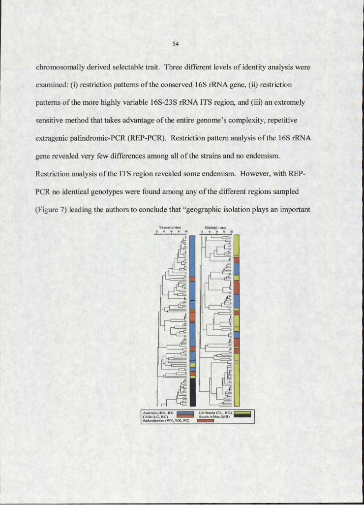
54
chromosomally derived selectable trait. Three different levels o f identity analysis were
examined: (i) restriction patterns o f the conserved 16S rRNA gene, (ii) restriction
patterns o f the more highly variable 16S-23S rRNA ITS region, and (iii) an extremely
sensitive method that takes advantage o f the entire genome’s complexity, repetitive
extragenic palindromic-PCR (REP-PCR). Restriction pattern analysis o f the 16S rRNA
gene revealed very few differences among all o f the strains and no endemism.
Restriction analysis o f the ITS region revealed some endemism. However, with REP-
PCR no identical genotypes were found among any o f the different regions sampled
(Figure 7) leading the authors to conclude that “geographic isolation plays an important
A ustra lia (B N . JD l IvA. l . “ " C o llfom ia (C L . H G )C h iIo (L C 1 R C ) M S M i B S o u th A tr ic a (M H )Saskatchewan (W V. W K. PC) BNBBBBI
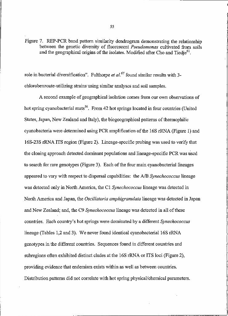
55
Figure 7. REP-PCR band pattern similarity dendrogram demonstrating the relationship between the genetic diversity o f fluorescent Pseudomonas cultivated from soils and the geographical origins o f the isolates. Modified after Cho and Tiedje61.
role in bacterial diversification”. Fulthorpe et al.61 found similar results with 3-
chlorobenzoate-utilizing strains using similar analyses and soil samples.
A second example of geographical isolation comes from our own observations of
hot spring cyanobacterial mats56. From 42 hot springs located in four countries (United
States, Japan, New Zealand and Italy), the biogeographical patterns o f thermophilic
cyanobacteria were determined using PCR amplification o f the 16S rRNA (Figure I) and
16S-23S rRNA ITS region (Figure 2). Lineage-specific probing was used to verify that
the cloning approach detected dominant populations and lineage-specific PCR was used
to search for rare genotypes (Figure 3). Each o f the four main cyanobacterial lineages
appeared to vary with respect to dispersal capabilities: the A/B Synechococcus lineage
was detected only in North America, the Cl Synechococcus lineage was detected in
North America and Japan, the Oscillatoria amphigranulata lineage was detected in Japan
and New Zealand; and, the C9 Synechococcus lineage was detected in all o f these
countries. Each country’s hot springs were dominated by a different Synechococcus
lineage (Tables 1,2 and 3). We never found identical cyanobacterial 16S rRNA
genotypes in the different countries. Sequences found in different countries and
subregions often exhibited distinct clades at the 16S rRNA or ITS loci (Figure 2),
providing evidence that endemism exists within as well as between countries.
Distribution patterns did not correlate with hot spring physical/chemical parameters.

The Ramifications of Population Isolation
From the examples above and numerous other studies, it is evident that physical
isolation can be an integral element in the biology o f microbial populations (symbiotic
and free-living), though its importance may vary among different bacteria. Therefore,
genetic drift, founder effects and/or neutral evolution should be considered when
studying microbial diversity, evolution and ecology. Population isolation may be more
common than is currently appreciated, as the ability to detect isolation depends upon the
resolution o f the genetic marker(s) used. The role o f isolation should be also be
considered when contemplating LGT, as physical proximity and barriers to gene flow are
realities that must affect the LGT process. In a general theory o f microbial evolutionary
ecology, isolation should be thought o f as one mechanism of speciation, not the only
mechanism of speciation. Adding isolation to a developing theoretical framework for
microbial evolution does not displace other mechanisms, such as adaptation arising from
mutation, LGT, or both. All o f these mechanisms must be considered to gain complete
insight into how microbes evolve.

57
REFERENCES CITED
1 Cohan, F.M. 1996. The role o f genetic exchange in bacterial evolution. Am. Soc. Microbiol. News. 62:631-636.
2 Cohan, F.M. 2001. Bacterial species and speciation. Syst. Biol. 50:513-524.
3 Hacker, J and Camiel E. 2000. Ecological fitness, genomic islands and bacterial pathogenicity: a Darwinian view of the evolution of microbes. EMBO Reports 2:376-381.
4 Rainey, P.B., Buckling, A., Kassen, R., and Travisano, M. 2000. The emergence and maintenance o f diversity: insights from experimental bacterial populations. Trends Ecol. Evol. 15:243-247.
5 Palys, T., Nakamura, L.K., and Cohan, F.M. 1997. Discovery and classification of ecological diversity in the bacterial world: the role o f DNA sequence data. Int. J. System. Bacterial. 47:1145-1156.
6 de la Cruz, F. and Davies, J. 2000. Horizontal gene transfer and the origin of species: lessons from bacteria. Trends Microbiol. 8:128-133.
7 Ochman,, H., Lawrence J.G., and Groisman, E.A. 2000. Lateral gene transfer and the nature o f bacterial innovation. Nature 405: 299-304.
8 Lawrence, J.G. 1997. Selfish operons and speciation by gene transfer. Trends Microbiol. 5:(9)355-359.
9 Lawrence, J.G. 1999, Gene transfer, speciation, and the evolution o f bacterial genomes. Cur. Opin. Microbiol. 2:519-523.
10 Ward, D M ., Ferris, M J., Nold, S.C., and Bateson, M M . 1998 A natural view of microbial biodiversity within hot spring cyanobacterial mat communities. Microbiol. Mol. Biol. Rev. 62: 1353-1370.
11 Ward5D. 1998. A natural species concept for prokaryotes. Curr. Opin. Microbiol. 1:271-277.
12 Avise, J. 2000. Phylogeography: TheHistory and Formation o f Species.Harvard University Press. Cambridge, MA.
58
13 Futuyma5D. 1986. Evolutionary Biology, 2ni td. Sinauer Associates, Inc. Sunderland, MA.
14 Boucher, Y. and Doolittle, W.F. 2000. The role of lateral gene transfer in the evolution o f isoprenoid biosynthesis pathways. Mol. Microbiol. 37: 703-718.
15 Lawrence, J.G., and Roth, J.R. 1996. Selfish operons: horizontal transfer may drive the evolution of gene clusters. Genetics 143:1843-1860.
16 Majewski, J. 2001. Sexual isolation in bacteria. FEMS Microbiol Lett. 199:161- 169.
17 Matic, I., Taddei, F., and Radman, M. 1996. Genetic barriers among bacteria. Trends Microbiol. 4:69-72.
18 Roberts, M. and Cohan, F. 1993. The effect o f DNA seqence divergence on sexual isolation in Bacillus. Genetics 134:401-408.
19 Vulic, M., Dionisio, F., Taddei, F., and Radman, M. 1997. Molecular keys to speciation: DNA polymorphism and the control of genetic exchange in enterobacteria. Proc. Natl. Acad. Sci. USA 94:9763-9767.
20 Datta, A., Hendrix, M., Lipsitch, M., and Jinks-Robertson, S. 1997. Duel roles for DNA sequence identity and the mismatch repair system in the regulation of mitotic crossing-over in Yeast. Proc. Natl. Acad. Sci. USA 94:9757-9762.
21 Rayssiguier, C., Thaler, D.S., and Radman, M. 1989. The barrier to recombination between Escherichia coli and Salmonella typhimurium is disrupted in mismatch-repair mutants. Nature 342:396-401.
22 Taddei, F., Vulic, M., Radman, M. and Matic, I. 1997. Genetic variability and adaptation to stress. EXS 83:271-290.
23 Muller, H.J. 1932. Some genetic aspects o f sex. Am. Nat. 8:118-138.
24 Atwood, K.C., Schneider, L.K. and Ryan, F.J. 1951. Periodic selection in Escherichia coli. Proc. Natl. Acad. Sci. USA 37:146-155.
25 Peek, A.S., Feldman, R A ., Lutz, R A ., and Vrijenhoek, R.C. 1998. Cospeciation of chemoautotrophic bacteria and deep sea clams. Proc. Natl. Acad. Sci. USA95:9962-9966.

59
26 Bauman, P., Lai, C., Bauman, L., Rouhbakhsh, D., Moran, N A ., and Clark, M.A. 1995. Mutalistic associations o f aphid and prokaryotes: Biology of the genus Buchnera. Appl Envirnon. Microbiol. 61:1-7.
27 Clark, M.A., Moran, N., Baumann, P., and Wemegreen, J. 2000. Cospeciatioh between bacterial endosymbionts (Buchnera) and a recent radiation o f aphids (Uroleucon) and pitfalls o f testing for phylogenetic congruence. Evolution 54:517-525.
28 Martinez-Torres, D., Bauades, C., Latorre, A., and Moya, A. 2001. Molecular systematics o f aphids and the primary endosymbionts. Mol. Phylogen. Evol. 20:437-449.
29 Spaulding, A.W. and von Dohlen, C.D. 2001. Psyllid endosymbionst exhibit patterns of co-speciation with hosts and desapilizing substitutions in ribosomal RNA. Insect Mol. Biol. 10:57-67.
30 Thao, MX., Moran, N A ., Abbot, P., Brennan, E.B., Burckhardt, D.H and Bauman, P. 2000. Cospeciation o f Psyllids and their primary prokaryotic endosymbionts. Appl Envirnon. Microbiol. 66:2898-2905.
31 Sauer, C., Stackebrandt, E., Gadau, J., Holldobler, B., and Gross, R. 2000. Systematic relationships and cospeciation of bacterial endosymbionts and their carpenter ant host species: proposal o f the new taxon Candidatus Blochmannia gen. nov. Int. J. Syst.-Evol Microbiol. 50:1877-1886.
32 Band!, C., Sironi, M., Damiani, G., Magrassi, L., Nalepa, C A ., Laudani, U., and Sacchi, L. 1995. The establishment o f intracellular symbiosis in an ancestor of cockroaches and termites. Proc. R. Soc. Land. B. 259:293-299.
33 Chen, X., Song, L., and Aksoy, S. 1999. Concordant evolution of a symbiont with its host insect species: molecular phylogeny of genus Glossina and its bacteriome-associated endosymbiont, Wigglesworthia glossinidia. J. Mol. Evol.48:49-58.
34 Dubilier, N., Giere, O., Distel, D.L., and Cavanaugh C.M. 1995.Characterization of chemoautotrophic bacterial symbionts in a gutless marine worm (Oligochaeta, Annelida) by phylogenetic 16S rRNA sequence analysis and in situ hybridization. Appl Envirnon. Microbiol. 61:2346-2350.
35 Ashen, J.B. and Goff, L.J. 2000. Molecular and ecological evidence for species specificity and coevolution in a group o f marine algal-bacterial symbioses. Appl Environ. Microbiol. 66:3024-3030.

60
36 Bucheli, E., Gautschi, B., and Shykoff, J.A. 2001. Differences in population structure of the anther smut fungus Microbotryum violceum on two closely related host species, Silene latifolia and S. dioica. Mol. EcoL 10:285-294.
37 Morzunov, S.P., Rowe, J.E., Ksiazek, T.G., Peters, C.J., St. Jeor, S.C., and Nichol, S.T. 1998. Genetic analysis o f the diversity and origin o f hantaviruses in Peromyscus leucopus mice in North America. J. Virol. 72:57-64.
38 McGeoch, D.J., Dolan, A., and Ralph, A.C. 2000. Toward a comprehensive phytogeny for mammalian and avian herpesviruses. J. Virol. 74:10401-10406.
39 Beijerinck, M.W. 1913. De infusies en de ontdekking der backterien. In Jaarboek van de Koninklijke Akademie v. Wetenschoppen. Muller. Amsterdam.
40 Baas-Becking L.G.M. 1934. Geobiologie o f Inleiding Tot de Milieukunde. The Hague: Van Stockum & Zon.
41 Estrada-de Ios Santos, P., Bustillos-Cristales, R., and Caballero-Mellado, J. 2001. Burkholderia, a genus rich in plant-associated nitrogen fixers with wide environmental and geographic distribution. Appl. Environ. Microbiol. 67:2790-2798.
42 Massana, R., DeLong E.F., and Pedros-Alio, C. 2000. A few cosmopolitan phytotypes dominate planktonic assemblages in widely different oceanic provinces. Appl. Environ. Microbiol. 66:1777-1787.
43 Darling, K., Wade, C., Stewart, I., Kroon, D., Dingle, R., and Leigh-Brown, A. 2000. Molecular evidence for genetic mixing of Arctic and Antarctic subpolar populations of planktonic foraminifers. Nature 405:43-47.
44 Zwart, G., Hioms, W.D., Methe, B A ., van Agterveld, M.P., Huismans, R., Nold, S.C., Zehr, J.P., and Laanbroek, H.J. 1998. Nearly identical 16S rRNA sequences recovered from lakes in North America and Europe indicate the existence o f clades o f globally distributed freshwater bacteria. SysL Appl. Microbiol. 21:546-556.
45 Finlay, B.J. and Clarke, K.J. 1999. Uhiquitious dispersal o f microbial species. Nature. 400, 828-828.
46 Kuske, C., Bams, S.M., and Busch, J.D. 1997. Diverse uncultivated bacterial groups from soils of the arid southwestern United States that are present in many geographic regions. Appl. Environ. Microbiol. 63:3614-3621.

61
47 Garcia-Pichel, F., Prafert-Bebout L. and Muyzer, G. 1996. Phenotypic and phylogenetic analyses show Microcoleus chthonoplastes to be a cosmopolitan cyanobacterium. Appl Environ. Microbiol. 62:3284-3291.
48 Castenholz, R.W. 1978. The biogeography o f hot spring algae through enrichment cultures. Mitt. Internal. Verein. Limnol. 21:296-315.
49 Castenholz, R.W. 1996. Endemism and biodiversity o f thermophilic cyanobacteria. Nova Hedwigia 112:33-47.
50 Beeder, J., Nielsen, R.K., Rosnes, J.T., Torsvik T., and Lien T. 1994. Archaelglobus fulgidus isolated from hot North Sea oil field waters. Appl. Environ. Microbiol. 60:1227-1234.
51 Stetter, K.O., Huber, R., Blochl, E., Kurr, M., Eden R.D. 1993. Hyperthermophilic archaea are thriving in deep North Sea and Alaskan oil reservoirs. Nature 365:743-745.
52 Godffay, H.C. and Lawton, J.H. 2001. Scale and species numbers. Trends Ecol. E vol 16:400-404.
53 Finlay, B.J., Esteban, G.F. and Fenchel, T. 1999. Global diversity and body size. Nature 383:132-133.
54 Fenchel, T 1993. There are more small than large species. Oikos 68, 375-378.
55 Wilson, E. O. 1992. The Diversity o f life. W.W. Norton & Co., New York.
56 Papke, R.T., Ramsing, N.B.,Bateson, M.M., and Ward D.M. Geographic isolation and the evolution of hot spring cyanobacteria. Submitted.
57 Pride, D. T., Meinersmann, R.J., and Blaser, MJ. 2001. Allelic variation within Helicobacter pylori babA and babB. Infect. Immun. 69:1160-1171.
58 Edmunds, S. 2001. Phylogeography of the intertidal copepod Tigriopus californicus reveals substantially reduced population differentiation at northern latitudes. Mol. Ecol. 10: 1743-1750.
59 Bertout, S., F. Renaud, RB., Symoens, F., Bumod, J., Piens, M.-A., Lebeau, B., Viviani, M., Chapuis, F., Bastide, L-M., Grillot, R., Mallie, M., and The European research group on biotype and genotype o l Aspergillus 2001. Genetic polymorphism of Aspergillus fumigatus in clinical samples from patients with invasive aspergillosis: investigation using multiple typing methods. J. Clin! Microbiol. 39:1731-1737.

62
60 Petursdottir S.K., Hreggvidsson, G.O., Da Costa, M.S., and Kristjansson, J.K. 2000. Genetic diversity analysis of Rhodothermus reflects geographical origin of the isolates. Extremophiles. 4:267-274.
61 Cho, J.-C. and Tiedje, J.M. 2000. Biogeography and degree o f endemicity of fluorescent Pseudomonas strains in soil. Appl Environ.Microhiol. 66:5448-5456.
62 Sanson, G. and Briones, M. 2000. Typing o f Candida glabrata in clinical isolates by comparative sequence analysis o f the cytochrome c oxidase subunit 2 gene distinguishes two clusters o f strains associated with geographical sequence polymorphisms. J. Clin. Microbiol. 38:227-235.
63 Beltran, E.C. and Neilan, B A 2000. Geographical segregation of neurotoxin- producing cyanobacterium Anabaena circinalis. Appl Environ. Microbiol. 66:4468-4474.
64 Staley, J. and Gosink, JJ. 1999. Poles apart: biodiversity and biogeography of sea ice bacteria. Annu. Rev. Microbiol. 53:189-215.
65 Habeshaw, G., Yao, Q., Bell, AT., Morton, D., and Rickinson, A. 1999. Epstein- Barr virus nuclear antigen I sequences in endemic and sporadic Burkisf s lymphoma reflect virus strains prevalent in different geographic areas. J. Virol. 73: 965-975.
66 Poussier, S., Vandewalle, P., and Luisetti, J. 1999. Genetic diversity of African and worldwide strains of Ralstonia solanacearum as determined by PCR- restriction fragment length polymorphism analysis of the hrp gene region. Appl Environ. Microbiol. 65:2184-2194.
67 Fulthorpe, R., Rhodes, A., and Tiedje, J. 1998. High levels o f endemicity o f 3- chlorobenzoate-degrading soil bacteria. Appl Environ. Microbiol. 64:1620-1627.
68 Wang G., van Dam, A.P., Spanjaard, L., and Dankert, J. 1998. Molecular typing of Borrelia burgdorferi sensu Iato by randomly amplified polymorphic DNA fingerprinting analysis. J. Clin. Microbiol. 36:768-776.
69 Williams, R A ., Smith, K.E., Welch, S.G., and Micallef, J. 1996. Thermus oshimai sp. nov., isolated from hot springs in Portugal, Iceland, and the Azores, and comment on the concept of a limited geographical distribution of Thermus species. Int. J. Syst. Bacterial. 46:403-408.
70 Yoshino, K., Ramamurthy, T., Nair, G.B., Fukushima, H., Ohtomo, Y., Takeda N., Kaneko, S., and Takeda, T. 1995. Geographical heterogeneity between Far

63
East and Europe in prevalence of ypm gene encoding the novel superantigen among Yersinia pseudotuberculosis strains. J. Clin. Microbiol. 33:3356-3358.
71 Vilgalys5 R. and Sun5 B.L. 1994. Ancient and recent patterns o f geographicspeciation in the oyster mushroom Pleurotus revealed by phylogenetic analysis of ribosomal DNA sequences. Proc. Natl. Aca. Sci. USA 91:4599-4603.
72 Saul5 D J., Rodrigo5 A.G., Reeves5 R A ., Williams, L.C., Borges5 K.M., Morgan5 H.W. and Bergquist5 P.L. 1993. Phytogeny o f twenty Thermus isolates constructed from 16S rRNA gene sequence data. Int. J. Syst Racteriol. 43:754- 760.
73 Hudson5 J., Morgan5 H. and Daniel R. 1989. Numerical classification .of Thermus isolates from globally distributed hot springs. System. Appl. Microbiol. 11:250-256.

64
CHAPTER 4
SUMMARY
Analyses o f the distribution patterns o f organisms can lead to valuable insights
into the causes o f those patterns. For instance, studies using the 16S rRNA molecule as a
molecular marker revealed that from a single model hot spring (Octopus Spring,
Yellowstone National Park) a group of closely related Synechococcus populations,
termed the A/B cluster, was surprisingly organized. Analysis o f their distribution
suggested adaptation to different habitats (e.g. temperatures and light quantities and/or
qualities)1,2. Though model systems can be extremely helpful for interpreting the natural
history o f the organisms studied, they provide an over-focused view o f just a part of
nature and its explanations. Therefore, it was the purpose o f this thesis to extend and
broaden the knowledge of the distribution o f hot spring Synechococcus populations and
improve our view o f the evolutionary forces that were and are acting upon them. To do
so, I analyzed global and local biogeographic distributional patterns using the conserved
16S rRNA gene and the adjacent, more variable, ITS region. My major contribution to
the study o f microbial evolution and ecology is that every recovered Synechococcus
genotype (16S rRNA and ITS) had a limited geographical range (and therefore restricted
dispersal capability) with many genotypes being restricted to single hot springs. Such
results are contrary to the microbial paradigm that “everything is everywhere and nature
selects”. Furthermore, analyses described in chapter 2 revealed that the geographic

65
distributions (both within and among countries) could not be correlated with hot spring
physical/chemical features (i.e. ecological factors that might determine population
ranges) suggesting that geographic isolation (possibly involving founder effects and
random genetic drift) plays a role in the diversification of thermophilic Synechococcus
species. By examining the broader distributional patterns, it becomes clear that natural
selection and adaptation are not the only evolutionary forces acting upon the A/B
radiation.
The implications o f my research are unlikely to be limited to just thermophilic
Syneehococcus species. Though there are relatively few examples o f microbial
population isolation, the phenomenon is now being discovered in many organisms living
in many habitats. Such evidence has led me to propose that population isolation and
genetic drift may be important to the divergence o f microbial populations (chapter 3).
This important aspect o f evolutionary theory is not generally considered in explanations
of microbial diversity and distribution, as microbiologists generally believe that the major
forces shaping microbial diversification and speciation are lateral gene transfer, and
natural selection3"6. That is to say, microbiologists generally believe that adaptation to
specific niches largely explains the divergence and distribution patterns o f microbes. The
complete scope and importance of population isolation to the evolution of microbes has
yet to be evaluated. The simple observation o f endemism suggests lateral gene transfer/
and natural selection are not the only forces affecting microbial evolution.
Geographic/physical isolation should be integrated within a general theory of bacterial
evolution.

REFERENCES CITED
Ferris, M., and Ward, D. 1997. Seasonal distributions o f dominant 16S rRNA- defined populations in a hot spring microbial mat examined by denaturing gradient gel electrophoresis. Appl Environ. Microbiol 63:1375-1381.
Ramsing, N., Ferris, M., and Ward, D. 2000. Highly ordered vertical structure o f Synechoccus populations within the one-millimeter-thick photic zone o f a hot spring cyanobactrial mat. Appl Environ. Microbiol. 66:1038-49.
Ochman, H., Lawrence, J.G., and Groisman, E.A. 2000. Lateral gene transfer and the nature of bacterial innovation. Nature 405: 299-304
Doolittle, W.F. 2000. Uprooting the tree o f life.. Sci. Am. 282: 90-95./
Hacker, J and Camiel E. 2000. Ecological fitness, genomic islands and bacterial pathogenicity: a Darwinian view o f the evolution o f microbes. EMBO Reports 2:376-381.
Cohan, F.M. 2001. Bacterial species and speciation. Syst. Biol. 50:513-524.

67
APPENDIX K
SUPPLEMENTAL TABLES

68
Table 3. Physical, chemical and biological data for all hot springs sampled.
R egion /Sam ple ^ t l e ^ pH (»M)
Cyanobacteria
, R elative A bundance (% )bM orph Clonedo lo g y genotypes „
C O C C U S
North A m erica I
Jack Stream (JS)- 60
4 2 °ir 20"N 120° 20' 41"E
59.8-61.2 8.3 2.5 R NACy03
JS-50 42°11' 20"N 120° 20' 41"E
49.7-50.2 8.4 0.7 R NACyOl 60.4±
4.16 ndd4.4861.39 65.3
Perpetual (Per) 42° 11' 20"N 120° 20' 41"E
60.1-60.9 8.2 4.6 R 83.3±
2.81nd
2.7660.00 86.1
Levee (Lev) 42° 11' 20"N 120° 20' 41"E
59.5-61.8 8.1 5.5 R 64.66
2.74 nd2.1160.69 66.7
North A m erica 2
Bozeman (Boz) 45° 39' 39"N 111° 11' 12"E
57.2-57.5 8.8 7.7 R ,f
NACy06,NACy08,NACylO
72.4610.8 nd
22.366.07 94.7
North A m erica 3
LaDuke (LaD) 45° 05' 25"N 111° 46' 28"E
59.8-60.2 6.7 nd R NACylO
91.9639.2
nd29.064.41 121
New Mound (NM)
44° 57' 90"N 110° 42' 50"E 56.4 6.6 25.4 R NACylO
88.6612.2 nd
0.6060.05 89.3
Bath Lake Vista (BLV)
44° 57' 90"N 110° 42' 50"E
57.0-57.3 6.6 39.3 R NACylO,
N A C yll86.161.74 nd nd 86.1
White Elephant (WE)
44° 57' 90"N 110° 42' 50"E
59.7-60.0 6.9 8.2 R NACyl 0,
NACy0984.363.41 nd nd 84.3
North A m ericaAClearwater East (ClwE)
44° 47' 19"N 110° 44' 21"E
57.3-61.9
5.2 1.3 R1.9260.08 nd
1.5160.50 3.43
Clearwater South (ClwS)
44° 47' 19"N 110° 44' 21"E
56.3-56.5 6.1 0.2 R
NACylO3 OS type-
C l*e
93.468.37
10.361.08
1.2760.11 105
Clearwater North (ClwN)
44° 47' 19"N 110° 44' 21"E
54.0-56.2 7.5 nd R NACyOS
87.1617.8 nd
1.6160.29 88.7

69
North America 5
Octopus (Oct) 44° 32' 35"N 110° 46' 06"E
54.0-64.0 8.2 0.1 R
NACy02, NACy04, NACy05, NACy06, NACy07, NACyl 0, NACyl I, NAC9a*
76.2±10.6 nd
1.2860.36 77.5
Twin Butte Vista (TBV)
44° 32' 35"N 110° 46' 06"E
59.3-60.7 8.4 0.6 R 88.8±
1.75 nd2.6260.24 91.4
Mushroom(Mush)
44° 32' 35"N 110° 46' 06"E
58.9-60.6 8.3 nd R 85.96
5.71 nd2.8860.24 88.7
North America 6
Mantrap (Man) 44° 24' 56"N 110° 34' 29"E
58.2-61.8 8.3 nd R
NACy04,NACy09,NACylO
92.5612.5 nd
1.2460.12 93.6
Double Spring (DS)
44° 24' 56"N 110° 34' 29"E
59.1-60.2 8.0 0.6 R NACy 10,
N A C yll81.567.52 nd nd 81.5
Heart Pool (HP) 44° 24' 56"N 110° 34' 29"E
56,6-58.3 9.2 19.5 R NACy05,
NACylO83.764.12 nd nd 83.7
Japan I
Geto-48 39° 12' 54"N 140° 52' 82"E 48.2 6.3 0.1 F JapanCy03 nd i nd nd nd
Zenikawa (Zen)- 62
40° 00' 76"N 140° 48' 20"E
62.0-62.4 8.1 0.2 R JapanCyOl
Zen-59 40° 00' 76"N 140° 48' 20"E
59.3-59.6 8.1 0.3 R JapanCyO I,
JapanCy05 nd89.8610.1
1.3960.09 91.2
Zen-57a 40° 00' 76"N 140° 48' 20"E
57.1-56.8 8.1 0.3 R JapanCyO I,
JapanCyOS
Zen-57b 40° 00' 76"N 140° 48' 20"E
57.5-57,7 8.3 0.3 R JapanCyO I,
JapanCyOS
Zen-55 40° 00' 76"N 140° 48' 20"E
56.1-56.3 8.4 0.3 R JapanCyOl
Magaroku (Mag)- 62
39° 48' 26"N 140° 48' 27"E 62.2 7.0 7.0 R JapanCyO I,
JapanCy02
Mag-60 39° 48' 26"N 140° 48' 27"E
59.8-59.9 7.1 19.7 R JapanCyO I,
JapanCy02 nd93.5616.6 nd 93.5
Mag-56 39° 48' 26"N 140° 48' 27"E 56.0 7.0 7.0 R JapanCyO I,
JapanCy02
Mag-51 39° 48' 26"N 140° 48' 27"E
51.1 7.9 4.1 R ,F JapanCy02
Mag-50 39° 48' 26"N 140° 48' 27"E
49 .7 -1 50.2
7.2 23.2 F JapanCy07, JapanCyl 2

70
Ganiba (Gan)-49 39° 48' 90"N 140° 47' 58"E 49.3 8.4 36.6 R ,F
JapanCyO I, JapanCyl2, JapanCyl4
nd 21.3± 3.55 nd 21.3
Mizusawa (Miz)- 55
39° 45' 92"N 140° 45' 20"E 55.0 7.6 36.7 a nd nd nd nd nd
Japan 2
Shionoyu (Shio)- 59
37° ll'N #f 139° 46'E
58.5-59.1 7.3 7.5 R JapanCyOS
Shio-58 37°11'N# 139° 46'E 58.0 7.3 7.5 R JapanCyOS,
JapanCy06
Shio-56 37° H'N# 139° 46'E 56.6 7.3 7.5 R JpnCyOS
Japan 3Nakafusa (Naka)-66
36° 23' 35"N 137° 44' 80"E
65.8-66.3 8.5 34.5 R JapanCy09
Naka-60 36° 23' 35"N 137° 44' 80"E
60.1-60.4 8.5 1.9 R
JapanCy09, JapanCyl 5, JapanC9a*, JapanC9b*, JapanC9c*
nd 141±28.7 nd 142
Naka-56 36° 23' 35"N 137° 44' 80"E
55.8-56.4 8.7 4.5 R ,F JapanCyOS,
JapanCy09Shinhokata (Shi)- 65
36° 15' 33"N 137° 34' 60"E
64.4-65.4 6.1 0.3 R JapanCy09,
JapanCylO
Shi-57a 36° 15' 33"N 137° 34' 60"E
57.2-57.6 6.9 0.4 R JapanCy09,
JapanCylO nd 100.26± 9 .09 nd 100
Shi-57b 36° 15' 33"N 137° 34' 60"E
57.4-57.6 8.2 0.2 R JapanCy09,
JapanCylO
Nakanoyu (Nak)- 64
36° 12' 34"N 137° 36' 27"E
63.3-61.9
7.5 1.0 R, o JapanCy09
Nak-60 36° 12' 34"N 137° 36' 27"E
59.4-60.0 7.8 5.8 R JapanCy09 nd 74.4±
3.60 nd 74.4
Nak-55 36° 12' 34"N 137° 36' 27"E
54.3-55.3 8.1 6.7 R JapanCy09
Nak-49 36° 12' 34"N 137° 36' 27"E
47.2-47.5 8.3 0.5 R , F , 0
Japan 4
Ninotaira (Nin)- 52
35° 15'N# 139° 09'E 52.0 7.3 7.5 R JapanCyOS
Nin-50 35° 15'N* 139° 09'E 50.6 7.3 7.5 R JapanCyOS,
Sokokura (Sok)- 60
35° 15'N# 139° 09'E
59.2-60.5 7.0 7.0 R JapanCyOS,
JapanCy06

71
Japan 5
Yunomine (Yun)- 62
33° 49' 75"N 135° 45' 47"E
61.7-61.9 7.8 0.9 R ,0
JapanCy09, JapanCylO5 JapanCy 15
Yun-55 33° 49' 75"N 135° 45' 47"E
53.8-56.3 8.4 4.1 R
JapanCy09, JapanCylO5 JapanCyl I
nd i i i i10.8 nd 111
Tousenji (Tou)-55 33° 59' 74"N 135° 48' 36"E
56.8-57.1 9.0 34.3 F nd nd nd nd nd
Tou-50 33° 59' 74"N 135° 48' 36"E
49.4-50.9 9.1 8.2 F JapanCyOS
Japan 6
Tsuetate (Tsu)-60 33° 10' 95"N 131° 02' 06"E
58.0-59.2 8.6 0.8 R JapanCyOS nd 87 0±
6.971.52i0.08 88.4
Tsu-55 33° 10' 95"N 131° 02' 06"E
55.8-55.9 8.8 0.4 F JapanCyOS5
JapanCylO
Tsu-50 33° 10' 95"N 131° 02' 06"E
51.9-53.4 8.8 0.5 F; o JapanCy04,
JapanCyOS
Tanoharu (Tan)- 53
33° 04' 86"N 131° 07' 54"E
52.4-52.9 7.6 0.6 F,r
JapanCyOS5 JapanCyl 3 nd 6 0 .0 i
2.931.72i0.07 61.7
New Zealand I
Kuirau Reserve (K)-1-4-5 3
38° 07' 74"S 176° 14' 65"E
48.7-52.9 7.8 1.3 F5 r
NZCy03,NZCyOl*,NZCla*,NZClb*
nd 1.16i0.19 1.16
K-l-4-50 38° 07' 74"S 176° 14' 65"E
47.1-48.4 8 1.1 R ,F NZCy03 X
K-l-3-60 38° 07' 74"S 176° 14' 65"E
58.7-60.9 7.7 1.3 R ,f NZCy03
K-l-3-50 38° 07' 74"S 176° 14' 65"E
47.4-53.4 7.9 1.3 R ,f
NZCyOl5NZCyOS5NZCy06
K3 38° 07' 74"S 176° 14' 65"E
50.7-51.7
6.4 1.5 F NZCy03 nd nd nd
K4 38° 07' 74"S 176° 14' 65"E
44.1-48.0 6.9 1.1 R ,f NZCyOl nd 4 6 .8 i
6.3 46.8
K-1-2-54 38° 07' 74"S 176° 14' 65"E
55.7-57.7
7.3 1.1 R5T5O NZCy02
K-1-2-48 38° 07' 74"S 176° 14' 65"E
46.3-50.6 7.2 1.9 R ,f
NZCyOS5NZCy07 nd 2 9 .0 i
1.60 29
Ohinemutu (OHS)-I '
38° 07' 71"S 176° 14' 83"E
50.4-51.7 8.3 R5F NZCy03 nd 1.13i
0.01 1.13
0HS-2 38° 07' 71"S 176° 14' 83 "E
50.4-54.2 8.9 48.6 R5F NZCy03 nd 2 0 .0 i
2.54 20
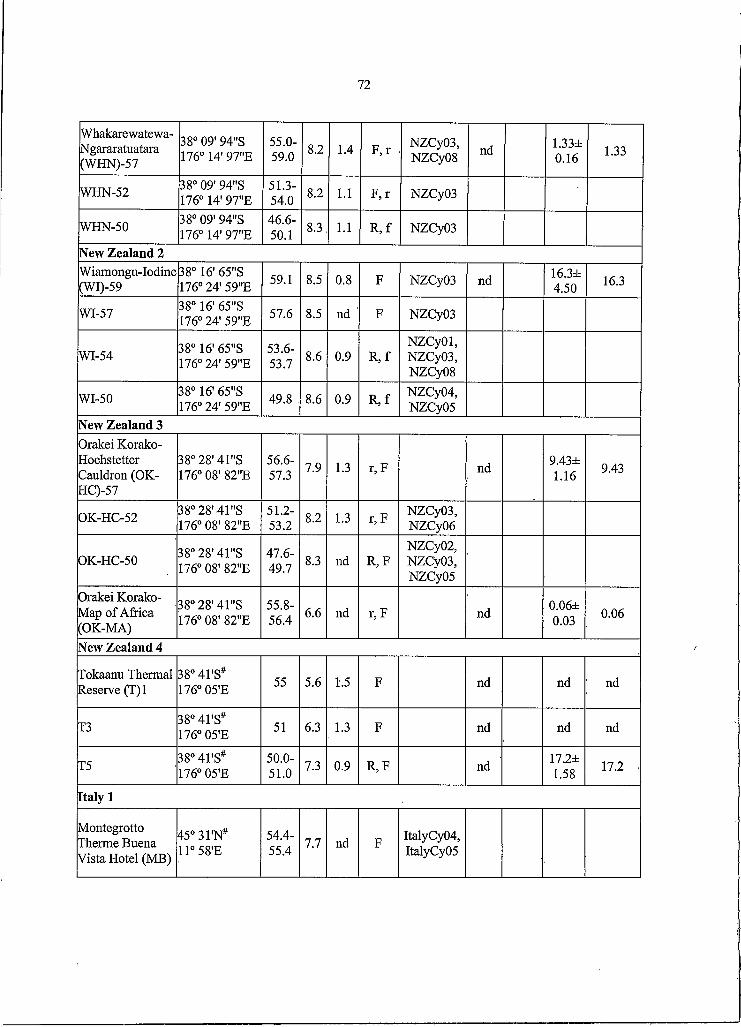
72
Whakarewatewa-Ngararatuatara(WHN)-57
38° 09' 94"S 176° 14' 97"E
55.0-59.0 8.2 1.4 F ,r NZCyOS5
NZCyOS nd 1.33±0.16 1.33
WHN-52 38° 09' 94"S 176° 14' 97"E
51.3-54.0 8.2 1.1 K r NZCyOS
WHN-50 38° 09' 94"S 176° 14' 97"E
46.6-50.1 8.3 1.1 R ,f NZCy03
New Zealand 2Wiamongu-Iodine(WI)-59
38° 16' 65"S 176° 24' 59"E 59.1 8.5 0.8 F NZCy03 nd 16.3±
4.50 16.3
WI-57 38° 16' 65"S 176° 24’ 59"E 57.6 8.5 nd F NZCyOS
WI-54 38° 16' 65"S 176° 24' 59"E
53.6-53.7 8.6 0.9 R ,f
NZCyOl5NZCyOS5NZCyOS
WI-50 38° 16' 65"S 176° 24' 59"E 49.8 8.6 0.9 R ,f
NZCy04,NZCyOS
New Zealand 3Orakei Korako- Hochstetter Cauldron (OK- HC)-57
38° 28' 41"S 176° 08' 82"E
56.6-57.3 7.9 1.3 r,F nd 9.43±
1.16 9.43
OK-HC-52 38° 28' 41"S 176° 08' 82"E
51.2-53.2 8.2 1.3 r,F NZCyOS5
NZCy06
OK-HC-50 38° 28' 41"S 176° 08' 82"E
47.6-49.7 8.3 nd R5F
NZCy02,NZCyOS5NZCyOS
Orakei Korako- Map o f Africa (OK-MA)
38° 28' 41"S 176° 08' 82"E
55.8-56.4 6.6 nd r,F nd 0.06±
0.03 0.06
New Zealand 4
Tokaanu Thermal Reserve (T)I
38° 4TS# 176° 05'E 55 5.6 1.5 F nd nd nd
T3 38°41'S# 176° 05'E 51 6.3 1.3 F nd nd nd
T5 38°41'S# 176° 05'E
50.0-51.0 7.3 0.9 R5F nd 17.2±
1.5817.2
Italy I
Montegrotto Therme Buena Vista Hotel (MB)
45°31'N# 11° 58'E
54.4-55.4 7.7 nd F ItalyCy04,
ItalyCyOS

73
Italy 2
Island o f Ischia- Cave Scurra (ICS)
40° 44'N# 13° 57'E
46.9-49.9 7.9 nd F ItalyCy02
Island o f Ischia- Sorgenta (IS)-57
40° 44'N# 13° 57'E
56.2-57.8 7.0 nd F ItalyCy03
IS-52 40° 44'N# 13° 57'E
50.7-51.9 7.0 nd F ItalyCy03
Italy 3
Agnano Terme (AT)-55
40° 49'N# 14° 07'E
54.2-54.9 7.3 nd F ItalyCy02
AT-5040 ° 49'N# 14° 07'E 50 7.1 nd F ItalyCyO I,
ItalyCy02a, R/r, unicellular rod; F/f, filamentous; O/o, unicellular, ovoid; a, aggregate (upper and lower cases
reflect predominance and presence o f the morphotype, respectively.b, Mean o f per-sample lineage-specific probe response relative to cyanobacterial lineage probe response.c, Sum o f means for all three lineagesd, not detected.e, *, genotype discovered using lineage-specific PCR technique, f #, estimated for the general area, but not the spring itself.

74
Table 4. Chemical measurements for all hot springs sampled
Spring3 P NH3b NOzb N 03b CaCOjc Ca" Clc Mgc SiOzc Nac SO / Asc Bc Cr0 Cuc Fec Mnc Znc
JS-60 0.37 2.48 0.57 0.54 64.0 9.00 130 0.50 73.0 190 270 0.15 7.40 0.01 0.01 0.02 0.01 0.03JS-50 0.04 2.48 0.57 0.54 65.0 9.00 130 0.50 74.0 200 280 0.02 7.50 0.01 0.01 0.02 0.01 0.03Per 0.37 4.12 0.57 0.54 65.0 10.0 120 0.50 73.0 190 260 0.15 7.40 0.01 0.01 0.02 0.01 0.03Lev 0.37 10.1 0.57 0.54 72.0 10.0 120 0.50 71.0 190 260 0.13 7.20 0.01 0.01 0.02 0.01 0.03Boz 0.45 2.48 0.57 0.54 87.0 1.00 53.0 0.50 34.0 130 130 0.11 0.60 0.01 0.01 0.02 0.01 0.03LaD 0.37 2.48 1.09 0.54 230 340 49.0 59.0 23.0 220 1300 0.02 0.93 0.01 0.01 0.19 0.01 0.03NM 0.37 24.1 0.57 0.54 720 350 160 65.0 27.0 120 600 0.82 4.30 0.01 0.01 0.70 0.05 0.03BLV 0.37 26.5 0.57 0.54 720 330 170 63.0 27.0 120 600 0.94 4.70 0.01 0.01 0.02 0.01 0.03WE 0.37 18.8 0.57 0.54 610 330 170 67.0 30.0 120 670 0.75 4.40 0.01 0.01 0.22 0.01 0.07ClwE 0.37 14.0 0.57 0.54 8.00 6.00 130 0.50 40.0 74.0 26.0 0.68 2.70 0.01 0.01 0.53 0.04 0.03ClwS 0.37 6.47 0.57 0.54 31.0 7.00 140 0.50 45.0 94.0 33.0 0.67 2.80 0.01 0.01 0.14 0.10 0.03ClwN 0.37 4.12 5.65 0.54 84.0 23.0 300 1.00 86.0 190 100 1.30 5.00 0.01 0.01 0.11 0.23 0.03Oct 0.37 35.3 1.09 6.13 290 0.50 290 0.50 HO 300 26.0 1.40 3.30 0.01 0.01 0.02 0.01 0.03TBV 0.37 2.48 0.57 0.54 270 0.50 320 0.50 no 290 21.0 2.10 3.30 0.01 0.01 0.02 0.01 0.03Man 0.37 2.48 0.57 1.12 350 0.50 180 0.50 94.0 270 29.0 1.00 2.50 0.01 0.01 0.02 0.01 0.03DS 0.37 2.48 0.57 0.54 390 0.50 290 0.50 85.0 350 42.0 1.80 3.60 0.01 0.01 0.02 0.01 0.03HP 0.37 2.48 0.57 2.58 480 0.50 350 0.50 120 410 44.0 2.10 4.30 0.01 0.01 0.02 0.01 0.08Geto 0.37 12.4 0.20 9.75 314 239 1535 49.0 92.0 574 408 0.01 20.6 0.05 0.05 0.23 0.08 0.03Mag 0.31 32.5 0.13 1.04 134 34.0 219 2.40 35.0 86.0 130 0.08 2.50 0.05 0.05 0.17 0.14 0.03Gan 0.31 14.4 0.05 0.73 14.0 121 204 0.50 11.0 45.0 454 0.01 0.12 0.05 0.05 0.12 0.03 0.05Miz 6.52 476 0.08 1.29 671 186 452 123 71.0 142 1026 0.03 1.90 0.05 0.05 0.05 2.20 0.03Naka 0.62 3.25 0.08 2.17 67.0 1.60 173 0.50 92.0 68.0 43.0 0.06 0.92 0.05 0.05 0.23 0.03 0.03Shi 0.19 4.20 0.99 11.2 54.0 3.50 142 0.50 102 48.0 40.0 0.21 1.30 0.05 0.05 0.05 0.03 0.03Nak 0.64 40.8 0.13 2.80 234 31.0 194 4.00 245 118 91.0 0.04 2.30 0.05 0.05 0.19 0.97 0.03Yun 0.99 19.7 0.05 1.02 648 8.60 308 1.70 112 374 43.0 0.00 3.00 0.05 0.05 0.17 0.09 0.03Tou 0.31 7.71 0.08 1.02 94.0 1.50 125 0.50 43.0 55.0 35.0 0.01 2.30 0.05 0.05 0.05 0.09 0.03Tsu 0.93 23.4 1.24 7.99 35.0 12.0 620 0.50 81.0 287 95.0 0.31 5.40 0.05 0.05 0.05 0.03 0.03Tan 1.18 21.8 0.43 24.4 231 14.0 259 17.0 245 171 222 0.07 2.60 0.05 0.05 0.12 0.10 0.08K-1-4 0.62 20.7 0.08 1.37 284 0.50 257 0.50 126 374 132 0.10 6.01 0.05 0.05 0.05 0.05 0.07K-1-3 0.62 7.94 0.13 1.97 292 0.50 282 0.50 127 385 130 0.10 6.18 0.05 0.05 0.05 0.05 0.06K3 0.87 51.3 0.13 1.10 78.0 0.50 230 0.50 147 293 262 0.09 5.16 0.05 0.05 0.47 0.05 0.07K4 0.50 13.6 0.18 1.09 280 0.50 251 0.50 130 343 127 0.09 5.85 0.05 0.05 0.05 0.05 0.07K-1-2 1.18 15.9 0.15 10.5 318 0.50 277 0.50 127 386 132 0.10 6.27 0.05 0.05 0.05 0.05 0.08OHS-I 2.24 7.45 0.43 0.27 334 0.50 262 0.50 123 376 77.0 0.04 5.47 0.05 0.05 0.05 0.05 0.07OHS-2 0.93 8.73 0.08 0.97 378 0.50 275 0.50 146 354 62.0 0.06 5.71 0.05 0.05 0.05 0.05 0.08WHN 0.62 24.5 0.18 1.57 127 0.50 490 0.50 122 395 86.0 0.74 5.28 0.05 0.05 0.05 0.05 0.07WI 0.99 19.4 0.50 1.24 202 0.50 667 0.50 133 512 112 0.86 5.92 0.05 0.05 0.05 0.05 0.07OKHC 0.37 3.09 0.35 2.27 160 0.50 193 0.50 129 239 100 0.38 2.72 0.05 0.05 0.05 0.05 0.07OKMA 1.18 6.24 0.45 2.21 131 0.50 161 0.50 222 235 102 0.31 2.27 0.05 0.05 0.05 0.05 0.08Tl 0.31 948 0.18 63.8 31.0 5.20 1960 0.50 233 1100 81.0 4.93 55.8 0.05 0.05 0.17 0.05 0.10T2 0.44 760 0.18 18.9 52.0 7.40 2140 0.50 244 1330 85.0 6.23 61.1 0.05 0.05 0.05 0.05 0.08T4 0.87 493 17.0 58.2 76.0 7.30 1940 0.50 241 1140 59.0 5.27 57.3 0.05 0.05 0.27 0.05 0.07

75
T5 0.25 548 0.03 2.19 67.0 6.90 3080 0.50 126 1810 74.0 8.04 89.7 0.05 0.05 0.05 0.05 0.07MB 0.16 111 13.5 43.8 130 198 1480 43.0 52.0 740 500 0.05 6.50 0.27 0.05 0.14 0.05 0.03[CS 3.75 16.8 1.45 2.53 940 12.0 1700 1.40 190 1540 680 1.38 3.40 0.26 0.05 0.27 0.51 0.06IS 0.59 0.24 16.9 56.4 310 14.0 2530 31.0 120 1790 660 0.42 4.20 0.27 0.05 0.14 0.05 0.10AT 2.02 437 0.16 1.51 1190 230 2870 65.0 160 1800 400 0.45 12.0 0.05 0.05 2.29 0.96 0.10a, see Appendix Supplemental Table 3 for full spring namesb, pMc, mg/L

- BOZEMAN