impact of routine completion angiography on the results of primary carotid endarterectomy: a...
TRANSCRIPT

Eur J Vasc Endovasc Surg (2011) 41, 579e588
Impact of Routine Completion Angiography on theResults of Primary Carotid Endarterectomy:A Prospective Study in a Teaching Hospital
J.-B. Ricco a,*, G. Regnault de la Mothe a, S. Fujita b, O. Page a,A. Valagier a, C. Marchand a
aVascular Surgery Department, Jean Bernard Hospital, University of Poitiers, Medical School, Rue de la Miletrie,Poitiers 86021, FrancebVascular Surgery service, Tsukuba Hospital, Ibaraki, Japan
Submitted 5 September 2010; accepted 18 January 2011Available online 18 February 2011
KEYWORDSCarotid endarterec-tomy;Carotid surgery;Completionangiography;Operative defects;Postoperative stroke
To access continuing medical
* Corresponding author.E-mail address: [email protected]
1078-5884/$36 ª 2011 European Sociedoi:10.1016/j.ejvs.2011.01.015
Abstract Objective: To assess the usefulness of completion angiography in the prevention ofstroke, carotid occlusion and residual stenosis after primary carotid endarterectomy (CEA) inthe setting of a teaching hospital.Material and Methods: From January 1995 to August 2009, 1055 consecutive patients having1179 CEAs were entered in a prospective study excluding patients with severe renal insuffi-ciency, allergy to contrast media and patients with repeat CEA or carotid bypass. In thiscohort, 552 patients (52.3%) were asymptomatic, 318 (30.2%) had a transient ischaemic attack(TIA) and 185 (17.5%) had a stroke. Routine completion angiography was obtained in all 1055patients. The decision to perform a surgical revision was decided for any of the followingdefects: (1) a residual stenosis of more than 50% of the internal carotid artery (ICA) or commoncarotid artery (CCA) and of more than 70% of the external carotid artery (ECA), (2) any flap and(3) any intraluminal-filling defect. A postoperative duplex scan was obtained within a weekafter surgery and thereafter on a yearly basis. Median follow-up was 7 years.Results: CEA was performed by a senior surgeon as first operator in 812 cases (69%) and bya trainee, with a scrubbed senior surgeon, in 367 cases (31%). Completion angiography re-vealed significant defects in 72 cases (6.1%) warranting revision for ECA flap (n Z 30),thrombus in contact with the patch (n Z 7), distal ICA flap or stenosis (n Z 20) and CCA flapor residual plaque (n Z 15).
Logistic regression analysis showed that total length of the carotid plaque >6 cm (p Z 0.02,Odds ratio: 2.31; 95% confidence interval (CI) (1.21e3.72)), eversion endarterectomy of theECA (p Z 0.01, Odds ratio 3.41; 95%CI (2.10e5.94)) and trainee as first operator (p Z 0.02,
education questions on this paper, please go to www.vasculareducation.com and click on ‘CME’
(J.-B. Ricco).
ty for Vascular Surgery. Published by Elsevier Ltd. All rights reserved.

580 J.-B. Ricco et al.
Odds ratio 2.42; 95%CI (1.81e4.23)) were independent predictors of operative defects seen oncompletion angiography. No complication in relation to carotid catheterisation or injection ofcontrast media occurred in this series.
The 30-day combined stroke and death rate was 1.5%, comparable between senior surgeonsand trainees (p Z 0.60). There was no significant difference in the combined stroke and deathrate observed in patients with normal completion angiography (1.4%) compared with that ofthe patients with a defect corrected (2.8%) (p Z 0.28, Odds ratio: 0.67; 95%CI (0.22e2.09)).But there was an increased incidence of postoperative TIA in the group with revision(p Z 0.001, odds ratio: 5.8, 95%CI: 1.8e18.9).
At7years, thefreedomratefrom>50%carotid restenosis orocclusionwas87.5�6.7% inpatientswith normal completion angiography and 92� 5.4% in patients, who undergo a surgical revision.Conclusion: In a single centre, CEA with routine completion angiography resulted in good perio-perative outcome. Plaque length, technique for external carotid artery (ECA) endarterectomyand trainee as first operator were independent predictors of operative defects seen on completionangiography.ª 2011 European Society for Vascular Surgery. Published by Elsevier Ltd. All rights reserved.
Figure 1 Flowchart of the study and follow-up status.
Introduction
The beneficial role of carotid endarterectomy (CEA) issupported by level 1 evidence in selected asymptomaticand in symptomatic patients with severe carotid sten-osis.1e4 However, the absolute benefit of CEA is limited bythe amount of surgical complications, and there is still noconsensus on whether the routine use of an intra-operativecompletion assessment should be performed to confirm thetechnical adequacy of CEA.5,6 This discrepancy is partiallydue to the favourable outcome of CEA even without theroutine use of completion imaging,1,3,7 and also due to thefact that certain intra- and postoperative strokes, includingintracerebral bleeding, as well as hyperperfusionsyndrome, are unrelated to the surgical technique. Never-theless, many authors have shown that detection andcorrection of significant technical defects by an intra-operative completion study lower the risk of postoperativestroke.8e16 The purpose of this prospective study was toevaluate the potential risk and benefit of routine comple-tion angiography for detection of significant intra-operativedefects in a prospective series of 1179 consecutive primaryCEA performed within a teaching hospital.
Material and Methods
From January 1995 to August 2009, a total of 1402 carotidrevascularisations for atheromatous diseasewere performedat our institution in 1278 patients. Patients with repeat CEAor carotid bypass, as well as patients with a contraindicationto angiography because of allergy or severe renal insuffi-ciency were excluded from this study (Fig. 1), leaving 1055patients having 1179 primary CEA procedures with comple-tion angiography, who were entered in a prospective study;those protocols were approved by the institutional reviewboard of the University of Poitiers. Bilateral staged CEAprocedures were performed in 124 patients. Demographics,risk factors, indications for surgery, characteristics of carotidlesions and technical details are reported in Table 1. Prior tosurgery, a neurologist independently assessed all symptom-atic patients. In this series, the findings of the intra-opera-tive completion angiography were entered into a predefined
database with details concerning the nature and location ofthe defects observed, together with the need for surgicalcorrection, the incidence of postoperative complicationsand the technique used for correction. In addition, thefollowing variables were recorded: surgeon (senior ortrainee), type of CEA, patch material, shunt insertion, totallength of the endarterectomy specimen measured in centi-meters in the common (CCA) and internal carotid artery (ICA)and technique used on the external carotid artery (ECA) withmeasurement of the length of the ECA specimen.
All patients received aspirin; 977 patients (93%) receivedaspirin alone and 72 patients received aspirin and clopi-dogrel (7%); and 852 patients (80.7%) received statins at

Table 1 Patients characteristics and risk factors in 1055patients having 1179 carotid endarterectomies.
N Prevalence (%)
Age (y)<70 580 54.9�70 475 45.1
GenderMale 728 69.0Female 327 31.0
Current smoker 696 65.9Hypertension 685 64.9Congestive heart failure 61 5.8Coronary artery disease 221 21.0Unstable angina 45 4.3Peripheral arterial disease 295 27.9Diabetes mellitus 232 22.0Renal insufficiency 57 5.4Surgical indicationsTransient ischemic attack 318 30.2Stroke 185 17.5Asymptomatic 552 52.3
First operatorSenior vascular surgeon 812 68.8Trainee supervised 367 31.2
Ipsilateral stenosis50e69% 212 17.970e99% 967 82.1
Contralateral stenosis<50% 354 30.150e69% 601 50.970e99% 224 19.0
Length of the plaque (ICA þ CCA)<6 cm (specimen) 858 72.8�6 cm (specimen) 321 27.2
Length of the plaque (ECA)<3 cm (specimen) 1041 88.3�3 cm (specimen) 138 11.7
Operated sideRight 611 51.9Left 568 48.1
ShuntYes 283 24.0No 896 76.0
Standard endarterectomy 1118 94.8Eversion endarterectomy 61 5.2Carotid closure for standard CEAPolyester patch 476 42.6Polyurethane patch 631 56.4Primary closure 11 1.0
Internal carotid artery e tacking suturesYes 203 17.2No 976 82.8
Common carotid artery e tacking suturesYes 935 79.3No 244 20.7
External carotid arteryFeathered end point at its origin 251 21.3Section of the plaque at its origin 516 43.7Eversion endarterectomy 412 35.0
ICA: Internal carotid artery, CCA: Common carotid artery, ECA:External carotid artery.
Impact of Routine Completion Angiography on the Results of Primary Carotid Endarterectomy 581
least 1 week before surgery. All patients were given generalanaesthesia, and attempts were made to maintain systolicblood pressure above 140 mmHg during carotid artery crossclamping. All patients received intravenous heparin50e70 IU kg�1 5 min before clamping. Half of this dose wasrepeated in patients who needed a surgical revision. Prot-amine was not administered at the end of the procedure.
Surgical technique
CEA was performed by a senior vascular surgeon (N Z 812,68.8%) or by a trainee (NZ 367, 31.2%) with a senior as firstassistant. All procedures were performed using magnifica-tion loops. On the CCA, at the proximal end of the endar-terectomy, feathering was uncommon and the plaque wasusually cut, leaving a small step. If the plaque was notadherent, a few U-shaped stitches were used to affix thisflap securely to the artery. The endarterectomy was thencarried to the ICA, trying to avoid any step at its distal end.Concerning the ECA, all efforts were made to obtaina feathered end point at its origin (N Z 51, 21.3%). Ifunsuccessful, the intima was sectioned at the origin of theECA with the use of tacking stitches (N Z 516, 43.7%). Ifa large atheromatous lesion was protruding into the ECA,eversion endarterectomy of the ECA was performed(N Z 412, 35.0%). The ECA specimen was measured andchecked for distal plaque fracture. Repair was notattempted at this point, but rather upon seeing the resultsof the completion angiography.
In this series, a shunt was used on a selective basis in 283patients (24%) in any of the following conditions: occlusionor stenosis >70% of the contralateral carotid artery(N Z 224), poor backflow from the ICA (N Z 13) or oper-ation within 7 days following a stroke (N Z 46). Conven-tionally accepted methods of determining the need forshunt insertion including electroencephalography (EEG),transcranial Doppler (TCD) or formal measurement of theICA back pressure were not used. A patch was tailored torestore the normal diameter of the ICA, and was used in allbut 11 patients (1%) having a standard endarterectomy. Thematerial used for the patch was polyester (Maquet, SARL,France) in 476 cases (42.6%) or polyurethane (B. Braun,SARL, France) in 631 cases (56.4%).
Completion angiography
Intra-operative angiography was performed after comple-tion of the procedure using an OEC mobile imaging system(General Electrics�) by direct needle puncture and retro-grade catheterisation of the CCA with manual injection of15 ml of contrast media. Two exposures of the carotidbifurcation were obtained with one frontal and one 45�
oblique view to avoid the superposition of the ICA and ECA. Athird angle was obtained to analyse the cerebral vessels. Avideodisc recorder was used to acquire the image sequenceand “freeze-frame” the best images for further analysis.
The angiography was examined immediately by theoperating surgeon, and the decision to perform a surgicalrevisionwas taken according to predefined criteria discussedand approved by the authors after a critical review of theliterature8,9,13,14 in any of the three following situations:

Table 2 Results of completion angiography in 1179carotid endarterectomies.
N %
No significant defectsa 1107 93.9Defects requiring revision 72 6.1ECA flapb 30 2.5ICA Filling defectc 7 0.6ICA flap 8 0.7ICA remaining plaque 12 1.0CCA flap/plaque 15 1.3
ICA: Internal carotid artery, ECA: external carotid artery, CCA:Common carotid artery.a Including 33 patients with a distal ICA spasm related to
clamping or shunt insertion that resolved after topical use ofpapaverine or diltiazem.b Including 6 patients having an ECA flap with a thrombus
protruding into the ICA.
582 J.-B. Ricco et al.
- a residual stenosis of more than 50% of the ICA or CCA andof more than 70% of the ECA;
- any flap; and- any intraluminal-filling defect.
All these defects were corrected and the angiographywas then resumed. Minor defects (N Z 39) including patchor arterial wall irregularities were ignored. All patients hada duplex scan and an independent neurological evaluationbefore leaving the hospital, followed by a clinical evalua-tion with a duplex scan of the carotid arteries on a yearlybasis. Median follow-up was 7 years (interquartile range4.3e9.7 years). Thirty-two patients (3.0%) were lost tofollow-up. The major end points of the study were theincidence and type of technical defects, and the 30-daycarotid occlusion and stroke rates. During follow-up, anyipsilateral stroke, ipsilateral carotid occlusion or restenosis(>50%) was recorded.
c Thrombus on the carotid patch.
Statistical analysis
Continuous data were expressed as mean � standarddeviation, or median and interquartile range in the eventof non-normal distribution. Categorical data were ana-lysed by the chi-square test. Odds ratios (ORs) with 95%confidence interval (CI) were calculated. The incidence ofrestenosis or occlusion of the CEA was calculated by life-table methods with comparisons made using the log-ranktest.
The effect of the various variables on the occurrence ofdefects, as seen on completion angiography, was studied byunivariate analysis. Univariate predictor variables withp < 0.10 were entered in a multivariate analysis usinga stepwise descending method. A HosmereLemeshowgoodness-of-fit test confirmed the model. Logistic regres-sion data were presented as OR with 95% CI. Statisticalsignificance was defined as p < 0.05. The statistical analysiswas conducted using the Statistical Package for SocialSciences (SPSS) v.18.
Results
Completion angiography
Carotid defects as seen on completion angiography arelisted in Table 2. Defects were found in 72 CEAs (6.1%). Nocomplication in relation to carotid catheterisation orinjection of contrast media occurred in this series.
No defects or minor abnormalities were seen in 1107CEAs (93.9%) including 33 (2.8%) transient spasm of the ICAdistal to the endarterectomy site (Fig. 2), 16 of themoccurring in patients who received a shunt during theprocedure. As seen on repeat angiogram, all these spasmsregressed after topical use of papaverine (60 mg) or dil-tiazem (10 mg). We wait 10 min after application of thedrug before proceeding to the final angiography.
In this series, seven patients had a stenosis of the ipsi-lateral intracranial carotid artery of more than 50%. Noadverse event occurred in these asymptomatic patientswho were not considered for angioplasty.
Abnormal completion angiography (N Z 72)
ECA (N Z 30)The most common site for defect was the ECA with a distalintimal flap (Fig. 3) in 30 cases (2.5%). In six of these cases(0.5%), the intimal flap was associated with a freshthrombus occluding the ECA and extending partly into theICA. Among these 30 defects, 22 occurred in 412 eversionendarterectomies of the ECA (5.3%), and eight defects (1%)occurred in 767 procedures with a feathered end point, orsection of the plaque with tacking sutures at the orifice ofthe ECA (p Z 0.01, OR: 5.3; CI: 2.3e12.3). To correct thesedefects, a vascular clamp was placed at the origin of theECA leaving the ICA open, and a separate arteriotomy wasmade in the ECA to affix the distal intima with tackingsutures. Transverse arteriotomy of the ECA was closedprimarily, and longitudinal arteriotomy was closed witha patch. In the six cases presenting with a thrombusextending from the ECA into the ICA, the carotid bifurca-tion required clamping with re-opening of the ICA patch forcomplete thrombus removal and correction of the intimalflap on the ECA by a separate arteriotomy with the use oftacking stitches.
ICA (N Z 27)An intimal flap was seen at the distal end of the endar-terectomy on the ICA in eight patients, who had a patch andno tacking sutures (0.7%), and was corrected by re-openingthe ICA with the use of tacking stitches. A thick remainingplaque >50% extending in the ICA above or at the level ofthe digastric muscle was seen in 12 cases (1%), and wascorrected by the use of a polytetrafluoroethylene (PTFE)bypass graft between the CCA and distal ICA. In addition,a non-occlusive thrombus, appearing as a filling defect, wasfound on the surface of the carotid patch with no otherabnormality, in seven patients (0.6%).
CCA (N Z 15)A flap or a significant step at the proximal end point of theendarterectomy was seen in 15 cases (1.3%) and was cor-rectedby further resectionof theplaqueandtacking stitches.

Figure 2 (a) Intraoperative completion angiography showing a significant spasm in the distal internal carotid artery. (b) Thespasm was released with the local use of diltiazem; no re-opening was necessary in any of these cases.
Impact of Routine Completion Angiography on the Results of Primary Carotid Endarterectomy 583
Logistic regression analysis (Table 3) showed that totallength of the carotid plaque >6 cm (p Z 0.02, OR: 2.3; 95%CI (1.2e3.7)), eversion endarterectomy of the ECA(p Z 0.01, OR 3.4; 95%CI (2.1e5.9)) and trainee as firstoperator (p Z 0.02, OR 2.4; 95%CI (1.8e4.2)) were inde-pendent predictors of operative defects seen on comple-tion angiography.
30-Day postoperative results
For the entire series (Table 4), the 30-day death and strokerate was 1.5%, with no significant difference among
Figure 3 Intraoperative completion angiography showing threeafter carotid endarterectomy with one complete occlusion, one intithree cases of eversion endarterectomy of the ECA.
trainees and senior surgeons. Three major strokes werefatal. There was no significant difference (p Z 0.23, OR:2.4, 95%CI: 0.5e10.8) when comparing the stroke rate inpatients with normal completion angiography (0.2%) to thatof the patients with a defect corrected (2.7%); however,there was a significant increase of postoperative transientischaemic attack (TIA) in the group with revision(p Z 0.001, OR: 5.8, 95% CI: 1.8e18.9). In this group, allTIAs but one occurred immediately or within 3 h aftersurgery. Cervical haematoma was also significantly morefrequent in patients with revision compared with thepatients with normal completion studies (p Z 0.006, OR:
different types of defect in the external carotid artery (ECA)mal flap with a thrombus, and one isolated flap. All occurred in

Table 3 Operative processes and characteristics of carotid lesions with defects found on completion angiography.
Univariate analysis Multivariate analysis
Odds ratio [CI] p value Odds ratio [CI] p valueTechnique N
Standard endarterectomy 1118 1.40 [0.54e3.61] 0.42Eversion endarterectomy 61Standard endarterectomy closureProsthetic patch 1107 1.45 [0.18e11.56] 0.52Primary closure 11
ShuntYes 283 1.14 [0.66e1.96] 0.67No 896
Length of the plaque (ICA þ CCA)<6 cm 858 3.46 [2.13e5.60] 0.001 2.31 [1.21e3.72] 0.02�6 cm 321
Internal carotid artery e tacking suturesYes 203 1.66 [0.95e2.89] 0.07 1.21 [0.78e3.13] 0.10No 976
ECA e plaque (length)<3 cm 1041 1.91 [1.03e3.53] 0.05 1.42 [0.78e3.53] 0.09�3 cm 138
ECASection of the plaque at its origin or feathered end point 767 5.35 [2.36e12.13] 0.001 3.41 [2.10e5.94] 0.01Eversion endarterectomy 412
CCA e tacking suturesYes 935 1.63 [0.96e2.79] 0.07 1.2 [0.75e3.2] 0.12No 244
First operatorSenior vascular surgeon 812 1.84 [1.13e2.98] 0.01 2.42 [1.81e4.23] 0.02Trainee supervised 367
ICA: Internal carotid artery, ECA: external carotid artery, CCA: Common carotid artery.The effect of the various variables on the occurrence of defects as seen on CA was studied by univariate analysis. Univariate predictorvariables with p < 0.10 were entered in a multivariate analysis using a stepwise descending method. Data are presented as odds ratiowith 95% confidence interval. Statistical significance was defined as p < 0.05. Statistical analysis showed that a plaque of more than 6 cmin length in the internal and common carotid artery, eversion endarterectomy of the ECA, or trainee as first operator were independentpredictors of operative defects seen on completion angiography.
584 J.-B. Ricco et al.
11.9, 95% CI: 2.6e54.6). Details concerning patients’characteristics, carotid lesions and operative processeswith the associated 30-day stroke rate are shown inTable 5.
Results of postoperative duplex scan are detailed inTable 6. ICA occlusions were seen in three patients witha normal completion angiography (0.27%) and in onepatient (1.38%) with a defect corrected after completionangiography (p Z 0.22, OR: 0.19; 95% CI: 0.02e1.99).
Long-term results
Freedom rate for restenosis (>50%) or occlusion of the ICA/CCA was 92.0 � 5.4% at 7 years, with no significant differ-ence between the patients with a normal completionangiography (87.5 � 6.7%) and those with a defect cor-rected after completion angiography (96.0 � 9.1%). ICArestenosis occurred in 23 patients (1.9%) and CCA restenosisin 15 patients (1.3%). There were six late strokes, includingtwo ipsilateral strokes, which were related to occlusion ofthe operated carotid artery.
ECA restenosis >50% or occlusion occurred in 60 patients(5.6%) despite correction of an initial defect in 12 of them.
No stroke or TIA occurred in these patients. In total, 126patients (12.0%) died during follow-up, mainly frommyocardial infarction.
Discussion
Because most complications of CEA are technical innature,17,18 it makes sense to ensure the effectiveness ofthe procedure upon its completion. In this study, comple-tion angiography identified 27 critical residual defects inthe ICA (2.3%), 15 in the CCA (1.3%) and 30 in the ECA(2.5%). They were all significant with flap, thrombus orloose atheromatous remains, which are a potential sourceof cerebral embolism.19 In addition, 33 spasms of the ICA(2.8%) that could have been harmful were relieved by intra-adventitial and topical papaverine infiltration. We haveshown in this study that completion angiography was anaccurate method of assessing the technical adequacy ofCEA, and as the series progressed, these findings helped usto improve our surgical technique. We became moreaggressive in the stitching of the sectioned intima of theCCA and extending the endarterectomy proximally on theCCA to avoid any significant step. We were also more

Table 4 30-day mortality and morbidity in 1055 patients having 1179 carotid endarterectomies.
All proceduresn Z 1179
No defectn Z 1107
Defect repairedn Z 72
p Oddsratio
Odds ratio95% CI
Strokeb 15 (1.2%) (3a) 13 (0.2%)(2a) 2 (2.7%)(1a) 0.23 2.4 0.5e10.8Death (all) 5 (0.4%) 4 (0.3%) 1 (1.4%) 0.27 3.8 0.4e35.2TIAc 15 (1.2%) 11 (0.9%) 4 (5.5%) 0.001 5.8 1.8e18.9Death and stroke 17 (1.5%) 15 (1.4%) 2 (2.8%) 0.27 2.0 0.4e9.3Carotid occlusion 4 (0.3%) 3 (0.3%) 1 (1.4%) 0.22 5.2 0.5e50.5Cervical hematoma 7 (0.6%) 4 (0.3%) 3 (4.2%) 0.006 11.9 2.6e54.6Myocardial infarction 6 (0.5%) 5 (0.4%) 1 (1.4%) 0.31 3.1 0.3e26.9Facial nerve injury 4 (0.3%) 4 (0.3%) 0 (0%) 1 1.7 0.1e31.7Hypoglossal nerve injury 4 (0.3%) 3 (0.2%) 1 (1.4%) 0.22 5.2 0.5e50.5Recurrent nerve injury 11 (0.9%) 9 (0.8%) 2 (2.8%) 0.14 3.5 0.7e16.4
TIA: transient ischemic attack.a Fatal strokes. Comparison of procedures without defect after completion angiography with procedures with a defect repaired after
completion angiography. Results are presented as numbers, percentages and odds ratio with 95% confidence interval (CI).b 10 strokes were intra-operative, apparent upon recovery from anesthesia 5 were postoperative (2 h, 3 h, 4 h, 6 h, 26 h).c 9 TIA were intra-operative, and 6 occurred within 8 h after surgery.
Impact of Routine Completion Angiography on the Results of Primary Carotid Endarterectomy 585
careful with the ECA in which a defect occurred in 30patients (3.0%), with a thrombus bulging into the ICA in sixpatients. We believe, with others,9,20 that the ECA isa potential site of thrombus formation with the risk ofembolic stroke. But we observed in this series that themajority of defects in the ECA occurred in patients havingan eversion endarterectomy of the ECA. In comparison,section of the plaque at the origin of the ECA or feather offthe origin of the ECA resulted in a significantly lower inci-dence of ECA defect. In addition, even when revisedsuccessfully, these severely obstructed ECAs had a poorlong-term patency, with a 40% rate of restenosis or occlu-sion comparable to that of 45.9% observed by Archie.21
Given these results, and as recommended by Ascheret al.,16 we decided to avert, as much as possible, anyattempt at external CEA and transect the plaque at theorigin of the ECA with the use of tacking stitches. In thisaspect also, completion angiography was an accuratemethod to assess and to improve our technique.
As shown by binary logistic regression analysis, extensivecarotid lesions with a carotid plaque extendingmore than 6 cmin length and eversion endarterectomy of the ECA were inde-pendent predictors of theoccurrenceof technical defects evenin well-trained hands. In addition, and independently from thecharacteristics of the carotid lesion, trainee as first operatorwas also an independent predictor of technical defect, sug-gesting that completion angiography should be considered asa routine quality-control procedure for trainees.
But, if completion angiography makes sense, immediaterevision could prove risky, as we observed in this group ofpatients an increased risk of TIA and cervical haematoma.These findings, with those of other authors,5,6 explain whythe ability of these completion studies to actually improveperioperative outcomes after carotid surgery remainscontroversial.
In a retrospective study, Courbier et al.13 compared theresults of 206 CEAs performed without completion angiog-raphy with 100 subsequent CEAs performed with comple-tion angiography and observed a non-significant reductionof the 30-day death and stroke rate from 4.8% to 1%(p Z 0.10).
Donaldson et al.9 reported a 16% revision rate, a 1.7%stroke rate and a 0.7% mortality rate after CEA with the useof routine completion angiography, and concluded withoutany control group that completion study contributed totheir low rates of perioperative complications.
Roon and Hoogerwerf8 compared 157 CEAs withoutcompletion angiography with 535 CEAs with completionangiography. The combined stroke and death rate was 4.5%in the control group and 1.3% in the group with completionangiography (p < 0.01). However, there were more symp-tomatic patients in the control group (19.8%) than in thegroup with completion angiography (13.5%, p Z 0.05).Woeffle et al.22 compared, in a prospective study, 115 CEAsperformed with completion angiography with 116 CEAswithout completion angiography, and demonstrated nosignificant difference in the 30-day death and stroke rates.
Other authors have also studied the value of duplex scanas an alternative to completion angiography to assess thetechnical result of CEA. Kinney et al.,23 in a series of 461CEAs, detected severe residual flow abnormalities in 26(5.6%) CEAs. Immediate revision was performed without anadverse event in 25 patients, but resulted in a stroke in onerevised reconstruction. Lennard et al.14 reported thata policy of transcranial Doppler and angioscopy has alsocontributed to a reduction of intra-operative strokefollowing CEA. In this series, no intra-operative strokeoccurred, but the rate of postoperative stroke was 2.8%.Recently, Ascher et al.16 reported the results of intra-operative duplex scanning in a series of 650 consecutiveprimary CEA procedures with a combined mortality andstroke rate of 0.8%. In this series, 15 major defects (2.3%)were identified and successfully repaired. No patient whounderwent revision had a postoperative stroke. Thesestudies suggest that routine intra-operative assessment ofCEA by duplex scan decreases the risk of postoperativestroke. However, its use has been limited by the need forequipment and by the technical expertise required in theoperative room to perform and interpret the study.
Recently, Pratesi et al.6 recommended a balanced policyof selective completion angiography at the surgeon’sdiscretion. They compared, in a single centre, 430 patients

Table 5 Patients characteristics, carotid lesions andoperative processes with associated 30-day stroke rate.
N Strokerate (%)
p value
Age (y)<70 580 1.72 0.44�70 475 1.05
GenderMale 728 1.24 0.57Female 327 1.83
Current smokerYes 696 1.58 0.78No 359 1.11
HypertensionYes 685 1.31 0.78No 370 1.62
Congestive heart failureYes 61 4.92 0.05No 994 1.21
Coronary artery diseaseYes 221 2.26 0.21No 834 1.20Unstable anginaYes 45 6.67 0.02No 1010 1.19
Peripheral arterial diseaseYes 295 1.69 0.58No 760 1.32
Diabetes mellitusYes 232 2.59 0.11No 823 1.09
Renal insufficiencyYes 57 1.75 0.57No 998 1.40
Surgical indicationsTransient ischemic attack 318 0.94 0.01Stroke 185 3.78Asymptomatic 552 0.91
Ipsilateral stenosis50e69% 212 1.89 0.3370e99% 967 1.14
Contralateral stenosis<60% 825 0.97 0.1660e99% 354 1.98
TechniqueStandard endarterectomy 1118 1.25 0.55Eversion endarterectomy 61 1.64
Carotid closurePolyester patch 476 1.05 0.47Polyurethane patch 631 1.27Primary closure 72 2.78
ShuntYes 283 1.77 0.37No 896 1.12
Length of the carotid plaque<6 cm 858 0.93 0.14�6 cm 321 2.18
Table 5 (continued )
N Strokerate (%)
p value
ICA e tacking suturesYes 203 1.97 0.30No 976 1.13
ECASection of the plaque orfeathered end point at itsorigin
767 1.04 0.41
Eversion endarterectomy 412 1.70CCA e tacking suturesYes 935 1.07 0.21No 244 2.05
First operatorSenior vascular surgeon 812 1.35 1.00Trainee supervised 367 1.09
ICA: Internal carotid artery, ECA: External carotid artery, CCA:Common carotid artery. The 30-day death and stroke rates arecompared using the Fisher’s exact test. Statistical significanceis defined as p < 0.05.
586 J.-B. Ricco et al.
who had a routine completion angiography with 484 patientswith selective completion angiography in 48 of them. Thedecision to proceed with completion angiography and torevisewas left to the surgeon’s discretionwithout predefinedcriteria. The 30-day combined death and stroke rate was1.9% for patients with routine completion angiography ascompared with 1.4% for patients with selective completionangiography. The authors suggest that routine completionangiography was useful only in some difficult situations andfor less-experienced surgeons. One review of this studymentions the lack of definition for the defects that requiredimmediate revision. This brings the issue of introducingquality control in carotid surgery, with objective criteria forrevision. We considered in our study that it was essential ina teaching hospital to set up guidelines to assess the effec-tiveness of CEA including predefined criteria for revision. Weused intra-operative angiography because it was quick, safeand produces excellent images without the need for anyspecific added expertise.
However, perhaps the challenge lies in the details, andZannetti et al.5 brought up another concern by reportinga subgroup analysis of 1305 (EVEREST) patients24 subjectedto an intra-operative completion study. Overall, 112defects (9%) were identified and 48 (4%) were revised.Logistic regression analysis showed that carotid plaqueextension >2 cm on the ICA was a positive independentpredictor of CEA defects (OR, 1.5; p Z 0.03). In this series,the 30-day combined death and stroke rate was low (1.6%),but the postoperative stroke rate was 14.3% (5/35) forpatients requiring correction for ICA and CCA defects,compared with 1.4% (17/1193) for patients whose intra-operative control was normal (p Z 0.0002). This strikingdifference could be related to the greater complexity ofrevising an eversion endarterectomy that sometimesrequires conversion to a bypass or to a standard CEA withpatching, adding more prolonged cerebral ischaemia. In ourseries, we did 61 eversion CEAs and two redo procedures fora flap on the CCA that was corrected by further resection of
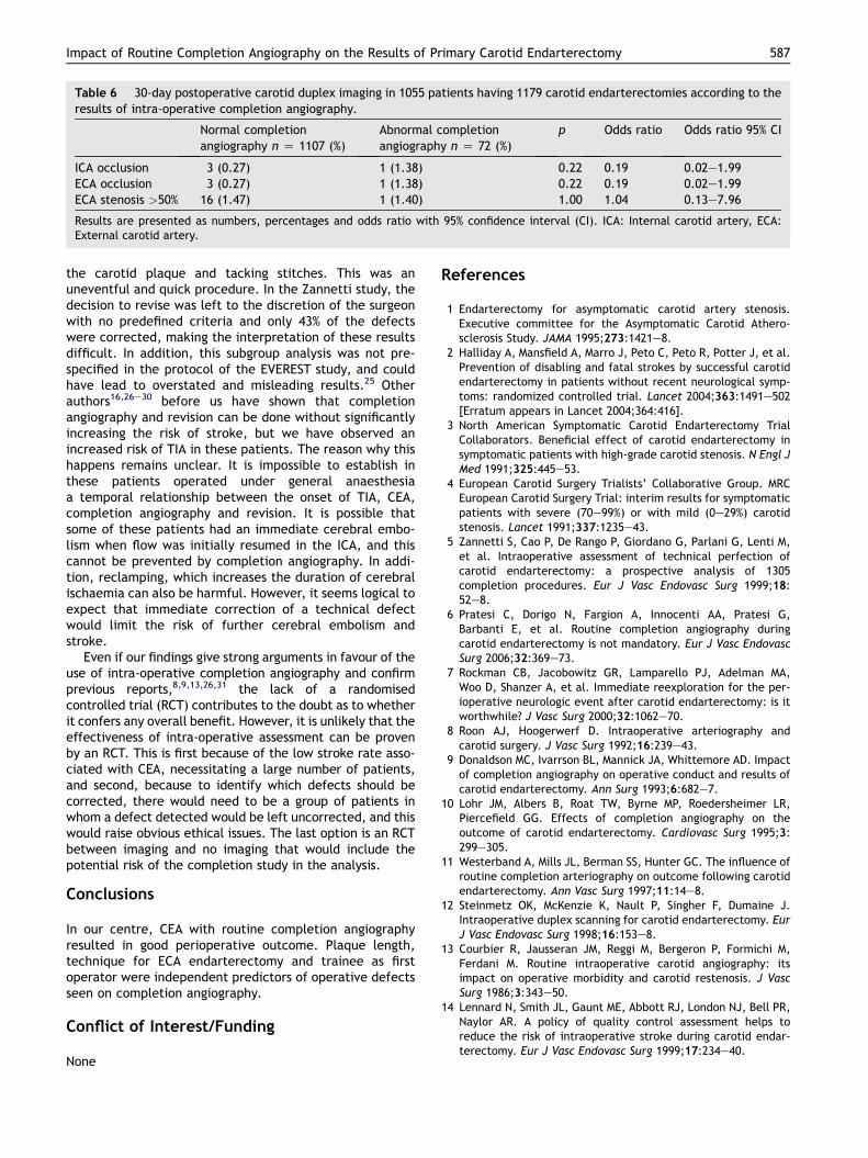
Table 6 30-day postoperative carotid duplex imaging in 1055 patients having 1179 carotid endarterectomies according to theresults of intra-operative completion angiography.
Normal completionangiography n Z 1107 (%)
Abnormal completionangiography n Z 72 (%)
p Odds ratio Odds ratio 95% CI
ICA occlusion 3 (0.27) 1 (1.38) 0.22 0.19 0.02e1.99ECA occlusion 3 (0.27) 1 (1.38) 0.22 0.19 0.02e1.99ECA stenosis >50% 16 (1.47) 1 (1.40) 1.00 1.04 0.13e7.96
Results are presented as numbers, percentages and odds ratio with 95% confidence interval (CI). ICA: Internal carotid artery, ECA:External carotid artery.
Impact of Routine Completion Angiography on the Results of Primary Carotid Endarterectomy 587
the carotid plaque and tacking stitches. This was anuneventful and quick procedure. In the Zannetti study, thedecision to revise was left to the discretion of the surgeonwith no predefined criteria and only 43% of the defectswere corrected, making the interpretation of these resultsdifficult. In addition, this subgroup analysis was not pre-specified in the protocol of the EVEREST study, and couldhave lead to overstated and misleading results.25 Otherauthors16,26e30 before us have shown that completionangiography and revision can be done without significantlyincreasing the risk of stroke, but we have observed anincreased risk of TIA in these patients. The reason why thishappens remains unclear. It is impossible to establish inthese patients operated under general anaesthesiaa temporal relationship between the onset of TIA, CEA,completion angiography and revision. It is possible thatsome of these patients had an immediate cerebral embo-lism when flow was initially resumed in the ICA, and thiscannot be prevented by completion angiography. In addi-tion, reclamping, which increases the duration of cerebralischaemia can also be harmful. However, it seems logical toexpect that immediate correction of a technical defectwould limit the risk of further cerebral embolism andstroke.
Even if our findings give strong arguments in favour of theuse of intra-operative completion angiography and confirmprevious reports,8,9,13,26,31 the lack of a randomisedcontrolled trial (RCT) contributes to the doubt as to whetherit confers any overall benefit. However, it is unlikely that theeffectiveness of intra-operative assessment can be provenby an RCT. This is first because of the low stroke rate asso-ciated with CEA, necessitating a large number of patients,and second, because to identify which defects should becorrected, there would need to be a group of patients inwhom a defect detected would be left uncorrected, and thiswould raise obvious ethical issues. The last option is an RCTbetween imaging and no imaging that would include thepotential risk of the completion study in the analysis.
Conclusions
In our centre, CEA with routine completion angiographyresulted in good perioperative outcome. Plaque length,technique for ECA endarterectomy and trainee as firstoperator were independent predictors of operative defectsseen on completion angiography.
Conflict of Interest/Funding
None
References
1 Endarterectomy for asymptomatic carotid artery stenosis.Executive committee for the Asymptomatic Carotid Athero-sclerosis Study. JAMA 1995;273:1421e8.
2 Halliday A, Mansfield A, Marro J, Peto C, Peto R, Potter J, et al.Prevention of disabling and fatal strokes by successful carotidendarterectomy in patients without recent neurological symp-toms: randomized controlled trial. Lancet 2004;363:1491e502[Erratum appears in Lancet 2004;364:416].
3 North American Symptomatic Carotid Endarterectomy TrialCollaborators. Beneficial effect of carotid endarterectomy insymptomatic patients with high-grade carotid stenosis. N Engl JMed 1991;325:445e53.
4 European Carotid Surgery Trialists’ Collaborative Group. MRCEuropean Carotid Surgery Trial: interim results for symptomaticpatients with severe (70e99%) or with mild (0e29%) carotidstenosis. Lancet 1991;337:1235e43.
5 Zannetti S, Cao P, De Rango P, Giordano G, Parlani G, Lenti M,et al. Intraoperative assessment of technical perfection ofcarotid endarterectomy: a prospective analysis of 1305completion procedures. Eur J Vasc Endovasc Surg 1999;18:52e8.
6 Pratesi C, Dorigo N, Fargion A, Innocenti AA, Pratesi G,Barbanti E, et al. Routine completion angiography duringcarotid endarterectomy is not mandatory. Eur J Vasc EndovascSurg 2006;32:369e73.
7 Rockman CB, Jacobowitz GR, Lamparello PJ, Adelman MA,Woo D, Shanzer A, et al. Immediate reexploration for the per-ioperative neurologic event after carotid endarterectomy: is itworthwhile? J Vasc Surg 2000;32:1062e70.
8 Roon AJ, Hoogerwerf D. Intraoperative arteriography andcarotid surgery. J Vasc Surg 1992;16:239e43.
9 Donaldson MC, Ivarrson BL, Mannick JA, Whittemore AD. Impactof completion angiography on operative conduct and results ofcarotid endarterectomy. Ann Surg 1993;6:682e7.
10 Lohr JM, Albers B, Roat TW, Byrne MP, Roedersheimer LR,Piercefield GG. Effects of completion angiography on theoutcome of carotid endarterectomy. Cardiovasc Surg 1995;3:299e305.
11 Westerband A, Mills JL, Berman SS, Hunter GC. The influence ofroutine completion arteriography on outcome following carotidendarterectomy. Ann Vasc Surg 1997;11:14e8.
12 Steinmetz OK, McKenzie K, Nault P, Singher F, Dumaine J.Intraoperative duplex scanning for carotid endarterectomy. EurJ Vasc Endovasc Surg 1998;16:153e8.
13 Courbier R, Jausseran JM, Reggi M, Bergeron P, Formichi M,Ferdani M. Routine intraoperative carotid angiography: itsimpact on operative morbidity and carotid restenosis. J VascSurg 1986;3:343e50.
14 Lennard N, Smith JL, Gaunt ME, Abbott RJ, London NJ, Bell PR,Naylor AR. A policy of quality control assessment helps toreduce the risk of intraoperative stroke during carotid endar-terectomy. Eur J Vasc Endovasc Surg 1999;17:234e40.

588 J.-B. Ricco et al.
15 Flanigan DP, Douglas DJ, Machi J, Siegel B, Schuler JJ,Buchbinder D. Intraoperative ultrasonic imaging of the carotidartery during carotid endarterectomy. Surgery 1986;100:893e8.
16 Ascher E, Markevich N, Kallakuri S, Schutzer RW, Hingorani AP.Intraoperative carotid artery duplex scanning in a modern seriesof 650 consecutive primary endarterectomy procedures. J VascSurg 2004;39:416e20.
17 Riles TS, Imparato AM, Jacobowitz GR, Lamparello PJ,Giangola G, AdelmanMA, et al. The cause of perioperative strokeafter carotid endarterectomy. J Vasc Surg 1994;19:206e14.
18 Naylor AR. Prevention of operation related stroke: are weasking the right questions? Cardiovasc Surg 1999;7:155e7.
19 Bandyk DF, Kaebnick HW, Adams MB, Towne JB. Turbulenceoccurring after carotid bifurcation endarterectomy: a harbingerof residual and recurrent carotid stenosis. J Vasc Surg 1988;7:261e74.
20 Moore WS, Marlello JY, Quinones-Baldrich WJ, Ahn SS. Etiologicimportance of the intimal flap of the external carotid artery inthe completion angiography in post carotid endarterectomystroke. Stroke 1990;21:1497e502.
21 Archie Jr JP. The outcome of external carotid endarterectomyduring routine carotid endarterectomy. J Vasc Surg 1998;28:585e90.
22 Woeffle KD, Bruijnen H, Neu J, Campbell P, Wack C,Loeprecht H. The role of intraoperative digital subtractionangioplasty for quality control of standard carotid endarterec-tomy using patch angioplasty. Cardiovasc Surg 2002;10:116e22.
23 Kinney EV, Seabrook GR, Kinney LY, Bandyk DF, Towne JB. Theimportance of intraoperative detection of residual flow
abnormalities after carotid endarterectomy. J Vasc Surg 1993;17:912e23.
24 Cao P, Giordano G, De Rango P, Zannetti S, Chiesa R, Coppi G,et al. Eversion versus conventional carotid endarterectomy.Late results of a prospective multicenter randomized trial. JVasc Surg 2000;31:19e30.
25 Wang R, Lagakos SW, Ware JH, Hunter DJ, Drazen JM. Statisticsin medicine e reporting of subgroup analyses in clinical trials.N Engl J Med 2007;357:2189e94.
26 Sala F, Hassen-Khodja R, Bouillanne PJ, Hussein H, Semlali C,Planchard P, et al. Importance of arteriography for intra-operative quality control during carotid artery surgery. Ann VascSurg 2002;16:730e5.
27 Panneton JM, Berger MW, Lewis BD, Hallett Jr JW, Bower TC,Gloviczki P, et al. Intraoperative duplex ultrasound duringcarotid endarterectomy. Vasc Surg 2001;35:1e9.
28 Dykes II JR, Bergamini TM, Lipski DA, Fulton RL, Garrison RN.Intraoperative duplex scanning reduces both residual stenosisand post-operative morbidity of carotid endarterectomy. AmSurg 1997;63:51e4.
29 Walker RA, Fox AD, Magee TR, Horrocks M. Intraoperativeduplex scanning as a means of quality control during carotidendarterectomy. Eur J Vasc Endovasc Surg 1996;11:364e7.
30 Bandyk DF, Mills JL, Gahtan V, Esses GE. Intraoperative duplexscanning of arterial reconstructions: fate of repaired andunrepaired defects. J Vasc Surg 1994;20:426e33.
31 Scott SM, Sethi GK, Bridgman AH. Perioperative stroke duringcarotid endarterectomy: the value of intraoperative angiog-raphy. J Cardiovasc Surg 1982;23:353e8.