impact of donor activating kir genes on hsct outcome in c1 ... filenoglobulin-like receptors (kir)...
TRANSCRIPT
RESEARCH ARTICLE
Impact of Donor Activating KIR Genes on
HSCT Outcome in C1-Ligand Negative Myeloid
Disease Patients Transplanted with Unrelated
Donors—A Retrospective Study
Christine Neuchel1,2, Daniel Furst1,2, Dietger Niederwieser3, Donald Bunjes4,
Chrysanthi Tsamadou1,2, Gerald Wulf5, Michael Pfreundschuh6, Eva Wagner7,
Gernot Stuhler8, Hermann Einsele9, Hubert Schrezenmeier1,2, Joannis Mytilineos1,2,10*
1 Institute of Clinical Transfusion Medicine and Immunogenetics Ulm, German Red Cross Blood Transfusion
Service, Baden Wuerttenberg–Hessen and University Hospital of Ulm, Ulm, Germany, 2 Institute of
Transfusion Medicine, University of Ulm, Ulm, Germany, 3 Department of Hematology/Oncology, University
of Leipzig, Leipzig, Germany, 4 Department of Hematology/Oncology, University Clinic Ulm, Ulm, Germany,
5 Department of Hematology/Oncology, Georg-August-University Gottingen, Gottingen, Germany,
6 Department of Internal Medicine I, Universitatsklinikum des Saarlandes, Homburg, Germany,
7 Department of Medicine III, Johannes Gutenberg-University Mainz, Mainz, Germany, 8 Centre for Bone
Marrow and Blood Stem Cell Transplantation, Deutsche Klinik fur Diagnostik, Wiesbaden, Germany,
9 Department of Medicine II, University Hospital Wurzburg, Wurzburg, Germany, 10 DRST–German
Registry for Stem Cell Transplantation, Essen, Germany
Abstract
Natural Killer cells (NK) are lymphocytes with the potential to recognize and lyse cells which
escaped T-cell mediated lysis due to their aberrant HLA expression profiles. Killer cell immu-
noglobulin-like receptors (KIR) influence NK-cell activity by mediation of activating or inhibi-
tory signals upon interaction with HLA-C (C1, C2) ligands. Therefore, absence of ligands for
donor inhibitory KIRs following hematopoietic stem cell transplantation (HSCT) may have an
influence on its outcome. Previous studies showed that C1 negative patients have a decreased
HSCT outcome. Our study, based on a cohort of 200 C1-negative patients, confirmed these
findings for the endpoints: overall survival (OS: HR = 1.41, CI = 1.14–1.74, p = 0.0012), dis-
ease free survival (DFS: HR = 1.27, CI = 1.05–1.53, p = 0.015), treatment related mortality
(TRM: HR = 1.41, CI = 1.01–1.96, p = 0.04), and relapse incidence (RI: HR = 1.33, CI = 1.01–
1.75, p = 0.04) all being inferior when compared to C1-positive patients (n = 1246). Subsequent
analysis showed that these findings applied for patients with myeloid malignancies but not
for patients with lymphoproliferative diseases (OS: myeloid: HR = 1.51, CI = 1.15–1.99,
p = 0.003; lymphoblastic: HR = 1.26, CI = 0.91–1.75, p = 0.16; DFS: myeloid: HR = 1.31,
CI = 1.01–1.70, p = 0.04; lymphoblastic: HR = 1.21, CI = 0.90–1.61, p = 0.21; RI: myeloid:
HR = 1.31, CI = 1.01–1.70, p = 0.04; lymphoblastic: HR = 1.21, CI = 0.90–1.61, p = 0.21).
Interestingly, within the C1-negative patient group, transplantation with KIR2DS2 resulted
in better OS (9/10 matched: HR = 0.24, CI = 0.08–0.67, p = 0.007) as well as DFS (9/10
matched: HR = 0,26, CI = 0.11–0.60, p = 0.002), and transplantation with KIR2DS1 positive
donors was associated with a decreased RI (HR = 0.30, CI = 0.13–0.69, p = 0.005). TRM
was increased when the donor was positive for KIR2DS1 (HR = 2.61, CI = 1.26–5.41,
PLOS ONE | DOI:10.1371/journal.pone.0169512 January 20, 2017 1 / 39
a1111111111
a1111111111
a1111111111
a1111111111
a1111111111
OPENACCESS
Citation: Neuchel C, Furst D, Niederwieser D,
Bunjes D, Tsamadou C, Wulf G, et al. (2017)
Impact of Donor Activating KIR Genes on HSCT
Outcome in C1-Ligand Negative Myeloid Disease
Patients Transplanted with Unrelated Donors—A
Retrospective Study. PLoS ONE 12(1): e0169512.
doi:10.1371/journal.pone.0169512
Editor: Golo Ahlenstiel, University of Sydney,
AUSTRALIA
Received: March 7, 2016
Accepted: December 19, 2016
Published: January 20, 2017
Copyright: © 2017 Neuchel et al. This is an open
access article distributed under the terms of the
Creative Commons Attribution License, which
permits unrestricted use, distribution, and
reproduction in any medium, provided the original
author and source are credited.
Data Availability Statement: All clinical data are
available either from the Deutsche Register fur
Stammzelltransplantation (DRST, http://www.drst.
de) or on-site at our institute, for researchers who
meet the criteria for confidential access to patient
data. In order to get access to the DRST clinical
data a project proposal has to be submitted to the
DRST. The committee for data access will consider
the proposal and either accept or decline it. KIR
typing-results and high resolution HLA typing
results, which are not listed in the DRST database,

p = 0.001). Our findings suggest that inclusion of KIR2DS1/2/5 and KIR3DS1-genotyping in
the unrelated donor search algorithm of C1-ligand negative patients with myeloid malignan-
cies may prove to be of clinical relevance.
Introduction
Haematopoietic stem cell transplantation (HSCT) has been established as a potentially curative
treatment for a variety of haematologic diseases. However, malignancy relapse after HSCT
remains the most frequent cause of treatment failure[1]. Therefore, introduction of a T-cell
mediated graft-versus-leukemia (GvL) effect is of great importance for a successful treatment
[2]. Unfortunately, there are downsides to this T-cell alloreactivity as it is associated with graft-
versus-host disease (GvHD)[3;4] which is a major complication following HSCT. Furthermore,
transformed cells may escape T-cell mediated lysis by down regulation of the Human Leukocyte
Antigen (HLA) expression on their cell surface[5–7]. On the other hand, Natural Killer cells
(NK) are lymphocytes that have the potential to recognize and lyse cells with aberrant HLA-
expression profiles, thereby mediating a GvL-effect of their own. It has been suggested that this
competence qualifies NK cells to conduct an anti-leukemic immune response without causing a
detrimental GvHD[8;9]. The functions of NK cells are controlled by interactions between NK
cell receptors and ligand molecules on the respective target cells. NK cell targeting of leukemic
cells is facilitated, among others, by the communication between killer cell immunoglobulin-
like receptors (KIR) on the surface of NK cells and HLA-A, -B, an -C molecules on the surface
of leukemic cells[10–12]. The KIR gene locus is characterized by high complexity, as it shows a
high genetic variation in regard of receptor functions (activation or inhibition of target cell kill-
ing), gene content, copy number variation, and sequence polymorphism[13]. KIR expression
profiles are further classified into two distinct haplotypes, A and B, based on the presence or
absence of certain KIR genes[14;15]. The A-haplotype is defined by the presence of the two
inhibitory KIRs KIR2DL1 and KIR2DL3, and KIR2DS4 as sole activating KIR. The B-haplotype
is characterized by the presence of a variety of different combinations of KIR genes, however,
the presence of at least one of the KIRs KIR2DL5, KIR2DS1/2/3/5, or KIR3DS1 is mandatory.
Possibly, the most important KIR-ligand interaction is that between HLA-C antigens and
their corresponding KIRs, in which both, inhibitory and activating, receptors are involved
[16]. However, HLA-C/KIR interactions appear to be even more complex, given that HLA-C
antigens are divided into two ligand groups based on specific amino acid residues [17]: Anti-
gens in the C1 group bear an asparagine residue on position 80 and interact with inhibitory
KIR2DL2 and KIR2DL3 as well as activating KIR2DS2, whereas C2 group ligands carry a
lysine residue at position 80 and interact with inhibitory KIR2DL1 and activating KIR2DS1
([18;19]. Activating signals and missing inhibition, both, lead to lysis of target cells[20;21].
Lack of interaction between a KIR and its respective HLA-C ligand in conjunction with acti-
vating signals may induce lysis of the target cell, a mechanism which is known as “missing self-
recognition”[22]. In fact, absence of one or more ligands for donor inhibitory KIRs in patients
with acute myeloid leukemia (AML) was shown to have a positive effect on HSCT outcome in
haploidentical transplantations[20;23]. Whereas this model is still controversially discussed in
the context of matched HSCT[24–26], it has been shown that in matched unrelated transplan-
tation the presence or absence of C1 and/or C2 ligands significantly affects outcome: patients
who expressed at least one C1 ligand showed superior overall survival and lower TRM rates
than patients who were homozygous for the C2 ligand[27].
Activating KIRs and Unrelated HSCT Outcome
PLOS ONE | DOI:10.1371/journal.pone.0169512 January 20, 2017 2 / 39
are available on-site at our institute for individuals
who meet the criteria for access to confidential
patient data. Please contact Dr Joannis Mytilineos
([email protected]) to request access to
confidential data.
Funding: This work was funded by the Deutsche
Jose Carreras Leukamie-Stiftung e.V. (Grant No.
DJCLS 11/10) (https://www.carreras-stiftung.de)
and the German Red Cross Blood Transfusion
Service, Baden-Wuerttemberg – Hessen (http://
www.blutspende.de/en/home/home.php). The
funders had no role in study design, data collection
and analysis, decision to publish, or preparation of
the manuscript.
Competing Interests: The authors have declared
that no competing interests exist
Abbreviations: AL, Unclassified acute leukemia;
ALL, Acute lymphoblastic leukemia; AML, Acute
myeloid leukemia; CI, Confidence interval; CLL,
Chronic lymphoblastic leukemia; CML, Chronic
myeloid leukemia; DFS, Disease free survival;
GvHD, Graft-versus-host disease; GvL, Graft-
versus-leukemia; HLA, Human Leukocyte Antigen;
HR, Hazard ratio; HSCT, Hematopoietic stem cell
transplantation; KIR, Killer cell immunoglobulin-like
receptors; MDS, Myelodysplastic syndrome; MM,
Multiple myeloma; NK, Natural Killer cell; OS,
Overall survival; RI, Relapse incidence; SBT,
sequence based typing; TRM, Treatment related
mortality.

In this multicentre study, we evaluated the C-ligand impact on unrelated HSCT outcome in a
large cohort of 1446 KIR & 5-loci-high resolution HLA-typed HSCT transplant pairs and thereby
confirmed the C1-ligand negative patient cohort to be at higher risk than C1-positive patients.
However, the incidence of patient C2 homozygosity is relatively low (in our cohort 14%). Trans-
plantation with KIR B-haplotype positive donors has already been shown to increase HSCT suc-
cess [28–31]. KIR haplotype analyses that were performed in our lab confirm the beneficial effect
of donor KIR B-haplotype on transplantation outcome (Abstract published in Biology of blood
and marrow transplantation in 2015[32]). NK cells and their activating KIR molecules have also
been shown to play a beneficial role in the immune response against various infectious agents
[33–35] as well as in the killing of T-cell blasts[36] and the prevention of leukaemia relapse[37].
KIR-Haplotype organisation, however, unvailed highly various phenotypes, especially among
individuals with KIR B-Haplotypes. In order to analyse the relative contribution of individual
B-Haplotype defining KIRs on HSCT outcome, sub-analyses on the level of single KIR genes are
required. In this study, we aimed to examine if C1-ligand negative patients, who were found to
be at risk post transplantation, could benefit from transplantations carried out from donors with
various activating KIRs.
Patients and Methods
Study cohort
The study population included a total of 1446 patients transplanted for malignant disorders
with T-cell repleted grafts from unrelated donors between 2000 and 2009. Benign diseases of
the hematopoietic system as well as inborn genetic diseases were excluded. Patients with AML
(acute myeloid leukemia), ALL (acute lymphoblastic leukemia), AL (unclassified acute leuke-
mia), CML (chronic myeloid leukemia), CLL (chronic lymphoblastic leukemia), MDS (myelo-
dysplastic syndrome), and MM (multiple myeloma) were eligible. Disease stages were adopted
from a previous report published by the European Society for Blood and Marrow Transplanta-
tion (EBMT) study group[38]: Early disease stage included AML, AL, and ALL transplanted
in first complete remission, CML in first chronic phase, and NHL, MM, and MDS trans-
planted either untreated or in first complete remission. Intermediate disease stage was defined
as AML, ALL, or NHL in second complete remission or first relapse, AL transplanted in sec-
ond complete remission, lymphoma and MM in second complete remission, partial remission
or stable disease, CML at all other stages than first chronic phase or blast crisis, and MDS in
second complete remission or in partial remission. Advanced stage was asigned to AML, AL,
ALL, and MDS in all other disease phases, CML in blast crisis, and lymphoma and MM in all
other disease stages than early or intermediate.
Graft source was either peripheral blood stem cells or bone marrow. Only first allogeneic
transplantations were included in the analysis. Transplant pairs were either 10/10 matched or
single HLA-A, -B, or -C mismatched. All patients received myeloablative or reduced intensity
conditioning. Myeloablative conditioning regimen was defined according to the EBMT rec-
ommendations as treatment with total body irradiation equal or above 10 Gy and/or cyclo-
phosphamide equal or greater than 120 mg/m2, and/or busulfan equal or greater than 16 mg/
kg[39;40]. Treatment with less intense regimen was classified as reduced intensity condition-
ing. All patients received T-cell replete grafts. Written recipient and donor consents for HLA
typing and for the analysis of clinical data were obtained. Clinical data was provided by the
German Registry for Stem Cell Transplantation (DRST) in pseudonymised form. This data
was collected by the transplant centers at day 100 post transplantation and yearly thereafter,
according to the EBMT guidelines. The study was approved by the ethical review board of the
University of Ulm (project number 263/09). Overall survival (OS) was defined as time to death
Activating KIRs and Unrelated HSCT Outcome
PLOS ONE | DOI:10.1371/journal.pone.0169512 January 20, 2017 3 / 39

from any cause and was censored at the time of last follow-up. Disease-free survival (DFS) was
defined as time to relapse of primary malignancy, or death from any cause, and was also cen-
sored at the time of last follow-up. Relapse incidence (RI) was defined as occurrence of relapse
of hematologic malignancy at any given time point. Treatment-related mortality (TRM) was
defined as mortality in complete disease remission.
HLA-Typing in patients and donors
Donor-recipient pairs from unrelated donor searches completed before May 2005 were low
resolution typed for HLA-A and -B, and high resolution typed for HLA-DRB1 and -DQB1.
Stored DNA material of these individuals was retrospectively high resolution typed for HLA-
A, -B, and -C. For unrelated donor searches which were initiated after 05/2005 5-loci high res-
olution HLA-typing was carried out upfront.
High resolution HLA-typing was performed by sequence based typing (SBT) using a CE-
certified in-house Kit for HLA-class I (H-Seq-ABC, DRK-BSD Baden- Wurttemberg–Hessen
gGmbH, Frankfurt am Main, Germany) and by in-house SBT (H-Seq-DR, DRK-BSD Baden-
Wurttemberg–Hessen gGmbH, Frankfurt am Main, Germany and H-Seq-DQB, DRK-BSD
Baden- Wurttemberg–Hessen gGmbH, Frankfurt am Main, Germany) or Luminex1 (Life
Technologies, Carlsbad, CA, USA) PCR-sequence specific oligonucleotide probe (PCR-SSO)
method for HLA-class II. Exons 2 and 3 were sequenced for HLA-class I typing, and exon 2
was analysed for HLA-class II testing. Additional typing for relevant non-expressed alleles was
performed according to the National Marrow Donor Program confirmatory typing require-
ments[41].
KIR-Typing in patients and donors
KIR-typing was performed using the commercially available “KIR Genotyping SSP Kit” from
Thermo Fisher Scientific (Waltham, MA, USA). The Kit uses sequence specific primer PCR to
assign presence or absence, respectively, of a given KIR gene. The method allows the identifica-
tion of the following KIR-genes: KIR2DL1/2/3/4/5A/5B, KIR2DS1/2/3/4/5, KIR3DL1/2/3,
KIR3DS1, KIR2DP1, and KIR3DP1.
Table 1. General patient characteristics.
n % n % n %
Age Disease stage Patient and donor sex combination
18–40 359 24.8% Early 556 38.5% Donor female, patient male 162 11.2%
41–60 1071 74.1% Intermediate 518 35.8% Others 1284 88.8%
61–76 16 1.1% Advanced 372 25.7%
HLA-compatibility Graft source Conditioning
10/10 1022 70.7% Bone Marrow 91 6.3% Myeloablative 870 39.8%
9/10 424 29.3% Peripheral Blood stem cells 1355 93.7% Reduced intensity 576 60.2%
Year of transplantation Diagnosis myeloid Diagnosis lymphoblastic
2000–2005 377 26.1% AML 447 30.9% ALL 189 13.1%
2006–2009 1069 73.9% CML 65 4.5% CLL 82 5.7%
MDS 271 18.7% MM 131 9.1%
NHL 176 12.2%
Diagnosis other
AL 85 5.9%
doi:10.1371/journal.pone.0169512.t001
Activating KIRs and Unrelated HSCT Outcome
PLOS ONE | DOI:10.1371/journal.pone.0169512 January 20, 2017 4 / 39

Assessment of missing KIR ligand status
KIR ligand status of patients was obtained from high resolution HLA-typing. HLA-C mole-
cules were grouped according to the presence of epitopes recognized by KIRs: HLA-C antigens
with an asparagine residue at amino acid sequence position 80 possess the C1 epitope, which
acts as ligand for the inhibitory KIR2DL2 and KIR2DL3 as well as for the activating KIR2DS2.
Accordingly, HLA-C antigens with a lysine residue at this position possess the C2 epitope,
which acts as ligand for the inhibitory KIR2DL1 as well as for the activating KIR2DS1.
The absence of C1 or C2 in patients who were transplanted with a KIR2DL2/3 or KIR2DL1
positive donor, respectively, was considered as “missing ligand”. Control patient groups car-
ried at least one ligand for any donor KIR2DL1 or KIR2DL2/3, respectively.
Statistical analysis
All statistical analyses were performed using the open source program for statistical computing
“R”, version 3.0.2. Cumulative estimates of overall survival (OS) and disease free survival (DFS)
were obtained using Kaplan-Meier analysis. Extended Cox regression models allowing for time-
dependent covariates were used to conduct multivariate analyses of OS and DFS. Univariate
analyses of relapse incidence (RI) and treatment related mortality (TRM) were performed using
competing risk analysis. Competing risk regression models for stratified data were used for mul-
tivariate analysis of RI and TRM. The categorical variable diagnosis showed different baselines
of hazard for the respective patient sub-groups. Hence, for both multivariate models stratifica-
tion for diagnosis was accomplished rather than integrating diagnosis as a covariate[42;43].
Centre effects were adjusted using a gamma frailty term (fitted with the R-package “survival”)
[44;45]. A stepwise backward exclusion procedure was used for model selection, where all vari-
ables have been included in the first model and then successively reduced (at each step the least
significant) until the loss of information became significant (analysis of missing ligand model
p�0.05; KIR analyses p�0.01)[43]. In order to address the problem of multiple testing and
Table 2. Distribution of activating KIR2DS1/2/3/5 and KIR3DS1 among donors in combination with patient C1-ligand status in the study cohort.
Myeloid malignancy Lymphoid malignancy
Donor Patient C1-positive Patient C1-negative Patient C1-positive Patient C1-negative
n = 666 (85.1%) n = 117 (14.9%) n = 501 (86.7%) n = 77 (13.3%)
KIR2DS1 positive 258 (38.7%) 45 (38.5%) 200 (39.9%) 28 (36.4%)
KIR2DS1 negative 408 (61.3%) 72 (61.5%) 301 (60.1%) 49 (63.6%)
KIR2DS2 positive 314 (47.1%) 63 (53.8%) 246 (49.1%) 35 (45.4%)
KIR2DS2 negative 352 (52.9%) 54 (45.2%) 255 (50.9%) 42 (54.5%)
KIR2DS3 positive 161 (24.2%) 41 (35.0%) 130 (25.9%) 19 (24.7%)
KIR2DS3 negative 505 (75.8%) 76 (65.0%) 371 (74.1%) 58 (75.3%)
KIR2DS5 positive 202 (30.3%) 34 (29.1%) 152 (30.3%) 21 (27.3%)
KIR2DS5 negative 464 (69.7%) 83 (70.9%) 349 (69.7%) 56 (72.7%)
KIR3DS1 positive 229 (34.4%) 45 (38.5%) 186 (37.1%) 30 (39.0%)
KIR3DS1 negative 437 (65.6%) 72 (61.5%) 315 (62.9%) 47 (61.0%)
doi:10.1371/journal.pone.0169512.t002
Table 3. Distribution of KIR2DL1, KIR2DL2 and KIR2DL3 as well as C1/C2 genotype in the study cohort.
KIR2DL1 KIR2DL2 KIR2DL3 KIR2DL2/3 Both ligands present Missing C2 ligand Missing C1 ligand
Patients 1393 762 1302 618 663 583 200
(96.3%) (52.7%) (90.0%) (42.7%) (45.9%) (40.3%) (13.8%)
Donors 1388 720 1307 581 673 578 195
(96.0%) (49.8%) (90.4%) (40.2%) (46.5%) (40.0%) (13.5%)
doi:10.1371/journal.pone.0169512.t003
Activating KIRs and Unrelated HSCT Outcome
PLOS ONE | DOI:10.1371/journal.pone.0169512 January 20, 2017 5 / 39

potentially associated type 1 errors we set the significance level for our KIR analyses to 0.01.
Covariates were assessed according to previously published recommendations of the EBMT
study group[42;43;46], including patient age, disease stage, graft manipulation, conditioning regi-
men, graft source, donor source (national vs. international), year of transplantation, time between
diagnosis and transplantation, and donor-recipient gender combination. For the clinical
Fig 1. Univariate OS analysis in C1-negative patients (missing C1 ligand) vs. C1-positive patients who were transplanted with KIR2DL2 or
KIR2DL3 positive donors. Dashed orange line: C1-negative patients (n = 200). Solid black line: C1-positive patients (n = 1239). p = 0.0014.
doi:10.1371/journal.pone.0169512.g001
Activating KIRs and Unrelated HSCT Outcome
PLOS ONE | DOI:10.1371/journal.pone.0169512 January 20, 2017 6 / 39

covariates analysed, the data was complete. All models were checked for interactions and propor-
tional hazards assumption. No significant interactions between covariates were found and viola-
tions of proportional hazards assumption were adjusted by using time dependent covariates.
Fig 2. Univariate DFS analysis in C1-negative patients (missing C1 ligand) vs. C1-positive patients who were transplanted with KIR2DL2 or
KIR2DL3 positive donors. Dashed orange line: C1-negative patients (n = 200). Solid black line: C1-positive patients (n = 1239). p = 0.006.
doi:10.1371/journal.pone.0169512.g002
Activating KIRs and Unrelated HSCT Outcome
PLOS ONE | DOI:10.1371/journal.pone.0169512 January 20, 2017 7 / 39

Results
Patient characteristics
General patient characteristics are shown in Table 1. The cohort included 1446 transplant
pairs. Median patient age was 52 years. Most patients were in early (38.5%) and intermediate
(35.8%) disease state. Stem cell source was peripheral blood stem cells in 93.7% cases. Almost
two thirds (60.2%) of the patients received myeloablative conditioning. An unfavourable
donor-patient sex mismatch (female donor for a male patient) was found in 11.2% of trans-
plant pairs. Most patients received a graft from a 10/10 matched HLA compatible donor
(70.7%), whereas 29.3% were transplanted with a 9/10 matched donor. Median post-transplan-
tation follow-up time was 40 months and maximum follow-up time was 9.5 years.
Table 2 lists the distribution of activating KIR2DS1/2/3/5 and KIR3DS1 among donors sub-
ject to subgroups of patients with myeloid or lymphoid malignancies. Between 54 and 60% of
donors were positive for KIR2DS1. KIR2DS2 was even more prevalent with up to 75% being
carriers. KIR2DS3 and KIR2DS5 were found in 40–50% of donors and KIR3DS1 was present
in more than 50% of donors.
C1-negative patients show significantly inferior HSCT outcome
Distribution of KIR2DL1/2/3 and HLA-C-ligands among patients and donors is shown in
Table 3. All individuals in the cohort were either positive for KIR2DL2 or KIR2DL3, or both
(approximately 40%). The corresponding C1 ligand for these KIRs was carried in 86% of the
individuals. Of these, 40% were C1 homozygous. KIR2DL1 was detected in 96% of patients
and donors. The C2 ligand was detected in 60% of the patients. Homozygosity for this ligand
was relatively rare with a prevalence of 13.8%.
Patients who did not possess the C1 ligand (i.e. C2 homozygous) showed significantly infe-
rior OS and DFS after 1, 3 and 5 years post transplantation (Fig 1 and Fig 2) when compared
to the C1 positive (C1 homozygous or C1C2 heterozygous) control group (OS: 60.7% vs.
49.7%; 47.0% vs. 36.0%; 41.6% vs. 29.2%, p = 0.0014. DFS: 51.1% vs. 40.0%; 37.6% vs. 27.1%;
30.3% vs. 26.6%, p = 0.006; Table 4). C2 Homozygosity also was associated with increased RI
and TRM rates as shown in Fig 3 and Fig 4. Multivariate analysis confirmed these results, since
OS (HR = 1.41, CI = 1.14–1.74, p = 0.0012), DFS (HR = 1.27, CI = 1.05–1.53, p = 0.015), RI
(HR = 1.33, CI = 1.01–1.75, p = 0.04) and TRM (HR = 1.41, CI = 1.01–1.96, p = 0.04) all were
negatively affected when the patients did not possess the C1 ligand (Table 4).
A significant impact of the missing ligand model described in previous studies was solely
found for myeloid malignancies. The four endpoints were therefore re-analysed in subgroups
of patients with myeloid and lymphoblastic diseases, respectively.
The protective effect of C1 could be observed only in the myeloid group (Table 5), where
absence of C1 was associated with reduced OS (myeloid: HR = 1.51, CI = 1.15–1.99, p = 0.003;
lymphoblastic: HR = 1.26, CI = 0.91–1.75, p = 0.16), reduced DFS (myeloid: HR = 1.31, CI =
Table 4. Multivariate analysis of OS, DFS, RI, and TRM in C1-negative patients vs. patients with at
least one C1 ligand.
Endpoint Cases HR 95% CI p
OS 200 1.41 1.14–1.74 0.0012
DFS 200 1.27 1.05–1.53 0.015
RI 200 1.33 1.01–1.75 0.04
TRM 200 1.41 1.01–1.96 0.04
doi:10.1371/journal.pone.0169512.t004
Activating KIRs and Unrelated HSCT Outcome
PLOS ONE | DOI:10.1371/journal.pone.0169512 January 20, 2017 8 / 39

1.01–1.70, p = 0.04; lymphoblastic: HR = 1.21, CI = 0.90–1.61, p = 0.21), and increased RI (mye-
loid: HR = 1.31, CI = 1.01–1.70, p = 0.04; lymphoblastic: HR = 1.21, CI = 0.90–1.61, p = 0.21).
There were no differences observed for TRM between the myeloid and lymphoblastic sub-
groups (myeloid: HR = 1.34, CI = 0.85–2.11, p = 0.20; lymphoblastic: HR = 1.27, CI = 0.75–
2.16, p = 0.38).
Fig 3. Univariate RI analysis in C1-negative patients (missing C1 ligand) vs. C1-positive patients who were transplanted with KIR2DL2 or
KIR2DL3 positive donors. Dashed orange line: C1-negative patients (n = 200). Solid black line: C1-positive patients (n = 1239). p = 0.04.
doi:10.1371/journal.pone.0169512.g003
Activating KIRs and Unrelated HSCT Outcome
PLOS ONE | DOI:10.1371/journal.pone.0169512 January 20, 2017 9 / 39

C1-negative patients with myeloid malignancies were thus identified as being at risk and
therefore this patient group is referred as such in subsequent analyses.
Fig 4. Univariate TRM analysis in C1-negative patients (missing C1 ligand) vs. C1-positive patients who were transplanted with KIR2DL2
or KIR2DL3 positive donors. Dashed orange line: C1-negative patients (n = 200). Solid black line: C1-positive patients (n = 1239). p = 0.06.
doi:10.1371/journal.pone.0169512.g004
Activating KIRs and Unrelated HSCT Outcome
PLOS ONE | DOI:10.1371/journal.pone.0169512 January 20, 2017 10 / 39

Missing C2-ligand has no effect on HSCT outcome
Missing C2-ligand does not affect HSCT outcome as shown by Kaplan-Meier analysis (Figs 5–8)
and multivariate analysis (OS: HR = 0.91, CI = 0.77–1.03, p = 0.22; DFS: HR = 0.94, CI = 0.81–
1.09, p = 0.41; RI: HR = 0.97, CI = 0.79–1.19, p = 0.74; TRM: HR = 0.90, CI = 0.69–1.16, p = 0.42).
Transplantation with KIR2DS2 positive donors significantly increases
OS and DFS in the C1-negative risk group
Since C1-deficiency was shown to negatively affect HSCT outcome in patients with myeloid,
but not lymphoblastic, malignancies, following analyses were conducted in the myeloid patient
group, only.
As KIR2DS4 is present in almost all individuals and is expressed on both, A- and B-Haplo-
types, this KIR gene was not taken into consideration for any analyses in our study. Addition-
ally, OS and DFS analyses were conducted in subgroups of 10/10 and 9/10 matched transplants.
OS and DFS were significantly improved when the donor was positive for KIR2DS2 and the
transplant pair was mismatched at a single HLA-class I gene: The OS rates were twice as high as
in the 9/10 matched patient group with a KIR2DS2 negative donor (10/10: 49.5% vs. 44.7%;
38.5% vs. 34.1%; 28.9% vs. 25.6%; 9/10: 57.3% vs. 23.5%; 40.9% vs. 17.6%; Fig 9 and Fig 10). Simi-
larly, donor KIR2DS2 positivity resulted in higher DFS rates in the 9/10 matched patient group
(10/10: 40.6% vs. 37.9%; 29.5% vs. 28.4%; 9/10: 51.6% vs. 21.1%; 24.6% vs. 15.8%; Fig 11 and Fig
12). Multivariate analysis confirmed these results (OS: 10/10: HR = 1.02, CI = 0.51–2.07, p =
0.95; 9/10: HR = 0.24, CI = 0.08–0.67, p = 0.007; DFS: 10/10: HR = 1.23, CI = 0.72–2.07, p = 0.44;
9/10: HR = 0.26, CI = 0.11–0.60, p = 0.002; Table 6 and 6B). Reduced intensity conditioning regi-
men was a significant predictor within our analysis (S1 Table) and it would have been interesting
to investigate whether the influence of KIR2DS2 could be observed in both, reduced intensity
and myeloablative conditioning, respectively. As individual disease stages (early, intermediate,
and advanced) are important predictors for HSCT outcome as well, it would have also been
interesting to investigate the effect of donor KIR2DS2 positivity in according to disease stage.
Unfortunately the number of cases in our study did not allow these analyses.
Presence of other activating KIR KIR2DS1/3/5 or KIR3DS1 in the donor did not have any
effect on OS or DFS (Tables 6 and 7).
Transplantation with KIR2DS1 positive donors is associated with a
significantly decreased RI in the C1-negative risk patient group
For RI and TRM analyses, case numbers were too small to conduct sub-analysis in 10/10 and
9/10 matched transplant pairs. Therefore the subsequent analyses of the impact of KIR2DS1/2/
Table 5. Multivariate analysis of OS, DFS, RI, and TRM in C1-negative patients with myeloid or lym-
phoid diseases vs. control group patients with at least one C1 ligand.
Endpoint Cases HR 95% CI p
OS myeloid 117 1.51 1.15–1.99 0.003
OS lymphoid 83 1.26 0.91–1.75 0.16
DFS myeloid 117 1.31 1.01–1.70 0.04
DFS lymphoid 83 1.21 0.90–1.61 0.21
RI myeloid 117 1.31 1.01–1.70 0.04
RI lymphoid 83 1.21 0.90–1.61 0.21
TRM myeloid 117 1.34 0.85–2.11 0.20
TRM lymphoid 83 1.27 0.75–2.16 0.38
doi:10.1371/journal.pone.0169512.t005
Activating KIRs and Unrelated HSCT Outcome
PLOS ONE | DOI:10.1371/journal.pone.0169512 January 20, 2017 11 / 39

3/5 or KIR3DS1 positive grafts on RI and TRM within the risk patient group were conducted
disregarding HLA matching degree (univariate analysis) or with stratification for HLA match-
ing (multivariate analysis).
Transplantation with KIR2DS1 positive donors significantly reduced RI in C1-negative
patients (HR = 0.30, CI = 0.13–0.69, p = 0.005) (Fig 13, Table 8). Transplantation with a
Fig 5. Univariate OS analysis in C2-negative (missing C2 ligand) vs. C2-positive patients who were transplanted with KIR2DL1-positive
donors. Dashed orange line: C2-negative patients (n = 823). Solid black line: C2-positive patients (n = 558). p = 0.08.
doi:10.1371/journal.pone.0169512.g005
Activating KIRs and Unrelated HSCT Outcome
PLOS ONE | DOI:10.1371/journal.pone.0169512 January 20, 2017 12 / 39
KIR2DS2-positive (HR = 1.87, CI = 0.86–4.08, p = 0.12), KIR2DS3-positive (HR = 1.60,
CI = 0.83–3.10, p = 0.16), KIR2DS5-positive (HR = 0.37, CI = 0.15–0.92, p = 0.03), or KIR3D-
S1-positive (HR = 0.51, CI = 0.24–1.10, p = 0.09) donor had no significant effect on RI (Figs
14–16, Table 8).
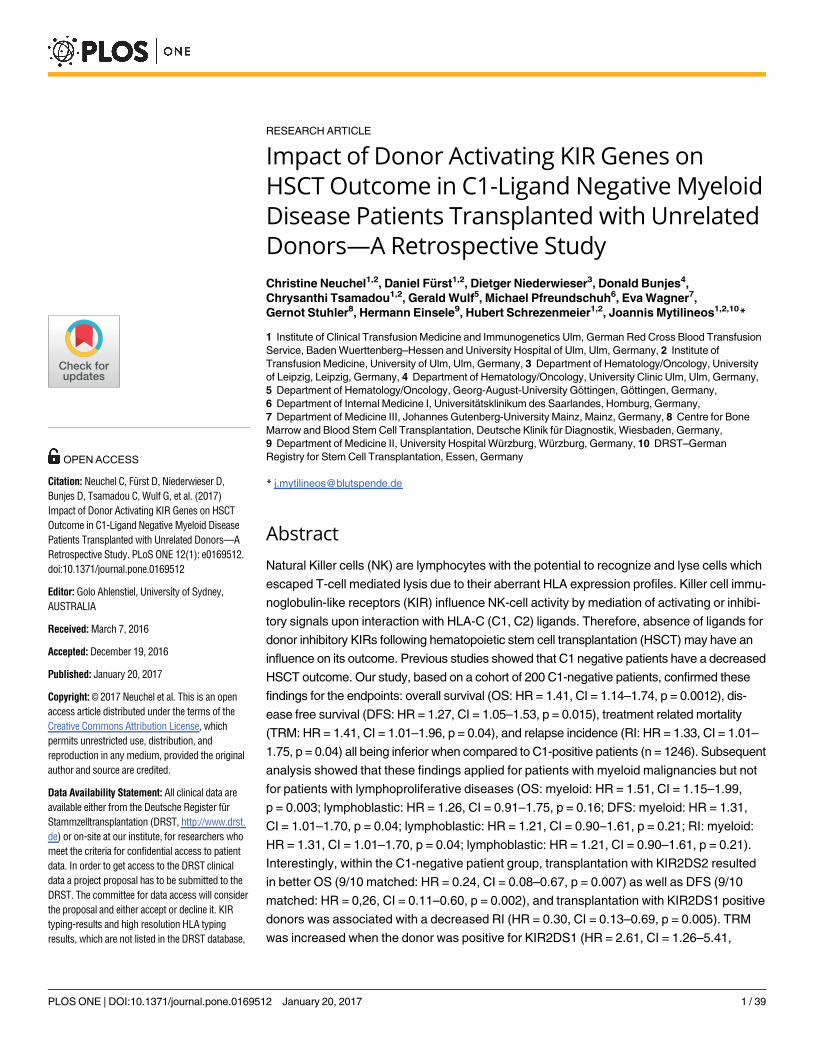
Fig 6. Univariate DFS analysis in C2-negative (missing C2 ligand) vs. C2-positive patients who were transplanted with KIR2DL1-positive
donors. Dashed orange line: C2-negative patients (n = 823). Solid black line: C2-positive patients (n = 558). p = 0.11.
doi:10.1371/journal.pone.0169512.g006
Activating KIRs and Unrelated HSCT Outcome
PLOS ONE | DOI:10.1371/journal.pone.0169512 January 20, 2017 13 / 39

Transplantation with KIR2DS1 positive donors is associated with a
significantly increased TRM in the C1-negative risk patient group
The results of the multivariate TRM analyses are summarised in Table 9. In the presence of a
single HLA class I mismatch, TRM was increased in C1 negative patients who were trans-
planted with a KIR2DS1 (HR = 2.61, CI = 1.26–5.41, p = 0.001; Fig 17) positive donor.
Fig 7. Univariate RI analysis in C2-negative (missing C2 ligand) vs. C2-positive patients who were transplanted with KIR2DL1-positive
donors. Dashed orange line: C2-negative patients (n = 823). Solid black line: C2-positive patients (n = 558). p = 0.47.
doi:10.1371/journal.pone.0169512.g007
Activating KIRs and Unrelated HSCT Outcome
PLOS ONE | DOI:10.1371/journal.pone.0169512 January 20, 2017 14 / 39

Transplantation with a KIR2DS5 positive donor had no effect on TRM (HR = 0.81, CI = 0.36–
1.86, p = 0.63, Fig 18) and a potential positive effect of a KIR2DS2 positive graft (HR = 0.31,
CI = 0.11–0.86, p = 0.03, Fig 19) as well as the effects observed on TRM by selecting a KIR2DS5
(HR = 3.33, CI = 1.20–9.29, p = 0.02), or KIR3DS1 (HR = 2.18, CI = 0.99–4.78, p = 0.05, Fig 20)
positive donor, respectively, did not reach the level of statistical significance (0.01).
Fig 8. Univariate TRM analysis in C2-negative (missing C2 ligand) vs. C2-positive patients who were transplanted with KIR2DL1-positive
donors. Dashed orange line: C2-negative patients (n = 823). Solid black line: C2-positive patients (n = 558). p = 0.30.
doi:10.1371/journal.pone.0169512.g008
Activating KIRs and Unrelated HSCT Outcome
PLOS ONE | DOI:10.1371/journal.pone.0169512 January 20, 2017 15 / 39

The KIR Better/Best model in C1-negative patients
The KIR locus is divided into a centromeric and a telomeric region, each bearing variable KIR
gene content motifs designated as cen-A, cen-B, tel-A, and tel-B. The B-motifs contain one or
Fig 9. Univariate OS analysis in C1-negative patients with myeloid malignancies who were transplanted with KIR2DS2 positive donors vs.
KIR2DS2 negative donors. Solid black line: donor KIR2DS2-negative. Dashed orange line: donor KIR2DS2-positive. 10/10 matched transplant pairs,
donor KIR2DS2-negative (n = 32) vs. donor KIR2DS2-positive (n = 45), p = 0.86.
doi:10.1371/journal.pone.0169512.g009
Activating KIRs and Unrelated HSCT Outcome
PLOS ONE | DOI:10.1371/journal.pone.0169512 January 20, 2017 16 / 39

more of the seven KIR B-haplotype specific genes. It has been shown by Cooley et al, that
C1-positive, but not C1-negative patients benefit from transplantation with donors who carry
two or more B-motifs. We used the B-content calculator that can be found at http://www.ebi.
ac.uk/ipd/kir/donor_b_content.html to assign the donors of our cohort accordingly to one of
Fig 10. Univariate OS analysis in C1-negative patients with myeloid malignancies who were transplanted with KIR2DS2 positive donors vs.
KIR2DS2 negative donors. Solid black line: donor KIR2DS2-negative. Dashed orange line: donor KIR2DS2-positive. 9/10 matched transplant pairs,
donor KIR2DS2-negative (n = 22) vs. donor KIR2DS2-positive (n = 18), p = 0.04.
doi:10.1371/journal.pone.0169512.g010
Activating KIRs and Unrelated HSCT Outcome
PLOS ONE | DOI:10.1371/journal.pone.0169512 January 20, 2017 17 / 39
three groups based on KIR B-content—neutral (n = 142), better (n = 40) or best (n = 18)—and
analysed the impact of this classification on the transplantation outcome of the C1-negative
patients. As case numbers in the “best” group were low, we decided to combine the “better”
and the “best” donors into the “better/best” group. Multivariate analyses showed that in 9/10
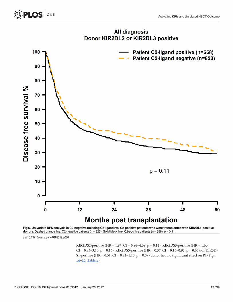
Fig 11. DFS analysis in C1-negative patients with myeloid malignancies who were transplanted with KIR2DS2 positive donors vs. KIR2DS2
negative donors. Solid black line: donor KIR2DS2-negative. Dashed orange line: donor KIR2DS2-positive. 10/10 matched transplant pairs, donor
KIR2DS2-negative (n = 32) vs. donor KIR2DS2-positive (n = 45), p = 0.88.
doi:10.1371/journal.pone.0169512.g011
Activating KIRs and Unrelated HSCT Outcome
PLOS ONE | DOI:10.1371/journal.pone.0169512 January 20, 2017 18 / 39

matched transplantations, “better/best” donors may improve DFS (HR = 0.39, CI = 0.15–0.99,
p = 0.048), but had no significant effect on OS (HR = 0.69, CI = 0.22–2.21, p = 0.52), RI
(HR = 0.48, CI = 0.07–3.29, p = 0.46), or TRM (HR = 0.76, CI = 0.23–2.49, p = 0.65). In 10/10
matched transplant pairs, however, the “better/best” KIR donor status had no effect on
Fig 12. DFS analysis in C1-negative patients with myeloid malignancies who were transplanted with KIR2DS2 positive donors vs. KIR2DS2
negative donors. Solid black line: donor KIR2DS2-negative. Dashed orange line: donor KIR2DS2-positive. 9/10 matched transplant pairs, donor
KIR2DS2-negative (n = 22) vs. donor KIR2DS2-positive (n = 18), p = 0.05.
doi:10.1371/journal.pone.0169512.g012
Activating KIRs and Unrelated HSCT Outcome
PLOS ONE | DOI:10.1371/journal.pone.0169512 January 20, 2017 19 / 39

Table 6. Multivariate analysis of OS in C1-negative patients transplanted with a KIR2DS1/2/3/5 or
KIR3DS1 positive donor vs. C1-negative patients transplanted with a KIR2DS1/2/3/5 or KIR3DS1 nega-
tive donor.
Cases HR 95% CI p
Donor KIR2DS1 negative 10/10 47 1 - -
Donor KIR2DS1 positive 10/10 30 0.75 0.37–1.52 0.42
Donor KIR2DS1 negative 9/10 25 1 - -
Donor KIR2DS1 positive 09/10 15 1.88 0.74–4.76 0.18
Donor KIR2DS2 negative 10/10 32 1 - -
Donor KIR2DS2 positive 10/10 45 1.04 0.51–2.12 0.91
Donor KIR2DS2 negative 9/10 22 1 - -
Donor KIR2DS2 positive 9/10 18 0.24 0.08–0.67 0.007
Donor KIR2DS3 negative 10/10 49 1 - -
Donor KIR2DS3 positive 10/10 28 1.38 0.68–2.79 0.37
Donor KIR2DS3 negative 9/10 27 1 - -
Donor KIR2DS3 positive 9/10 13 0.85 0.31–2.37 0.76
Donor KIR2DS5 negative 10/10 56 1 - -
Donor KIR2DS5 positive 10/10 21 0.55 0.25–1.24 0.15
Donor KIR2DS5 negative 9/10 27 1 - -
Donor KIR2DS5 positive 9/10 13 2.19 0.78–6.12 0.13
Donor KIR3DS1 negative 10/10 46 1 - -
Donor KIR3DS1 positive 10/10 31 0.96 0.47–1.97 0.92
Donor KIR3DS1 negative 9/10 26 1 - -
Donor KIR3DS1 positive 9/10 14 1.74 0.66–4.56 0.26
doi:10.1371/journal.pone.0169512.t006
Table 7. Multivariate analysis of DFS in C1-negative patients transplanted with a KIR2DS1/2/3/5 or
KIR3DS1 positive donor vs. C1-negative patients transplanted with a KIR2DS1/2/3/5 or KIR3DS1 nega-
tive donor.
Cases HR 95% CI p
Donor KIR2DS1 negative 10/10 47 1 - -
Donor KIR2DS1 positive 10/10 30 0.65 0.38–1.11 0.11
Donor KIR2DS1 negative 9/10 25 1 - -
Donor KIR2DS1 positive 09/10 15 1.31 0.66–2.59 0.44
Donor KIR2DS2 negative 10/10 32 1 - -
Donor KIR2DS2 positive 10/10 45 1.23 0.72–2.07 0.44
Donor KIR2DS2 negative 9/10 22 1 - -
Donor KIR2DS2 positive 9/10 18 0.26 0.11–0.60 0.002
Donor KIR2DS3 negative 10/10 49 1 - -
Donor KIR2DS3 positive 10/10 28 1.27 0.75–2.13 0.37
Donor KIR2DS3 negative 9/10 27 1 - -
Donor KIR2DS3 positive 9/10 13 0.67 0.33–1.37 0.27
Donor KIR2DS5 negative 10/10 56 1 - -
Donor KIR2DS5 positive 10/10 21 1.14 0.64–2.02 0.66
Donor KIR2DS5 negative 9/10 27 1 - -
Donor KIR2DS5 positive 9/10 13 1.36 0.65–2.88 0.41
Donor KIR3DS1 negative 10/10 46 1 - -
Donor KIR3DS1 positive 10/10 31 0.84 0.49–1.45 0.54
Donor KIR3DS1 negative 9/10 26 1 - -
Donor KIR3DS1 positive 9/10 14 1.24 0.62–2.51 0.54
doi:10.1371/journal.pone.0169512.t007
Activating KIRs and Unrelated HSCT Outcome
PLOS ONE | DOI:10.1371/journal.pone.0169512 January 20, 2017 20 / 39

transplantation outcome (OS: HR = 0.98, CI = 0.46–2.10, p = 0.95; DFS: HR = 1.13, CI = 0.63–
2.02, p = 0.69; RI: HR = 1.60, CI = 0.72–3.55, p = 0.25; TRM: HR = 0.80, CI = 0.17–3.80,
p = 0.78).
Fig 13. Univariate RI analysis in C1-negative patients with myeloid malignancies who were transplanted with KIR2DS1 positive vs.
KIR2DS1 negative donors. Solid black line: donor KIR2DS1 negative. Dashed orange line: donor KIR2DS1 positive. Donor KIR2DS1-negative
(n = 72) vs. donor KIR2DS1-positive (n = 45), p = 0.008.
doi:10.1371/journal.pone.0169512.g013
Activating KIRs and Unrelated HSCT Outcome
PLOS ONE | DOI:10.1371/journal.pone.0169512 January 20, 2017 21 / 39

Proposed algorithm for the selection for donors for C1-negative patients
If our findings prove to be correct, and can be confirmed by others, then a possible selection
algorithm of donors for C1-negative patients with myeloid malignancies could be as displayed
in Fig 21.
Discussion
The missing ligand model is based on the premise that underrepresentation of inhibiting signals
will increase donor NK-alloreactivity against remaining malignant cells in the patient. As already
described in the results section, all patients and donors within our cohort where positive for at
least one inhibitory KIR that binds to HLA-C ligands. These results are consistent with the KIR-
frequencies in Caucasoid and German populations provided by allelefrequencies.net[47]. We
subsequently investigated if there was any effect of the missing ligand model on unrelated HSCT
outcome. Deficiency for the C1-ligand had significantly negative effects on all analysed survival
endpoints (OS, DFS, RI, and TRM). In contrast, C2-negative patient status did not affect any
of these survival endpoints. Our findings are supported by studies done by Fischer et al.[48]
(patients with CML), Giebel et al.[49] (various malignancies), and Hsu et al.[50] (AML and
MDS) who also showed a negative impact of patient C1-ligand absence in HSCT outcome. Sub-
sequent analyses confirmed that these findings applied for patients with myeloid malignancies
but not for lymphoproliferative diseases. The C1-negative patient group with myeloid malignan-
cies was therefore identified as being at higher risk than C1-positive patients and those with lym-
phatic diseases. Our findings can be explained by an impaired donor derived NK-cell activity in
C1-ligand negative patients. During immune reconstitution the first KIRs to be expressed are
KIR2DL2 and KIR2DL3, which recognize C1-ligands[48]. The early onset of donor NK-cells in
these patients is consequently non-reactive against C1-ligand negative recipient cells. Further-
more, C1-restricted NK-cells form bigger populations, react faster to interferon γ secretion, and
degranulate CD107a more potently than C2-restricted NK cells[51;52]. Additionally, the devel-
opment of NK-cell activity is dependent on a process called NK-cell licensing which is mediated
by interaction with an inhibitory receptor and its ligand[27;53]. Missing inhibition fails NK-cell
licensing and therefore the NK-cells remain hypo-responsive. Interestingly, non-licensed NK-
cells react extremely hyper-responsive against malignant cells in the early post-transplantation
phase[53]. The late occurrence of KIR2DL1 cell surface expression therefore may impair NK-cell
activity in two ways: by the failure to take advantage of the hyper-responsive early phase, and by
the prolonged hypo-responsiveness thereafter. A further difference between C1-KIR2DL2/3 and
Table 8. Multivariate analysis of RI in C1-negative patients transplanted with a KIR2DS1/2/3/5 or
KIR3DS1 positive donor vs. C1-negative patients transplanted with a KIR2DS1/2/3/5/or KIR3DS1 nega-
tive donor.
Cases HR 95% CI p
Donor KIR2DS1 negative 72 1 - -
Donor KIR2DS1 positive 45 0.30 0.13–0.69 0.005
Donor KIR2DS2 negative 54 1 - -
Donor KIR2DS2 positive 63 1.87 0.86–4.08 0.12
Donor KIR2DS3 negative 76 1 - -
Donor KIR2DS3 positive 41 1.60 0.83–3.10 0.16
Donor KIR2DS5 negative 83 1 - -
Donor KIR2DS5 positive 34 0.37 0.15–0.92 0.03
Donor KIR3DS1 negative 72 1 - -
Donor KIR3DS1 positive 45 0.51 0.24–1.40 0.09
doi:10.1371/journal.pone.0169512.t008
Activating KIRs and Unrelated HSCT Outcome
PLOS ONE | DOI:10.1371/journal.pone.0169512 January 20, 2017 22 / 39

C2-KIR2DL1 interactions lies within the binding affinity of the receptors to their ligands. Herein,
C1-KIR2DL 2/3 interactions are considerably weaker than those between C2 and KIR2DL1
[18;51;54;55]. A weak binding affinity may promote overwriting of inhibitory by activating signals
and therefore make the NK-cell more potent in killing of malignant cells[18]. The dependence of
the observed effect from disease group, however, may be explained by sophisticated defence
Fig 14. Univariate RI analysis in C1-negative patients with myeloid malignancies who were transplanted with KIR2DS2 positive vs.
KIR2DS2 negative donors. Solid black line: donor KIR2DS2 negative. Dashed orange line: donor KIR2DS2 positive. Donor KIR2DS2-negative
(n = 54) vs. donor KIR2DS2-positive (n = 63), p = 0.33.
doi:10.1371/journal.pone.0169512.g014
Activating KIRs and Unrelated HSCT Outcome
PLOS ONE | DOI:10.1371/journal.pone.0169512 January 20, 2017 23 / 39

mechanisms of lymphatic blasts against NK-cell activity. CLL-samples have been shown to pro-
duce high amounts of tolerogenic HLA-G1 which promotes the acquisition of inhibitory NK cell
receptors[56]. Furthermore, CLL and ALL patients show a selective down-regulation of non-Bw4
HLA-molecules (HLA-A and -B molecules with the Bw4 motif are ligands for inhibitory KIR)
[6]–a mechanism which impairs both, NK and T-cell activity. The already impaired immune
Fig 15. Univariate RI analysis in C1-negative patients with myeloid malignancies who were transplanted with KIR2DS5 positive vs.
KIR2DS5 negative donors. Solid black line: donor KIR2DS5 negative. Dashed orange line: donor 5 positive. Donor KIR2DS5-negative (n = 83) vs.
donor KIR2D5-positive (n = 34), p = 0.05.
doi:10.1371/journal.pone.0169512.g015
Activating KIRs and Unrelated HSCT Outcome
PLOS ONE | DOI:10.1371/journal.pone.0169512 January 20, 2017 24 / 39

response against malignant cells therefore seems not to be further influenced by the additional
impairment caused by the absence of the C1-ligand.
Transplantation with B-haplotype positive donors has been shown to have beneficial effects
on HSCT[28;29]. Since gene content in B-haplotypes is highly variable and the main difference
to A-haplotypes is the presence of other activating KIRs than KIR2DS4, we focused our analysis
Fig 16. Univariate RI analysis in C1-negative patients with myeloid malignancies who were transplanted with KIR3DS1 positive vs.
KIR3DS1 negative donors. Solid black line: donor KIR3DS1 negative. Dashed orange line: donor KIR3DS1 positive. Donor KIR3DS1-negative
(n = 72) vs. donor KIR3DS1-positive (n = 45), p = 0.06.
doi:10.1371/journal.pone.0169512.g016
Activating KIRs and Unrelated HSCT Outcome
PLOS ONE | DOI:10.1371/journal.pone.0169512 January 20, 2017 25 / 39

on the effects of the remaining activating KIRs on transplantation outcome in the identified risk
group. Overall survival and disease free survival were greatly improved upon transplantation
with a KIR2DS2-positive donor in the single HLA-class I mismatched setting. Furthermore,
again in the presence of a single HLA-class I mismatch, RI was significantly reduced in risk
group patients who had received a graft from a KIR2DS1 -positive donor when compared to
risk group patients who were transplanted with KIR2DS1-negative donor cells. However, trans-
plantation with a donor who carried KIR2DS1, resulted in a higher TRM, respectively. These
results are surprising, if one considers that activated NK cells have been shown to mediate graft-
versus-leukemia effects (i.e. lower relapse rates) in conjunction with a suppression of graft-ver-
sus-host disease (a major cause for higher TRM)[8;9]. Therefore, we expected lower relapse
rates without an increase in TRM rates. Given that all patients in our study cohort received T-
cell repleted grafts, a T-cell mediated effect may explain these controversial findings. Indeed, it
has been shown that activated NK cells are able to directly communicate with CD4 positive T-
cells via OX40—OX40 ligand interactions[57] and T-cells which have been activated by OX40
ligand binding have been detected in peripheral inflammatory sites of GvHD[58].
To our knowledge, we are the first to describe a beneficial effect of KIR2DS2 positive
donors in HSCT, whereas a beneficial effect of transplantation with KIR2DS1-positive donors
has been already described by several study groups[36;37;59]. However, in these studies the
impact of HLA-matching grade in this context had not been taken into account. Taking
advantage of a large multicentre study cohort of 1446 transplant pairs we were able to identify
the 9/10 HLA-class I mismatched transplants as the decisive patient subgroup. We assume
that, since activating signalling by KIR2DS1-C2 interaction is dominated by inhibiting
KIR2DL1-C2[18;36], the effect of KIR2DS1-expressing donor NK-cells may be neutralized in
10/10 matched transplantations. In 9/10 matched transplant pairs, however, this domination
could be shifted towards NK-cell activation by the stimulation of additional activating NK-
receptors besides KIR, which enables KIR2DS1 (and maybe KIR2DS5) positive NK-cells to
unfold their anti-leukemic reactivity.
There is some evidence for the potential of KIR2DS2-expressing NK-cells to control inflam-
matory processes[19], which would explain the positive influence of these cells on overall sur-
vival and disease free survival, but not on relapse incidence. Admittedly, the positive impact of
KIR2DS2 in a patient group, which is negative for this receptors ligand seems to be hard to
explain. However, the interaction potential of this receptor to ligands of the C1-group is yet
not fully proven. Binding between KIR2DS2 and the C1-molecule HLA-C�16:01 but not with
Table 9. Multivariate analysis of TRM in C1-negative patients transplanted with a KIR2DS1/2/3/5 or
KIR3DS1 positive donor vs. C1-negative patients transplanted with a KIR2DS1/2/3/5/or KIR3DS1 nega-
tive donor.
Cases HR 95% CI p
Donor KIR2DS1 negative 72 1 - -
Donor KIR2DS1 positive 45 2.61 1.26–5.41 0.001
Donor KIR2DS2 negative 54 1 - -
Donor KIR2DS2 positive 63 0.31 0.11–0.86 0.03
Donor KIR2DS3 negative 76 1 - -
Donor KIR2DS3 positive 41 0.81 0.36–1.86 0.63
Donor KIR2DS5 negative 83 1 - -
Donor KIR2DS5 positive 34 3.33 1.20–9.29 0.02
Donor KIR3DS1 negative 72 1 - -
Donor KIR3DS1 positive 45 2.18 0.99–4.78 0.05
doi:10.1371/journal.pone.0169512.t009
Activating KIRs and Unrelated HSCT Outcome
PLOS ONE | DOI:10.1371/journal.pone.0169512 January 20, 2017 26 / 39

further C1-ligands has been shown to be significant[19]. Additionally, there is evidence of pep-
tide-dependant binding capacities between KIR2DS2 and genes of the HLA-A�11 group[60].
There may be additional ligands for this receptor which enable its activation, even in C1-nega-
tive patients.
Fig 17. Univariate TRM analysis in C1-negative patients with myeloid malignancies who were transplanted with KIR2DS1 positive vs.
KIR2DS1 negative donors. Solid black line: donor KIR2DS1 negative. Dashed orange line: donor KIR2DS1 positive. Donor KIR2DS1-negative
(n = 72) vs. donor KIR2DS1-positive (n = 45), p = 0.06.
doi:10.1371/journal.pone.0169512.g017
Activating KIRs and Unrelated HSCT Outcome
PLOS ONE | DOI:10.1371/journal.pone.0169512 January 20, 2017 27 / 39
Cooley et al. investigated the effect on donor KIR B-haplotype status on transplantation
outcome in unrelated matched HSCT in AML patients with respect to the centromeric and
telomeric A- and B-haplotype motifs on the KIR locus[30]. According to their B-motif con-
tent, donors were grouped into “better/best” and “neutral”, wherein the patients profited from
transplantation with “better/best” donors. However, they could only show a significant effect
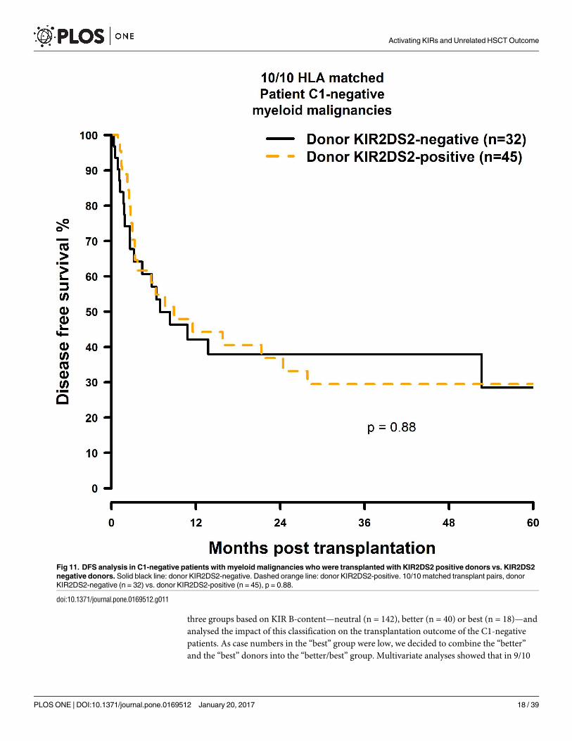
Fig 18. Univariate TRM analysis in C1-negative patients with myeloid malignancies who were transplanted with KIR2DS5 positive vs.
KIR2DS5 negative donors. Solid black line: donor KIR2DS5 negative. Dashed orange line: donor KIR2DS5 positive. Donor KIR2DS5-negative
(n = 83) vs. donor KIR2D5-positive (n = 34), p = 0.45.
doi:10.1371/journal.pone.0169512.g018
Activating KIRs and Unrelated HSCT Outcome
PLOS ONE | DOI:10.1371/journal.pone.0169512 January 20, 2017 28 / 39
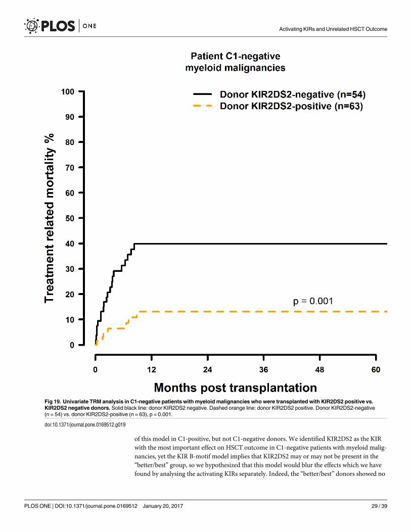
of this model in C1-positive, but not C1-negative donors. We identified KIR2DS2 as the KIR
with the most important effect on HSCT outcome in C1-negative patients with myeloid malig-
nancies, yet the KIR B-motif model implies that KIR2DS2 may or may not be present in the
“better/best” group, so we hypothesized that this model would blur the effects which we have
found by analysing the activating KIRs separately. Indeed, the “better/best” donors showed no
Fig 19. Univariate TRM analysis in C1-negative patients with myeloid malignancies who were transplanted with KIR2DS2 positive vs.
KIR2DS2 negative donors. Solid black line: donor KIR2DS2 negative. Dashed orange line: donor KIR2DS2 positive. Donor KIR2DS2-negative
(n = 54) vs. donor KIR2DS2-positive (n = 63), p = 0.001.
doi:10.1371/journal.pone.0169512.g019
Activating KIRs and Unrelated HSCT Outcome
PLOS ONE | DOI:10.1371/journal.pone.0169512 January 20, 2017 29 / 39

beneficial effect on transplantation outcome in the C1-negative patients. Hence, the KIR B-
motif model indeed showed a diminishing impact on C1-negative patients and therefore for
this patient group complete KIR locus typing seems to bear no advantage. Instead, our results
recommend donor typing for KIR2DS2 only (and possibly also for KIR2DS1).
Fig 20. Univariate TRM analysis in C1-negative patients with myeloid malignancies who were transplanted with KIR3DS1 positive vs.
KIR3DS1 negative donors. Solid black line: donor KIR3DS1 negative. Dashed orange line: donor KIR3DS1 positive. Donor KIR3DS1-negative
(n = 72) vs. donor KIR3DS1-positive (n = 45), p = 0.35.
doi:10.1371/journal.pone.0169512.g020
Activating KIRs and Unrelated HSCT Outcome
PLOS ONE | DOI:10.1371/journal.pone.0169512 January 20, 2017 30 / 39

In summary, our study shows, that activating signals derived from KIR2DS1 and possibly
KIR2DS5 can overcome the impaired NK-cell response in the C1-negative risk patient group,
but also provoke the risk for TRM. These findings provide new insights for the selection of a
suitable donor for the C1 negative risk patient group: for patients who are at high risk for
relapse a KIR2DS1 donor positive may be an adequate choice, whereas a graft which is nega-
tive for this KIR may be an option when a high probability for treatment related complications
is anticipated. The most important finding of our study, however, is the association of donor
KIR2DS2 positivity with a better overall outcome after HSCT in the C1-negative risk patient
group.
C1-negative patients with myeloid malignancies may therefore benefit from the inclusion
of KIR2DS1- and more importantly KIR2DS2-genotyping in the search algorithm. Our find-
ings certainly need to be confirmed by further studies in an independent cohort and also possi-
bly by a prospective study, prior to conduct changes in the search algorithm.
Limitations
Our approach was to identify beneficial donor KIRs in the relatively small group of C1-nega-
tive patients (14% of all patients in our cohort). In this context, small subgroups were inevita-
ble. However, while our study was limited by small sample sizes it nonetheless demonstrated
statistically significant findings with a robust effect size.
Supporting Information
S1 Fig. Univariate OS analysis in C1-negative patients (missing C1 ligand) vs. C1-positive
patients who were transplanted with KIR2DL2 or KIR2DL3 positive donors. Dashed red
line: C1-negative patients (n = 200), fine red lines: corresponding confidence intervals. Solid
black line: C1-positive patients (n = 1239), fine black lines: corresponding confidence intervals.
p = 0.0014.
(TIFF)
S2 Fig. Univariate DFS analysis in C1-negative patients (missing C1 ligand) vs. C1-positive
patients who were transplanted with KIR2DL2 or KIR2DL3 positive donors. Dashed red
line: C1-negative patients (n = 200), fine red lines: corresponding confidence intervals. Solid
black line: C1-positive patients (n = 1239), fine black lines: corresponding confidence intervals.
p = 0.006.
(TIFF)
Fig 21. Proposed KIR dependant search algorithm for C1-negative patients with myeloid
malignancies.
doi:10.1371/journal.pone.0169512.g021
Activating KIRs and Unrelated HSCT Outcome
PLOS ONE | DOI:10.1371/journal.pone.0169512 January 20, 2017 31 / 39

S3 Fig. Univariate RI analysis in C1-negative patients (missing C1 ligand) vs. C1-positive
patients who were transplanted with KIR2DL2 or KIR2DL3 positive donors. Dashed red
line: C1-negative patients (n = 200), fine red lines: corresponding confidence intervals. Solid
black line: C1-positive patients (n = 1239), fine black lines: corresponding confidence intervals.
p = 0.04.
(TIFF)
S4 Fig. Univariate TRM analysis in C1-negative patients (missing C1 ligand) vs. C1-posi-
tive patients who were transplanted with KIR2DL2 or KIR2DL3 positive donors. Dashed
red line: C1-negative patients (n = 200), fine red lines: corresponding confidence intervals.
Solid black line: C1-positive patients (n = 1239), fine black lines: corresponding confidence
intervals. p = 0.06.
(TIFF)
S5 Fig. Univariate OS analysis in C2-negative (missing C2 ligand) vs. C2-positive patients
who were transplanted with KIR2DL1-positive donors. Dashed red line: C2-negative
patients (n = 823), fine red lines: corresponding confidence intervals. Solid black line: C2-posi-
tive patients (n = 558), fine black lines: corresponding confidence intervals. p = 0.08.
(TIFF)
S6 Fig. Univariate DFS analysis in C2-negative (missing C2 ligand) vs. C2-positive patients
who were transplanted with KIR2DL1-positive donors. Dashed red line: C2-negative
patients (n = 823), fine red lines: corresponding confidence intervals. Solid black line: C2-posi-
tive patients (n = 558), fine black lines: corresponding confidence intervals. p = 0.11.
(TIFF)
S7 Fig. Univariate RI analysis in C2-negative (missing C2 ligand) vs. C2-positive patients
who were transplanted with KIR2DL1-positive donors. Dashed red line: C2-negative
patients (n = 823), fine red lines: corresponding confidence intervals. Solid black line: C2-posi-
tive patients (n = 558), fine black lines: corresponding confidence intervals. p = 0.47.
(TIFF)
S8 Fig. Univariate TRM analysis in C2-negative (missing C2 ligand) vs. C2-positive
patients who were transplanted with KIR2DL1-positive donors. Dashed red line: C2-nega-
tive patients (n = 823), fine red lines: corresponding confidence intervals. Solid black line:
C2-positive patients (n = 558), fine black lines: corresponding confidence intervals. p = 0.30.
(TIFF)
S9 Fig. Univariate OS analysis in C1-negative patients with myeloid malignancies who
were transplanted with KIR2DS2 positive donors vs. KIR2DS2 negative donors. Solid black
line: donor KIR2DS2-negative, fine black lines: corresponding confidence intervals. Dashed
red line: donor KIR2DS2-positive, fine red lines: corresponding confidence intervals. 10/10
matched transplant pairs, donor KIR2DS2-negative (n = 32) vs. donor KIR2DS2-positive
(n = 45), p = 0.86.
(TIFF)
S10 Fig. Univariate OS analysis in C1-negative patients with myeloid malignancies who
were transplanted with KIR2DS2 positive donors vs. KIR2DS2 negative donors. Solid black
line: donor KIR2DS2-negative, fine black lines: corresponding confidence intervals. Dashed
red line: donor KIR2DS2-positive, fine red lines: corresponding confidence intervals. 9/10
matched transplant pairs, donor KIR2DS2-negative (n = 22) vs. donor KIR2DS2-positive
Activating KIRs and Unrelated HSCT Outcome
PLOS ONE | DOI:10.1371/journal.pone.0169512 January 20, 2017 32 / 39

(n = 18), p = 0.04.
(TIFF)
S11 Fig. DFS analysis in C1-negative patients with myeloid malignancies who were trans-
planted with KIR2DS2 positive donors vs. KIR2DS2 negative donors. Solid black line:
donor KIR2DS2-negative, fine black lines: corresponding confidence intervals. Dashed red
line: donor KIR2DS2-positive, fine red lines: corresponding confidence intervals. 10/10
matched transplant pairs, donor KIR2DS2-negative (n = 32) vs. donor KIR2DS2-positive
(n = 45), p = 0.88.
(TIFF)
S12 Fig. DFS analysis in C1-negative patients with myeloid malignancies who were trans-
planted with KIR2DS2 positive donors vs. KIR2DS2 negative donors. Solid black line:
donor KIR2DS2-negative, fine black lines: corresponding confidence intervals. Dashed red
line: donor KIR2DS2-positive, fine red lines: corresponding confidence intervals. 9/10
matched transplant pairs, donor KIR2DS2-negative (n = 22) vs. donor KIR2DS2-positive
(n = 18), p = 0.05.
(TIFF)
S13 Fig. Univariate RI analysis in C1-negative patients with myeloid malignancies who
were transplanted with KIR2DS1 positive vs. KIR2DS1 negative donors. Solid black line:
donor KIR2DS1 negative, fine black lines: corresponding confidence intervals. Dashed red
line: donor KIR2DS1 positive, fine red lines: corresponding confidence intervals. Donor
KIR2DS1-negative (n = 72) vs. donor KIR2DS1-positive (n = 45), p = 0.008.
(TIFF)
S14 Fig. Univariate RI analysis in C1-negative patients with myeloid malignancies who
were transplanted with KIR2DS2 positive vs. KIR2DS2 negative donors. Solid black line:
donor KIR2DS2 negative, fine black lines: corresponding confidence intervals. Dashed red
line: donor KIR2DS2 positive, fine red lines: corresponding confidence intervals. Donor
KIR2DS2-negative (n = 54) vs. donor KIR2DS2-positive (n = 63), p = 0.33.
(TIFF)
S15 Fig. Univariate RI analysis in C1-negative patients with myeloid malignancies who
were transplanted with KIR2DS5 positive vs. KIR2DS5 negative donors. Solid black line:
donor KIR2DS5 negative, fine black lines: corresponding confidence intervals. Dashed red
line: donor 5 positive, fine red lines: corresponding confidence intervals. Donor KIR2DS5-ne-
gative (n = 83) vs. donor KIR2D5-positive (n = 34), p = 0.05.
(TIFF)
S16 Fig. Univariate RI analysis in C1-negative patients with myeloid malignancies who
were transplanted with KIR3DS1 positive vs. KIR3DS1 negative donors. Solid black line:
donor KIR3DS1 negative, fine black lines: corresponding confidence intervals. Dashed red
line: donor KIR3DS1 positive, fine red lines: corresponding confidence intervals. Donor
KIR3DS1-negative (n = 72) vs. donor KIR3DS1-positive (n = 45), p = 0.06.
(TIFF)
S17 Fig. Univariate TRM analysis in C1-negative patients with myeloid malignancies who
were transplanted with KIR2DS1 positive vs. KIR2DS1 negative donors. Solid black line:
donor KIR2DS1 negative, fine black lines: corresponding confidence intervals. Dashed red
line: donor KIR2DS1 positive, fine red lines: corresponding confidence intervals. Donor
Activating KIRs and Unrelated HSCT Outcome
PLOS ONE | DOI:10.1371/journal.pone.0169512 January 20, 2017 33 / 39

KIR2DS1-negative (n = 72) vs. donor KIR2DS1-positive (n = 45), p = 0.06.
(TIFF)
S18 Fig. Univariate TRM analysis in C1-negative patients with myeloid malignancies who
were transplanted with KIR2DS5 positive vs. KIR2DS5 negative donors. Solid black line:
donor KIR2DS5 negative, fine black lines: corresponding confidence intervals. Dashed red
line: donor KIR2DS5 positive, fine red lines: corresponding confidence intervals. Donor
KIR2DS5-negative (n = 83) vs. donor KIR2D5-positive (n = 34), p = 0.45.
(TIFF)
S19 Fig. Univariate TRM analysis in C1-negative patients with myeloid malignancies who
were transplanted with KIR2DS2 positive vs. KIR2DS2 negative donors. Solid black line:
donor KIR2DS2 negative, fine black lines: corresponding confidence intervals. Dashed red
line: donor KIR2DS2 positive, fine red lines: corresponding confidence intervals. Donor
KIR2DS2-negative (n = 54) vs. donor KIR2DS2-positive (n = 63), p = 0.001.
(TIFF)
S20 Fig. Univariate TRM analysis in C1-negative patients with myeloid malignancies who
were transplanted with KIR3DS1 positive vs. KIR3DS1 negative donors. Solid black line:
donor KIR3DS1 negative, fine black lines: corresponding confidence intervals. Dashed red
line: donor KIR3DS1 positive, fine red lines: corresponding confidence intervals. Donor
KIR3DS1-negative (n = 72) vs. donor KIR3DS1-positive (n = 45), p = 0.35.
(TIFF)
S1 Table.
(DOCX)
S2 Table.
(DOCX)
S3 Table.
(DOCX)
S4 Table.
(DOCX)
Acknowledgments
This work was supported by the Deutsche Jose Carreras Leukamie-Stiftung e.V. (Grant No.
DJCLS 11/10), and the German Red Cross Blood Transfusion Service, Baden-Wuerttemberg–
Hessen.
We are grateful to the following colleagues for contribution of their patients:
• Department of Hematology/Oncology, University of Leipzig, Germany: Niederwieser Diet-
ger, Buhner Alexander
• Department of Internal Medicine III, University of Ulm, Germany: Bunjes Donald, Missel
Lucia
• Hematology, Oncology and Pneumology, University Medical Center Mainz, Germany:
Wagner Eva, Waldeck Birgit
• Division of Stem Cell Transplantation and Immunotherapy, 2nd Department of Medicine,
University of Kiel, Germany: Gramatzki Martin, Helweg Marianne
Activating KIRs and Unrelated HSCT Outcome
PLOS ONE | DOI:10.1371/journal.pone.0169512 January 20, 2017 34 / 39

• Centre for Bone Marrow and Blood Stem Cell Transplantation, Deutsche Klinik fur Diag-
nostik, Wiesbaden, Germany: Stuhler Gernot, Eichler Susanne
• Hematology/Oncology Department, Charite Campus Virchow Berlin, Germany: Arnold
Renate
• Department of Medicine II, University Hospital Wurzburg, Germany: Einsele Hermann,
Grigoleit Gotz Ulrich, Bonig Heidrun
• Department of Hematology/Oncology, Georg-August-University Gottingen, Germany:
Trumper Lorenz, Gerald Wulf
• Department Internal Medicine I, Universitatsklinikum des Saarlandes, Homburg, Germany:
Pfreundschuh Michael, Martin Renate
• Department of Hematology/Oncology, Asklepios Klinik St. Georg, Hamburg, Germany:
Dingeldein Susanne, Zeis Matthias
• Medical Clinic 5, Hematology and Oncology, Klinikum Nuremberg, Nuremberg, Germany:
Schafer-Eckart Kerstin
• Medical Clinic III, Hematology and Oncology, University Clinic r.d.Isar, Munich, Germany:
Peschel Christian
• Medical Clinic 5, Hematology and Oncology, University of Erlangen-Nuremberg, Germany:
Mackensen Andreas
• Hematology/Oncology Department, Robert-Bosch Clinic,Stuttgart, Germany: Aulitzky Wal-
ter Erich, Kaufmann Martin, Eier Nicole
• Hematology/Oncology Department, University of Rostock, Germany: Freitag Sebastian
• Hematology/Oncology Department, Clinic Oldenburg, Germany: Casper Jochen
• Clinic for Stem Cell Transplantation, University Medical Center Hamburg- Eppendorf, Ger-
many: Zander Axel, Kasper Anja
• Department of Hematology/Oncology, University of Regensburg, Germany: Herr Wolfgang,
Holler Ernst
• Department of Hematology/Oncology, Evangelisches KKH Essen-Werden, Germany: Wat-
tad Mohammed, Manicone Julia
• Hematology/Oncology Department, Clinic Bremen-Mitte, Germany: Hertenstein Bernd
• Hematology/Oncology Department, Clinic Idar-Oberstein, Germany: Gregor Sebastian
• Medical Clinic II, Hematology and Oncology, J.W.-Goethe-University, Frankfurt/Main,
Germany: Martin Hans, Serve Hubert
• Department of Oncology/Hematology, Martin Luther University Halle, Germany: Muller-
Tidow Carsten, Korholz Dieter, Bach Jana
• Hematology/Oncology Department, Clinic Karlsruhe, Germany: Ringhoffer Mark, Schiefer
Julie
• Department of Hematology/Oncology, University Hospital Schleswig-Holstein, Lubeck,
Germany: Biersack Harald, Marx Rudina
Activating KIRs and Unrelated HSCT Outcome
PLOS ONE | DOI:10.1371/journal.pone.0169512 January 20, 2017 35 / 39

• Hematology/Oncology Department, University Hospital Mannheim, Germany: Klein Ste-
fan, Folz Christine
• Transplantation Immunology Department, German Red Cross Blood Transfusion Service,
Baden-Wuerttemberg–Hessen, Ulm, Germany and Institute of Transfusion Medicine, Uni-
versity of Ulm, Germany: Marlene Fischer, Sabine Simon, Renate Hanold, Martina
Baumann
Ethics Statement
The study was approved by the ethical review board of the University of Ulm (project number
263/09).
Author Contributions
Conceptualization: CN.
Data curation: DF.
Formal analysis: CN.
Funding acquisition: DF.
Investigation: CN.
Methodology: CN DF.
Project administration: JM.
Resources: DN DB GW MP EW GS HE.
Supervision: JM.
Writing – original draft: CN DF JM.
Writing – review & editing: DF DN DB CT GW MP EW GS HE HS JM.
References1. Barrett AJ, Battiwalla M. Relapse after allogeneic stem cell transplantation. Expert Rev Hematol 2010
Aug; 3(4):429–41. doi: 10.1586/ehm.10.32 PMID: 21083034
2. Zinkernagel RM, Bachmann MF, Kundig TM, Oehen S, Pirchet H, Hengartner H. On immunological
memory. Annu Rev Immunol 1996; 14:333–67. doi: 10.1146/annurev.immunol.14.1.333 PMID:
8717518
3. Horowitz MM, Gale RP, Sondel PM, Goldman JM, Kersey J, Kolb HJ, et al. Graft-versus-leukemia reac-
tions after bone marrow transplantation. Blood 1990 Feb 1; 75(3):555–62. PMID: 2297567
4. Deol A, Lum LG. Role of donor lymphocyte infusions in relapsed hematological malignancies after stem
cell transplantation revisited. Cancer Treat Rev 2010 Nov; 36(7):528–38. doi: 10.1016/j.ctrv.2010.03.
004 PMID: 20381970
5. Riemersma SA, Jordanova ES, Schop RF, Philippo K, Looijenga LH, Schuuring E, et al. Extensive
genetic alterations of the HLA region, including homozygous deletions of HLA class II genes in B-cell
lymphomas arising in immune-privileged sites. Blood 2000 Nov 15; 96(10):3569–77. PMID: 11071656
6. Demanet C, Mulder A, Deneys V, Worsham MJ, Maes P, Claas FH, et al. Down-regulation of HLA-A
and HLA-Bw6, but not HLA-Bw4, allospecificities in leukemic cells: an escape mechanism from CTL
and NK attack? Blood 2004 Apr 15; 103(8):3122–30. doi: 10.1182/blood-2003-07-2500 PMID:
15070694
7. Brouwer RE, van der Heiden P, Schreuder GM, Mulder A, Datema G, Anholts JD, et al. Loss or downre-
gulation of HLA class I expression at the allelic level in acute leukemia is infrequent but functionally rele-
vant, and can be restored by interferon. Hum Immunol 2002 Mar; 63(3):200–10. PMID: 11872238
Activating KIRs and Unrelated HSCT Outcome
PLOS ONE | DOI:10.1371/journal.pone.0169512 January 20, 2017 36 / 39

8. Ruggeri L, Capanni M, Casucci M, Volpi I, Tosti A, Perruccio K, et al. Role of natural killer cell alloreac-
tivity in HLA-mismatched hematopoietic stem cell transplantation. Blood 1999 Jul 1; 94(1):333–9.
PMID: 10381530
9. Asai O, Longo DL, Tian ZG, Hornung RL, Taub DD, Ruscetti FW, et al. Suppression of graft-versus-
host disease and amplification of graft-versus-tumor effects by activated natural killer cells after alloge-
neic bone marrow transplantation. J Clin Invest 1998 May 1; 101(9):1835–42. doi: 10.1172/JCI1268
PMID: 9576746
10. Vitale M, Sivori S, Pende D, Augugliaro R, di DC, Amoroso A, et al. Physical and functional indepen-
dency of p70 and p58 natural killer (NK) cell receptors for HLA class I: their role in the definition of differ-
ent groups of alloreactive NK cell clones. Proc Natl Acad Sci U S A 1996 Feb 20; 93(4):1453–7. PMID:
8643653
11. Moretta A, Bottino C, Pende D, Tripodi G, Tambussi G, Viale O, et al. Identification of four subsets of
human CD3-CD16+ natural killer (NK) cells by the expression of clonally distributed functional surface
molecules: correlation between subset assignment of NK clones and ability to mediate specific alloanti-
gen recognition. J Exp Med 1990 Dec 1; 172(6):1589–98. PMID: 2147946
12. Wagtmann N, Rajagopalan S, Winter CC, Peruzzi M, Long EO. Killer cell inhibitory receptors specific
for HLA-C and HLA-B identified by direct binding and by functional transfer. Immunity 1995 Dec; 3
(6):801–9. PMID: 8777725
13. Middleton D, Gonzelez F. The extensive polymorphism of KIR genes. Immunology 2010 Jan; 129(1):8–
19. doi: 10.1111/j.1365-2567.2009.03208.x PMID: 20028428
14. Robinson J, Halliwell JA, Hayhurst JD, Flicek P, Parham P, Marsh SG. The IPD and IMGT/HLA data-
base: allele variant databases. Nucleic Acids Res 2015 Jan 28; 43(Database issue):D423–D431. doi:
10.1093/nar/gku1161 PMID: 25414341
15. Uhrberg M, Valiante NM, Shum BP, Shilling HG, Lienert-Weidenbach K, Corliss B, et al. Human diver-
sity in killer cell inhibitory receptor genes. Immunity 1997 Dec; 7(6):753–63. PMID: 9430221
16. Parham P. MHC class I molecules and KIRs in human history, health and survival. Nat Rev Immunol
2005 Mar; 5(3):201–14. doi: 10.1038/nri1570 PMID: 15719024
17. Biassoni R, Falco M, Cambiaggi A, Costa P, Verdiani S, Pende D, et al. Amino acid substitutions can
influence the natural killer (NK)-mediated recognition of HLA-C molecules. Role of serine-77 and lysine-
80 in the target cell protection from lysis mediated by "group 2" or "group 1" NK clones. J Exp Med 1995
Aug 1; 182(2):605–9. PMID: 7629517
18. David G, Djaoud Z, Willem C, Legrand N, Rettman P, Gagne K, et al. Large spectrum of HLA-C recogni-
tion by killer Ig-like receptor (KIR)2DL2 and KIR2DL3 and restricted C1 SPECIFICITY of KIR2DS2:
dominant impact of KIR2DL2/KIR2DS2 on KIR2D NK cell repertoire formation. J Immunol 2013 Nov 1;
191(9):4778–88. doi: 10.4049/jimmunol.1301580 PMID: 24078689
19. Moesta AK, Parham P. Diverse functionality among human NK cell receptors for the C1 epitope of HLA-
C: KIR2DS2, KIR2DL2, and KIR2DL3. Front Immunol 2012; 3:336. doi: 10.3389/fimmu.2012.00336
PMID: 23189078
20. Ruggeri L, Capanni M, Mancusi A, Martelli MF, Velardi A. The impact of donor natural killer cell alloreac-
tivity on allogeneic hematopoietic transplantation. Transpl Immunol 2005 Aug; 14(3–4):203–6. doi: 10.
1016/j.trim.2005.03.008 PMID: 15982564
21. Moretta A, Bottino C, Vitale M, Pende D, Cantoni C, Mingari MC, et al. Activating receptors and core-
ceptors involved in human natural killer cell-mediated cytolysis. Annu Rev Immunol 2001; 19:197–223.
doi: 10.1146/annurev.immunol.19.1.197 PMID: 11244035
22. Ljunggren HG, Karre K. In search of the ’missing self’: MHC molecules and NK cell recognition. Immu-
nol Today 1990 Jul; 11(7):237–44. PMID: 2201309
23. Ruggeri L, Capanni M, Urbani E, Perruccio K, Shlomchik WD, Tosti A, et al. Effectiveness of donor natu-
ral killer cell alloreactivity in mismatched hematopoietic transplants. Science 2002 Mar 15; 295
(5562):2097–100. doi: 10.1126/science.1068440 PMID: 11896281
24. Farag SS, Bacigalupo A, Eapen M, Hurley C, Dupont B, Caligiuri MA, et al. The effect of KIR ligand
incompatibility on the outcome of unrelated donor transplantation: a report from the center for interna-
tional blood and marrow transplant research, the European blood and marrow transplant registry, and
the Dutch registry. Biol Blood Marrow Transplant 2006 Aug; 12(8):876–84. doi: 10.1016/j.bbmt.2006.
05.007 PMID: 16864058
25. Morishima Y, Yabe T, Matsuo K, Kashiwase K, Inoko H, Saji H, et al. Effects of HLA allele and killer
immunoglobulin-like receptor ligand matching on clinical outcome in leukemia patients undergoing
transplantation with T-cell-replete marrow from an unrelated donor. Biol Blood Marrow Transplant 2007
Mar; 13(3):315–28. doi: 10.1016/j.bbmt.2006.10.027 PMID: 17317585
Activating KIRs and Unrelated HSCT Outcome
PLOS ONE | DOI:10.1371/journal.pone.0169512 January 20, 2017 37 / 39

26. Davies SM, Ruggieri L, DeFor T, Wagner JE, Weisdorf DJ, Miller JS, et al. Evaluation of KIR ligand
incompatibility in mismatched unrelated donor hematopoietic transplants. Killer immunoglobulin-like
receptor. Blood 2002 Nov 15; 100(10):3825–7. doi: 10.1182/blood-2002-04-1197 PMID: 12393440
27. Fischer JC, Kobbe G, Enczmann J, Haas R, Uhrberg M. The impact of HLA-C matching depends on the
C1/C2 KIR ligand status in unrelated hematopoietic stem cell transplantation. Immunogenetics 2012
Dec; 64(12):879–85. doi: 10.1007/s00251-012-0648-7 PMID: 22923051
28. Cooley S, Trachtenberg E, Bergemann TL, Saeteurn K, Klein J, Le CT, et al. Donors with group B KIR
haplotypes improve relapse-free survival after unrelated hematopoietic cell transplantation for acute
myelogenous leukemia. Blood 2009 Jan 15; 113(3):726–32. doi: 10.1182/blood-2008-07-171926
PMID: 18945962
29. Zhou H, Bao X, Wu X, Tang X, Wang M, Wu D, et al. Donor selection for killer immunoglobulin-like
receptors B haplotype of the centromeric motifs can improve the outcome after HLA-identical sibling
hematopoietic stem cell transplantation. Biol Blood Marrow Transplant 2014 Jan; 20(1):98–105. PMID:
24516895
30. Cooley S, Weisdorf DJ, Guethlein LA, Klein JP, Wang T, Marsh SG, et al. Donor killer cell Ig-like recep-
tor B haplotypes, recipient HLA-C1, and HLA-C mismatch enhance the clinical benefit of unrelated
transplantation for acute myelogenous leukemia. J Immunol 2014 May 15; 192(10):4592–600. doi: 10.
4049/jimmunol.1302517 PMID: 24748496
31. Knorr DA, Wang H, Aurora M, MacMillan ML, Holton SG, Bergerson R, et al. Loss of T Follicular Helper
Cells In the Peripheral Blood Of Patients With Chronic Graft vs. Host Disease. Biol Blood Marrow Trans-
plant 2016 Jan 12.
32. Furst D, Zollikofer C, Muller C, Niederwieser D, Bunjes DW, Wagner E, et al. Donors with KIR-Bx Haplo-
types Improve Outcome of Unrelated Hematopoietic Stem Cell Transplantation for Recipients with a
Myeloid Malignant Disease and a C1 Ligand Phenotype. Biol Blood Marrow Transplant 2015 Feb; 21
(2):S31.
33. Bonagura VR, Du Z, Ashouri E, Luo L, Hatam LJ, DeVoti JA, et al. Activating killer cell immunoglobulin-
like receptors 3DS1 and 2DS1 protect against developing the severe form of recurrent respiratory papil-
lomatosis. Hum Immunol 2010 Feb; 71(2):212–9. doi: 10.1016/j.humimm.2009.10.009 PMID:
19861144
34. Alter G, Martin MP, Teigen N, Carr WH, Suscovich TJ, Schneidewind A, et al. Differential natural killer
cell-mediated inhibition of HIV-1 replication based on distinct KIR/HLA subtypes. J Exp Med 2007 Nov
26; 204(12):3027–36. doi: 10.1084/jem.20070695 PMID: 18025129
35. Khakoo SI, Thio CL, Martin MP, Brooks CR, Gao X, Astemborski J, et al. HLA and NK cell inhibitory
receptor genes in resolving hepatitis C virus infection. Science 2004 Aug 6; 305(5685):872–4. doi: 10.
1126/science.1097670 PMID: 15297676
36. Sivori S, Carlomagno S, Falco M, Romeo E, Moretta L, Moretta A. Natural killer cells expressing the
KIR2DS1-activating receptor efficiently kill T-cell blasts and dendritic cells: implications in haploidentical
HSCT. Blood 2011 Apr 21; 117(16):4284–92. doi: 10.1182/blood-2010-10-316125 PMID: 21355085
37. Venstrom JM, Pittari G, Gooley TA, Chewning JH, Spellman S, Haagenson M, et al. HLA-C-dependent
prevention of leukemia relapse by donor activating KIR2DS1. N Engl J Med 2012 Aug 30; 367(9):805–
16. doi: 10.1056/NEJMoa1200503 PMID: 22931314
38. Iacobelli S. Suggestions on the use of statistical methodologies in studies of the European Group for
Blood and Marrow Transplantation. Bone Marrow Transplant 2013 Mar; 48 Suppl 1:S1–37.
39. EBMT.org [Internet]. European Group for Blood and Marrow Transplantation. MED-AB forms manual.
A guide to the completion of the EBMT HSCT MED-AB forms. appendix iii. [Updated 2015 August 18;
cited 2015 September 12]. Available from: http://www.ebmt.org/Contents/Data-Management/
Registrystructure/MED-ABdatacollectionforms/Documents/MED-ABFormsManual.pdf.
40. Bacigalupo A, Ballen K, Rizzo D, Giralt S, Lazarus H, Ho V, et al. Defining the intensity of conditioning
regimens: working definitions. Biol Blood Marrow Transplant 2009 Dec; 15(12):1628–33. doi: 10.1016/j.
bbmt.2009.07.004 PMID: 19896087
41. bioinformatics.bethematchclinical.org [Internet]. National Marrow Donor Program. NMDP Policy for
HLA Confirmatory Typing Requirements for Unrelated Adult Donors and Patients. [Updated 2015 Janu-
ary; cited 2015 September 12]. Available from: https://bioinformatics.bethematchclinical.org/WorkArea/
DownloadAsset.aspx?id=10528
42. Gratwohl A, Stern M, Brand R, Apperley J, Baldomero H, de WT, et al. Risk score for outcome after allo-
geneic hematopoietic stem cell transplantation: a retrospective analysis. Cancer 2009 Oct 15; 115
(20):4715–26. doi: 10.1002/cncr.24531 PMID: 19642176
43. EBMT.org [Internet]. Labopin M, Iacobelli S. Statistical guidelines for EBMT. [Updated October 2003;
cited 2015 September 12]]. Available from: http://www.ebmt.org/1WhatisEBMT/Op_Manual/OPMAN_
StatGuidelines_oct2003.pdf.
Activating KIRs and Unrelated HSCT Outcome
PLOS ONE | DOI:10.1371/journal.pone.0169512 January 20, 2017 38 / 39

44. Glidden DV, Vittinghoff E. Modelling clustered survival data from multicentre clinical trials. Stat Med
2004 Feb 15; 23(3):369–88. doi: 10.1002/sim.1599 PMID: 14748034
45. CRAN.R-project.org [Internet]. Therneau T. A Package for Survival Analysis in S. R package version
2.38. [updated 2015 July 02; cited 2015 September 12]. Available from: http://CRAN.R-project.org/
package=survival
46. Gratwohl A. The EBMT risk score. Bone Marrow Transplant 2012 Jun; 47(6):749–56. doi: 10.1038/bmt.
2011.110 PMID: 21643021
47. Gonzalez-Galarza FF, Christmas S, Middleton D, Jones AR. Allele frequency net: a database and
online repository for immune gene frequencies in worldwide populations. Nucleic Acids Res 2011 Jan;
39(Database issue):D913–D919. doi: 10.1093/nar/gkq1128 PMID: 21062830
48. Fischer JC, Ottinger H, Ferencik S, Sribar M, Punzel M, Beelen DW, et al. Relevance of C1 and C2 epi-
topes for hemopoietic stem cell transplantation: role for sequential acquisition of HLA-C-specific inhibi-
tory killer Ig-like receptor. J Immunol 2007 Mar 15; 178(6):3918–23. PMID: 17339492
49. Giebel S, Locatelli F, Wojnar J, Velardi A, Mina T, Giorgiani G, et al. Homozygosity for human leucocyte
antigen-C ligands of KIR2DL1 is associated with increased risk of relapse after human leucocyte anti-
gen-C-matched unrelated donor haematopoietic stem cell transplantation. Br J Haematol 2005 Nov;
131(4):483–6. doi: 10.1111/j.1365-2141.2005.05797.x PMID: 16281939
50. Hsu KC, Keever-Taylor CA, Wilton A, Pinto C, Heller G, Arkun K, et al. Improved outcome in HLA-identi-
cal sibling hematopoietic stem-cell transplantation for acute myelogenous leukemia predicted by KIR
and HLA genotypes. Blood 2005 Jun 15; 105(12):4878–84. doi: 10.1182/blood-2004-12-4825 PMID:
15731175
51. Ahlenstiel G, Martin MP, Gao X, Carrington M, Rehermann B. Distinct KIR/HLA compound genotypes
affect the kinetics of human antiviral natural killer cell responses. J Clin Invest 2008 Mar; 118(3):1017–
26. doi: 10.1172/JCI32400 PMID: 18246204
52. Wilson MJ, Torkar M, Trowsdale J. Genomic organization of a human killer cell inhibitory receptor gene.
Tissue Antigens 1997 Jun; 49(6):574–9. PMID: 9234478
53. Yu J, Venstrom JM, Liu XR, Pring J, Hasan RS, O’Reilly RJ, et al. Breaking tolerance to self, circulating
natural killer cells expressing inhibitory KIR for non-self HLA exhibit effector function after T cell-
depleted allogeneic hematopoietic cell transplantation. Blood 2009 Apr 16; 113(16):3875–84. doi: 10.
1182/blood-2008-09-177055 PMID: 19179302
54. Jamil KM, Khakoo SI. KIR/HLA interactions and pathogen immunity. J Biomed Biotechnol 2011;
2011:298348. doi: 10.1155/2011/298348 PMID: 21629750
55. Vales-Gomez M, Reyburn H, Strominger J. Interaction between the human NK receptors and their
ligands. Crit Rev Immunol 2000; 20(3):223–44. PMID: 10968372
56. Maki G, Hayes GM, Naji A, Tyler T, Carosella ED, Rouas-Freiss N, et al. NK resistance of tumor cells
from multiple myeloma and chronic lymphocytic leukemia patients: implication of HLA-G. Leukemia
2008 May; 22(5):998–1006. doi: 10.1038/leu.2008.15 PMID: 18288133
57. Zingoni A, Sornasse T, Cocks BG, Tanaka Y, Santoni A, Lanier LL. NK cell regulation of T cell-mediated
responses. Mol Immunol 2005 Feb; 42(4):451–4. doi: 10.1016/j.molimm.2004.07.025 PMID: 15607797
58. Tittle TV, Weinberg AD, Steinkeler CN, Maziarz RT. Expression of the T-cell activation antigen, OX-40,
identifies alloreactive T cells in acute graft-versus-host disease. Blood 1997 Jun 15; 89(12):4652–8.
PMID: 9192792
59. Chewning JH, Gudme CN, Hsu KC, Selvakumar A, Dupont B. KIR2DS1-positive NK cells mediate allor-
esponse against the C2 HLA-KIR ligand group in vitro. J Immunol 2007 Jul 15; 179(2):854–68. PMID:
17617576
60. Liu J, Xiao Z, Ko HL, Shen M, Ren EC. Activating killer cell immunoglobulin-like receptor 2DS2 binds to
HLA-A*11. Proc Natl Acad Sci U S A 2014 Feb 18; 111(7):2662–7. doi: 10.1073/pnas.1322052111
PMID: 24550293
Activating KIRs and Unrelated HSCT Outcome
PLOS ONE | DOI:10.1371/journal.pone.0169512 January 20, 2017 39 / 39