immunopathology ahmad shihada silmi hematologist & immunologist iug
Post on 21-Dec-2015
227 views
TRANSCRIPT
Immunopathology
Ahmad Shihada Silmi
Hematologist & Immunologist
IUG
Section A
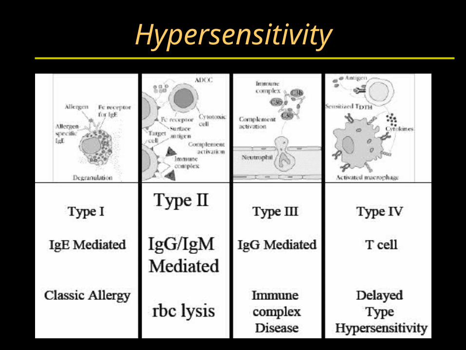
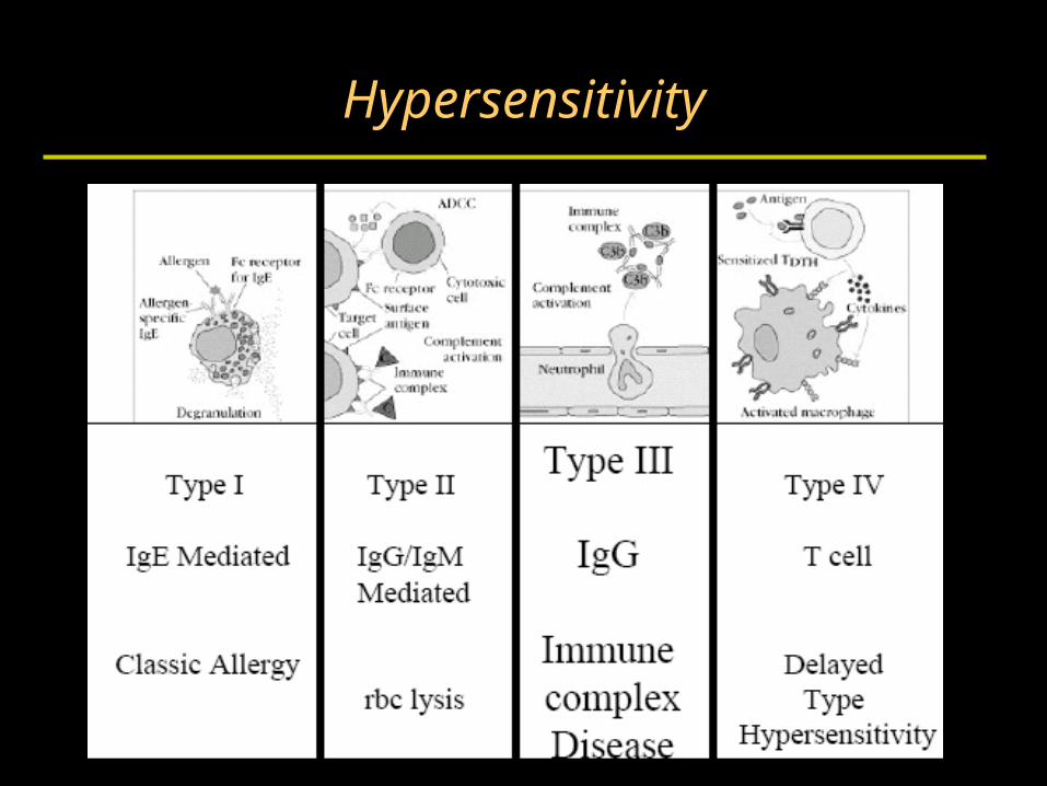
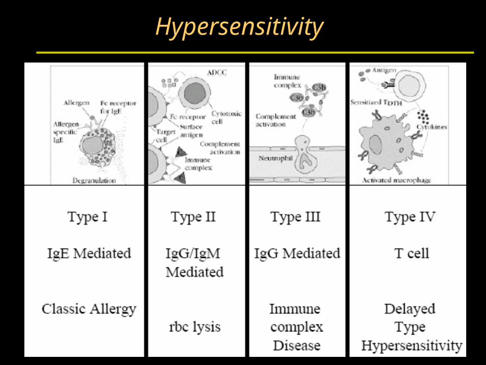
Hypersensitivity

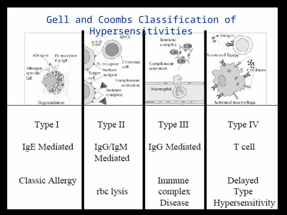
Gell and Coombs Classification of Hypersensitivities
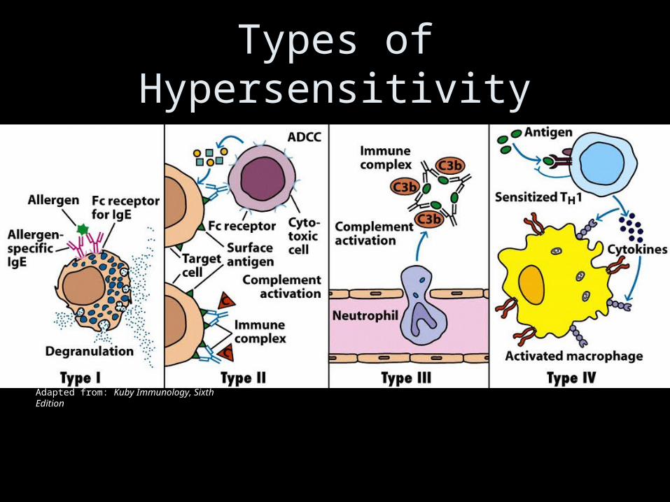
Types of Hypersensitivity
Adapted from: Kuby Immunology, Sixth Edition
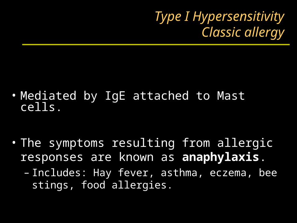
Type I HypersensitivityClassic allergy
• Mediated by IgE attached to Mast cells.
• The symptoms resulting from allergic responses are known as anaphylaxis.– Includes: Hay fever, asthma, eczema, bee stings,
food allergies.
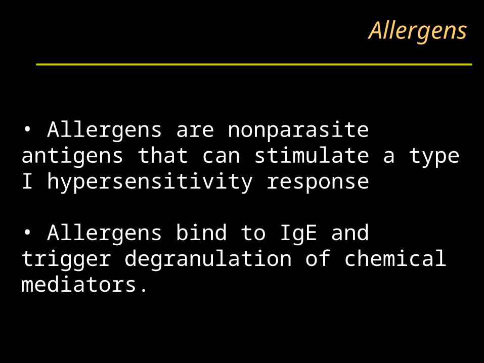
Allergens
• Allergens are nonparasite antigens that can stimulate a type I hypersensitivity response
• Allergens bind to IgE and trigger degranulation of chemical mediators.
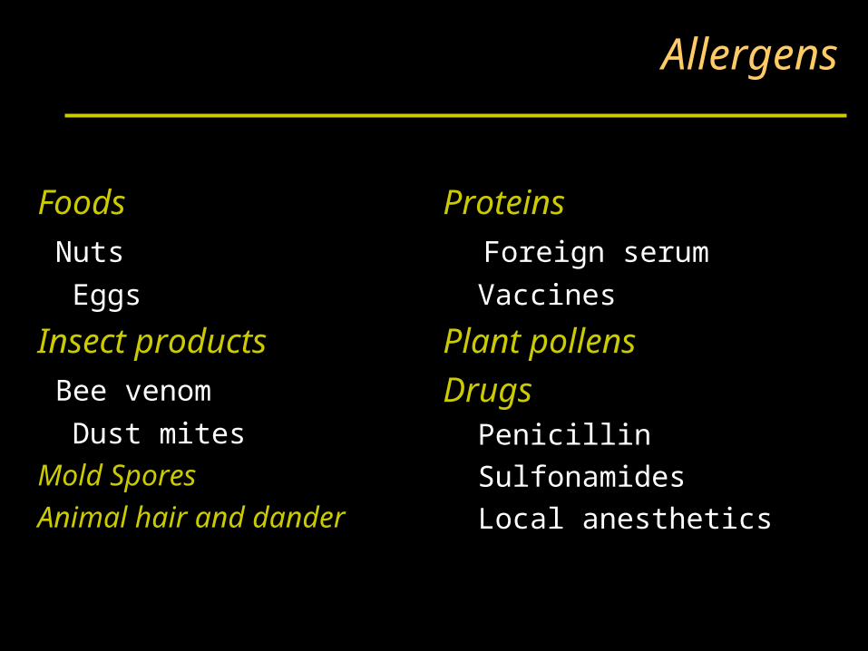
Allergens
Proteins
Foreign serum
Vaccines
Plant pollens
Drugs Penicillin
Sulfonamides
Local anesthetics
Foods
Nuts
Eggs
Insect products
Bee venom
Dust mites
Mold Spores
Animal hair and dander
Characteristics of allergens
• Small 15-40,000 MW proteins.• Specific protein components
• Often enzymes.• Low dose of allergen• Mucosal exposure.• Most allergens promote a Th2 immune response.
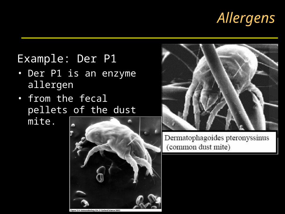
Example: Der P1• Der P1 is an enzyme
allergen• from the fecal pellets of
the dust mite.
Allergens
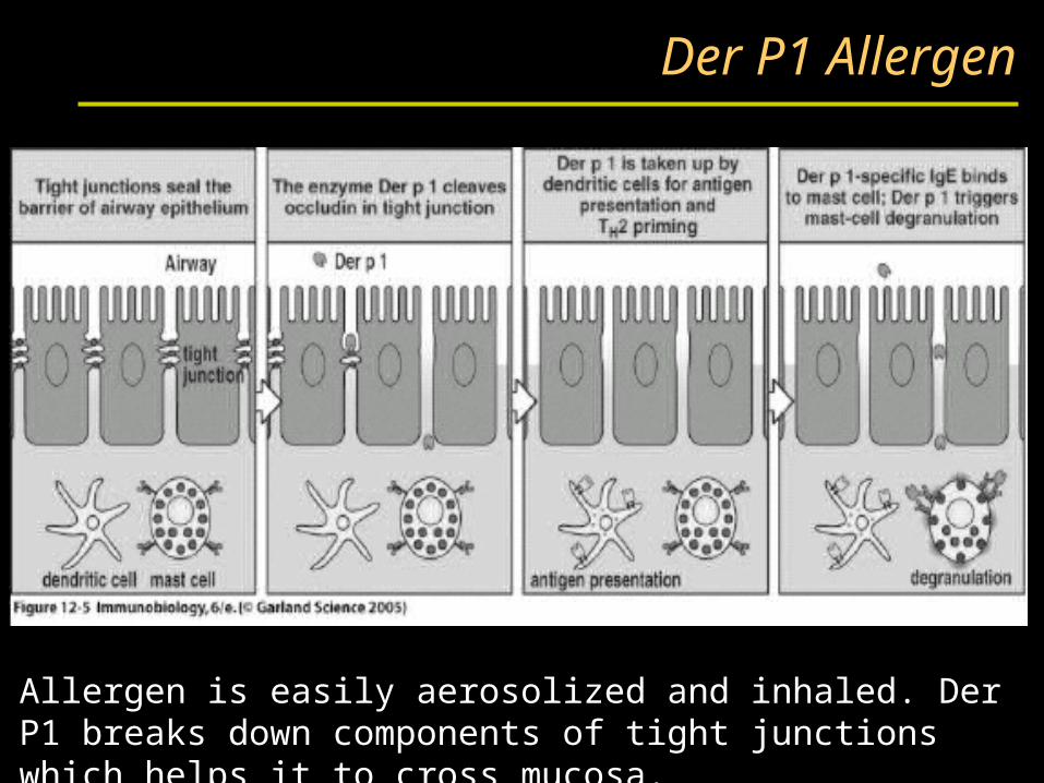
Der P1 Allergen
Allergen is easily aerosolized and inhaled. Der P1 breaks down components of tight junctions which helps it to cross mucosa.
Atopy
• Atopy is the term for the genetic trait to have a predisposition for localized anaphylaxis.
• Atopic individuals have higher levels of IgE and eosinophils.
Genetic PredispositionType I hypersensitivity
• Candidate polymorphic genes include:– IL-4 Receptor.– IL-4 cytokine (promoter region).– FcεRI. High affinity IgE receptor.– Class II MHC (present peptides promoting Th2 response).
– Inflammation genes.
Mechanisms of allergic response
Sensitization
• Repeated exposure to allergens initiates immune response that generates IgE isotype.
• Th2 cells required to provide the IL-4required to get isotype switching to IgE.
Mechanisms of allergic response
Sensitization
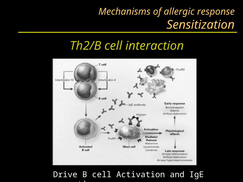
Th2/B cell interaction
Drive B cell Activation and IgE secretion
Mechanisms of allergic response
Sensitization
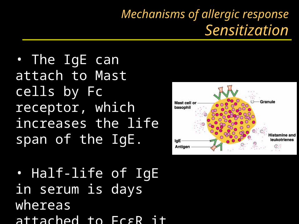
• The IgE can attach to Mast cells by Fc receptor, which increases the life span of the IgE.
• Half-life of IgE in serum is days whereasattached to FcεR it is increased to months.
Mechanisms of allergic response
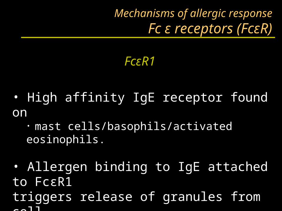
Fc ε receptors (FcεR)
• High affinity IgE receptor found on• mast cells/basophils/activated eosinophils.
• Allergen binding to IgE attached to FcεR1triggers release of granules from cell.
FcεR1
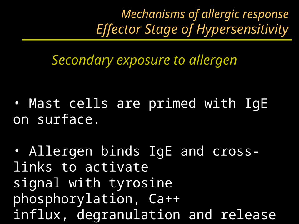
Mechanisms of allergic responseEffector Stage of Hypersensitivity
• Mast cells are primed with IgE on surface.
• Allergen binds IgE and cross-links to activatesignal with tyrosine phosphorylation, Ca++influx, degranulation and release of mediators.
Secondary exposure to allergen
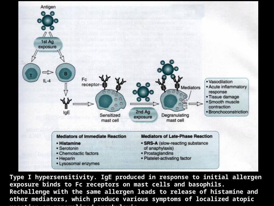
Type I hypersensitivity. IgE produced in response to initial allergen exposure binds to Fc receptors on mast cells and basophils. Rechallenge with the same allergen leads to release of histamine and other mediators, which produce various symptoms of localized atopic reaction or generalized anaphylaxis.
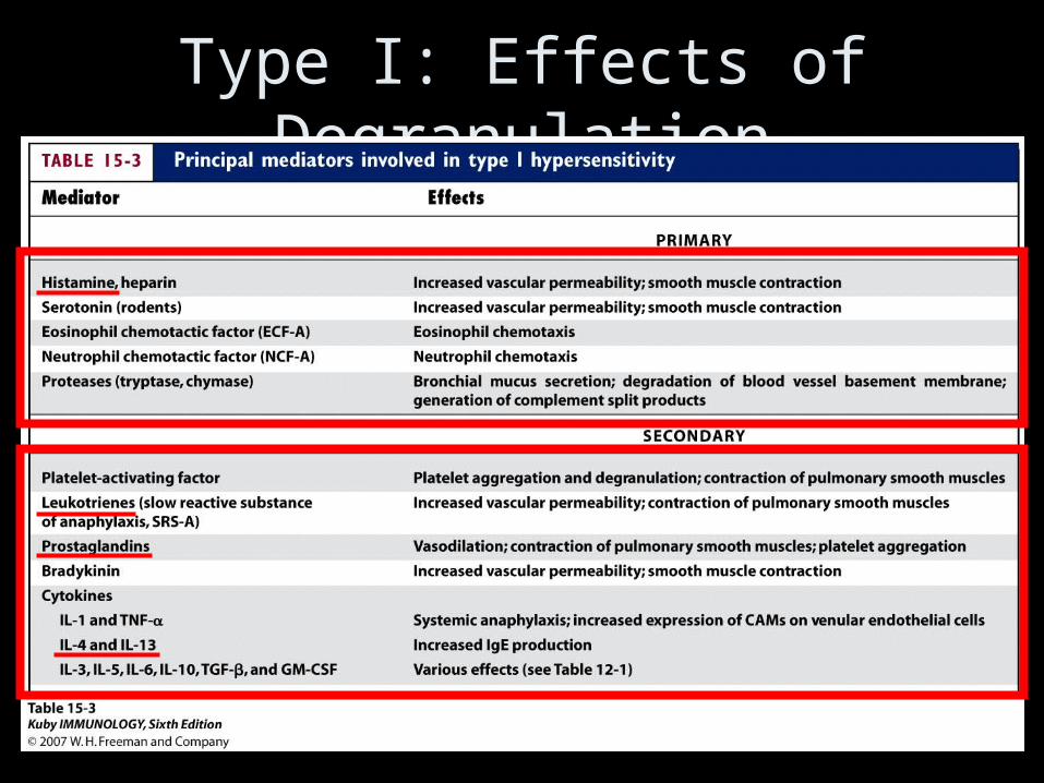
Type I: Effects of Degranulation
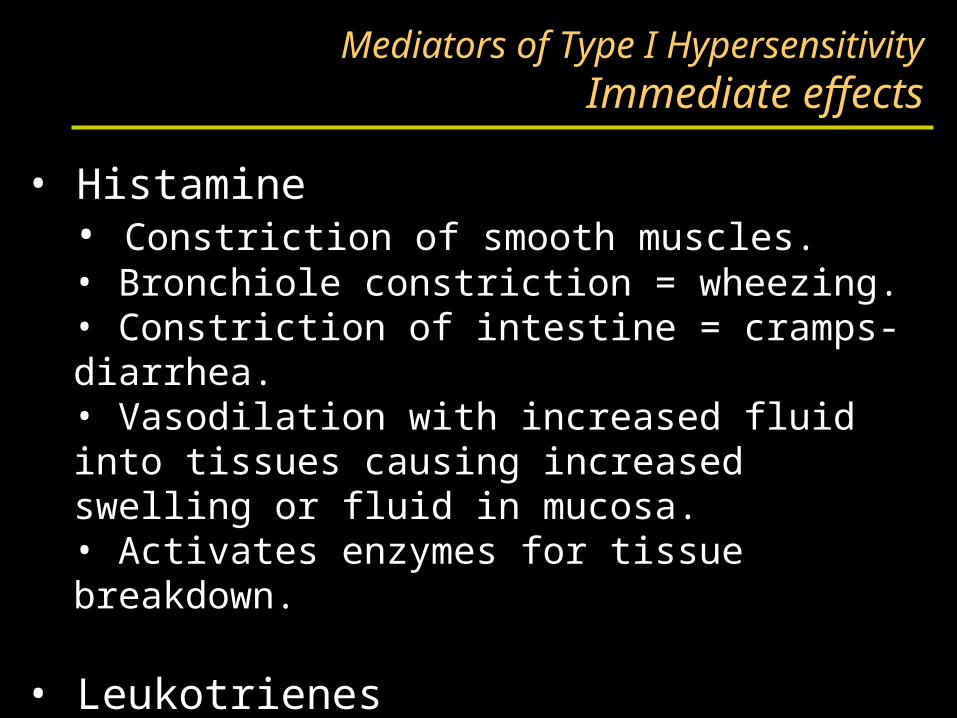
Mediators of Type I HypersensitivityImmediate effects
• Histamine• Constriction of smooth muscles.• Bronchiole constriction = wheezing.• Constriction of intestine = cramps-diarrhea.• Vasodilation with increased fluid into tissues causing increased swelling or fluid in mucosa.• Activates enzymes for tissue breakdown.
• Leukotrienes
• Prostaglandins
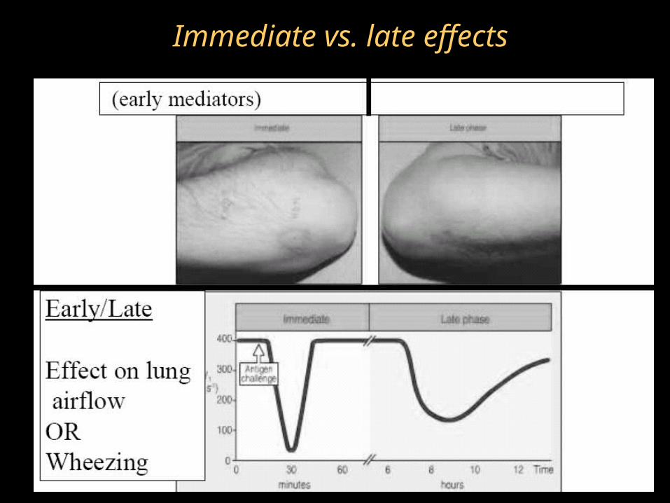
Immediate vs. late effects
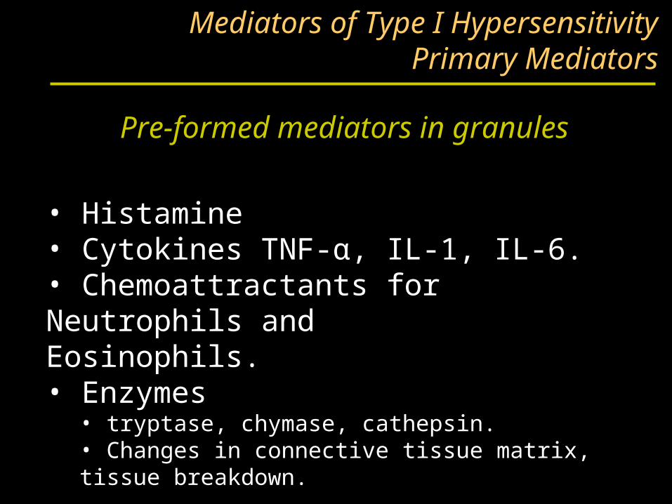
Pre-formed mediators in granules
Mediators of Type I HypersensitivityPrimary Mediators
• Histamine• Cytokines TNF-α, IL-1, IL-6.• Chemoattractants for Neutrophils andEosinophils. • Enzymes
• tryptase, chymase, cathepsin.• Changes in connective tissue matrix, tissue breakdown.
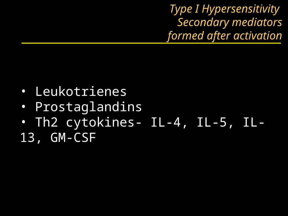
Type I Hypersensitivity Secondary mediators
formed after activation
• Leukotrienes• Prostaglandins• Th2 cytokines- IL-4, IL-5, IL-13, GM-CSF
Continuation of sensitization cycle
• Mast cells control the immediate response.
• Eosinophils and neutrophils drive late or chronic response.
• More IgE production further driven by activated Mast cells, basophils, eosinophils.
Continuation of sensitization cycleEosinophils
• Eosinophils play key role in late phase reaction.
• Eosinophils make:
• enzymes,
• cytokines (IL-3, IL-5, GM-CSF),
• Lipid mediators (LTC4, LTD4, PAF)
• Eosinophils can provide CD40L and IL-4for B cell activation.
Localized anaphylaxis
• Target organ responds to direct contact with allergen.
• Digestive tract contact results in vomiting, cramping, diarrhea.
• Skin sensitivity usually reddened inflamed area resulting in itching.
• Airway sensitivity results in sneezing and rhinitis OR wheezing and asthma.
Systemic anaphylaxis
• Systemic vasodilation and smooth muscle contraction leading to severe bronchiole constriction, edema, and shock.
• Similar to systemic inflammation.
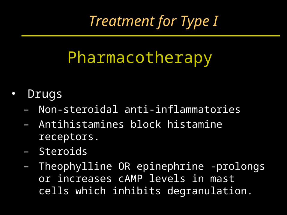
Treatment for Type I
Pharmacotherapy
• Drugs– Non-steroidal anti-inflammatories– Antihistamines block histamine receptors.– Steroids– Theophylline OR epinephrine -prolongs or
increases cAMP levels in mast cells which inhibits degranulation.
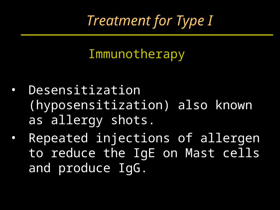
Treatment for Type I
Immunotherapy
• Desensitization (hyposensitization) also known as allergy shots.
• Repeated injections of allergen to reduce the IgE on Mast cells and produce IgG.
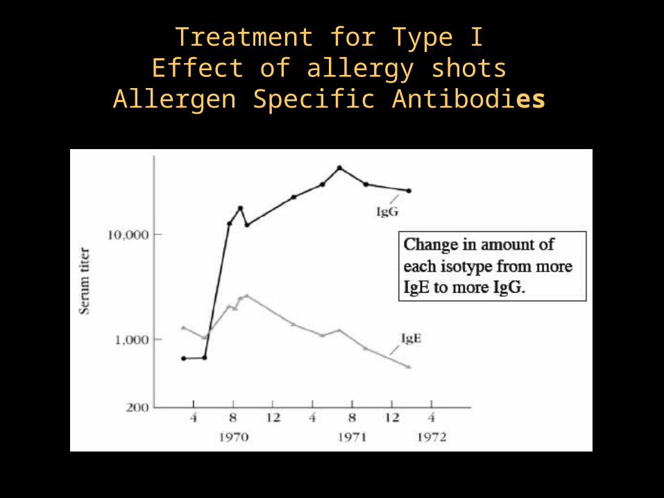
Treatment for Type IEffect of allergy shots
Allergen Specific Antibodies
Hypersensitivity
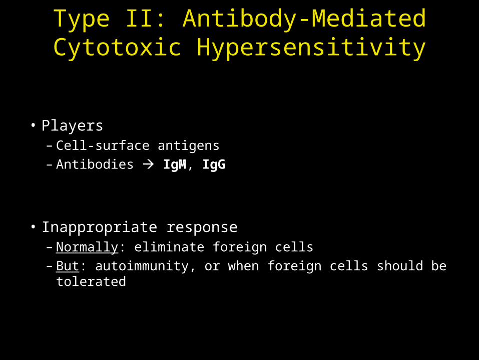
Type II: Antibody-Mediated Cytotoxic Hypersensitivity
• Players– Cell-surface antigens– Antibodies IgM, IgG
• Inappropriate response– Normally: eliminate foreign cells– But: autoimmunity, or when foreign cells should be tolerated
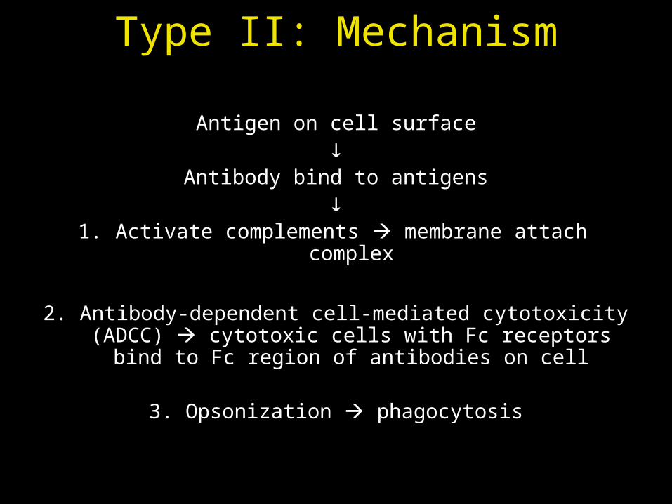
Type II: Mechanism
Antigen on cell surface
Antibody bind to antigens
1. Activate complements membrane attach complex
2. Antibody-dependent cell-mediated cytotoxicity (ADCC) cytotoxic cells with Fc receptors bind to Fc region of
antibodies on cell
3. Opsonization phagocytosis
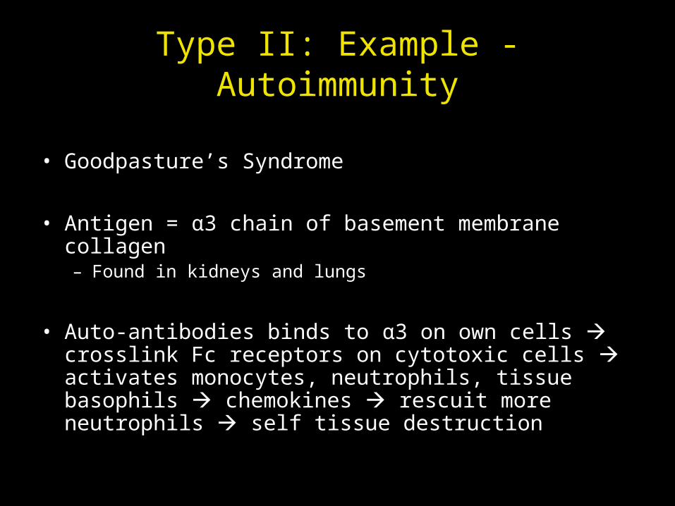
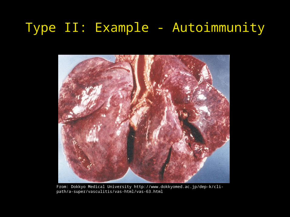
Type II: Example - Autoimmunity
• Goodpasture’s Syndrome
• Antigen = α3 chain of basement membrane collagen– Found in kidneys and lungs
• Auto-antibodies binds to α3 on own cells crosslink Fc receptors on cytotoxic cells activates monocytes, neutrophils, tissue basophils chemokines rescuit more neutrophils self tissue destruction
Type II: Example - Autoimmunity
From: Dokkyo Medical University http://www.dokkyomed.ac.jp/dep-k/cli-path/a-super/vasculitis/vas-html/vas-63.html
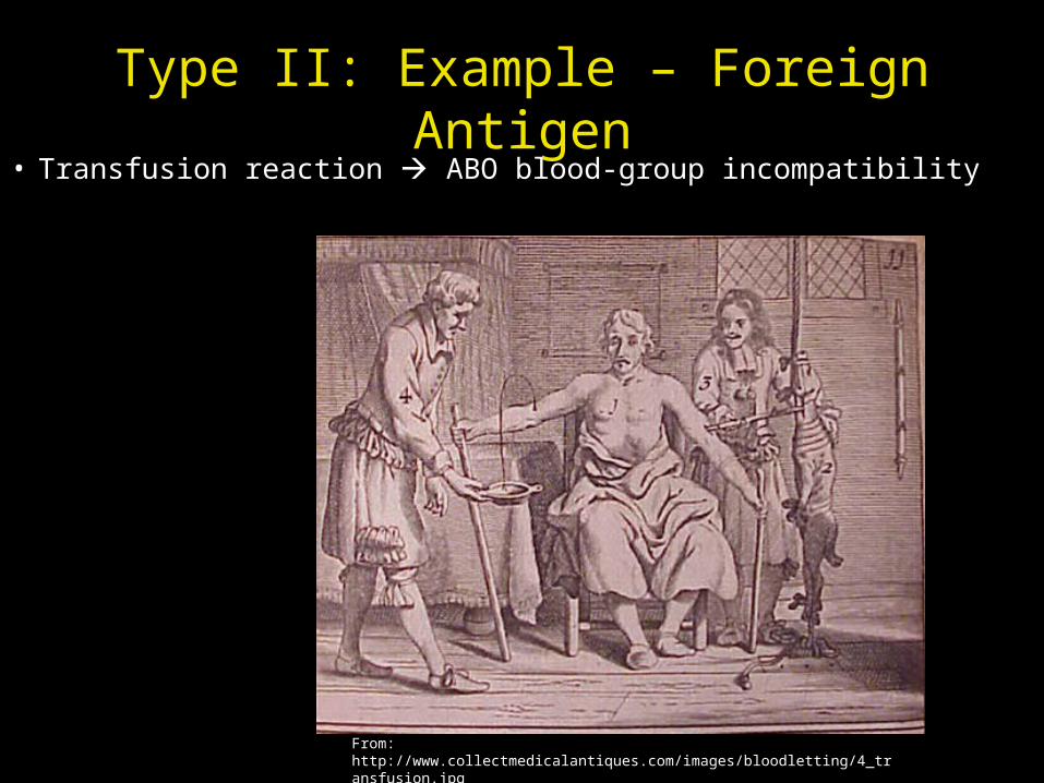
Type II: Example – Foreign Antigen• Transfusion reaction ABO blood-group incompatibility
From: http://www.collectmedicalantiques.com/images/bloodletting/4_transfusion.jpg
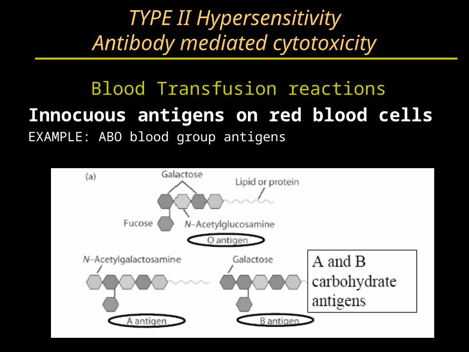
TYPE II HypersensitivityAntibody mediated cytotoxicity
Blood Transfusion reactions
Innocuous antigens on red blood cellsEXAMPLE: ABO blood group antigens
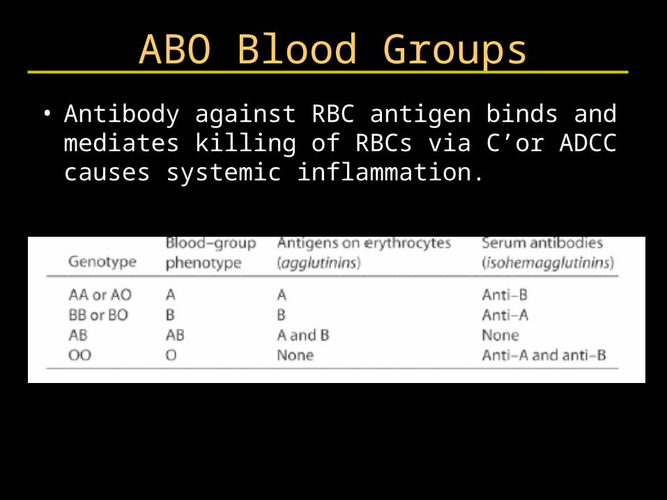
ABO Blood Groups
• Antibody against RBC antigen binds and mediates killing of RBCs via C’or ADCC causes systemic inflammation.
TYPE IIAntibody mediated cytotoxicity
Drug reactions
• Drug binds to RBC surface and antibody against drug binds and causes lysis of RBCs.
• Immune system sees antibody bound to "foreign antigen" on cell. ADCC
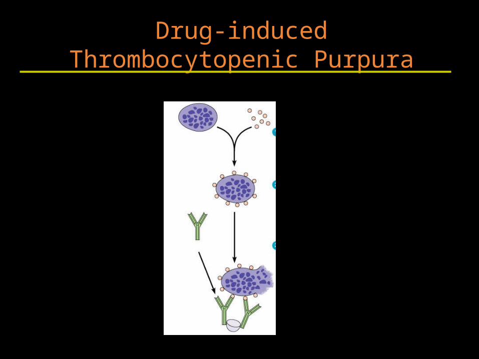
Drug-induced Thrombocytopenic Purpura
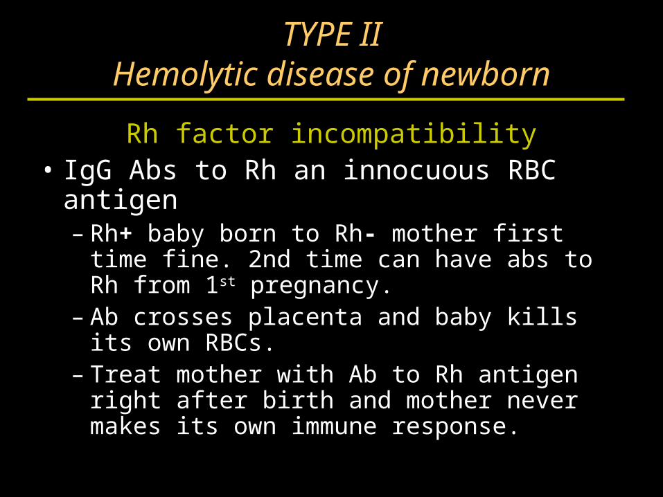
TYPE IIHemolytic disease of newborn
Rh factor incompatibility• IgG Abs to Rh an innocuous RBC antigen
– Rh+ baby born to Rh- mother first time fine. 2nd time can have abs to Rh from 1st pregnancy.
– Ab crosses placenta and baby kills its own RBCs.
– Treat mother with Ab to Rh antigen right after birth and mother never makes its own immune response.
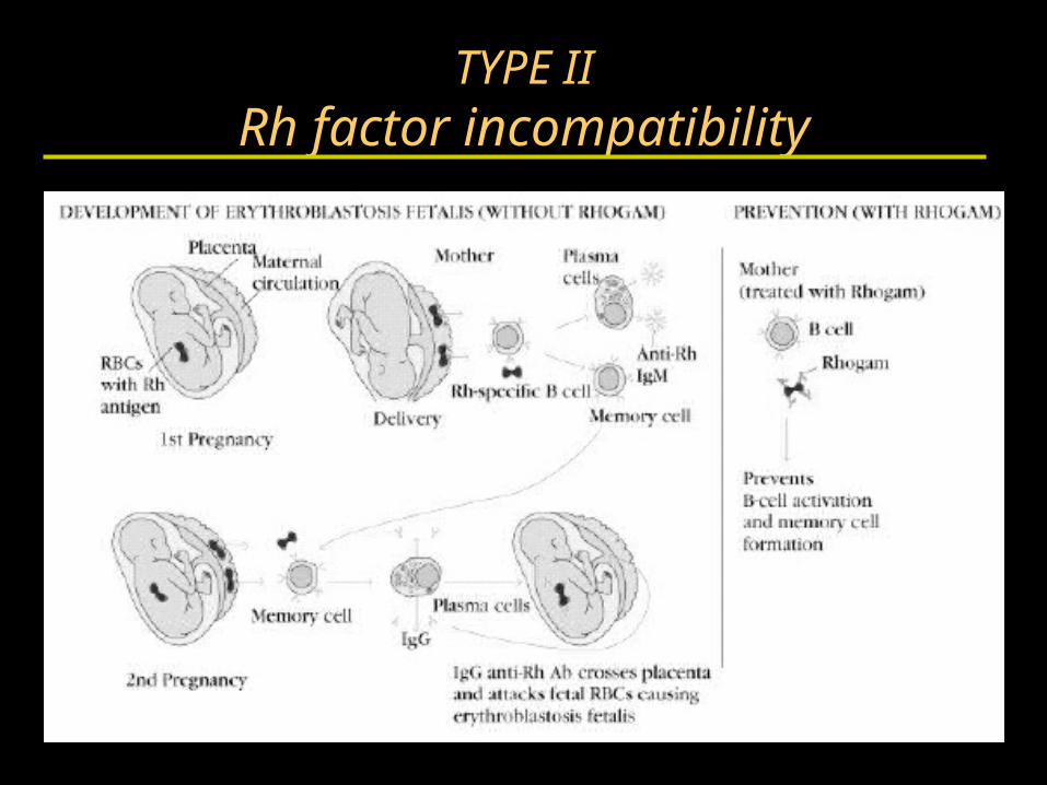
TYPE IIRh factor incompatibility
Hypersensitivity
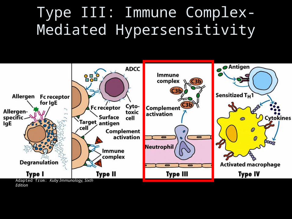
Type III: Immune Complex-Mediated Hypersensitivity
Adapted from: Kuby Immunology, Sixth Edition
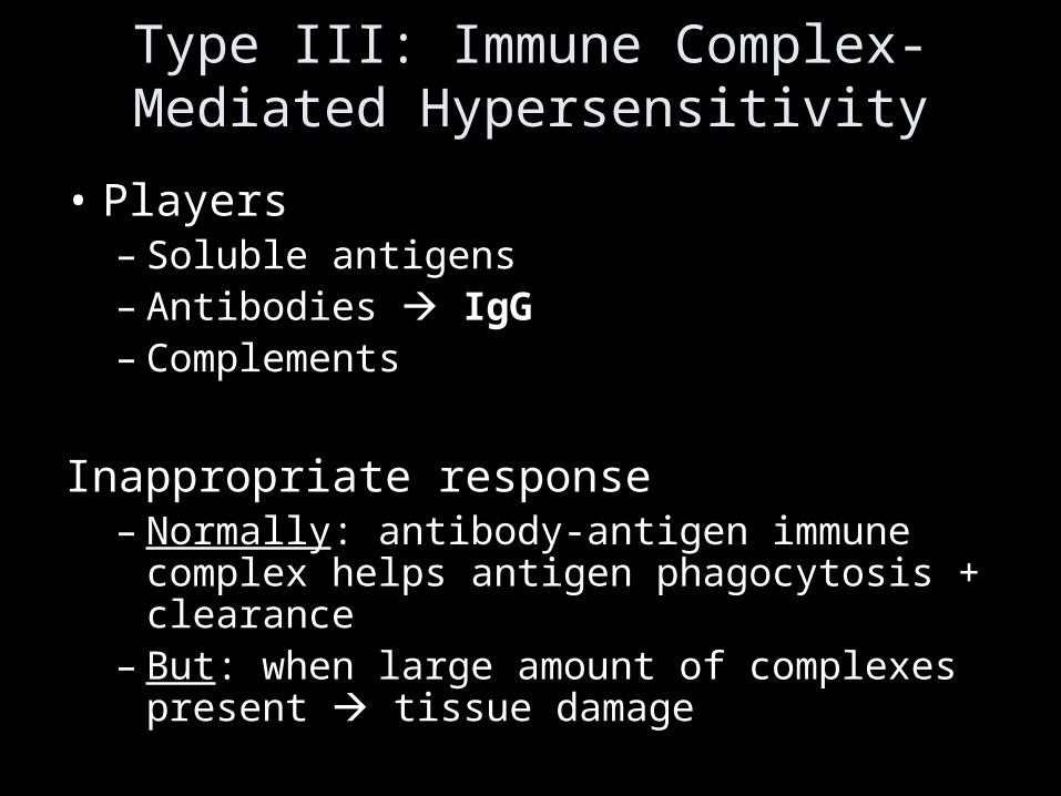
Type III: Immune Complex-Mediated Hypersensitivity
• Players– Soluble antigens– Antibodies IgG– Complements
Inappropriate response– Normally: antibody-antigen immune complex
helps antigen phagocytosis + clearance– But: when large amount of complexes present
tissue damage
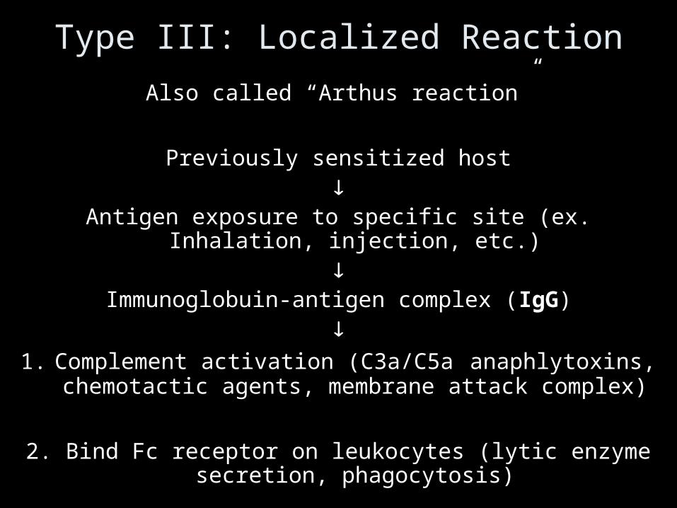
Type III: Localized Reaction
Also called “Arthus reaction”
Previously sensitized host
Antigen exposure to specific site (ex. Inhalation, injection, etc.)
Immunoglobuin-antigen complex (IgG)
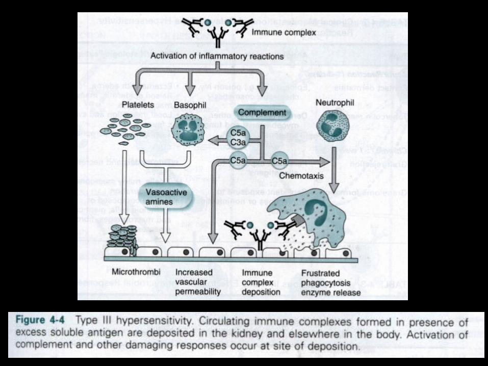
1. Complement activation (C3a/C5a anaphlytoxins, chemotactic agents, membrane attack complex)
2. Bind Fc receptor on leukocytes (lytic enzyme secretion, phagocytosis)
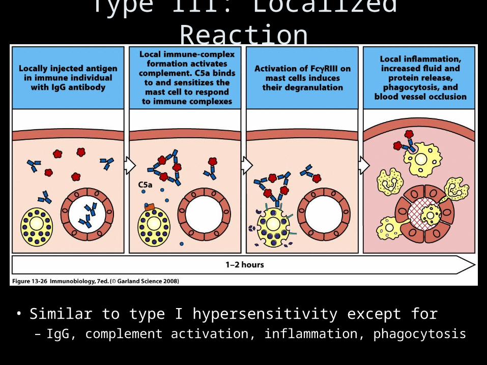
Type III: Localized Reaction
• Similar to type I hypersensitivity except for– IgG, complement activation, inflammation, phagocytosis
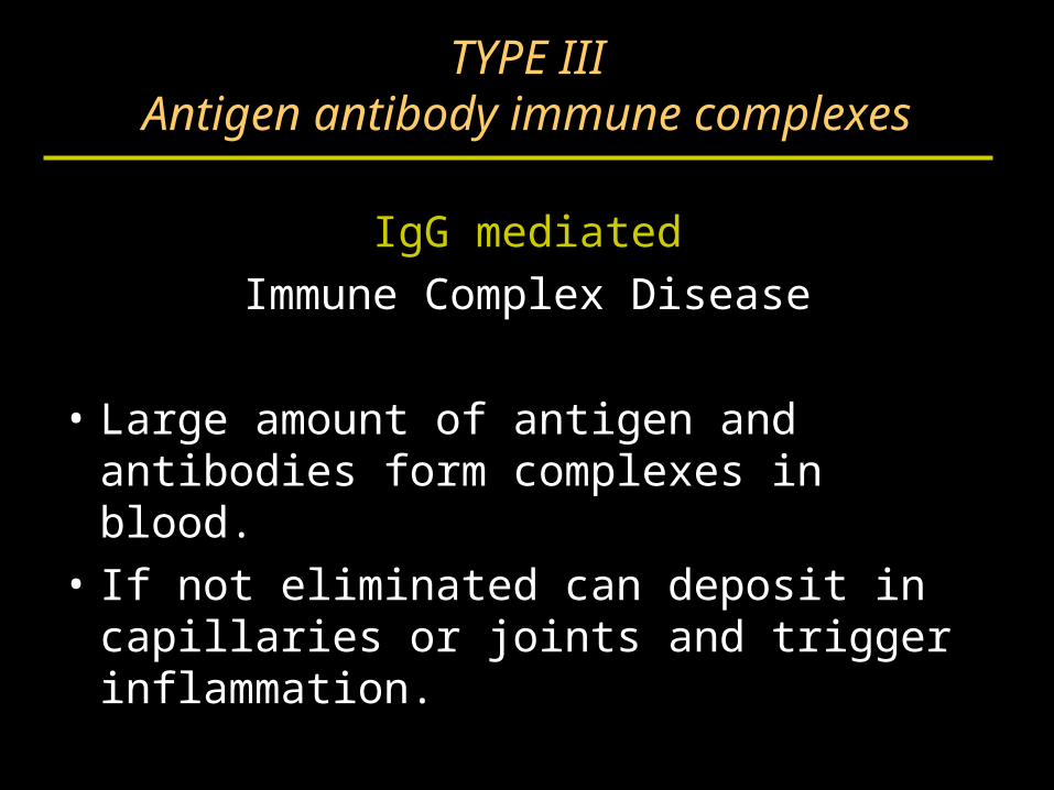
TYPE IIIAntigen antibody immune complexes
IgG mediated
Immune Complex Disease
• Large amount of antigen and antibodies form complexes in blood.
• If not eliminated can deposit in capillaries or joints and trigger inflammation.
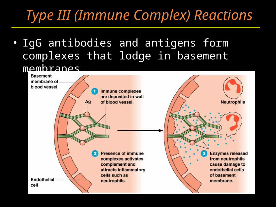
• IgG antibodies and antigens form complexes that lodge in basement membranes.
Type III (Immune Complex) Reactions
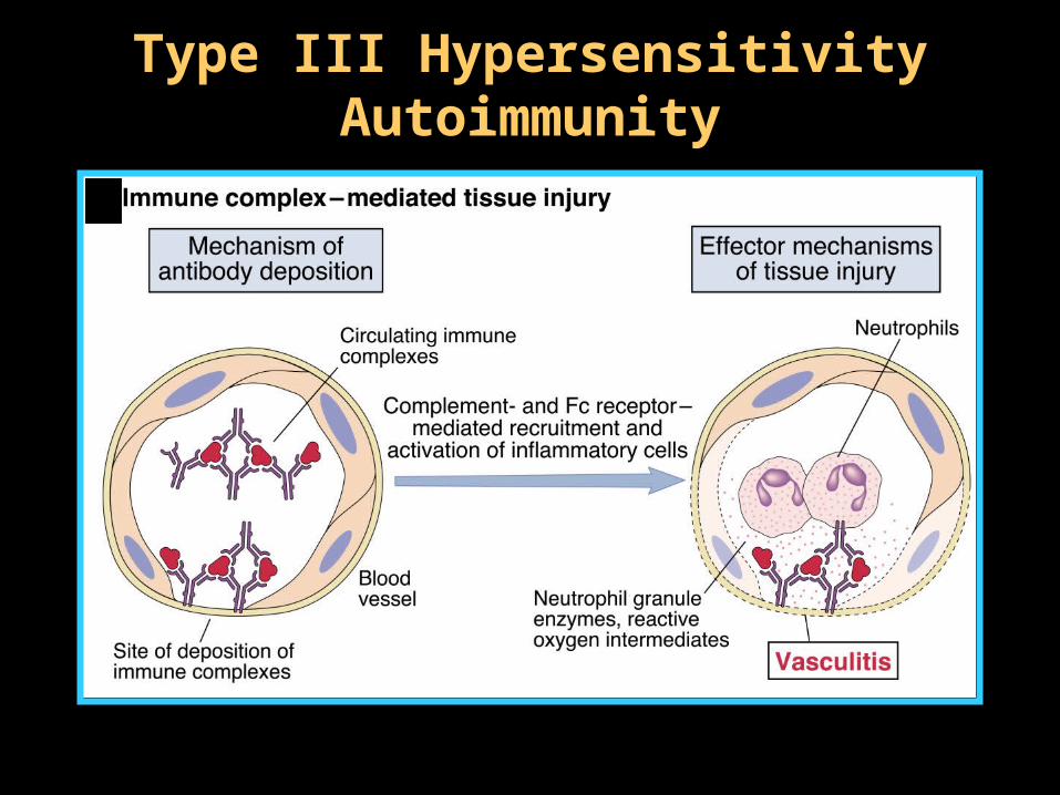
Type III HypersensitivityType III HypersensitivityAutoimmunityAutoimmunity
TYPE IIIImmune Complexes
• PMNs and macrophages bind to immune complexes via FcR and phagocytize the complexes.
BUT
• If unable to phagocytize the immune complexes can cause inflammation via C’ activation C3a C4a, C5a and "frustrated phagocytes".
TYPE IIIImmune Complex Disease
"Frustrated Phagocytes”
• If neutrophils and macrophages are unable to phagocytize the immune complexes these cells will degranulate in the area of immune complex deposition and trigger inflammation.
• Unable to eat -------try to digest outside cell.
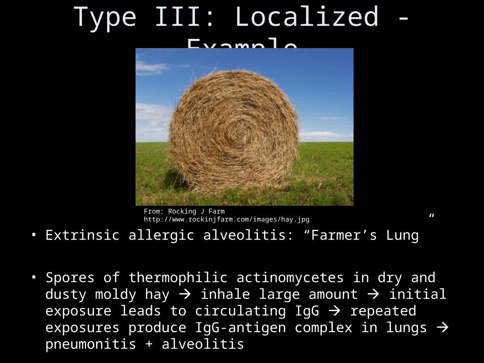
Type III: Localized - Example
• Extrinsic allergic alveolitis: “Farmer’s Lung”
• Spores of thermophilic actinomycetes in dry and dusty moldy hay inhale large amount initial exposure leads to circulating IgG repeated exposures produce IgG-antigen complex in lungs pneumonitis + alveolitis
From: Rocking J Farm http://www.rockinjfarm.com/images/hay.jpg
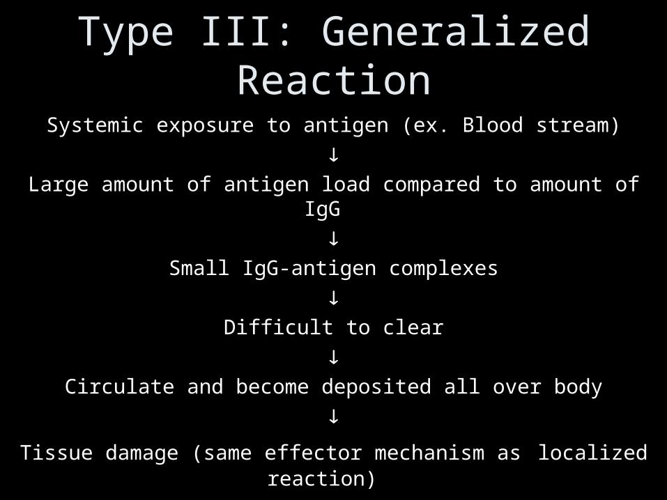
Type III: Generalized Reaction
Systemic exposure to antigen (ex. Blood stream)
Large amount of antigen load compared to amount of IgG
Small IgG-antigen complexes
Difficult to clear
Circulate and become deposited all over body
Tissue damage (same effector mechanism as localized reaction)
Type III: Generalized – Foreign Antigen
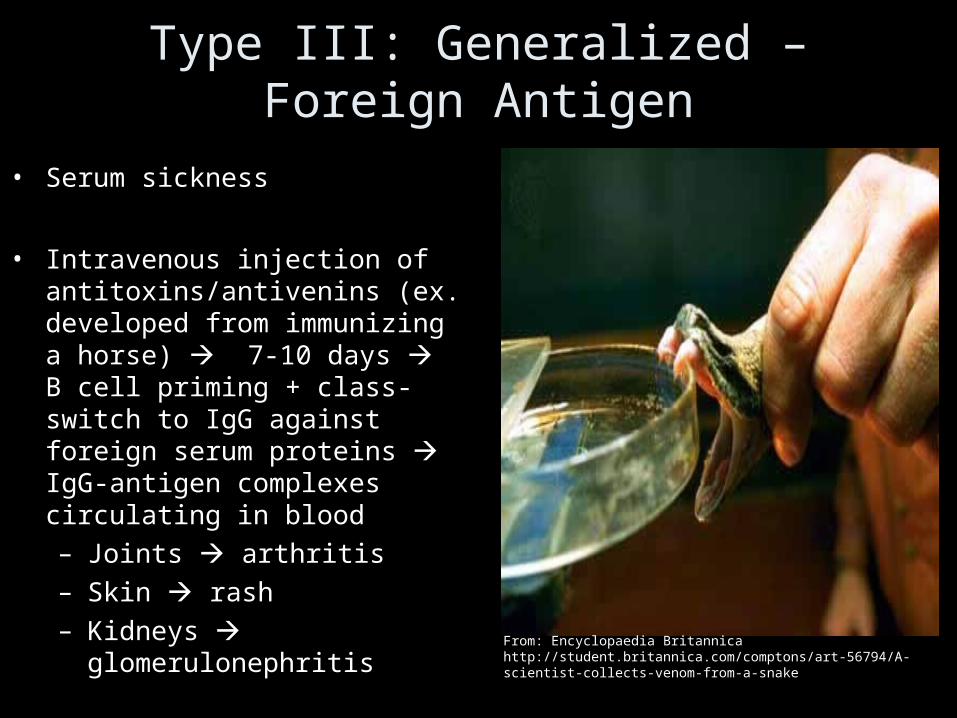
• Serum sickness
• Intravenous injection of antitoxins/antivenins (ex. developed from immunizing a horse) 7-10 days B cell priming + class-switch to IgG against foreign serum proteins IgG-antigen complexes circulating in blood– Joints arthritis– Skin rash– Kidneys glomerulonephritis
From: Encyclopaedia Britannica http://student.britannica.com/comptons/art-56794/A-scientist-collects-venom-from-a-snake
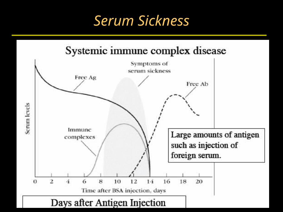
Serum Sickness
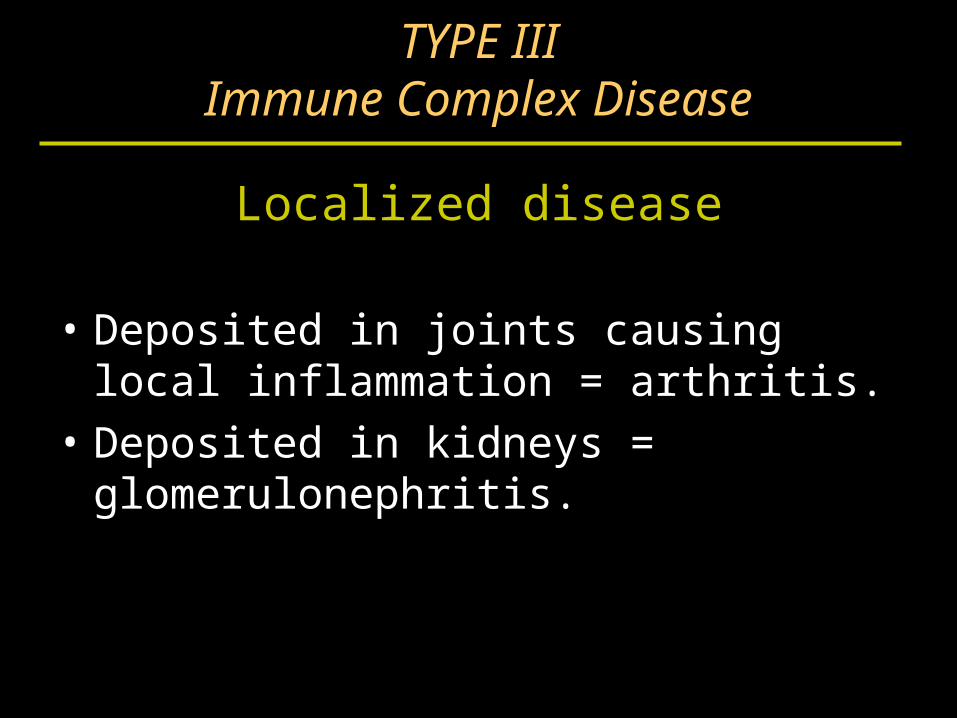
TYPE IIIImmune Complex Disease
Localized disease
• Deposited in joints causing local inflammation = arthritis.
• Deposited in kidneys = glomerulonephritis.
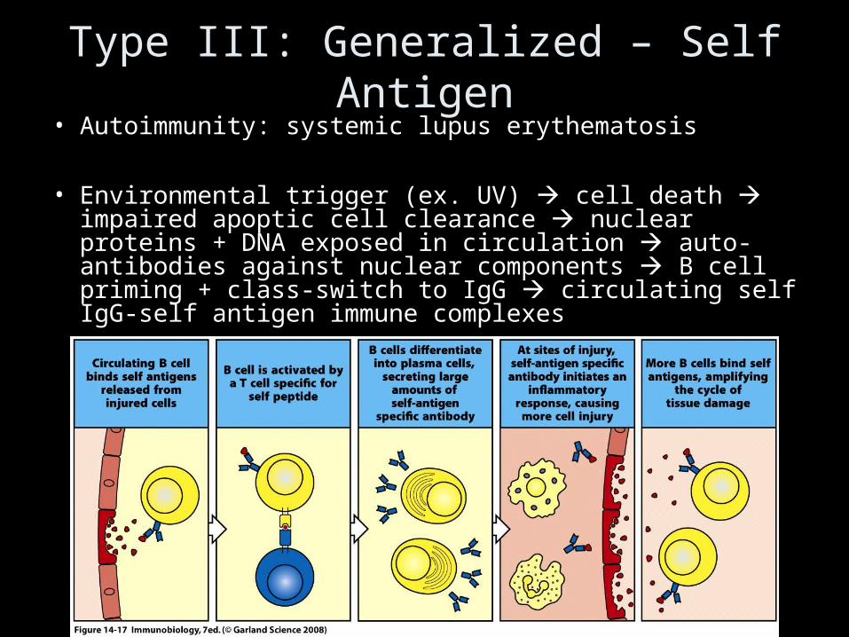
Type III: Generalized – Self Antigen• Autoimmunity: systemic lupus erythematosis
• Environmental trigger (ex. UV) cell death impaired apoptic cell clearance nuclear proteins + DNA exposed in circulation auto-antibodies against nuclear components B cell priming + class-switch to IgG circulating self IgG-self antigen immune complexes
Hypersensitivity
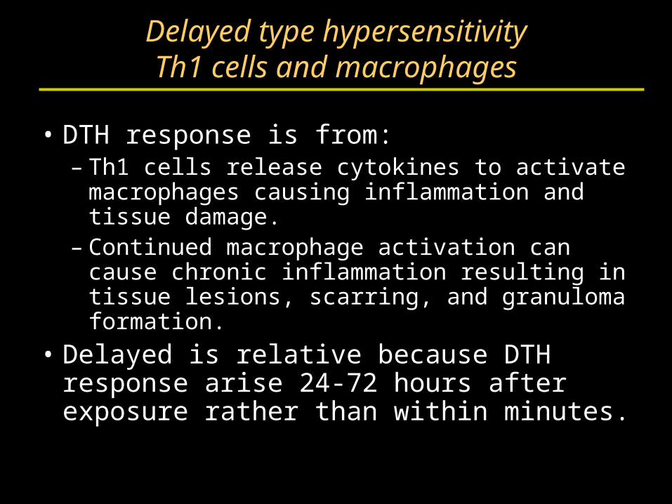
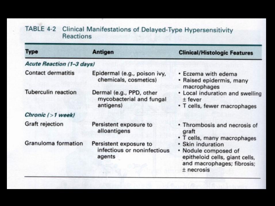
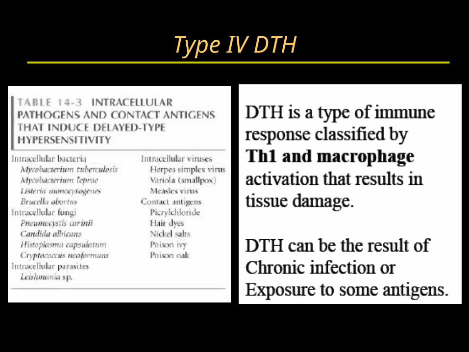
Delayed type hypersensitivityTh1 cells and macrophages
• DTH response is from:– Th1 cells release cytokines to activate
macrophages causing inflammation and tissue damage.
– Continued macrophage activation can cause chronic inflammation resulting in tissue lesions, scarring, and granuloma formation.
• Delayed is relative because DTH response arise 24-72 hours after exposure rather than within minutes.
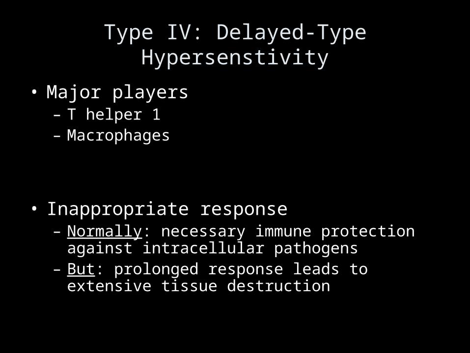
Type IV: Delayed-Type Hypersenstivity
• Major players– T helper 1– Macrophages
• Inappropriate response– Normally: necessary immune protection against
intracellular pathogens– But: prolonged response leads to extensive tissue
destruction
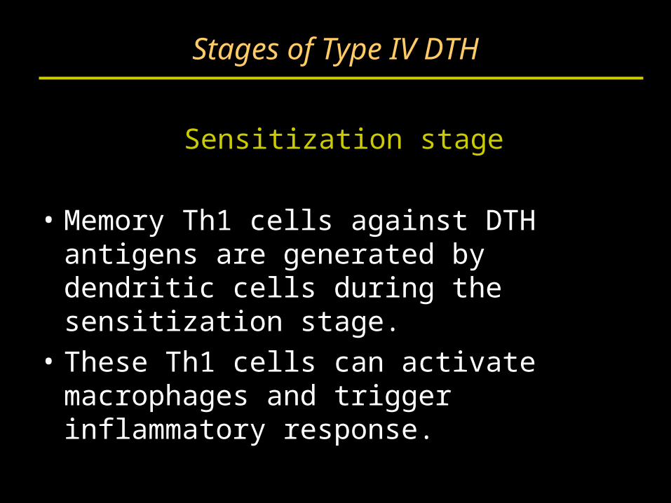
Stages of Type IV DTH
Sensitization stage
• Memory Th1 cells against DTH antigens are generated by dendritic cells during the sensitization stage.
• These Th1 cells can activate macrophages and trigger inflammatory response.
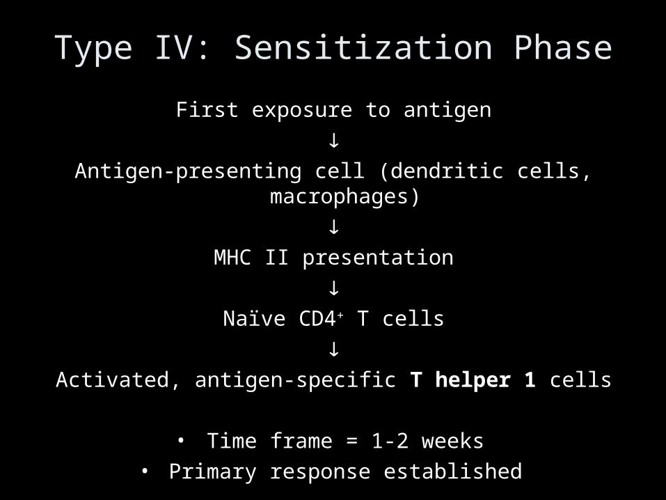
Type IV: Sensitization Phase
First exposure to antigen
Antigen-presenting cell (dendritic cells, macrophages)
MHC II presentation
Naïve CD4+ T cells
Activated, antigen-specific T helper 1 cells
• Time frame = 1-2 weeks• Primary response established
Stages of Type IV DTH
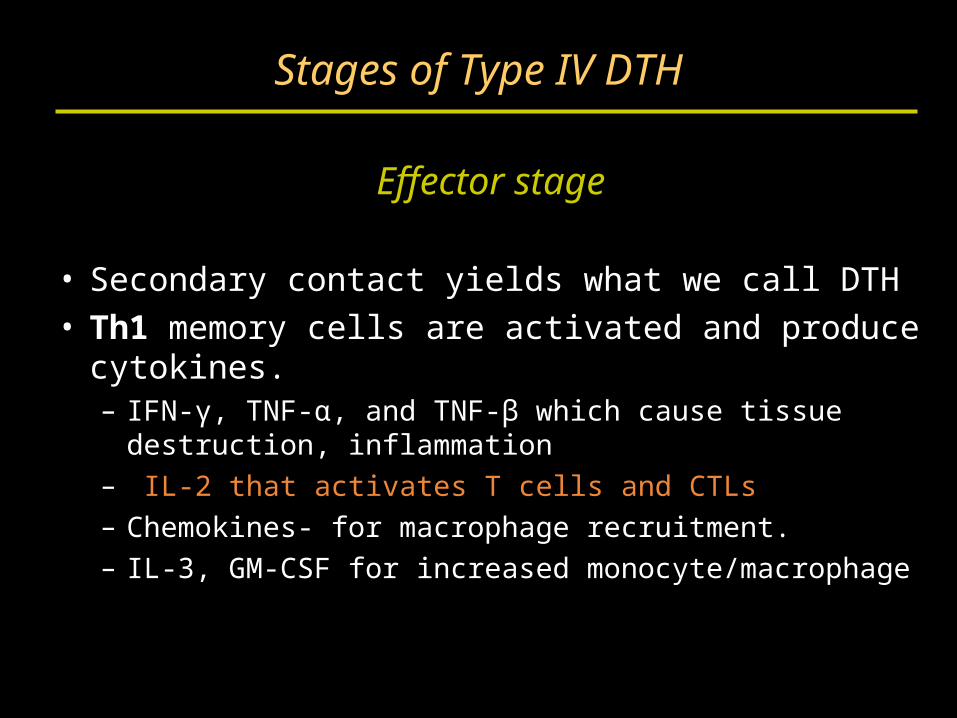
Effector stage
• Secondary contact yields what we call DTH• Th1 memory cells are activated and produce
cytokines.– IFN-γ, TNF-α, and TNF-β which cause tissue
destruction, inflammation– IL-2 that activates T cells and CTLs– Chemokines- for macrophage recruitment.– IL-3, GM-CSF for increased monocyte/macrophage
Stages of Type IV DTH
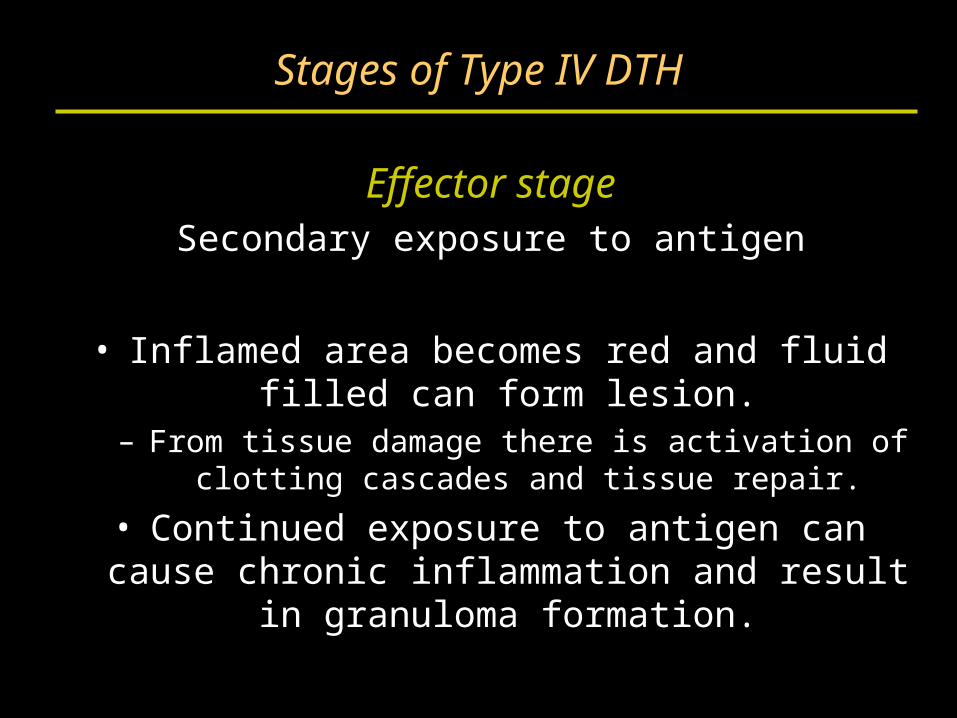
Effector stageSecondary exposure to antigen
• Inflamed area becomes red and fluid filled can form lesion.
– From tissue damage there is activation of clotting cascades and tissue repair.
• Continued exposure to antigen can cause chronic inflammation and result in granuloma formation.
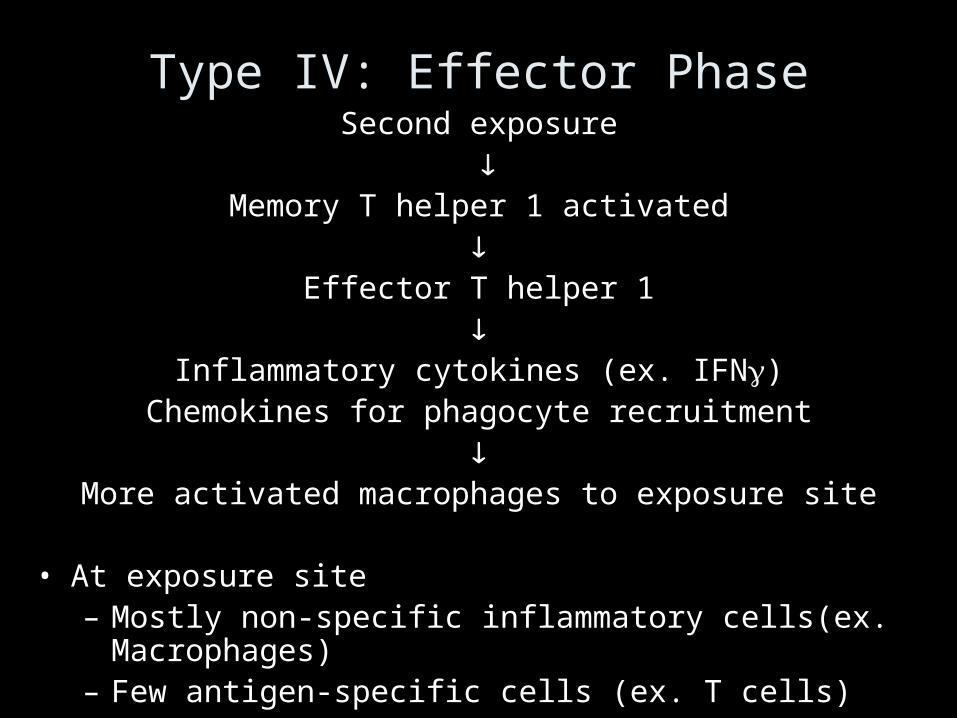
Type IV: Effector PhaseSecond exposure
Memory T helper 1 activated
Effector T helper 1
Inflammatory cytokines (ex. IFN)
Chemokines for phagocyte recruitment
More activated macrophages to exposure site
• At exposure site– Mostly non-specific inflammatory cells(ex. Macrophages)– Few antigen-specific cells (ex. T cells)
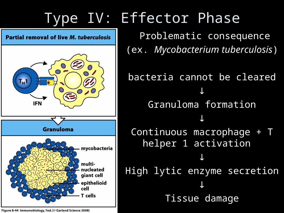
Type IV: Effector PhaseProblematic consequence
)ex. Mycobacterium tuberculosis(
bacteria cannot be cleared
Granuloma formation
Continuous macrophage + T helper 1
activation
High lytic enzyme secretion
Tissue damage
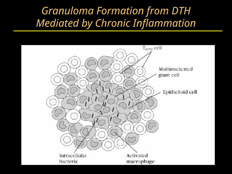
Granuloma Formation from DTHMediated by Chronic Inflammation
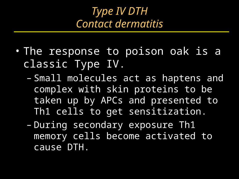
Type IV DTHContact dermatitis
• The response to poison oak is a classic Type IV.– Small molecules act as haptens and complex
with skin proteins to be taken up by APCs and presented to Th1 cells to get sensitization.
– During secondary exposure Th1 memory cells become activated to cause DTH.
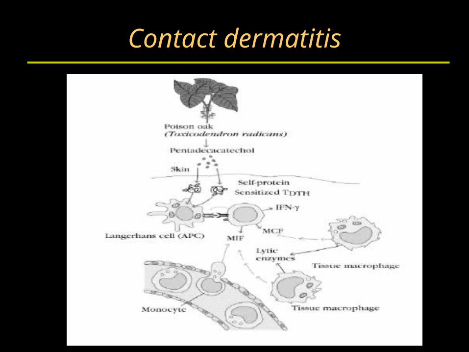
Contact dermatitis
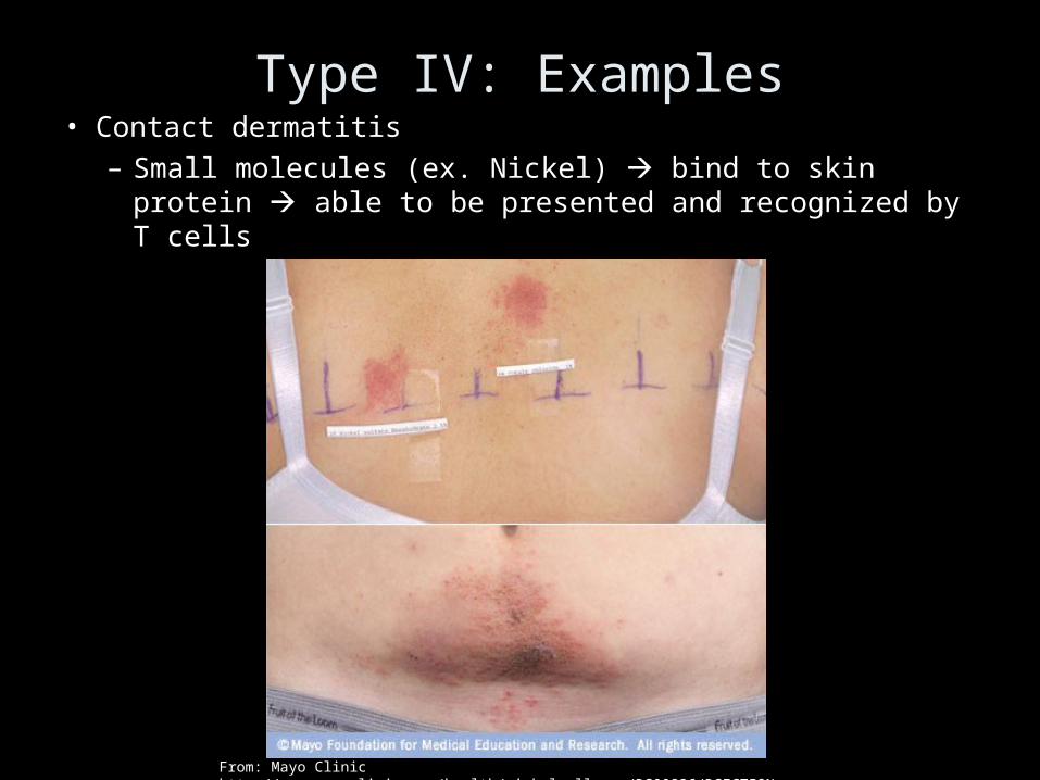
Type IV: Examples• Contact dermatitis
– Small molecules (ex. Nickel) bind to skin protein able to be presented and recognized by T cells
From: Mayo Clinic http://www.mayoclinic.com/health/nickel-allergy/DS00826/DSECTION=causes
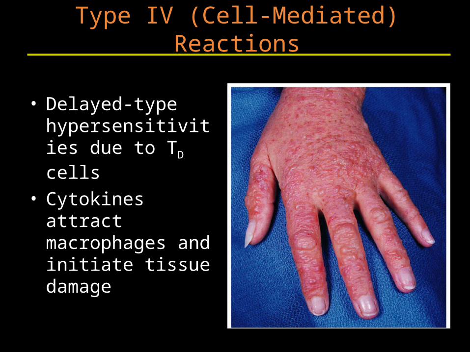
• Delayed-type hypersensitivities due to TD cells
• Cytokines attract macrophages and initiate tissue damage
Type IV (Cell-Mediated) Reactions
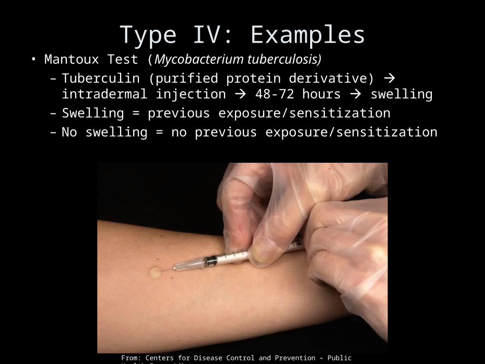
Type IV: Examples• Mantoux Test (Mycobacterium tuberculosis)
– Tuberculin (purified protein derivative) intradermal injection 48-72 hours swelling
– Swelling = previous exposure/sensitization– No swelling = no previous exposure/sensitization
From: Centers for Disease Control and Prevention – Public Health Image Library
Type IV DTH
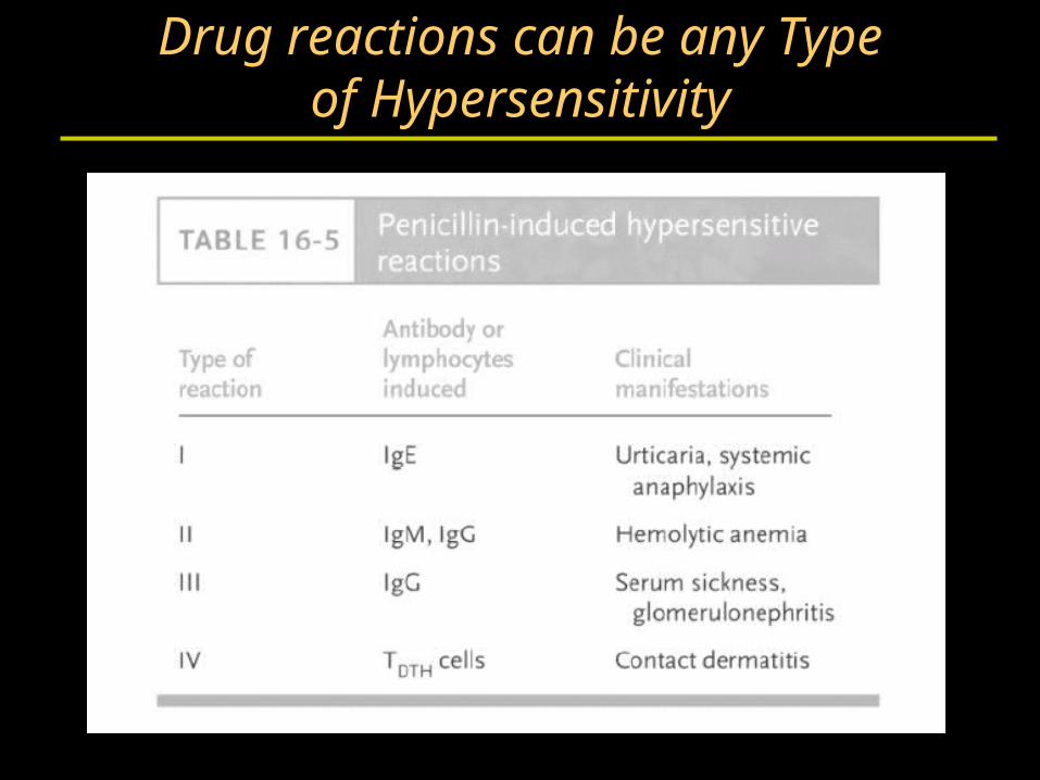
Drug reactions can be any Typeof Hypersensitivity
Section B
Autoimmunity
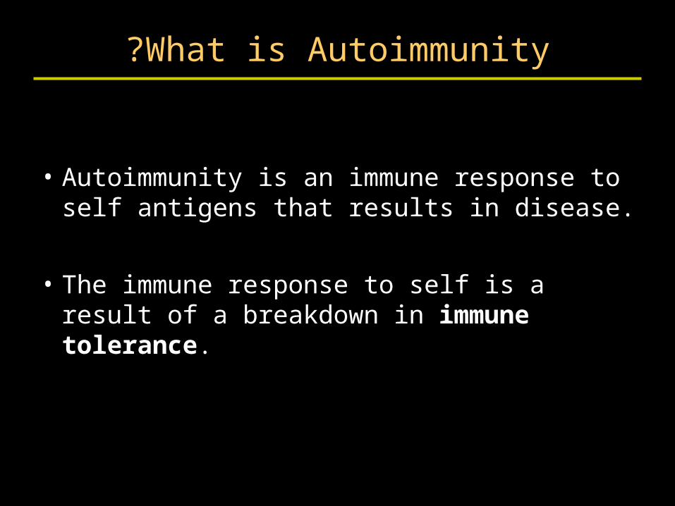
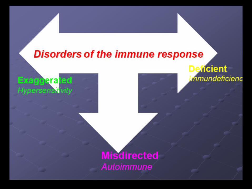
What is Autoimmunity?
• Autoimmunity is an immune response to self antigens that results in disease.
• The immune response to self is a result of a breakdown in immune tolerance.
Immune Tolerance
• Tolerance of self is a hallmark of adaptive immune response.
• B cell tolerance vs. T cell tolerance.
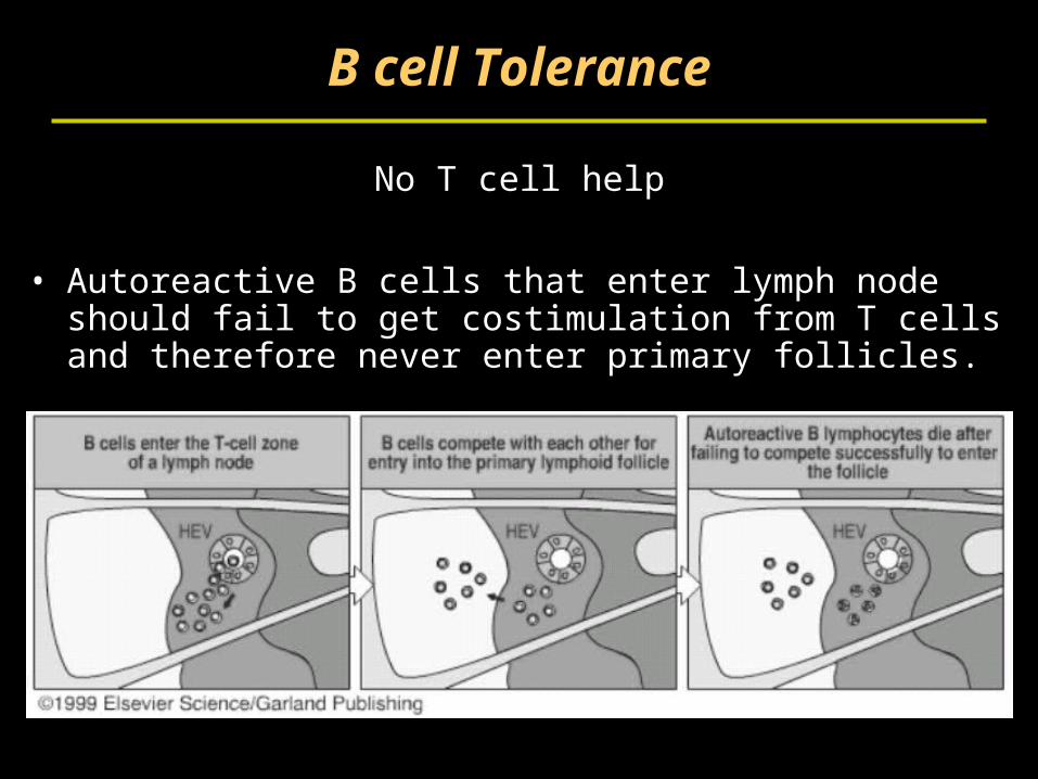
B cell Tolerance
No T cell help
• Autoreactive B cells that enter lymph node should fail to get costimulation from T cells and therefore never enter primary follicles.
Maintenance of T cell tolerance
• Clonal deletion– negative selection in the thymus, deletion in
the periphery.
• Sequestration of antigens– Inside nucleus– Inaccessible to immune system (brain, eye,
testes)
• Immunological ignorance– self antigens at low density on APCs– or T cells do not cross barrier.
Maintenance of T cell tolerance
• Anergy– Lack of co-stimulation or second signal to T
cells results in anergy.
• Suppression– T-cell cytokine mediated suppression.– Regulatory T cells. CD4+CD25+ CTLA4+ T
cells that produce suppressive cytokines.
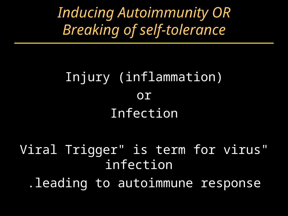
Inducing Autoimmunity ORBreaking of self-tolerance
Injury (inflammation)
or
Infection
"Viral Trigger" is term for virus infection
leading to autoimmune response.
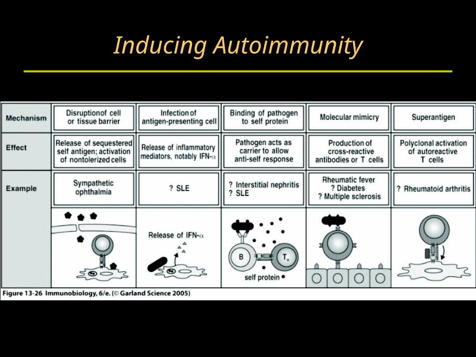
Inducing Autoimmunity
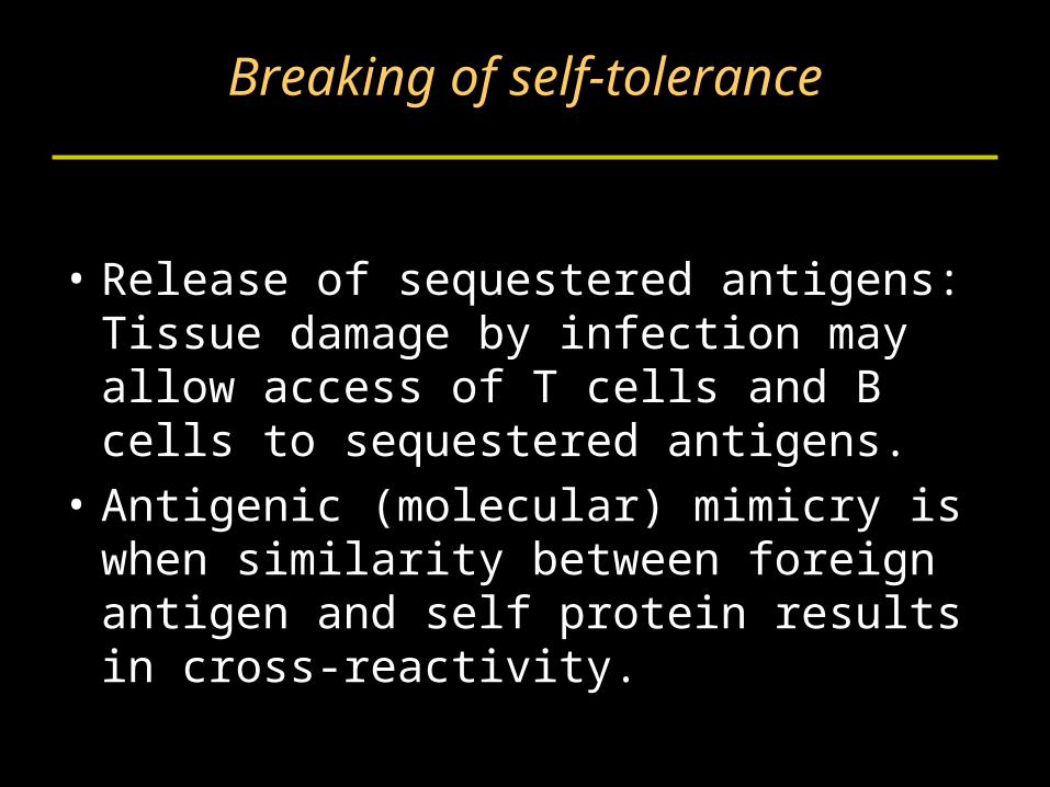
Breaking of self-tolerance
• Release of sequestered antigens: Tissue damage by infection may allow access of T cells and B cells to sequestered antigens.
• Antigenic (molecular) mimicry is when similarity between foreign antigen and self protein results in cross-reactivity.
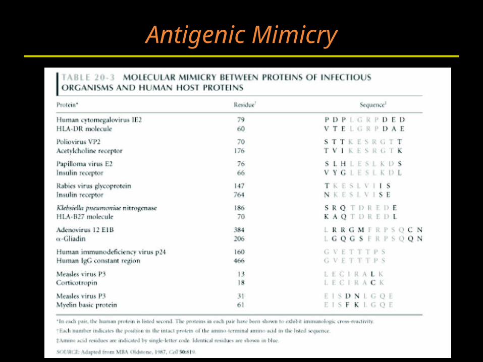
Antigenic Mimicry
Breaking of self-tolerance
• Inappropriate expression of Class II MHC.– Abnormal expression of class II molecules can
lead to presentation of self antigens that were not presented in thymus or periphery.
– "non-APC" becomes APC with inflammation.
Classification ofautoimmune diseases
Autoantibody or
T cell mediated
autoimmune diseases
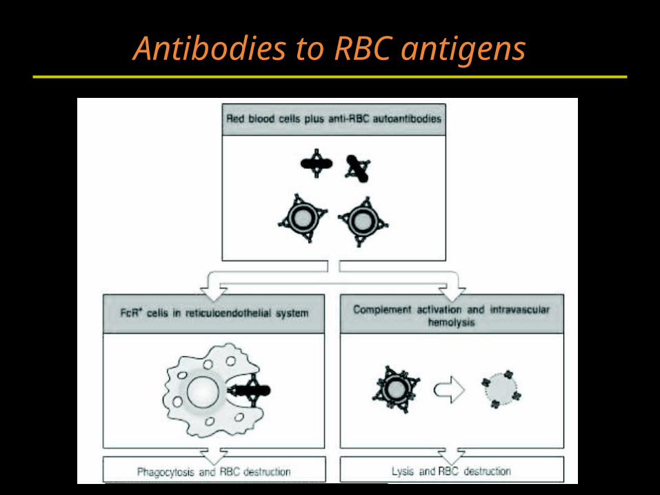
Antibodies to RBC antigens
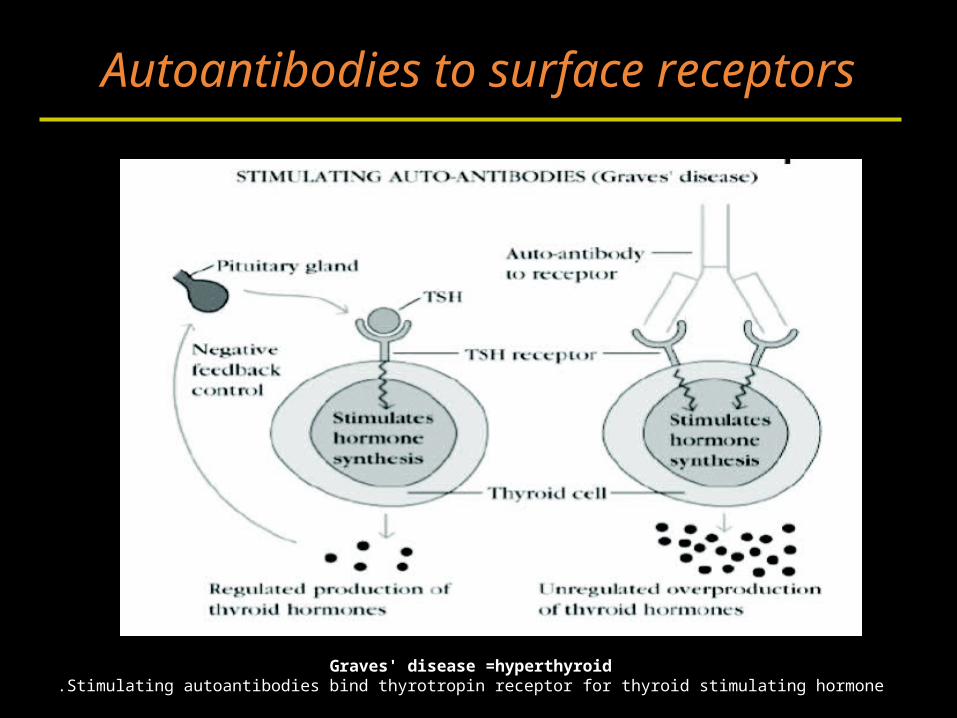
Autoantibodies to surface receptors
Graves' disease =hyperthyroidStimulating autoantibodies bind thyrotropin receptor for thyroid stimulating hormone.
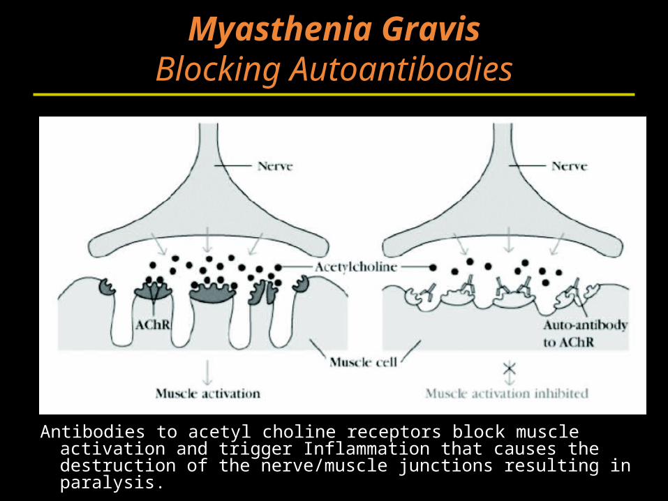
Myasthenia GravisBlocking Autoantibodies
Antibodies to acetyl choline receptors block muscle activation and trigger Inflammation that causes the destruction of the nerve/muscle junctions resulting in paralysis.
Autoantibodies to surface receptorsBlocking autoantibodies
• Hashimoto's thyroiditis =hypothyroid– Blocking autoantibodies inhibit thyroid
function.
Goodpasture's Syndrome
• Autoantibodies to type IV collagen and noncollagenous basement membrane
• Antibodies bind in lung and kidney causing inflammation and destruction.
Rheumatoid ArthritisImmune Complex Disease
• Autoantibodies to ubiquitous antigens– IgM against IgG is called "rheumatoid factor"– IgG against glucose-6-phosphate isomerase.
• Primary disease manifestation– immune complexes get deposited in joints and
trigger inflammatory response.– Complement and FcγRs play large role.
Systemic lupus erythematosus (SLE)Immune complex disease
• Chronic IgG production to intracellular proteins.
• Disease symptoms are widespread and varied.– kidney damage, lung disease, skin, eye, etc.
Systemic lupus erythematosus (SLE)
• Autoantibodies against nucleoprotein particles:– Nucleosome– Spliceosome– Ribonucleoprotein complexes
• Th response to one epitope can drive auto Abs.
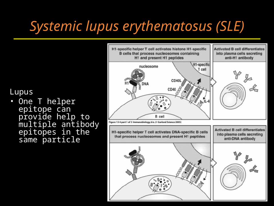
Systemic lupus erythematosus (SLE)
Lupus• One T helper
epitope can provide help to multiple antibody epitopes in the same particle
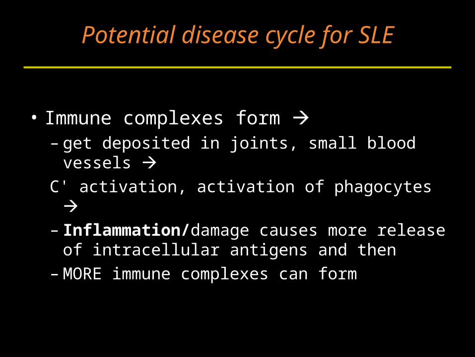
Potential disease cycle for SLE
• Immune complexes form – get deposited in joints, small blood vessels C' activation, activation of phagocytes – Inflammation/damage causes more release
of intracellular antigens and then– MORE immune complexes can form
T cell Mediated Autoimmune DiseasesMultiple sclerosis (MS)
• T cell responses to myelin basic protein (MBP).
• The destruction of the myelin sheath results in neurological symptoms
Multiple sclerosis (MS)
• The cause remains unknown, but autoimmunity possibly triggered during an inflammatory response to a viral infection is implicated.
• MBP has high sequence homology with measles protein and Hepatitis B virus protein.
Antigenic mimicry?
Insulin-dependent (type I)diabetes mellitus (IDDM)
Type I diabetes
• Selective destruction of insulin-producing β cells in the islets of Langerhans of the pancreas.
• Autoantibodies and self-reactive T cells have been found in human patients with IDDM.
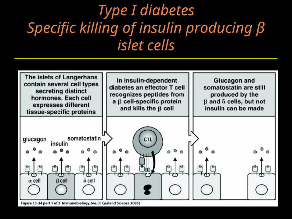
Type I diabetesSpecific killing of insulin producing β
islet cells
Diabetes
• CD8+ CTLs are thought to be responsible for the actual killing of the islet cells.
• Autoantibodies are present in IDDM.– However, animal models of IDDM have shown
that these autoantibodies alone cannot cause IDDM.
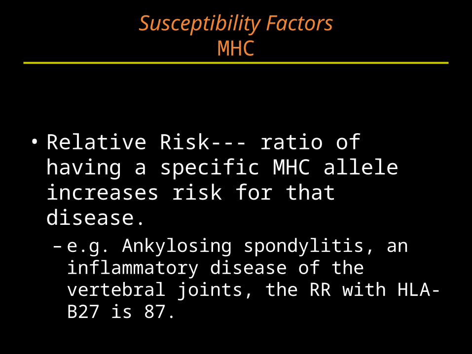
Susceptibility FactorsMHC
• Relative Risk--- ratio of having a specific MHC allele increases risk for that disease.– e.g. Ankylosing spondylitis, an inflammatory
disease of the vertebral joints, the RR with HLA-B27 is 87.
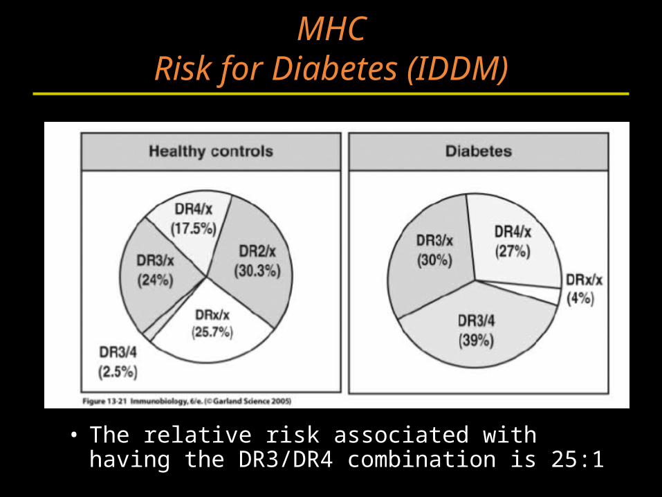
MHCRisk for Diabetes (IDDM)
• The relative risk associated with having the DR3/DR4 combination is 25:1
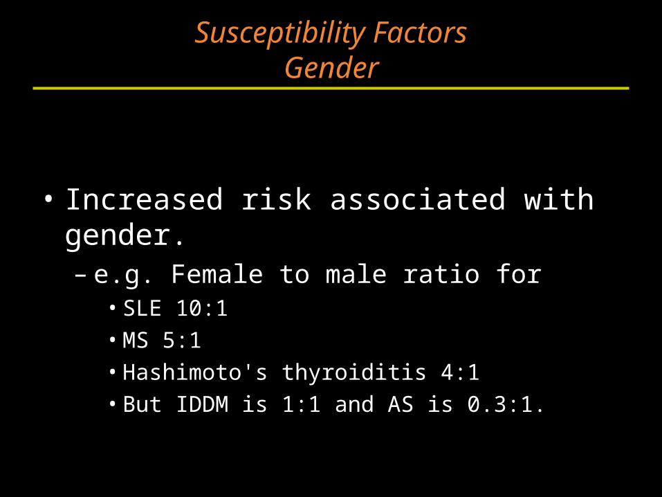
Susceptibility FactorsGender
• Increased risk associated with gender.– e.g. Female to male ratio for
• SLE 10:1• MS 5:1• Hashimoto's thyroiditis 4:1• But IDDM is 1:1 and AS is 0.3:1.
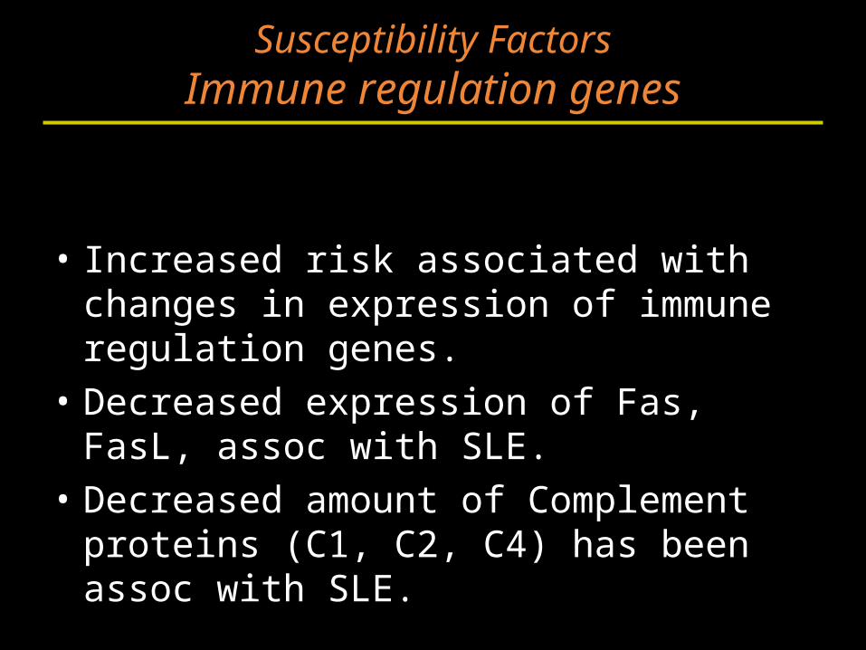
Susceptibility FactorsImmune regulation genes
• Increased risk associated with changes in expression of immune regulation genes.
• Decreased expression of Fas, FasL, assoc with SLE.
• Decreased amount of Complement proteins (C1, C2, C4) has been assoc with SLE.
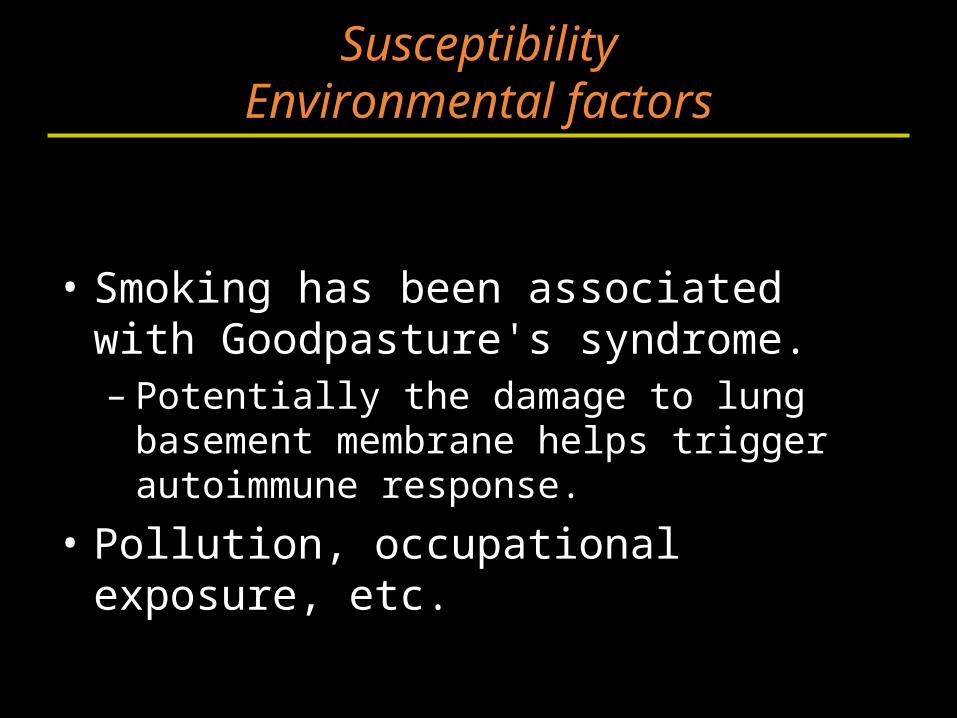
SusceptibilityEnvironmental factors
• Smoking has been associated with Goodpasture's syndrome.– Potentially the damage to lung basement
membrane helps trigger autoimmune response.
• Pollution, occupational exposure, etc.
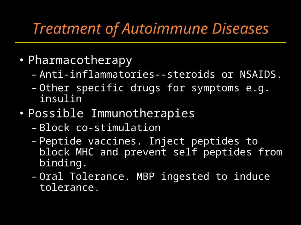
Treatment of Autoimmune Diseases
• Pharmacotherapy– Anti-inflammatories--steroids or NSAIDS.– Other specific drugs for symptoms e.g. insulin
• Possible Immunotherapies– Block co-stimulation – Peptide vaccines. Inject peptides to block
MHC and prevent self peptides from binding.– Oral Tolerance. MBP ingested to induce
tolerance.
Section C
Immunodeficiencies
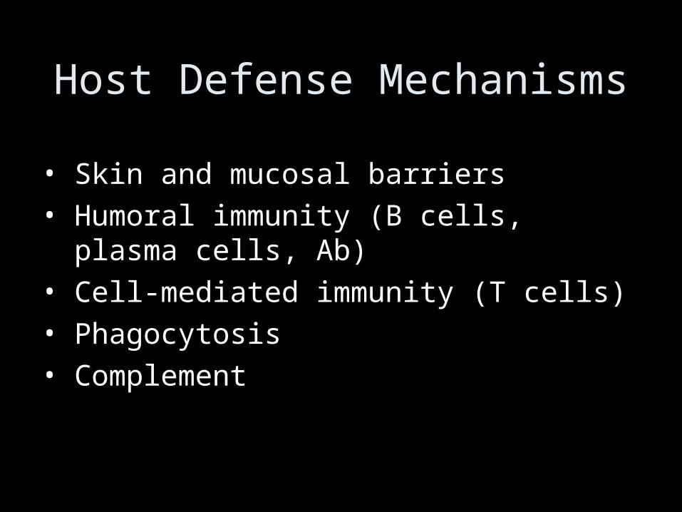
Host Defense Mechanisms
• Skin and mucosal barriers
• Humoral immunity (B cells, plasma cells, Ab)
• Cell-mediated immunity (T cells)
• Phagocytosis
• Complement
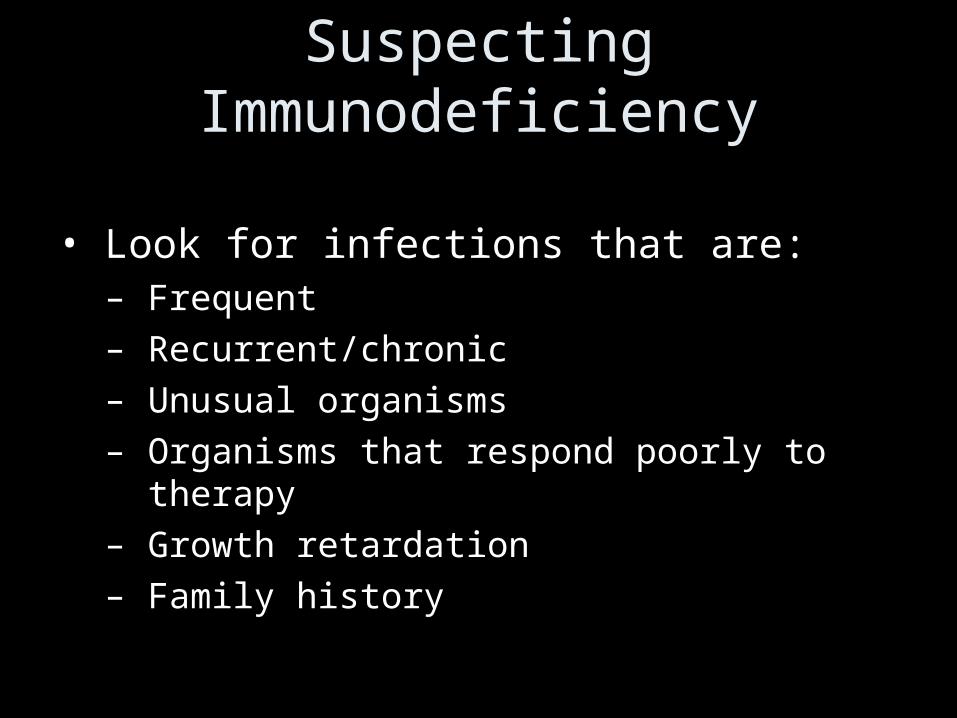
Suspecting Immunodeficiency
• Look for infections that are:– Frequent– Recurrent/chronic– Unusual organisms– Organisms that respond poorly to therapy– Growth retardation– Family history
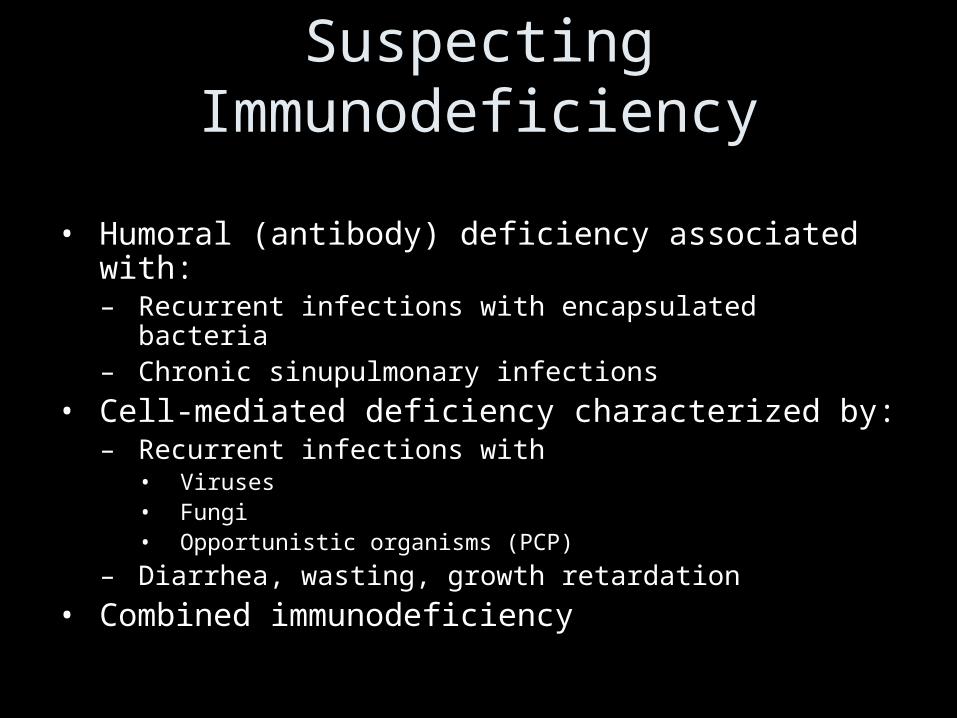
Suspecting Immunodeficiency
• Humoral (antibody) deficiency associated with:– Recurrent infections with encapsulated bacteria– Chronic sinupulmonary infections
• Cell-mediated deficiency characterized by:– Recurrent infections with
• Viruses• Fungi• Opportunistic organisms (PCP)
– Diarrhea, wasting, growth retardation
• Combined immunodeficiency
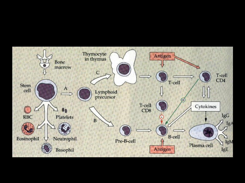
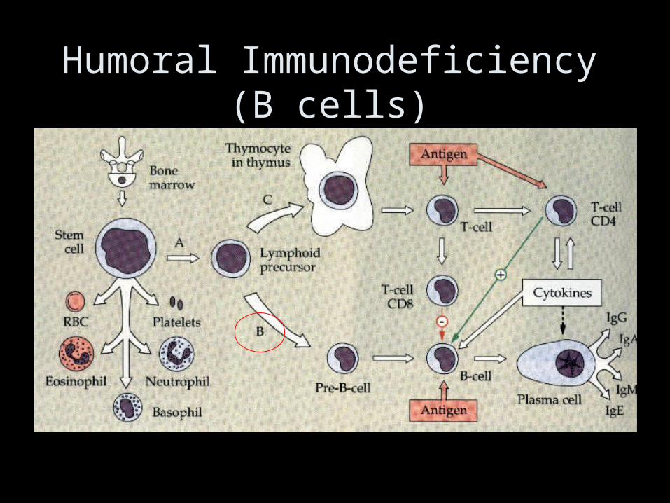
Humoral Immunodeficiency (B cells)
Humoral Immunodeficiency (B cells)
• Transient hypogammaglobulinemia of infancy– Slow to develop normal levels of antibody– Asymptomatic, minor infections– Low levels of IgG, IgA (IgM usually normal)– Resolves by 3-6 yo
• IgA deficiency– Most common humoral antibody deficiency– 50-80% asymptomatic– Recurrent sinopulmonary infections most frequent
manifestation– May have severe malabsorption (chronic diarrhea)– Isolated low IgA level– Increased risk of autoimmune disorders
Bruton’s X-linked Agammaglobulinemia• No B cells• Child clinically well for first 6 months of life• Recurrent upper/lower respiratory tract
infections with encapsulated bacteria (S. pneumo, H.flu)
– Bronchiectasis chronic cough/increased sputum• Sepsis, meningitis, skin infections• Paucity of lymphoid tissue (tonsils, adenoids)• Markedly decreased IgG, IgA, IgM• Treatment: IVIG, antibiotic therapy
Common Variable Immunodeficiency
• B lymphs don’t differentiate into plasma cells
• Recurrent sinopulmonary infections
• Low IgG, IgA, IgM
• Treatment: IVIG
• Associated with autoimmune disease, lymphoma
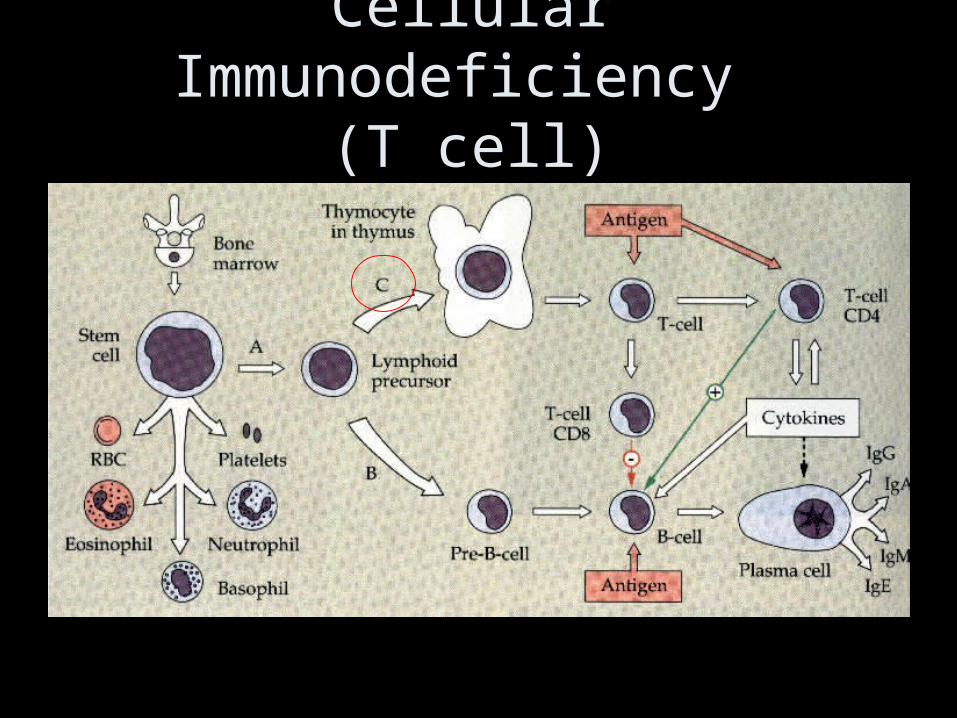
Cellular Immunodeficiency (T cell)
DiGeorge Syndrome
• No T cells secondary to thymic hypoplasia
• “CATCH 22”
• Overwhelming infections with viruses, fungi, bacteria
• Treatment: correct hypocalcemia, cardiac defects, fetal thymus transplant
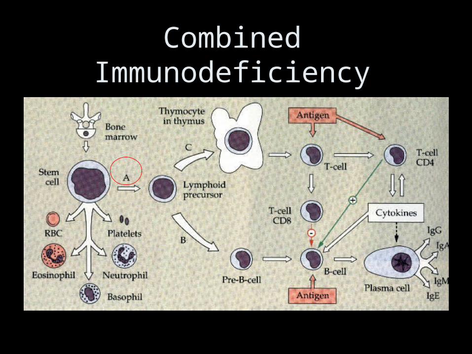
Combined Immunodeficiency
SCID
• Defects in stem cell maturation• Adenosine deaminase deficiency (toxic insult to
T and B cells)• Manifestations seen in first 3 months of life
– Recurrent, severe bacterial, viral, fungal, and protozoan infections (usually respiratory infections)
– Failure to thrive, diarrhea, dermatitis, candidiasis• Most have lymphopenia, decreased IgG, IgA,
and IgM– Diagnosis made by analysis of T, B, and NK cell
subsets• Treatment: isolation, treat underlying
infections, bone marrow transplant
Wiskott-Aldrich Syndrome• X-linked recessive• Symptoms in infancy
– Recurrent, severe infections– Eczema– Thrombocytopenia (petechiae)
• Low levels of IgM• Increased risk for hematologic malignancy • Treatment: manage bleeding/infections,
BMT
Ataxia Telangiectasia
• Autosomal recessive deficiency in DNA repair affecting T and B cells
• Progressive ataxia, telangiectasia, variable immunodeficiency (recurrent sinopulmonary infections common)
• Increased risk of malignancy (leukemia, lymphoma)
Hyper IgE (Job) syndrome
• Autosomal recessive
• Symptoms/signs– Coarse facial features/skeletal abnormalities– Recurrent staph infections
• Impetigo (resistant)• Pneumonia with pneumatocele formation
– 3 E’s: Elevated IgE, Eosinophilia, Eczema
Hyper IgM Syndrome
• T cell abnormality preventing IgM IgG
• X-linked recessive (males 6 mo-1 year)
• Frequent sinopulmonary infections, diarrhea, opportunistic infections (PCP)
• Low levels of IgG/IgA, high levels of IgM
• Treatment: Ig replacement
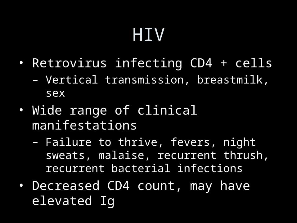
HIV
• Retrovirus infecting CD4 + cells– Vertical transmission, breastmilk, sex
• Wide range of clinical manifestations– Failure to thrive, fevers, night sweats,
malaise, recurrent thrush, recurrent bacterial infections
• Decreased CD4 count, may have elevated Ig
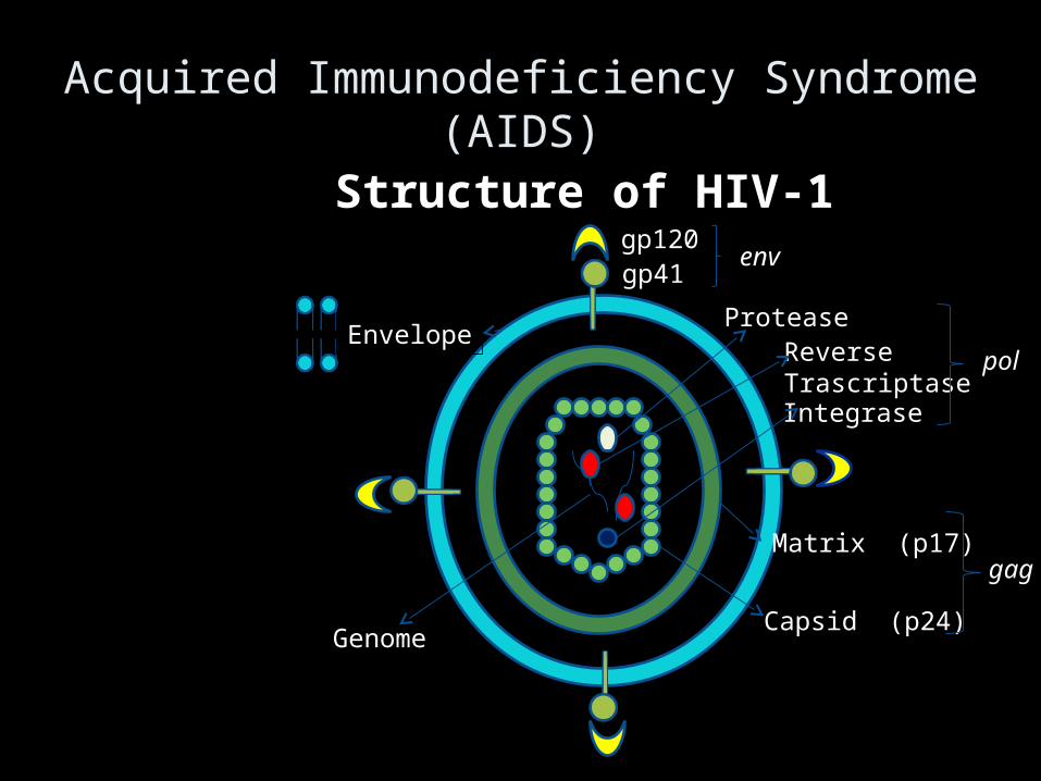
Acquired Immunodeficiency Syndrome (AIDS)
gp120gp41
Envelope
Capsid (p24)Genome
Structure of HIV-1
Matrix (p17)
Reverse Trascriptase
env
gag
Integrase
Protease
pol
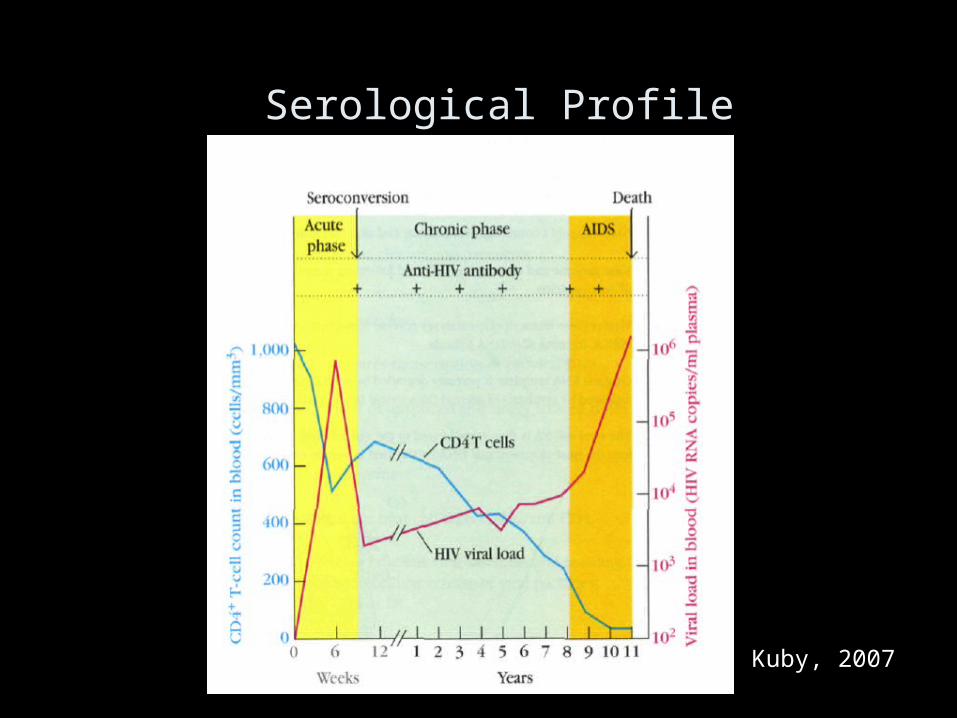
Serological Profile
Kuby, 2007
Immunological Abnormalities
• Infection and destruction of dendritic cells, macrophages and Th cells
• Late decrease in Th cell numbers (200/mm3 blood)

Phagocytic Disorders
Chronic Granulamatous Disease (CGD)
• Defective NADPH oxidase• 75% X-linked recessive, 25% autosomal
recessive• Severe, recurrent staph aureus infections of
lymph nodes, and skin (granulomas, heal slowly), pneumonitis, osteo, hepatosplenomegaly
• Dx: Nitroblue tetrazolium (NBT) test • Treatment: antimicrobial prophylaxis, IFN-
gamma, BMT
Leukocyte adhesion deficiency (LAD)
• Deficient chemotaxis
• Recurrent soft tissue, skin, respiratory infections, impaired wound healing (typically no pus, minimal inflammation)
• Delayed umbilical separation
• Increased WBC count
• Treatment: BMT
Complement System Disorders• Defects of early components (C1-C4) associated
with infections with encapsulated bacteria– Present similarly to humoral immune deficiencies
• Defects of late components (C5-C9) associated with Neisseria infections
• Also associated with autoimmune-like conditions• CH50 functional assay assesses entire
complement cascade– Also may use individual components
• Treatment: treat infectious and autoimmune sequelae
Summary
• Primary immunodeficiencies are inherited• They can affect hematopoietic stem cells,
lymphoid or myeloid cells.• Secondary immunodeficiencies are due to
infections, aging, cancer or chemical exposure• HIV affects immune system by eliminating CD4+ T
cells