imaging with pet and spect - tum filethorsten poethko nuklearmedizinische klinik und poliklinik und...
TRANSCRIPT
Thorsten Poethko
Nuklearmedizinische Klinik und Poliklinikund
Institut für RadiochemieTechnische Universität München
Imaging with PET and SPECT

Basics of Radionuclide Production
Knowledge of production process
Purification
Recovery of target material
Analytical methods (quality control)

Radionuclide Production
(A) Cyclotron (ca. 250 world-wide)
(Ion accelerator)
(B) Reactor (ca. 250 Research reactors world-wide)
(C) Generators

General Aspect
Neutronenübers
Protonenüberschuss
18F15O
13N11C
Reactor-nuclides:neutron rich
Emission of electron ß--decayAZ
Z+1A
Neutron number N = A-Z
Prot
on n
umbe
rP =
Z
AZ-1
AZ Positron emission: ß+-decay
Electron capture: EC-decay
Cyclotron-nuclides:mostly neutron pure

H+
Production of commonly used PET-Nuclidesvia Compact Cyclotron
Ion Source
Negatively ChargedHydrogen Ions
Stripping Foil
Direction of Magnetic Field
Extraction Radius
Dees
Vacuum
Cyclotron Tank
He-Cooling
Vacuum Foil Target Foil
Target
2.0
10.0
20.4
EnergyNuclear-Reaction
T1/2(min)
Nuclide Targetproduct
Mode of
decay[%]
(MeV)- Target
productMode
of decay
[%]11C
13N
15O
11C 109.6
14N(p,α)
16O(p,α)
15N(p,n)
18O(p,n)
13 → 3
16 → 7
16 → 3
10 → 0
β+
β+
β+
β+
(99.8)
(100)
(99.9)
(97)
11CO211CO13NO2
-
13NO3-
18Faq-
18F2
15O2

Selection of Radionuclide
Half-life comparable with kinetics of physiological process ?(too short, too long)
Do the nuclear and physical properties fulfill the special demands ?(e.g. pure β+ - Emitter)
Labeling chemistry compatible with targeting ?(e.g. metalated CNS-ligands)
Patient dose acceptable ?(CNS-ligands: C-11 or F-18)
Nuclide availability ?(Generator, cyclotron, energy range)

PET-Radionuclides
2,32,1423100,3124I
5,44,59561,35120gI
G: 110Sn / 4,1 h3,02,20621,15110mIn
1,83,143314,786Y
5,33,945416,276Br
T½ ,D = 120 d2,21,72711,675Br
G: 68Ge / 271d 2,41,90891,1468Ga
-4,15579,566Ga
0,70,651812,764Cu
1,51,22613,461Cu
T½ ,D = 2,6 y1,61,507617,555Co
0,70,6396,91,818F
CommentsIntrinsic spacial
resolution loss[mm]
Maximumpositron energy[MeV]Eβ+
max
Positron percentage branching
%β+
Half-life[h]
Isotope

<β+>= 0,242 MeV
<β+>= 0,735 MeV
<β+>= 1.409 MeV
18F
15O
82Rb
Resolution Limitation due to Positron Energy
10 5 005
10
mm
Compact bone Soft tissue Lung tissue

Positron-Emission
18F9 ß+
511keV
635 keV ß+ e-
18O8 νe+
CoincidenceDetection
[ns]
ANNIHILATIONRADIATION
511keV
18F9 ß+
511keV
635 keV ß+ e-
18O8 νe+
CoincidenceDetection
[ns]
ANNIHILATIONRADIATION
511keV
O-180,200
σ 0.00016
F-18109.7 min
β+ 0.6 no γ
O-180,200
σ 0.00016
F-18109.7 min
β+ 0.6 no γ
F-18109.7 min
β+ 0.6 no γ
β+
β+

Production of F-18
• Mainly produced via 18O(p,n)18F-reaction• Product: 18Faq
- with high specific activity and up to 100% radiochemical yield,compared to [18F]F2 with low specific activityand max. 50% radiochemical yield
Nuclear reaction 18O(p,n)18F 16O(3He,n)18F 20Ne(d,α)18F 18O(p,n)18F
Target H218O H2O Ne (0.1-0.2% F2,
18 bar)18O2 (20 bar)
Energy range of bombarding particle
[MeV]16 → 0 36 → 0 11.2 → 0 10 → 0
Chemical form [18F]Faq- [18F]Faq
- [18F]F2 [18F]F2
Thick target yield [MBq µA-1 h-1) 2.200 250 350-450 ~350
Specific activity [TBq mmol-1] 40×103 40×103 0.04–0.4 0.04-2
(2. + 0.1% F2)

18F-Fluoride Chemistry
18F- [18F]F2
18F-19F
18F-
δ−R
[18F- + 19F+]
δ+Rδ−
R
R-18F R-18F R-19F
[19F- + 18F+]
(+ F2)
Nucleophilic Substitution Electrophilic Substitution
z.B. [18F]Fluor-Choline z.B. [18F]Dopa
Non-dilutedwith unlabeled
tracer=
„No-carrier-added“(n.c.a)
dilutedwith unlabeled
fluorine=
„carrier-added“(c.a)
Only for targets with high capacity(e.g. enzymsubstrates);
not suitable saturableprocesses (e.g.
peptide-receptor-ligands)

Production Scheme: Radiopharmaceutical Production
Application
Automatic production
Quality control
Cyclotron und on-line synthesis

Automatic Synthesis
11C-Methionine Production
1. Homocysteine suspension on Al2O3/KF in EtOH2. Bubbling of 11CH3I in He-Flow (2min)3. Filtration, dilution (isot. NaCl), sterile filtration

3350β+ (96%), EC (4%)1.3 min82Rb
EC (100%)25 d82Sr
2930β+ (98%), EC(2%)9.7 min62Cu
660β+ (93%), EC (7%)9.2 h62Zn
1900β+ (90%), EC (10%)68 min68Ga
EC (100%)271 d68Ge
Eβ+[keV]Mode of decayHalf-lifeIsotope
Generators for Positron Emission

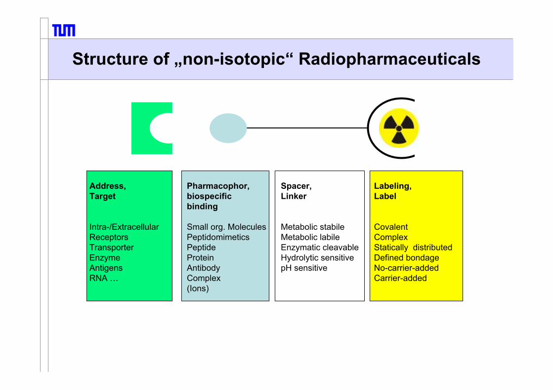
Structure of „non-isotopic“ Radiopharmaceuticals
Pharmacophor,biospecificbinding
Small org. MoleculesPeptidomimeticsPeptideProteinAntibodyComplex(Ions)
Address,Target
Intra-/ExtracellularReceptorsTransporterEnzymeAntigensRNA …
Spacer,Linker
Metabolic stabileMetabolic labileEnzymatic cleavableHydrolytic sensitivepH sensitive
Labeling,Label
CovalentComplexStatically distributedDefined bondageNo-carrier-addedCarrier-added

Peptide, Protein and Macromolecule Labelingwith Radiometals
In, Ga, Cu …Lu, Y, Bi, Ac …Fe, Gd, …..

Radionuclide Production for SPECTSPECT = Single-Photon-Emission-Computer Tomography
All these nuclides are commercially produced and sold
as radionuclide,
generators
or readily-prepared radiopharmaceuticals
Most important SPECT-Isotope is Technetium (99mTc) !

SPECT - Isotopes
Radio T1/2 Mode of Main- Productions datanuclide decay γ-Lines Nuclear Energy Yield
reaction range [MBq/µAh][MeV]
67Ga 3.26 d EC (100) 93 (27%) 68Zn(p,2n) 26→18 18599Mo 2.75 d b- (100) 181 (6%) 235U(n,f)↓ 740 (12%) 98Mo(n,g) σth= 0.14b
99mTc 6.0 h IT (100) 141 (87%)111In 2.8 d EC (100) 173 (91%) 112Cd(p,2n) 25→18 166
247 (94%)123I 13.2 h EC (100) 159 (83%) 123Te(p,n) 14.5→10 137
124Xe(p,x)123Xe 29→23 414127I(p,5n)123Xe 65→45 777
201Tl 3.06 d EC (100) 68-82 (RL) 210Tl(p,3n)201Pb 28→20
166 (10.2%)

99mTc-Generator (1)1
2
3
4
5
6
1
2
3
4
5
6
(1) Simple HandlingInsert vacuum flask and elute desired volume.
(2) Transport-Security-valvePrevent an elution after production and during transport
(3) Shifted needleReduction of energy rich Mo-radiation
(4) Max. protection against radiationProtection from all side.99Mo-column is covered at least with 52 mm Pb.Supplementary shielding with overall 98 mm Pb.
(5) High concentrated activityTotal 99mTc-activity is less than 5 mL volume.
(6) Ready for useSterile, closed system.
NaCl
Glass woolSilica
99MoO3
on Al2O3
Fritte
99mTcO4-
NaCl
Glass woolSilica
99MoO3
on Al2O3
Fritte
99mTcO4-

Labeling of Tc-Radiopharmaceuticals

Tracer Properties: Influence of the Nuclide
11C 18F 123I 99mTc 68Ga / 111In
WAY 100638 (5-HT1A-receptor ligand) 99mTc-WAY 100638-analog(as 5-HT1A-receptor ligand)
Change in structure
Complexity of radiopharmaceutical development
Predictability of biological integrity

Molecular Imaging by Radiotracer
Targeting biochemical processes in living organismon basis of molecular interaction between tracer und target
by radiolabeled, „molecular probes“, changing their concentration at the target in a specific way
and suitable detection systems for non-invasive, quantitativeand repetitive measurement
with the aim of exploring physiological processes,diagnostics und therapy as well as therapy monitoring

Target Structures in Oncology
FDG
Choline
Antibodies
Peptide Receptors
TK
Receptors Transporters
HSV-TkAcetate
Nucleosides
Nucleosides
O2
Local Concentration
Local Concentration
PeptidesPS
Amine Precursors
Hypoxia
ApoptosisAmino Acids
Transport, Protein Synthesis
Transport, Choline Kinase
Integrin Expression,Neovascularization
Antigen Expression
Transport, Hexokinase
Lipid Synthesis
APUD-System
Neuropep. Receptors Monitoring Gene Expression
Proliferation
InhibitorsGrowth Factor Receptors
Characterization of the tumor biology by molecular imaging

2-[18F]Fluoro-2-Deoxy-Glucose-Synthesis ([18F]FDG)
18F
CH2OAc
OAc
OAcOAc
OO
18F
CH2OH
OH
OHOH
H218O
18O(p,n)18F18Faq
-A.P.E. /K2CO3
[K/(2.2.2)]+18F-
(~1h) PurificationDrying (10 min)
Substitution7 min
Hydrolysis(25 min)
Purification(10 min)
Sterile filtration(∼ 50 min EOB)
1000 mCi
400-500 mCi
∼ 50 min
CH2OAc
OAc
OAcOAc
OOTf

GMP Production of FDG2-[18F]Fluor-2-deoxy-D-Glucose, TU Munich

Scheme of [18F]FDG Uptake
O
18FOH
Hexokinase (II)
GLUT-3,5GLUT-2,4,7
OHOH
G6-Phosphat-Isomerase (II)
X
X
O
HOCH2
OH
OHOH
18F
CH2OPO32-
GLUT-1
FDG-Aufnahme abhängig von:1. GLUT-Expression 2. Hexokinase-Expression
O
18FOH
Hexokinase (II)
GLUT-3,5GLUT-2,4,7
OHOH
G6-Phosphat-Isomerase (II)
X
X
O
HOCH2
OH
OHOH
18F
O
HOCH2
OH
OHOH
18F
CH2OPO32-
GLUT-1
FDG-Aufnahme abhängig von:1. GLUT-Expression 2. Hexokinase-Expression
FDG uptake correlates with:GLUT-ExpressionHexokinase-Activity
OH

[18F]FDG: Therapy control –gastric carcinoma-

Amino Acid Transport
Primary target: Rate measurement of regional protein synthesisBut: During the short time period after injection the accumulation of tracer represent
only the accumulation of amino acid by special transport systems
HO CH2 CH
NH2
COOH
18F
HO CH2 CH
NH2
11COOH
O CH2 CH
NH2
COOH18F CH
NH2
COOHS
H311C
2-Fluoro-L-Tyrosin 2-[18F]Tyr L-Tyrosin 1-[11C]Tyr
O-(2-Fluoroethyl)-L-Tyrosin [18F]FET L-Methionine [11C]MET

[18F]FDG-PET versus [11C]MET-PET -Brain Tumor-
MRT (T2) FDG-PET [11C]Methionine-PET

O-(2-[18F]Fluoroethyl)-L-Tyrosin: [18F]FET (2)
Squamous cellcarcinoma
tumorinflammation tumor inflammation
Osteolyse
[18F]FET[18F]FDG
FDG also accumulates in inflammation,amino acids and especially FET does not accumulate in inflammation.

11C-Acetate: Prostate cells undergo metabolic transformation from citrate producing normalcells to citrate oxidizing malignant cells(Costello LC et al. Urology 1997)
18F/11C-Cholines: malignant transformation is associated withinduction of choline kinase activity(e.g. Kotzerke et al. and refs herein; J Nucl Med. 2002)
(18F-Fluoide): (for bone metastasis)
Imaging of Metastatic Prostate Cancer

Biochemical Model: Choline
Choline
Choline kinase
Phosphorylcholine
Choline dehydrogenase
Betain-aldehyde
Betain
Betainaldehyd-Dehydrogenase
Acetylcholine
NC H3
H3CC H3
CH2-CH2-O CO
CH3+
NC H3
H3CC H3
CH2-CH2-O PO
OHOH+
NC H3
H3CC H3
C H2- C
O
H+
NH3CC H3
CH2-CH2-OH+C H3
NC H3
H3CC H3
C H2- C
O
O-
+
N+
OH
18FN+
OH
11CH3
FEC
N+
OH
18FCholine FMC[18F]FMC
„Fluoromethylcholine“[18F]FEC
„Fluoroethylcholine“[11C]CHO

11C-Choline-PET
Bladder cancer metastasis
Prostate carcinoma
PET/CT PET
H311C-N-CH2-CH2-OH
CH3
CH3 T1/2(11C)=20 min
H3C-N-CH2-CH2-OH
CH218F
CH3 T1/2(11C)= 109.7 min
18F-Choline-PET
Tracer accumulation with:
1. Cellular accumulation bycholine transporter;(Signal in the first minutesafter injection)
2. and followed byphosphorylationby choline kinase.(Signal by late imaging)

thymidinekinase 1
Di-phosphokinase tri-phosphokinase
TK1 activity correlates with cell proliferation
DNAnucleoside transporter
18F
OHO
H3C
N
O
NH
O
18F
OOPO3
2-
H3C
N
O
NH
O
X
X X
„Fluorothymidine“[18F]FLT
3´-[18F]Fluoro-3´-Deoxy-Thymidine: [18F]FLT
OH
OHO
H3C
N
O
NH
O
One Nucleoside

Metabolic Scheme for [11C]Thymidine and FLT
DNA DNA
11C-labeled metabolites(ca. 80 %)
11CO2 (ca. 50 %)
no significant degradation
DNA
OH
OHO
H311C
N
O
NH
O
OH
OHO
H3C
N
O
NH
O
18F
OHO
H3C
N
O
NH
O*
FLT-PO4TK1 activity:
TdR / FLT =0.3

Visualization of Cell Proliferation with 18F-thymidine (FLT) in Esophageal Cancer
tumor
bone marrow
OH
OHO
H3C
N
O
NH
O
18F
OHO
H3C
N
O
NH
O
thymidine [18F]FLT
tumor
bone marrow

Critical O2 Levels
Genome changesClonal heterogeneityClonal selection
Proteome changes via gene expressionPosttranscriptional and posttranslational changesProtein degradation
10-1 100 101
AggressivenessTumor Progression
pO2 [mm Hg]
Vaupel P. et al.
Distribution of medium pO2 levels for prostatic tumor tissue versus muscle
Nitroimidazoles (pos. <15mm Hg)
NormoxiaHypoxia

The Mechanism of Non-Invasive Detection of Tumor Hypoxia
tumor cell
NO2
N NCH2
18F
OH
18F-MISO -
reductasee-
18F-MISO
oxidationO2- O2
covalent linkage tointracellular proteins
TRAPPING
tracer accumulation should correlate
with partial oxygen pressure (ptO2)
deliveryPerfusionhypoperfused
normally orhyperperfused
Blood flow
Experimental Determination /
Validationmicroelectrode, correlation
with radioresistance
othertissues
clearance
blood clearance
excretion
t1/2 of radionuclide
[18F]FMISO

Serial, transaxiale PET images -nude mice- with A431 tumors.
Evaluation of [18F]FAZA
Exp.1 Exp.1
Exp.2 Exp.2
8 h 100% O2 21% O2
Exp.1 T/Bk 3.7 / 1 4.7 / 1Exp.2 T/Bk 5.4 / 1 11.3 / 1
Synthesis of [18F]FAZA:Radiochemical Yield: 20 ± 4%, 50 min
(Reischl et al., 2005)

cell
Peptide receptors
Intracellular trapping (lysosomes)
Peptide receptor imaging: Diagnosis and therapy
monitoring; reporter system
sst GRP CCKB NTS1 NK1 α−MSH
+
externalization
internalization
degradation
Peptide Receptor Imaging: PRI

Somatostatin-R neuroendocrine tumors, small cell lung cancer,medullary thyroid carcinoma, lymphoma (NHL)
Integrins melanoma, breast tumor, osteosarcoma, glioblastoma
VIP-R adenocarcinomas, small cell lung cancer, neuroendocrine tumors, lymphoma
CCK-B-R medullary thyroid carcinoma, small cell lung cancer, stromal ovarian cancer, astrocytoma
Substance P-R medullary thyroid cancer, small cell lung cancer, breast tumors
Bombesin/ GRP-R colon cancer, small cell lung cancer, glioblastoma
Neurotensin-R pancreatic cancer, prostate cancer,small cell lung cancer
Targets for Radiolabeled Peptides in Human Tumor Tissue

Influence of the Carbohydrateon the Biodistribution of Octreotides/-tates
(Mice, AR42J, n=3-5, 60 min p.i.)
blood liver kidney tumor t/k t/l t/b
0
10
20
30
40
50
60
70
0
10
20
30
40
50
60
70 Mtr-TOC Malt-TOC TOC Gluc-TOC TOCA Glucoron-TOCA Mtr-TOCA Malt-TOCA Gluc-TOCA Gal-merc-TOCA Gluc-merc-TOCA
blood liver kidney tumor t/kidney t/liver t/blood
activity uptake %ID/g tissue uptake ratiosMtr-TOC +2Malt-TOC +2 TOC +2Gluc-TOC +2TOCA +2/-1 +1Glucoron-TOCA +2/-2 0Mtr-TOCA +2/-1 +1Malt-TOCA +2/-1 +1Gluc-TOCA +2/-1 +1Gal-mercapto-TOCA +1/-1 0Gluc-mercapto-TOCA +1/-1 0
Schottelius, Wester et al. Clin Cancer Res. (2004)

Chemical Structure of Gluc-FP[18F]-TOCA
Gluc-Lys([18F]FP)-TOCA
O
18F
NHO
H3C
OH
HOOHO
HO
NH
O
NH
NNH
NH
O
O
SS
H ONH
H
O
O NH
NH
NH2
N
OH
NHO
OH
OH
O
HO
[18F]FP = 2-[18F]Fluoropropionic acidTOCA = Tyr3-octreotate, -DPhe-Cys-Tyr-DTrp-Lys-Thr-Cys-Thr-OH

Gluc-FP[18F]-TOCA
Male patient, 50 yrs, carcinoid with multiple metastases

Imaging of Neoangiogenesis
Inhibitors of Angiogenesis
VEGF antagonistsSolubleVEGFR
VEGFR tryosinekinase inhibitors
anti-MMPs
Metastasis
Direct ECAntagonists
AngiostatinEndostatin…
anti-MMPs
anti-αvβ3
ECM degradationadhesion antagonists
Tumor
α
Inhibitors of Angiogenesis
VEGF antagonistsSolubleVEGFR
VEGFR tryosinekinase inhibitors
anti-MMPs
Metastasis
Direct ECAntagonists
AngiostatinEndostatin…
anti-MMPs
anti-αvβ3
ECM degradationadhesion antagonists
Tumor
αO
O
HOOH
NH
O
18F
OHNCO NH
NH
LysD-Phe
Asp
Gly
Arg
HO
OOO
HOOH
NH
O
18F
OHNCO NH
NH
LysD-Phe
Asp
Gly
Arg
HO
R
Arg-Gly-Asp (RGD)-containing pentapeptides
with high affinity and selectivity to the αvβ3-integrin

tumor cell
endothelial cell
extracellular matrix
Cytocines
Inhibition ofAngiogenesis
Inhibition ofMetastasis
Fluorinated “Galacto-RGD”-Peptide
O
HOHO OH
NH
O
18F
NH
LysD-Phe
O
HN
AspGly
ArgCO
CONH
O
HOHO OH
NH
O
18F
NH
LysD-Phe
O
HN
AspGly
ArgCO
CONH
Carbohydrated RGD-Peptide : [18F]Galacto-RGD

*I
Lys
OHNH
O
OHHO
HO
AcHN O
Sugar Amino Acid
D-Tyr
HN
AspGly
ArgCO
CONH
Carbohydrated RGD-Peptides with Improved Pharmacokinetics
c(Arg-Gly-Asp-D-(I)Tyr -Val)
c(Arg-Gly-Asp-D-Phe-Val)
O
HOHO OH
NH
O
18F
NH
LysD-Phe
O
HN
AspGly
ArgCO
CONH
Fluorinated “Galacto-RGD”-Peptide

kidney
blood
tumor
liver
%iD/g
0
1
2
0 60 120 180 240
%iD/g
time p.i. [min]
0
1
2
3
0 60 120 180 240
%iD/g
time p.i. [min]
0
5
10
15
0 60 120 180 240time p.i. [min]
0
10
20
0 60 120 180 240
%iD/g
time p.i. [min]
c(Arg-Gly-Asp-D-(I)Tyr -Val): Log P = -1.89I-Gluco-RGD: Log P = -2.45
Comparison of the Pharmacokinetics in Selected Tissues
BALB/c mice,osteosarcoma model;comparable data obtained with M21melanoma bearing mice

control(unspecific MAb)
nude mouse bearing a human squamous cell carcinoma at the right shoulder
anti human αvβ3MAb LM609
no expression of αvβ3 on cells
anti murine β3MAb 2C9.G2
expression of murineβ3 on vasculature
18 mg/kg c(RGDfV), 10 min prior
n.c.a. experiment
1.5h p.i
Non-invasive Monitoring of αvβ3 Expression on the Tumor Vasculature

blood vessel
CBA
sagittal section, 170 min p.i. , PET/CT image fusion; Immunohistochemistry, circular tracer uptake, uptake corresponds with MAb LM609, staining of max. SUV=10 the tumor-wall blood vessels
soft tissue sarcoma
PET PET/CT IHC
blood vessel
lymph node
FED
malignant melanoma
axial section, 140 min p,i, PET/CT image fusion Immunohistochemistry, focal uptake in the lymph node MAB LM609, staining of
tumor cells and blood vessels
Determination of αvβ3-Expression in vivo

Multiple-Ligand-Tumortargeting (MLT)
tumor-cell
radionuclide*
strong cooperative binding
substance P-R
gastrin/CCK-B-R
αvβ3
sst
VIPR
BB-R
αvβ3-ligands

Structure of the Extracellular Domain of αvβ3
J.-P. Xiong, et. al, Science 2001.
av-subunitβ3-subunit
Arg
GlyAsp
Val
DPhe

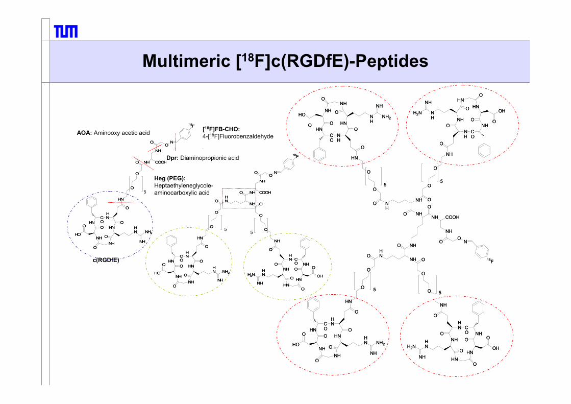
Multimeric [18F]c(RGDfE)-Peptides
NH
CO
NHN
HN
NH
O
HO
OO
O
O
H
HN NH2
NH
O
HN
O
O
O
5
O NH COOH
NH
OO
N
NH COOH
NH
OO
N
18F(24)
NH
CO
NHN
HN
NH
O
HO
OO
O
O
H
HN NH2
NH
O
HN
O
O
O
5
O NH COOH
NH
OO
N
NH COOH
NH
OO
N
18F(24)
NH
CO
NHN
HN
NH
O
HO
OO
O
O
H
HN NH2
NH
O
NH
CO
NHN
HN
NH
O
HO
OO
O
O
H
HN NH2
NH
O
HN
O
O
O
5
HN
CO
NNH
NH
HN
O
OH
OO
O
O
H
HNH2N
NH
O
NH
O
O
O
5
HN
NH
O NH COOH
NH
OO
N
18F
(25)
NH
CO
NHN
HN
NH
O
HO
OO
O
O
H
HN NH2
NH
O
NH
CO
NHN
HN
NH
O
HO
OO
O
O
H
HN NH2
NH
O
HN
O
O
O
5
HN
CO
NNH
NH
HN
O
OH
OO
O
O
H
HNH2N
NH
O
NH
O
O
O
5
HN
NH
O NH COOH
NH
OO
N
18F
(25)
c(RGDfE)
Dpr: Diaminopropionic acid
[18F]FB-CHO:4-[18F]Fluorobenzaldehyde
Heg (PEG):Heptaethyleneglycole-aminocarboxylic acid5
55
AOA: Aminooxy acetic acid
NH
CO
NHN
HN
NH
O
HO
OO
O
O
H
HN NH2
NH
O
HN
O
O
O
5
HN
CO
NNH
NH
HN
O
OH
OO
O
O
H
HNH2N
NH
O
NH
O
O
O
5
HN
NH
O NH
NH
CO
NHN
HN
NH
O
HO
OO
O
O
H
NH
NH2
NH
O
HN
O
O
O
5
HN
CO
NNH
NH
HN
O
OH
OO
O
O
H
NH
H2N
NH
O
NH
O
O
O
5
NH
NH
ONH COOH
NH
OO
N
18F
NHO
NH
CO
NHN
HN
NH
O
HO
OO
O
O
H
HN NH2
NH
O
HN
O
O
O
5
HN
CO
NNH
NH
HN
O
OH
OO
O
O
H
HNH2N
NH
O
NH
O
O
O
5
HN
NH
O NH
NH
CO
NHN
HN
NH
O
HO
OO
O
O
H
NH
NH2
NH
O
HN
O
O
O
5
HN
CO
NNH
NH
HN
O
OH
OO
O
O
H
NH
H2N
NH
O
NH
O
O
O
5
NH
NH
ONH COOH
NH
OO
N
18F
NHO

[K⊂2.2.2]+[18F]-
CF3SO3-
N
HO
+
Labeling Purification
90°C, 5min
60-75% RCA(35 min)
Peptide ON
18F
Route suitable for a variety of radiolabeled aldehydes and ketones
Cartridges(1. SCX, 2. RP-18)
HPLC (+RP-18
Cartridge)
Peptide-O-NH2
Conjugation
18F
HO
90 % - 99 % Radiochem.
Purity
18F-Labeling via Oxime Formation
[18F]FB-CHO
> 99 % Radiochem.
Purity

Binding Affinities of Multimeric RGD-Peptide Constructs
IC50[nM]
Heg
as
Heg
as
Heg
as
Heg
as
Heg
as
Heg
as
Heg
as
I-BAI-BA
I-BA
RGD RGDRGD RGDRGDRGDRGD
0.23.020
5.0
0
10
20
18F-Heg-Monomer18F-Heg-Dimer
18F-Heg-Tetramer18F-Galacto-RGD

Tumor to Organ Ratios
Nude Mice, M21-(αvβ3+)-Melanoma, 2 h p.i. (n=3-5)
0
10
20
30
Tum
or /
Org
an R
atio
s
serum lung kidney spleen muscle M21-Lblood heart liver adrenals intestine femur
RGDfluorine

PET Imaging of [18F]RGD-Mono-, Di- and Tetramers
ECAT Exact HR+, 90 min p.i.
M21L(low) αvβ3
Monomer MRIDimer
Tracer only Tracer only
Tetramer
Blockade Tracer only+ 18 mg/kg c(RGDfV)
0
40kBq
M21(high)αvβ3

Affinities of Multimeric RGD-Peptides with/without RAD-Sequences
0
5
10
15
20
25
30Ahx
10 2021 2521 25
iodine
0.3 0.41(I: 0.35) (I: 0.2)
fluorine
0.3 0.41(I: 0.35) (I: 0.2)
fluorine
Hegas
IC50 [nM]αvβ3
c(RGDfE)c(RADfE)
Thumshirn et al. Eur Chem. 2003

PET Imaging of Tetrameric [18F]RGDwith and without „Knockout“ Sequences
(18F)
(PEG)
0
40kBq
90 min p.i.
ECAT Exact HR+, 90 min p.i.
0,7%ID/g (2h)Biodistibution: 2.0%ID/g (2h)
Poethko et al. Radiochimica Acta 2004

Target
+ Tracer
+ Technology ____________________________
= Suitable Radiopharmaceuticaland Contrast Agent
Radiopharmaceutical Chemistry and Development of Radiopharmaceuticals
Triple TTriple T

Thorsten Poethko
Nuklearmedizinische Klinik und Poliklinikund
Institut für RadiochemieTechnische Universität München
Imaging with PET and SPECT

Basics of Radionuclide Production
Knowledge of production process
Purification
Recovery of target material
Analytical methods (quality control)

Radionuclide Production
(A) Cyclotron (ca. 250 world-wide)
(Ion accelerator)
(B) Reactor (ca. 250 Research reactors world-wide)
(C) Generators

General Aspect
Neutronenübers
Protonenüberschuss
18F15O
13N11C
Reactor-nuclides:neutron rich
Emission of electron ß--decayAZ
Z+1A
Neutron number N = A-Z
Prot
on n
umbe
rP =
Z
AZ-1
AZ Positron emission: ß+-decay
Electron capture: EC-decay
Cyclotron-nuclides:mostly neutron pure

H+
Production of commonly used PET-Nuclidesvia Compact Cyclotron
Ion Source
Negatively ChargedHydrogen Ions
Stripping Foil
Direction of Magnetic Field
Extraction Radius
Dees
Vacuum
Cyclotron Tank
He-Cooling
Vacuum Foil Target Foil
Target
2.0
10.0
20.4
EnergyNuclear-Reaction
T1/2(min)
Nuclide Targetproduct
Mode of
decay[%]
(MeV)- Target
productMode
of decay
[%]11C
13N
15O
11C 109.6
14N(p,α)
16O(p,α)
15N(p,n)
18O(p,n)
13 → 3
16 → 7
16 → 3
10 → 0
β+
β+
β+
β+
(99.8)
(100)
(99.9)
(97)
11CO211CO13NO2
-
13NO3-
18Faq-
18F2
15O2

Selection of Radionuclide
Half-life comparable with kinetics of physiological process ?(too short, too long)
Do the nuclear and physical properties fulfill the special demands ?(e.g. pure β+ - Emitter)
Labeling chemistry compatible with targeting ?(e.g. metalated CNS-ligands)
Patient dose acceptable ?(CNS-ligands: C-11 or F-18)
Nuclide availability ?(Generator, cyclotron, energy range)

PET-Radionuclides
2,32,1423100,3124I
5,44,59561,35120gI
G: 110Sn / 4,1 h3,02,20621,15110mIn
1,83,143314,786Y
5,33,945416,276Br
T½ ,D = 120 d2,21,72711,675Br
G: 68Ge / 271d 2,41,90891,1468Ga
-4,15579,566Ga
0,70,651812,764Cu
1,51,22613,461Cu
T½ ,D = 2,6 y1,61,507617,555Co
0,70,6396,91,818F
CommentsIntrinsic spacial
resolution loss[mm]
Maximumpositron energy[MeV]Eβ+
max
Positron percentage branching
%β+
Half-life[h]
Isotope

<β+>= 0,242 MeV
<β+>= 0,735 MeV
<β+>= 1.409 MeV
18F
15O
82Rb
Resolution Limitation due to Positron Energy
10 5 005
10
mm
Compact bone Soft tissue Lung tissue

Positron-Emission
18F9 ß+
511keV
635 keV ß+ e-
18O8 νe+
CoincidenceDetection
[ns]
ANNIHILATIONRADIATION
511keV
18F9 ß+
511keV
635 keV ß+ e-
18O8 νe+
CoincidenceDetection
[ns]
ANNIHILATIONRADIATION
511keV
O-180,200
σ 0.00016
F-18109.7 min
β+ 0.6 no γ
O-180,200
σ 0.00016
F-18109.7 min
β+ 0.6 no γ
F-18109.7 min
β+ 0.6 no γ
β+
β+

Production of F-18
• Mainly produced via 18O(p,n)18F-reaction• Product: 18Faq
- with high specific activity and up to 100% radiochemical yield,compared to [18F]F2 with low specific activityand max. 50% radiochemical yield
Nuclear reaction 18O(p,n)18F 16O(3He,n)18F 20Ne(d,α)18F 18O(p,n)18F
Target H218O H2O Ne (0.1-0.2% F2,
18 bar)18O2 (20 bar)
Energy range of bombarding particle
[MeV]16 → 0 36 → 0 11.2 → 0 10 → 0
Chemical form [18F]Faq- [18F]Faq
- [18F]F2 [18F]F2
Thick target yield [MBq µA-1 h-1) 2.200 250 350-450 ~350
Specific activity [TBq mmol-1] 40×103 40×103 0.04–0.4 0.04-2
(2. + 0.1% F2)

18F-Fluoride Chemistry
18F- [18F]F2
18F-19F
18F-
δ−R
[18F- + 19F+]
δ+Rδ−
R
R-18F R-18F R-19F
[19F- + 18F+]
(+ F2)
Nucleophilic Substitution Electrophilic Substitution
z.B. [18F]Fluor-Choline z.B. [18F]Dopa
Non-dilutedwith unlabeled
tracer=
„No-carrier-added“(n.c.a)
dilutedwith unlabeled
fluorine=
„carrier-added“(c.a)
Only for targets with high capacity(e.g. enzymsubstrates);
not suitable saturableprocesses (e.g.
peptide-receptor-ligands)

Production Scheme: Radiopharmaceutical Production
Application
Automatic production
Quality control
Cyclotron und on-line synthesis

Automatic Synthesis
11C-Methionine Production
1. Homocysteine suspension on Al2O3/KF in EtOH2. Bubbling of 11CH3I in He-Flow (2min)3. Filtration, dilution (isot. NaCl), sterile filtration

3350β+ (96%), EC (4%)1.3 min82Rb
EC (100%)25 d82Sr
2930β+ (98%), EC(2%)9.7 min62Cu
660β+ (93%), EC (7%)9.2 h62Zn
1900β+ (90%), EC (10%)68 min68Ga
EC (100%)271 d68Ge
Eβ+[keV]Mode of decayHalf-lifeIsotope
Generators for Positron Emission
Structure of „non-isotopic“ Radiopharmaceuticals
Pharmacophor,biospecificbinding
Small org. MoleculesPeptidomimeticsPeptideProteinAntibodyComplex(Ions)
Address,Target
Intra-/ExtracellularReceptorsTransporterEnzymeAntigensRNA …
Spacer,Linker
Metabolic stabileMetabolic labileEnzymatic cleavableHydrolytic sensitivepH sensitive
Labeling,Label
CovalentComplexStatically distributedDefined bondageNo-carrier-addedCarrier-added

Peptide, Protein and Macromolecule Labelingwith Radiometals
In, Ga, Cu …Lu, Y, Bi, Ac …Fe, Gd, …..

Radionuclide Production for SPECTSPECT = Single-Photon-Emission-Computer Tomography
All these nuclides are commercially produced and sold
as radionuclide,
generators
or readily-prepared radiopharmaceuticals
Most important SPECT-Isotope is Technetium (99mTc) !

SPECT - Isotopes
Radio T1/2 Mode of Main- Productions datanuclide decay γ-Lines Nuclear Energy Yield
reaction range [MBq/µAh][MeV]
67Ga 3.26 d EC (100) 93 (27%) 68Zn(p,2n) 26→18 18599Mo 2.75 d b- (100) 181 (6%) 235U(n,f)↓ 740 (12%) 98Mo(n,g) σth= 0.14b
99mTc 6.0 h IT (100) 141 (87%)111In 2.8 d EC (100) 173 (91%) 112Cd(p,2n) 25→18 166
247 (94%)123I 13.2 h EC (100) 159 (83%) 123Te(p,n) 14.5→10 137
124Xe(p,x)123Xe 29→23 414127I(p,5n)123Xe 65→45 777
201Tl 3.06 d EC (100) 68-82 (RL) 210Tl(p,3n)201Pb 28→20
166 (10.2%)

99mTc-Generator (1)1
2
3
4
5
6
1
2
3
4
5
6
(1) Simple HandlingInsert vacuum flask and elute desired volume.
(2) Transport-Security-valvePrevent an elution after production and during transport
(3) Shifted needleReduction of energy rich Mo-radiation
(4) Max. protection against radiationProtection from all side.99Mo-column is covered at least with 52 mm Pb.Supplementary shielding with overall 98 mm Pb.
(5) High concentrated activityTotal 99mTc-activity is less than 5 mL volume.
(6) Ready for useSterile, closed system.
NaCl
Glass woolSilica
99MoO3
on Al2O3
Fritte
99mTcO4-
NaCl
Glass woolSilica
99MoO3
on Al2O3
Fritte
99mTcO4-

Labeling of Tc-Radiopharmaceuticals

Tracer Properties: Influence of the Nuclide
11C 18F 123I 99mTc 68Ga / 111In
WAY 100638 (5-HT1A-receptor ligand) 99mTc-WAY 100638-analog(as 5-HT1A-receptor ligand)
Change in structure
Complexity of radiopharmaceutical development
Predictability of biological integrity

Molecular Imaging by Radiotracer
Targeting biochemical processes in living organismon basis of molecular interaction between tracer und target
by radiolabeled, „molecular probes“, changing their concentration at the target in a specific way
and suitable detection systems for non-invasive, quantitativeand repetitive measurement
with the aim of exploring physiological processes,diagnostics und therapy as well as therapy monitoring

Target Structures in Oncology
FDG
Choline
Antibodies
Peptide Receptors
TK
Receptors Transporters
HSV-TkAcetate
Nucleosides
Nucleosides
O2
Local Concentration
Local Concentration
PeptidesPS
Amine Precursors
Hypoxia
ApoptosisAmino Acids
Transport, Protein Synthesis
Transport, Choline Kinase
Integrin Expression,Neovascularization
Antigen Expression
Transport, Hexokinase
Lipid Synthesis
APUD-System
Neuropep. Receptors Monitoring Gene Expression
Proliferation
InhibitorsGrowth Factor Receptors
Characterization of the tumor biology by molecular imaging

2-[18F]Fluoro-2-Deoxy-Glucose-Synthesis ([18F]FDG)
18F
CH2OAc
OAc
OAcOAc
OO
18F
CH2OH
OH
OHOH
H218O
18O(p,n)18F18Faq
-A.P.E. /K2CO3
[K/(2.2.2)]+18F-
(~1h) PurificationDrying (10 min)
Substitution7 min
Hydrolysis(25 min)
Purification(10 min)
Sterile filtration(∼ 50 min EOB)
1000 mCi
400-500 mCi
∼ 50 min
CH2OAc
OAc
OAcOAc
OOTf

GMP Production of FDG2-[18F]Fluor-2-deoxy-D-Glucose, TU Munich

Scheme of [18F]FDG Uptake
O
18FOH
Hexokinase (II)
GLUT-3,5GLUT-2,4,7
OHOH
G6-Phosphat-Isomerase (II)
X
X
O
HOCH2
OH
OHOH
18F
CH2OPO32-
GLUT-1
FDG-Aufnahme abhängig von:1. GLUT-Expression 2. Hexokinase-Expression
O
18FOH
Hexokinase (II)
GLUT-3,5GLUT-2,4,7
OHOH
G6-Phosphat-Isomerase (II)
X
X
O
HOCH2
OH
OHOH
18F
O
HOCH2
OH
OHOH
18F
CH2OPO32-
GLUT-1
FDG-Aufnahme abhängig von:1. GLUT-Expression 2. Hexokinase-Expression
FDG uptake correlates with:GLUT-ExpressionHexokinase-Activity
OH

[18F]FDG: Therapy control –gastric carcinoma-

Amino Acid Transport
Primary target: Rate measurement of regional protein synthesisBut: During the short time period after injection the accumulation of tracer represent
only the accumulation of amino acid by special transport systems
HO CH2 CH
NH2
COOH
18F
HO CH2 CH
NH2
11COOH
O CH2 CH
NH2
COOH18F CH
NH2
COOHS
H311C
2-Fluoro-L-Tyrosin 2-[18F]Tyr L-Tyrosin 1-[11C]Tyr
O-(2-Fluoroethyl)-L-Tyrosin [18F]FET L-Methionine [11C]MET

[18F]FDG-PET versus [11C]MET-PET -Brain Tumor-
MRT (T2) FDG-PET [11C]Methionine-PET

O-(2-[18F]Fluoroethyl)-L-Tyrosin: [18F]FET (2)
Squamous cellcarcinoma
tumorinflammation tumor inflammation
Osteolyse
[18F]FET[18F]FDG
FDG also accumulates in inflammation,amino acids and especially FET does not accumulate in inflammation.

11C-Acetate: Prostate cells undergo metabolic transformation from citrate producing normalcells to citrate oxidizing malignant cells(Costello LC et al. Urology 1997)
18F/11C-Cholines: malignant transformation is associated withinduction of choline kinase activity(e.g. Kotzerke et al. and refs herein; J Nucl Med. 2002)
(18F-Fluoide): (for bone metastasis)
Imaging of Metastatic Prostate Cancer

Biochemical Model: Choline
Choline
Choline kinase
Phosphorylcholine
Choline dehydrogenase
Betain-aldehyde
Betain
Betainaldehyd-Dehydrogenase
Acetylcholine
NC H3
H3CC H3
CH2-CH2-O CO
CH3+
NC H3
H3CC H3
CH2-CH2-O PO
OHOH+
NC H3
H3CC H3
C H2- C
O
H+
NH3CC H3
CH2-CH2-OH+C H3
NC H3
H3CC H3
C H2- C
O
O-
+
N+
OH
18FN+
OH
11CH3
FEC
N+
OH
18FCholine FMC[18F]FMC
„Fluoromethylcholine“[18F]FEC
„Fluoroethylcholine“[11C]CHO

11C-Choline-PET
Bladder cancer metastasis
Prostate carcinoma
PET/CT PET
H311C-N-CH2-CH2-OH
CH3
CH3 T1/2(11C)=20 min
H3C-N-CH2-CH2-OH
CH218F
CH3 T1/2(11C)= 109.7 min
18F-Choline-PET
Tracer accumulation with:
1. Cellular accumulation bycholine transporter;(Signal in the first minutesafter injection)
2. and followed byphosphorylationby choline kinase.(Signal by late imaging)

thymidinekinase 1
Di-phosphokinase tri-phosphokinase
TK1 activity correlates with cell proliferation
DNAnucleoside transporter
18F
OHO
H3C
N
O
NH
O
18F
OOPO3
2-
H3C
N
O
NH
O
X
X X
„Fluorothymidine“[18F]FLT
3´-[18F]Fluoro-3´-Deoxy-Thymidine: [18F]FLT
OH
OHO
H3C
N
O
NH
O
One Nucleoside

Metabolic Scheme for [11C]Thymidine and FLT
DNA DNA
11C-labeled metabolites(ca. 80 %)
11CO2 (ca. 50 %)
no significant degradation
DNA
OH
OHO
H311C
N
O
NH
O
OH
OHO
H3C
N
O
NH
O
18F
OHO
H3C
N
O
NH
O*
FLT-PO4TK1 activity:
TdR / FLT =0.3

Visualization of Cell Proliferation with 18F-thymidine (FLT) in Esophageal Cancer
tumor
bone marrow
OH
OHO
H3C
N
O
NH
O
18F
OHO
H3C
N
O
NH
O
thymidine [18F]FLT
tumor
bone marrow

Critical O2 Levels
Genome changesClonal heterogeneityClonal selection
Proteome changes via gene expressionPosttranscriptional and posttranslational changesProtein degradation
10-1 100 101
AggressivenessTumor Progression
pO2 [mm Hg]
Vaupel P. et al.
Distribution of medium pO2 levels for prostatic tumor tissue versus muscle
Nitroimidazoles (pos. <15mm Hg)
NormoxiaHypoxia

The Mechanism of Non-Invasive Detection of Tumor Hypoxia
tumor cell
NO2
N NCH2
18F
OH
18F-MISO -
reductasee-
18F-MISO
oxidationO2- O2
covalent linkage tointracellular proteins
TRAPPING
tracer accumulation should correlate
with partial oxygen pressure (ptO2)
deliveryPerfusionhypoperfused
normally orhyperperfused
Blood flow
Experimental Determination /
Validationmicroelectrode, correlation
with radioresistance
othertissues
clearance
blood clearance
excretion
t1/2 of radionuclide
[18F]FMISO

Serial, transaxiale PET images -nude mice- with A431 tumors.
Evaluation of [18F]FAZA
Exp.1 Exp.1
Exp.2 Exp.2
8 h 100% O2 21% O2
Exp.1 T/Bk 3.7 / 1 4.7 / 1Exp.2 T/Bk 5.4 / 1 11.3 / 1
Synthesis of [18F]FAZA:Radiochemical Yield: 20 ± 4%, 50 min
(Reischl et al., 2005)

cell
Peptide receptors
Intracellular trapping (lysosomes)
Peptide receptor imaging: Diagnosis and therapy
monitoring; reporter system
sst GRP CCKB NTS1 NK1 α−MSH
+
externalization
internalization
degradation
Peptide Receptor Imaging: PRI

Somatostatin-R neuroendocrine tumors, small cell lung cancer,medullary thyroid carcinoma, lymphoma (NHL)
Integrins melanoma, breast tumor, osteosarcoma, glioblastoma
VIP-R adenocarcinomas, small cell lung cancer, neuroendocrine tumors, lymphoma
CCK-B-R medullary thyroid carcinoma, small cell lung cancer, stromal ovarian cancer, astrocytoma
Substance P-R medullary thyroid cancer, small cell lung cancer, breast tumors
Bombesin/ GRP-R colon cancer, small cell lung cancer, glioblastoma
Neurotensin-R pancreatic cancer, prostate cancer,small cell lung cancer
Targets for Radiolabeled Peptides in Human Tumor Tissue

Influence of the Carbohydrateon the Biodistribution of Octreotides/-tates
(Mice, AR42J, n=3-5, 60 min p.i.)
blood liver kidney tumor t/k t/l t/b
0
10
20
30
40
50
60
70
0
10
20
30
40
50
60
70 Mtr-TOC Malt-TOC TOC Gluc-TOC TOCA Glucoron-TOCA Mtr-TOCA Malt-TOCA Gluc-TOCA Gal-merc-TOCA Gluc-merc-TOCA
blood liver kidney tumor t/kidney t/liver t/blood
activity uptake %ID/g tissue uptake ratiosMtr-TOC +2Malt-TOC +2 TOC +2Gluc-TOC +2TOCA +2/-1 +1Glucoron-TOCA +2/-2 0Mtr-TOCA +2/-1 +1Malt-TOCA +2/-1 +1Gluc-TOCA +2/-1 +1Gal-mercapto-TOCA +1/-1 0Gluc-mercapto-TOCA +1/-1 0
blood liver kidney tumor t/k t/l t/b
0
10
20
30
40
50
60
70
0
10
20
30
40
50
60
70 Mtr-TOC Malt-TOC TOC Gluc-TOC TOCA Glucoron-TOCA Mtr-TOCA Malt-TOCA Gluc-TOCA Gal-merc-TOCA Gluc-merc-TOCA
blood liver kidney tumor t/kidney t/liver t/blood
activity uptake %ID/g tissue uptake ratiosMtr-TOC +2Malt-TOC +2 TOC +2Gluc-TOC +2TOCA +2/-1 +1Glucoron-TOCA +2/-2 0Mtr-TOCA +2/-1 +1Malt-TOCA +2/-1 +1Gluc-TOCA +2/-1 +1Gal-mercapto-TOCA +1/-1 0Gluc-mercapto-TOCA +1/-1 0
Schottelius, Wester et al. Clin Cancer Res. (2004)

Chemical Structure of Gluc-FP[18F]-TOCA
Gluc-Lys([18F]FP)-TOCA
O
18F
NHO
H3C
OH
HOOHO
HO
NH
O
NH
NNH
NH
O
O
SS
H ONH
H
O
O NH
NH
NH2
N
OH
NHO
OH
OH
O
HO
[18F]FP = 2-[18F]Fluoropropionic acidTOCA = Tyr3-octreotate, -DPhe-Cys-Tyr-DTrp-Lys-Thr-Cys-Thr-OH

Gluc-FP[18F]-TOCA
Male patient, 50 yrs, carcinoid with multiple metastases

Imaging of Neoangiogenesis
Inhibitors of Angiogenesis
VEGF antagonistsSolubleVEGFR
VEGFR tryosinekinase inhibitors
anti-MMPs
Metastasis
Direct ECAntagonists
AngiostatinEndostatin…
anti-MMPs
anti-αvβ3
ECM degradationadhesion antagonists
Tumor
α
Inhibitors of Angiogenesis
VEGF antagonistsSolubleVEGFR
VEGFR tryosinekinase inhibitors
anti-MMPs
Metastasis
Direct ECAntagonists
AngiostatinEndostatin…
anti-MMPs
anti-αvβ3
ECM degradationadhesion antagonists
Tumor
αO
O
HOOH
NH
O
18F
OHNCO NH
NH
LysD-Phe
Asp
Gly
Arg
HO
OOO
HOOH
NH
O
18F
OHNCO NH
NH
LysD-Phe
Asp
Gly
Arg
HO
R
Arg-Gly-Asp (RGD)-containing pentapeptides
with high affinity and selectivity to the αvβ3-integrin

tumor cell
endothelial cell
extracellular matrix
Cytocines
Inhibition ofAngiogenesis
Inhibition ofMetastasis
Fluorinated “Galacto-RGD”-Peptide
O
HOHO OH
NH
O
18F
NH
LysD-Phe
O
HN
AspGly
ArgCO
CONH
O
HOHO OH
NH
O
18F
NH
LysD-Phe
O
HN
AspGly
ArgCO
CONH
Carbohydrated RGD-Peptide : [18F]Galacto-RGD

*I
Lys
OHNH
O
OHHO
HO
AcHN O
Sugar Amino Acid
D-Tyr
HN
AspGly
ArgCO
CONH
Carbohydrated RGD-Peptides with Improved Pharmacokinetics
c(Arg-Gly-Asp-D-(I)Tyr -Val)
c(Arg-Gly-Asp-D-Phe-Val)
O
HOHO OH
NH
O
18F
NH
LysD-Phe
O
HN
AspGly
ArgCO
CONH
Fluorinated “Galacto-RGD”-Peptide

kidney
blood
tumor
liver
%iD/g
0
1
2
0 60 120 180 240
%iD/g
time p.i. [min]
0
1
2
3
0 60 120 180 240
%iD/g
time p.i. [min]
0
5
10
15
0 60 120 180 240time p.i. [min]
0
10
20
0 60 120 180 240
%iD/g
time p.i. [min]
c(Arg-Gly-Asp-D-(I)Tyr -Val): Log P = -1.89I-Gluco-RGD: Log P = -2.45
Comparison of the Pharmacokinetics in Selected Tissues
BALB/c mice,osteosarcoma model;comparable data obtained with M21melanoma bearing mice

control(unspecific MAb)
nude mouse bearing a human squamous cell carcinoma at the right shoulder
anti human αvβ3MAb LM609
no expression of αvβ3 on cells
anti murine β3MAb 2C9.G2
expression of murineβ3 on vasculature
18 mg/kg c(RGDfV), 10 min prior
n.c.a. experiment
1.5h p.i
Non-invasive Monitoring of αvβ3 Expression on the Tumor Vasculature

blood vessel
CBA
sagittal section, 170 min p.i. , PET/CT image fusion; Immunohistochemistry, circular tracer uptake, uptake corresponds with MAb LM609, staining of max. SUV=10 the tumor-wall blood vessels
soft tissue sarcoma
PET PET/CT IHC
blood vessel
lymph node
FED
malignant melanoma
axial section, 140 min p,i, PET/CT image fusion Immunohistochemistry, focal uptake in the lymph node MAB LM609, staining of
tumor cells and blood vessels
Determination of αvβ3-Expression in vivo

Multiple-Ligand-Tumortargeting (MLT)
tumor-cell
radionuclide*
strong cooperative binding
substance P-R
gastrin/CCK-B-R
αvβ3
sst
VIPR
BB-R
αvβ3-ligands

Structure of the Extracellular Domain of αvβ3
J.-P. Xiong, et. al, Science 2001.
av-subunitβ3-subunit
Arg
GlyAsp
Val
DPhe
Multimeric [18F]c(RGDfE)-Peptides
NH
CO
NHN
HN
NH
O
HO
OO
O
O
H
HN NH2
NH
O
HN
O
O
O
5
O NH COOH
NH
OO
N
NH COOH
NH
OO
N
18F(24)
NH
CO
NHN
HN
NH
O
HO
OO
O
O
H
HN NH2
NH
O
HN
O
O
O
5
O NH COOH
NH
OO
N
NH COOH
NH
OO
N
18F(24)
NH
CO
NHN
HN
NH
O
HO
OO
O
O
H
HN NH2
NH
O
NH
CO
NHN
HN
NH
O
HO
OO
O
O
H
HN NH2
NH
O
HN
O
O
O
5
HN
CO
NNH
NH
HN
O
OH
OO
O
O
H
HNH2N
NH
O
NH
O
O
O
5
HN
NH
O NH COOH
NH
OO
N
18F
(25)
NH
CO
NHN
HN
NH
O
HO
OO
O
O
H
HN NH2
NH
O
NH
CO
NHN
HN
NH
O
HO
OO
O
O
H
HN NH2
NH
O
HN
O
O
O
5
HN
CO
NNH
NH
HN
O
OH
OO
O
O
H
HNH2N
NH
O
NH
O
O
O
5
HN
NH
O NH COOH
NH
OO
N
18F
(25)
c(RGDfE)
Dpr: Diaminopropionic acid
[18F]FB-CHO:4-[18F]Fluorobenzaldehyde
Heg (PEG):Heptaethyleneglycole-aminocarboxylic acid5
55
AOA: Aminooxy acetic acid
NH
CO
NHN
HN
NH
O
HO
OO
O
O
H
HN NH2
NH
O
HN
O
O
O
5
HN
CO
NNH
NH
HN
O
OH
OO
O
O
H
HNH2N
NH
O
NH
O
O
O
5
HN
NH
O NH
NH
CO
NHN
HN
NH
O
HO
OO
O
O
H
NH
NH2
NH
O
HN
O
O
O
5
HN
CO
NNH
NH
HN
O
OH
OO
O
O
H
NH
H2N
NH
O
NH
O
O
O
5
NH
NH
ONH COOH
NH
OO
N
18F
NHO
NH
CO
NHN
HN
NH
O
HO
OO
O
O
H
HN NH2
NH
O
HN
O
O
O
5
HN
CO
NNH
NH
HN
O
OH
OO
O
O
H
HNH2N
NH
O
NH
O
O
O
5
HN
NH
O NH
NH
CO
NHN
HN
NH
O
HO
OO
O
O
H
NH
NH2
NH
O
HN
O
O
O
5
HN
CO
NNH
NH
HN
O
OH
OO
O
O
H
NH
H2N
NH
O
NH
O
O
O
5
NH
NH
ONH COOH
NH
OO
N
18F
NHO

[K⊂2.2.2]+[18F]-
CF3SO3-
N
HO
+
Labeling Purification
90°C, 5min
60-75% RCA(35 min)
Peptide ON
18F
Route suitable for a variety of radiolabeled aldehydes and ketones
Cartridges(1. SCX, 2. RP-18)
HPLC (+RP-18
Cartridge)
Peptide-O-NH2
Conjugation
18F
HO
90 % - 99 % Radiochem.
Purity
18F-Labeling via Oxime Formation
[18F]FB-CHO
> 99 % Radiochem.
Purity

Binding Affinities of Multimeric RGD-Peptide Constructs
IC50[nM]
Heg
as
Heg
as
Heg
as
Heg
as
Heg
as
Heg
as
Heg
as
I-BAI-BA
I-BA
RGD RGDRGD RGDRGDRGDRGD
0.23.020
5.0
0
10
20
18F-Heg-Monomer18F-Heg-Dimer
18F-Heg-Tetramer18F-Galacto-RGD

Tumor to Organ Ratios
Nude Mice, M21-(αvβ3+)-Melanoma, 2 h p.i. (n=3-5)
0
10
20
30
Tum
or /
Org
an R
atio
s
serum lung kidney spleen muscle M21-Lblood heart liver adrenals intestine femur
RGDfluorine

PET Imaging of [18F]RGD-Mono-, Di- and Tetramers
ECAT Exact HR+, 90 min p.i.
M21L(low) αvβ3
Monomer MRIDimer
Tracer only Tracer only
Tetramer
Blockade Tracer only+ 18 mg/kg c(RGDfV)
0
40kBq
M21(high)αvβ3

Affinities of Multimeric RGD-Peptides with/without RAD-Sequences
0
5
10
15
20
25
30Ahx
10 2021 2521 25
iodine
0.3 0.41(I: 0.35) (I: 0.2)
fluorine
0.3 0.41(I: 0.35) (I: 0.2)
fluorine
Hegas
IC50 [nM]αvβ3
c(RGDfE)c(RADfE)
Thumshirn et al. Eur Chem. 2003

PET Imaging of Tetrameric [18F]RGDwith and without „Knockout“ Sequences
(18F)
(PEG)
0
40kBq
90 min p.i.
ECAT Exact HR+, 90 min p.i.
0,7%ID/g (2h)Biodistibution: 2.0%ID/g (2h)
Poethko et al. Radiochimica Acta 2004

Target
+ Tracer
+ Technology ____________________________
= Suitable Radiopharmaceuticaland Contrast Agent
Radiopharmaceutical Chemistry and Development of Radiopharmaceuticals
Triple TTriple T