imaging tips for the fast exam - american society of … 12 (saturday... · 2014-03-24 · imaging...
TRANSCRIPT

10/7/2013
1
Imaging Tips for the FAST exam Point of Care Basics
October 12, 2013 9:10am
FAST defined
• Focused Assessment with Sonography for Trauma
• Quickly survey the abdomen, dependent regions for potential spaces of to detect blood from organ or vascular injuries.
• Detect 100 to 150ml of intraperitoneal fluid.
• CT is more accurate but may not be practical.
• Hemodynamically unstable patients-hypotension cause
• Intoxicated Patients
• Observation and re-examination
• CT not available
FAST in the past
• Goldberg et al – investigated the sensitivity of ultrasound to detect free intraperitoneal fluid in 1970
• Kristensen et al – performed first U/S to assess an injured patient in 1971
• Asher et al – U/S had 80% sensitivity in screening for splenic injury in 1976
• Tiling et al reported sensitivity 89%, specificity 100% & accuracy 98% in 1990
10/7/2013
2
FAST in the present
• Jang 2004 – “Residents should not independently perform FAST after 10 training exams”
• <20 exams: Sensitivity 51-89%/Specificity 92-99%
• >31 exams: Sensitivity 89-98%/Specificity 94-99%
• No patient required laparotomy was missed by an operator with > 31 exams (J US Med 2004;23:793-797)
• eFAST Extended Focused Assessment with Sonography for Trauma
• Hemothorax, Pneumothorax (occult)
• Includes additional views of the lung
• The MORE exams the better!
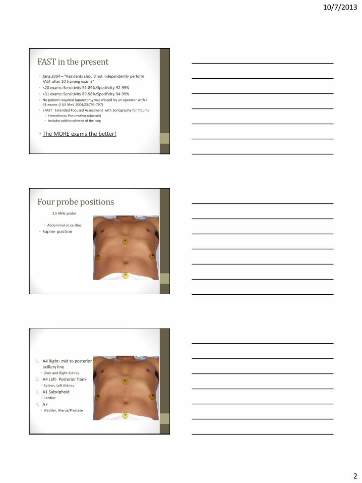
Four probe positions
3.5 MHz probe
• Abdominal or cardiac
• Supine position
1. A4 Right- mid to posterior axillary line Liver and Right Kidney
2. A4 Left- Posterior flank
Spleen, Left Kidney
3. A1 Subxiphoid Cardiac
4. A7 Bladder, Uterus/Prostate
10/7/2013
3
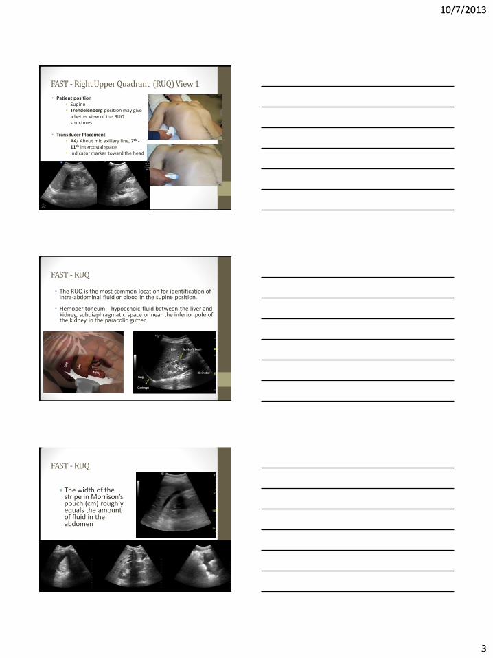
FAST - Right Upper Quadrant (RUQ) View 1
• Patient position • Supine • Trendelenberg position may give
a better view of the RUQ structures
• Transducer Placement
• A4/ About mid axillary line, 7th - 11th intercostal space
• Indicator marker toward the head
FAST - RUQ
• The RUQ is the most common location for identification of intra-abdominal fluid or blood in the supine position.
• Hemoperitoneum - hypoechoic fluid between the liver and
kidney, subdiaphragmatic space or near the inferior pole of the kidney in the paracolic gutter.
FAST - RUQ
The width of the stripe in Morrison’s pouch (cm) roughly equals the amount of fluid in the abdomen
Morrison’s Pouch
Diaphragm

10/7/2013
4
FAST tip for RUQ
• Trendelenberg position may facilitate visualization of fluid in upper abdomen.
• Lung, Liver Kidney
• Multiple windows and repeat exams maybe necessary to confirm presence of free fluid.
• Counter clock rotation help eliminate the rib shadows.
• Most of the time the transducer has to be moved more superiorly and posteriorly.
FAST - Left Upper Quadrant (LUQ)
• Transducer Placement
• A4/ About the mid – posterior axillary line, 7th-11h intercostal space
• Indicator marker toward
the head
FAST - LUQ
• Fluid most often collects between the diaphragm and the spleen in the left upper quadrant.
• Hemoperitoneum - hypoechoic fluid between the spleen and kidney, sub-diaphragmatic
or near the inferior pole of the kidney in the paracolic gutter
10/7/2013
5
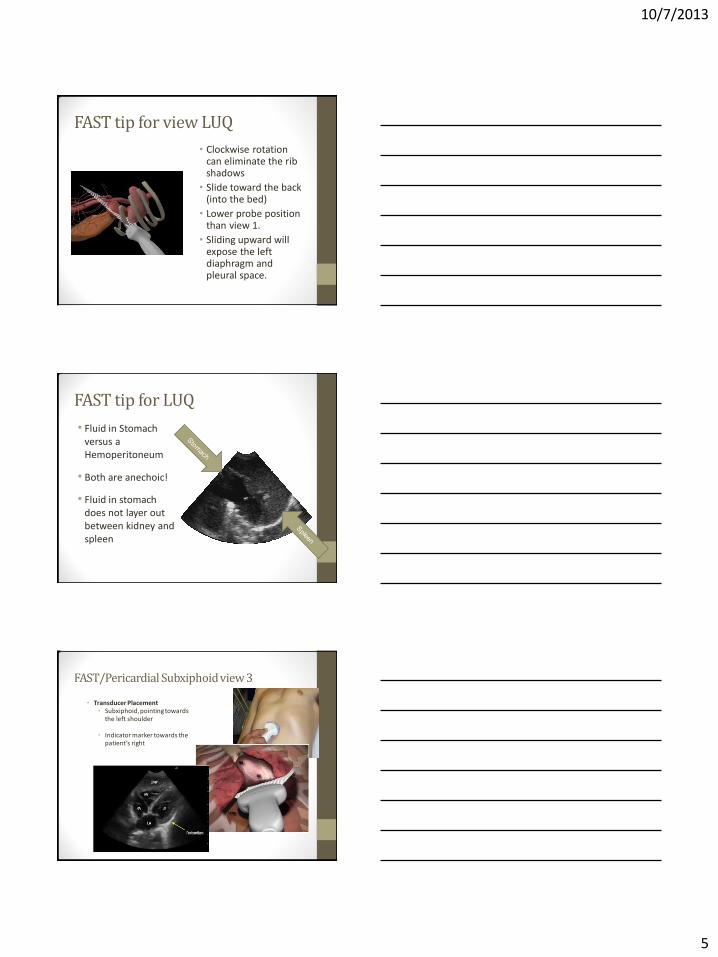
FAST tip for view LUQ
• Clockwise rotation can eliminate the rib shadows
• Slide toward the back (into the bed)
• Lower probe position than view 1.
• Sliding upward will expose the left diaphragm and pleural space.
FAST tip for LUQ
• Fluid in Stomach versus a Hemoperitoneum
• Both are anechoic!
• Fluid in stomach does not layer out between kidney and spleen
FAST/Pericardial Subxiphoid view 3
• Transducer Placement • Subxiphoid, pointing towards
the left shoulder
• Indicator marker towards the patient’s right

10/7/2013
6
FAST
Pericardial Effusion
• Detection of echo-free rim around the heart within the hyperechoic parietal pericardium
• Minimal fluid accumulations that occur rapidly can be hemodynamically significant while chronic accumulations can be large without causing hemodynamic compromise
FAST/ Tamponade
• Right atrium and right ventricle collapse
• Insert movie 72
FAST tip for view 3
• Pericardial anechoic stripes that are circumferential usually represent fluid while an anterior stripe may be pericardial fat.
• Multiple windows and repeat exams maybe necessary to confirm presence of free fluid.
• Use the liver to enhance the cardiac image.
• Hand on TOP of the probe allows you to tilt the probe under the ribs.
• Hand on top of the probe also sets you up for the next view…

10/7/2013
7
FAST – Suprapubic view 4
• Transducer Placement
• Above pubis angled inferiorly. • Hand on top of probe. • Obtain both the transverse
and longitudinal views.
• Longitudinal View: Indicator marker toward the head and fan up and down.
• Transverse view : Indicator marker pointing towards the patient’s right and fan right to left.
FAST/Suprapubic
Structures to be identified
• Bladder • Uterus • Prostate • Cul De Sac • Retrovesical space
• Sonographic Findings • Better to perform the US on a
full bladder • Accumulated fluid will be found
as a hypoechoic strip in the cul de sac or retrovesicular space on either side of the bladder
• Reverse Trendelenberg may help visualize the pelvic regions.
• Long and short axis views required
Short and long bladder Male
Anterior Pelvic
Left side of Patient
Right side of Patient
Posterior Region
Patient’s head
Posterior Region
Anterior Pelvic

10/7/2013
8
Long axis bladder female
Patient’s head
Right side of Patient
Left side of Patient
FAST tips
• Dimming room lights may aid visualization.
• Trendelenberg position may facilitate visualization of fluid in upper abdomen.
• Reverse Trendelenberg may facilitate visualization in the pelvis.
• Pericardial anechoic stripes that are circumferential usually represent fluid while an anterior stripe may be pericardial fat.
• Multiple windows and repeat exams maybe necessary to confirm presence of free fluid.
• Always best to confirm with clinical/physical exam.

10/7/2013
9
FAST points to ponder
• FAST should be coupled with the patient assessment and other diagnostic modalities to reach a final decision.
• Always best to confirm with clinical/physical exam.
• Bedside ultrasound cannot determine the etiology of the fluid solely on the ultrasound findings.
• False positive results may be seen in the setting of ascites or non-traumatic causes of bleeding.
Thank you
• If time permits… three slides on e-FAST
E-FAST Lung & Pleural interface
• To detect the presence of pneumothorax or pleural effusion
• Transducer Type & Placement
• Phased array, Linear or Curvilinear
• L1 2nd-4th intercostal spaces, anterior chest wall
• L2 5th-8th intercostal spaces, anterior chest wall
• L3 4th-10th intercostal spaces, between the anterior & posterior axillary lines
• Transducer Placement
• Transducer marker pointing cephalad
• The exam should be performed bilaterally
• Depth about 15-20 cm
10/7/2013
10
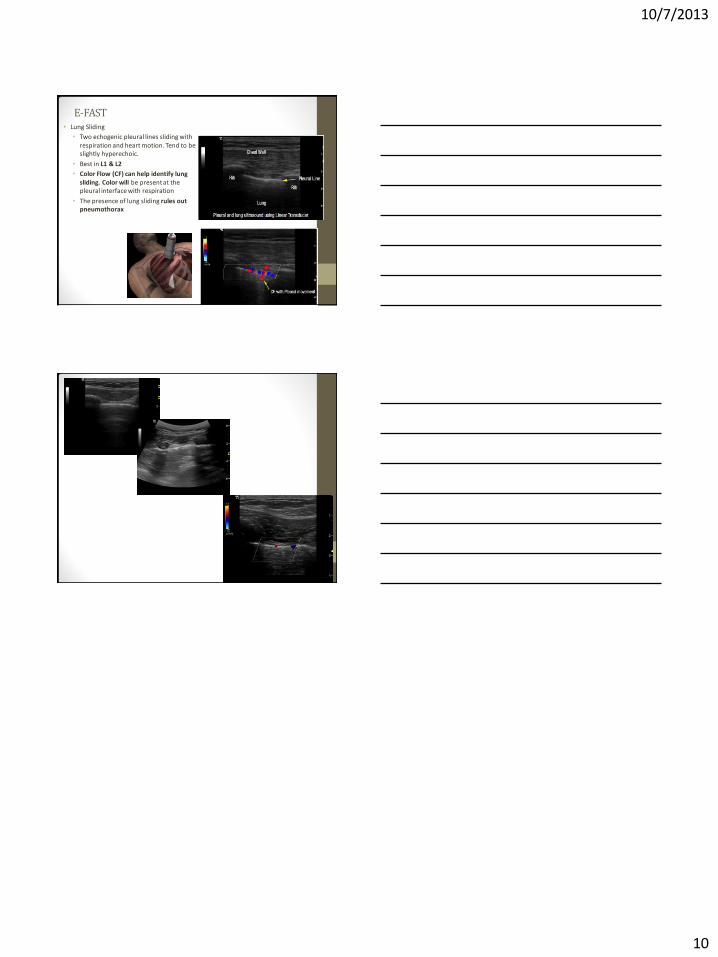
E-FAST • Lung Sliding
• Two echogenic pleural lines sliding with respiration and heart motion. Tend to be slightly hyperechoic.
• Best in L1 & L2
• Color Flow (CF) can help identify lung sliding. Color will be present at the pleural interface with respiration
• The presence of lung sliding rules out pneumothorax