imaging of sinonasal inflammatory disease
DESCRIPTION
orlTRANSCRIPT

Imaging of Sinonasal Inflammatory Disease’
David M. Yousem, MD
303
Changes in imaging sinonasal in-flammatory disease have paralleledchanges in the treatment of chronicsinusitis. As functional endoscopicsinus surgery has become a morewidespread technique, coronal com-puted tomography (CT) has becomethe primary imaging modality, re-placing plain radiography. Knowl-edge of the plethora of sinonasal ana-tomic variations and the inherentsurgical implications is critical to theinterpretation of the CT scans and tothe safe performance of endoscopicsurgery. Currently, the role of mag-netic resonance imaging is restrictedto the evaluation of complicated si-nusitis, intraorbital and intracranialmanifestations of aggressive sinus-itis, and sinonasal neoplasms.
Index terms: Nose, diseases, 26.24 #{149}Paranasal
sinuses, diseases, 23.25 #{149}Sinusitis, 23.25
State-of-art reviews
Radiology 1993; 188:303-314
‘From the Department of Radiology, Neuro-radiology section, Hospital of the University ofPennsylvania, 3400 Spruce St. Philadelphia, PA19104. Received January26, 1993; revision re-quested February 18; revision received March 4;
accepted March 8. Address reprint requests tothe author.
� RSNA, 1993
S INONASAL imaging has progressedmethodically as each new gener-
ation of imaging modality has en-croached on the domain of the formergeneration. While plain radiographywas once the study ordered mostcommonly to evaluate the sinonasalcavity, computed tomography (CT)has now supplanted plain radiogra-phy because the endoscopic sinus sur-geon has required greater anatomicprecision. The 1970s and 1980s sawthe introduction of the Messerklingertechnique-subsequently coinedfunctional endoscopic sinus surgery(FESS)-replace the more traditionalCaldwell-Luc and maxillary antros-tomy procedures for treating chronicsinusitis. At the same time, coronal CThas become the study of choice forchronic sinusitis, since it simulates theendoscopist’s view of the sinonasalcavity and provides a road map ofbone for surgery. Finally, previouslyunresectable sinonasal malignanciesinvading the skull base can now betreated by a team of otorhinolaryngol-ogists and neurosurgeons performingaggressive craniofacial surgery eveninto the cavernous sinus. The needfor optimal mapping of such tumorshas led to the ascendancy of magneticresonance (MR) imaging for sinonasalneoplasms.
This article will review the perti-nent anatomy of the sinonasal cavity,discuss imaging of uncomplicatedand complicated sinusitis, and ana-lyze the current role of each imagingmodality.
BASIC ANATOMY
To understand the pathogenesis ofsinusitis, one must understand thenormal pathway of mucociliary clear-ance in the paranasal sinuses. Thecilia within the maxillary sinus propelthe mucus stream in a starlike patternfrom the floor of the maxillary sinustoward the ostium situated superome-dially. In approximately 30% of pa-
tients there is a second accessory Os-tium to the maxillary sinus inferior tothe major opening (1). From the max-illary sinus ostium, mucus from themaxillary antrum gets swept superi-orly through the infundibulum, whichis located lateral to the uncinate pro-cess and medial to the inferomedialborder of the orbit (Fig 1). The unci-nate process, a sickle-shaped bone ex-tension of the lateral nasal wall ex-tending from anterosuperiorly toposteromnferiorly, is rarely (less than2.5% of patients) pneumatized (1).Occasionally the uncinate processattaches to the lamina papyracea (themedial wall of the orbit). If it does so,the infundibulum will not have a su-perior opening creating a blind pouch,the recessus terminalis. Posterior to theuncinate process, at the terminationof the infundibulum, mucus is pro-pelled to the hiatus semilunaris, an air-filled space just anterior and inferiorto the largest anteroinferior ethmoidair cell, the ethmoidal bulla. The mucusis then passed posteromedially via themiddle meatus, a channel passing me-dial and superior to the uncinate pro-cess, into the back of the nasal cavityto the nasopharynx, where it is subse-quently swallowed.
The OMC refers to the maxillarysinus ostium, the infundibulum, andthe middle meatus, the commondrainage pathway of the frontal, max-illary, and anterior ethmoid air cells.
The frontal sinuses drain inferome-dially via the nasofrontal duct. Theterm nasofrontal duct has been re-placed with the term frontoethmoidalrecess, since a true circular duct is usu-ally not present and since frontoeth-moidal recess connotes the commondrainage of the frontal sinus and theanterior ethmoidal sinuses (Fig 2).The frontoethmoidal recess is the
Abbreviations: FESS = functional endoscopic
sinus surgery, OMC = ostiomeatal complex.

304 #{149}Rad� ‘!ogv August 1993
space between the inferomedial fron-tal sinus and the anterior middle me-atus. The frontal sinus and the ante-nor ethmoidal sinuses usually drainthrough the recess directly into themiddle meatus via the frontoethmoi-dal recess or, less commonly, into thesuperior ethmoidal infundibulum be-fore passing to the middle meatus.
The anteriormost ethmoid air cells,located in front of the attachment ofthe middle turbinate to the cribriformplate, are termed agger nasi cells. Ag-ger nasi cells lie anterior, lateral, andinferior to the frontoethmoidal recess.They are present in over 90% of pa-tients (1,2) and lie deep to the lacrimalbone (the anteromedial margin of theorbit). Another set of anterior ethmoi-dal air cells, which are variably pre-sent in patients, are supraorbital eth-moid air cells. These are important toidentify, as these air cells are difficultto access through the endoscope be-cause of their superolateral locationand proximity to the orbit.
As stated earlier, ethmoidal bulla isthe term used for the ethmoid air celldirectly above and posterior to theinfundibulum and hiatus semilunaris.A very large ethmoidal bulla can ob-struct the infundibulum and hiatussemilunaris, leading to obstruction ofthe drainage of the maxillary and an-tenor ethmoid sinuses. When anteriorethmoid air cells are located inferolat-eral to the bulla, along the inferiormargin of the orbit protruding intothe maxillary sinus, they are termedHaller cells or maxilloethmoidal cells.Haller cells have been reported to bepresent in 10%-45% of patients un-dergoing sinus CT (1-3). Whengreatly enlarged, these Haller cellsmay narrow the infundibulum ormaxillary sinus ostium.
Between the ethmoidal bulla andthe basal lamella (the lateral attach-ment of the middle turbinate to thelamina papyracea of the orbit) is thesinus lateralis. The sinus lateralis mayopen into the frontoethmoidal recessor into a space posterior to the bulla,the hiatus semilunaris posterioris. Thesinus lateralis is important to identifyas a potential area for residual sinus-itis after ethmoidectomy, shielded asit is by the bulla. The bulla, Hallercells, and sinus lateralis are all part ofthe anterior ethmoid complex, ante-rior to the basal lamella.
The posterior ethmoid air cells arelocated behind the basal (or ground)lamella of the middle turbinate anddrain via the superior meatus, the su-preme meatus, or other tiny ostia justunder the superior turbinate. Ulti-mately, these ostia drain into the
sphenoethmoidal recess of the nasalcavity, from which the secretions passto the nasopharynx. In some patients,the most posterior ethmoid air cellmay pneumatize into the sphenoidbone, superior and lateral to the si-nus. This is termed an “Onodi cell.”
The sphenoid sinus drains anteri-orly into the nasal cavity through itsostium into the sphenoethmoidal re-cess, just medial to the superior turbi-nate of the nose. This ostium is bestseen in the axial plane, since it passesposterior to anterior (Fig 3). The sphe-noid sinuses may have aerated exten-sions into the pterygoid plates (44%of patients) or into the clinoid pro-cesses (13% of patients) (4,5).
The nasal cavity typically has threesets of turbinates-the superior, mid-dle, and inferior turbinates-divided
by the midline nasal septum. Occa-sionally, one may identify a fourthsuperiormost turbinate, the supremeturbinate. An aerated middle turbi-nate, which usually communicateswith the anterior ethmoid air cells, istermed a concha bullosa and is seenin approximately 34%-53% of pa-tients (1-3). The vertical attachment ofthe middle turbinate is pneumatizedmore commonly than its inferior bul-bous portion, and an air cell in thisarea is termed an “intralamellar cell.”Pneumatization of the inferior or su-perior turbinates is much less com-mon (less than 5% of patients) (1).
Paradoxical middle turbinates areones that are concave medially ratherthan laterally. In our experience thisoccurs in less than 10% of the popula-tion (3), but another study has foundthis variation in 26% of cases (1).Large paradoxical turbinates havebeen implicated as a possible cause ofmiddle meatus obstruction.
The nasal septum is composed ofthree parts. There is a cartilaginousanteroinferior portion, a bony pos-teroinferior portion known as the vo-mer, and a superoposterior bony por-tion, the perpendicular plate of theethmoid bone. The nasal septum isonly rarely aerated. Nasal septal devi-ation, however, is quite common, andbone spurs often develop at the apexof the deviation, usually at the junc-tion of the perpendicular plate, carti-laginous septum, and vomer. Spursmay cause the sensation of nasal ob-struction.
The nasolacrimal duct coursesdownward from the lacrimal sacalong the medial canthus, where it isin proximity with agger nasi air cells.Inflammation of agger nasi cells maybe associated with epiphora becauseof this close relationship (6). The duct
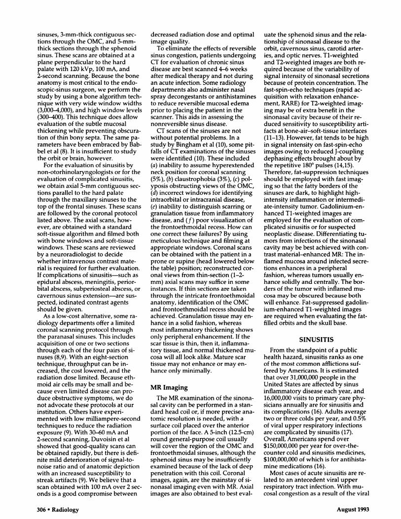
Figure 1. Ostiomeatal complex (OMC) on
coronal CT scan. A 3-mm-thick CT section
obtained with 120 kVp, 100 mA, and 2-sec-
ond scanning in the coronal plane with a
bone algorithm and filmed with window
width of 3,000 and level of 300 demonstrates
bone anatomy of the OMC well. The left
maxillary sinus ostium (solid arrow) and in-
fundibulum (arrowheads) are below thelarge ethmoidal bulla (B). Medial to the right
uncinate process (open arrow) one identifies
the middle meatus air channel, bounded me-
dially by the middle turbinate (t). The inser-
tion of the right middle turbinate into the
lamina papyracea of the orbit can be seen.
This patient has mild nasal septal deviation
with a small spur on the left.
subsequently runs in the anterior por-tion of the lateral nasal wall. Its end-ing opens below the inferior turbinateat the inferior meatus.
IMAGING MODALITIES
Plain Radiography
The plain radiographic examinationin sinonasal imaging consists of acqui-sition of Waters, Caldwell, lateral, andsubmental vertex views. While theutility of the plain radiographic seriesin evaluating sinonasal malignanciesis relatively limited, it is still orderedfrequently in the evaluation of rou-tine sinusitis. For the identification ofair-fluid levels suggestive of acutesinusitis in the maxillary antrum,plain radiography is still a relativelyeasy and sensitive examination. How-ever, for the evaluation of chronicsinusitis where mucosal thickeningalone may be present, the drawbackof overlapping structures makes theevaluation of the OMC, anterior eth-moid sinus, middle meatus, and sphe-noid sinus somewhat limited.
If one is searching for a localizedcause of sinusitis or if surgical inter-vention is contemplated, a more exactlook at the anatomy is required. Thisis typically obtained with CT scans.

a. b.Figure 2. Frontoethmoidal recess, agger nasi cell, and Haller cell on coronal CT scans. (a) A
wide frontoethmoidal recess (arrow) is seen on the left side with communication to an ante-
nor ethmoid air cell (e). As this section is anterior to the middle turbinate’s cribriform plate
attachment, the ethmoid air cells seen are agger nasi cells (a on right). Note their proximity tothe nasolacrimal fossa (arrowhead). (b) A Haller cell (arrow) is seen below the left orbit. This
cell contributes to the narrowing of the left infundibulum. Associated OMC and superior max-illary sinus inflammatory change are present.
Vnliine 1RR #{149}Number 2 Radiology #{149}305
Figure 3. Sphenoethmoidal recess on axial
CT scan. The exits from the sphenoid sinus
into the posterior nasal cavity are seen bilat-
erally (arrowheads). The ostia leading from
posterior ethmoid air cells are usually not
definable.
The decline in the role of plain radi-ography is exemplified by a recentstudy that showed the plain radio-graphs ordered by ophthalmologistswere useless 98.5% of the time, led tounnecessary additional studies in 20%of “abnormal” plain radiographicstudies, and were normal in 86% ofstudies ordered (7).
The advantages of plain radiogra-phy of the paranasal sinuses are its
low cost, small radiation dose, ease ofperformance, and capability for porta-ble examination. At our institution,where an active FESS team is present,plain radiography is ordered infre-quently. Sinonasal endoscopy hasreplaced plain radiography as ascreening test in patients with chronicor recurrent sinusitis. Plain radiogra-phy is most often ordered at our hos-pital in the evaluation of a fever ofunknown origin in patients in theintensive care units who are unable tobe transported for CT. For the generalpractitioner suspicious of sinusitis in apatient with equivocal clinical find-ings, a positive plain radiographicexamination may be a low-cost confir-matory test for sinusitis. However, anegative radiographic examinationshould not eliminate sinusitis fromthe differential diagnosis. Endoscopy,CT, or both must follow if suggestivesymptoms persist.
CT Scanning
The role of CT in providing a roadmap for the functional endoscopicsinus surgeon is well known. To un-derstand the purpose of CT examina-tion of the sinonasal cavity, one mustunderstand the rationale for FESS.
The goal of FESS is to maintain thenormal mucosa of the sinonasal cavityin order to preserve the normal path-way of mucociliary clearance. There-fore, rather than create an alternateegress of mucus from the maxillarysinus, as in an inferior meatal antros-
tomy (the Caldwell-Luc procedure),FESS enlarges the natural ostia andpassageways of the paranasal sinuses.Whereas in the past maxillary andfrontal sinusitis was thought to be aprimary process in patients withchronic sinusitis, it is now believedthat these sinuses are secondarily ob-structed due to disease in the ostio-meatal complex. Disease at the ostiumand inferior infundibulum obstructsthe maxillary sinus, whereas disease inthe middle meatus and anterior in-fundibulum obstructs the frontal andanterior ethmoid sinuses. Therefore,surgery is directed toward removingpotential obstacles to mucociliaryclearance at the OMC. Persistence ofchronic sinusitis after nasoantral win-dows (nasal antrostomies) is usuallydue to anterior ethmoid disease. There-fore, amputation of the uncinate pro-cess, enlargement of the infundibulumand maxillary sinus ostia, and creationof a common unobstructed channelfor the anterior ethmoid air cells arecommon practices at FESS. UsuallyFESS also includes complete or partialethmoidectomy. In a similar vein,FESS does not attempt to strip themucosa clean, as in a Caldwell-Lucprocedure. Mucociliary motility ispreserved.
The surgery is done by means of anintranasal endoscope rather than withan external approach, so knowledgeof the bone landmarks is essential toplanning surgery. Posterior ethmoidand sphenoid sinus access can also beobtained with the nasal endoscopebut requires greater expertise. Poste-nor ethmoidectomy and sphenoid-otomy are not performed routinelyexcept by very experienced FESSpractitioners. The surgeon must knowwhere he or she is at all times in orderto prevent complications such as or-bital or intracranial entry, particularlywhen operating posteriorly in the si-nonasal cavity.
For the FESS surgeon, coronal CTscans are ideal, as they simulate theappearance of the sinonasal cavityfrom the point of view of the endo-scope. Because the otorhinolaryngolo-gists have used CT more frequently astheir screening and mapping study ofthe sinonasal cavity, an impetus forreducing the cost of the study andlimiting the radiation dose has led toproposals for new protocols for per-forming sinus CT (8,9).
At our institution, CT scanning per-formed for cases of uncomplicatedsinusitis referred by an otorhinolaryn-gologist consists of acquisition of
5-mm-thick contiguous coronal sec-tions through the anterior frontal
306 #{149}Radiology August 1993
sinuses, 3-mm-thick contiguous sec-tions through the OMC, and 5-mm-thick sections through the sphenoidsinus. These scans are obtained at aplane perpendicular to the hardpalate with 120 kVp, 100 mA, and2-second scanning. Because the boneanatomy is most critical to the endo-scopic-sinus surgeon, we perform thestudy by using a bone algorithm tech-nique with very wide window widths(3,000-4,000), and high window levels(300-400). This technique does allowevaluation of the subtle mucosalthickening while preventing obscura-tion of thin bony septa. The same pa-rameters have been embraced by Bab-bel et al (8). It is insufficient to studythe orbit or brain, however.
For the evaluation of sinusitis bynon-otorhinolaryngologists or for theevaluation of complicated sinusitis,we obtain axial 5-mm contiguous sec-tions parallel to the hard palatethrough the maxillary sinuses to thetop of the frontal sinuses. These scansare followed by the coronal protocollisted above. The axial scans, how-ever, are obtained with a standardsoft-tissue algorithm and filmed bothwith bone windows and soft-tissuewindows. These scans are reviewedby a neuroradiologist to decidewhether intravenous contrast mate-rial is required for further evaluation.If complications of sinusitis-such asepidural abscess, meningitis, perior-bital abscess, subpenosteal abscess, orcavernous sinus extension-are sus-pected, iodinated contrast agentsshould be given.
As a low-cost alternative, some ra-diology departments offer a limited
coronal scanning protocol throughthe paranasal sinuses. This includesacquisition of one or two sectionsthrough each of the four pairs of si-nuses (8,9). With an eight-sectiontechnique, throughput can be in-creased, the cost lowered, and theradiation dose limited. Because eth-moid air cells may be small and be-cause even limited disease can pro-duce obstructive symptoms, we donot advocate these protocols at ourinstitution. Others have experi-mented with low milliampere-secondtechniques to reduce the radiationexposure (9). With 30-60 mA and2-second scanning, Duvoisin et alshowed that good-quality scans canbe obtained rapidly, but there is defi-nite mild deterioration of signal-to-noise ratio and of anatomic depictionwith an increased susceptibility tostreak artifacts (9). We believe that ascan obtained with 100 mA over 2 sec-onds is a good compromise between
decreased radiation dose and optimalimage quality.
To eliminate the effects of reversiblesinus congestion, patients undergoingCT for evaluation of chronic sinusdisease are best scanned 4-6 weeksafter medical therapy and not duringan acute infection. Some radiologydepartments also administer nasalspray decongestants or antihistaminesto reduce reversible mucosal edemaprior to placing the patient in thescanner. This aids in assessing thenonreversible sinus disease.
CT scans of the sinuses are notwithout potential problems. In astudy by Bingham et al (10), some pit-falls of CT examinations of the sinuseswere identified (10). These included(a) inability to assume hyperextendedneck position for coronal scanning(5%), (b) claustrophobia (3%), (c) pol-yposis obstructing views of the OMC,
(d) incorrect windows for identifyingintraorbital or intracranial disease,(e) inability to distinguish scarring orgranulation tissue from inflammatorydisease, and (f ) poor visualization ofthe frontoethmoidal recess. How canone correct these failures? By usingmeticulous technique and filming atappropriate windows. Coronal scanscan be obtained with the patient in aprone or supine (head lowered belowthe table) position; reconstructed cor-onal views from thin-section (1-2-mm) axial scans may suffice in someinstances. If thin sections are takenthrough the intricate frontoethmoidalanatomy, identification of the OMCand frontoethmoidal recess should beachieved. Granulation tissue may en-hance in a solid fashion, whereasmost inflammatory thickening showsonly peripheral enhancement. If the
scar tissue is thin, then it, inflamma-tory tissue, and normal thickened mu-cosa will all look alike. Mature scartissue may not enhance or may en-hance only minimally.
MR Imaging
The MR examination of the sinona-sal cavity can be performed in a stan-dard head coil or, if more precise ana-tomic resolution is needed, with asurface coil placed over the anteriorportion of the face. A 5-inch (12.5-cm)round general-purpose coil usuallywill cover the region of the OMC andfrontoethmoidal sinuses, although thesphenoid sinus may be insufficientlyexamined because of the lack of deeppenetration with this coil. Coronalimages, again, are the mainstay of si-nonasal imaging even with MR. Axialimages are also obtained to best eval-
uate the sphenoid sinus and the rela-tionship of sinonasal disease to theorbit, cavernous sinus, carotid arter-ies, and optic nerves. Ti-weightedand T2-weighted images are both re-quired because of the variability ofsignal intensity of sinonasal secretionsbecause of protein concentration. Thefast-spin-echo techniques (rapid ac-quisition with relaxation enhance-ment, RARE) for T2-weighted imag-ing may be of extra benefit in thesinonasal cavity because of their re-duced sensitivity to susceptibility arti-facts at bone-air-soft-tissue interfaces(11-13). However, fat tends to be highin signal intensity on fast-spin-echoimages owing to reduced J-couplingdephasing effects brought about bythe repetitive 180#{176}pulses (14,15).Therefore, fat-suppression techniquesshould be employed with fast imag-ing so that the fatty borders of thesinuses are dark, to highlight high-intensity inflammation or intermedi-ate-intensity tumor. Gadolinium-en-hanced Ti-weighted images areemployed for the evaluation of com-plicated sinusitis or for suspectedneoplastic disease. Differentiating tu-mors from infections of the sinonasalcavity may be best achieved with con-trast material-enhanced MR: The in-flamed mucosa around infected secre-tions enhances in a peripheralfashion, whereas tumors usually en-hance solidly and centrally. The bor-ders of the tumor with inflamed mu-cosa may be obscured because bothwill enhance. Fat-suppressed gadolin-ium-enhanced Ti-weighted imagesare required when evaluating the fat-filled orbits and the skull base.
SINUSITIS
From the standpoint of a publichealth hazard, sinusitis ranks as oneof the most common afflictions suf-fered by Americans. It is estimatedthat over 31,000,000 people in theUnited States are affected by sinusinflammatory disease each year, and16,000,000 visits to primary care phy-sicians annually are for sinusitis andits complications (16). Adults averagetwo or three colds per year, and 0.5%of viral upper respiratory infectionsare complicated by sinusitis (17).Overall, Americans spend over$150,000,000 per year for over-the-counter cold and sinusitis medicines,$100,000,000 of which is for antihista-mine medications (16).
Most cases of acute sinusitis are re-lated to an antecedent viral upperrespiratory tract infection. With mu-cosal congestion as a result of the viral

Volume 188 #{149}Number 2 Radiology #{149}307
infection, apposition of mucosal sur-faces occurs in the paranasal sinuses.Once this occurs, obstruction of thenormal flow of mucus results in a re-tention of secretions, creating a favor-able environment for bacterial super-infection. The ethmoid sinuses aremost commonly involved in sinusitis,possibly because of their position inthe “line of fire” as inspired particlesimpact and irritate the fragile ethmoidsinus lining. The bacterial pathogensresponsible for acute sinusitis includeStreptococcus pneumoniae, Haemophilusinfluenzae, beta hemolytic streptococ-cus, and Moraxella catarrhalis (16,18).In the chronic phase, staphylococcus,streptococcus, corynebacteria, bacte-roides, fusobacteria, and other anaer-obes may be responsible. The fungithat may infect the sinuses includeAspergillus species, mucormycosis,bipolaris, drechslera, curvularia, andCandida species (19,20).
The workup of a patient with sinus-itis has changed over recent years be-cause of the advent of experiencedintranasal endoscopists. While acutebouts of sinusitis are typically treatedmedically and the diagnosis is usuallymade on a clinical basis, patients withrecurrent or intractable sinusitis oftenare referred to the otorhinolaryngolo-gist for evaluation. In a patient withrecurrent or chronic symptoms (debil-itating headache, facial pain, or con-gestion) or persistent symptoms aftermedical therapy, intranasal endoscopyis usually performed. On the basis ofthe endoscopic findings and the clini-cal symptoms, the patient may betreated with a second long-term courseof antibiotics. If medical therapy failsagain and the endoscopic findingssuggest surgically correctable abnor-malities, CT scanning is requested.
Why not order plain radiographyinstead? Besides its inability to showthe relevant anatomy to guide thesurgeon, plain radiography has lim-ited accuracy. Using air-fluid levelsand opacification as criteria of diseaseand antroscopy as the standard ofreference, Kuhn found that the sensi-tivity of plain radiography was 54%and specificity was 92% (21). With thecriterion of mucosal thickening, thenumbers were nearly reversed: sensi-tivity was 99% and specificity was46%. These maxillary sinus accuracyrates are higher than expected in theother sinuses, since soft-tissue overlapis less of a problem in the antra. Withantral aspiration used as a standard,another study found plain radiogra-phy to be accurate in only 63%-82%of cases of maxillary sinus infection(17). This has led to wider use of CT.
When evaluating a CT scan in apreoperative patient before FESS, sev-eral issues must be addressed (22). Isthe uncinate process apposed to themedial orbital wall (an atelectatic in-fundibulum)? If so, its vigorous re-moval may result in orbital penetra-hon. Are there areas of dehiscence inthe lamina papyracea or do the orbitalcontents protrude into the ethmoidsinus, both of which may lead to un-intentional orbital entry from the eth-moid sinus? Defects in the laminapapyracea have been reported in 5%-10% of autopsy specimens (5,23). Be-cause orbital hematomas are the mostcommon orbital complication of FESS,it is important to identify any gaps inthe lamina papyracea (24-26). CT ob-viously is the best means for identify-ing the thin medial bony wall of theorbit. However, orbital (and cribri-form plate) perforations at the time ofFESS often occur through areas ofnondehiscence because of overly vig-
orous surgical technique.Is the internal os of the frontal sinus
occluded with bone? Bone disease ofthe frontal sinus typically precludesan endoscopic approach. Are thereareas of dehiscence in the cribriformplate? Another uncommon but seri-ous complication of FESS is the pro-duction of a dural tear leading to acerebrospinal fluid leak. By identify-ing areas of dehiscence in the cribri-form plate, reported in 8.5%-14% ofpatients at autopsy, the radiologisteffectively warns the endoscopic si-nus surgeon (5,23).
Are the bony walls between thesphenoid sinus and the optic nerveintact? It has been shown that in 4%of patients there is no bony borderbetween the optic nerve and thesphenoid sinus and in 78%-88% ofpatients the bone is less than 0.5 mmthick (4,5). The posterior ethmoid aircells contact the optic canal in 48%and also may have a very thin boneborder (4). There have been limitedreports of optic nerve transection dur-ing sphenoethmoidectomy from anintranasal approach, and dehiscenceof the sphenoid wall may be a predis-posing factor. Additionally, the rela-tionship of the posteriormost ethmoidair cell to the sphenoid sinus and tothe optic nerve is important, since thesphenoid sinus is typically enteredthrough this cell (5). If an Onodi cellextends far lateral to the lateral wallof the sphenoid sinus, the endosco-pist must remain close to the midlineas he or she perforates into the sphe-noid sinus. Otherwise orbital entrymay occur near the orbital apex, a sitewhere the risk of injury to the extra-
ocular muscles or nerves is very high.By the same token, the angle at whichthe sphenoid sinus should be entered,based on the superior and inferiorrelationships of the Onodi cell andsphenoid sinus, may be determinedon the basis of the preoperative CTscan (and best with a sagittal recon-struction).
In 8%-i4% of autopsy cases, thereis no bone between the internal ca-rotid artery and the sphenoid sinus(23). An intersinus septum in thesphenoid sinus, which attaches to thecarotid canal, is important to recog-nize preoperatively and is typicallybest identified in the axial view. Re-moval of such an intersinus septumduring surgery may result in carotidlaceration. Consideration shouldtherefore be given to routine acquisi-tion of axial CT scans when sphenoidsinus surgery is contemplated.
Preoperatively, the sinus surgeonexamines the height of the ethmoidroof in relationship to the cribriformplate (5). Normally the cribriformplate is lower than the ethmoid roof,and therefore the surgeon can dissectmore superiorly if he or she proceedslaterally along the ethmoid roof dur-ing ethmoidectomy. Variations in thisanatomy should be cited in radiologicreports. The presence of middle turbi-nate pneumatization (concha bullosa)and/or opacification will be inappar-ent to the endoscopic sinus surgeonlooking at the mucosal surface only.The radiologist can identify this nor-ma! variant and determine if it is com-promising the middle meatus.
Because of all of these factors thatthe endoscopic sinus surgeon mustevaluate prior to and during the sur-gery, good-quality CT scans are criti-cal at the time of the operation. Weprovide “operating room copies” ofthe coronal sinus scans for all surgicalcases. Teatini et al stated “few organsof the human body are liable to suchremarkable intersubject and intrasub-ject variations as the paranasal si-nuses” (5). This is certainly true and,unfortunately, creates considerableperil for the endoscopic surgeon. Theradiologist and the operating roomCT scan can be the navigators.
In addition to commenting in theCT report on the normal anatomicvariations, the radiologist shouldidentify areas of mucosal thickeningand sinus passageway opacification. Ithas come to be accepted that the loca-tion of sinusitis is more important inproducing a patient’s symptoms thanthe extent of the sinusitis. Therefore, asubtle area of opacification in the in-fundibulum of the OMC may cause
308 #{149}Radiology August 1993
more pain and discomfort than nearcomplete opacification of the maxil-lary sinus with a mucous retentioncyst and/or polyp. According toKennedy et al, CT almost alwayscauses underestimation of sinus mu-cosal thickening compared with whatis seen at endoscopy, despite high-resolution imaging with appropriatewindows and section thicknesses (27).
OMC opacification correlates wellwith the development of sinusitis (Fig4). Yousem et al found that the posi-tive predictive value of infundibularopacification for the presence of max-illary sinus inflammatory disease in100 consecutive patients was 79% (3).When the middle meatus was opaci-fled, the maxillary and ethmoid si-nuses showed inflammatory changein 84% and 82% of patients, respec-tively (3). The specificity of middlemeatus opaciflcation for maxillary orethmoid sinus disease was 93%. An-other study found frontal or maxillarysinus disease in 84% of patients whohad OMC opacification (1). Bolger etal demonstrated that when the infun-dibulum was clear, the ipsilateralmaxillary and frontal sinuses werefree of disease in 77% of cases (1). An-terior ethmoid disease was present in84% of patients with chronic sinusitisand 19% without sinus symptoms (1).Zinreich et al found middle meatusopacification in 72% of patients withchronic sinusitis, and, of these pa-tients, 65% had maxillary sinus muco-periosteal thickening (28). IsolatedOMC disease without frontal, maxil-lary, or ethmoid disease was uncom-mon (28). Of patients with frontal si-nus inflammation reported in thestudy of Zinreich et al, all had opacifl-cation of the frontoethmoidal recess.These findings support the conten-tion that obstruction of the narrowdrainage pathways will lead to subse-quent sinus inflammation.
After analyzing CT scans of 500 pa-tients, Babbel et al categorized recur-rent inflammatory sinonasal diseaseinto five patterns: infundibular, ostio-meatal unit, sphenoethmoidal recess,sinonasal polyposis, and sporadic orunclassifiable disease (29). The infun-dibular pattern was seen in 26% ofpatients and referred to isolated ob-struction of the inferior infundibu-lum, just above the maxillary sinusostium. Limited maxillary sinus dis-ease often coexisted with this pattern,whereas the ostiomeatal unit pattern,seen in 25% of cases, often had con-comitant frontal and ethmoidal dis-ease (29). The ostiomeatal unit patternwas designated when middle meatusopacification was present. Sphenoeth-
moidal recess obstruction occurred in6% of cases and led to sphenoid orposterior ethmoid sinus inflamma-tion. When the sinonasal polyposispattern was present, enlargement ofthe ostia, thinning of adjacent bone,and air-fluid levels were often seen.
In the study of Yousem et al, thepresence of a greater degree of nasalseptal deviation and a horizontallyoriented uncinate process correlatedwith an increased prevalence of sinusinflammation (3). The presence of aconcha bullosa did not increase therisk of sinusitis. This work has beenduplicated in another study in whichthe presence of a concha bullosa, para-doxical turbinates, Haller cells, anduncinate pneumatization were notshown to have a significantly higherrate in patients with chronic sinusitisthan in patients without sinus symp-toms (1). It appears that the size, notthe presence, of these normal varia-tions is the critical factor. Even whensmall, however, these variations areworth reporting, since they helpguide endoscopy.
The presence of air-fluid levels onplain radiographs or CT scans of thesinuses are more typically associatedwith acute sinusitis than chronic in-flammatory disease. Of course, acutesinusitis may be superimposed onchronic changes. The findings sugges-tive of chronic sinusitis include muco-sal thickening, bone remodeling,polyposis, and bone thickening sec-ondary to osteitis from adjacentchronic mucosal inflammation.
Hyperattenuating secretions on CTscans may be due to inspissated secre-tions, fungal sinusitis, or hemorrhagein the sinus. The hyperattenuatingsinus may be the only clue to fungalsinusitis and is an important featureto note. However, chronic sinusitisinfected with bacteria occasionallywill be hyperattenuating at CT, par-ticularly in patients who have verylongstanding disease or cystic fibrosis.The hyperattenuating sinus often cor-responds to the hypointense sinus onT2-weighted MR images.
What then is the role for MR imag-ing in the evaluation of a patient withchronic uncomplicated sinusitis? Ibelieve that MR is more sensitive tomucosal thickening than CT (and thepotential for improvement is thereaccording to Dr Kennedy’s assess-ment of CT). Sinus thickening is usu-ally seen as high signal intensity onthe T2-weighted image against theblack background of air and/or corti-cal bone. On Ti-weighted images themucosal thickening is low in inten-sity.
Figure 4. Bilateral OMC disease in a patientwith chronic sinusitis. Bilateral opacification
of the infundibula and maxillary sinus ostiacan be seen in this patient with chronic sinusdrainage. The left hiatus semilunaris and
middle meatus are also opacified at the level
of the middle turbinate (cf normal Fig la).
This patient also has a nasal septal spur and
deviation to the left. Maxillary sinus muco-
periosteal thickening is present bilaterally,
and the right bulla (b) is obstructed.
The sensitivity of MR to mucosalthickening accounts for the visualiza-tion of the normal nasal cycle as de-scribed by Zinreich et al and Kennedyet al (30,31). There is cyclical passivecongestion and decongestion of eachside of the nasal turbinates, nasal sep-tum, and ethmoid air cell mucosaover a 50-minute to 6-hour period inhumans. Thus, 1-2 mm of ethmoidsinus mucosal thickening may not bedue to inflammatory disease but mayreflect the normal intermittent con-gestion of the nasal cycle. In a similarvein, asymmetry in the size and in-tensity of the nasal turbinates is nor-mal. Because many radiologists arenot aware of the presence of this na-sal cycle (it is not readily apparent ordescribed on routine CT scans), therehas been a gross tendency to interpretincreased signal intensity from theturbinates and ethmoid sinus as apathologic finding on MR images.This accounts for some of the studiesin the literature claiming that MR isan inaccurate study for the evaluationof sinusitis.
This overcalling of the normal nasalcycle as ethmoid sinusitis notwith-standing, there is a high incidence ofsinus inflammatory mucosal diseasein asymptomatic patients. The preva-lence of incidental sinus mucosalthickening on CT scans of the parana-sal sinuses in asymptomatic patientsranges from 39% to 43% (32,33). Inthe asymptomatic population the

a. b. c.
Volume 188 #{149}Number 2 Radiology 309
Figure 5. Squamous cell carcinoma of the maxillary sinus with obstructed secretions. (a) Patient with squamous cell carcinoma demonstrates a
variety of signal intensities in her sinonasal cavity on this Ti-weighted (repetition time msec/echo time msec [600/17]), coronal image. One can
see intermediate-intensity tumor (t) in the nasal cavities and along the midline septum. In the right maxillary sinus (m) there is central high
signal intensity and peripheral low to intermediate signal intensity obstructed secretions and mucosa, respectively. In the left maxillary antrum
the secretions are low in intensity centrally and intermediate peripherally. The lateral ethmoid sinuses bilaterally also show air cells with high
intensity (*). The intensity variations reflect different protein concentrations in the sinuses and tissue differences between secretions, tumor,
and mucosa. (b) On this T2-weighted (3,000/90) image, one identifies intermediate-intensity tumor (t), high-intensity secretions centrally in the
right maxillary antrum (m), and low-intensity secretions centrally with bright mucosa peripherally in the left maxillary antrum. (c) The gadolin-ium-enhanced fat-suppressed short repetition time image shows solid enhancement in the tumor (t) and peripheral enhancement in the maxil-lary antrum (m). The high-intensity secretions in the right maxillary antrum (m) simulate contrast material enhancement.
presence of ethmoid sinus disease(28.4%) predominates over maxillary
sinus disease (24.8%), which is morecommon than sphenoid (7%-1i%)and frontal sinus (4.8%) mucosalchanges (32,34). Of all the sinuses,sphenoid sinus opacification is mostlikely to cause symptoms (34). Muco-sal thickening is the most common
finding, but polyps and/or mucousretention cysts may often be found in
asymptomatic patients. In asymptom-atic patients with a history of seasonalallergies, the prevalence of abnormalsinus CT scans is 54.4% (32). In chil-dren younger than 1 year old, 70%will have opacified sinuses, with themaxillary sinus more commonly af-fected than the ethmoid sinus (35).The significance of sinus opacificationin children is limited, since redundantmucosa, congestion from crying, andtears can cause opacification.
When asymptomatic patients areevaluated with MR for sinus inflam-matory changes, approximately 13%-63% of patients have abnormal si-nuses (36-39). The predominantfinding is mucoperiosteal thickening,just as at CT, followed by mucous re-tention cysts (38). Again, maxillary(27%-39%), ethmoid (6%-25%), or
combined maxillary and ethmoid(43%) sinus inflammatory disease pre-
dominates (36,37). In the presence ofan upper respiratory tract viral infec-tion, the sinuses are abnormal on MRimages in 65% of patients, whereas ifthere is no history of a cold, the MRimage is abnormal in 34% of cases
(37). Even when ethmoid sinus thick-
ening of 1-2 mm is eliminated fromconsideration (to account for the na-sal cycle), over 35% of asymptomaticpatients have changes of mucosalthickening (39). Polyps are seen in asmaller percentage, and, in approxi-mately 3%-5% of asymptomatic pa-tients, air-fluid levels or total sinusopacification may be found within the
paranasal sinuses (37-39). Sphenoidsinus disease can be seen in 6%-8% ofpatients undergoing MR or CT of thebrain; of these, approximately 25%have symptoms including visualproblems and meningitis (34).
Why is MR not employed in theroutine evaluation of patients sus-pected of having sinusitis? Why notspare the patient ionizing radiation?Again, this harks back to the primary
goal of imaging prior to FESS: to mapthe bony anatomy for preoperativeplanning. Because bone and air areseen as signal voids on MR images,MR has less utility for surgical plan-fling. A surgical map is not possible.
Another drawback of MR is theproblem of signal intensity and sinussecretions. Sinonasal secretions arenot simply bright on T2-weighted im-ages and dark on Ti-weighted im-ages. Som et al described the changesin signal intensity of sinonasal secre-tions on the basis of protein concen-tration and mobile water protons (40).The changes are probably due to theincreased cross-linking of glycopro-teins in hyperproteinaceous secre-tions, which leads to fewer free water
protons available and more boundprotons (bound to glycoproteins).Som et al found that as protein con-centration increased, the signal inten-sity on Ti-weighted images of sinus
secretions changed from hypointenseto hyperintense to hypointense again.These changes paralleled changes inviscosity. On the T2-weighted images,
hypoproteinaceous watery secretionswere initially bright, but as the pro-tein concentration and viscosityincreased, the signal intensity on T2-weighted images decreased. There-fore, the authors were able to describefour combinations of intensity pat-terns for sinus secretions: (a) hypo-
intense on Ti-weighted images andhyperintense on T2-weighted imagesin the most liquid form (total proteinconcentration less than 9%); (b) hyper-intense on Ti-weighted images andhyperintense on T2-weighted imagesin the mildly to moderately protein-aceous form (total protein concentra-
tion, 20%-25%); (c) hyperintense on
Ti-weighted images and hypointenseon T2-weighted images in a highlyproteinaceous form (total protein con-centration, 25%-28%); and (d) hypo-intense on Ti-weighted images andhypointense on T2-weighted imageswhen the secretions are in a nearsolid form (total protein concentrationgreater than 28%) (Fig 5) (40). Becauseof the last combination of signal in-tensities, there is the potential for mis-diagnosing completely opacified si-nuses at MR, since the concretions ofvery hyperproteinaceous secretions

a. b.Figure 6. Hypointense secretions of fungal sinusitis on MR images. (a) T2-weighted image ofa patient with documented aspergillus infection shows low-intensity areas amidst the high-
intensity secretions of the right maxillary antrum. This probably represents saprophytic
growth of the fungus in the sinus. (b) Gadolinium-enhanced Ti-weighted image demon-
strates a peripheral pattern of enhancement in the infected sinus and low-intensity secretions.
310 #{149}Radiology August 1993
may show a signal void simulating
aeration on both Ti-weighted andT2-weighted images (41,42). This is
also a potential pitfall when evaluat-ing a patient with a hyperprotein-aceous mucocele (41,43,44).
Other potential pitfalls in which asignal void may be encountered onboth Ti-weighted and T2-weightedimages include osteomas, odonto-
genic lesions, osteochondromas, fibro-sis, and fungal sinusitis mycetomas(42). The hypointensity of fungal si-nusitis is thought to be secondary to
the paramagnetic effects of either ironor manganese produced by the fungi(Fig 6) (19,20). Fungi are also a causeof a hyperattenuating sinus on CTscans (19).
Since chronically obstructed sinussecretions may have any combinationof signal intensities, it may be difficultto distinguish inflammation from neo-plasm solely on the basis of intensitypatterns. For this reason, the presenceof rim gadolinium enhancement is areassuring finding that suggests in-flammation rather than neoplasm(which enhances centrally).
Postoperatively, neither CT nor MRis highly accurate in distinguishing
fibrosis from inflammation. Both will
enhance and appear as mucosal thick-ening. The absence of disease on apostoperative study is reliable, butfalse-positive studies occur frequently(45).
COMPLICATED SINUSITIS
While MR has a limited role in theevaluation of uncomplicated sinusitis,MR has great potential for evaluatingintracranial and intraorbital complica-tions of sinusitis. The complications of
sinusitis, including meningitis, throm-
bophlebitis, subdural empyemas, in-tracranial abscesses, and perineural orperivascular spread of infection, arewell depicted with MR.
MR has been shown to be superiorto CT in the evaluation of the menin-ges in many other settings. For de-tection of meningitis, gadolinium isrequired in order to identify contrast-enhancing abnormal meninges (Fig7). A subdural or epidural collectionassociated with infection will be seenwithout gadolinium but may also im-bibe contrast material and is generallymuch thicker than pachymeningitis.Epidural or subdural abscesses maybe drained surgically; meningitis istreated medically, so this distinctionmay be significant. Occasionally onewill identify pial enhancement deepinto the sulci of the brain. While thisfinding is relatively uncommon, it is
fairly specific for meningeal irritation(46).
Thrombophlebitis may occur due toadjacent sinusitis, but it is more com-monly seen with mastoiditis. Sigmoid
sinus venous thrombosis and venous
infarction may complicate mastoiditisand petrous apicitis. In the paranasalsinuses, the most common venouschannel involved is the cavernoussinus, and thrombosis is usually asso-ciated with sphenoid sinus inflamma-tion (47). A less common cause of si-nus thrombosis is frontal sinusitis. On
the other hand, frontal sinusitis maylead to superficial vein thrombosisand or meningeal spread of the sinusinfection. MR is superior to CT for thediagnosis of sinus thrombosis and its
complication, venous infarction.There are advantages and disad-
vantages to both CT and MR in theevaluation of the orbital complica-tions of sinusitis. Because of the low-
density fat in the orbit, orbital celluli-
tis is usually well seen with CT. MR,with its high-intensity fat on Ti-weighted images, is competitive withCT for the evaluation of orbital celluli-tis. While CT is superior for identify-ing defects in the lamina papyraceaand or breaches of the bony walls ofthe ethmoid sinus by inflammation, itis not as useful in the evaluation ofthe orbital apex. When CT and surgi-cal findings were compared in 19cases of orbital complications of sinus-itis, surgery corroborated CT findingsin 16 abscesses (84%), but there weretwo false-positive and one false-nega-tive cases (48).
Because of the lack of signal inten-sity from bone at the orbital apex, in-flammation in the orbit is well evalu-ated with MR. If an inflammatory
process extends from the orbital apex
into the cavernous sinus, MR also hasthe advantage, since bone creates CTartifacts there. Both Ti-weighted im-
ages, which demonstrate bright or-bital fat and dark inflammation, andfat-suppressed T2-weighted images,
which demonstrate dark fat andbright inflammation, may depict or-
bital and cavernous sinus complica-tions of sinusitis. Additionally, thesuperior ophthalmic vein may beevaluated noninvasively with MRthrough the use of vascular flow gra-dient-echo imaging. Differentiation ofoptic nerve from optic nerve sheathabnormalities are also best seen withT2-weighted MR images. Typically,CT does not allow the sheath to bedistinguished from the nerve.
These advantages of MR are of par-
ticular value in a patient who has anaggressive fulminant fungal sinus in-fection, such as mucormycosis or as-pergillosis (19,20,47). These fungal
infections have a propensity for inva-
sion of the orbit, the cavernous sinus,
and the neurovascular structures. Nu-merous case reports have described
the ability of MR to demonstrate fun-gal spread of sinusitis into the intra-cranial cavity, resulting in vascularabnormalities including thrombosesor parenchymal infarcts. Mucormyco-sis in particular appears to spread in-tracranially along the vessels. Thisfungus grows in the internal elasticmembrane of blood vessels; its hy-
phae may penetrate the lumen of thevessel to occlude it. Prompt detectionof this complication can lead to life- ororbit-saving therapy.
With regard to fungal sinus infec-tions, one must understand that thereare different levels of aggressiveness

a. b. c.
Volume 188 #{149}Number 2 Radiology
Figure 7. Sinusitis, mucocele formation, meningitis, and venous thrombosis demonstrated at MR. (Courtesy of Nazareth Hospital, Rhode Is-
land.) (a) Sagittal TI-weighted image through the right side of the brain demonstrates relatively hyperintense secretions in the right frontalsinus (f) compared with the secretions in the maxillary antrum (m). There is an area of intracranial hemorrhage with surrounding edema (ar-row). (b) Axial long repetition time image shows extensive edema in the right frontal lobe, the right frontal mucocele (f), and an air-fluid level
in the left frontal sinus. (c) Gadolinium-enhanced image shows peripheral enhancement of the mucocele (f), as well as meningeal enhance-ment along the right side (arrowheads). The high signal intensity in the right frontal lobe was due to hemorrhage. At surgery, the lesion in theright frontal lobe was not infected and represented a benign intracranial hematoma, presumed to be due to cortical venous thrombosis from
the mucocele and associated meningitis.
to mycotic infections. Sinonasal my-cotic infection is similar in this regard
to pulmonary fungal disease. Ex-tramucosal fungal infection may man-ifest as polypoid lesions (due to sapro-phytic growth on retained secretions
in patients with atopy) or as fungusballs. These are benign conditions
usually caused by aspergillus. Steroid
therapy and local excision are suffi-cient treatment for extramucosal fun-gal infections. Infiltrating fu �zgal sinus-itis occurs in an immune-competent
host but is not as aggressive as the
f ulminant infection seen in the im-mune-compromised individual. The
fulminant disease is the lethal form ofinfection and may be caused by mu-cormycosis or aspergillus. Wide local
excision and intravenous administra-tion of amphotericin B are requiredfor extirpation of the invasive mycoticinfections. Orbital exenteration andradical surgical therapy often are nec-essary for fulminant cases, and, eventhen, the prognosis is grim.
Another complication of sinusitis isa mucocele. While CT best demon-strates the bone distortion associatedwith mucocele formation, as well asremodeling of the osseous structuressuggesting chronicity, MR can best
depict its interface with intracranial orintraorbital structures (Fig 7). The sig-nal intensity of mucoceles may varyconsiderably because of protein con-tent (40,43,44). Mucoceles are mostcommon in the frontal and ethmoidsinuses; sphenoid sinus mucoceles are
the least common. Lanzieri et al have
described a peripheral enhancement
pattern seen with mucoceles on MR
images (43). This pattern is useful for
distinguishing mucoceles and ob-
structed secretions from neoplasms
such as inverted papillomas or malig-
nancies, which may also remodel
bone but show solid gadolinium en-hancement (Figs 5, 7). The sensitivity
of enhanced MR for distinguishing
mucoceles from neoplasms or both
together was 83%-93% in the report
of Lanzieri et al; the specificity was
86%-95% (43). Simultaneous carci-
noma and mucocele remains a diffi-
cult diagnosis to make. As stated ear-
her, signal intensities on MR images
may not be as helpful as enhance-ment patterns in distinguishing the
two. Some neoplasms may be hypoin-
tense on Ti-weighted images and hy-
perintense on T2-weighted images,just like nonviscous secretions (Fig 8)
(49). However, the tumor will en-
hance in a solid fashion.
Encephaloceles are better distin-
guished from sinonasal inflammation
with MR than with CT (50). Encepha-loceles may occur from congenital
defects or may arise in the postopera-
tive setting when the cribriform platehas been violated (24). Particularly if
brain herniates downward within thedefect, the distinction between in-flammation and encephaloceles maybe difficult to make with CT. MR usu-ally is very accurate, since brain-tissuesignal intensity and sinusitis signal
intensity do not overlap. Often, rhi-
norrhea is the manifestation that elic-
its the imaging study, but this may
occur as late as 18 months after sur-gery (24). Is it due to a dural tear or anencephalocele in the postethmoidec-tomy patient? MR seems to be thebest study.
One of the manifestations of aller-gic sinusitis is sinonasal polyposis.
Polyps may also occur in the absence
of allergies and are due to nonneo-plastic hyperplasia of inflamed mu-
cous membranes. Nasal polyps occur
in approximately 25% of patients withallergic rhinitis and 15% of patients
with asthma (50). From an imagingstandpoint, this entity is somewhatproblematic because the lesion,though benign, may demonstratefairly aggressive bone distortion. AtCT, the findings suggestive of polypo-sis include enlargement of sinus ostia,rounded masses within the nasal cay-ity, expanded sinuses or portions of
the nasal cavity, thinning of bony tra-beculae, and, less commonly, erosive
bone changes at the anterior skullbase (29,51-53). Because the aggres-
sive skull base erosion might suggest
a malignancy, it would be useful ifthere were specific findings at MR orCT to distinguish polyposis from can-cer. Unfortunately, this is not always
possible. While the signal intensity
of most sinonasal polyps on T2-weighted MR images is high, thisoverlaps with some of the neoplasmsseen in the paranasal sinuses includ-ing sarcomas, adenoid cystic carcino-mas, and other less common minorsalivary gland neoplasms (54-58). Pol-

312 #{149}Radiology August 1993
a. b. c.Figure 8. Adenoid cystic carcinoma simulating sinusitis by signal intensities. (Courtesy of Greg Weinstein, MD, Hospital of the University ofPennsylvania, Philadelphia.) (a) Coronal CT scan demonstrates an enlarged opacified maxillary sinus with soft tissue widening the ostium. Thedifferential diagnosis included polyps, a mucocele, and a slow-growing neoplasm. (b) Axial T2-weighted MR image shows high signal intensityin the expanded left maxillary antrum. This was presumed to be due to inflammatory disease, thus favoring the diagnosis of a mucocele orpolyp. (c) On gadolinium-enhanced coronal MR image, the lesion shows a solid enhancement pattern, which would be highly atypical for in-flammation. At surgery an adenoid cystic carcinoma was found filling the maxillary antrum and anterior ethmoid air cells.
yps usually enhance peripherally,
since they represent hypertrophied
mucosa, but occasionally they may
enhance solidly, as do neoplasms.Antrochoanal polyps arise in the
maxillary sinus but may protrude intothe nasal cavity or the nasopharyn-geal airway. They are smooth massesthat often remodel bone and enlargethe maxillary sinus ostium on CTscans. On T2-weighted MR images
they are bright and have a variableamount of contrast enhancement.
Mucous retention cysts developdue to obstruction of small seromuci-nous glands, usually in the maxillarysinus. In most cases it is impossible to
distinguish a polyp from a retentioncyst. The distinction is of little clinicalrelevance. If the endoscopist is toldthat a patient has a mucous retentioncyst, he or she will pay particular at-tention to that OMC for possible in-termittent obstruction. The cyst itselfmay not be addressed unless obstruc-
tive in size.
POSTOPERATIVE
COMPLICATIONS AND
OUTCOMES
CT is the study of choice in theacute setting of visual symptoms afterFESS. To diagnose an orbital hema-toma rapidly, assess for compromiseof the optic nerve, and plan therapyto relieve the intraorbital pressure, CTis fast and reliable, and is readily per-formed in an emergency setting withless risk of eye-motion artifact. Often,however, if vision is deterioratingrapidly, this complication is treatedon the basis of clinical findings. Acute
hematomas are usually hyperattenu-ating on CT scans; occasionally, dif-
fuse orbital fat edema may be themost salient finding. Usually, orbitalhematomas are due to transection of
an ethmoidal artery at FESS. The ar-tery then retracts into the orbit andcontinues to bleed. The intraorbitalpressure rises with the expanding he-
matoma, leading to compromise ofthe flow of the retinal artery and isch-emia of the optic nerve (26). Decom-pression is required rapidly (withinhours), or irreversible nerve damage
will occur.Orbital cellulitis should also be con-
sidered if fatty infiltration alone isseen on MR or CT studies in the post-
operative setting. Orbital cellulitismay simulate postoperative edema or
diffuse retrobulbar hemorrhage. With
regard to another orbital complicationof surgery-extraocular muscular in-jury-both MR and CT should be reli-
able in diagnosis. The muscles appearthickened, edematous, and often ir-regular in shape.
Postoperative cerebrospinal fluidleaks that are caused by dural tears
are best assessed with a combinationof studies. Dural tears occur mostcommonly on a patient’s right sidebecause most surgeons are right-
handed, making the left sinonasalcavity more easily accessed. Nuclear
medicine studies in which indiumdiethylenetriaminepentaacetic acid is
injected intrathecally are highly accu-rate for detecting leaks when activeflow is present, even at a slow rate.Pledgets are placed in the nose, andcounts are recorded from the rightand left sides for comparison, as wellas from superior and inferior quad-
rants. However, the anatomic defini-tion afforded with CT makes a post-cisternographic CT study more useful
to the clinician (24). If there is rapidactive cerebrospinal fluid leakage, theCT study may show the contrast dyein the sinonasal cavity at the site of
intracranial opening. The endoscopic
treatment of this complication canbest be guided by the CT scan, since
the bony dehiscence can usually beseen and the landmarks to the site
delimited. If, however, there is a pos-sibility of brain herniating through
the dehiscent area, MR is of value. Itmay also distinguish postoperativescar tissue or inflammation from me-
ninges, cerebrospinal fluid, and brain.
Despite the relative contributions ofimaging, direct endoscopic visualiza-
tion of intrathecally instilled floures-
cein leaking from the cribriform plateis the most reliable test when per-
formed by a skilled endoscopist.Very rarely, pseudoaneurysms may
occur as a postoperative complication
of FESS. These may occur in the cay-ernous carotid artery (when posterior
ethmoid or sphenoid sinus surgery, orboth, has been performed) or in the
anterior cerebral artery branches as a
result of anterior intracranial entry
(24). Conventional arteriography is
the standard and should be per-formed if subarachnoid hemorrhage
is present; MR angiography may beuseful as a screening study.
Are the benefits of FESS worth therisks of complications? The data from
several reports suggest this is so. In
children with chronic sinusitis, thesurgical failure rate is 36% (59) for
nasal antral windows but just 8% for
FESS (60). Medical management is still

Volume 188 #{149}Number 2 Radiology #{149}313
the mainstay of treatment for childrenwith sinusitis; however, FESS can beperformed safely and effectively with
smaller endoscopes. In adults, thesuccess of FESS is usually assessed by
grading the patient’s symptomaticimprovement rather than residualmucosal disease. In one recent study,97.5% of patients reported symptom-atic improvement at a mean fol-low-up of 18 months (61). Eighty-fivepercent graded the improvement asmarked and 12.5% as mild. Symptom-atic improvement does not necessar-ily correlate with endoscopic or CTevidence of resolution of mucosal dis-ease. In the 97.5% of symptomaticallyimproved patients, endoscopic evi-dence of drainage, scarring, inflam-mation, or mucosal hypertrophy waspresent in 45% of sinus cavities. Thepatients with residual disease tendedto be those who preoperatively hadpolyposis. Another FESS study wasless successful: Good or moderate im-provement in symptoms was presentonly in 73% of patients (62). By com-parison, studies of the success of
Caldwell-Luc surgery report favor-able results in 70%-88% of cases (63-65), with complete resolution ofsymptoms in 53% (63). Intranasalnonendoscopic ethmoid surgery isassociated with a success rate of ap-proximately 69%, judged by subjec-tive improvement in patients’ symp-toms (66). The success rate of surgery
clearly depends on the level of experi-ence of the surgeon. Nonetheless, theprevailing opinion in the otorhinolar-yngology literature is that FESS is as
successful, if not more so, than non-endoscopic surgery. The FESS compli-cation rates of less than 1% (61) to1.8% (62) compare favorably withthose of nonendoscopic sinus surgery,2.8% (67) to 19% (64). FESS has also
gained acceptance because of the im-proved patient tolerance for the pro-cedure and the shortened patienthospitalization.
Endoscopic repeat operation afterprevious sinus surgery is fraught with
potential problems. The normal ana-tomic landmarks (turbinates, ethmoidsepta, uncinate processes, bulla walls)may have been removed, and thebone contours may be thicker or thin-ner than expected depending on thedegree of postoperative osteitis andintraoperative skeletonization of thebone. Scar tissue may obscure grooves
and ostia that would normally directthe endoscopist. The radiologist whois able to identify anatomic signpoststo direct the surgeon to occult resid-ual disease is a valuable FESS teammember.
CONCLUSIONS
The advent of FESS has led togreater demand for coronal CT scan-fling of the paranasal sinuses and forradiologists familiar with the ana-tomic and surgical considerations inanalyzing the images. Knowledge ofnormal anatomic variants and poten-tial pitfalls at surgery will lead tomore valuable reporting of thesestudies. While the role of MR is lim-
ited in uncomplicated sinusitis, MR isof benefit in diagnosing intracranial,meningeal, vascular, and intraorbitalcomplications of sinusitis. #{149}
Acknowledgments: The author gratefully
acknowledges the assistance of David W.Kennedy, MD, Donald Lanza, MD, S. James Zin-reich, MD, Richard Hayden, MD, William Bol-
ger, MD, and Eugenia Vining, MD, in the prepa-
ration of this article.
References1. Bolger WE, Butzin CA, Parsons DS. Para-
nasal sinus bony anatomic variations andmucosal abnormalities: CT analysis for en-doscopic sinus surgery. Laryngoscope1991; 1:56-64.
2. Kennedy DW, Zinreich SJ. Functionalendoscopic approach to inflammatory si-nus disease: current perspectives and tech-
nique modifications. Am J Rhinology 1988;2:89-96.
3. Yousem DM, Kennedy DW, Rosenberg S.
Ostiomeatal complex risk factors for sinus-itis: CT evaluation. J Otorhinolaryngol1991; 20:419-424.
4. Bansberg SF, Harner SC, Forbes C. Rela-tionship of the optic nerve to the paranasalsinuses as shown by CT. Otolaryngol HeadNeck Surg 1987; 96:331-335.
5. Teatini C, Simonetti C, Salvolini U, et al.Computed tomography of the ethmoidlabyrinth and adjacent structures. AnnOtol Rhinol Laryngol 1987; 96:239-250.
6. FosterJA, Wulc AE, Yousem DM, et al.Computed tomography in lacrimal outflowobstruction. Presented at the ESOPRSmeeting, London, England, 1992.
7. Moseley IF. The plain radiograph in oph-
thalmology: a wasteful and potentiallydangerous anachronism. J R Soc Med 1991;84:76-80.
8. Babbel R, Harnsberger HR. Nelson B, et al.Optimization of techniques in screeningCT of the sinuses. AJR 1991; 157:1093-1098.
9. Duvoisin B, Landry M, Chapuis U, Krayen-
buhl M, Schnyder P. Low-dose CT andinflammatory disease of the paranasal si-nuses. Neuroradiology 1991; 33:403-406.
10. Bingham B, Shankar U, Hawke M. Pitfallsin computed tomography of the paranasalsinuses. J Otolaryngol 1991; 20:414-418.
11. HennigJ, Nauerth A, Friedburg H. RAREimaging: a fast imaging method for clinicalMR. Magn Reson Med 1986; 3:823-833.
12. HennigJ, Friedburg H. Clinical applica-tions and methodological developments ofthe RARE technique. Magn Reson Imaging1988; 6:391-395.
13. Jones KM, Mulkern RV, Mantell MT, et al.Brain hemorrhage: evaluation with fastspin-echo and conventional dual spin-echoimages. Radiology 1992; 182:53-58.
14. Jones KM. Mulkern RV, Schwartz RB, et al.Fast spin-echo MR imaging of the brainand spine: current concepts. AJR 1992; 158:1313-1320.
15. Melki PS, Mulkern RV, Panych LP, Jolesz
FA. Comparing the FAlSE method withconventional dual-echo sequences. JMRI
1991; 1:319-326.16. Zinreich SJ. Paranasal sinus imaging.
Otolaryngol Head Neck Surg 1990; 103:863-868.
17. Friedman RA, Harris JP. Sinusitis. AnnuRev Med 1991; 42:471-489.
18. Evans FO, SydnorJB, Moore WEC, et al.Sinusitis of the maxillary antrum. N EngIMed 1974; 290:135-140.
19. Zinreich SJ, Kennedy DW, MalatJ, et al.Fungal sinusitis: diagnosis with CT andMR imaging. Radiology 1988; 169:439-444.
20. Terk MR. Underwood DJ, Zee CS, CollettiPM. MR imaging in rhinocerebral andintracranial mucormycosis with CT andpathologic correlation. Magn Reson Imag-ing 1992; 10:81-87.
21. Kuhn JP. Imaging of the paranasal sinus-es: current status. J Allergy Clin Immunol1986; 77:6-9.
22. Stammberger H, Posawetz W. Functional
endoscopic sinus surgery. Concept, indica-tions and results of the Messerklinger tech-nique. Eur Arch Otorhinolaryngol 1990;247:63-76.
23. Fuji K, Chambers SM, Rhoton AU Jr. Neu-rovascular relationships of the sphenoidsinus. J Neurosurg 1979; 50:31-39.
24. Hudgins PA, Browning DG, Gallups J, et al.Endoscopic paranasal sinus surgery: radio-graphic evaluation of severe complications.AJNR 1992; 13:1161-1 167.
25. Clary RA, Cunningham MK, Eavey RD.Orbital complications of acute sinusitis:comparison of computed tomography scanand surgical findings. Ann Otol RhinolLaryngol 1992; 101:598-600.
26. Buus DR, Tse DT, Farris BK. Ophthalmiccomplications of sinus surgery. Ophthal-
mology 1990; 97:612-619.27. Kennedy DW, Zinreich SJ, Rosenbaum AE,
Johns ME. Functional endoscopic sinussurgery: theory and diagnostic evaluation.Arch Otolaryngol 1985; 111:576-582.
28. Zinreich SJ, Kennedy DW, Rosenbaum AE,et al. Paranasal sinuses: CT imaging re-
quirements for endoscopic surgery. Radiol-ogy 1987; 163:769-775.
29. Babbel RW, Harnsberger HR, Sonkens J,Hunt S. Recurring patterns of inflamma-tory sinonasal disease demonstrated on
screening sinus CT. AJNR 1992; 13:903-912.30. Zinreich SJ, Kennedy DW, Kumar AJ,
Rosenbaum AE, Arrington JA, Johns ME.MR imaging of normal nasal cycle: compar-ison with sinus pathology. J Comput AssistTomogr 1988; 12:1014-1019.
31. Kennedy DW, Zinreich SJ, Kumar AJ,Rosenbaum AE, Johns ME. Physiologicmucosal changes within the nose and eth-moid sinus: imaging of the nasal cycle byMRI. Uaryngoscope 1988; 98:928-933.
32. Havas TE, Motbey JA, Gullane PJ. Preva-lence of incidental abnormalities on corn-puted tornographic scans of the paranasalsinuses. Arch Otolaryngol Head Neck Surg1988; 114:856-859.
33. Lloyd CA. CT of the paranasal sinuses:study of a control series in relation to en-doscopic sinus surgery. J Laryngol Otol1990; 104:477-481.
34. Digree KB, Maxner CE, Crawford S, YuhWT. Significance of CT and MR findings
in sphenoid sinus disease. AJNR 1989; 10:603-606.
35. Glasier CM, Mallory GB Jr, Steele RW.Significance of opacification of the maxil-lary and ethrnoid sinuses in infants. J Pedi-atr 1989; 114:45-50.
36. Conner BU, Roach ES, Laster W, GeorgitisJW. Magnetic resonance imaging of the
314 #{149}Radiology August 1993
paranasal sinuses: frequency and type ofabnormalities. Ann Allergy 1989; 62:457-460.
37. Cooke LD, Hadley DM. MRI of the para-nasal sinuses: incidental abnormalities andtheir relationship to symptoms. J LaryngolOtol 1991; 105:278-281.
38. Moser FG, Panush D, Rubin JS, HonigsbergRM, Sprayregen 5, Eisig SB. Incidental
paranasal sinus abnormalities on MRI ofthe brain. Clin Radiol 1991; 43:252-254.
39. Rak KM, NewellJD II, Yakes WF, DamianoMA, Luethke JM. Paranasal sinuses onMR images of the brain: significance of mu-cosal thickening. AJR 1991; 156:381-384.
40. Som PM, Dillon WP, Fullerton GD, Zim-merman RA, Rajagopalan B, Marom Z.Chronically obstructed sinonasal secre-tions: observations on Ti and T2 shorten-ing. Radiology 1989; 172:515-520.
41. Dillon WP, Som PM, Fullerton GD. Hypo-intense MR signal in chronically inspis-sated sinonasal secretions. Radiology 1990;174:73-78.
42. Som PM, Dillon WP, Curtin HD, FullertonCD, Lidov M. Hypointense paranasal si-
nus foci: differential diagnosis with MRimaging and relation to CT findings. Radi-ology 1990; 176:777-781.
43. Lanzieri CF, Shah M, Krauss D, Lavertu P.
Use of gadolinium-enhanced MR imagingfor differentiating mucoceles from neo-plasms in the paranasal sinuses. Radiology1991; 178:425-428.
44. Van Tassel P, Lee YY, Jing BS, De Pena CA.
Mucoceles of the paranasal sinuses: MRimaging with CT correlation. AJR 1989; 153:407-412.
45. Katsantonis GP, Friedman WH, Sivore MC.The role of computed tomography in revi-sion sinus surgery. Laryngoscope 1990;
100:811-816.
46. Mittl RU Jr, Yousem DM, Turner RS. Fre-
quency of unexplained meningeal en-hancement in the brain following lumbarpuncture (abstr). Radiology 1992; 185(P):
270.
47. Yousem DM, Galetta SL, Gusnard DA,
Goldberg HI. MR findings in rhinocere-bral mucormycosis. J Comput Assist To-rnogr 1989; 13:878-882.
48. Clary RA, Cunningham MK, Eavey RD.Orbital complications of acute sinusitis:comparison of computed tomography scanand surgical findings. Ann Otol RhinolLaryngol 1992; 101:598-600.
49. Lloyd GAS, Lund VJ, Phelps PD, HowardDJ. MRI in the evaluation of nose and
paranasal sinus diseases. BrJ Radiol 1987;60:957-968.
50. Malen I, Lindahl L, Andeasson L, Rund-crantz H. Chronic maxillary sinusitis: def-inition, diagnosis, and relations to dentalinfections and nasal polyposis. Acta Oto-laryngol 1986; 101:320-327.
51. Sorn PM, Lawson W, Lidov MW. Simu-lated aggressive skull base erosion in re-sponse to benign sinonasal disease. Radiol-ogy 1991; 180:755-759.
52. Drutman J, Babbel RW, Harnsberger HR,Sonkens JW, Braby 0. Sinonasal polypo-sis. Semin Ultrasound CT MR 1991; 12:561-574.
53. Harnsberger HR, Babbel RW, Davis WL.
The major obstructive inflammatory pat-terns of the sinonasal region seen onscreening sinus computed tomography.Semin Ultrasound CT MR 1991; 12:541-560.
54. Sorn PM, Dillon WP, Sze G, et al. Benignand malignant sinonasal lesions with intra-cranial extension: differentiation with MRimaging. Radiology 1989; 172:763-766.
55. Som PM, Shapiro MD, Biller HF, Sasaki C,Lawson W. Sinonasal tumors and inflam-matory tissues: differentiation with MRimaging. Radiology 1988; 169:803-808.
56. Sigal R, Monnet 0, de Baere T, et al. Ade-noid cystic carcinomas of the head and
neck: evaluation with MR imaging andclinical-pathologic correlation in 27 pa-tients. Radiology 1992; 184:95-101.
57. Yousern DM, Lexa FJ, Bilaniuk LT, Zimmer-man RI. Rhabdomyosarcornas in the headand neck: MR imaging evaluation. Radiol-
ogy 1990; 177:683-686.58. Shapiro MD, Sorn PM. MRI of the para-
nasal sinuses and nasal cavity. Radiol Clin
North Am 1989; 27:447-475.59. Tarkkanen J, Pasila M, Backman A, et al.
Adenotomy, antrostomy and Caldwell-Lucoperation in the treatment of chronic bron-chitis in children. Acta Otolaryngol 1972;
74:371-374.
60. Lusk RP, Muntz HR. Endoscopic sinus
surgery in children with chronic sinusitis: apilot study. Laryngoscope 1990; 100:654-
659.61. Kennedy DW. Prognostic factors, out-
comes and staging in ethrnoid sinus sur-gery. Laryngoscope 1992; 102 (Suppl 57):1-18.
62. Vleming M, de Vries N. Endoscopic para-nasal sinus surgery: results. Am J Rhinol1990; 4:13-17.
63. Yarington CT. The Caldwell-Luc opera-tion revisited. Ann Otol Rhinol Laryngol1984; 93:380-384.
64. DeFreitas J, Lucente FE. The Caldwell-Luc procedure: institutional review of 670cases. Laryngoscope 1988; 98:1297-1300.
65. Macbeth R. Caldwell-Luc operation 1952-1966. Arch Otolaryngol 1968; 87:630-636.
66. Sogg A. Long-term results of ethmoid
surgery. Ann Otol Rhinol Laryngol 1989;
98:699-701.67. Freedman HM, Kern EB. Complications
of intranasal ethmoidectomy: a review of1,000 consecutive operations. Laryngo-
scope 1979; 89:421-440.