imaging of pineal masses
TRANSCRIPT

1
Imaging of Pineal Masses
John Hegde, Harvard Medical School Year IIIGillian Lieberman, MD
October/November 2010John Hegde, MSIIIGillian Lieberman, MD

2
Clinical Presentation: Patient 1•
A 26 year-old man with a past history of chronic headaches, previous Percocet addiction and asthma presented to the ED at BIDMC, after transfer from an outside hospital, with an increasing headache and nausea over a period of several days.
•
His chronic headaches began 8 years prior to presentation.
•
Vital signs were stable, and physical exam was unremarkable.
John Hegde, MSIIIGillian Lieberman, MD

3
Outside Hospital Findings
•
At the outside hospital, he had received a CT head due to concern for acute head processes.
•
A calcified pineal mass and hydrocephalus in the lateral ventricles and the 3rd
ventricle were found.
John Hegde, MSIIIGillian Lieberman, MD
4
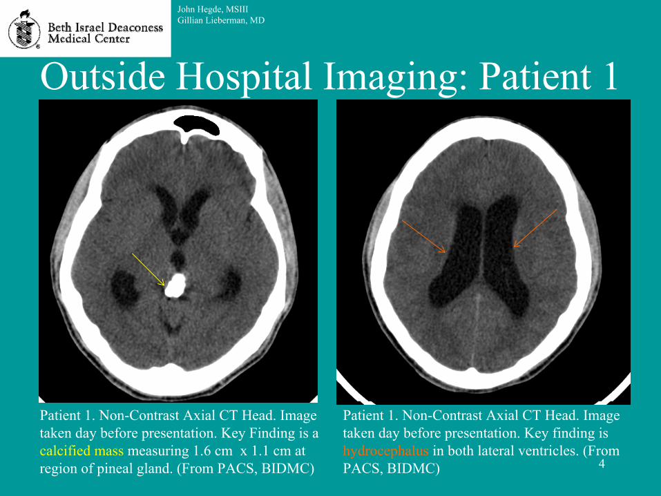
Outside Hospital Imaging: Patient 1
John Hegde, MSIIIGillian Lieberman, MD
Patient 1. Non-Contrast Axial CT Head. Image taken day before presentation. Key Finding is a calcified mass measuring 1.6 cm x 1.1 cm at region of pineal gland. (From PACS, BIDMC)
Patient 1. Non-Contrast Axial CT Head. Image taken day before presentation. Key finding is hydrocephalus in both lateral ventricles. (From PACS, BIDMC)
5
BIDMC Imaging Work-Up: Patient 1
•
MR imaging was performed at BIDMC to better characterize the pineal mass.
John Hegde, MSIIIGillian Lieberman, MD

6
Imaging on Day 1: Patient 1Patient 1. Non-Contrast Sagittal
T1-Weighted MR Head. Image taken on day 1 of presentation. There is hydrocephalus, with dilated lateral and 3rd
ventricles but a normal 4th
ventricle. Also, a hypo-intense to iso-intense signal to gray matter pineal mass
along
the posterior margin of the 3rd
ventricle narrowing the Sylvian aqueduct is seen. The mass is
estimated to be 2 cm x 2 cm x 1.1 cm (4400 mm3), which far exceeds the normal dimensions and volume of the pineal gland.
John Hegde, MSIIIGillian Lieberman, MD
(From PACS, BIDMC)

7
Anatomy: Normal Pineal Gland•
Pineal Gland Facts:–
Originates embryologically
as part of the brain
–
Receives innervation
from peripheral sympathetic nerves
–
Makes melatonin which regulates sleep and circadian cycles
•
Pineal Gland Imaging–
Can appear crescent-like, nodule-
like, or ring-like–
Iso-dense to gray matter on T1 –
Several mm in height, length, and width
–
Volume varies, but about 60 mm3
is common for young adult
Comparison Patient 2. Non-Contrast Sagittal
T1-Weighted MR Head . All structures, including pineal gland,
are normal. (From PACS, BIDMC)
John Hegde, MSIIIGillian Lieberman, MD

8
Additional View of Day 1 Imaging: Patient 1
Patient 1. Non-Contrast Axial T2-Weighted MR Head. Image taken on day 1 of admission. Key finding is an iso-intense to hyper-intense mass
in
the region of the pineal gland. (From PACS, BIDMC)
John Hegde, MSIIIGillian Lieberman, MD

9
Immediate Treatment: Patient 1
•
In order to treat the patient’s hydrocephalus caused by the pineal mass, a ventriculoperitoneal
(VP) shunt was placed.
•
This resulted in improving hydrocephalus over a number of days.
John Hegde, MSIIIGillian Lieberman, MD
10
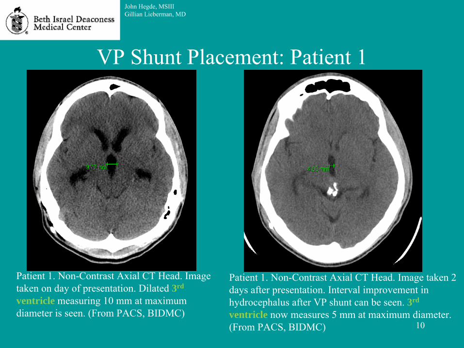
VP Shunt Placement: Patient 1
John Hegde, MSIIIGillian Lieberman, MD
Patient 1. Non-Contrast Axial CT Head. Image taken 2 days after presentation. Interval improvement in hydrocephalus after VP shunt can be seen. 3rd
ventricle now measures 5 mm at maximum diameter. (From PACS, BIDMC)
Patient 1. Non-Contrast Axial CT Head. Image taken on day of presentation. Dilated 3rd
ventricle measuring 10 mm at maximum diameter is seen. (From PACS, BIDMC)

11
Further Imaging Work-Up: Patient 1
•
In order to further characterize the pineal mass, MR head with contrast enhancement was performed.
•
The pineal mass was found to have punctate contrast enhancement and mild overall
enhancement.
John Hegde, MSIIIGillian Lieberman, MD
12
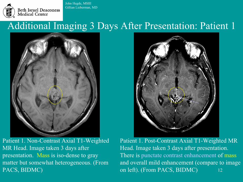
Additional Imaging 3 Days After Presentation: Patient 1
John Hegde, MSIIIGillian Lieberman, MD
Patient 1. Non-Contrast Axial T1-Weighted MR Head. Image taken 3 days after presentation. Mass
is iso-dense to gray matter but somewhat heterogeneous. (From PACS, BIDMC)
Patient 1. Post-Contrast Axial T1-Weighted MR Head. Image taken 3 days after presentation. There is punctate
contrast enhancement
of mass
and overall mild enhancement (compare to image on left). (From PACS, BIDMC)

13
Calcification Distribution: Pineal Masses•
Two types of Calcifications:–
Exploded to periphery•
Pineal parenchymal
tumors–
Engulfed•
Germinomas
John Hegde, MSIIIGillian Lieberman, MD
Comparison Patient 4.
Engulfed
Calcifications (black arrow)
in a Germinoma. Non-Contrast Axial CT.
Comparison Patient 3. Calcifications Exploded to the Periphery (white arrows) in a Pineoblastoma. Non-Contrast Axial CT.
(Images taken from Smith et al. “Lesions of the Pineal Region: Radiologic-Pathologic Correlation”)

14
Calcification Distribution: Patient 1
•
For patient 1, calcification distribution was indeterminate.
Patient 1. Post-Contrast Axial CTA Head. Image taken 2 days after presentation. Calcifications present
heterogeneously throughout mass. (From PACS, BIDMC)
John Hegde, MSIIIGillian Lieberman, MD

15
Summary of Imaging Findings: Patient 1•
CT: –
Well-demarcated, iso-
to
hyper-attenuated mass in region of the pineal gland
–
Obstructive hydrocephalus at Sylvian
aqueduct, improved
after VP shunt placement–
Equal to or < 2 cm in length in any dimension
–
Heterogeneous calcifications of mass, not in periphery or engulfed centrally
•
MR:–
On T1-weighted images, hypo-
to iso-intense to gray
matter–
On T2-weighted images, iso-
to hyper-intense to
gray matter–
Post-contrast punctate
enhancement and some mild overall enhancement
John Hegde, MSIIIGillian Lieberman, MD

16
Symptoms and Signs: Pineal Masses•
Related to mass effect on adjacent structures or invasion of tissues
•
Headache/Nausea/Vomiting–
Occurs from ↑ICP from hydrocephalus
•
Parinaud
syndrome–
Invasion of tectal
plate
–
Symptoms:•
Failed conjugate vertical eye movements
•
Mydriasis•
Failed ocular convergence
•
Blepharospasm
•
Precocious Puberty–
Germ Cell Tumors (increased beta-HCG)
•
Pineal Apoplexy–
Hemorrhage into tumor/cyst
–
Sudden decrease in consciousness associated with a headache
•
Secondary Parkinsonism–
Cause unknown
John Hegde, MSIIIGillian Lieberman, MD

17
Differential Diagnosis: Pineal Masses•
Pineal Parenchymal
Tumors–
Pineocytoma
–
Pineal Parenchymal Tumor of Intermediate
Differentiation–
Pineoblastoma
•
Non-Pineal Parenchymal Tumors
–
Metastasis–
Glioma
–
Germinoma–
Lipoma
–
Meningioma–
Astrocytoma
–
Trilateral Retinoblastoma–
Ependymoma
John Hegde, MSIIIGillian Lieberman, MD
• Pineal Cysts
18
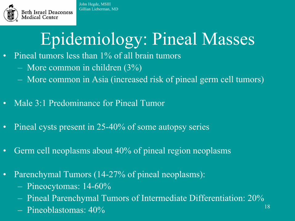
Epidemiology: Pineal Masses•
Pineal tumors less than 1% of all brain tumors–
More common in children (3%)
–
More common in Asia (increased risk of pineal germ cell tumors)
•
Male 3:1 Predominance for Pineal Tumor
•
Pineal cysts present in 25-40% of some autopsy series
•
Germ cell neoplasms
about 40% of pineal region neoplasms
•
Parenchymal
Tumors (14-27% of pineal neoplasms):–
Pineocytomas: 14-60%
–
Pineal Parenchymal
Tumors of Intermediate Differentiation: 20% –
Pineoblastomas: 40%
John Hegde, MSIIIGillian Lieberman, MD

19
Pineocytoma: Comparison Patient 5
Comparison Patient 5. Post-Contrast Sagittal
T1-Weighted MR Image. Pineocytoma
is
enhancing mass in pineal region, resulting in hydrocephalus.
John Hegde, MSIIIGillian Lieberman, MD
(Image above from Smith et al. “Lesions of the Pineal Region: Radiologic-Pathologic Correlation”)

20
Pineal Parenchymal
Tumor of Intermediate Differentiation: Comparison Patient 6
John Hegde, MSIIIGillian Lieberman, MD
Comparison Patient 6. Post- Contrast Axial T1-Weighted
MR Image. Pineal parenchymal
tumor of
intermediate differentiation has enhancement of solid
component of mass.
(Image above from Smith et al. “Lesions of the Pineal Region: Radiologic-Pathologic Correlation”)

21
Pineoblastoma: Comparison Patient 7
John Hegde, MSIIIGillian Lieberman, MD
Comparison Patient 7. Post-Contrast Axial T1-
Weighted MR Image. Pineoblastoma
is the ill-
defined enhancing mass.
(Image above from Smith et al. “Lesions of the Pineal Region: Radiologic-Pathologic Correlation”)

22
Pineal Cyst: Comparison Patient 8
John Hegde, MSIIIGillian Lieberman, MD
Comparison Patient 8.
Post- Contrast Axial T1-Weighted
MR Image. A round, low- intensity cyst (white arrow)
with
an incompletely- enhancing rim is shown. No
nodularity
or hydrocephalus is present.
(Image above from Smith et al. “Lesions of the Pineal Region: Radiologic-Pathologic Correlation”)
23
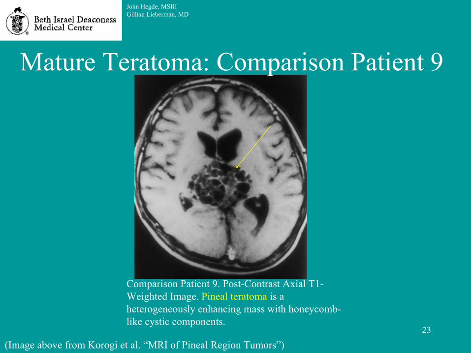
Mature Teratoma: Comparison Patient 9
Comparison Patient 9. Post-Contrast Axial T1-
Weighted Image. Pineal teratoma
is a heterogeneously enhancing mass with honeycomb-
like cystic components.
John Hegde, MSIIIGillian Lieberman, MD
(Image above from Korogi
et al. “MRI of Pineal Region Tumors”)

24
Pineal Germinoma: Comparison Patient 10
John Hegde, MSIIIGillian Lieberman, MD
Comparison Patient 10. Non-Contrast Sagittal
T2-
Weighted MR Image. Pineal Germinoma has
a solid component iso-dense with gray matter and a cystic component iso-dense with CSF.
(Image above from Liang et al. “MRI of Intracranial Germ-Cell Tumours”)

25
Meningioma: Comparison Patient 11
Comparison Patient 11. Non-Contrast Sagittal
T1-
Weighted MR Image. Meningioma (black arrow tips) results in compression of the midbrain, cerebellum, and corpus collosum.
John Hegde, MSIIIGillian Lieberman, MD
(Image above from Korogi
et al. “MRI of Pineal Region Tumors”)

26
Diagnosis Summary of Selected Pineal Mass Imaging FindingsPineocytoma CT: well-demarcated, iso-
to hyperattenuating
lesion, peripheral calcifications MR: well-circumscribed, hypo-
to iso-intense on T1 and hyper-intense on T2. Avid, homogeneous enhancement post-contrast
PPTID No specific findings separate PPTID from pineoblastoma
or pineocytoma
Pineoblastoma No specific findings separate pineoblastoma
from pineocytoma
or PPTID CT: may be large (>3 cm), lobulated
and hyperattenuatedMR: Heterogeneous enhancement with sold portion hypo-
to iso-intense on T1 and iso-
to hyper-intense on T2. Heterogeneous enhancement post-contrast
Germ Cell Tumor
CT: sharply circumscribed, hyperattenuating
lesion, engulfing calcificationsMR: solid mass with cystic components. Iso-
to hyper-intense on T1 and T2 images. Avid, homogeneous enhancement post-contrast
Teratoma CT: multi-loculated, lobulated
lesion with foci of fat attenuation, calcification, and cystMR: foci of T1 shortening due to fat and variable signal intensity from calcification. T2 images have soft-tissue iso-
to hypo-intensity. Soft tissue has post-contrast enhancement
Pineal Cyst CT: round or oval, thin-walled, well-circumscribed MR: Intensity similar to CSF on T1& T2. Post-contrast: incomplete cyst wall enhancement
Meningioma CT: hyperattenuating, calcifications in 15-20%. Avid enhancement post-contrast. Dural tail.MR: Hypo-
to iso-intense on T1 and iso-
to hyper-intense on T2
John Hegde, MSIIIGillian Lieberman, MD
(Information from Smith et al. “Lesions of the Pineal Region: Radiologic-Pathologic Correlation”)

27
Diagnosis: Patient 1
•
No diagnostic imaging findings for the mass were seen.
•
For any suspected pineal parenchymal tumor, a biopsy is necessary to diagnose
tumor type.
John Hegde, MSIIIGillian Lieberman, MD

28
Diagnosis: Brain Biopsy Results for Patient 1•
Highly cellular tumor, monomorphic
cells in sheets and rosettes,
with mild atypia
and rare prominent nucleoli
•
No Mitotic Figures
•
Tumors positive for neuronal tumor markers
•
Tumor classified as a Pineal Parenchymal Tumor of Intermediate Differentiation
•
However, given small pathology specimen, pineocytoma
or pineoblastoma
could not be ruled-out
John Hegde, MSIIIGillian Lieberman, MD
29
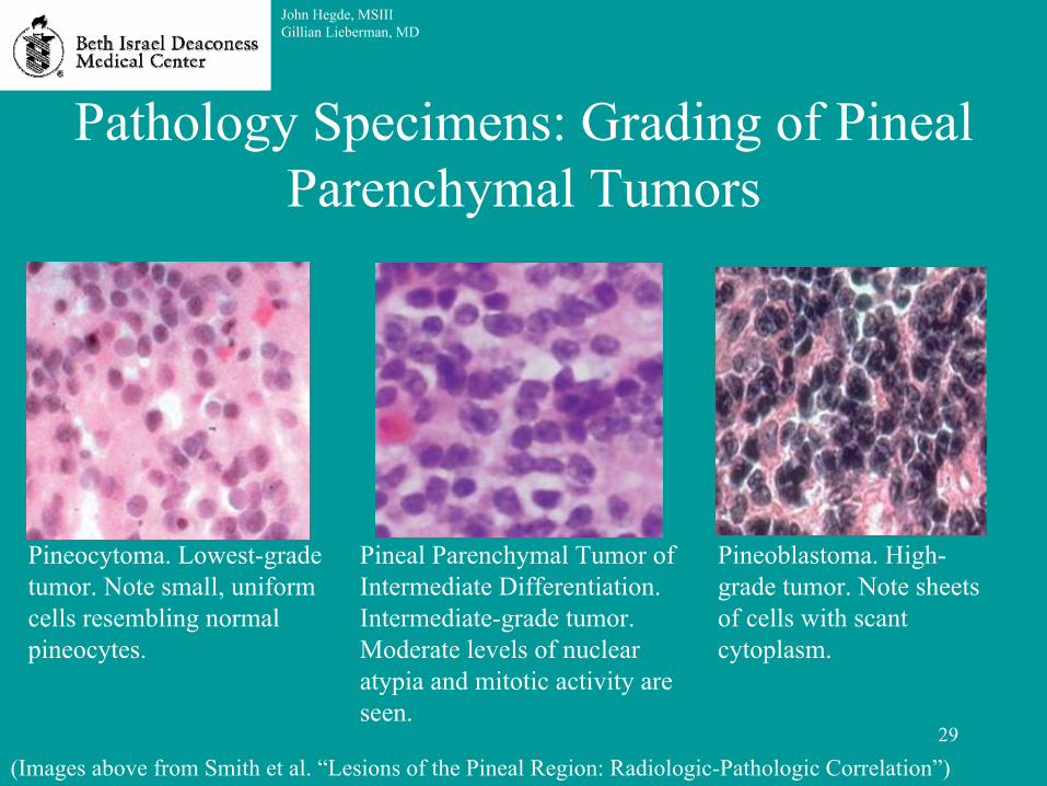
Pathology Specimens: Grading of Pineal Parenchymal
Tumors
John Hegde, MSIIIGillian Lieberman, MD
Pineocytoma. Lowest-grade tumor. Note small, uniform cells resembling normal pineocytes.
Pineal Parenchymal
Tumor of Intermediate Differentiation. Intermediate-grade tumor. Moderate levels of nuclear atypia
and mitotic activity are seen.
Pineoblastoma. High-
grade tumor. Note sheets of cells with scant cytoplasm.
(Images above from Smith et al. “Lesions of the Pineal Region: Radiologic-Pathologic Correlation”)

30
Treatment: Pineal Parenchymal
Tumors•
Acute Hydrocephalus–
VP Shunt
•
Pineocytoma/PPTID–
Surgical Resection +/-
Radiotherapy
–
Radiotherapy often only recommended if tumor appears to behave like pineoblastoma
•
Pineoblastoma–
Usually Surgical Resection + Radiotherapy + Chemotherapy
–
Craniospinal
irradiation important to reduce risk of metastasis or potential to seed CSF
–
Radiotherapy not universally recommended–
Chemotherapy used to reduce common leptomeningeal
seeding
John Hegde, MSIIIGillian Lieberman, MD

31
Emerging Treatment: Stereotactic Radiosurgery•
Emerging as alternative to surgical resection and traditional radiotherapy for pineocytomas
•
Precise radiation fields defined by MRI or CT to reduce damage to surrounding brain are used
•
Many non-parallel beams of radiation converge on a small area of brain to target tumor
•
Craniotomy and general anesthesia avoided•
Preliminary results suggest higher survival rates than using surgical resection and/or traditional radiotherapy
John Hegde, MSIIIGillian Lieberman, MD

32
Clinical Course Update: Patient 1•
Following diagnosis with PPTID:–
CSF found to be negative for malignant cells
–
Clinical correlation is with an intermediate grade pineal tumor – unlikely to be pineoblastoma
–
Surgical risk deemed too high given patient’s young age
–
Stereotactic radiosurgery
also considered risky given patient’s young age, as there is risk of damage to tectum
and for late
sequelae
–
Stereotactic radiotherapy suggested
John Hegde, MSIIIGillian Lieberman, MD

33
Prognosis: Pineal Tumors•
In SEER Data from 1973-2005:–
5 year survival for all tumors 65%
–
Germ Cell Tumors 79%–
Gliomas
61%
–
Pineal Parenchymal
Tumors 47%
(Information taken from Al-Hussaini
et al., “Pineal Gland Tumors: Experience from the SEER Database”)
John Hegde, MSIIIGillian Lieberman, MD
34
Summary•
Pineal masses are rare lesions (<1% of primary brain tumors)
•
Pineal tumors can have various presentations, including headaches, nausea/vomiting, and visual defects (Parinaud
syndrome)
•
MRI is most helpful for characterizing brain lesions like pineal masses, although CT can help for calcification distribution
•
No pathognomonic
imaging findings to differentiate between parenchymal
pineal masses
•
Pineal parenchymal
tumors usually iso-
to hyperattenuating
on CT with calcifications “exploded to the periphery”
•
Pineal parenchymal
tumors usually hypo-
to iso-intense on T1, iso-
to hyper-intense on T2, and enhancing post-contrast
•
Correlation with other data can narrow differential diagnosis•
Brain masses are difficult to evaluate given limited biopsy potential
•
Stereotactic radiosurgery
is increasingly being used for pineal masses
John Hegde, MSIIIGillian Lieberman, MD

35
References•
Al-Hussaini
M, I Sultan, N Abuirmileh, I Jaradat, I Qaddoumi. Pineal Gland Tumors: Experience from the SEER Database. Journal of Neuro-Oncology. 2009; 94(3): 351-358
•
Chumas
P, A Tyagi, J Livingston. Hydrocephalus--What's New?. Arch Dis Child Fetal Neonatal Ed. 2001; 85:F149
•
Drake JM, JR Kestle, R Milner, G Cinalli, F Boop, J Piatt, S Haines, SJ Schiff, DD Cochrane, P Steinbok, N MacNeil. Randomized Trial of Cerebrospinal Fluid Shunt Valve Design in Pediatric Hydrocephalus. Neurosurgery. 1998; 43(2):294-303
•
Korogi
Y, M Takahashi, Y Ushio. MRI of Pineal Region Tumors. Journal of Neuro-Oncology. 2001; 54: 251-261
•
Liang L, Y Korogi, T Sugahara, I Ikushima, Y Shigematsu, T Okuda, M Takahashi, M Kochi, Y Ushio. MRI of Intracranial Germ-Cell Tumors. Neuroradiology. 2002; 44: 382-388
•
Mori Y, T Kobayashi, T Hasegawa, et al. Stereotactic Radiosurgery
for Pineal and Related Tumors. Prog Neuro Surg. 2009; 23: 106
•
Moschovi
M, GP Chrousos. Pineal Gland Masses. www.uptodate.com. Accessed 11/10/2010•
Nakamura M, N Saeki, Y Iwadate, K Sunami, K Osato, A Yamaura. Neuroradiological
Characteristics of Pineocytoma
and Pineoblastoma. Neuroradiology. 2000; 42: 509-514•
Smith AB, EJ Rushing, JG Smirniotopoulos. Lesions of the Pineal Region: Radiologic Pathologic Correlation. RadioGraphics. 2010; 30(7): 2001-2020
•
Sumida M, AJ Barkovich, TH Newton. Development of the Pineal Gland: Measurement with MR. American Journal of Neuroradiology. 1996;
17: 233-236
John Hegde, MSIIIGillian Lieberman, MD

36
Acknowledgements
•
Dr. Gul
Moonis, Staff Neuroradiologist, Beth Israel Deaconess Medical Center
•
Dr. Gillian Lieberman, Core Radiology Clerkship Director, Beth Israel Deaconess Medical Center
•
Emily Hanson, Radiology Clerkship Medical Student Education Coordinator, Beth Israel Deaconess Medical Center
John Hegde, MSIIIGillian Lieberman, MD