imaging infection prof john buscombe - higher education b lectures 2016/infection... · • tc-99m...
TRANSCRIPT

Imaging infection Prof John Buscombe

inflammation in·flam·ma·tion (ĭn'fləә-mā'shəәn) n.
A localised protective reaction of tissue to irritation, injury or infection,characterized by pain, redness, swelling, and somet
imes loss of function.
infection in·fec·tion (ĭn-fěk'shəәn) n.
Invasion by and multiplication of pathogenic microorganisms in a
bodily part or tissue, which may produce subsequent tissue injury and progress to overt disease through a variety of cellular
or toxic mechanisms.
The American Heritage® Stedman's Medical Dictionary Copyright © 2002, 2001, 1995 by Houghton Mifflin Company
Inflamma&on and Infec&on

Slide 3

• Defensive host response to invading foreign bodies and necro4c 4ssue • Capable of causing 4ssue damage.
• Components: vascular reac4on and a cellular response • Both ac4vated by mediators derived from plasma proteins and various inflammatory
cells.
• The steps of the inflammatory response can be remembered as the five Rs: ü recogni4on of the injurious agent ü recruitment of leukocytes ü removal of the agent ü regula4on (control) of the response ü resolu4on (repair)
Inflamma&on : General Features


Increasing vascular permeability leads to the movement of protein-‐rich fluid and even blood cells into the extravascular 4ssues
Vascular permeability

The sequence of events in the recruitment of leukocytes from the vascular lumen to the extravascular space consists of:
• margina4on and rolling along the vessel wall;
• firm adhesion to the endothelium;
• transmigra4on between endothelial cells;
• migra4on in inters44al 4ssues toward a chemotac,c s,mulus
Leukocyte recruitment


Acute inflamma4on is rapid in onset and of short dura4on, las4ng from a few minutes to as long as a few days, and is characterized by fluid and plasma protein exuda4on and a predominantly neutrophilic leukocyte accumula&on.
Chronic inflamma4on may be more insidious, is of longer dura&on (days to years), and is typified by influx of lymphocytes and macrophages with associated vascular prolifera4on and fibrosis (scarring).
Acute or chronic inflamma&on

• Infec4ous diseases are causes of death among the every young, the elderly, people with AIDS or chronic diseases and pa4ents receiving immunosuppressive drugs.
• In developing countries, unsanitary living condi4ons and malnutri4on contribute to a massive burden of infec4ous diseases that kills more than 10 million people each year.
• Infec&ous agents belong to a wide range of classes and vary greatly in size, ranging from prion protein aggregates of under 20 nm to 10 m tapeworms.
Infec&on: microbial pathogenesis

Chronic (mononuclear cell-‐mediated)
inflamma4on
Acute (granulocyte-‐mediated)
inflamma4on
Nuclear Medicine imaging of inflamma&on/infec&on

Pagina 12
Inflamma4on is part of the complex biological response of vascular 4ssues to harmful s4muli, such as pathogens, damaged cells, or irritants.
Imaging “inflamma4on, in nuclear medicine, means to image all those non specific chemical, vascular and cellular phenomena associated to inflammatory diseases.
Infec4on is the invasion of a host organism's bodily 4ssues by disease-‐causing organisms, their mul4plica4on, and the reac4on of host 4ssues to these organisms and the toxins they produce.
Imaging “infec4on” in nuclear medicine, means to specifically detect the presence of pathogens.
Nuclear medicine techniques aim at differen&a&ng “sterile inflamma&on” from “infec&on” and the two terms cannot be used as synonyms
Signore A. EJNMMI Research 2013;3:8
Inflamma&on vs infec&on

Radiopharmaceuticals Targe&ng the host immune system
HIG
Labelled White Blood Cells
Monoclonal An4bodies against Granulocytes
IL-‐8 (acute) IL-‐1, IL-‐2, Monoclonal An4bodies against TNF α (chronic) 18Fluorodeoxyglucose (FDG) (images the hypermetabolic state) 67Gallium Citrate, 68Gallium Citrate
Slide 13
Targeting the infectious agent
67Gallium Citrate, 68Gallium Citrate
Labelled anti-microbials
Labelled vitamins
Labelled antimicrobial peptides

Single photon imaging of infection
• Success depends on number of factors • Understanding the clinical background of the patient
immunocompetent vs immunodeficient • Understanding when a sensitive test is needed and
when a specific agent is needed • Being pragmatic, look at availability, cost and time • Always do the best study you can for a specific clinical
problem and situation in 2016 that means SPECT/CT or PET/CT
• Some infections only seen by some techniques • Every patient is different

Is there a role for fusion imaging in infection/inflammation?
• Potential for better localisation • Potential for improved specificity • Is time taken justified • Will use of machines be taken up by more
“trendy” topics such as cancer/endocrine • Will it be worth the effort

Gallium-67 citrate • No cell labelling • 90keV, 190keV, 300keV, 394keV gammas med energy
collimator • Poor dosimetry limits activity that can be given • Indications now limited
– Sarcoid – Spinal infections – Immunocompremised
• FDG PET-CT can do some of these • SPECT-CT of suspect areas at 24 or 48 hours

Ga-67 in sarcoid Panda sign, lacrimal and salivary glands
Lamba sign medias4num and hilar nodes
Diffuse lung uptake
Lymphadanopathy (symmetrical)
Joints
Liver-‐diffuse

More specific agents; In-111 WBCs
• In-111 WBC – Limited access, needs cell labelling – 174 keV and 247 keV needs medium energy
collimator – Poor dosimetry limits activity to 20MBq – Gold standard good specificity – All but spinal infections – Imaging 4 and 20 hours p.i. – SPECT/CT at 24 hours

Tc-99m HMPAO labelled WBCs
• Tc-99m HMPAO WBC – Needs cell labelling – Max 200MBq but lower radiation dose – Theoretically less specific than In-111 WBC
but no real evidence in skilled hands – All but spinal infections – Imaging normally 30 mins and 3-4 hours – Can perform late imaging at 24 hours – SPECT-CT normally at 3-4 hours
Slide 19
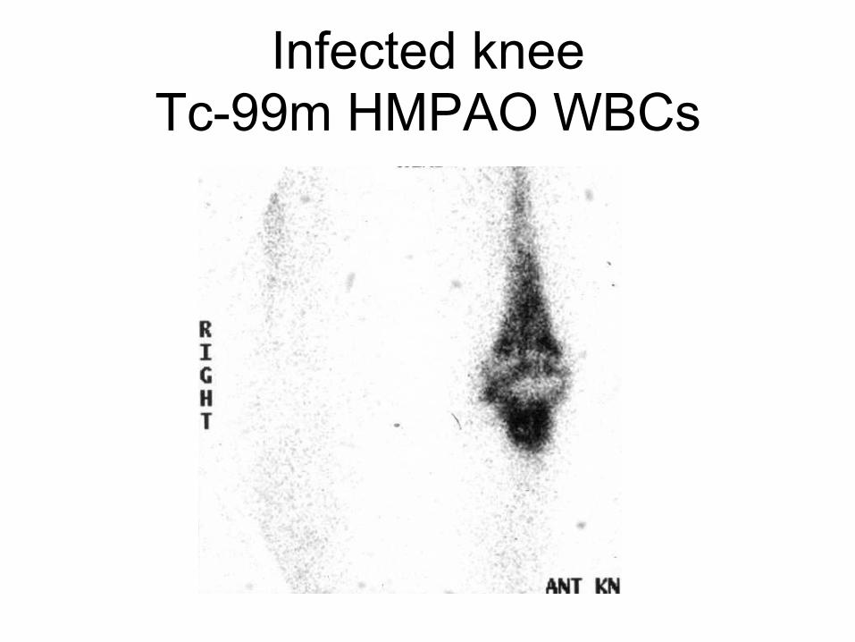
Infected knee Tc-99m HMPAO WBCs

Antibodies • Tc-99m granuolscint
– Anti-CD66 on granulocytes – No cell labelling – Use similar to Tc-99m HMPAO WBC – Widely used in Europe
• Tc-99m leucoscan – No cell labelling – Mechanism not clear – No Fc on antibody so can do repeat scanning – Mainly in bone/joint infection
• Both agents imaging normally 1 and 4 hours (sometimes 24hours)
• SPECT-CT normally at 24 hours

A. PET/CT whole body MIP projection showing high 18 F-FDG uptake around the pre peritoneal part and cutaneous exit of the driveline(red arrow) and in the LVAD pocket (green arrow). B. CT scout view showing LVAD pocket and driveline. C. Anterior planar scintigraphy 24hours after injection of the Tc 99m-anti-leucocyte antibodies showing uptake along the driveline
AGAB more specific than FDG
Images from Dr A Boubaker

Why spend $600,000 on this
Slide 23

SPECT-CT in infection imaging should be our
standard method. • Roach et al 2006 NMC • Looked at 50 scans including bone and
Ga-67 SPECT-CT • 16% of patients had minor change 11%
major change c/w SPECT alone • Almost all to do with localisation and
improved specificity • Specificity itself improved by 26%

Specific results identifying infection
• Inquie et al J Comp Assist Tom 2007 • 16 patients (11 In-111 WBC and 6 Ga--67) • SPECT/CT images yielded "added value"
for anatomical localization in 65%, diagnostic confidence in 71%, and altered interpretations in 47% of cases

WBC SPECT-CT showing an infected iliac graft Bar Shalom et et JNM 2006 48% more accurate than planar WBC
imaging

Ga-67 citrate in an infected renal transplant; Nowosinska et al
WJNM 2015

Other use of Ga-67 is discitis • 85 year old man • Severe back pain • CRP 250 • Gram positive rods in
blood • Pacemaker

Ga-67 citrate
• In PCKD residual infection can occur in native cysts
• Ga-67 after 24 hours has no normal renal uptake
• Therefore focal uptake in cyst infection
• Helped by SPECT-CT

Tc-WBC scintigraphy vs conventional radiological imaging in management of late, low-grade vascular prosthesis infections
Erba et al, EJNMMI 2014
55 patients, susp. late & low grade graft infection } Tc-WBC (planar +SPECT/CT) } 47 graft infection, 8 extra-graft infectious foci } Tc-WBC positive: 90% (43/47, 20/43 also extra-graft) } SPECT/CT: reduced # FP in 37% patients
Test Sensitivity Specificity
SPECT 85% 63% SPECT/CT 100% 100% US 34% 75% CT 49% 83% Clinical criteria 68% 63%

In-WBC imaging of infected vascular grafts
Two different clinical cases .Sometimes uptake is obvious in the heavily pre-treated patient it can be subtle
NB. The specificity of In-111 WBCs and the 24 hour image can be advantageous

In-111 WBC SPECT-CT in infected THR Though planar image was positive the SPECT-CT images allow for good localisation of the labelled WBCs and show where the infection is sited so drainage and anti-biotics used

The Diabetic Foot – the Value of WBC-SPECT/CT
Slide 33
WBC scan: • Pros: Diagnosis of infection • Cons: not good enough [poor] for localization (to soft tissues and/or bone) Solved with SPECT/CT! • Single study • Accurate spatial localization
• extremities are less prone to motion • close proximity of structures in a small anatomic region
• Decreased radiation exposure; lower cost

Skin ulcer, pus secreting, tenderness & swelling 1st right toe
1h
4h
24h SPECT MIP
Infected soft tissue ulcer, plantar aspect 1st right toe No evidence of osteomyelitis 5 months follow up
Tc-WBC SPECT/CT Diabetic Foot

WBC Scan in Diabetic Foot Potential Pitfalls
Slide 35
Tc-WBC uptake in hyperdense foreign body secondary to soft tissue infection – no OM!

44 year old male
diabetic since aged 6
Had carpet changed
pain in heel
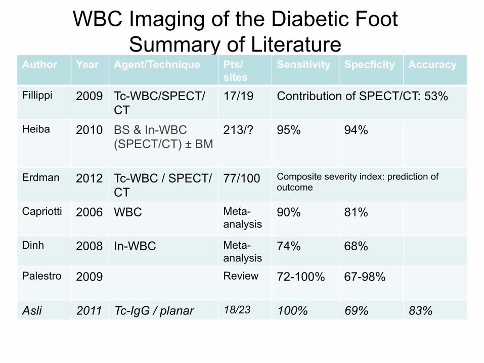
WBC Imaging of the Diabetic Foot Summary of Literature
Author Year Agent/Technique Pts/sites
Sensitivity Specficity Accuracy
Fillippi 2009 Tc-WBC/SPECT/CT
17/19 Contribution of SPECT/CT: 53%
Heiba 2010 BS & In-WBC (SPECT/CT) ± BM
213/? 95% 94%
Erdman 2012 Tc-WBC / SPECT/CT
77/100 Composite severity index: prediction of outcome
Capriotti 2006 WBC Meta-analysis
90% 81%
Dinh 2008 In-WBC Meta-analysis
74% 68%
Palestro 2009 Review 72-100% 67-98%
Asli 2011 Tc-IgG / planar 18/23 100% 69% 83%

• Routine use of In-111 WBC SPECT-CT
• 4 year old female multiple problems
• Temp after bilateral cochleal implants
• ?infected • 24 hr SPECT/CT only • Retropharyngeal
abscess
When specificity is needed

Glucose uptake into tumours

FDG and inflammation or infection
• Increased uptake of FDG occurs when lymphocytes activated. Ishimori JNM 2002
• Uptake not just related to perfusion but active uptake when FDG increased compared to FLT where no increased uptake van Gaarde JNM 2004
• Uptake of FDG related to hypoxia and presence of cytokines Matsui JNM 2009

Uptake and cytokines-Matsui JNM
Macrophages Neutrophils
Fibroblasts

Imaging inflammation
• Most inflammatory diseases can be imaged using scintigraphic technique
• Some techniques are blood flow dependent such as 2 phase bone and Tc-99m HIG
• Some methods dependent on bone turnover such as Tc-99m bone scintigraphy and F-18 NaF
• Other methods image inflammation more directly eg Labelled WBCs, and F-18 FDG
• All can be quantified so useful in research

Sarcoid
• Disseminated inflammatory disease • Characterised by granuloma • Various patterns
– Salivary/lacrimal glands – Lymph nodes – CNS – Skin – Joint – Pulmonary- the most dangerous

Imaging in sarcoid • Normally diagnosis clinical followed by biopsy • 50% of patients have raised serum ACE • If lymph nodes involved may see symmetrical enlarged
mediastinal/hilar nodes the lambda pattern • Since 1966 Ga-67 citrate used
– Not very trendy – High radiation dose

Use of F-18 FDG • Lymphocytes very FDG avid • Much improved resolution • Lower radiation dose (5mSv vs 18mSv) • Confirm sites of active disease esp in the abdomen • Quantify uptake which may be useful in treatment
monitoring

FDG vs Ga-67 • Nishiyama et el JNM
2006 • 18 sarcoid patients
imaged with Ga-67 and FDG.
• Pulmonary disease Ga-67 81%, FDG 100% - mean SUVmax 7
• Extra-pulmonary disease Ga 48%, FDG 90% mean SUVmax 5
A= Ga-‐67 B= F-‐18 FDG C= F-‐18 FDG post therapy

Radiotracer utilisation in RA Indication MDP
Bone Scan
Sodium Flouride PET/CT
IgG (HIG) imaging
FDG PET/CT
Confirmation of suspected diagnosis
Yes Yes Yes Probably
Depiction of joints involved
Yes Yes Yes Yes
Extra-articular disease
No No No Yes
Therapy response assessment
Maybe No Yes Yes
Suitability for radiosynovectomy
Yes Yes No No

Bone scintigraphy in arthritis • For local issues 2-phase imaging is useful though not
good for axial skeleton • Can image whole body for same radiation dose – 2mSv • Normally extra images of hands and feet • Pattern may be useful • Personal view SIJ quant not very helpful

PALM VIEW to depict small joints
Whole Body Bone Scan in RA
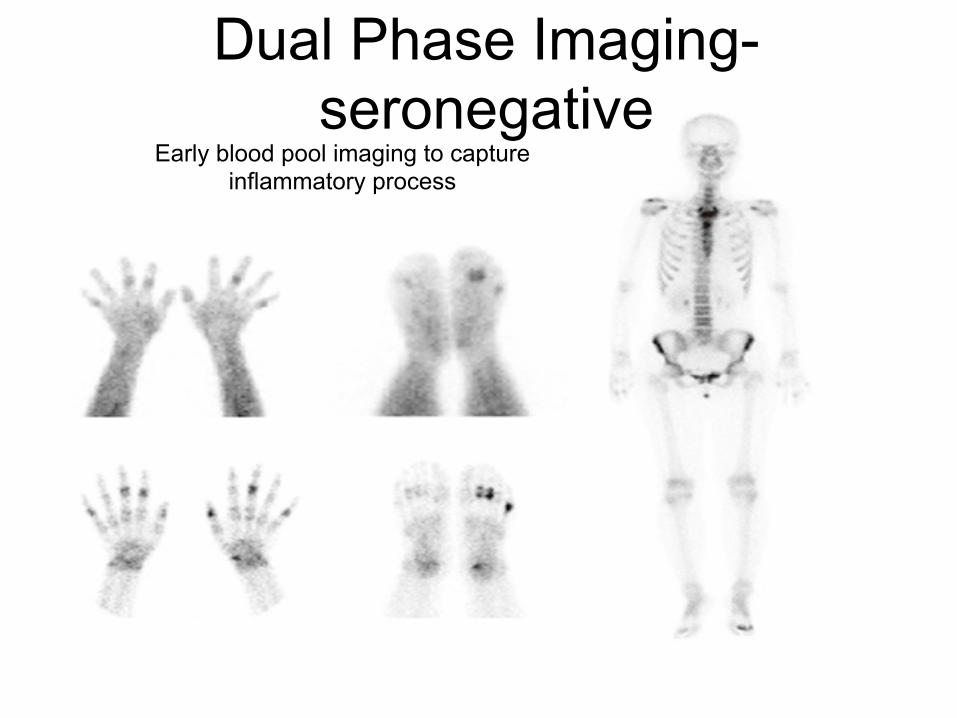
Early blood pool imaging to capture inflammatory process
Dual Phase Imaging-seronegative

FDG -psoriatic arthritis

Atlantoaxial synovitis Extra-articular disease
18F FDG PET/CT-‐staging a ligle like cancer

Using FDG in RA • Beckers et al JNM 2004 • 21 patients with active RA • FDG imaging with views of knees and hands • FDG positive in 68% joints though 75% of joints swollen
and 79% painful • Good correlation with increased blood flow on Doppler
ultrasound

FDG uptake in RA Beckers et al JNM 2004
Normal
Pa4ent with RA

Monitoring response • Vijavant et al WJR 2012. 17 newly diagnosed RA and 11
newly diagnosed sero-neg arthropathy • Good correlation between symptoms and sites of
increased uptake of FDG • Change in SUVmax correlated well with clinical response
and change in CRP

FDG before and after Tx Vijavant et al WJR 2012

F-18 FDG in RPF • Small volume of published work • Concentrates on the use of F-18 FDG in following
inflammation in RPF • Jansen et al E J Int Med 2012 • 26 patients with iRPF 20 positive with FDG PET
correlated with high initial CRP • F-18 FDG reduction correlated with reduction in
inflammatory markers not CT thickness of RPF
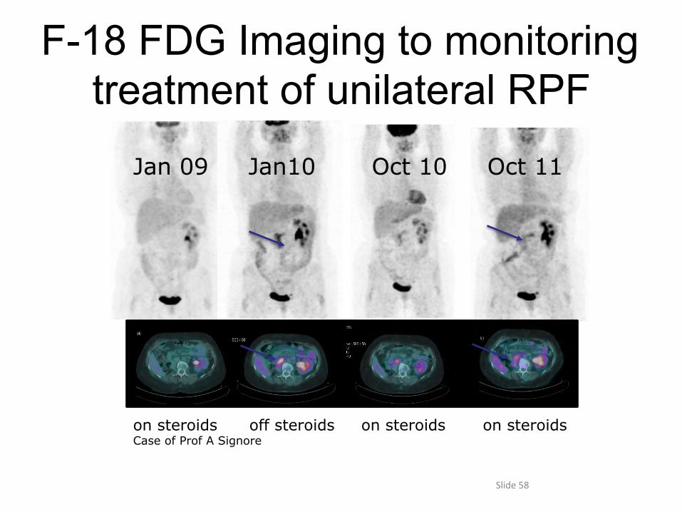
F-18 FDG Imaging to monitoring treatment of unilateral RPF
Slide 58
Jan 09 Jan10 Oct 10 Oct 11
on steroids off steroids on steroids on steroids Case of Prof A Signore
F-18 FDG in vasculitis • Walter et al EJNMMI 2005 used F-18 FDG imaging in 26
patients with giant cell artertitis • Good correlation with wall thickness on CT, ESR and
CRP • Papathanasiou et al from UCL BJR imaged 16 patients
with GCA before and after their forst dose of steroids • Mean SUVmax dropped from 3.38 to 2.32 with treatment

Giant cell arteritis

Aortitis

FDG Imaging in Infection Image Interpretation – False Positive
Physiologic FDG uptake • oropharynx, vocal cords • Cervical muscle, fat uptake (vs.
LN) • GIT (focal or segmental) • Ureter • Salivary glands, lymphoid tissue • LN proximal to tissue injection • Skin folds & sweat gland in
axilla • Bone marrow uptake • Ovarian & endometrial uptake • Brown Fat • Lactating Breasts
Benign FDG uptake & artifacts
• Artifacts (e.g. injection, AC, contamination, metallic devices)
• Benign bone lesions (fracture, degenerative changes)
• Uptake in foreign body aseptic reaction (e.g. implants, grafts, stents)
• After treatment (e.g. healing scar, chemo/radiation & distorted anatomy
• Uptake in [un]known malignancies
Slide 62

FDG Imaging of Infection Pitfalls Associated with Adminstration of Drugs
• Antibiotics - thought to lower FDG uptake in infection (no studies confirming this)
• Metformin (antihyperglycemic) - associated with intense diffuse FDG uptake in small & large bowel, could mask infectious/inflammatory lesions (=FN) or be misinterpreted as severe colitis (=FP). Resolved by 2 days discontinuation.
• Steroids - may result in FN, should be avoided or on low dose if possible. Potential mechanisms: – resolution of inflammation – Inhibition of peripheral glucose uptake (reduce GLUT expression
on cell surface) – effect on liver uptake with lower FDG availability
Slide 63

M, 67, advanced parotid ca s/a total parotidectomy &
radiotherapy (1y) FUO
Focal FDG uptake in lt. maxilla
Dental abscess
FDG Imaging of Infection

PET and FUO • Bleeker-Rovers et al EJNMMI 2004 • Nijmegen group • 35 patients with FUO imaged • Diagnosis conformed in 19 • 37% of scans clinically useful • 65% of the positive scans clinically useful • PPV 87%, NPV 95%

Peritonitis

FDG in infective discitis
F-18 FDG in neutropaenic patients
• Vos et al EJNMMI 2102 • 28 patients neutropaenic following
chemotherapy • 26 patients FDG positive • 18 in GI Tract • 9 around CVC lines • 7 in the lungs • Found bacterial and fungal disease

Cyptococcus in patient post BMT

Diabetes & Infection • Diabetics – increased propensity to infections • Unclear if hyperglycemia is an independent risk factor • Host-specific factors predisposing diabetics to infection:
� impairment of immune response induced by hyperglycemia � vascular insufficiency (local tissue ischemia) � neuropathy (unnoticed, ignored skin ulcers, urinary stasis) � skin & mucosa pathogens (Staph, candida)
• Frequent type of infections: � Foot � Urinary tract � Fungal, malignant otitis externa � Cholecystitis , Pyomyositis, Necrotizing fasciitis

Diabetic foot blood glucose – 10.6 mmol/l TP study
Diabetic patient, vascular graft blood glucose – 4.7 mmol/l FN study
Osteomyelitis 4th metatarsus Infected surgical wound
FDG Imaging in Infection Diabetes & Hyperglycemia, Specific Considerations

Diagnostic Accuracy of FDG-PET/CT in Hyperglycemia & Diabetes [n=443 Patients]
Rivkin et al, JNM 2010
• Hyperglycemia but not DM affect FDG-PET/CT detection rate of cancer (p<0.05) • Neither DM nor hyperglycemia had a significant impact on false negative rate of FDG imaging in infection
Infection & Inflammation Cancer p
No. pts False negative rate No. pts False negative rate
Hyperglycemia 19/123 0/11 (0%) 84/320 6/56 (10%) NS
Normo-glycemia 104/123 4/54 (7%) 236/320 7/181 (4%) NS
P NS P<0.05 Diabetes Mellitus 42/123 2/26 (8%) 183/320 8/122 (7%) NS
No diabetes 83/123 2/39 (5%) 137/320 5/115 (4%) NS
P NS NS

FDG-PET/CT Accuracy in Hyperglycemic & Diabetes Rabkin et al, JNM 2010
Neither DM nor hyperglycemia had an impact on FN rate of FDG in infection [High glucose levels but not DM affected FDG detection rate of cancer (p<0.05)] Different response: ? different intracellular glycogen storage • inflammatory cells: can mobilize intracellular glycogen when
plasma glucose levels are low • [tumor cells: low storage capabilities, need for extracellular glucose
supply] These data were used as recommendations in the EANM/SNM guidelines for FDG imaging in infection-inflammation

M, 60, Diabetes, non-healing wound lt. foot, susp. osteomyelitis
Early: increased blood pool in region of 3rd left toe
Delayed and Late: Focal uptake in 3rd & 5th left toes
3-phase Tc-99m MDP Bone Scintigraphy
2H 24H Blood Pool

M, 60, Diabetes, non-healing wound lt. foot, susp. osteomyelitis
SPECT: • Focal uptake 2nd, 3rd , 5th lt. metatarsus CT: • Wound between 2nd and 3rd toe • Wound at lateral aspect of the foot • Bone destruction and periosteal reaction
Tc99m-MDP SPECT/CT
2H BP

FDG-PET/CT: Focal FDG uptake 3rd metatarsus Single site of osteomyelitis
M, 60, Diabetes, non-healing wound lt. foot, susp. osteomyelitis
Bone SPECT/CT FDG
PET/CT
BP
Surgery (2 days later): Osteomyelitis & fracture 3rd metatarsus Additional metatarsal fractures

FDG –PET/CT Performance Indices for Vascular Graft Infection
Bruggink JL et al, Eur J Vasc Endovasc Surg, 2010, 25 patients
} FDG sensitivity 93%, specificity 70%, PPV 82%, NPV 88% } CT 56% 57% 60%
58%
Spacek et al., EJNMMI, 2009, 76 patients Diagnostic criteria (quantitation lesion/aorta>1.7)
} Focal intense uptake + irregular CT boundaries: 97% PPV } No uptake and regular CT boundaries: 95% NPV } Inhomogeneous uptake + irregular CT boundaries: 77%
PPV
Keidar et al, JNM 2007, 39 patients
Sens: 93%, Spec:91%, PPV: 88%, NPV:96% } Accurate diagnosis of infection } Differential diagnosis } Precise localization to soft tissues ± graft

Infected Vascular Graft Additional Findings on FDG-PET/CT
Slide 78
M, 62, s/a aorto-bifem, fem-fem & bilateral fem-pop graft insertion FDG-PET/CT uptake of mild intensity in soft tissues of left thigh at the margins of a hypodense soft tissue lesion, with a “cold” center: consistent with post-surgery seroma Resolution of the findings on the study performed 6 mo later
Early infection 6 mo later

Warning FDG is not specific
Slide 79
41 year old swinging fever. FDG looks like an abscess but is Hodgkin’s Lymphoma in adrenals Case supplied by Dr Gnanasegaran

Guidelines

Summary-single photon vs FDG PET
• Single photon • A choice of agents for
different types of patients and infections
• Immunocompetent • SPECT-CT is now our
standard • Need cell labelling • Time to do scan • Offers specificity
• FDG PET-CT • FDG can find infection
and inflammation • Though more expensive
in England paid by NHS England
• May be quicker to get results
• Immunocompetent and immunodeficient
• Sensitivity better, specificity may be an issue