imaging in prgnancy
TRANSCRIPT

Seeing Through a Pregnant Seeing Through a Pregnant MotherMother: : The Balance between The Balance between Downright Hazards and Upright Downright Hazards and Upright
BenefitsBenefitsDr. Chai Ming Cheng
Obstetrician and Gynaecologist

Scenarios…Scenarios…36-year-old pregnant
lawyer at 34 weeks POAK/C bronchial asthmaFever, cough, SOB for 3/7O/E: Tachypnoeic,
crepitation and ronchiCXR TRO pneumonia but
strongly refused by her


Scenarios…Scenarios…16-year-old girl complaining of
abdominal distension for the past 5 month.
Ass. with nausea, vomiting. She has no BO for the past four days
O/E: distended abdomen, soft but tense
AXR TRO Intestinal obstruction


ContentContentIntroductionIonizing Radiation
◦X-ray◦CT-scan◦Nuclear Medicine
MRISGH O&G Radiology ConsentsTake Home Messages

IntroductionIntroductionImaging studies are important
adjuncts in the diagnostic evaluation of acute and chronic conditions
Confusion about the safety in pregnancy results in unnecessary avoidance of useful diagnostic tests
Health care providers should weight the risks and benefits

IONIZING RADIATIONIONIZING RADIATION
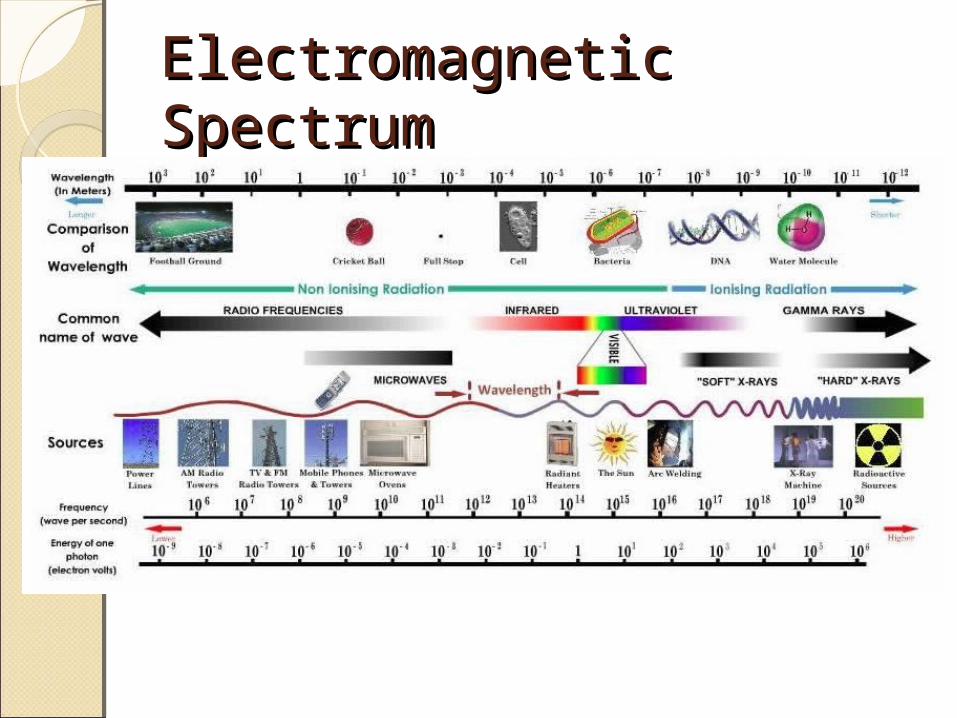
Electromagnetic SpectrumElectromagnetic Spectrum

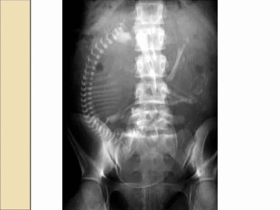
X-ray – DiagnosisX-ray – Diagnosis

X-ray – DiagnosisX-ray – Diagnosis

X-ray - DiagnosisX-ray - Diagnosis

Ionizing RadiationIonizing RadiationAbsorbed dose
◦Amount of energy deposited per kilogram of tissue
◦1 Rad = 10 mGyAccepted background cumulative
dose of ionizing radiation = 5 Rad (50 mGy)

Ionizing RadiationIonizing RadiationBackground radiation to fetus
during entire pregnancy = 1mGy (0.5-1.6 mGy)◦Cosmic Rays◦Radioactive substance from buiding
materials◦Radiation emitting from
TV/handphoneSingle CXR ≈ 10 days natural
exposure to background radiation

Radiation Radiation EffectsEffects
DeterminisDeterministictic
EffectsEffectsStochasticStochastic
EffectsEffects Damage to multiple
cells Effects are seen
above a threshold dose
Severity increased with radiation dose
Eg: malformations
(teratogenic), mental or growth
retardation, death
Damage to single cell
No dose threshold Eg:
childhood cancer (carcinogenic),
mutagenic

Deterministic EffectsDeterministic EffectsCommonest teratogenic effects
of exposure to high dose radiation are CNS changes
Greatest risk of microcephaly or mental retardation is during10-17 week of gestation (no proven risk <10wk, >27wk)

Deterministic EffectsDeterministic EffectsBackground risks:
◦Miscarriage: 15%◦Growth Restriction: 4%◦Major Malformation: 3%
NO increased risk from raidation dose < 5 Rad

Stochastic EffectsStochastic EffectsLifetime risk of developing cancer is
estimated to be < 1 in 10,000 per 1mGy (vs Natural risk of 1 in 500)
Exposure to 50 mGy radiation◦Childhood leukaemia: Additional risk of
<1-3 per 1,000,000/year (background risk 40 per 1,000,000)
20 years after Chernobyl Disaster◦No evidence of increase risk of solid
cancer or leukaemia


Stochastic EffectsStochastic EffectsRisk of inducing hereditary ds
(congenital defects) is negligible◦0.012% if exposed to 25 mGy (vs 1-3% Natural risk)
Japanese atomic bomb survivors◦NO increase incidence of genetic
disorders


IV contrast media in CT-IV contrast media in CT-scanscanMost commonly used: Iodinated mediaLow Risks
◦Nausea, vomiting, flushing, pain at injection site
◦Anaphylactoid reactionsCross placenta but no reported
teratogenic or mutagenic effectsTheoretical risk of free iodide on fetal
thyroid gland◦To check neonatal TFT in 1st wk of life
Nuclear Medicine ImagingNuclear Medicine ImagingPerformed by “tagging” a chemical
agent with a radioisotopeEg: V/Q scan, bone scan, renal scanTechnetium 99m
◦Most commonly used ◦Radiation exposure dose < 5 mGy◦Half life: 6 hrs◦Safe in pregnancy
Iodine (I131) is contraindicated in pregnancy
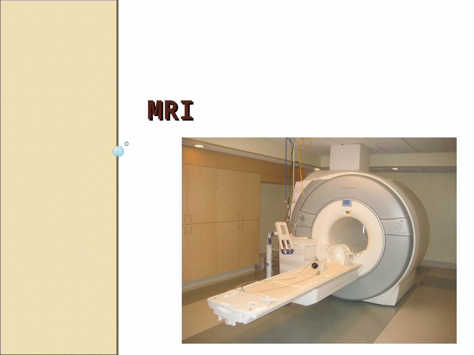
MRIMRI

MRIMRIMagnet used in MRI is measured
according to SI unit of Tesla (T)Generally range 0.5-3.0 T (Earth’s
magnetic field: 0.0005 T)Advantages:
◦ Image deep soft tissue structures◦ Not operator dependent◦ Does not use ionizing radiation◦ No evidence of actual harm
Terotogenesis Tissue heating Acoustic damage *
*Reeves et al, Neonatal Cochlear Function: Measurement after Exposure to Acoustic Noise during in Utero MR Imaging. Radiology, Dec 2010 Vol 257 Issue 3

ContraindicationsContraindicationsMagnetically sensitive
equipments◦Cardiac pacemaker◦Implantable defibrillators◦Intraocular metals◦Cochlear implants◦Intracranial aneurysm clips
MRI is not advisable in the 1st trimester

Contrast Enhancement in Contrast Enhancement in MRIMRIGadolinium-based agents
◦Use in pregnancy is controversial◦Water soluble, cross the placenta into fetal
circulation and amniotic fluid◦Prospective study on pregnant women who
received gadolinium in 1st trimester reported no adverse perinatal or neonatal outcomes
◦Thus, use should be limited to situation in which the benefits clearly outweigh the possible risks
(De Santis M, Straface G, Cavaliere AF, Carducci B, Caruso A. Gadolinium periconceptional exposure: pregnancy and neonatal outcome.
Acta Obstet Gynecol Scand 2007;86: 99-101.)
KKM RADIOLOGY KKM RADIOLOGY CONSENTSCONSENTS
(Please Find the Consents in SGH O&G Website)http://www.sgh-og.com/downloads/kkm-radiology-consents-for-pregnancy/
Take Home MessagesTake Home MessagesMRI is safe during pregnancyRadiation exposure through
Radiography, CT scan or Nuclear Medicine imaging is very low to cause harm to fetus
Concern on ionising radiation should not prevent or delay medically indicated radiological procedures
Clinicians should weigh the risks and benefits of any radiographic study

ReferencesReferences1. ACOG , Guidelines for Diagnostic Imaging During Pregnancy and
Lactation. Committee Opinion Number 656, Feb 20162. The American College of Radiology, Practice Guideline for Imaging
Pregnant or Potentially Pregnant Adolescents and Women with Inizing Radiation. 2008
3. SOGC Clinical Practice Guideline No. 306. The Use of Magnetic Resonance Imaging in the Obstetric Patient. April 2014
4. Review Safety of Diagnostic Imaging in pregnancy. The Obstetrician and Gynaecologist. 2010
5. Medicines and Healthcare Products Regulatory Agency, Safety Guidelines for Magnetic Resonance Imaging Equipment in Clinical Use. March 2015
6. The College of Radiographers and The Royal Colelge of Radiologists. Pregnancy and Work in Diagnostic Imaging Departments. 2nd Edition 2009
7. Health Protection Agency, The Royal College of Radiologists and The College of Radiographers. Protection of Pregnant Patients dueing Diagnostic Medical Exposures to Ionising Radiation. March 2009

THANK YOUTHANK YOU