imaging choices in the management of colorectal cancer part 1 · pdf fileimaging choices in...
TRANSCRIPT
Imaging Choices in the Management of Colorectal
Cancer Part 1
Patrick Vos Department of Radiology
St. Paul�s Hospital Vancouver, BC
Imaging Choices in the Management of Colorectal Ca
Review staging Colorectal Ca Local staging Lung and liver lesions
PART 2: PET/CT Dr. Pete Tonseth
No time
Colon CaDetails local Rectal staging New Imaging Techniques (MR) Tumor regression post Ch/RT
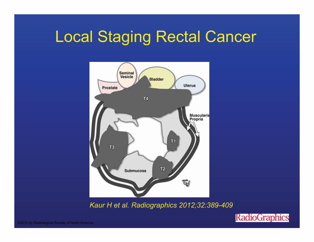
Local Staging Rectal Cancer
Kaur H et al. Radiographics 2012;32:389-409
©2012 by Radiological Society of North America

Rectal Ca Local Staging
Accuracy DRE T staging 58-88% EUS Staging information changed the
surgeon�s original treatment plan based on CT in 31% of patients
Schaffzin et al. Clin Colorectal Cancer. 2004;4:124-132.
Harewood GC. Gastroenterology 2002; 123:24-32
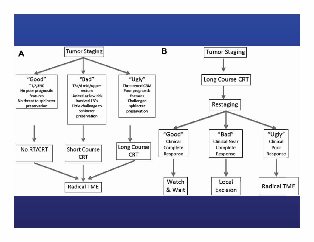
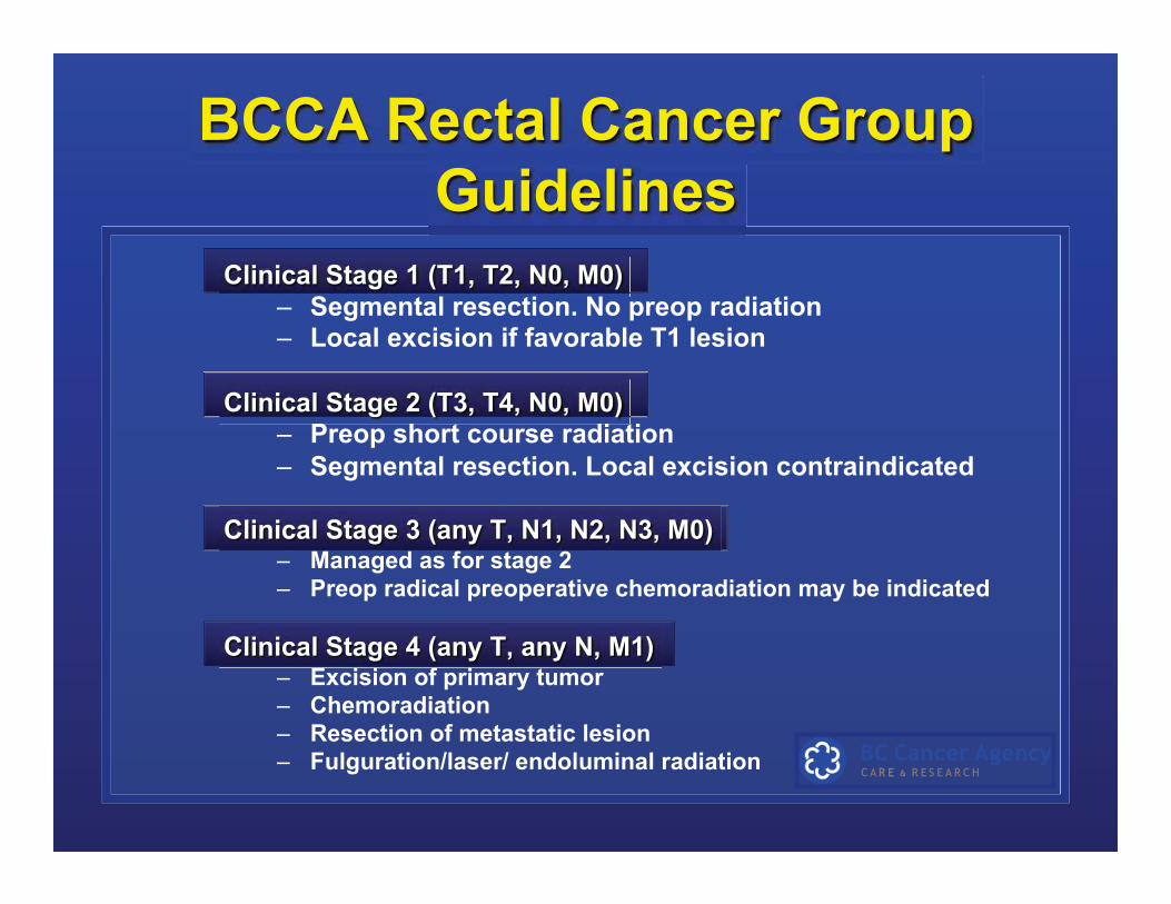
Clinical Stage 1 (T1, T2, N0, M0) – Segmental resection. No preop radiation – Local excision if favorable T1 lesion
Clinical Stage 2 (T3, T4, N0, M0) – Preop short course radiation – Segmental resection. Local excision contraindicated
Clinical Stage 3 (any T, N1, N2, N3, M0)
– Managed as for stage 2 – Preop radical preoperative chemoradiation may be indicated
Clinical Stage 4 (any T, any N, M1) – Excision of primary tumor – Chemoradiation – Resection of metastatic lesion – Fulguration/laser/ endoluminal radiation
BCCA Rectal Cancer Group Guidelines

BCCA Rectal Cancer Group Cancer Management Guidelines • Complete colonoscopy • Tumour height • Accurate preoperative staging
• Preoperative CEA • PET scan not recommended • Core biopsy in patients with unresectable disease
Accurate preoperative staging
• Location (height)
• TNM staging • Free resection Margin TME
Tumor Location
• Surgical planning
• Determine pre-op management
• Most distal location of the tumour is used to define tumour location
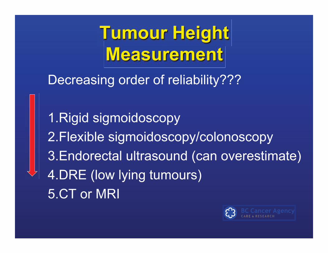
Tumour Height Measurement
Decreasing order of reliability??? 1. Rigid sigmoidoscopy 2. Flexible sigmoidoscopy/colonoscopy 3. Endorectal ultrasound (can overestimate) 4. DRE (low lying tumours) 5. CT or MRI

Relationship to anal sphincter
Kaur H et al. Radiographics 2012;32:389-409
©2012 by Radiological Society of North America
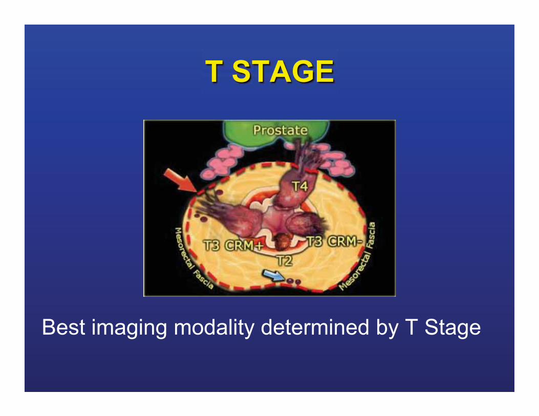
T STAGE
Best imaging modality determined by T Stage
http://www.medscape.org/viewarticle/

Rectal Ca
T0
TEM
T1
TEM
T2
TME
T3
Rth
T4
Ch-Rt
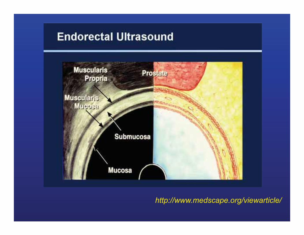
ERUS
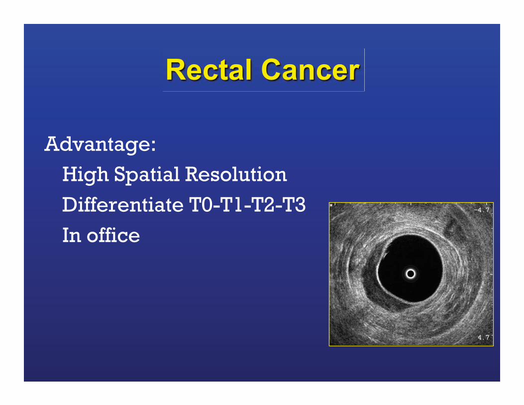
Advantage:
High Spatial Resolution
Differentiate T0-T1-T2-T3
In office
Rectal Cancer
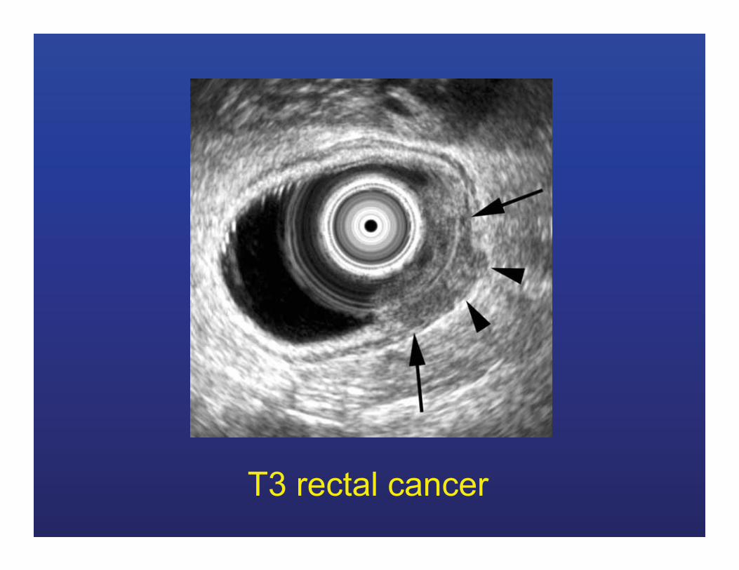
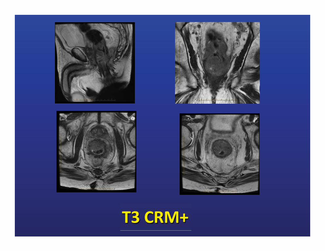
T3 rectal cancer
ERUS Disadvantage:
Availability/Expertise High/low/obstructing tumors Discomfort Cannot see MRF May overestimate distance Overstaging: 20% T3-T4 actually T2
Sauer R, N Engl J Med. 2004;351:1731-1740.
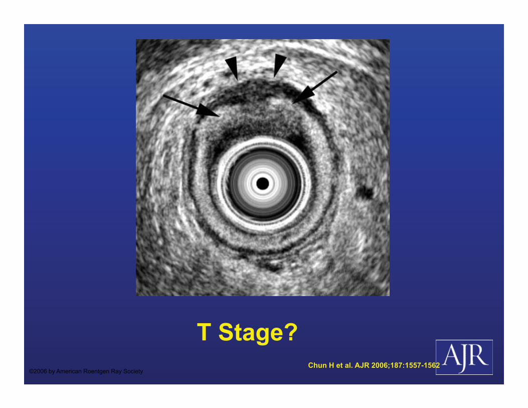
T Stage? Chun H et al. AJR 2006;187:1557-1562
©2006 by American Roentgen Ray Society
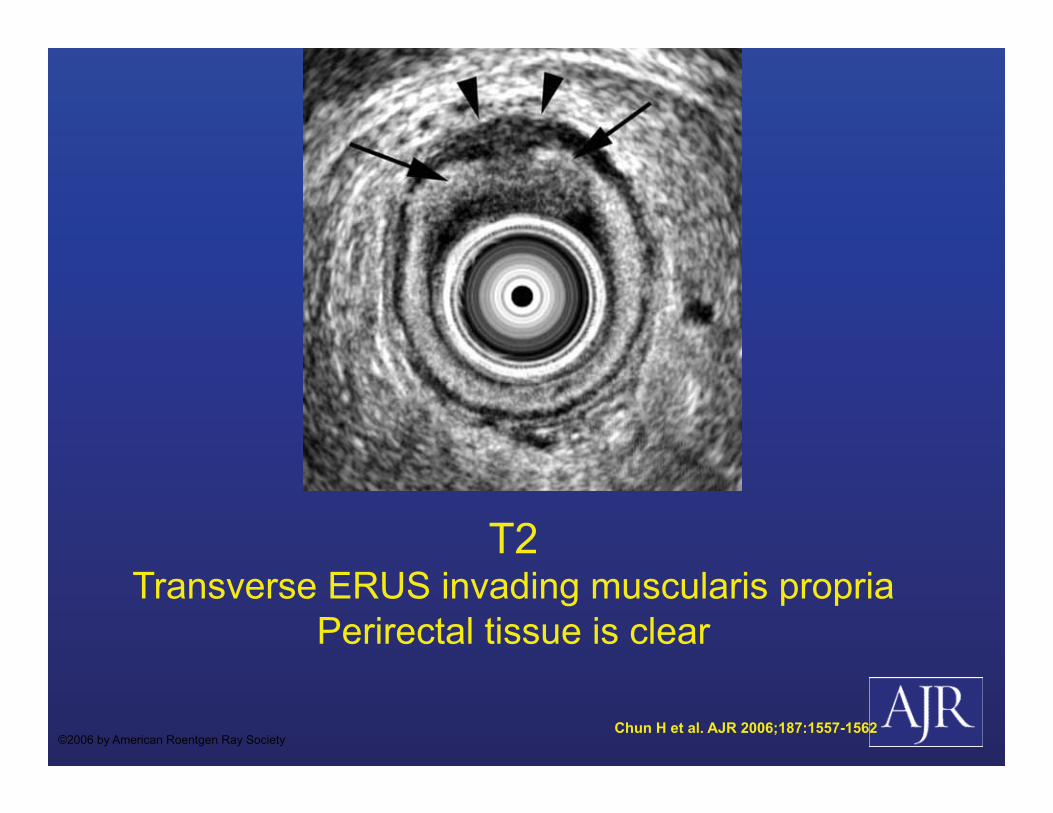
T2 Transverse ERUS invading muscularis propria
Perirectal tissue is clear
Chun H et al. AJR 2006;187:1557-1562 ©2006 by American Roentgen Ray Society
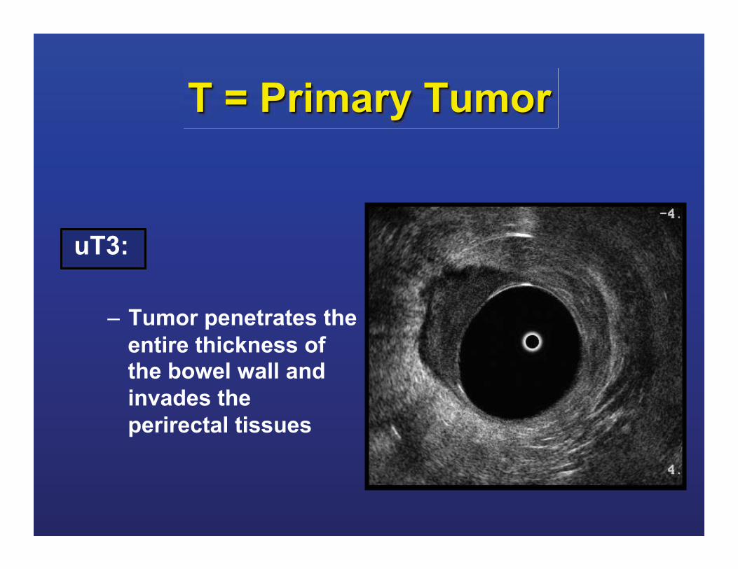
T = Primary Tumor uT3:
– Tumor penetrates the entire thickness of the bowel wall and invades the perirectal tissues
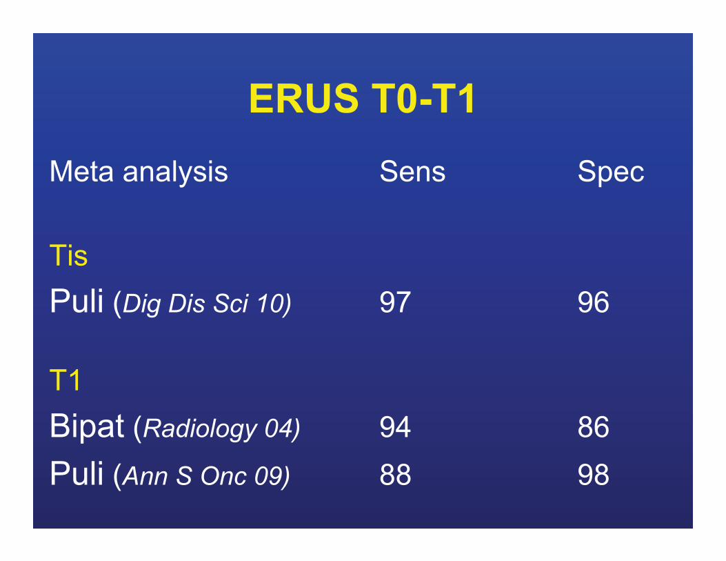
ERUS T0-T1 Meta analysis Sens Spec Tis Puli (Dig Dis Sci 10) 97 96
T1 Bipat (Radiology 04) 94 86 Puli (Ann S Onc 09) 88 98

MRI
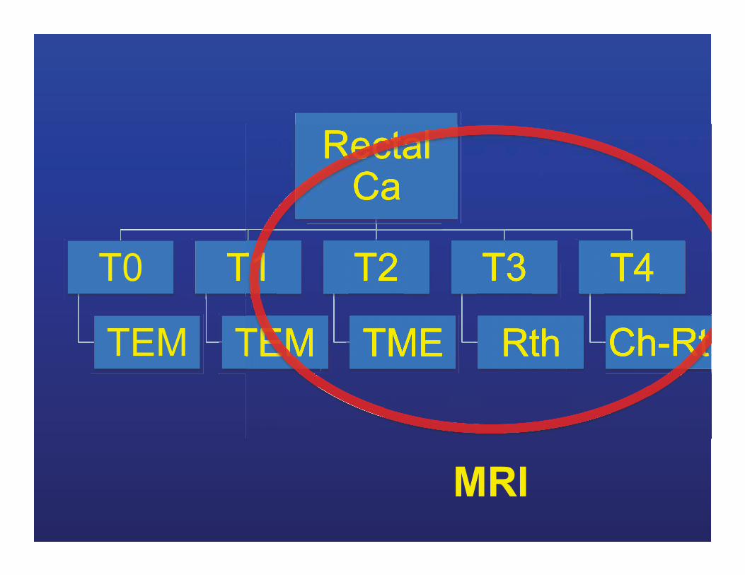
Rectal Ca
T0
TEM
T1
TEM
T2
TME
T3
Rth
T4
Ch-Rt
MRI
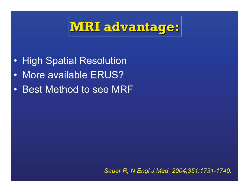
MRI advantage:
• High Spatial Resolution • More available ERUS? • Best Method to see MRF
Sauer R, N Engl J Med. 2004;351:1731-1740.

Musc propria
Levator ani
Puborectalis
MRF
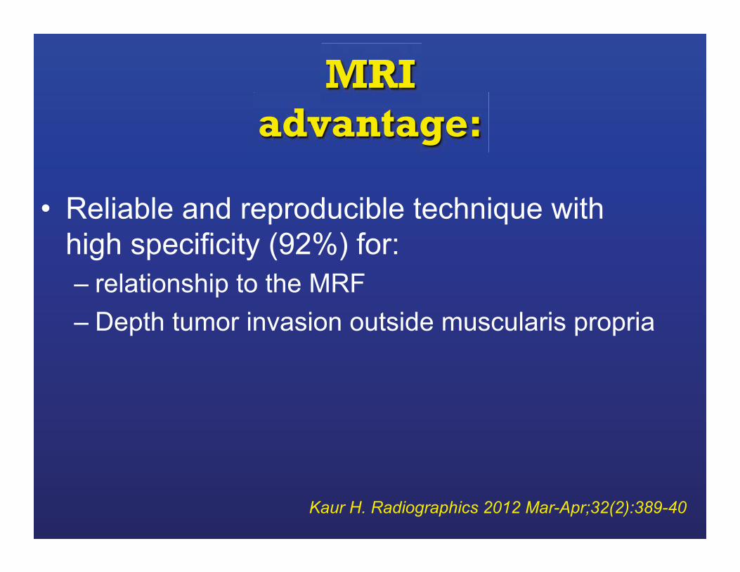
MRI advantage:
• Reliable and reproducible technique with
high specificity (92%) for: – relationship to the MRF – Depth tumor invasion outside muscularis propria
Kaur H. Radiographics 2012 Mar-Apr;32(2):389-40
MRI Disadvantage:
• Availability • Claustrophobia etc • No staging outside pelvis
Muthusamy VR, Chang KJ. Clin Cancer Res. 2007
MRI Disadvantage:
• Expertise • Interobserver variability • Need High Resolution Images
• Limitations borderline T2-T3 • Overstaging T2 29-40%
Sauer R, N Engl J Med. 2004;351:1731-1740.
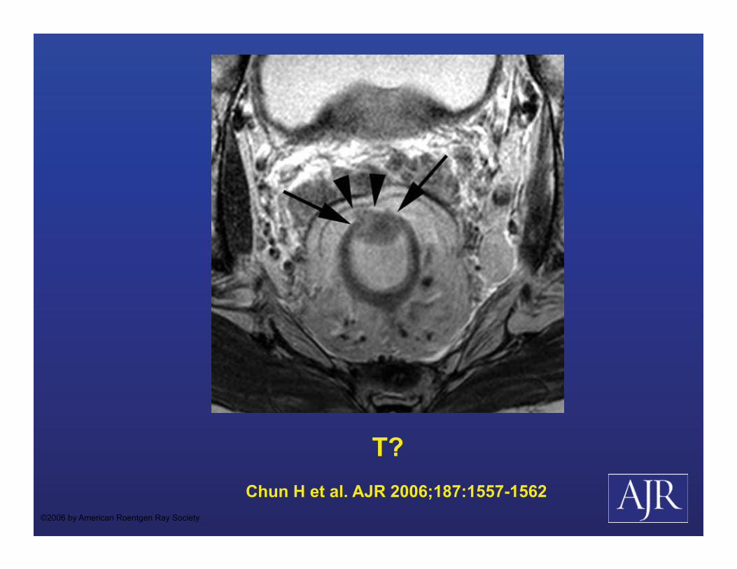
T? Chun H et al. AJR 2006;187:1557-1562
©2006 by American Roentgen Ray Society
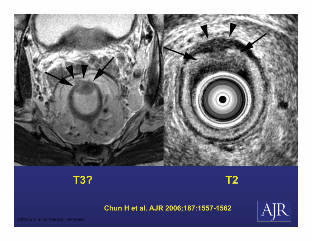
T3?
Chun H et al. AJR 2006;187:1557-1562 ©2006 by American Roentgen Ray Society
T2
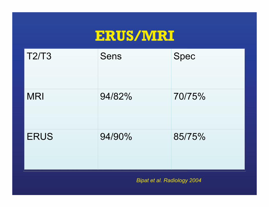
ERUS/MRI T2/T3 Sens Spec
MRI 94/82% 70/75%
ERUS 94/90% 85/75%
Bipat et al. Radiology 2004

Rectal Ca
T0
TEM
T1
TEM
T2
TME
T3
Rth
T4
Ch-Rt
MRI=CT
CT advantage:
• Fast • Available • Staging entire chest/abd/pelvis
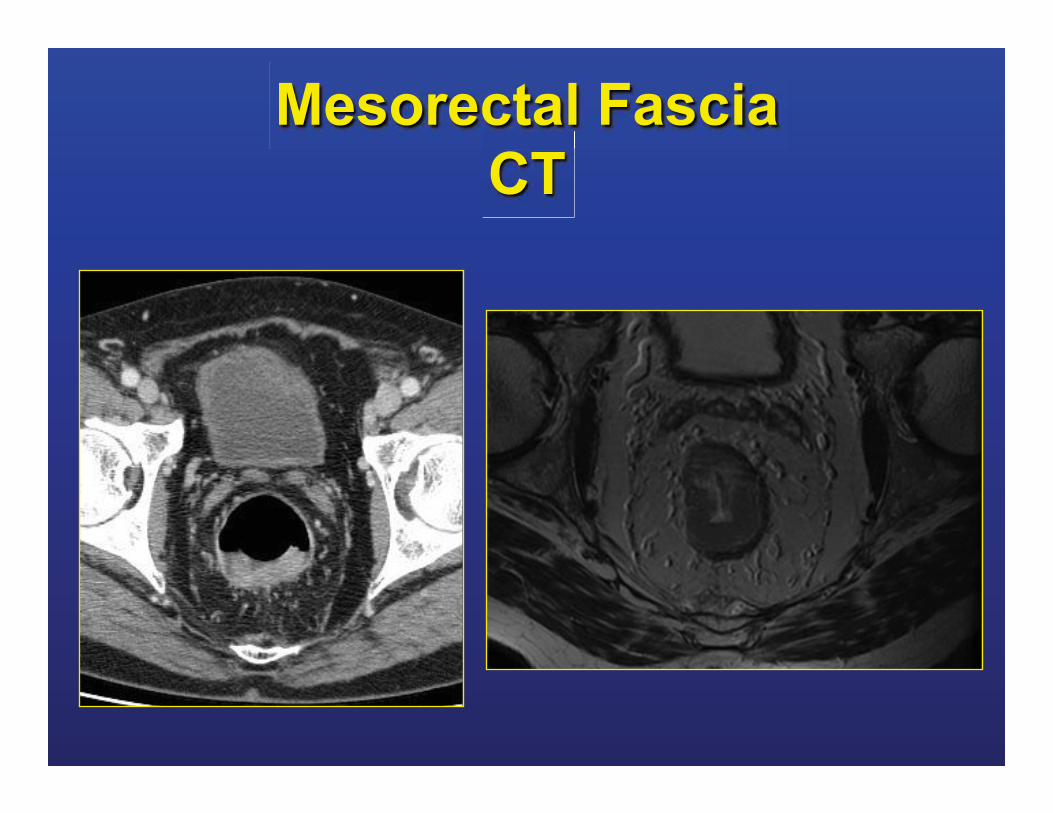
Mesorectal Fascia CT
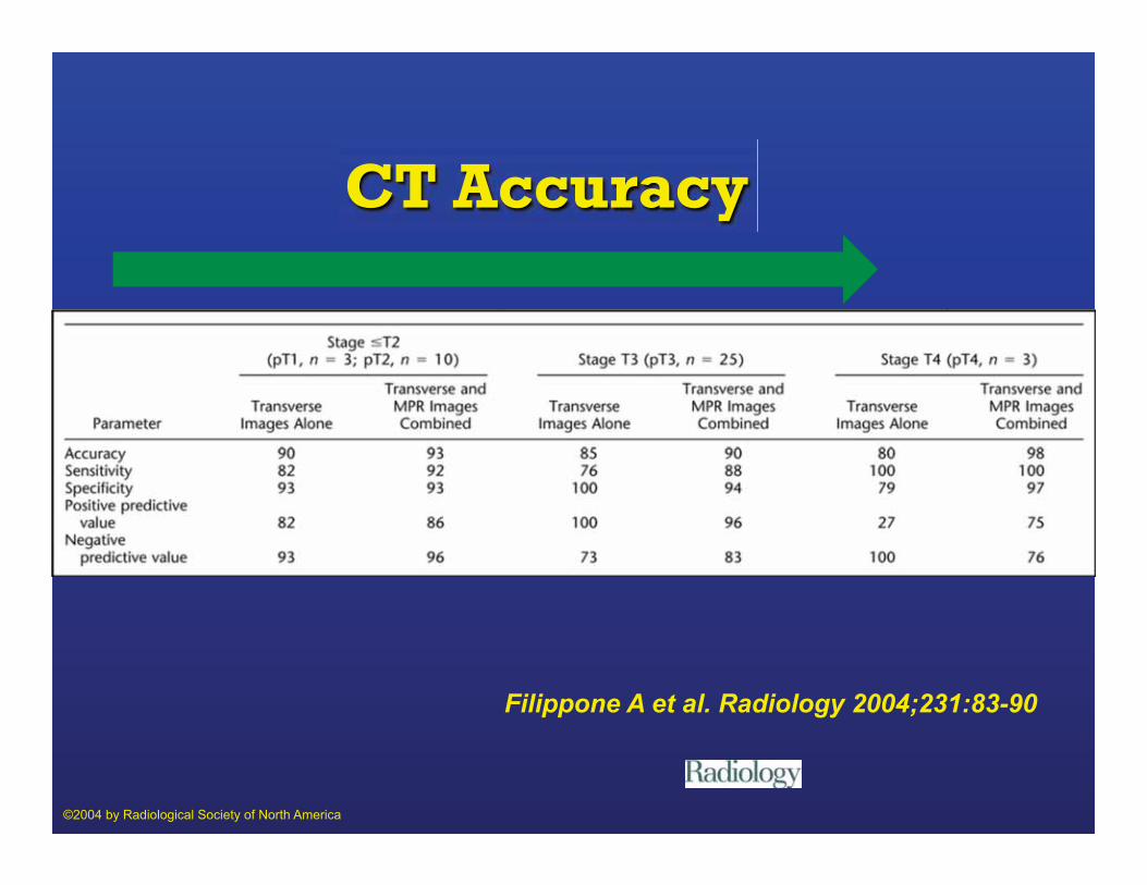
Filippone A et al. Radiology 2004;231:83-90
©2004 by Radiological Society of North America
CT Accuracy
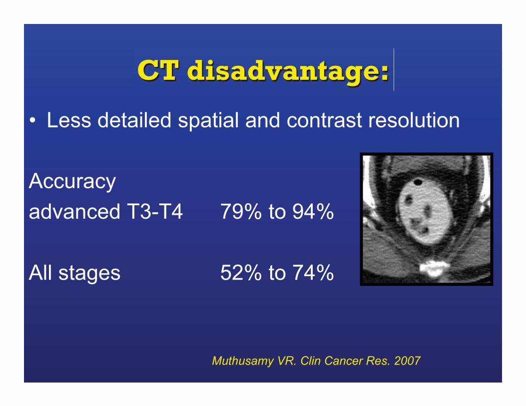
CT disadvantage:
• Less detailed spatial and contrast resolution
Accuracy advanced T3-T4 79% to 94% All stages 52% to 74%
Muthusamy VR. Clin Cancer Res. 2007
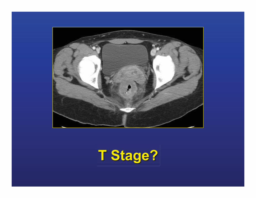
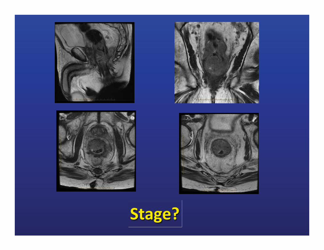
T Stage?

T4 Lesions
Sacral invasion Loss of fat plane between tumor and lower uterine segment

Nodes

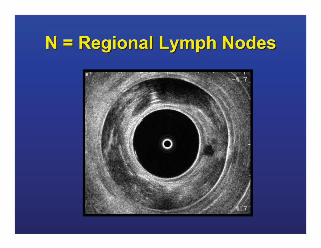
NX Regional lymph nodes cannot be assessed
N0 No regional lymph node metastasis
N1 Metastasis in 1 to 3 regional lymph nodes
N2 Metastasis in 4 or more regional lymph nodes
N3 Metastasis in a lymph node along the course of a named vascular trunk
N = Regional Lymph Nodes
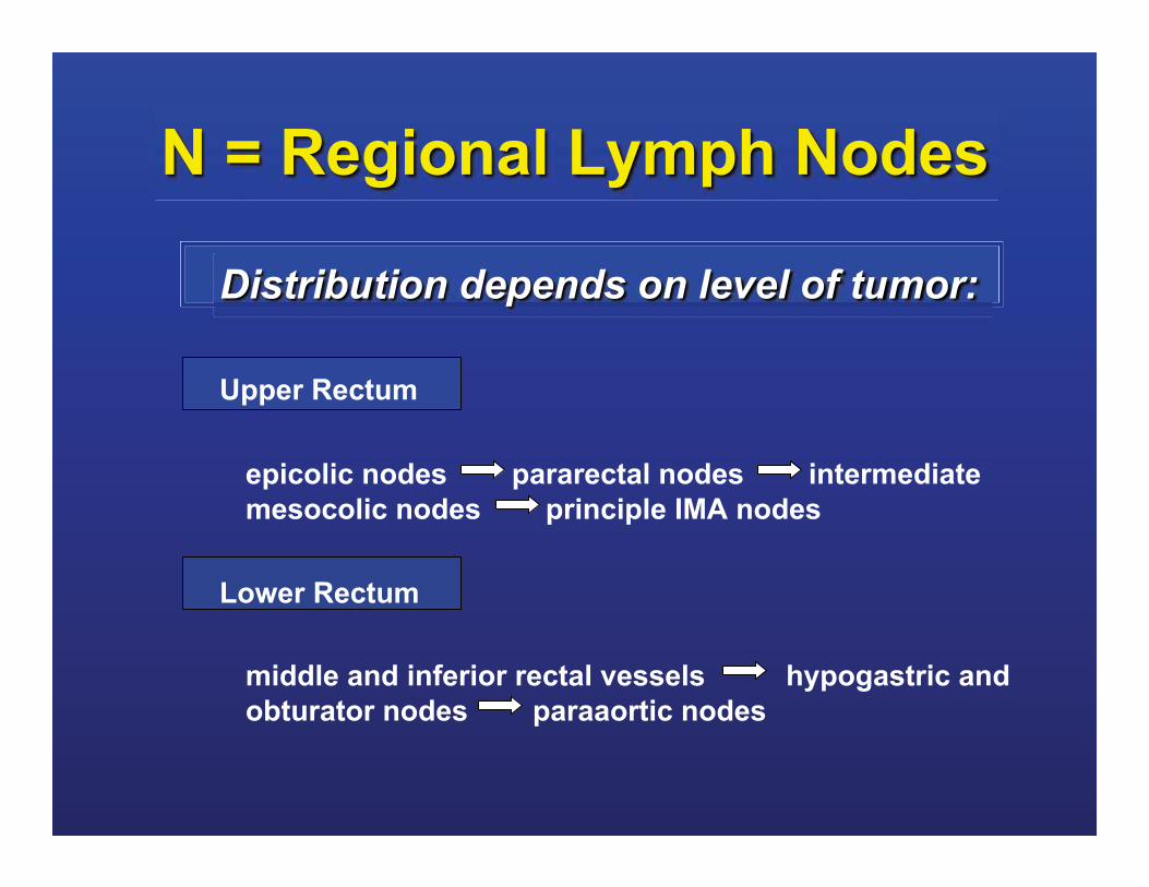
Distribution depends on level of tumor:
Upper Rectum
epicolic nodes pararectal nodes intermediate mesocolic nodes principle IMA nodes
Lower Rectum
middle and inferior rectal vessels hypogastric and obturator nodes paraaortic nodes
N = Regional Lymph Nodes
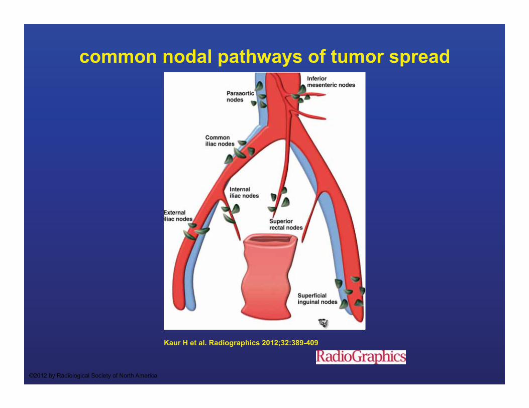
common nodal pathways of tumor spread
Kaur H et al. Radiographics 2012;32:389-409
©2012 by Radiological Society of North America

Nodal Criteria for Size?
N = Regional Lymph Nodes
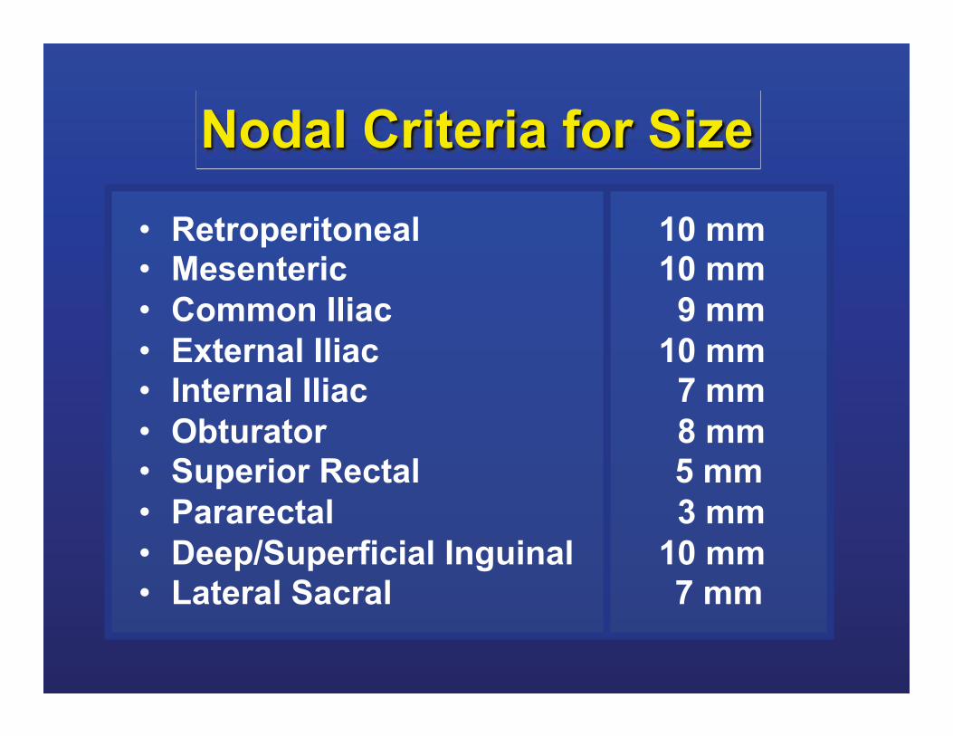
• Retroperitoneal 10 mm • Mesenteric 10 mm • Common Iliac 9 mm • External Iliac 10 mm • Internal Iliac 7 mm • Obturator 8 mm • Superior Rectal 5 mm • Pararectal 3 mm • Deep/Superficial Inguinal 10 mm • Lateral Sacral 7 mm
Nodal Criteria for Size
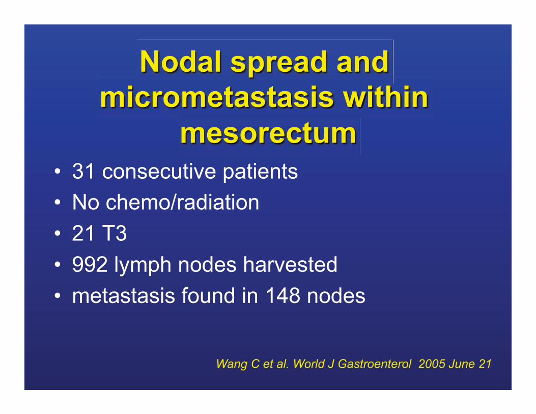
Nodal spread and micrometastasis within
mesorectum
Wang C et al. World J Gastroenterol 2005 June 21
• 31 consecutive patients • No chemo/radiation • 21 T3 • 992 lymph nodes harvested • metastasis found in 148 nodes

Nodal spread and micrometastasis within
mesorectum <1mm 7% <2mm 24% <5mm 70%
Wang C et al. World J Gastroenterol 2005 June 21
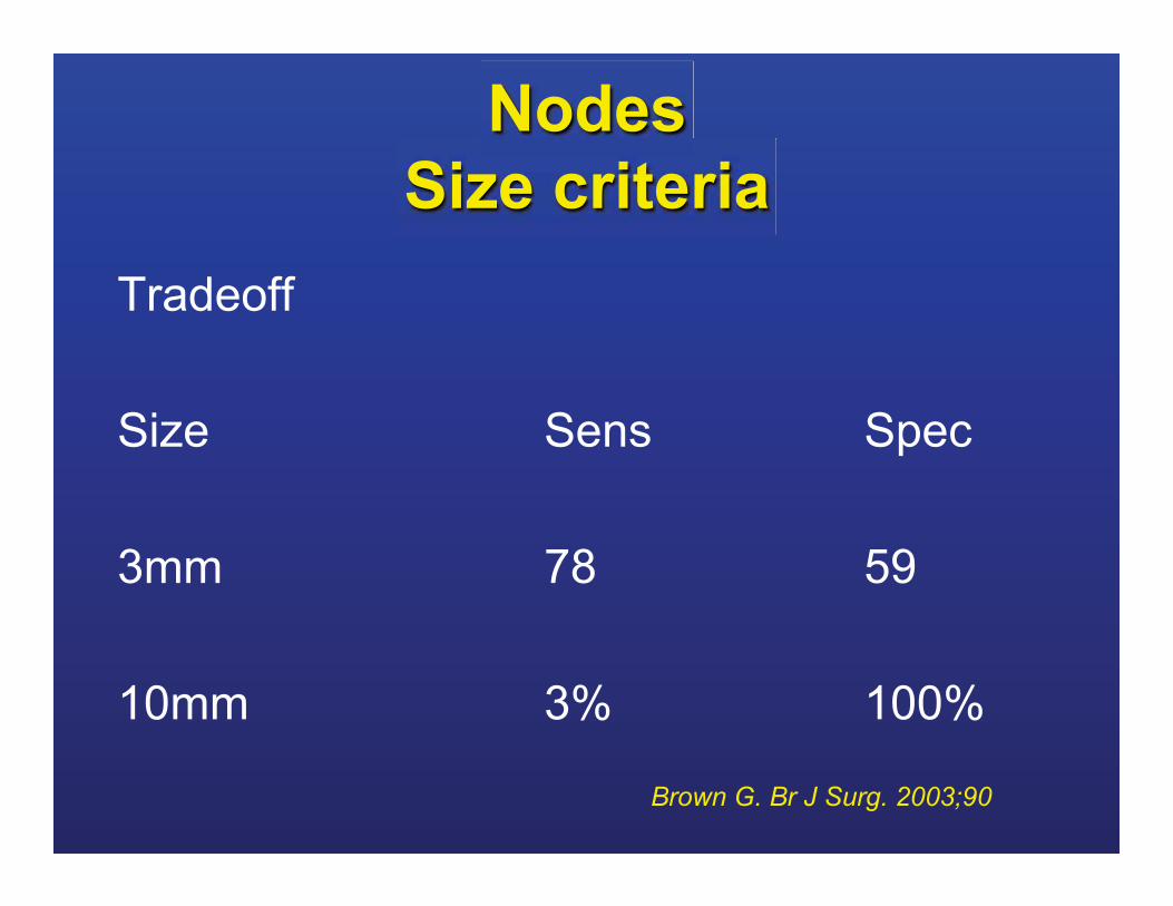
Nodes Size criteria
Tradeoff Size Sens Spec 3mm 78 59 10mm 3% 100%
Brown G. Br J Surg. 2003;90
N=188 EUS/MR staged T3 N0
• Multicenter • 188 pts • T3 N0 ERUS/MRI • preop Ch-RT
Guillem JG. J Clin Oncol. 2008 Jan 20

N=188 EUS/MR staged T3 N0
• 22% of patients undetected mesorectal LN involvement despite Ch-RT
Guillem JG. J Clin Oncol. 2008 Jan 20

Nodal spread
Overall accuracy 60-80% No differences ERUS/MR/CT T stage correlates with LN positivity T stage correlates with accuracy LN staging
Wang C et al. World J Gastroenterol 2005 June 21

Other criteria
Amount not helpful sens spec
Spiculated Indistinct Heterogeneous 85% 98%
Kim JH. Eur J Radiol. 2004 Oct;52(1):78-83.

Irregular Border and Mixed Signal Intensity
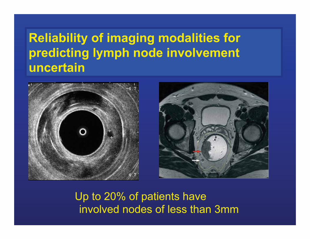
Reliability of imaging modalities for predicting lymph node involvement uncertain
Up to 20% of patients have involved nodes of less than 3mm
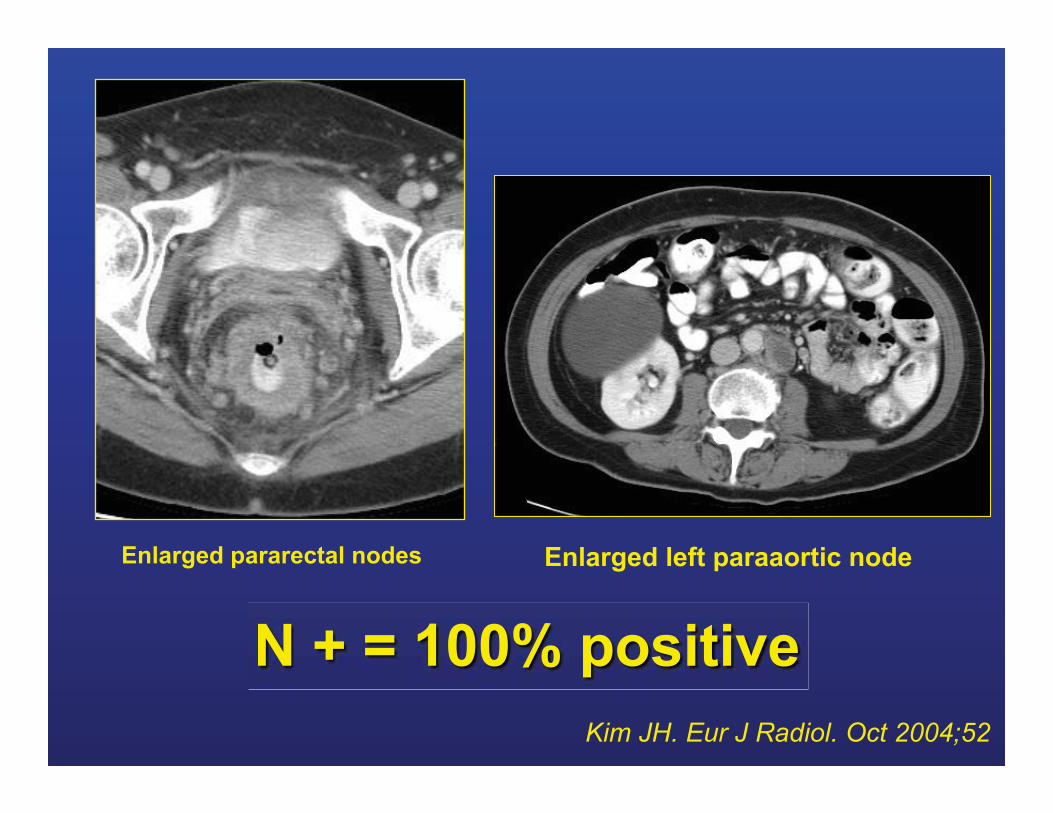
N + = 100% positive
Enlarged pararectal nodes Enlarged left paraaortic node
Kim JH. Eur J Radiol. Oct 2004;52

T stage assessment is fairly accurate N stage is only moderately effective
whatever modality is used
Conclusion
• New techniques
– DWI – Specific contrast agents – USPIO, Gadofosveset
– PET/CT PET/MR ??
Conclusion
M = Distant Metastases
MX = Distant metastases cannot be assessed M0 = No distant metastases M1 = Distant metastases
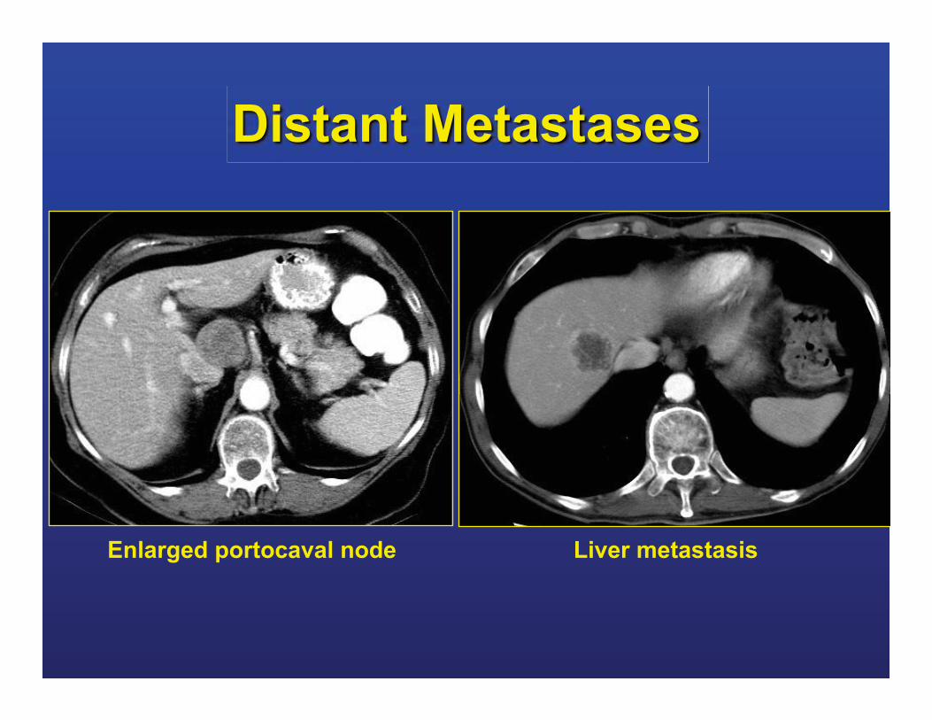
Distant Metastases
Liver metastasis Enlarged portocaval node
Distant disease and Follow-up
• Generally CT sufficient • Follow-up: How often? How long?
• What to do with incidental findings? – Liver: subcentimeter lesions TSTC – Lung: small nodules ILN
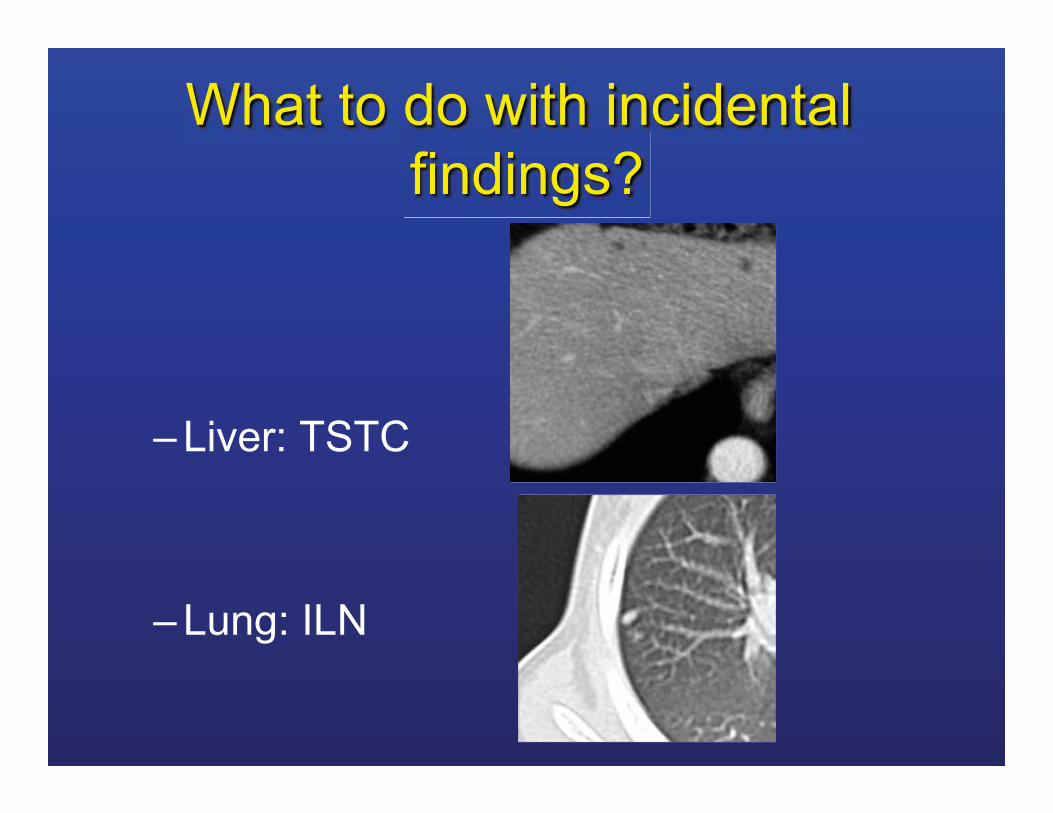
What to do with incidental findings?
– Liver: TSTC
– Lung: ILN
Prevalence and importance of small hepatic lesions found at CT in
patients with cancer • CT 2,978 patients with cancer • Benign: 303/2978 (80.2%) patients • Malignant 44 (11.6%) patients • Indeterminate 31 (8.2%) (short FU)
• CRC: mets in 14% pts with CRC
Schwartz LH. Radiology. 1999 Jan;210(1):71-4.

Prevalence and importance of small hepatic lesions found at CT in
patients with cancer
• CONCLUSION:
• small hepatic lesions in patients with cancer majority is benign
• metastases in 14 % of patient
Schwartz LH. Radiology. 1999 Jan;210(1):71-4.

Natural history of small, "indeterminate" hepatic lesions in
patients with colorectal cancer • 70/419 patients (16.7%) small liver lesions TSTC
• 46 patients (65.7%) subsequent imaging of their liver lesions
• 41 (89.1%) stable likely benign • 5 (10.9%) progression suggestive of mets
Lim GH. Dis Colon Rectum. 2009 Aug;52(8)
CT follow-up hypoattenuating small liver lesions in patients with rectal ca • 616 consecutive patients • 70 patients with 163 hepatic lesions • Patients stable 80% • Lesions Stable 90.8%
• No significant difference in results was found for patients stratified according to T-stage
Tan CH. Am J Clin Oncol. 2011 Aug;34(4)
CT follow-up hypoattenuating small liver lesions in patients with rectal ca • CONCLUSION
• majority of small hypoattenuating liver lesions remain stable and treated as benign lesions
• Closely followed for at least 1 year after completion of therapy
Tan CH. Am J Clin Oncol. 2011 Aug;34(4)
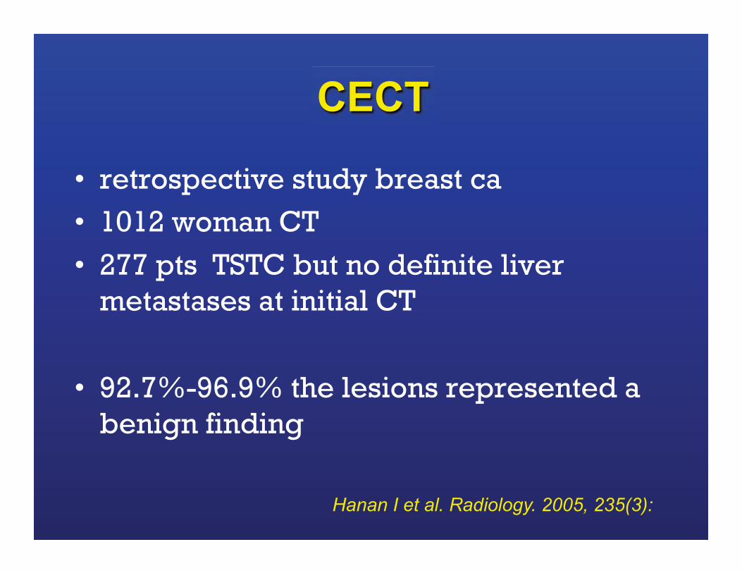
CECT
• retrospective study breast ca
• 1012 woman CT
• 277 pts TSTC but no definite liver metastases at initial CT
• 92.7%-96.9% the lesions represented a benign finding
Hanan I et al. Radiology. 2005, 235(3):
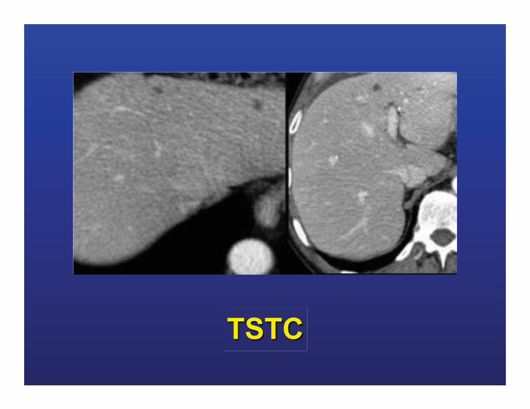
TSTC
Problem solving
• US: small cysts
• MRI: hepatocyte-specific contrast agents Gd-EOB-DTPA (Primovist)
• Follow-up
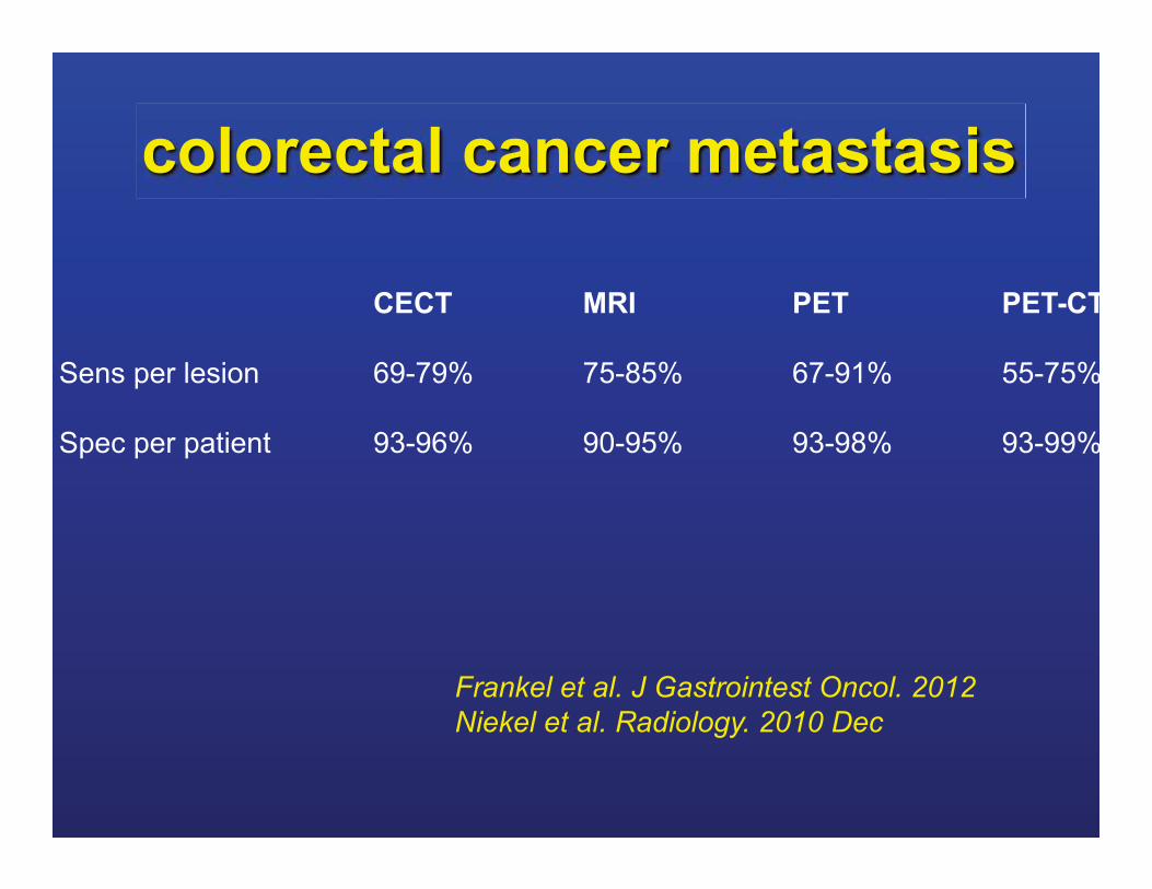
colorectal cancer metastasis
CECT MRI PET PET-CT
Sens per lesion 69-79% 75-85% 67-91% 55-75%
Spec per patient 93-96% 90-95% 93-98% 93-99%
Frankel et al. J Gastrointest Oncol. 2012 Niekel et al. Radiology. 2010 Dec
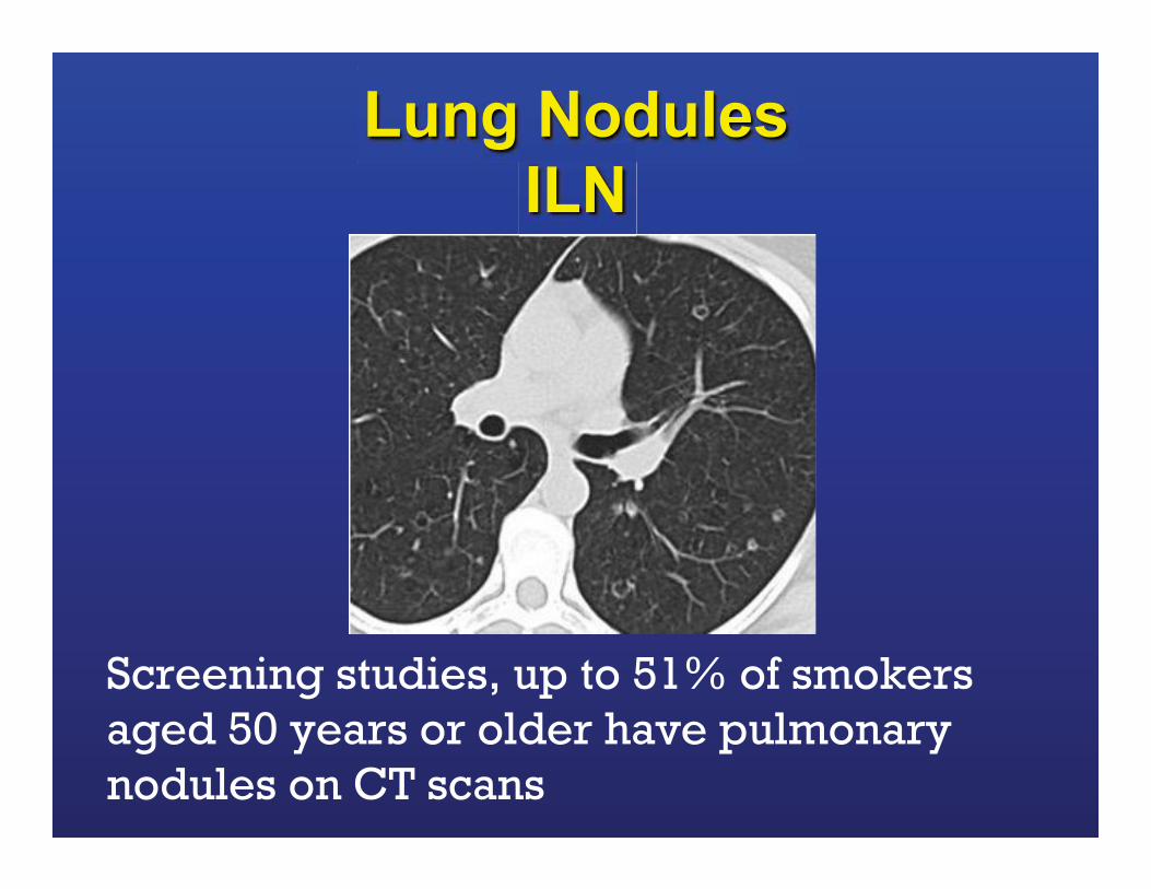
Lung Nodules ILN
Screening studies, up to 51% of smokers aged 50 years or older have pulmonary nodules on CT scans
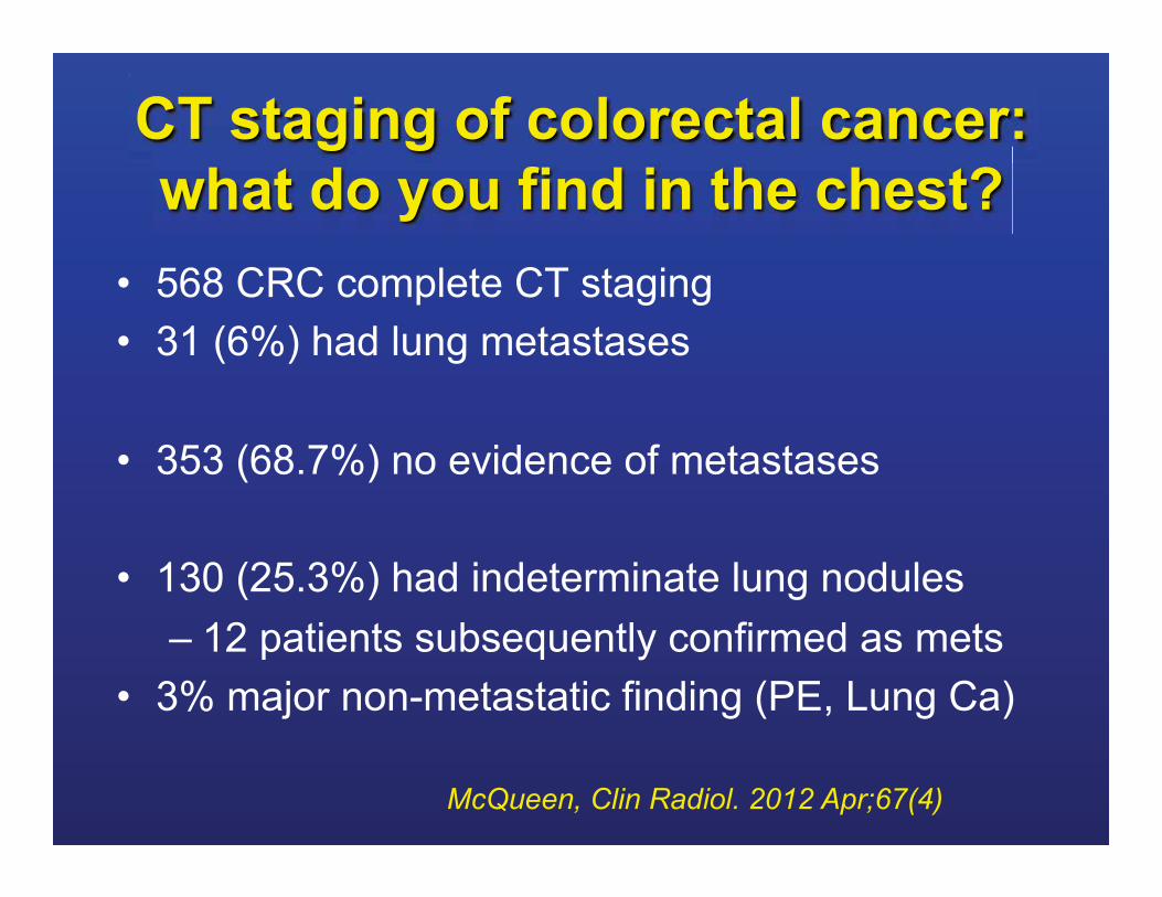
CT staging of colorectal cancer: what do you find in the chest?
• 568 CRC complete CT staging • 31 (6%) had lung metastases
• 353 (68.7%) no evidence of metastases
• 130 (25.3%) had indeterminate lung nodules – 12 patients subsequently confirmed as mets
• 3% major non-metastatic finding (PE, Lung Ca)
McQueen, Clin Radiol. 2012 Apr;67(4)
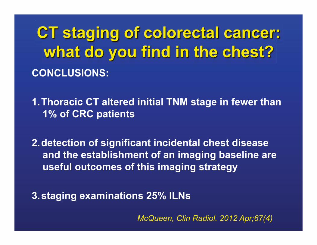
CT staging of colorectal cancer: what do you find in the chest?
CONCLUSIONS:
1. Thoracic CT altered initial TNM stage in fewer than 1% of CRC patients
2. detection of significant incidental chest disease and the establishment of an imaging baseline are useful outcomes of this imaging strategy
3. staging examinations 25% ILNs
McQueen, Clin Radiol. 2012 Apr;67(4)
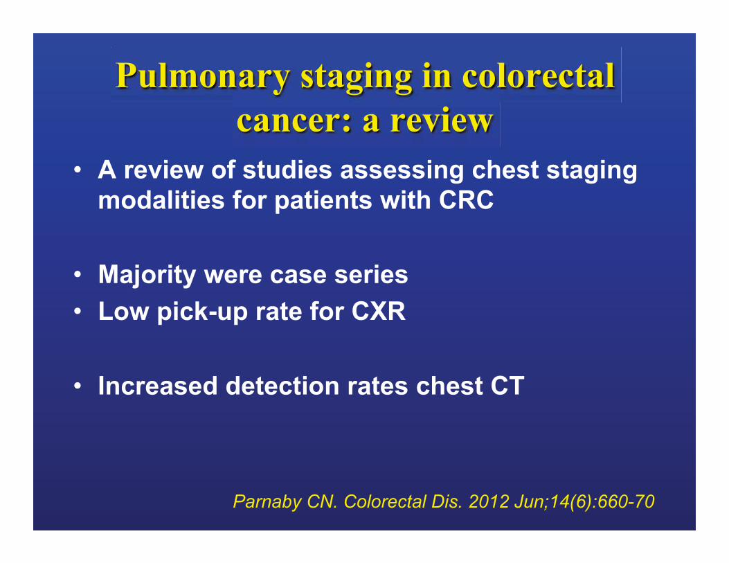
Pulmonary staging in colorectal cancer: a review
• A review of studies assessing chest staging modalities for patients with CRC
• Majority were case series • Low pick-up rate for CXR
• Increased detection rates chest CT
Parnaby CN. Colorectal Dis. 2012 Jun;14(6):660-70
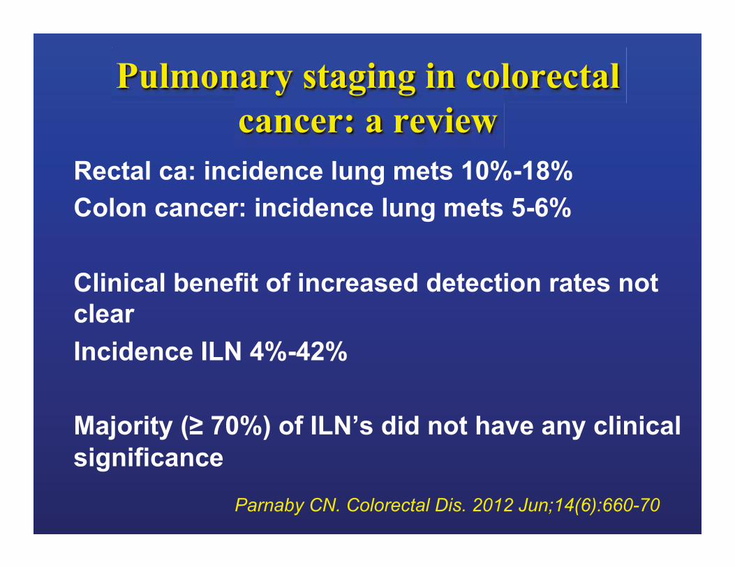
Pulmonary staging in colorectal cancer: a review
Rectal ca: incidence lung mets 10%-18% Colon cancer: incidence lung mets 5-6% Clinical benefit of increased detection rates not clear Incidence ILN 4%-42% Majority (≥ 70%) of ILN’s did not have any clinical significance Parnaby CN. Colorectal Dis. 2012 Jun;14(6):660-70
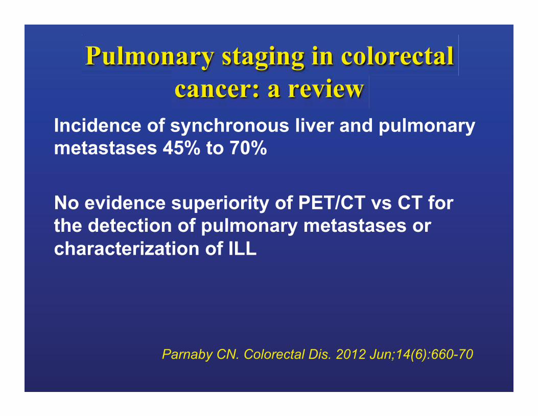
Pulmonary staging in colorectal cancer: a review
Incidence of synchronous liver and pulmonary metastases 45% to 70% No evidence superiority of PET/CT vs CT for the detection of pulmonary metastases or characterization of ILL
Parnaby CN. Colorectal Dis. 2012 Jun;14(6):660-70
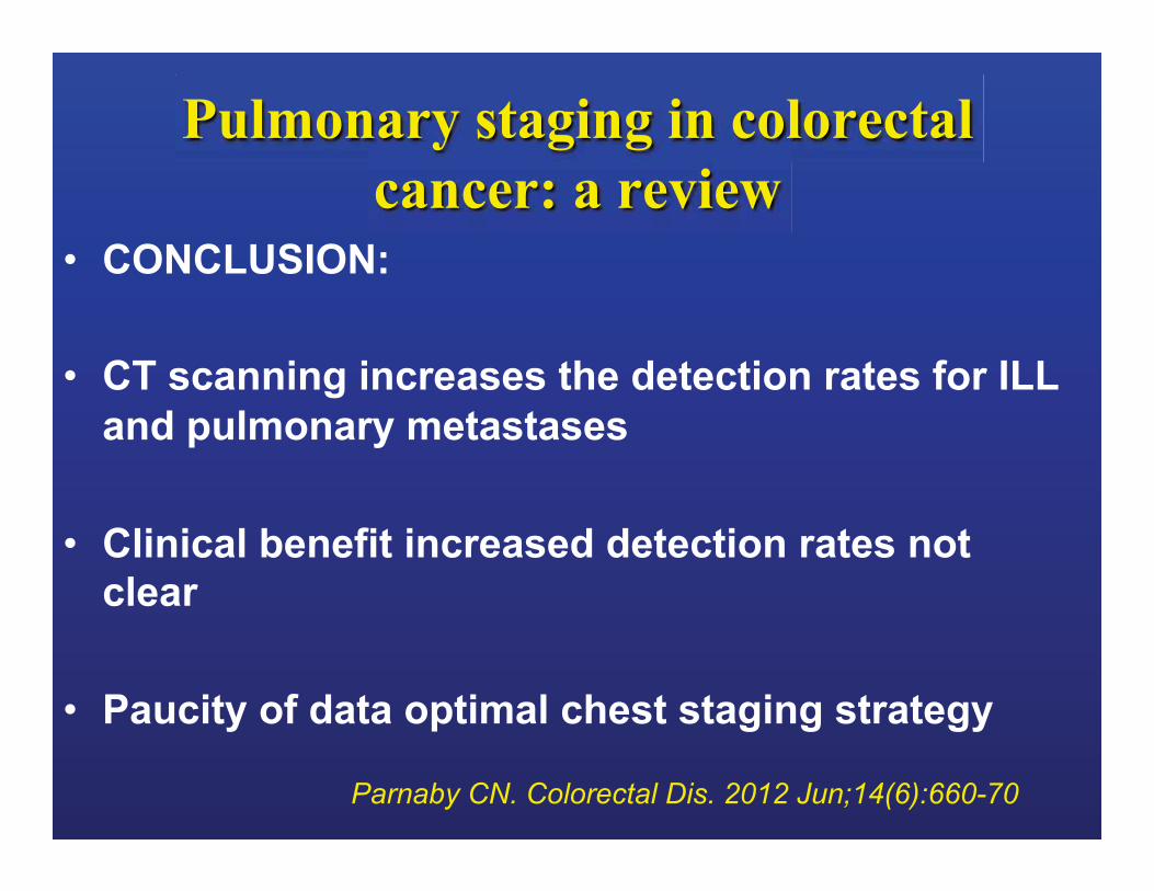
Pulmonary staging in colorectal cancer: a review
• CONCLUSION:
• CT scanning increases the detection rates for ILL and pulmonary metastases
• Clinical benefit increased detection rates not clear
• Paucity of data optimal chest staging strategy
Parnaby CN. Colorectal Dis. 2012 Jun;14(6):660-70

Mets Colon ca

Summary

Summary
• Best choice Imaging depends on T stage
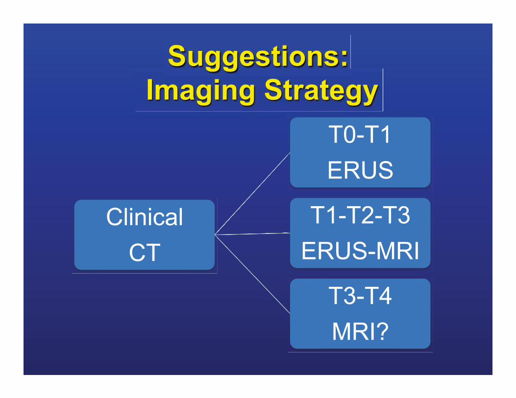
Suggestions: Imaging Strategy
Clinical CT
T0-T1 ERUS
T1-T2-T3 ERUS-MRI
T3-T4 MRI?
Summary
• Imaging often complimentary
• Overstaging: ERUS + MRI
• Accuracy LN 60-80%
Summary
• Have a plan: – Liver: TSTC – Lung: ILN’s
• Clinical benefit of increased detection rates not clear
Summary
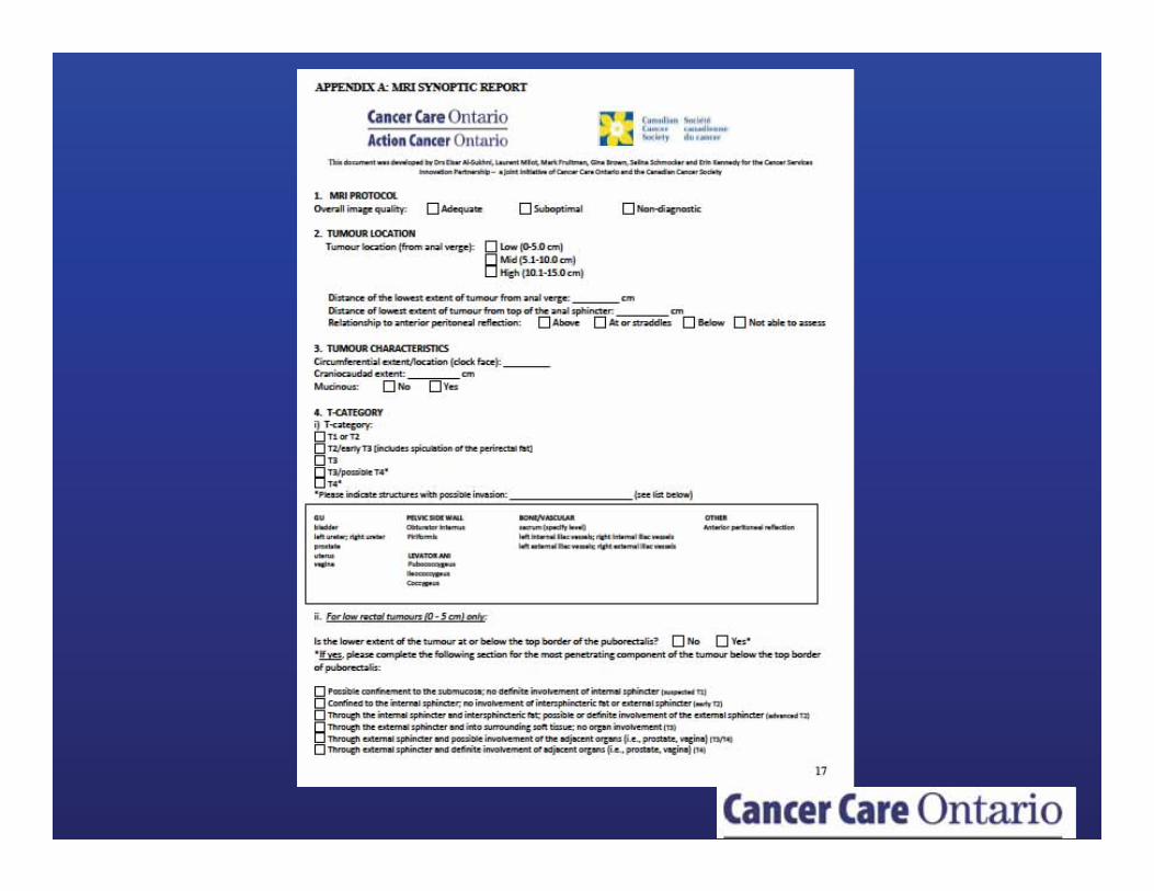
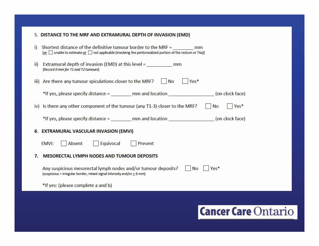
• Standardized/Template reporting?
�������
��� ��