image comm. lab ee/nthu digital image/video processing
TRANSCRIPT

Image Comm. Lab EE/NTHUImage Comm. Lab EE/NTHU 1
Digital Image/Video ProcessingDigital Image/Video Processing
• One picture is worth more than ten thousand words.
• Information or media is the most valuable development tool for human intelligence.
• Multimedia includes text, images, video, graphic, animation, and audio.

Image Comm. Lab EE/NTHUImage Comm. Lab EE/NTHU 2
Digital Image/Video ProcessingDigital Image/Video ProcessingFive Aspects of IP
Image,Video
Pattern Recognition
Computer Vision
Conversion, Enhancement
Transmission, storage
Restoration, Generation
Image,Video
Name
DescriptionData
Data

Image Comm. Lab EE/NTHUImage Comm. Lab EE/NTHU 3
Digital Image/Video ProcessingDigital Image/Video Processing
Relationship with multimedia
Relationship with multimedia
Speech recognition Text-to-speech
Audio
multimedia
Text Images/video
Lip synch, Face animation, Speech-driven talking head, Joint A/V coding
Sign language Lip-Reading
Compression, Graphic, Database indexing/retrieval
Natural language translation
Compression on synthesis 3D sound
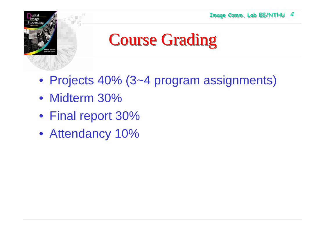
Image Comm. Lab EE/NTHUImage Comm. Lab EE/NTHU 4
Course GradingCourse Grading
• Projects 40% (3~4 program assignments)• Midterm 30%• Final report 30%• Attendancy 10%

Image Comm. Lab EE/NTHUImage Comm. Lab EE/NTHU 5
Chapter 1: IntroductionChapter 1: Introduction
• An image is defined as a two-dimensional function f(x, y), where x and y are spatial plane coordinates, the amplitude of f(x, y) is the intensity or gray level.
• When x, y, and the amplitude values of f are all finite and discrete quantities, the image is called a digital image
• A digital image is composed of a finite number of elements, each of which has a particular location and value.
• These elements are referred as picture elements, image elements, pixels, or pels.
• A video is defined as a three-dimensional function f(x, y, t), where the number a pictures displayed in time domain is called the frame rate.

Image Comm. Lab EE/NTHUImage Comm. Lab EE/NTHU 6
Chapter 1: Introduction
• Images are related to Human perceptions
• Human perception → Artificial Intelligence ←Computer Vision
• Image Processing are related to Computer Vision.
• Low-level process : inputs and outputs are images.
• Mid-level process : inputs are images, and outputs are the attributes extracted from the image (e.g., edges, contour, segmented objects).
• High-level process : perform the cognitive function normally associated with the vision.

Image Comm. Lab EE/NTHUImage Comm. Lab EE/NTHU 7
1.2: The original of Digital Image processing1.2: The original of Digital Image processing

Image Comm. Lab EE/NTHUImage Comm. Lab EE/NTHU 8
1.2: The original of Digital Image processing1.2: The original of Digital Image processing

Image Comm. Lab EE/NTHUImage Comm. Lab EE/NTHU 9
1.2: The original of Digital Image processing1.2: The original of Digital Image processing
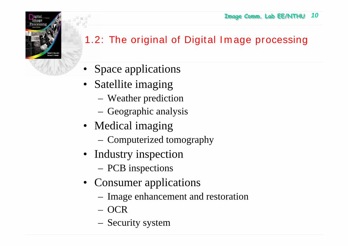
Image Comm. Lab EE/NTHUImage Comm. Lab EE/NTHU 10
1.2: The original of Digital Image processing
• Space applications• Satellite imaging
– Weather prediction– Geographic analysis
• Medical imaging– Computerized tomography
• Industry inspection– PCB inspections
• Consumer applications– Image enhancement and restoration– OCR– Security system
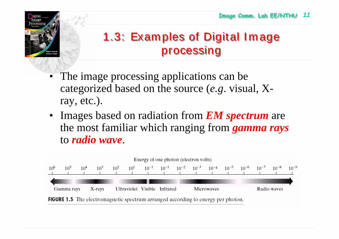
Image Comm. Lab EE/NTHUImage Comm. Lab EE/NTHU 11
1.3: Examples of Digital Image processing
1.3: Examples of Digital Image processing
• The image processing applications can be categorized based on the source (e.g. visual, X-ray, etc.).
• Images based on radiation from EM spectrum are the most familiar which ranging from gamma raysto radio wave.

Image Comm. Lab EE/NTHUImage Comm. Lab EE/NTHU 12
1.3.1: Gamma-Ray Imaging1.3.1: Gamma-Ray Imaging
• Nuclear imaging: inject a patient with a radioactive isotope that emits gamma ray as it decays.
• Images are produced from the emission collected by gamma ray detector.
• Positron emission tomography (PET): The radioactive isotope emits positron as it decays. When a positron meets an electron, both annihilate and two gamma rays are given off.

Image Comm. Lab EE/NTHUImage Comm. Lab EE/NTHU 13
1.3.1: Gamma-Ray Imaging1.3.1: Gamma-Ray Imaging

Image Comm. Lab EE/NTHUImage Comm. Lab EE/NTHU 14
1.3.2: X-Ray Imaging1.3.2: X-Ray Imaging
• X-rays are the oldest EM radiation used for imaging.
• X-ray tube (source) : a vacuum tube• The intensity of X-rays is modified by absorption
as they pass through the patient, and the resulting energy falling on the film develops it.
• Digital radiography– Digitizing X-ray film– X-ray fall on a device (e.g. Phosphor screen)
that convert X-ray to light. The light signal is captured by light-sensitive digitizing system.

Image Comm. Lab EE/NTHUImage Comm. Lab EE/NTHU 15
1.3.2: X-Ray Imaging
• Angiography – contrast enhancement radiography– To obtain the images of blood vessel
(angiograms)– A Catheter ( a small flexible hollow tube) is
inserted into an artery or vein in the groin. The Catheter is threaded into the blood vessel and guided to the area to be studied.
– When the Catheter reaches the site under investigation, an X-ray contrast medium is injected through the catheter to enhance the contrast of the blood vessel.

Image Comm. Lab EE/NTHUImage Comm. Lab EE/NTHU 16
1.3.2: X-Ray Imaging
• Computerized Axial Tomography (CAT)• Each CAT image is a slice taken
perpendicularly through the patient• Numerous slices are generated as the patient
is moved in longitudinal direction.• Industrial inspection using X-ray• Industrial CAT scans• X-ray imaging in Astronomy

Image Comm. Lab EE/NTHUImage Comm. Lab EE/NTHU 17

Image Comm. Lab EE/NTHUImage Comm. Lab EE/NTHU 18
1.3.3: Imaging in the Ultraviolet Band1.3.3: Imaging in the Ultraviolet Band
• Applications of Ultraviolet lights:– Lithography– Industrial inspection– Fluorescence Microscopy (for studying
material)– Lasers– Biological imaging– Astronomical observation

Image Comm. Lab EE/NTHUImage Comm. Lab EE/NTHU 19
Chapter 1: IntroductionChapter 1: Introduction
Fluorescence Microscopy
Astronomical observation

Image Comm. Lab EE/NTHUImage Comm. Lab EE/NTHU 20
1.3.4: Imaging in the visible and infrared Bands1.3.4: Imaging in the visible and infrared Bands
• Visual band is the most familiar in our activity– inspection
• Infrared band for satellite image• Astronomy• Light microscope
– Pharmaceuticals– Micro-inspection
• Satellite image for remote sensing– Thermal bands for LANDSAT– Multi-spectral image

Image Comm. Lab EE/NTHUImage Comm. Lab EE/NTHU 21
Chapter 1: IntroductionChapter 1: Introduction

Image Comm. Lab EE/NTHUImage Comm. Lab EE/NTHU 22
1.3.4: Imaging in the visible and infrared Bands1.3.4: Imaging in the visible and infrared Bands
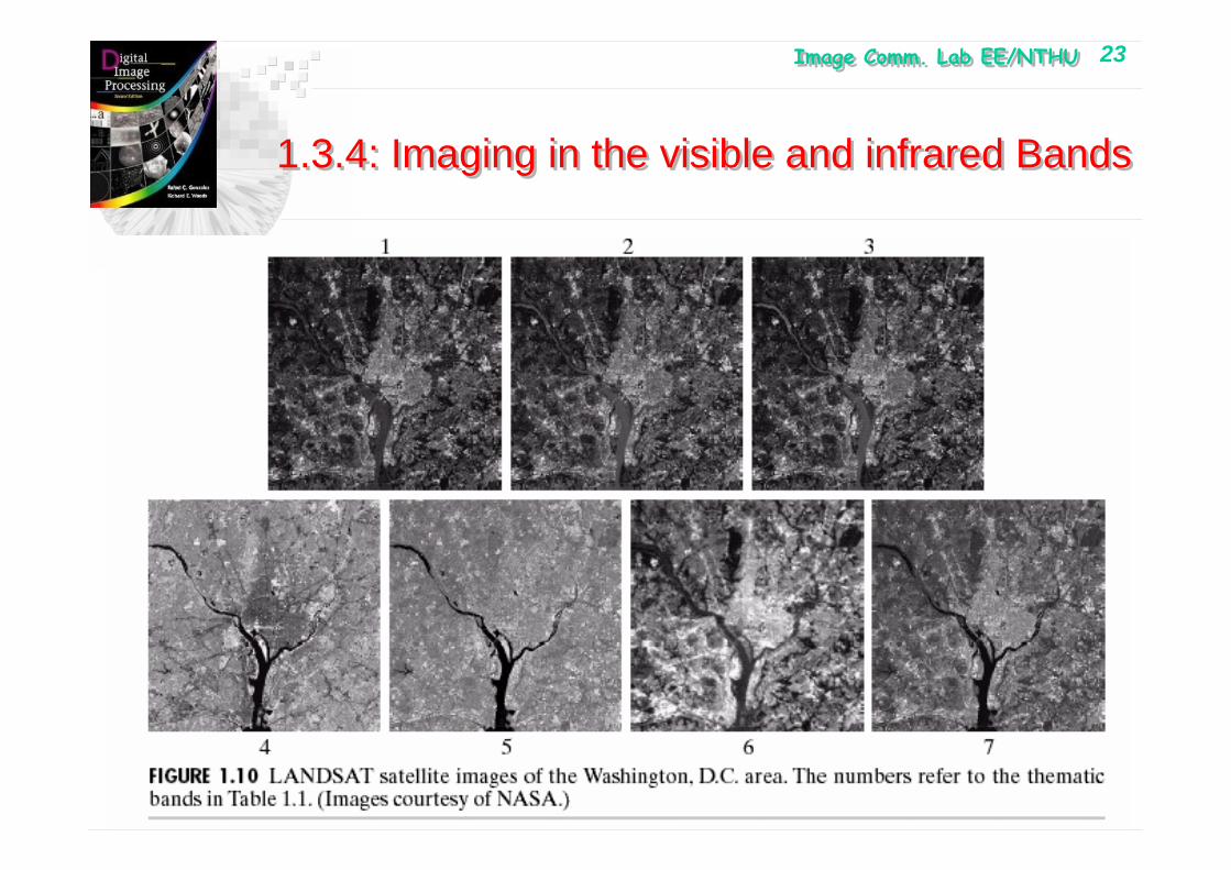
Image Comm. Lab EE/NTHUImage Comm. Lab EE/NTHU 23
1.3.4: Imaging in the visible and infrared Bands1.3.4: Imaging in the visible and infrared Bands

Image Comm. Lab EE/NTHUImage Comm. Lab EE/NTHU 24
1.3.4: Imaging in the visible and infrared Bands1.3.4: Imaging in the visible and infrared Bands
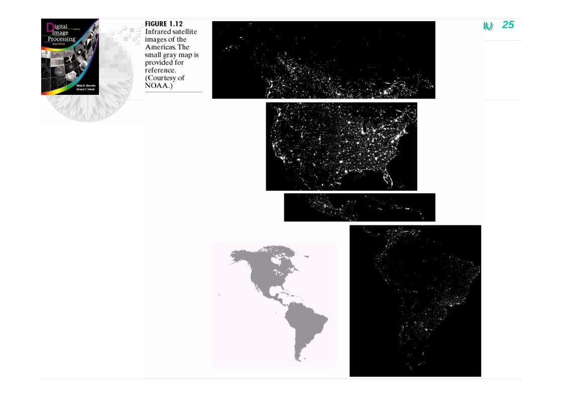
Image Comm. Lab EE/NTHUImage Comm. Lab EE/NTHU 25
Chapter 1: IntroductionChapter 1: Introduction

Image Comm. Lab EE/NTHUImage Comm. Lab EE/NTHU 26
Chapter 1: IntroductionChapter 1: Introduction
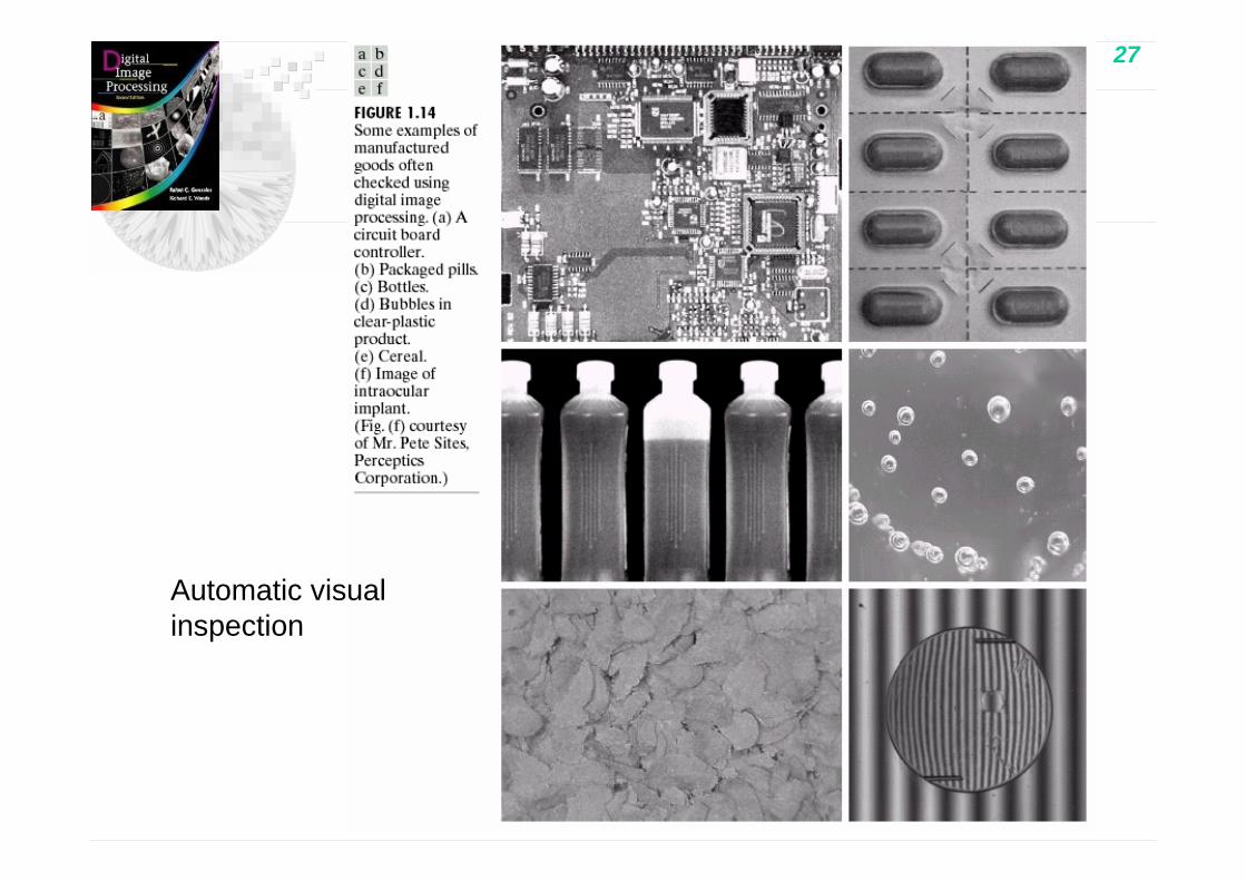
Image Comm. Lab EE/NTHUImage Comm. Lab EE/NTHU 27
Chapter 1: IntroductionChapter 1: Introduction
Automatic visual inspection
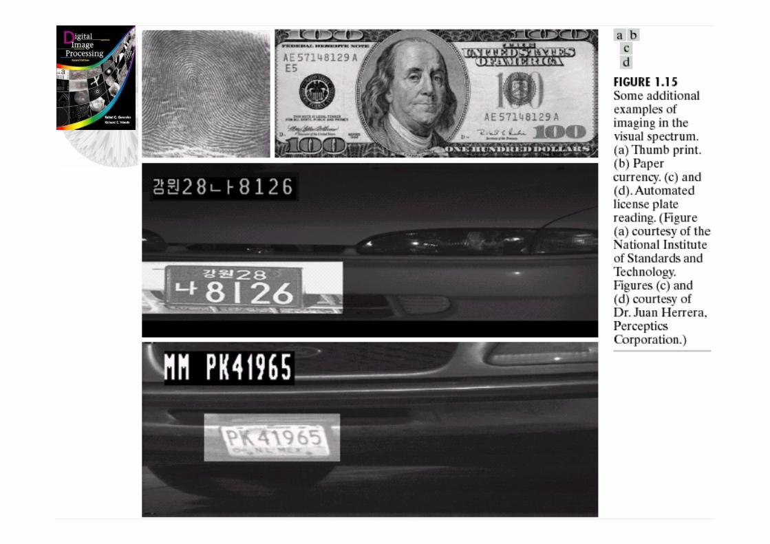
Image Comm. Lab EE/NTHUImage Comm. Lab EE/NTHU 28
Chapter 1: IntroductionChapter 1: Introduction
Image Comm. Lab EE/NTHUImage Comm. Lab EE/NTHU 29
1.3.5: Imaging in the microwave Bands
• Radar image– Collect data over virtually any region at any
time regardless of weather and ambient lightning conditions.
– Like a flash camera using its own illumination.
– Using antenna and digital image processing techniques to record its image

Image Comm. Lab EE/NTHUImage Comm. Lab EE/NTHU 30
1.3.5 Imaging in the microwave Bands1.3.5 Imaging in the microwave Bands

Image Comm. Lab EE/NTHUImage Comm. Lab EE/NTHU 31
1.3.6: Imaging in the radio bands
• Medicine– Magnetic resonance imaging (MRI)– Patient is placed in a magnet– Passing a radio wave through his body– Each pulse causes a responding pulse of radio
wave emitted from the patient tissue– The location and strength of the signal is detected
and then calculated by the computer to generate the image.
• Astonomy

Image Comm. Lab EE/NTHUImage Comm. Lab EE/NTHU 32
1.3.6: Imaging in the radio bands1.3.6: Imaging in the radio bands

Image Comm. Lab EE/NTHUImage Comm. Lab EE/NTHU 33
1.3.6: Imaging in the radio bands1.3.6: Imaging in the radio bands

Image Comm. Lab EE/NTHUImage Comm. Lab EE/NTHU 34
1.3.7: Examples in which other image modalities are used
• Acoustic imaging– Geological exploration– Industry– medicine
• Electron microscopy– Transmission electron microscopy (TEM)
• For thin sample– Scanning electron microscopy (SEM)
• For “bulky” sample• Synthetic image
– Fractal image– 3-D rendering of the computerized tomography

Image Comm. Lab EE/NTHUImage Comm. Lab EE/NTHU 35
1.3.7: Examples in which other image modalities are used
Seismic interpretation looks for these “bright spots” to find oil and gas.
The target is brighter than the surrounding layer

Image Comm. Lab EE/NTHUImage Comm. Lab EE/NTHU 36
1.3.7: Examples in which other image modalities are used
• Ultrasound image is generated as follows:– Ultrasound system (computer, source, receiver,
display) transmits high freq. sound (1~5MHz) pulse into the body
– Sound hits the boundary between tissues (between fluid and soft tissue, soft tissue and bone)
– The reflected wave is picked up by the probe.– Machine calculates the distance from the probe to
the tissue or organ boundaries using the speed of sound in tissue (1540m/sec) and time of the each echo return.
– Display the distance and intensities of the echoes on the screen, forming a 2-D image

Image Comm. Lab EE/NTHUImage Comm. Lab EE/NTHU 37
1.3.7: Examples in which other image modalities are used
1.3.7: Examples in which other image modalities are used
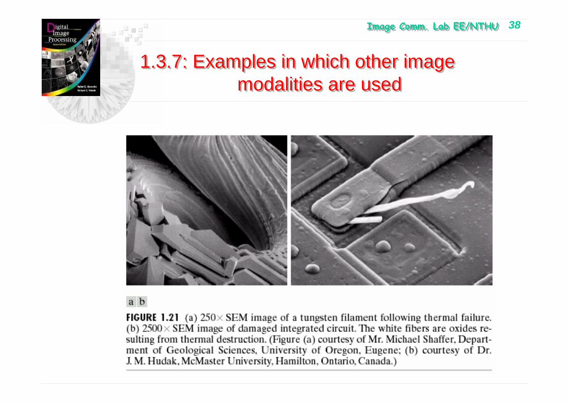
Image Comm. Lab EE/NTHUImage Comm. Lab EE/NTHU 38
1.3.7: Examples in which other image modalities are used
1.3.7: Examples in which other image modalities are used
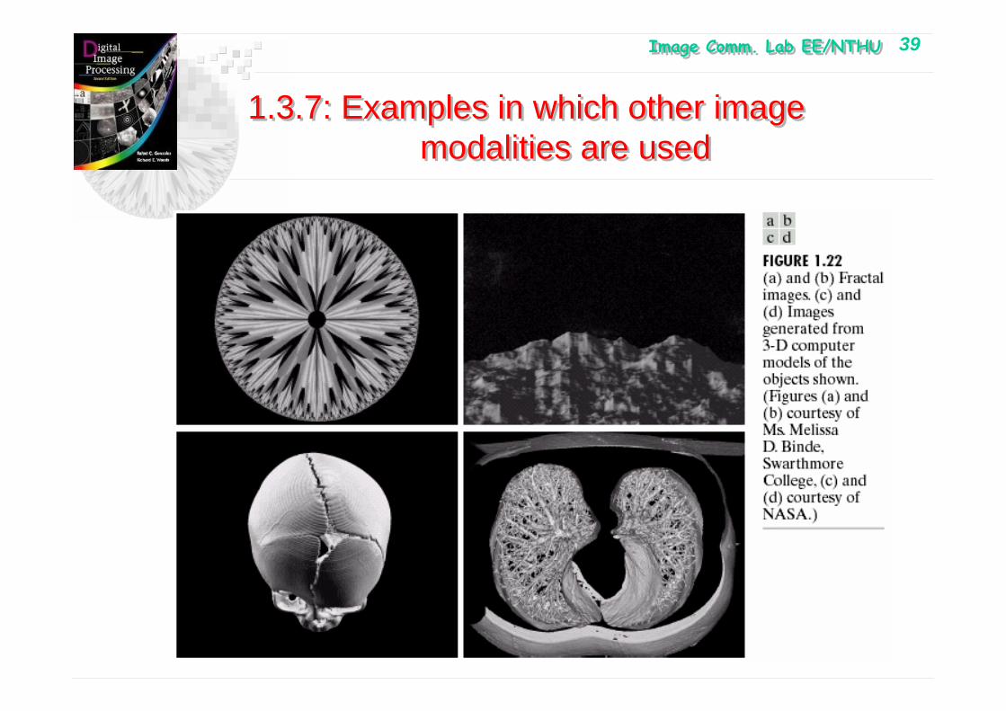
Image Comm. Lab EE/NTHUImage Comm. Lab EE/NTHU 39
1.3.7: Examples in which other image modalities are used
1.3.7: Examples in which other image modalities are used
Image Comm. Lab EE/NTHUImage Comm. Lab EE/NTHU 40
1.4: Fundamental Steps in Digital Image Processing
• Image Acquisition• Image Enhancement• Image Restoration• Color Image Processing• Wavelet• Compression• Morphological Processing• Segmentation• Representation and description• Recognition
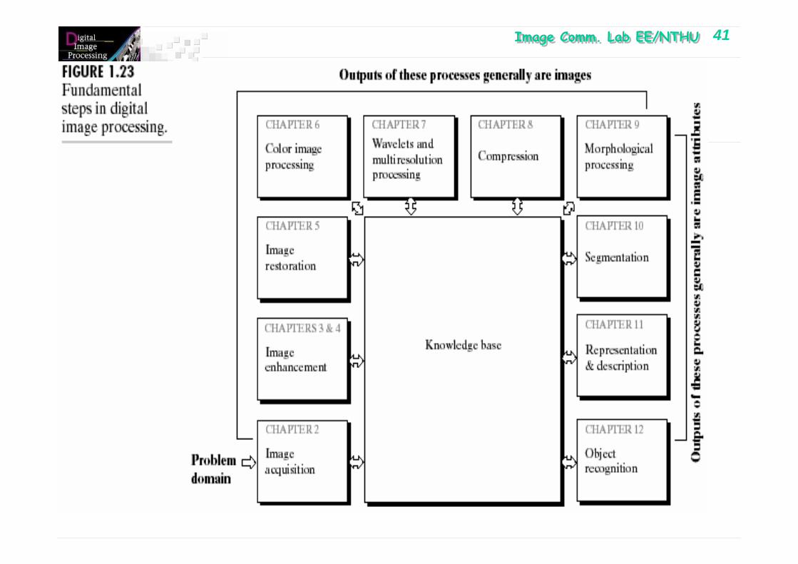
Image Comm. Lab EE/NTHUImage Comm. Lab EE/NTHU 41
Chapter 1: IntroductionChapter 1: Introduction

Image Comm. Lab EE/NTHUImage Comm. Lab EE/NTHU 42
1.5: Components of an image processing system
• Sensing• Specialized Image processing hardware• Computer• Software• Mass storage• Display• Hardcopy• Network
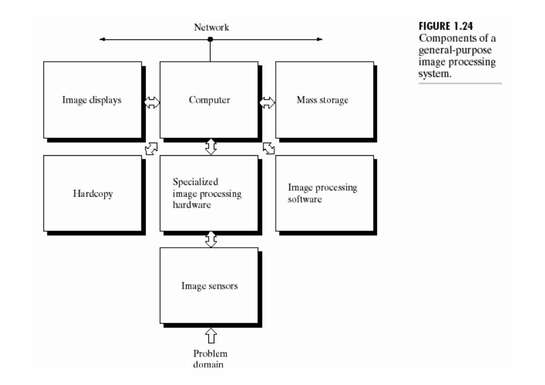
Image Comm. Lab EE/NTHUImage Comm. Lab EE/NTHU 43
Chapter 1: IntroductionChapter 1: Introduction