im - apps.dtic.mil
TRANSCRIPT

-AP-AIIO 630 ARMY RESEARCH INST OF ENVIROUMNTAL MEDICINE NATICK MA P/6 6/5ANATOMIC PERSPECTIVE OF THE PENALE AT)4.ETEI AN APPROACH TO MUSC--ECrcU)JAN 82 C BAUMAN. J J KNAPIK. 6 H JONESUNCLASSIFIED USARIEM-N-&/S NL
IM

L
11111I.O2 5 1111 L
MICROCOPY RESOLUTION TEST CHART
NATIONAL BUREAU Of STANDARDS 1963-A.

UNCLASSIFIEDSECURITY CLASSIFICATION OF THIS PACf ("on, Dat. Entered)
REPORT DOCUMEN', 4TION PAGE BEFORE CPLTGFORMI. REPORT NUMBER - 2. GOVT ACCESSION NO. 3. RECIPIENT'S CATALOG NUMB3ER
J ' /, .____AA__'SG
4. TITLE (and Subtitle) S. TYPE OF REPORT & PERIOD COVERED
Anatomic Perspective of the Female Athlete: AnApproach to Musculoskeletal Profiling of Women inSports In6. PERFORMING ORG. REPORT NUMBER
7. AUTHORfe) 140F S. CONTRACT OR GRANT NUMBER(&)
0Connie Bauman, ATC, MS, J.J. Knapik, MS, B.H.Jones, M.D., J.M. Harris, M.D. and L.K. Vaughn,
IBBMI Ph.D.__9 . PERFORMING ORGANIZATION NAME AND ADDRESS 10. PROGRAM ELEMENT. PROJECT, TASKVBMBI US Army Research Institute of Environmental AE OKUI UBR
Medicine, Natick, MA 01760
11. CONTROLLING OFFICE NAME AND ADDRESS 12. REPORT DATE
US Ary Medical Research and Development Command 11 Jan 82Fort Detrick 13. NUMBER OF PAGES
Frederick, MD 21701 20 pages74. MONITORING AGENCY NAME & ADORESS' ifferen.,t hum. Canm*IIn Office) 1S. SECURITY CLASS. (af 11,1. repout)
IS. OECLAS I FICAflON/ DOWNGRADINGr r SCHEDULE
1S. DISTRIBUTION STATEMEt~W
DISTRIBUTION OF THIS DOCUMENT IS UNLIMITED X
IS. SUPPLEMENTARY NOTES
N/A
>ee 19. KEY WORDS (Continue an revrs. side If noeea and Identify by block tmnbef)
0. N/A
20. r A~ACT ('CWMtoa M reevW0 h Nf ,,~eei md Ideewify by block abr
~~ N/A0
DD I JA 141 MDTIO% OFt I IOV 65 5 OOLETR UNCLASSIFIED
SECURIY CLASSIFICATION OF THIS PACE fWhn Dataneredlm

SECURITY CLASSIFICATION OF THIS PAGE(l rn, Does Emitewd)
SECURITY CLASSIFICATION OF THIS PAGEtIhef Daes Eft*
* i ,ll : , |
Anatomic Perspective of the Female Athlete:
An Approach to Musculoskeletal Profiling
of Women in Sports
Connie Bauman, ATC, MS*Joseph 3. Knapik, MS **
Bruce H. Jones, M.D.**John M. Harris, M.D.***
*Wellesley College, Wellesley, MA**US Army Research Institute of Environmental Medicine, Natick, MA***Veterans Administration Hospital, Boston, MA
7wY -

An approach to musculoskeletal profiling of women in sports
Vnki 5 duction
Women's sports did not begin to grow and gain public recognition until the
early 1970's. Since then, the number of women participating in intercollegiate
sports has doubled and the number of girls participating in interscholastic sports
programs has increased almost threefold. As women flood the sports arena,
many questions arise regarding the female athlete. Their performance
capabilities are constantly compared with male athletic records and standards,
and explanations for the difference in their level of performance are being
sought L.
As e as differences in performance, it has also been suggested that
there are differences in susceptibility to injury between males and females.
Because of suspected differences, many questions arise concerning the medical
care of the female athlete. Should women and men be trained in the same way if
injury is of concern? Does the female anatomical structure predispose her to
certain types of injuries? Are there distinct patterns of injury for women
athletes? Can potential injuries be detected prior to their occurrence through a
comprehensive musculoskeletal profile screening system? These are questions
which should be addressed in order to reduce the number and severity of injuries
incurred by women in sports competition.
This paper will focus on women and the injuries they incur as a result of
physical training and athletic competition. First the literature pertaining to
training injuries in both males and females will be reviewed. Next a
muscujoskeletal profiling system presently used in a study of women athletes at
Wellesley College will be outlined. Then the potential applications of such a
system will be explored.
I>
-I
-.--.-- '- -u
Review of the literature
It has been speculated that peculiarities of female anatomy may account
for their different propensity for certain types of injuries. Anatomically, it has
been stated (1,2) that the female has a wider pelvis, and therefore, the angle of
the femur to the pelvis is more acute than in the male. Albohm 3) and Haycock
(4] feel that the wider pelvis contributes to the potential problem of subluxation
or dislocation of the patella, which can ultimately result in chondromalacia. It
has been shown DI that women have somewhat slighter bone structure and a
smaller proportion of muscle to adipose tissue than the male. Females also
possess a greater degree of elasticity in the connective tissue which increases
flexibility and many contribute to a greater susceptibility to ligamentous injury
[3).
The two major sources of information on injuries to women resulting from
physical activity are the military and interscholastic athletic programs. More
women have been entering the military in the last ten years and as a result, their
physical aptitudes and injury rates have been more closely scruitinized. Several
studies have been conducted involving male and female basic trainees. Reinker
and Ozburne C5) noted that in basic training, the injury rate for women was twice
that of the men. The incidence of stress fractures was 10 percent in women as
compared with I percent for men. Stress fractures accounted for an alarmingly
high number (one-third) of the total injuries sustained by women. Other
significant injuries which involved time loss from training were chondromalacia
of the patella, achilles tendinitis, and sprains.
In an attempt to decrease the incidence of injury a company of 280 females
was placed on a revised exercise program D.I during basic training at Fort
3ackson. The strategy was to introduce physical stresses gradually and to avoid
known pathogenic varieties of training. As a result, the incidence of stress
2
V ____ ______ _____

fractures alone was reduced markedly from over 10 percent to 1.5 percent and
lower extremity musculoskeletal injuries as a whole were decreased by
approximately 50 percent.
In another study, Kowal (6) found that approximately 25 percent of males,
and almost 60 percent of females suffered overuse injuries to the lower
extremities during the 8 week basic training cycle. The injury data was
correlated with prior fitness measures, suggesting that the major causes of injury
in women might be attributed to the lack of conditioning, greater percent body
fat, and to the rapid introduction of heavy training, which does not allow for a
progressive exposure to stress. It appeared that women in the military were not
in condition to perform to male standards at the onset of training and needed
additional time for their bodies to respond to the stresses of a vigorous training
program.
Thus, nearly all of the military studies to date (5,6,7] clearly demonstrate
that female trainees are highly susceptible to stress related injuries in basic
training. However, it is interesting to compare the similarities and
dissimilarities in injury patterns between women in the military and women in
sport. The National Athletic Injury/Illness Reporting System (NAIRS) is a
national surveillance system that compiles intercollegiate and interscholastic
injury data submitted by athletic trainers or other health care personnel. A
preliminary overview [@ of injuries among collegiate women athletes during its
first three operational years (1975-78) indicated that injuries to women athletes
are essentially sport related; not gender related. The data reveals that matched
sports demonstrated similar patterns of injury for men and women and that the
major differences of injury patterns were between sports. It appeared from this
study that there are more dissimilarities between women's sports than between
comparable men and women's sports.
3
,q /.;. ..

A study which supports the NAIRS findings [91 was conducted to compare
injury rates between male and female high school athletic participants in eight
similar sports. The study revealed that sprains and strains were the most
frequent injuries. Females had a significantly greater number of knee injuries
and major ankle injuries than males, but the pattern of injury for other major
anatomical sites was similar. There were no significant differences in overall
incidence of injury between males and females, however, it was interesting to
note that females had a greater number of serious injuries.
Graham E10 conducted an injury survey among 29 colleges in Virginia
involving 129 female athletic teams during the 1974-75 school year. The study
indicated that the most common injuries in all sports were sprains, strains,
contusions, and simple fractures. Of these injuries, 44 percent were reinjuries
and nearly twice as many injuries occurred to the right side of the body as
opposed to the left side. The results of the survey support previous findings r9]
which indicate that the most common injuries involve the lower extremity,
specifically the ankle and knee.
Another study reviewing the clinical records of two sports physicians [11i
identified 1819 injuries in 1650 running patients during 1978-80. The records
revealed that both sexes had a similar distribution of injuries by major
anatomical sites (hips, thigh, knee, leg, ankle, foot), but certain specific
conditions were observed more frequently in one sex or the other. The knee was
the most common site of complaint accounting for 42 percent of all injuries and
patello-femoral pain syndrome was the most frequent disorder, accounting for 26
percent of all injuries. This study found that men sustained more achilles
tendinitis and patellar tendinitis, while women suffered more from "patellar pain
syndrome" and "tibial stress syndrome".
4
7* .'r
Pagliano Ci2) found that the knee accounted for 30 percent of the 1077
injuries among runners in a ten month study of running related injuries.
Chondromalacia was the most common diagnosis, accounting for almost one third
of those complaining of knee pain. The clinical diagnosis of chondromalacia
patella is a matter of debate and there are no specific criteria which are agreed
upon. Nevertheless, it appears that women are subject to more patello-femoral
complaints (which are construed to be chondromalacia) than men. Pagliano f12)stated that women outnumber men two to one in the general population in terms
of having symptomatic "chondromalacia." Brody ri claims that the typical
patient complaining of "chondromalacia" is one who possesses a wide pelvis,
knock-knees and hyperpronated feet. Therefore, it may be that the anatomic
structure of women which is more like this may predispose them to patello-
femoral complaints when subject to the stress of competition and weight bearing
physical training.
In summary, the data from civilian studies shows some differences in the
d4istribution and incidence of injuries to males and females competing in the
same sports. However, these differences are not as marked as those noted in
military studies of basic training populations. What this contrast may suggest is
that when women are expected to train and perform with men, as they are in the
military, they are more likely to suffer injuries than when allowed to train and
compete at their own level (at least initially).
A system of musculoskeletal profiling
It would certainly be advantageous to the athlete if potential injuries could
be predicted prior to their occurrence. A pre-season musculoskeletal
examination could identify abnormal physical characteristics and the athletic
trainer could then prescribe a preventive conditioning program to help decrease
the possibility of future injury.
__ . .!4

|I
In an effort to establish a selective musculoskeletal athletic profile
system, a collaborative four year longitudinal study was launched in 1980 by
Wellesley College and the US Army Research Institute of Environmental
Medicine (USARIEM). The study involves ten female intercollegiate varsity
teams. The purpose of the study is to determine whether any relationship exists
between various physical characteristics and the occurrence of injury and if so to
attempt to develope a battery of tests that would help predict injury.
The pre-season screening exam involves many components: medical
history, anthropometry, muscle strength, orthopedic alignment, joint laxity, and
flexibility. These components are measured and recorded to provide a baseline
for each student athlete. Injuries sustained during the season are documented by
the athletic trainer and then appropriate follow-up care is administered by the
College infirmary, athletic trainer, or the team orthopedic physician.
1. Medical History
Biographical information is recorded concerning age, sport, years of
participation and injury history.
2. Anthropometry
The subjects height and weight are recorded. Skinfold measurements are
taken from the subscapular, tricep, bicep, and suprailiac areas. Percent body fat
is estimated from skinfold thicknesses according to the formulas of Durnin and
Womersley (20).
3. Muscle Strength
Maximal isometric strength of the upper torso, the legs and back are
measured on a static strength machine designed and used extensively by
6

USARIEM (211. The Cybex 11 dynamometer is used to measure maximal dynamic
strength of both the left and right knee flexors and extensors at both 30
degrees/second and 180 degrees/second. On all strength tests three maximum
voluntary contractions are performed with at least 30 seconds rest between
contractions. If the strength values are not within + 10% of each other they are
repeated up to a maximum of five contractions. The mean of the three highest
values is recorded as the criterion strength score.
4. Orthopedic Alignment
The purpose of orthopedic alignment measures is to construct a lower body
model as a means of individual comparison. Our plan is to eventually have
sufficient data for a computer to generate a lower body structural profile for
each athlete to permit comparisons between injured and uninjured subjects and
thus identify predisposing structural differences.
For most of the following measurements of the lower extremities, subjects
are measured while standing with their feet in 5 degrees of external rotation.
Body length and width measurements are recorded in centimeters while angular
measurements are recorded in degrees. The measurements taken are as follows
(16,17)
a. The anterior superior iliac spine (ASIS) is identified and designated
with a marking implement. Leg length is determined by 'measuring each leg from
the ASIS to the shelf of the medial malleolus.
b. The Q-angle is measured with a long arm goniometer. The axis of the
goniometer is centered on the mid-patella and the arms aligned with the ASIS
and the center of the tibial tuberosity (figure 1-1).
c. An anthropometer is used to measure the distance between the ASIS.
71
• -
d. Obstetric calipers are used to compress the adipose tissue and
measure the distance between the right and left greater trochanters.
e. Obstetric calipers are placed on the right and left outer femoral
condyles and the distance measured and recorded.
f. The distance between the right and left inner femoral condyles is
measured.
g. The distance is measured between the right and left medial malleoli.
h. The distance is measured from the greater trochanter to the lateral
malleolus.
i. The distance is measured from the greater trochanter to the outer
femoral condyle.
j. The distance is measured from the outer femoral condyle to the
lateral malleolus.
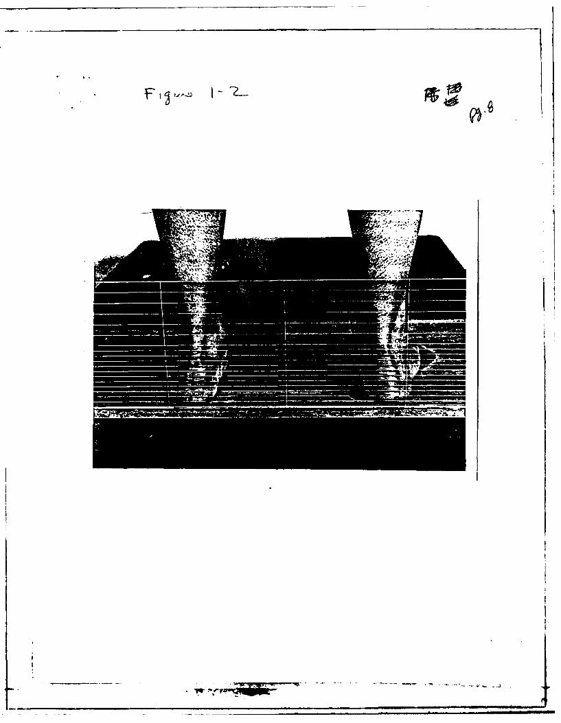
k. Foot pronation/supination is measured with the subject standing with
toes pointing straight ahead. The insertion of the achilles tendon into the
calcaneous and the midline of the calcaneous are identified and marked. The
heel is then placed against a plexiglas grid and the angle measured with a
protractor (figure 1-2).
5. Joint Laxity
Joint laxity varies throughout the population of normal healthy adults (22).
It has been suggested that "supple" individuals are more likely to suffer
dislocations than tight or less flexible individuals. Four hypermobility tests are
administered to determine overall joint laxity. Individuals who demonstrate
excessive joint mobility in 3 out of 4 of the following tests are suspected to have
an increased susceptibility toward joint related injuries.
• , ~qluS
Wi
a. The subject attempts to touch the thumb to the ventral forearm with
the wrist flexed and the thumb extended.
b. The subject attempts to apply gentle pressure with the non-tested
hand in order to extend the fingers so that they are parallel to the dorsal aspect
of the forearm.
c. Elbow hyperextension is measured with the athlete's arm extended
and hand supinated. The goniometer is centered on the lateral epicondyle and
the goniometer arms are aligned with the long axis of the humerus and the radial
styloid, and degrees of deviation from 1800 are noted.
d. Hyperextension of the knee joint is measured while the subject is
supine with the knee extended and the heel resting on a three inch platform. The
goniometer is centered at the knee on the lateral femoral condyle with the arms
aligned with the greater trochanter and the lateral malleolus, and deviation from
1800 is recorded.
6. Flexibility
Flexibility below a certain level is suspected to predispose a muscle to
injury when stressed. In order to test this assumption, flexibility measurements
are obtained from 7 major muscle groups of the lower extremity, so that injury
rates in "flexible" and "inflexible" athletes may be compared 18). A goniometer
is used to measure active flexion in each joint, as follows:
a. Hamstring flexibility is measured with the subject lying in a supine
position with legs extended. The leg being tested is actively flexed at the hip
with the knee locked. The axis of the goniometer is placed on the greater
trochanter with one arm aligned with the lateral midline of the thigh (parallel to
the femur) and the other arm parallel to the measuring surface.
b. Adductor flexibility is measured with the subject lying supine with
9

the tested leg extended and the non-tested flexed and hanging over the side of
the table to stabalize the pelvis. One arm of the goniometer is placed on a line
across the right and left ASIS, and the other arm placed on the anterior midline
of the thigh. The leg is actively abducted with the toes and knees pointing up to
avoid lateral rotation of the hip.
c. Quadricep flexibility is measured with the subject in a prone position
with leg extended. The knee is actively flexed and measured with one
goniometer arm aligned with the greater trochanter and the other arm aligned
with the lateral malleolus (parallel to the crest of the tibia).
d. Hip extension flexibility is measured with the subject in a prone
position with the knee flexed at 90 degrees. The thigh is actively extended with
the goniometer arms aligned with the lateral midline of the thigh and an
imaginary line which parallels the table surface.
e. Gastrocnemius flexibility is measured with the subject in a long
sitting position with knees extended. The foot is actively dorsiflexed with one
goniometer arm placed parallel to the fifth metatarsal and the other arm aligned
with the lateral midline of the leg (parallel to the shaft of the tibia).
f. Soleus flexibility is measured with the subject in a long sitting
position with the knee flexed at 90 degrees. The foot is actively dorsiflexed with
goniometer arms aligned parallel to the fifth metatarsal and the lateral midline
of the leg (parallel to the shaft of the tibia).
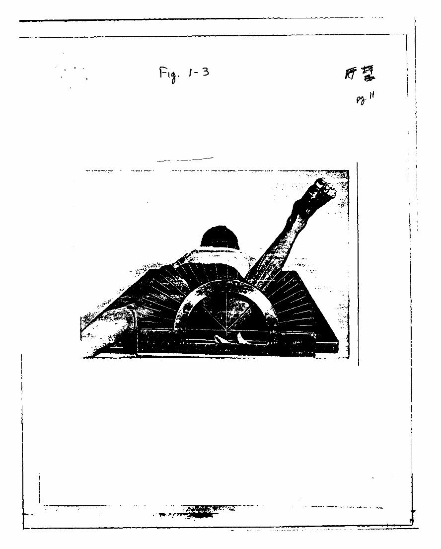
g. Hip rotation is measured with the subject in a prone position with the
knee flexed to 90 degrees and the midpatelia resting against the axis of a large
plexiglas protractor. As the lower leg externally rotates, internal hip rotation
takes place and is measured (figure 1-3). Care is taken to keep the subject's hips
on the table. The procedure is repreated allowing the leg to fall into internal
rotation, so external hip rotation can be measured.
10
.. . . . . . .

Practical use of musculoskeletal profiling
The intent of the collaborative study between Wellesley College and
USARIEM is to examine the possible value and utility of developing a
musculoskeletal profile which could identify biomechanical abnormalities as well
as strength imbalances, and to see if these may predispose women to certain
types of injuries. We are particularly interested in identifying physical
characteristics which either exceed or fall short of the accepted norms and
determining if certain characteristics or combinations of characteristics are
predictive of specific injuries.
Musculoskeletal profiling may be useful as other than a research tool. For
instance, historical information can alert the trainer to potential sources of
future injury. When a history of injury dictates, a preventive conditioning
program can be prescribed with the aim of reducing the chances of reinjury.
Also, if particular muscle groups are found to be relatively inflexible during pre-
season screening a remedial static stretching program may be prescribed.
In regard to the characteristics of muscles, Klafs and Arnheim (143 have
stated that imbalances in muscle strength and inflexibility are precipitating
factors leading to muscle strains. Burkett 04 in a prospective study found that
67% of athletes with a muscular strength imbalance of 10% or greater between
the right and left knee flexors suffered hamstring strains. But, Laird [13) was
unable to show that knee flexor or knee extensor strength imbalances
predisposed athletes to muscle strains. Despite the contradictory literature at
this point, however, it is felt that athletes who demonstrate a 10 percent or
greater muscle imbalance between flexors and/or extensors should be placed on a
strength conditioning program. It is hoped that a reduction of strength
imbalance will be useful in the prevention of potential muscle strains.
Nevertheless, it is important to recognize that 10 percent only represents a
11 N
- .~

guideline and that strength imbalance is not always solely responsible for the
occurrence of a muscle strain. There are athletes with a muscle imbalance of 10
percent or greater who will not sustain a muscle strain. The type of sport,
position, motivation, and intensity of effort may be important factors to
consider when looking at muscle strains and all injury statistics in general.
A musculoskeletal profile can also be important insurance that an athlete
is not returned to competition before she (or he) is fully rehabilitated. Preseason
screening establishes objective norms for a particular athlete and the decision
regarding when the athlete may return to practice can be based on these norms.
It is important that the athlete be tested prior to the competitive season to
insure test scores that are not confounded by injuries. Sapega and Nicholas C19)
have stated that the common practice of assuming that physical symmetry is the
norm and using the athletes opposite side as a comparative standard is not
always desirable.
Summary and conclusions
It has been suggested that women athletes sustain similar types of injuries
as their male counterparts. The mechanisms responsible for injury are similar
for both sexes and it appears that injuries are more sport related, than sex
related. But there is a continued need to focus on the patterns of injury, and to
determine whether there is a causal relationship between measurably aberrant
musculoskeletal variables and injury. Also, there is a further need to ascertain
whether susceptibility to injury is indeed similar in males and females. By using
the precompetition examination outlined above, we hope to develop a system for
predicting with some certainty predisposition to injury. If predisposing factors
can be identified, precompetition training and equipment can be individualized
where practical to minimize the chance of injury.
12
i1
REFERENCES
I. Harris, V. Physical sex differences: A matter of degree. The Counseling
Psychologist 6:9-11, 1976.
2. Haycock, E., Gillette, 3. . Susceptibility of women athletes to injury.
JAMA 236:163-165, 1976.
3. Albohm, M. How injuries occur in girls' sports. Phys. and Sportsmed. 5:44-
48, Feb., 1976.
4. Haycock, E. Health concerns of women in sports. Athletic Purchasing and
Facilities 62-65, March, 1981.
5. Reinker, A., Ozburne, S. A comparison of male and female orthopedic
pathology in basic training. Military Medicine 532-536, August, 1979.
6. Kowal, D.M. Nature and causes of injuries in women resulting from an
endurance training program. Am. 3. of Sports Med. 8:265-269, 1980.
7. Protzman, R. Can women be overextended in physical conditioning
programs? Am. 3. of Sports Med. 7:145-146, 1979.
8. Clarke, K.S. Women's injuries in collegiate sports. Am. 3. of Sports Med.
8:187-191, 1980.
9. Shively, R.A., Grana W.A., Ellis, D. High school sports injuries. Phys. and
Sportsmed. 9:46-49, 1981.
13
-V ,"'.

wI
10. Graham, F.P., Bruce, 3. Survey of intercollegiate athletic injuries to
women. Res. Quart. 48:217-
11. Clement, D.B., Taunton, J.E., Smart, G.W., McNicol, K.L. A survey of
overuse running injuries, Phys. and Sportsmed. 9:47-48, 1981.
12. Pagliano 3., Jackson D. The ultimate study of running injuries. Runner's
World 42-50, Nov., 1980.
13. Brody, D., Konecke, S., Day, S.W., Kryder, S. A study of 4,000 running
injuries. Running Times 22-29, July, 1981; 20-27 Sept., 1981.
14. Burkett, L.N. Causative factors in hamstring strains. Med. Sci. Sports
2:39-42, 1970.
15. Laird, D.E. Comparison of quad to ham strength ratios of an
intercollegiate soccer team. Athletic Training 66-68, Spring, 1981.
16. Enneking, W.F., Brower, T.D., Ralston, E.L., American Orthopaedic
Association. Manual of Orthopaedic Surgery, United States of America, 1979.
17. Heck, C.V., Hendryson, I.E., Rowe, C.R. Joint Motion: Method of
Measuring and Recording. Chicago, Illinois: American Academy of Orthopaedic
Surgeons, 1965.
18. ports Medicine Section, American Physical Therapy
Association. Guidelines for Pre-season Athletic Participation Evaluation.
Columbus, Georgia: Diversified Printing Services, 1979.
_14
I i ' I

19. Sapega, A.A., Nicholas, J.A. The clinical use of musculoskeletal profiling
in orthopedic sportsmedicine. Phys. and Sportsmed. 2:39-42, April, 1981.
20. Durnin, 3.V.G.G., Wormersley, 3. Body fat assessment from total body
density and its estimation from skinfold thickness: measurements on 481 men
and women aged from 16 to 72 years. Brit. 3. Nutr. 32:77-97, 1974.
21. Knapik, 3.3., Wright, 3.E., Kowal, D.M., Vogel, J.A. The influence of US
Army Basic Initial Entry Training on the muscular strength of men and women.
Aviat. Space Environ. Med. 51:1086-1090, 1980.
22. Marshall, 3.L., Johanson, N., Wickiewicz, T.L., Tischler, H. M., Koslin,
B.L., Zeno, S., Meyers, A. Joint loosness: a function of the person and the joint.
Med. Sci. Sports 12:189-194, 1980.
13
w•

BIBLIOGRAPHY
Buchbinder, M.R.; Napora, N.U., Biggs, E.W. The relationship of abnormal
pronation to chondromalacia of the patella in distance runners. Podiatric Sports
Medicine 69:159-162, February 1979.
Eggold, James F. Orthotics in the prevention of runners overuse injuries. Phys.
and Sportsmed. 9:125-128, March, 1981.
Glick, James M. Muscle strains: prevention and treatment. Phys. and
Sportsmed. 8:73-77, November, 1980.
Godshall, Richard W. The predictability of athletic injuries: an eight year study.
3. of Sports Med 3:50-54, 1975.
Hayes, Don. Risk factors in sport. Human Factors 16:454-458, 1974.
Krissoff, W.B., Ferris, W.D. Runners' injuries. Phys. and Sportsmed. 7:55-64,
1979.
16
1~

Fi~i I -I
'- ~
:74'-if
-
______F-
-. ....
1,~~.'* - -~
* 1-3
I
The views, opinions, and/or findings contained in this report are those ofthe author(s) and should not be construed as an official department of theArmy position, policy, or decision, unless so designated by other officialdocumentation.
Human subjects participated in these studies after giving their free and
informed voluntary consent. Investigators adhered to AR 70-25 and USAMRDCRegulation 70-25 on Use of Volunteers in Research.
oI* *. -'-.

'IiI