illustrating human development by computer graphics …€¦ · · 2014-09-12illustrating human...
TRANSCRIPT

Illustrating Human Development by Computer Graphics for Education in Embryology
Koh KAKUSHO1, Shinobu MIZUTA2, Yutaka MINEKURA1, Michihiko MINOH1
Tomoko NAKATSU3 and Kohei SHIOTA4
1 Center for Information and Multimedia Studies 2 Deptartment of Systems Science, Graduate School of Informatics
3 Congenital Anomaly Research Center, Graduate School of Medicine 4 Department of Anatomy and Developmental Biology, Graduate School of Medicine
Kyoto University, Kyoto, Japan e-mail: [email protected]
Abstract This article describes teaching materials (TM) developed for education in embryology, which is one of the basic subjects in medicine to learn the process of human development especially from fertilization to birth. Since the human body takes various kinds of three-dimensional (3D) shapes by complicated deformations in this process, it is difficult to understand those shapes and deformations properly from conventional TM with static two-dimensional (2D) representations, such as photographs and illustrations. We produced novel TM, which illustrate the 3D shapes and deformations by an animation using computer graphics (CG). In order to produce CG with the reality sufficient for education in embryology, we employed data of the 3D shapes from specimens of human embryos by magnetic resonance microscopy (MRM). Those specimens have individual differences in shape, and the measurement by MRM cannot extract all the important features in shape due to the limitation in resolution. We normalized those individual differences and recovered the features important for education based on our expertise in embryology. The teaching materials based on the result are highly valued in the community of medicine and receiving a lot of requests for distribution. Keywords Teaching Materials, Medical Education, Modeling, Magnetic Resonance Microscopy 1. Introduction
Through the spread of information technology, it becomes quite usual to use teaching materials (TM) in the form of the data for Web browsers or application software, in university educations. However, those TM are often not much more useful than conventional TM such as textbooks, slides and videotapes, but only a kind of “digitized” conventional TM. Both of them employ the same kind of materials: images, illustrations, videos, audios and texts. Only the difference is whether they are displayed by a computer or not.
We started a project to develop novel TM that have never appeared as conventional TM, so that production of the TM becomes really worthwhile. As the first trial in this project, we chose to illustrate the development of human embryos for education in embryology, which is one of the basic subjects in medicine to learn the process of human development especially from fertilization to birth. Human embryos take various kinds of shapes changing through complicated deformations when they are developed after fertilization. In traditional education, static and two-dimensional (2D) representations such as photographs and illustrations have been used to show the shapes and deformations. Dynamic and three-dimensional (3D) representations, such as animations, should be employed for better understandings.
Recently, various kinds of novel TM have been proposed for medical educations[1-5]. Some of them employ the data from a real specimen of the human body to attain the reality sufficient for medical research

and education[4,5]. For embryology, 3D data of magnetic resonance microscopy (MRM) obtained from specimens of human embryos have been released[6]. However, raw MRM data are not preferable representations for beginners to learn the 3D shapes of human embryos, compared with animations. For learning the average case of human development, raw MRM data are not appropriate either, because specimens of human embryos include various kinds of individual differences. In addition, the MRM data do not cover the very early stages of human development, for which specimens are not available.
We have produced an animation that illustrates human development using computer graphics (CG). It covers all the stages that include the change in shape of embryos. We also employed MRM data from specimens of human embryos at the stages for which the specimens are available, for sufficient reality of medical education. The individual differences among the specimens are normalized in the process of modeling the 3D shapes of embryos from the MRM data, based on our expertise in embryology.
In the remainder of this article, we first give a brief explanation about the process of human development and the problem of conventional TM to illustrate the process in section 2. In section 3, we describe the process of modeling the 3D shapes of human embryos based on MRM data. The animation produced by CG using the 3D models is shown in section 4. In section 5, we discuss our future work. 2. Process of Human Development It is well known that the human body takes about 40 weeks to complete its development after fertilization. Human embryos change their shapes during the first eight weeks before taking a human shape. The remaining 32 weeks are spent mainly for development of the brain and organs, and cause the increase in volume without any change in shape. The process of human development during the first eight weeks has 23 stages, which are called “Carnegie stages” in embryology. Each Carnegie stage includes a considerable change in the shapes of human embryos. As visual aids for beginners to learn the change, it is important to illustrate the 3D shape of a human embryo at each Carnegie stage.
In conventional TM in embryology, the shapes of human embryos during the Carnegie stages have been visualized by static 2D illustrations in textbooks. In those figures, the shapes are too complicated for beginners to understand correctly. It is difficult to imagine the deformation between the shapes of consecutive Carnegie stages from these illustrations. Nevertheless, it is very important to understand the deformations of human embryos in order to know how the human body originates from each part of the embryo and how the defects in each part of the embryo affect the development of the human body.
The objective of our work is to illustrate the 3D shapes of human embryos and their deformations by 3D CG. By using 3D CG, we can not only produce an animation that describes the deformations from a specific viewpoint, but also allow the users to specify their viewpoints interactively while they are looking at a human embryo. Both capabilities are much more useful to illustrate the complicated shapes and deformations of human embryos than the conventional TM. 3. Modeling 3D Shapes of Human Embryos from Specimens
In order to produce CG with a realism sufficient for medical education, we developed 3D models from specimens of actual human embryos. We have access to more than 40,000 specimens of human embryos from Carnegie stage 13 to 23 of their development. This collection is quite rich and includes various individuals at different Carnegie stages with or without abnormality. We carefully selected 11 normal specimens from this collection so that each specimen represents a different Carnegie stage.
The selected specimens are measured by a device for MRM with a probe specially designed for measurement of small objects with a size around 3 millimeters. By using this device, we can get MRM data of human embryos represented by 128128128 ×× voxels with the cubic size of 50×50×50 micrometers. A slice of the MRM data obtained by this device from a specimen of a human embryo and the 3D shape directly obtained from those data are shown in Fig.1 (a), (b), respectively.
Unfortunately, the MRM data include noise as well as measurement errors. Another problem is that the MRM specimen data do not describe all the important features to be illustrated for education in embryology, due to the limitation in resolution. To cope with these problems, we carefully produced initial 3D models, so that they precisely describe the 3D shapes obtained from MRM data, while neglecting the noise and errors, and recovering the important features lost in the MRM measurement. The initial 3D model from Fig.1 (b) is shown in (c).

(a) MRI data (b) 3D shape from (a) (c) Initial 3D model of (b) (d) Final 3D model
Fig. 1: Modeling from a Specimen of a Human Embryo.
The specimens have their own individual characteristics, which make them different from one another. The sizes and proportions of different specimens are not the same even if they are at the same Carnegie stage. It is also often the case that different parts of a specimen are not at the same Carnegie stage due to individual differences in growth. We normalized the individual differences by carefully modifying the shapes of the initial 3D models based on our expertise in embryology, so that the series of the resultant 3D models show an average case of human development without loosing the details originated from MRM data. The 3D model obtained by this procedure from the initial 3D model in Fig.1 (c) is shown in (d). In this example, the positions of joints and the width of limbs of the model in (c) do not describe the average case. Those features are normalized as shown in (d).
Fig. 2: Illustrating Development of a Human Embryo by an Animation.
4. Illustrating the Development of Human Embryos
We produced an animation by generating the appearance of each 3D models obtained in section 3 from appropriate viewpoints. Since the specimens do not cover the very early stages of their development, we produced CG for those stages based on our expertise in embryology. The representative scenes of the animation is shown in Fig.2.
We also transformed the 3D models into the form of VRML (Virtual Reality Markup Language) files, so that instructors can specify the viewpoint to display the 3D models interactively when they explain the human development in their lectures of embryology. Since the shapes of the human embryo under development are often very complicated, it can then be difficult to see all the surface of the embryo due to
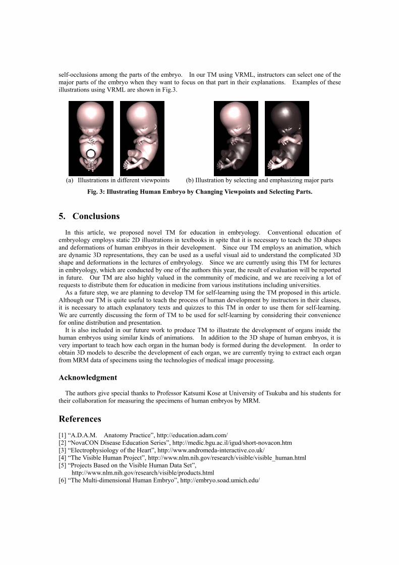
self-occlusions among the parts of the embryo. In our TM using VRML, instructors can select one of the major parts of the embryo when they want to focus on that part in their explanations. Examples of these illustrations using VRML are shown in Fig.3.
(a) Illustrations in different viewpoints (b) Illustration by selecting and emphasizing major parts
Fig. 3: Illustrating Human Embryo by Changing Viewpoints and Selecting Parts.
5. Conclusions
In this article, we proposed novel TM for education in embryology. Conventional education of embryology employs static 2D illustrations in textbooks in spite that it is necessary to teach the 3D shapes and deformations of human embryos in their development. Since our TM employs an animation, which are dynamic 3D representations, they can be used as a useful visual aid to understand the complicated 3D shape and deformations in the lectures of embryology. Since we are currently using this TM for lectures in embryology, which are conducted by one of the authors this year, the result of evaluation will be reported in future. Our TM are also highly valued in the community of medicine, and we are receiving a lot of requests to distribute them for education in medicine from various institutions including universities.
As a future step, we are planning to develop TM for self-learning using the TM proposed in this article. Although our TM is quite useful to teach the process of human development by instructors in their classes, it is necessary to attach explanatory texts and quizzes to this TM in order to use them for self-learning. We are currently discussing the form of TM to be used for self-learning by considering their convenience for online distribution and presentation.
It is also included in our future work to produce TM to illustrate the development of organs inside the human embryos using similar kinds of animations. In addition to the 3D shape of human embryos, it is very important to teach how each organ in the human body is formed during the development. In order to obtain 3D models to describe the development of each organ, we are currently trying to extract each organ from MRM data of specimens using the technologies of medical image processing. Acknowledgment
The authors give special thanks to Professor Katsumi Kose at University of Tsukuba and his students for their collaboration for measuring the specimens of human embryos by MRM. References [1] “A.D.A.M. Anatomy Practice”, http://education.adam.com/ [2] “NovaCON Disease Education Series”, http://medic.bgu.ac.il/igud/short-novacon.htm [3] “Electrophysiology of the Heart”, http://www.andromeda-interactive.co.uk/ [4] “The Visible Human Project”, http://www.nlm.nih.gov/research/visible/visible_human.html [5] “Projects Based on the Visible Human Data Set”,
http://www.nlm.nih.gov/research/visible/products.html [6] “The Multi-dimensional Human Embryo”, http://embryo.soad.umich.edu/