i’ll cry instead: the neural correlates of empathy
TRANSCRIPT

ResearchOnline@JCU
This file is part of the following work:
Krivan, Sarah (2020) I'll cry instead: the neural correlates of empathy. PhD
Thesis, James Cook University.
Access to this file is available from:
https://doi.org/10.25903/6ym6%2D7291
© 2020 Sarah Krivan.
The author has certified to JCU that they have made a reasonable effort to gain
permission and acknowledge the owners of any third party copyright material
included in this document. If you believe that this is not the case, please email
I’ll Cry Instead: The Neural Correlates of Empathy
Dissertation submitted by
Sarah Krivan B. Psych (Hons)
March, 2020.
For the degree of Doctor of Philosophy
College of Healthcare Sciences
James Cook University
Cairns, Australia
ii
Abstract
Tears demand attention. A compelling form of emotional expression, tears fascinate
both scientists and lay people alike (Trimble, 2012). Despite this fascination, surprisingly
little is known about the functions of tears, as crying has been neglected by emotion
researchers relative to other expressions (Vingerhoets, 2013). Emotional tears are believed to
signal sadness and distress, which in turn reliably elicit help and empathic responses from
observers (Balsters, Krahmer, Swerts, & Vingerhoets, 2013; Hendriks & Vingerhoets, 2006;
Lockwood, Millings, Hepper, & Rowe, 2013; Provine, Krosnowski, & Brocato, 2009).
However, it is unknown why tears elicit such strong responses from observers. To put it
simply, why do we care when others cry?
This thesis details a series of studies investigating the way that unconscious
physiological responses influence the way we outwardly respond to emotional tears. First, it
was established that tears signal sadness in the absence of context, regardless of the valence
of the facial expression. Moreover, tearful displays of sadness, and happy faces without tears
are particularly distinctive displays that are rapidly interpreted. Therefore, tears modulate the
way that emotional faces are perceived at the behavioural level.
Next, I sought to explore whether these pronounced responses to tears had a
biological basis. To address this aim, I explored the way that mimicry, early event-related
potentials (ERP), and neural mirroring were influenced by the presence of tears on a face.
Chapter 4 demonstrated that mimicry largely was not affected by tear presence. As such, it
was concluded that mimicry of tearful displays may not be adaptive. Conversely, Chapter 5
demonstrated that tears were preferentially processed at the early neural level. Moreover,
tearful sadness and happy faces without tears elicited larger face-related ERPs than other
expressions. This increased responding is in line with the social relevance hypothesis, which
posits that emotional expressions with increased social relevance are preferentially processed.
iii
Therefore, the results in Chapter 5 demonstrate the same pattern of results as the behavioural
data reported in Chapter 3, demonstrating a biological basis for the behavioural results.
Finally, Chapter 6 details one of the first studies to explore whether neural mirroring is
modulated by tearful displays. The results obtained were the inverse of that reported in
Chapters 3 and 5, where more ambiguous expressions (i.e. neutral, happy-tear, and sad tear-
free) elicited greater neural mirroring. This result is in accord with recent research, which has
demonstrated that ambiguous faces require greater neural mirroring. Therefore, tearful
expressions—particularly those of sadness—are interpreted rapidly. Moreover, humans
appear to be biologically hard-wired to respond to socially relevant information. This thesis
has provided both behavioural and psychophysiological evidence to demonstrate that tears
enhance the social relevance of sad faces. However, what is it about tears that make them
such an effective signal?
The final aim of this thesis was to explore the type of stimuli used in tear research.
Tears have been touted as an honest signal of emotion; however, there is limited empirical
evidence to support this assertion. Chapter 8 details the results of some of the first
experiments exploring perceptions of genuineness in tearful displays. It was demonstrated
that people are sensitive to the difference between posed and genuine crying, and that tears
increase the perception of genuineness for posed displays. Thus, this research has provided a
rationale for continued investigation of the ideal type of stimuli to be used in tear research
and has highlighted the potential to use tears as a means of determining how humans
distinguish between sincere and insincere emotion.
Overall, the results of this thesis detail the way that subconscious physiological
responses influence the way we outwardly respond to emotional tears. Through the use of
novel methodologies, this thesis has demonstrated the unique way that tears shape responses
to facial expressions. Therefore, this research affords a new understanding of a uniquely

iv
human phenomenon, and by extension, provides insight into the development of empathy and
prosocial behaviour in society as a whole.
Wordcount: 650 words (min 500)

v
Declaration
I declare that this dissertation is my own work and has not been submitted in any other form
for another degree or diploma at any other university or other institution of tertiary education.
Information derived from published or unpublished work of others has been acknowledged in
the text and a list of references given.
_________________
Sarah Krivan
28/03/2020

vi
Statement of Contributions
This thesis is made up of my own work, except for the normal contributions of a supervisory
panel. Chapters 1, 2, and 9 are completely my own work. Chapters 3, 4, 5, 6, 7, and 8 are
prepared as manuscripts for publication and the contribution of co-authors is outlined below
and at the beginning of each chapter. I also acknowledge the aid of Aidan Possemiers in
implementing the Python script for the processing of the EEG data in Chapter 6. Table 1
denotes the intellectual and financial support received throughout my candidature.
Table 1.
Overarching support throughout doctoral candidature.
Nature of Assistance
Contribution Names, Titles and Affiliation of Co-Contributors
Financial support Fee offset/waiver Research costs RTP Stipend JCU Stipend
Australian Government Research Training Program and James Cook University
Intellectual Support Supervision Conceptualisation of the research Revision of manuscripts
A/Prof. Nerina Caltabiano (James Cook University), Dr. David Cottrell (James Cook University – Adjunct), and Dr. Nicole Thomas (Monash University)
Publications arising from this dissertation and the contributions of the candidate and
co-authors
One scientific paper has already been published:
Krivan, S. J., & Thomas, N. A. (2020). A call for the empirical investigation of tear stimuli. Frontiers in Psychology, 11, 52. doi:10.3389/fpsyg.2020.00052
This paper is included in the thesis exactly as it is reported in the published version (except for minor formatting changes). The text in this thesis and the published document is identical.
I conceptualized and designed the article and wrote the first draft of the manuscript. I re-drafted subsequent manuscript versions, implemented the changes recommended by reviewers and approved the submitted version.
vii
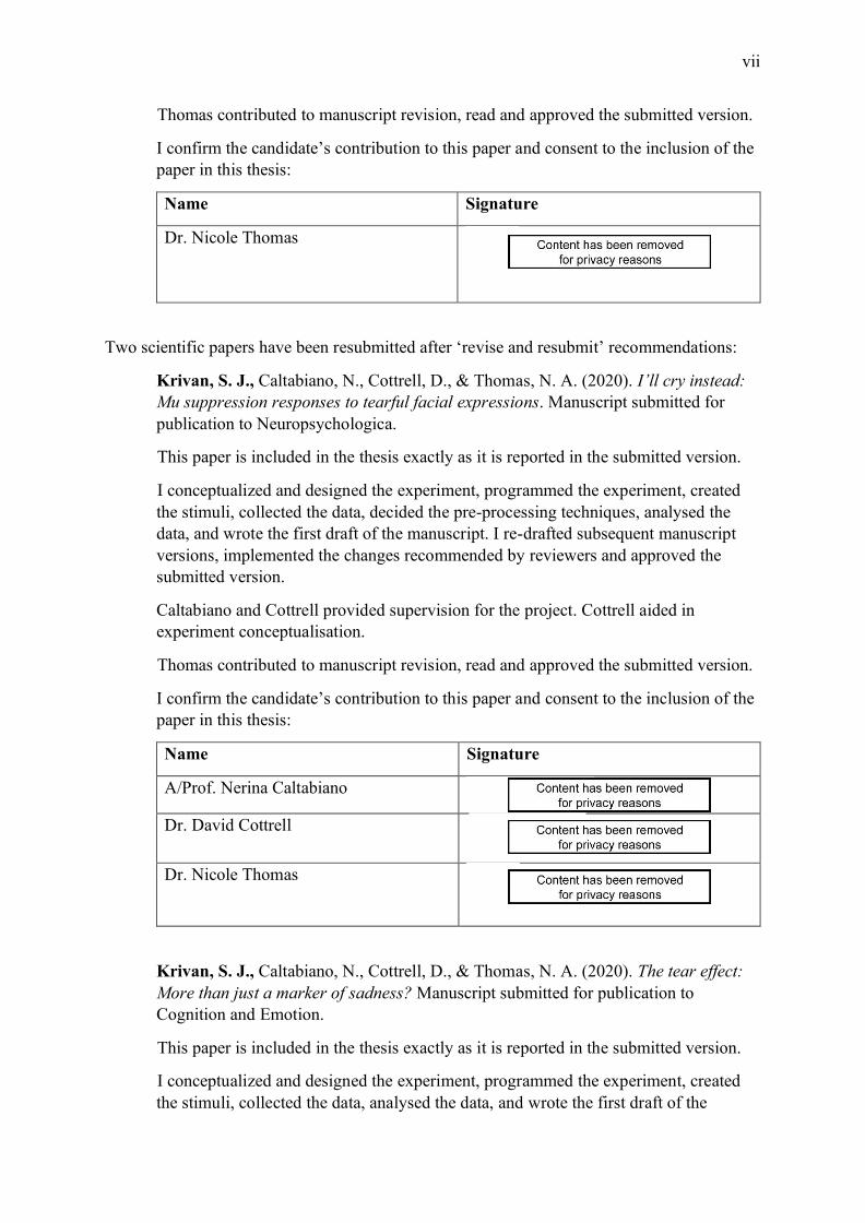
Thomas contributed to manuscript revision, read and approved the submitted version.
I confirm the candidate’s contribution to this paper and consent to the inclusion of the paper in this thesis:
Name Signature
Dr. Nicole Thomas
Two scientific papers have been resubmitted after ‘revise and resubmit’ recommendations:
Krivan, S. J., Caltabiano, N., Cottrell, D., & Thomas, N. A. (2020). I’ll cry instead: Mu suppression responses to tearful facial expressions. Manuscript submitted for publication to Neuropsychologica.
This paper is included in the thesis exactly as it is reported in the submitted version.
I conceptualized and designed the experiment, programmed the experiment, created the stimuli, collected the data, decided the pre-processing techniques, analysed the data, and wrote the first draft of the manuscript. I re-drafted subsequent manuscript versions, implemented the changes recommended by reviewers and approved the submitted version.
Caltabiano and Cottrell provided supervision for the project. Cottrell aided in experiment conceptualisation.
Thomas contributed to manuscript revision, read and approved the submitted version.
I confirm the candidate’s contribution to this paper and consent to the inclusion of the paper in this thesis:
Name Signature
A/Prof. Nerina Caltabiano
Dr. David Cottrell
Dr. Nicole Thomas
Krivan, S. J., Caltabiano, N., Cottrell, D., & Thomas, N. A. (2020). The tear effect: More than just a marker of sadness? Manuscript submitted for publication to Cognition and Emotion.
This paper is included in the thesis exactly as it is reported in the submitted version.
I conceptualized and designed the experiment, programmed the experiment, created the stimuli, collected the data, analysed the data, and wrote the first draft of the

viii
manuscript. I re-drafted subsequent manuscript versions, implemented the changes recommended by reviewers and approved the submitted version.
Caltabiano provided supervision for the project.
Cottrell aided in experiment conceptualisation and provided supervision for the project.
Thomas contributed to manuscript revision, read and approved the submitted version.
I confirm the candidate’s contribution to this paper and consent to the inclusion of the paper in this thesis:
Name Signature
A/Prof. Nerina Caltabiano
Dr. David Cottrell
Dr. Nicole Thomas
Three papers are in preparation for submission:
Krivan, S. J., Cottrell, D., & Thomas, N. A. (2020). Unconscious mimicry of tearful expressions. Manuscript in preparation.
I conceptualized and designed the experiment, programmed the experiment, created the stimuli, collected the data, processed the psychophysiological data, analysed the data, and drafted the manuscript.
Cottrell aided in conceptualisation of the project and provided supervision.
Thomas contributed to manuscript revision and provided supervision.
I confirm the candidate’s contribution to this paper and consent to the inclusion of the paper in this thesis:
Name Signature
Dr. David Cottrell
Dr. Nicole Thomas
Krivan, S. J., Cottrell, D., & Thomas, N. A. (2020). Early neural processing of tearful faces. Manuscript in preparation.

ix
I conceptualized and designed the experiment, programmed the experiment, created the stimuli, collected the data, processed the EEG data, analysed the data, and drafted the manuscript.
Cottrell aided in conceptualisation of the project and provided supervision.
Thomas contributed to manuscript revision and provided supervision.
I confirm the candidate’s contribution to this paper and consent to the inclusion of the paper in this thesis:
Name Signature
Dr. David Cottrell
Dr. Nicole Thomas
Krivan, S. J., & Thomas, N. A. (2020). Tears alter the perceived genuineness of emotional displays. Manuscript in preparation.
I conceptualized and designed the experiment, programmed the experiment, created the stimuli, collected the data, analysed the data, and drafted the manuscript.
Thomas contributed to experiment conceptualisation, provided the funding for the experiments, and aided in manuscript revision.
I confirm the candidate’s contribution to this paper and consent to the inclusion of the paper in this thesis:
Name Signature
Dr. Nicole Thomas

x
Acknowledgements
Undertaking a PhD is a lonely experience and one which I could not have completed without
some influential people. First, I thank my advisors, David, Nerina and Nicole. I appreciate
your guidance and unwavering support throughout my candidature. Next, I thank all the
students who have passed through the Cognitive Sciences Laboratory and the A2P2
Laboratory. The inspiration that comes from collaborative discussion made the project feel a
lot less lonely. I thank my friends, and my family members, Mum, Dad, and Leanna. You
listened to my triumphs and failures over the past four years, with a caring ear and
reassurance that it would be okay in the end. Thank you to my partner Aidan. Completing our
PhDs at the same time meant that I always had someone who uniquely understood the
challenge of completing the PhD journey. Thank you for all the time you have spent patiently
listening to me talk about my thesis for the past four years. Finally, I thank David Bowie,
whose music always made me feel like everything was Hunky Dory.
xi
Table of Contents Chapter 1: Responses to Emotional Facial Expressions and Crying ________________ 1
1.1 Introduction Overview ____________________________________________________ 1 1.2 Facial Expressions ________________________________________________________ 2 1.3 Emotional Theories _______________________________________________________ 2 1.4 Positivity and Negativity Biases ____________________________________________ 11 1.5 The Communicative Value of Tears ________________________________________ 21 1.6 Summary ______________________________________________________________ 26
Chapter 2: The Psychophysiological Measurement of Empathy __________________ 27 2.1 Chapter Introduction ____________________________________________________ 27 2.2 Empathy _______________________________________________________________ 27
2.2.1 What is empathy? _____________________________________________________________ 28 2.2.2 The perception-action model (PAM) of empathy ____________________________________ 29 2.2.3 The measurement of empathy ___________________________________________________ 31 2.2.4 Catching emotions ____________________________________________________________ 34 2.2.5 Imitating others ______________________________________________________________ 35
2.3 Mirror Neurons _________________________________________________________ 39 2.3.1 What are mirror neurons?_______________________________________________________ 39 2.3.2 The empathic brain what have we learned from neuroimaging? _________________________ 41 2.3.3 Beyond neuroimaging: Mirror neuron research with mu rhythm ________________________ 45
2.4 Contributions of this Thesis _______________________________________________ 48 2.4.1 2.4.1 Chapter overview ________________________________________________________ 50 2.4.2 2.4.2 Thesis aim ______________________________________________________________ 52
Chapter 3: The Tear Effect: More than just a Marker of Sadness? _______________ 53 3.1 Chapter Overview _______________________________________________________ 53 3.2 Publication Status _______________________________________________________ 53 3.3 Author Contributions ____________________________________________________ 53 3.4 Manuscript _____________________________________________________________ 53
3.4.1 Abstract ____________________________________________________________________ 53 3.4.2 Introduction _________________________________________________________________ 54 3.4.3 Experiment 1 ________________________________________________________________ 59
3.4.3.1 Method ________________________________________________________________ 59 3.4.3.1.1 Participants __________________________________________________________ 59 3.4.3.1.2 Stimuli ______________________________________________________________ 60 3.4.3.1.3 Procedure ____________________________________________________________ 61
3.4.3.2 Results ________________________________________________________________ 62 3.4.3.2.1 Accuracy ____________________________________________________________ 62 3.4.3.2.2 Reaction time _________________________________________________________ 64 3.4.3.2.3 Neutral face analyses ___________________________________________________ 64 3.4.3.2.4 Discussion ___________________________________________________________ 65
3.4.4 Experiment 2 ________________________________________________________________ 67 3.4.4.1 Method ________________________________________________________________ 68
3.4.4.1.1 Participants __________________________________________________________ 68 3.4.4.1.2 Stimuli ______________________________________________________________ 68 3.4.4.1.3 Procedure ____________________________________________________________ 69
3.4.4.2 Results ________________________________________________________________ 70 3.4.4.2.1 Reaction time task _____________________________________________________ 71 3.4.4.2.2 Rating task ___________________________________________________________ 73
xii
3.4.4.3 Discussion______________________________________________________________ 75 3.4.5 General Discussion____________________________________________________________ 76
Chapter 4: Unconscious Mimicry of Tearful Expressions _______________________ 82 4.1 Chapter Overview _______________________________________________________ 82 4.2 Publication Status _______________________________________________________ 82 4.3 Author Contributions ____________________________________________________ 82 4.4 Manuscript _____________________________________________________________ 83
4.4.1 Abstract ____________________________________________________________________ 83 4.4.2 Introduction _________________________________________________________________ 83 4.4.3 Method _____________________________________________________________________ 89
4.4.3.1 Participants _____________________________________________________________ 89 4.4.3.2 Stimuli ________________________________________________________________ 89 4.4.3.3 Procedure ______________________________________________________________ 90
4.4.3.3.1 Phase 1: Masked trials __________________________________________________ 91 4.4.3.3.2 Phase 2: Shown face trials _______________________________________________ 92
4.4.3.4 EMG recording and analysis _______________________________________________ 93 4.4.3.5 Data analysis ____________________________________________________________ 94
4.4.4 Results _____________________________________________________________________ 95 4.4.4.1 Physiological responses to masked primes _____________________________________ 95
4.4.4.1.1 ZMaj and ZMin _______________________________________________________ 95 4.4.4.1.2 CS _________________________________________________________________ 96
4.4.4.2 Physiological responses to supraliminal faces __________________________________ 96 4.4.4.2.1 ZMaj and ZMin _______________________________________________________ 96 4.4.4.2.2 CS. _________________________________________________________________ 97
4.4.5 Discussion __________________________________________________________________ 97 Chapter 5: Early Neural Processing of Tearful Faces _________________________ 104
5.1 Chapter Overview ______________________________________________________ 104 5.2 Publication Status ______________________________________________________ 104 5.3 Author Contributions ___________________________________________________ 104 5.4 Manuscript ____________________________________________________________ 104
5.4.1 Abstract ___________________________________________________________________ 104 5.4.2 Introduction ________________________________________________________________ 105 5.4.3 Method ____________________________________________________________________ 111
5.4.3.1 Participants ____________________________________________________________ 111 5.4.3.2 Stimuli _______________________________________________________________ 112 5.4.3.3 Procedure _____________________________________________________________ 113 5.4.3.4 EEG recording and analysis _______________________________________________ 114 5.4.3.5 Data exclusions _________________________________________________________ 115
5.4.4 Results ____________________________________________________________________ 115 5.4.4.1 Behavioural data ________________________________________________________ 115 5.4.4.2 ERP data ______________________________________________________________ 116
5.4.4.2.1 N170 mean amplitude _________________________________________________ 117 5.4.4.2.2 N170 peak latency. ___________________________________________________ 118
5.4.5 Discussion _________________________________________________________________ 119 Chapter 6: I’ll cry Instead: Mu Suppression Responses to Tearful Facial Expressions 125
6.1 Chapter Overview ______________________________________________________ 125 6.2 Publication Status ______________________________________________________ 125 6.3 Author Contributions ___________________________________________________ 125
xiii
6.4 Manuscript ____________________________________________________________ 126 6.4.1 Abstract ___________________________________________________________________ 126 6.4.2 Introduction ________________________________________________________________ 126 6.4.3 Method ____________________________________________________________________ 133
6.4.3.1 Participants ____________________________________________________________ 133 6.4.3.2 Materials ______________________________________________________________ 134 6.4.3.3 Procedure _____________________________________________________________ 136
6.4.3.3.1 Discrimination task ___________________________________________________ 136 6.4.3.3.2 Execution task _______________________________________________________ 137
6.4.3.4 EEG recording and analysis _______________________________________________ 138 6.4.4 Results ____________________________________________________________________ 139
6.4.4.1 Data exclusions _________________________________________________________ 139 6.4.4.2 Behavioural results ______________________________________________________ 140 6.4.4.3 EEG results ____________________________________________________________ 141
6.4.4.3.1 Central electrodes ____________________________________________________ 142 6.4.4.3.2 Occipital electrodes ___________________________________________________ 145
6.4.4.4 Correlations between central electrodes and self-report empathy __________________ 146 6.4.5 Discussion _________________________________________________________________ 146
Chapter 7: Future Directions in Tear Research: Part I ________________________ 153 7.1 Chapter Overview ______________________________________________________ 153 7.2 Publication Status ______________________________________________________ 153 7.3 Author Contributions ___________________________________________________ 154 7.4 Manuscript ____________________________________________________________ 154
7.4.1 Abstract ___________________________________________________________________ 154 7.4.2 Introduction ________________________________________________________________ 154 7.4.3 Genuine emotional displays ____________________________________________________ 156 7.4.4 The artificial tear ____________________________________________________________ 158 7.4.5 The genuine tear _____________________________________________________________ 161 7.4.6 Discussion _________________________________________________________________ 162
Chapter 8: Future Directions in Tear Research: Part II _______________________ 166 8.1 Chapter Overview ______________________________________________________ 166 8.2 Publication Status ______________________________________________________ 166 8.3 Author Contributions ___________________________________________________ 166 8.4 Manuscript ____________________________________________________________ 166
8.4.1 Abstract ___________________________________________________________________ 166 8.4.2 Introduction ________________________________________________________________ 167 8.4.3 Pilot experiment _____________________________________________________________ 171
8.4.3.1 Method _______________________________________________________________ 171 8.4.3.1.1 Participants _________________________________________________________ 171 8.4.3.1.2 Stimuli _____________________________________________________________ 172 8.4.3.1.3 Procedure ___________________________________________________________ 172
8.4.3.2 Results and discussion ___________________________________________________ 173 8.4.3.2.1 Genuineness ratings ___________________________________________________ 174 8.4.3.2.2 Intensity ratings ______________________________________________________ 175 8.4.3.2.3 Valence ratings ______________________________________________________ 176 8.4.3.2.4 Emotion classification task _____________________________________________ 177
8.4.4 Experiment 1 _______________________________________________________________ 178 8.4.4.1 Method _______________________________________________________________ 178
8.4.4.1.1 Participants _________________________________________________________ 178 8.4.4.1.2 Stimuli _____________________________________________________________ 178 8.4.4.1.3 Procedure ___________________________________________________________ 178
8.4.4.2 Results and discussion ___________________________________________________ 179 8.4.5 Experiment 2 _______________________________________________________________ 185

xiv
8.4.5.1 Method _______________________________________________________________ 187 8.4.5.1.1 Participants _________________________________________________________ 187 8.4.5.1.2 Procedure ___________________________________________________________ 188
8.4.5.2 Results and discussion ___________________________________________________ 188 8.4.6 General Discussion___________________________________________________________ 190
Chapter 9: Why Do We Care When Others Cry? _____________________________ 195 9.1 Summary _____________________________________________________________ 195 9.2 Tears as a Signal _______________________________________________________ 195
9.2.1 Findings and implications _____________________________________________________ 195 9.2.2 Limitations and future directions ________________________________________________ 198 9.2.3 Conclusions ________________________________________________________________ 201
9.3 Psychophysiological Responses to Tears ____________________________________ 201 9.3.1 Findings and implications _____________________________________________________ 201 9.3.2 Limitations and future directions ________________________________________________ 205 9.3.3 Conclusions ________________________________________________________________ 207
9.4 The Type of Stimuli used in Crying Research _______________________________ 208 9.4.1 Findings and implications _____________________________________________________ 208 9.4.2 Limitations and future directions ________________________________________________ 210 9.4.3 Conclusions ________________________________________________________________ 213
9.5 The Big Picture ________________________________________________________ 213
xv
Table of Figures Figure 1.1. Circumplex model of affect with happy and sad plotted as bipolar emotions. ....... 4
Figure 1.2. Happy and angry schematic expressions replicated from Fox et al. (2000). ........ 17
Figure 1.3. a) Examples of happy and sad schematic expressions replicated from White
(1995); b) examples of happy, sad, and angry schematic expressions replicated from Calvo et
al. (2006). ................................................................................................................................. 20
Figure 2.1. The perception-action model of empathy, and the distinction from the automatic
nature of feeling ‘with’ another, to the conscious ability to feel ‘for’ another. ....................... 30
Figure 3.1. Example RT trial procedure. The “Too Slow” message was only presented if the
participant failed to respond within 1800 ms. The KDEF image presented here is F01SAS,
which has been edited to include tears. .................................................................................... 62
Figure 3.2. Mean scores for each emotion condition for the a) accuracy and b) response time
data. Error bars are SEM. ......................................................................................................... 64
Figure 3.3. Mean scores for each emotion condition for a) accuracy and b) reaction time.
Error bars are SEM. ................................................................................................................. 72
Figure 3.4. Interaction between tears and emotion for the intensity ratings. Error bars are
SEM. ........................................................................................................................................ 74
Figure 3.5. a) Interaction between emotion and stimulus sex; and b) interaction between tears
and stimulus sex for the intensity ratings. Error bars are SEM. .............................................. 75
Figure 4.1. Example trial structure for the masked prime and supraliminal face trials. Arrows
denote onset of the target stimulus for each trial. KDEF stimulus is AF01. ........................... 92
Figure 5.1. Example trial structure and KDEF stimuli used in the study. KDEF stimuli are
F01HAS and M11HAS. The correct response for this trial was ‘same’. ............................... 114
Figure 5.2. Grand-averaged ERPs as a function of facial expression, for left hemisphere
electrode P7 (pictured on left), and right hemisphere electrode P8 (pictured on right). The

xvi
waveforms were low pass filtered prior to plotting, with a half-amplitude cut-off of 30 Hz for
clarity of ERP figures............................................................................................................. 117
Figure 6.1. Experimental layout and timing of a) the discrimination task and b) the execution
task. In the execution task the circle was completely green on go trials and completely red on
no-go trials. KDEF stimulus in image is AF01...................................................................... 137
Figure 6.2. The interaction effect between task, emotion, and tears at the central electrode
sites (averaged C3 and C4). a) depicts the mu enhancement observed during the
discrimination task, and b) depicts mu suppression observed during the execution task. Error
bars are SEM. ......................................................................................................................... 143
Figure 6.3. Planned contrast between the emotional faces and the neutral control condition
during the execution task at central electrodes. Error bars are SEM. .................................... 145
Figure 8.1. Tears by Face Type interaction effects for the genuineness ratings. Error bars are
SEM. Higher scores indicate greater perceived genuineness. ............................................... 175
Figure 8.2. Tears by Face Type interaction effects for the valence ratings. Error bars are
SEM. More negative scores indicate greater perceived negative valence. ............................ 177
Figure 8.3. The interaction between judgement condition and tears for the first sensitivity
analysis (genuine vs. KDEF faces). Error bars are SEM. Scores closer to 0.5 (dashed line)
indicate scores significantly closer to chance, with higher scores indicative of better
performance. .......................................................................................................................... 184
Figure 8.4. A graphical depiction of the second sensitivity analysis (genuine faces versus
YouTube faces). Error bars are SEM. Scores closer to 0.5 (dashed line) indicate scores
significantly closer to chance, with higher scores indicative of better performance. ............ 185
Figure 8.5. Mean reaction time to categorise words as a function of expression type and tear
presence. Error bars are SEM. ............................................................................................... 190

xvii
Figure 9.1. Electrode placement for the muscles typically reported to index sadness, and
anger (left), and the muscles Duchenne attributed to these expressions (right). ................... 206
Figure 9.2. A proposed curvilinear model for responses to tearful expressions on an intensity
continuum. ............................................................................................................................. 212

xviii
List of Tables
Table 1.1 An Overview of the Basic Emotions; Their Associated AUs in the FACS, and the
Muscles associated with each AU (Adapted from Ekman and Rosenberg, 2005). .................... 7
Table 1.2 Facial Expressions and their Associated Emotions under the BET, and Functions
under the BECV (Adapted from Fridlund, 1994)....................................................................... 8
Table 2.1 Neural Correlates of the Mirror Neuron System from Action Observation to
Empathy ................................................................................................................................... 42
Table 3.1 Mean (SDs) Pilot Intensity and Valence Ratings for the 10 Facial Identities ......... 60
Table 3.2 Mean (SD’s) Classification Accuracy and Reaction Time across Emotion
Categories. ............................................................................................................................... 63
Table 3.3 Mean (SDs) Pilot Generalised Intensity and Valence ratings for the 20 Facial
Identities ................................................................................................................................... 69
Table 3.4 Mean (SDs) for Each Emotion Condition for the Reaction Time and Rating Tasks in
Experiment 2. ........................................................................................................................... 71
Table 4.1 Mean (SDs) Pilot Intensity and Valence Ratings for the 10 Facial Identities ......... 90
Table 4.2 Mean (SDs) Mimicry Responses for the Masked Trials by Emotion and Muscle.... 96
Table 5.1 Mean (SDs) Pilot Generalised Intensity and Valence Ratings for the 20 Facial
Identities ................................................................................................................................. 112
Table 5.2 One Sample t-test Results of Accuracy Scores per Emotion Condition during the
Discrimination Task. .............................................................................................................. 116
Table 5.3 Mean Amplitudes and Local Peak Latencies (SD) of the N170, for each Emotion at
the Left and Right Occipitotemporal Electrodes. .................................................................. 116
Table 6.1 Mean (SDs) Pilot Intensity and Valence ratings for the 20 Facial Identities........ 135
Table 6.2 One Sample t-test Results of Accuracy Scores per Emotion Condition during the
Discrimination Task. .............................................................................................................. 141
xix
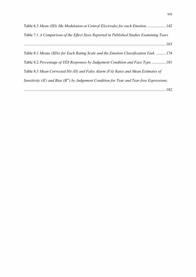
Table 6.3 Mean (SD) Mu Modulation at Central Electrodes for each Emotion. ................... 142
Table 7.1 A Comparison of the Effect Sizes Reported in Published Studies Examining Tears
................................................................................................................................................ 163
Table 8.1 Means (SDs) for Each Rating Scale and the Emotion Classification Task. .......... 174
Table 8.2 Percentage of YES Responses by Judgement Condition and Face Type. .............. 181
Table 8.3 Mean Corrected Hit (H) and False Alarm (FA) Rates and Mean Estimates of
Sensitivity (𝐴′) and Bias (𝐵′′) by Judgement Condition for Tear and Tear-free Expressions.
................................................................................................................................................ 182
xx
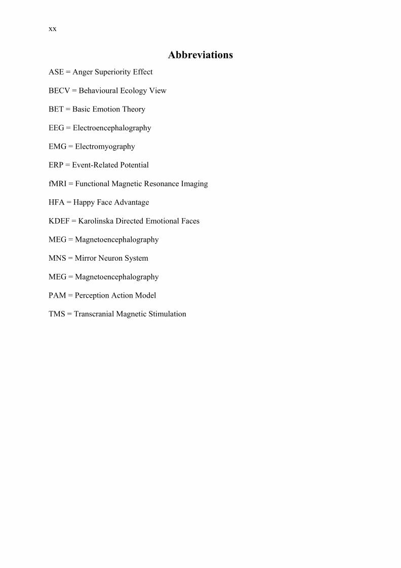
Abbreviations ASE = Anger Superiority Effect
BECV = Behavioural Ecology View
BET = Basic Emotion Theory
EEG = Electroencephalography
EMG = Electromyography
ERP = Event-Related Potential
fMRI = Functional Magnetic Resonance Imaging
HFA = Happy Face Advantage
KDEF = Karolinska Directed Emotional Faces
MEG = Magnetoencephalography
MNS = Mirror Neuron System
MEG = Magnetoencephalography
PAM = Perception Action Model
TMS = Transcranial Magnetic Stimulation

1
Chapter 1: Responses to Emotional Facial Expressions and Crying
The land of tears is so mysterious.
- Antoine de Saint-Exupéry, The Little Prince.
1.1 Introduction Overview
Tears demand attention. A compelling form of emotional expression, tears fascinate
both scientists and lay people alike (Trimble, 2012). Despite this fascination, surprisingly
little is known about the functions of tears, as crying has been neglected by emotion
researchers relative to other universal expressions (Vingerhoets, 2013). Tears act as a cue for
empathic responses; they are a salient distress signal, which elicit attention and prosocial
responses from those witness to the display (Hasson, 2009; Kret, 2015). The communicative
functions of emotion are increasingly being investigated to determine why tears elicit these
responses from observers. Put simply, why do we care when others cry?
The overarching aim of this thesis was to investigate how people perceive and
respond to emotional tears. Specifically, I sought to extend the existing research, which has
identified that tears elicit pro-social responses, by using a combination of
psychophysiological and behavioural measures. The use of psychophysiology provides the
opportunity to validate earlier self-report studies, and additionally provide a biological basis
for the communicative function of tears. The introduction of this thesis is comprised of two
chapters. Chapter 1 reviews the existing literature exploring facial emotional processing and
the communicative functions of tears. Chapter 2 explores the psychophysiological
measurement of empathy, through the lens of a perception-action model. Furthermore,
Chapter 2 discusses the neural correlates that are believed to underpin the sharing of others’
affective states. Finally, Chapter 2 will conclude by bringing these two lines of inquiry
2
together and outline how this thesis will explore the way we perceive and respond to adult
emotional tears.
1.2 Facial Expressions
The face is a rich canvas through which communication with others can occur without
words. Therefore, the rapid and accurate decoding of facial displays is integral to adaptive
social behaviour and survival (Batty & Taylor, 2003). However, there are several competing
theories about what facial expressions convey. Dimensional theory, as it stands today, argues
that facial expressions of emotion can be categorised on a two-dimensional circumplex,
wherein affective states are distributed on the basis of valence and arousal (Russell, 1980).
Basic Emotion Theory asserts that there are six expressions of emotion which are universally
recognisable (Ekman & Friesen, 1969). Finally, the Behavioural Ecology View Theory states
that facial expressions are unrelated to emotion and instead are communicative signals
(Fridlund, 1994). This review of the facial expression literature begins by exploring the tenets
of these three theories, as well as their associated criticisms. Next, the focus shifts to the
development of the view that facial expressions are both affective and communicative with a
review of the literature that details a processing advantage for positive and negative affective
states. Finally, the review concludes with a summary of the empirical research exploring the
communicative functions of emotional tears.
1.3 Emotional Theories
Emotions are inherently complex. The main goal of emotion theorists is to understand
emotion—whether that be expression, perception, or feeling. However, understanding
emotion is a complicated endeavour. For this reason, the majority of emotion research has
attempted to reduce emotion into constituent components. This approach has given rise to
most of the models of emotion we have today—wherein emotions (in all their complexity)

3
can be reduced to bipolar dimensions in dimensional theory; basic expressions, in Basic
Emotion Theory; and targeted signals under the Behavioural Ecology View Theory.
The idea of affective dimensions began with Wundt (1897), wherein emotions could-
be expressed by way of three bipolar dimensions: pleasant-unpleasant, excitement-inhibition,
tension-relaxation. Since this early work, the dimensional theory typically adopts a two-
dimensional structure, due to the overlap between the excitement-inhibition and tension-
relaxation dimensions (Larsen & Diener, 1992; Watson & Tellegen, 1985; Yik, Russell, &
Feldman Barrett, 1999). Schlosberg (1952) proposed that facial expressions (e.g. expressions
of love/ happiness; and fear/ suffering), could be organised on a circular model, wherein
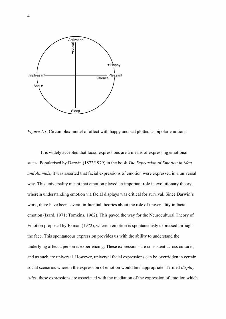
emotions could be represented as bipolar, rather than singular states. Russell (1980) built
upon this theory and developed the circumplex model of affect. The circumplex model of
affect proposes that emotional states are expressed via two affective dimensions: a hedonic
valence dimension encompassing the pleasantness or unpleasantness of a stimulus; and an
arousal dimension encompassing alertness from arousal to sleep (Russell, 1980). In this way,
facial expressions of emotion vary on the basis of these two dimensions, and as such can then
be transformed into specific labels. Therefore, emotions are not independent of one and
other—rather they are interrelated according to these two bipolar dimensions (Russell &
Barrett, 1999). For example, happiness is considered a pleasant, positive emotion, whereas
sadness is considered an unpleasant negative emotion. For this reason, happiness and sadness
sit at opposite ends of the valence dimension. However, happiness is more arousing than
sadness, thus they sit in opposite quadrants according to the circumplex model (see Figure
1.1). In this way, emotions are structured according to these dimensions, rather than as
discrete categories.
4
Figure 1.1. Circumplex model of affect with happy and sad plotted as bipolar emotions.
It is widely accepted that facial expressions are a means of expressing emotional
states. Popularised by Darwin (1872/1979) in the book The Expression of Emotion in Man
and Animals, it was asserted that facial expressions of emotion were expressed in a universal
way. This universality meant that emotion played an important role in evolutionary theory,
wherein understanding emotion via facial displays was critical for survival. Since Darwin’s
work, there have been several influential theories about the role of universality in facial
emotion (Izard, 1971; Tomkins, 1962). This paved the way for the Neurocultural Theory of
Emotion proposed by Ekman (1972), wherein emotion is spontaneously expressed through
the face. This spontaneous expression provides us with the ability to understand the
underlying affect a person is experiencing. These expressions are consistent across cultures,
and as such are universal. However, universal facial expressions can be overridden in certain
social scenarios wherein the expression of emotion would be inappropriate. Termed display
rules, these expressions are associated with the mediation of the expression of emotion which
5
can vary across cultures. In this way, facial expressions can be both universal and culture
specific.
It has been hypothesised that there are six facial expressions of emotion that are
universally expressed, and, as such, are universally recognisable (Ekman & Friesen, 1969).
Known as Basic Emotion Theory (BET), these basic emotions include: happiness, sadness,
anger, fear, disgust, and surprise (though occasionally other categories, such as contempt, are
included; Ekman, 1992). Ekman and Friesen (1969) showed prototypical photographs of
basic emotions to a remote Papua New Guinea culture known as the South Fore. All Fore
individuals were capable of classifying basic emotional displays in their own language. In
another study, Fore individuals were asked to pose emotions from their own language, and
these emotions were accurately decoded by other Fore observers, as well as western
populations, albeit classification accuracy was lower in the former group (Ekman, Sorenson,
& Friesen, 1969). Further evidence for universality was demonstrated, as the least
westernised Fore members were capable of correctly selecting a facial display that
accompanied an emotional story (i.e. a happy facial display was selected for a happy story)
for most basic expressions (Ekman & Friesen, 1971). These studies provide evidence for the
universality of emotion, wherein facial affect is both expressed and recognised across
cultures.
By contrast, cultural specificity has been demonstrated through the experimental
investigation of the facial displays of United States and Japanese students, which were
activated in response to films (Ekman, 1972). While facial displays were largely similar
between US and Japanese students in a solitary condition, Japanese students were less likely
to express negative facial emotions in the presence of an observer. This finding fits within the
concept of display rules, wherein negative emotion is inhibited or masked (Ekman, 1972).
Additionally, in further studies of American and Japanese subjects, there was agreement

6
across the type of emotion expressed (Ekman et al., 1987); however, Japanese participants
attributed lower intensity ratings to the basic emotions, with the exception of disgust
(Matsumoto & Ekman, 1989). Thus, facial expressions can be both universal and culturally
specific, by way of expressive intensity.
As the universality movement demonstrated that prototypical expressions were
associated with emotional states, there was a growing need for a measurement system that
was capable of indexing the anatomical structures associated with facial movements (Ekman
& Rosenberg, 2005). The Facial Action Coding System (FACS) is a anatomically based
system that distinguishes facial activity on the basis of muscular movements (Ekman &
Friesen, 1978). Facial activity is broken down into singular action units (AUs), where each
AU has a unique code, and is associated with a corresponding muscular movement. There are
44 AU, 13 of which are associated with basic emotions (Ekman & Rosenberg, 2005). For
example, when one smiles, the corners of the lips are drawn upwards towards the ears, by the
Zygomaticus Major muscle (Duchenne 1862/1990). This movement is associated with AU
12, or lip corner puller (Ekman & Rosenberg, 2005). In this way facial movements are
categorised based on the outward appearance of the underlying facial muscle. Each basic
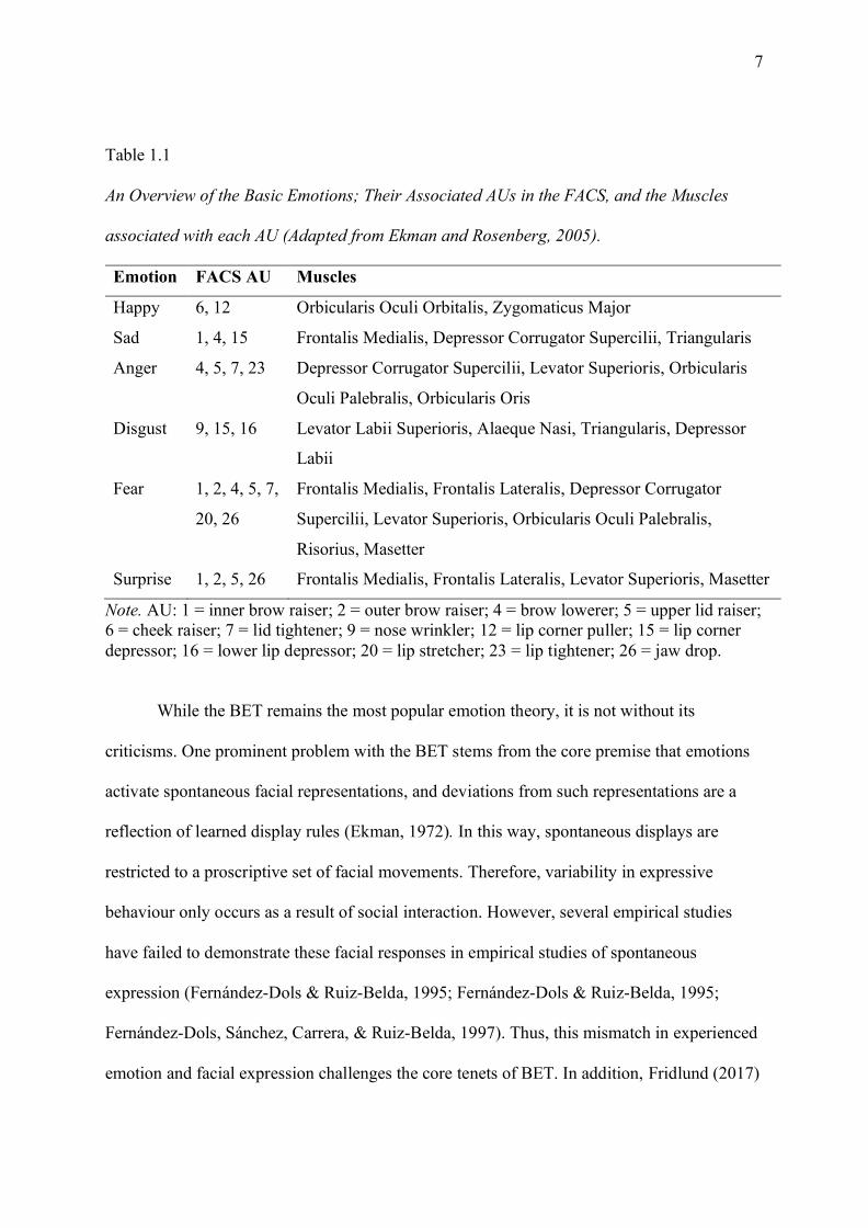
emotion is associated with a selection of AUs (see Table 1.1).
7
Table 1.1
An Overview of the Basic Emotions; Their Associated AUs in the FACS, and the Muscles
associated with each AU (Adapted from Ekman and Rosenberg, 2005).
Emotion FACS AU Muscles
Happy 6, 12 Orbicularis Oculi Orbitalis, Zygomaticus Major
Sad 1, 4, 15 Frontalis Medialis, Depressor Corrugator Supercilii, Triangularis
Anger 4, 5, 7, 23 Depressor Corrugator Supercilii, Levator Superioris, Orbicularis
Oculi Palebralis, Orbicularis Oris
Disgust 9, 15, 16 Levator Labii Superioris, Alaeque Nasi, Triangularis, Depressor
Labii
Fear 1, 2, 4, 5, 7,
20, 26
Frontalis Medialis, Frontalis Lateralis, Depressor Corrugator
Supercilii, Levator Superioris, Orbicularis Oculi Palebralis,
Risorius, Masetter
Surprise 1, 2, 5, 26 Frontalis Medialis, Frontalis Lateralis, Levator Superioris, Masetter
Note. AU: 1 = inner brow raiser; 2 = outer brow raiser; 4 = brow lowerer; 5 = upper lid raiser; 6 = cheek raiser; 7 = lid tightener; 9 = nose wrinkler; 12 = lip corner puller; 15 = lip corner depressor; 16 = lower lip depressor; 20 = lip stretcher; 23 = lip tightener; 26 = jaw drop.
While the BET remains the most popular emotion theory, it is not without its
criticisms. One prominent problem with the BET stems from the core premise that emotions
activate spontaneous facial representations, and deviations from such representations are a
reflection of learned display rules (Ekman, 1972). In this way, spontaneous displays are
restricted to a proscriptive set of facial movements. Therefore, variability in expressive
behaviour only occurs as a result of social interaction. However, several empirical studies
have failed to demonstrate these facial responses in empirical studies of spontaneous
expression (Fernández-Dols & Ruiz-Belda, 1995; Fernández-Dols & Ruiz-Belda, 1995;
Fernández-Dols, Sánchez, Carrera, & Ruiz-Belda, 1997). Thus, this mismatch in experienced
emotion and facial expression challenges the core tenets of BET. In addition, Fridlund (2017)
8
fundamentally disagreed with the BET, due to the circular nature of matching select
emotional terms with emotional images, and select emotional images with emotional terms.
In this way, the technical procedures responsible for popularising BET were flawed. It was
these criticisms that resulted in the development of the Behavioural Ecology View (BECV).
By contrast, the BECV is based upon the idea that facial expressions function as
social signals (Fridlund, 1991, 1994, 2017). Under the BECV facial expressions are not
related to an underlying emotional state. Instead, human emotional experience is unrelated to
facial expression. Moreover, Fridlund (1994) asserts that fundamental emotions, and their
accompanying expressions, do not exist. Rather, facial displays are dependent on intention
and context. As a result, a facial expression of a frown or a scowl, which would be associated
with the expression of anger under BET, is associated with signalling intention to attack, or
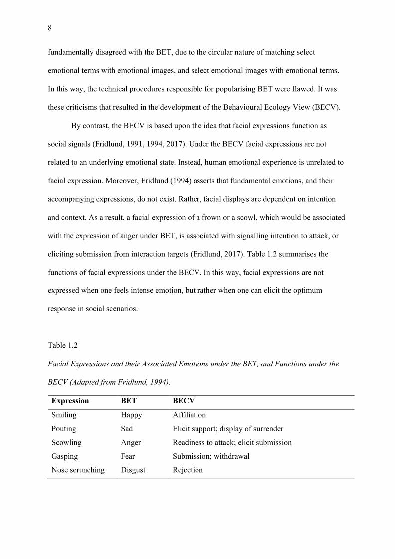
eliciting submission from interaction targets (Fridlund, 2017). Table 1.2 summarises the
functions of facial expressions under the BECV. In this way, facial expressions are not
expressed when one feels intense emotion, but rather when one can elicit the optimum
response in social scenarios.
Table 1.2
Facial Expressions and their Associated Emotions under the BET, and Functions under the
BECV (Adapted from Fridlund, 1994).
Expression BET BECV
Smiling Happy Affiliation
Pouting Sad Elicit support; display of surrender
Scowling Anger Readiness to attack; elicit submission
Gasping Fear Submission; withdrawal
Nose scrunching Disgust Rejection

9
The BECV has predominantly been bolstered through research demonstrating that
emotional expressions are maximal during social interaction. Therefore, the context in which
the facial expression occurs contributes more to outward expression than the emotion
experienced. For example, humans smile for a variety of reasons—to signal affiliation, to
engage in pleasant social interactions (i.e. a polite smile), to engage in social dominance
behaviours (Martin, Rychlowska, Wood, & Niedenthal, 2017; Niedenthal, Mermillod,
Maringer, & Hess, 2010)—and these smiles need not be accompanied by an underlying
positive affective state. Thus, it is insufficient to propose that basic emotions accompany
prototypical smiling faces. This argument has received experimental support wherein positive
facial displays occur more frequently in the presence of social others, as opposed to alone
(Fridlund, 1991; Hess, Banse, & Kappas, 1995; Jakobs, Manstead, & Fischer, 1999).
Young and Fry (1966) demonstrated that laughter and smiling in response to
humorous jokes occurred more in the presence of peers than when alone. Further evidence for
this conclusion was demonstrated in a series of studies wherein increased smiles were
exhibited to films when accompanied by friends, as opposed to being alone or with strangers
(Jakobs et al., 1999; Jakobs, Manstead, & Fischer, 1999). Similarly, Fridlund (1991)
concluded that humorous content viewed with friends elicits greater smiling responses than
content viewed alone. A similar conclusion was demonstrated from naturalistic observations
of victorious athletes (Fernández-Dols & Ruiz-Belda, 1995). Moreover, gold medal winners
exhibited increased smiling behaviour during social interaction contexts (i.e. podium
waving), than during non-interactive stages (i.e. waiting to proceed to the podium). Relatedly
Crivelli, Carrera, and Fernández-Dols (2015) demonstrated a greater occurrence of genuine
smiles when judo winners were engaging with the audience after victory, as opposed to a
non-social interaction. In this way, social presence increases expressiveness.
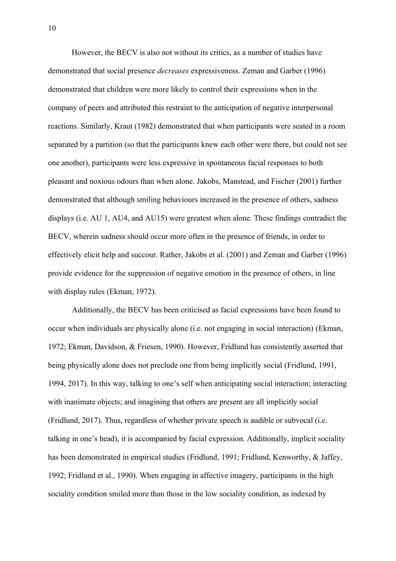
10
However, the BECV is also not without its critics, as a number of studies have
demonstrated that social presence decreases expressiveness. Zeman and Garber (1996)
demonstrated that children were more likely to control their expressions when in the
company of peers and attributed this restraint to the anticipation of negative interpersonal
reactions. Similarly, Kraut (1982) demonstrated that when participants were seated in a room
separated by a partition (so that the participants knew each other were there, but could not see
one another), participants were less expressive in spontaneous facial responses to both
pleasant and noxious odours than when alone. Jakobs, Manstead, and Fischer (2001) further
demonstrated that although smiling behaviours increased in the presence of others, sadness
displays (i.e. AU 1, AU4, and AU15) were greatest when alone. These findings contradict the
BECV, wherein sadness should occur more often in the presence of friends, in order to
effectively elicit help and succour. Rather, Jakobs et al. (2001) and Zeman and Garber (1996)
provide evidence for the suppression of negative emotion in the presence of others, in line
with display rules (Ekman, 1972).
Additionally, the BECV has been criticised as facial expressions have been found to
occur when individuals are physically alone (i.e. not engaging in social interaction) (Ekman,
1972; Ekman, Davidson, & Friesen, 1990). However, Fridlund has consistently asserted that
being physically alone does not preclude one from being implicitly social (Fridlund, 1991,
1994, 2017). In this way, talking to one’s self when anticipating social interaction; interacting
with inanimate objects; and imagining that others are present are all implicitly social
(Fridlund, 2017). Thus, regardless of whether private speech is audible or subvocal (i.e.
talking in one’s head), it is accompanied by facial expression. Additionally, implicit sociality
has been demonstrated in empirical studies (Fridlund, 1991; Fridlund, Kenworthy, & Jaffey,
1992; Fridlund et al., 1990). When engaging in affective imagery, participants in the high
sociality condition smiled more than those in the low sociality condition, as indexed by
11
increased zygomaticus major activity measured via electromyography (Fridlund et al., 1990).
Similarly, participants smiled more during a humorous film when they were informed that
their friend was also watching the film in a separate location (Fridlund, 1991). In this way,
sociality can be implied, and as such facial expressions are inherently social.
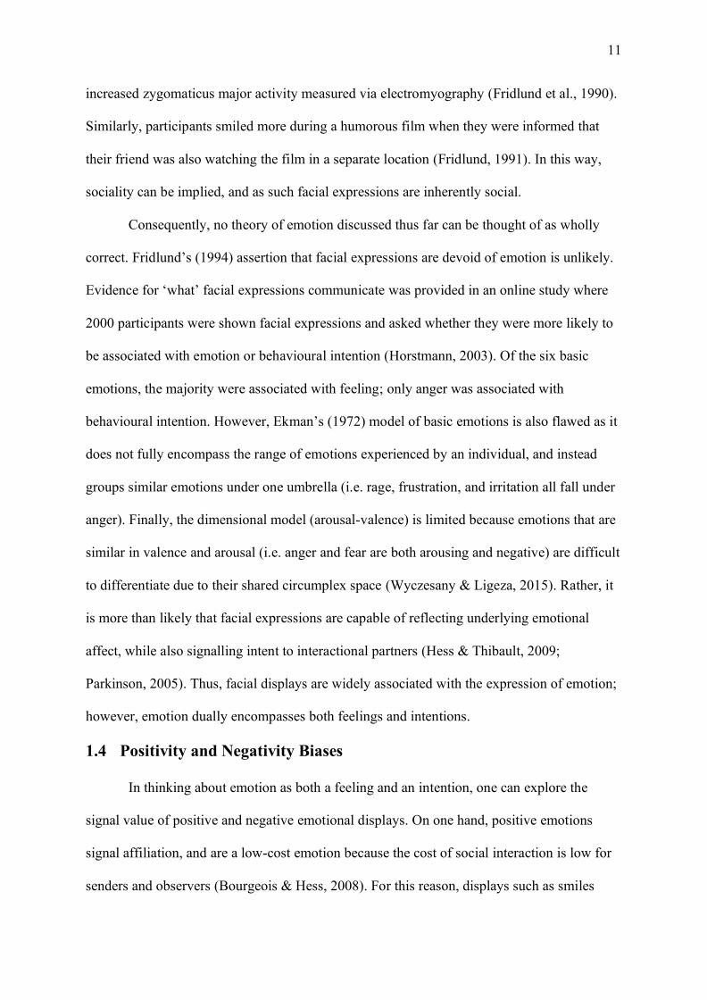
Consequently, no theory of emotion discussed thus far can be thought of as wholly
correct. Fridlund’s (1994) assertion that facial expressions are devoid of emotion is unlikely.
Evidence for ‘what’ facial expressions communicate was provided in an online study where
2000 participants were shown facial expressions and asked whether they were more likely to
be associated with emotion or behavioural intention (Horstmann, 2003). Of the six basic
emotions, the majority were associated with feeling; only anger was associated with
behavioural intention. However, Ekman’s (1972) model of basic emotions is also flawed as it
does not fully encompass the range of emotions experienced by an individual, and instead
groups similar emotions under one umbrella (i.e. rage, frustration, and irritation all fall under
anger). Finally, the dimensional model (arousal-valence) is limited because emotions that are
similar in valence and arousal (i.e. anger and fear are both arousing and negative) are difficult
to differentiate due to their shared circumplex space (Wyczesany & Ligeza, 2015). Rather, it
is more than likely that facial expressions are capable of reflecting underlying emotional
affect, while also signalling intent to interactional partners (Hess & Thibault, 2009;
Parkinson, 2005). Thus, facial displays are widely associated with the expression of emotion;
however, emotion dually encompasses both feelings and intentions.
1.4 Positivity and Negativity Biases
In thinking about emotion as both a feeling and an intention, one can explore the
signal value of positive and negative emotional displays. On one hand, positive emotions
signal affiliation, and are a low-cost emotion because the cost of social interaction is low for
senders and observers (Bourgeois & Hess, 2008). For this reason, displays such as smiles

12
should be readily shared, as there is an evolutionary advantage to adaptive pro-social
behaviours (Johnston, Miles, & Macrae, 2010). By contrast, negative emotions, such as anger
and fear, signal potentially threatening and dangerous situations (Hajcak, Weinberg,
MacNamara, & Foti, 2011; Ito, Larsen, Smith, & Cacioppo, 1998). In this way, the rapid
interpretation of threat related signals would be biologically advantageous for survival
(LeDoux, 2007; Liddell, Williams, Rathjen, Shevrin, & Gordon, 2004). Thus, both positive
and negative facial displays serve an evolutionary purpose. This evolutionary purpose has
seen the development of two research arguments: one is a positivity advantage (i.e., the
Happy Face Advantage, HFA) and the other is a negativity bias (e.g., the Anger Superiority
Effect, ASE). In both theories, it is argued that a particular display is facilitated, enhanced, or
preferentially processed as a result of increased evolutionary relevance. The following
paragraphs will outline the research contributing to the development of both theories and the
bounds and limitations of each argument.
The preferential processing of positive stimuli has been evidenced using faces (Calvo,
Avero, Fernández-Martín, & Recio, 2016; Leppänen, Tenhunen, & Hietanen, 2003; Palermo
& Coltheart, 2004), images (Lehr, Bergum, & Standing, 1966), and words (Bayer & Schacht,
2014; Feyereisen, Malet, & Martin, 1986; Stenberg, Wiking, & Dahl, 1998). In this way,
humans are quick to respond, preferentially process, and retain positive information, relative
to unpleasant information. This concept is largely conveyed by the Pollyanna principle
(Matlin & Stang, 1979). The Pollyanna principle stems from looking on the bright side and
remaining optimistic. For example, according to the Pollyanna principle, persons would
convey positive information more than negative information, use positive terms rather than
negative terms, and favour positive events more than negative events. In this way, there is a
positivity bias that encompasses memory, language, and perception (Matlin, 2017). By far the
most evidence for a positivity bias stems from the research exploring the HFA, wherein
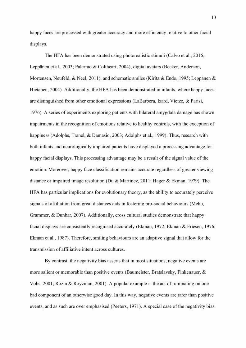
13
happy faces are processed with greater accuracy and more efficiency relative to other facial
displays.
The HFA has been demonstrated using photorealistic stimuli (Calvo et al., 2016;
Leppänen et al., 2003; Palermo & Coltheart, 2004), digital avatars (Becker, Anderson,
Mortensen, Neufeld, & Neel, 2011), and schematic smiles (Kirita & Endo, 1995; Leppänen &
Hietanen, 2004). Additionally, the HFA has been demonstrated in infants, where happy faces
are distinguished from other emotional expressions (LaBarbera, Izard, Vietze, & Parisi,
1976). A series of experiments exploring patients with bilateral amygdala damage has shown
impairments in the recognition of emotions relative to healthy controls, with the exception of
happiness (Adolphs, Tranel, & Damasio, 2003; Adolphs et al., 1999). Thus, research with
both infants and neurologically impaired patients have displayed a processing advantage for
happy facial displays. This processing advantage may be a result of the signal value of the
emotion. Moreover, happy face classification remains accurate regardless of greater viewing
distance or impaired image resolution (Du & Martinez, 2011; Hager & Ekman, 1979). The
HFA has particular implications for evolutionary theory, as the ability to accurately perceive
signals of affiliation from great distances aids in fostering pro-social behaviours (Mehu,
Grammer, & Dunbar, 2007). Additionally, cross cultural studies demonstrate that happy
facial displays are consistently recognised accurately (Ekman, 1972; Ekman & Friesen, 1976;
Ekman et al., 1987). Therefore, smiling behaviours are an adaptive signal that allow for the
transmission of affiliative intent across cultures.
By contrast, the negativity bias asserts that in most situations, negative events are
more salient or memorable than positive events (Baumeister, Bratslavsky, Finkenauer, &
Vohs, 2001; Rozin & Royzman, 2001). A popular example is the act of ruminating on one
bad component of an otherwise good day. In this way, negative events are rarer than positive
events, and as such are over emphasised (Peeters, 1971). A special case of the negativity bias
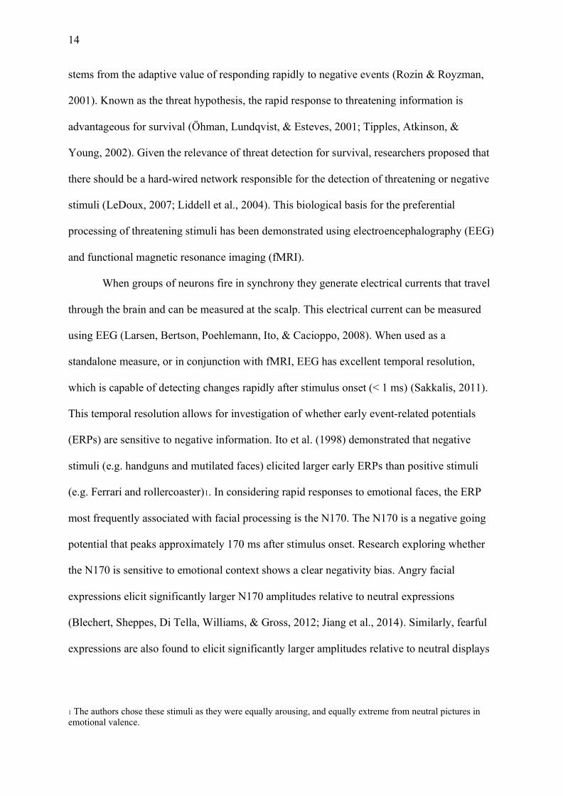
14
stems from the adaptive value of responding rapidly to negative events (Rozin & Royzman,
2001). Known as the threat hypothesis, the rapid response to threatening information is
advantageous for survival (Öhman, Lundqvist, & Esteves, 2001; Tipples, Atkinson, &
Young, 2002). Given the relevance of threat detection for survival, researchers proposed that
there should be a hard-wired network responsible for the detection of threatening or negative
stimuli (LeDoux, 2007; Liddell et al., 2004). This biological basis for the preferential
processing of threatening stimuli has been demonstrated using electroencephalography (EEG)
and functional magnetic resonance imaging (fMRI).
When groups of neurons fire in synchrony they generate electrical currents that travel
through the brain and can be measured at the scalp. This electrical current can be measured
using EEG (Larsen, Bertson, Poehlemann, Ito, & Cacioppo, 2008). When used as a
standalone measure, or in conjunction with fMRI, EEG has excellent temporal resolution,
which is capable of detecting changes rapidly after stimulus onset (< 1 ms) (Sakkalis, 2011).
This temporal resolution allows for investigation of whether early event-related potentials
(ERPs) are sensitive to negative information. Ito et al. (1998) demonstrated that negative
stimuli (e.g. handguns and mutilated faces) elicited larger early ERPs than positive stimuli
(e.g. Ferrari and rollercoaster)1. In considering rapid responses to emotional faces, the ERP
most frequently associated with facial processing is the N170. The N170 is a negative going
potential that peaks approximately 170 ms after stimulus onset. Research exploring whether
the N170 is sensitive to emotional context shows a clear negativity bias. Angry facial
expressions elicit significantly larger N170 amplitudes relative to neutral expressions
(Blechert, Sheppes, Di Tella, Williams, & Gross, 2012; Jiang et al., 2014). Similarly, fearful
expressions are also found to elicit significantly larger amplitudes relative to neutral displays
1 The authors chose these stimuli as they were equally arousing, and equally extreme from neutral pictures in emotional valence.
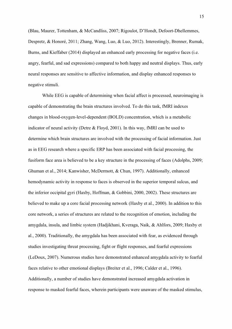
15
(Blau, Maurer, Tottenham, & McCandliss, 2007; Rigoulot, D’Hondt, Defoort-Dhellemmes,
Despretz, & Honoré, 2011; Zhang, Wang, Luo, & Luo, 2012). Interestingly, Brenner, Rumak,
Burns, and Kieffaber (2014) displayed an enhanced early processing for negative faces (i.e.
angry, fearful, and sad expressions) compared to both happy and neutral displays. Thus, early
neural responses are sensitive to affective information, and display enhanced responses to
negative stimuli.
While EEG is capable of determining when facial affect is processed, neuroimaging is
capable of demonstrating the brain structures involved. To do this task, fMRI indexes
changes in blood-oxygen-level-dependent (BOLD) concentration, which is a metabolic
indicator of neural activity (Detre & Floyd, 2001). In this way, fMRI can be used to
determine which brain structures are involved with the processing of facial information. Just
as in EEG research where a specific ERP has been associated with facial processing, the
fusiform face area is believed to be a key structure in the processing of faces (Adolphs, 2009;
Ghuman et al., 2014; Kanwisher, McDermott, & Chun, 1997). Additionally, enhanced
hemodynamic activity in response to faces is observed in the superior temporal sulcus, and
the inferior occipital gyri (Haxby, Hoffman, & Gobbini, 2000, 2002). These structures are
believed to make up a core facial processing network (Haxby et al., 2000). In addition to this
core network, a series of structures are related to the recognition of emotion, including the
amygdala, insula, and limbic system (Hadjikhani, Kveraga, Naik, & Ahlfors, 2009; Haxby et
al., 2000). Traditionally, the amygdala has been associated with fear, as evidenced through
studies investigating threat processing, fight or flight responses, and fearful expressions
(LeDoux, 2007). Numerous studies have demonstrated enhanced amygdala activity to fearful
faces relative to other emotional displays (Breiter et al., 1996; Calder et al., 1996).
Additionally, a number of studies have demonstrated increased amygdala activation in
response to masked fearful faces, wherein participants were unaware of the masked stimulus,
16
but an increased BOLD response still occurred (Kim et al., 2010; Pessoa, Japee, Sturman, &
Ungerleider, 2006; Whalen et al., 1998; Williams et al., 2006). Therefore, the amygdala plays
an important role in the processing of threat-related information, in line with the preferential
processing of negative information.
The most support for a negativity bias in behavioural tasks stems from ASE research.
The ASE is typically investigated using visual search tasks where faces are arranged in a
matrix or array to simulate a crowd of faces. Angry faces are said to be automatically
detected wherein angry expressions “pop out” from facial arrays depicting happy and neutral
distractor expressions. First explored in a series of three experiments, Hansen and Hansen
(1988) demonstrated that regardless of whether the matrix consisted of four or nine faces,
angry faces attracted greater attention and participants took less time to determine their
presence or absence than happy and neutral faces in visual search tasks. Since this seminal
research, a series of experiments using schematic facial expressions have replicated the ASE
(Calvo, Avero, & Lundqvist, 2006; Fox et al., 2000; Öhman et al., 2001). These schematic
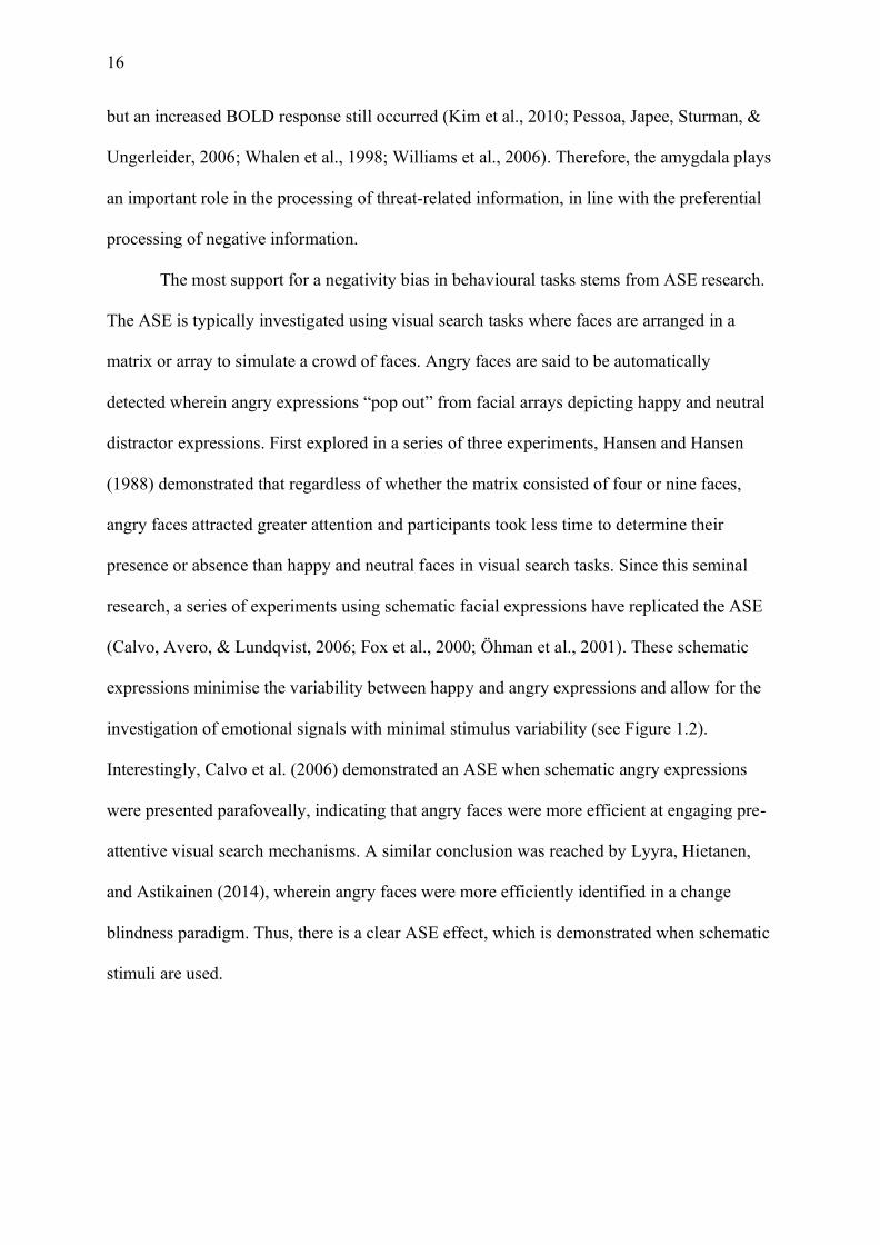
expressions minimise the variability between happy and angry expressions and allow for the
investigation of emotional signals with minimal stimulus variability (see Figure 1.2).
Interestingly, Calvo et al. (2006) demonstrated an ASE when schematic angry expressions
were presented parafoveally, indicating that angry faces were more efficient at engaging pre-
attentive visual search mechanisms. A similar conclusion was reached by Lyyra, Hietanen,
and Astikainen (2014), wherein angry faces were more efficiently identified in a change
blindness paradigm. Thus, there is a clear ASE effect, which is demonstrated when schematic
stimuli are used.
17
Figure 1.2. Happy and angry schematic expressions replicated from Fox et al. (2000).
In addition to the evidence for an ASE provided by schematic expressions, several
studies have demonstrated the ASE using photorealistic stimuli (Ceccarini & Caudek, 2013;
Horstmann & Bauland, 2006; Lipp, Price, & Tellegen, 2009; Pinkham, Griffin, Baron,
Sasson, & Gur, 2010). Lipp et al. (2009) demonstrated an ASE when schematic and
photorealistic stimuli were used; however, an overall negativity bias was only demonstrated
in the schematic condition. Similarly, Pinkham et al. (2010) demonstrated that when black
and white arrays of happy face distractors were presented, angry faces were detected faster
and more accurately than when happy faces were used as targets. Additionally, when
comparing angry faces and happy faces in a neutral distractor condition, angry expressions
were again detected more efficiently than happy expressions. Ceccarini and Caudek (2013)
further explored the ASE, using dynamic stimuli which were matched in intensity and
salience. They concluded that the ASE was present amongst dynamic, but not static displays.
Furthermore, they attribute the discrepancy between their results and Pinkham et al. (2010) to
control over low level stimulus properties such as colour and salience. A recent meta-analysis
concluded that an ASE is well evidenced in arrays depicting schematic expressions; however,
the HFA is typically observed when realistic stimuli are used (Nummenmaa & Calvo, 2015).
Thus, there is both facilitated detection of angry facial expressions, and facilitated
18
recognition of happy facial expressions (Kauschke, Bahn, Vesker, & Schwarzer, 2019;
Öhman et al., 2001).
Unlike the research exploring happy, angry, and fearful expressions, sad facial
displays have not received the same inquiry despite being a communicative signal that elicits
aid from observers (Reed & DeScioli, 2017). One possible explanation is that sad facial
displays are typically less intense or arousing than other negative displays (Russell, 1980).
Therefore, their use in facial expression recognition and detection tasks is limited, as they are
naturally less salient than angry, fearful, and happy displays. Additionally, sad displays are
more costly to respond to (Bavelas, Black, Lemery, & Mullett, 1986). In this way providing
comfort to a sad person comes at a much greater cost to the observer, compared to
reciprocating a smile (Bourgeois & Hess, 2008). As such, sadness displays are considered a
high cost emotion, which lack the social relevance of angry, fearful, and happy displays.
In line with this cost associated with responding to sad facial displays, several
experiments have demonstrated that mimicry of a sad person (wherein mimicry is designed to
demonstrate understanding and facilitate interaction) is limited to personally relevant others
(Bourgeois & Hess, 2008; Häfner & IJzerman, 2011). Moreover, Bourgeois and Hess (2008)
demonstrated that unlike affiliative displays of happiness, displays of sadness were only
mimicked if the expresser was an ingroup member. Similarly, Häfner and IJzerman (2011)
demonstrated that participants were more likely to mimic a partner’s display of sadness (as
opposed to anger), but only if the mimicker was high in communal strength (i.e. a greater
attendance to the partner’s needs). However, other studies have demonstrated enhanced
physiological responses in healthy controls to posed displays of sadness. Turetsky et al.
(2007) demonstrated that participants’ sad displays elicited larger N170 amplitudes relative to
neutral expressions. Similarly, Lynn and Salisbury (2008) demonstrated that healthy control
participants had significantly larger N170 ERPs for sad faces, relative to fearful expressions.
19
In addition, Blair, Morris, Frith, Perrett, and Dolan (1999) observed greater left amygdala
activity in response to sad faces, which was not present for angry faces. In this way, enhanced
physiological responses to sadness have been emphasised in studies indexing neural activity;
however, in the case of outward expressions, the relationship between the mimicker and the
person being mimicked has a particular importance in the response to sadness.
The behavioural studies exploring the recognition and detection of sad displays have
also yielded mixed results. Sad displays have been explored through schematic detection and
recognition tasks, in an effort to match the stimulus qualities as much as possible to isolate
the signal value of sad expressions (see Figure 1.3a for example schematic stimuli). White
(1995) demonstrated that sad schematic facial expressions of emotion are responded to more
quickly than happy facial expressions. Eastwood, Smilek, and Merikle (2001) reached a
similar conclusion, wherein visual search of schematic displays is more efficient for sad than
happy expressions. However, in order to disentangle the negativity bias from the threat
hypothesis, it is expected that under the negativity bias sad facial displays should have the
same prioritisation as angry displays. This is due to both angry and sad faces sharing negative
affective valence; although angry displays are threat-related, sad displays are not (LoBue,
2009). Calvo et al. (2006) explored this idea using identical schematic expressions with
upturned eyebrows for sad expressions, and downturned eyebrows for angry expressions (see
Figure 1.3b). A series of three experiments revealed that angry faces were detected faster in
visual search tasks than happy and sad expressions. In this way, Calvo et al. (2006)
demonstrated support for the threat detection hypothesis, rather than an overarching
negativity bias. Similarly, LoBue (2009) demonstrated in a series of five experiments, using
photographs and schematic expressions, that threat-related negative expressions were
detected faster than non-threatening negative expressions (i.e. sad faces). Importantly,

20
negative expressions (i.e. angry, fearful, and sad) were detected faster overall than positive
displays.
Figure 1.3. a) Examples of happy and sad schematic expressions replicated from White
(1995); b) examples of happy, sad, and angry schematic expressions replicated from Calvo et
al. (2006).
Additionally, some experiments have demonstrated that happy and angry expressions
are detected faster than sad facial displays (Williams, Moss, Bradshaw, & Mattingley, 2005).
In this way, rather than evidencing the HFA or the ASE, they demonstrated a social relevance
effect, wherein more socially relevant expressions (i.e. those expressions requiring rapid
interpretation: happy and angry) were detected faster than less socially relevant displays (i.e.
sadness). These findings are interesting, given that the HFA is more pronounced when sad
expressions are used as a comparison category (Feyereisen et al., 1986). In this way, it may
be that the majority of research exploring responses to sad displays, has used stimuli which
were not matched in distinctiveness to happy and angry displays. Evidence for this
conclusions stems from empirical research that has demonstrated that sad faces are responded

21
to more slowly (Calvo & Nummenmaa, 2008; Kirita & Endo, 1995; Williams et al., 2005),
and are more likely to be confused for other expressions of emotion in identification tasks
(Calvo & Marrero, 2009; Palermo & Coltheart, 2004; Prkachin, 2003). Therefore, further
research into sad facial displays would benefit from the use of a particularly distinctive sad
signal—tears.
1.5 The Communicative Value of Tears
The signal value of tears can be investigated through the lens of biological signalling
(Hasson, 2009). One of the functions of biological signals is to communicate information that
subsequently changes the behaviour of others (Hasson, 1997; Hasson, 2009). This signalling
function has been explored under the BECV wherein sad expressions, particularly those of a
tearful “cry-face” signal readiness to receive succour and attention from observers (Fridlund,
1994). However, biological signals must be an honest display, wherein they reliably elicit the
intended response from observers (Scott-Phillips, 2008). These signals are kept honest, via
the handicap principle. The handicap principle asserts that signals must be accompanied by a
cost in order to convey their honesty to observers (Zahavi, 1975, 1977). In the case of tears,
tears blur one’s vision and, as such, make the expresser more vulnerable. In this way, tearful
displays are costly; however, the beneficial outcomes of increased support and reduced
aggression are greater than the costs that are associated with the signal (Hasson, 2009;
Hendriks, Croon, & Vingerhoets, 2008; Kottler & Montgomery, 2001). This review will
focus on Hasson’s (2009) assertion that for tears to function as effective signals they must
provide information to individuals, and the reception of such information should change the
individual’s behaviour.
The information that emotional tears convey to observers has been investigated from a
range of different viewpoints. Where some studies have focused on how tears influence the
perception of crying individuals, others have focused on exploring tears as a signal of
22
sadness. The former is typically investigated according to two universal dimensions: warmth
and competence (Fiske, Cuddy, & Glick, 2007). Born under social cognition theory, the
warmth dimension encapsulates traits such as sincerity, trustworthiness, and friendliness,
whereas the competence dimension is associated with ability and efficacy (Fiske et al., 2007).
In this way, numerous studies have demonstrated that crying individuals are perceived as
warmer than the same individuals without tears (Fischer, Eagly, & Oosterwijk, 2013; Ven de
Ven, Meijs, & Vingerhoets, 2017; Zickfeld & Schubert, 2018; Zickfeld, van de Ven,
Schubert, & Vingerhoets, 2018).
Fischer et al. (2013) used vignette contexts and demonstrated that males who cried in
the workplace were perceived as less competent than females who cried. Conversely, there
were no gender differences in perceptions of competence when either gender cried in a
relationship context at home. Fischer et al. (2013) concluded that perceptions of competence
were dependent on the context in which crying is encountered. Recently, a series of studies
investigated perceptions of warmth and competence in the absence of context via still
photographs that included tears (Ven de Ven et al., 2017; Zickfeld & Schubert, 2018;
Zickfeld et al., 2018). Ven de Ven et al. (2017) used a sample of five male and five female
faces and found that the presence of tears made individuals seem warmer and less competent
than the same individuals with the tears removed. In replication studies using a larger sample
of the same faces, the effect of warmth has been replicated; however, the effect of
competence has not (Zickfeld & Schubert, 2018; Zickfeld et al., 2018). Zickfeld et al. (2018)
concluded that the original effect of competence observed by Ven de Ven et al. (2017) was
target specific (i.e. limited to 10 faces), and therefore not generalisable.
Tearful individuals are not only perceived as warmer, but are also perceived as more
sincere (Picó et al., 2020; Zeifman & Brown, 2011). Zeifman and Brown (2011) explored the
perception of crying infants, children, and adults, and concluded that tears made no difference
23
in perception of sincerity when exhibited by infants. By contrast, tears moderately increased
perceptions of sincerity when exhibited by children, and the greatest sincerity was observed
for crying adults. Thus, sincerity increased as a function of age, wherein crying adults were
perceived as the most sincere. Similarly, Picó et al. (2020) demonstrated that when
participants are instructed to rate the sincerity of identical vignette statements, sincerity was
increased after priming participants with a tearful individual. In a similar vein, Hornsey et al.
(2019) demonstrated that tears increased the perception of remorse during public apologies.
Thus, tearful individuals are perceived as warmer and more sincere than the same individuals
without tears.
The representation of tears as a signal of sadness has predominantly stemmed from a
series of studies exploring the tear effect (Provine et al., 2009). Provine et al. (2009) used
tearful images and digitally removed the tears to create tear-free duplicate images.
Participants perceived the tearful expressions as significantly sadder than the tear-free
duplicates. Furthermore, the tear-free images were perceived as ambiguous in emotional
valence. Therefore, the tear effect states that tears facilitate the recognition of sadness. Since
this seminal work, numerous studies have demonstrated support for the tear effect across
different age categories (Zeifman & Brown, 2011), emotions (Ito, Ong, & Kitada, 2019;
Reed, Deutchman, & Schmidt, 2015), and even using the isolated eye region of digital avatars
(Küster, 2018). In addition, Balsters et al. (2013) demonstrated the tear effect when using
brief (50 ms) presentation times, showing that tears change rapid responses to faces.
Participants’ reaction times were significantly faster to sad tearful expressions, compared to
both tear-free sad and neutral expressions (Balsters et al., 2013). Takahashi et al. (2015)
extended the paradigm originally developed by Provine et al. (2009) to include an additional
neutral condition. Crying female adults were found on Google and Flickr, and their tears were
digitally removed to create tear-free images. Images were rated by a separate sample on the
24
intensity of sadness, and half of the images were classified as sad expressions, with the
remaining half classified as neutral. In their main experiment, they concluded that although
tearful expressions were perceived as sadder, the magnitude of the influence of tears (i.e. the
increased perception of sadness as a result of the tears) was greater for neutral expressions.
As such, tears serve as a marker of sadness and resolve the ambiguity associated with neutral
expressions.
Interestingly, support for the tear effect is demonstrated even when exploring a
second perspective, known as the general enhancement perspective. Under the general
enhancement perspective, it is postulated that the presence of tears makes all expressions
more intense, rather than simply signalling sadness (i.e. an angry face is angrier with tears)
(Ito et al., 2019). However the two studies exploring this perspective have demonstrated
generalised support for the tear effect (Ito et al., 2019; Reed et al., 2015). Ito et al. (2019)
demonstrated that when tears are added to other negative emotional expressions such as
anger, fear, and disgust, participants are significantly more likely to rate the images as sadder
in appearance. They further validated this conclusion by examining the ratings in
multidimensional space and demonstrated that tearful, negatively valenced faces are more
tightly clustered around sadness than tear-free expressions. However, tears are not elicited
solely in response to sadness (Miceli & Castelfranchi, 2003).
Although most people typically associate tears with negative events such as death,
breakups and separation, tears are also elicited in response to their positive counterparts:
birth, weddings, and reunion (Vingerhoets, 2013). Reed et al. (2015) demonstrated that in the
case of genuine, dynamic smiling expressions (i.e. Duchenne smiles), the presence of tears
increased the overall intensity and perceived sadness of the image. Conversely, non-
Duchenne smiles with tears were perceived as less positive, less happy, sadder, angrier, and
more fearful. In this way, there is mixed evidence, with a generalised tear effect present,

25
however the generalised enhancement perspective cannot be ruled out. However, it must be
cautioned that Reed et al. (2015) used a single actress and as such it is unclear whether the
results would generalise to a larger sample of faces. Therefore, it is unclear what role tears
play in the perception of happy expressions. Furthermore, studies that have used naturalistic
tearful images of joy as stimuli have demonstrated that context is essential for tears of joy to
be perceived as positive (Aragón, 2017; Aragón & Bargh, 2018). Thus, in the absence of
context, tears seem to make all expressions seem sadder, which is in line with the tear effect,
meaning tears serve as a sincere signal of sadness.
To be a biological signal, tears must also convey information that leads an individual
to change their behaviour. This effect has been well documented in the literature, wherein
tearful displays are responded to with greater prosocial behaviour, reduced aggression, and
empathy relative to tear-free expressions (Hendriks & Vingerhoets, 2006; Hendriks et al.,
2008; Vingerhoets, van de Ven, & Velden, 2016). As such, tears elicit helping responses
from observers. Hendriks and Vingerhoets (2006) were some of the first to demonstrate that
tearful photographs of crying persons were responded to with greater emotional support and
lower ratings of avoidance relative to tear-free expressions. Since then, several studies have
demonstrated that tearful expressions foster approach behaviour (relative to avoidance) in
reaction time tasks (Gračanin, Krahmer, Rinck, & Vingerhoets, 2018; Riem, van Ijzendoorn,
De Carli, Vingerhoets, & Bakermans-Kranenburg, 2017). Similarly, Vingerhoets et al. (2016)
showed that participants attribute greater helping responses to individuals with tears, than the
same expressions without tears. Furthermore, they concluded that these helping behaviours
stemmed from a perception of closeness with the tearful individual, wherein feeling
connected increased one’s willingness to help.
Although tears are known to elicit helping responses, several studies have identified
limitations to these pro-social behaviours. Stadel, Daniels, Warrens, and Jeronimus (2019)
26
identified an increased willingness to help tearful individuals but concluded that this
willingness was the strongest between female and mixed interactional dyads and reduced
during male dyad interactions. Furthermore, Hendriks et al. (2008) showed that participants
were significantly more likely to provide help to tearful individuals in negative scenarios, but
not positive ones. Similarly, Reed, Matari, Wu, and Janaswamy (2019) identified that this
increased pro-sociality was only present when tearful individuals were capable of
reciprocating the assistance. Thus, pro-social responses were dependent on reciprocity.
Furthermore, a number of studies have reported that although participants are more likely to
aid tearful individuals, they are also more likely to feel personal distress or negative emotion
during the interaction (Hendriks & Vingerhoets, 2006; Hendriks et al., 2008; Küster, 2018).
In this way, it is unclear whether helping responses to tearful displays are based on altruistic
or egoistic motives. Therefore, further research can validate the self-report ‘willingness to
help’ measures via the use of psychophysiological indices of empathy, thus determining
whether tears elicit increased help and empathy from observers.
1.6 Summary
Throughout this review, it is evident that tears meet the criterion proposed by Hasson
(2009) to function as a biological signal. Moreover, the presence of tears on a face sincerely
communicates distress and sadness to those who are witness to the display. This sadness and
distress, in turn, changes the way that people respond, wherein tears elicit succour and aid
from observers. However, exploring the psychophysiological responses to emotional tears has
been largely neglected, despite the opportunity for validation of the existing self-report
research. Therefore, in Chapter 2, the movement towards the adoption of psychophysiological
indices of empathy will be explored. This exploration will conclude with a discussion on the
role of the mirror neuron system in empathic responses and highlight how the intersection of
emotion and empathy research will afford a greater understanding of human crying.

27
Chapter 2: The Psychophysiological Measurement of Empathy
2.1 Chapter Introduction
Have you ever witnessed a loved one slice their finger with a knife by mistake and
winced in pain? Or have you found yourself in a room full of happy people and instantly felt
your mood lift? If your answer to these questions was yes, then you are not alone. Humans
are instinctively social animals. A species dependent on others, from the helpless stages of
infancy to late stages of life, daily existence is punctuated with human interaction. How this
interaction occurs and the mechanisms behind it will make up this review of the science of
empathy.
Understanding those around us is instinctive and automatic. Often, we can “read” the
intent of those around us through the smirk of a politician or the grin of a friend. Thousands
of these deductions each day, without even considering or acknowledging the mechanisms
that drive this behaviour. Even more astounding is the fact that this understanding has
puzzled scientists for years. How do we understand what the people around us are thinking
and feeling? Empathy, an umbrella term for the ability to resonate with others, is one
proposed mechanism for shared affective states (Preston & Hofelich, 2012). The present
review aims to clarify the role that empathy plays in human interaction. Through focusing on
deconstructing the mechanisms that are known antecedents of empathy, a greater
understanding of how we share the affective states of others will be obtained. To investigate
these mechanisms, psychophysiology will be used as a lens, to explore the way that affective
states are shared through the central and peripheral nervous systems. It is intended that this
review will provide an understanding of the way that the brain is inherently social and
provide a platform for future research into the neuroscience of empathy.
2.2 Empathy
28
2.2.1 What is empathy?
There is consensus that empathy is an important component of resonating with others
(Decety & Jackson, 2004; Jankowiak-Siuda, Rymarczyk, & Grabowska, 2011); however, it is
notoriously difficult to define (van Baaren, Decety, Dijksterhuis, van der Leij, & van
Leeuwen, 2009). Originating from the term “feeling into” (or Einfühlung in German),
empathy was initially conceptualised as a means to explain shared aesthetic experiences,
typically in the form of beautiful art (Vischer, 1873/1994). Both Titchener (1909) and Lipps
(1903) built upon this conceptualisation extending it into the interpersonal domain with the
proposition that empathy is the mechanism by which we understand those around us. After
over a century of research, there is still no universally agreed upon definition of empathy
(Zahavi, 2012). However, Preston and Hofelich’s (2012) review argues that empathy research
is largely consistent. Moreover, empathy is commonly used as an “umbrella term for states of
feeling “with” or resonating with the other, which can occur at any level – neural to
phenomenological, conceptual to affective” (p. 71).
Empathy is comprised of a myriad of interrelated phenomena, all which constitute
distinct psychological states (Batson, 2009). Although not the first to theorise the importance
of exploring empathy’s constituent states (see also Reik (1948) and Scheler (1913/1970)),
Batson (2009) posited that exploring the eight components of empathy are critical to human
sharing of affective states. These eight components of empathy include: knowing another’s
internal state; adopting the posture or matching the neural responses of another; feeling as the
other person feels; projecting oneself into another’s situation; imagining how another is
thinking and feeling; imagining how oneself would think and feel in the other’s place; feeling
distress at another’s suffering; and feeling for another who is suffering. These components
can be roughly categorised into three overarching phenomena: understanding another

29
person’s cognitive and affective state; feeling for and as the other person feels (or emotional
contagion); and matching the affective state of another (or mimicry).
These eight affective states tap into both the cognitive and the affective components
of empathy (Preston & de Waal, 2002). Broadly, cognitive empathy encompasses the ability
to understand and infer what another is thinking, similar to the concepts of perspective taking,
mentalizing, and theory of mind (Preston & Hofelich, 2012). In contrast, affective empathy
does not require cognitive understanding; rather, it is primarily concerned with the sharing or
matching of emotional states, sometimes termed emotional empathy or emotional contagion
(Decety & Lamm, 2006; Rankin, Kramer, & Miller, 2005). Batson (2009) continues that
further convolution of the matter stems from the questions empathy research attempts to
answer. Two fundamental questions exist in empathy research: 1) how do we understand
what those around us are thinking and feeling; and 2) what leads one to respond to another?
These two key questions pose an interesting conundrum for empathy researchers. On
one hand, they lend themselves to independent investigation, that is, researchers are capable
of addressing one without the other (Batson, 2009). On the other hand, the two questions are
undeniably related. Research that has adopted a psychophysiological approach to empathy
has primarily focused on exploring the first question (Decety & Jackson, 2004; Gallese,
Keysers, & Rizzolatti, 2004; Jackson, Meltzoff, & Decety, 2005). Alternately, in addressing
the second question, researchers have focused on the factors that motivate a response
(Dovidio, 1991; Eisenberg & Fabes). Undoubtedly, understanding empathy will require
investigation of both these questions; however, the focus of this review is on exploring how
we understand what those around us are thinking and feeling.
2.2.2 The perception-action model (PAM) of empathy
The PAM of empathy is one explanation of how we interact with those around us
(Preston & de Waal, 2002). The PAM of empathy is based upon the perception-action model
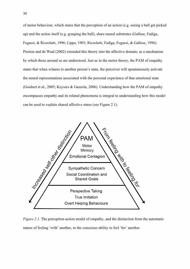
30
of motor behaviour, which states that the perception of an action (e.g. seeing a ball get picked
up) and the action itself (e.g. grasping the ball), share neural substrates (Gallese, Fadiga,
Fogassi, & Rizzolatti, 1996; Lipps, 1903; Rizzolatti, Fadiga, Fogassi, & Gallese, 1996).
Preston and de Waal (2002) extended this theory into the affective domain, as a mechanism
by which those around us are understood. Just as in the motor theory, the PAM of empathy
states that when witness to another person’s state, the perceiver will spontaneously activate
the neural representations associated with the personal experience of that emotional state
(Goubert et al., 2005; Keysers & Gazzola, 2006). Understanding how the PAM of empathy
encompasses empathy and its related phenomena is integral to understanding how this model
can be used to explain shared affective states (see Figure 2.1).
Figure 2.1. The perception-action model of empathy, and the distinction from the automatic
nature of feeling ‘with’ another, to the conscious ability to feel ‘for’ another.
31
In the PAM of empathy, phenomena such as motor mimicry, imitation, and emotional
contagion are all antecedents to empathic behaviour. In this sense, empathy is an evolved
function that is supported by automatic representations of shared affective states (Goubert et
al., 2005; Preston & de Waal, 2002). A key specification is that this process occurs
automatically, or without cognitive effort. Although cognition is an integral component of the
later stages of empathy (i.e. helping behaviours), conscious awareness is not required for core
components such as mimicry and contagion. As a result, the PAM of empathy is typically
used as a framework for psychophysiological studies.
2.2.3 The measurement of empathy
Empathy is typically measured using two methods: behavioural self-report scales and
psychophysiology. Self-report measures typically ask (either directly or indirectly) for a
participant to respond to a particular stimulus—usually in an attempt to gage the attitudes or
feelings related to the stimulus (Paulhus & Vazire, 2009). Conversely, psychophysiological
methods study the physiological signals underlying the reported psychological processes
(Cacioppo, Tassinary, & Berntson, 2007; Larsen et al., 2008). Empathy research has
traditionally been dominated by self-report measures, and investigation using
psychophysiological techniques has, by comparison, only recently gained popularity
(Neumann & Westbury, 2011; Paulhus & Vazire, 2009). Exploring the two key limitations of
the self-report approach will provide a justification for why psychophysiology is increasingly
becoming the preferred approach to investigate empathy.
The first limitation to self-report measures is by no means a new criticism. The
objectivity of self-report measures has been questioned since the development of
psychological assessment (Paulhus & Vazire, 2009). Self-report measures require trust in the
participant to respond truthfully, which does not always occur. This problem is of particular
relevance to empathy research as empathic responses are likely to be exaggerated because it
32
is socially desirable to help others, and thus respondents might seek to conform with
perceived social norms (Mauss & Robinson, 2009). An example of this social desirability is
evident in a study conducted by Kämpfe, Penzhorn, Schikora, Dünzl, and Schneidenbach
(2009) with delinquent or incarcerated participants. It was concluded that delinquent
participants were higher in cognitive empathy than non-delinquent controls when assessed
using a self-report measure. However, an implicit association empathy test (an indirect
measure of empathy) indicated higher empathy in the non-delinquent controls compared to
delinquent participants. Critically, there was a significant weak correlation between self-
report empathy and social desirability in the delinquent sample, but not the control sample.
As such, self-report empathy is susceptible to biased responses evidenced via social
desirability. However, a further and perhaps more serious limitation of self-report measures
stems from when a participant believes they are responding accurately and truthfully, but in
fact they are not.
Self-report measures require cognition. It is this cognition that allows for the
collection of information-rich data that is obtained from self-report measures (Paulhus &
Vazire, 2009). However, as was identified using the PAM, the core components of empathy,
such as mimicry and contagion, are processed unconsciously. Thus, even if participants
believe they are reporting honestly, there may be a discrepancy between unconscious
processes and cognitive awareness. For example, Preis, Kröner-Herwig, Schmidt-Samoa,
Dechent, and Barke (2015) used both behavioural and psychophysiological measures to index
empathy responses to pain images, and found that although self-report pain ratings remained
constant over repeated presentations, neurological responses decreased over repeated
exposure. In other words, psychophysiological measures showed habituation to the stimuli,
but self-report measures did not (Preis et al., 2015). Thus, a participant may believe they are
reporting consistently and accurately, however, psychophysiological measures are sensitive
33
to these non-conscious processes and the corresponding data indicate otherwise. Therefore, it
is proposed that future empathy research should adopt a complementary approach, as
physiological and self-report measures need not be mutually exclusive. Rather, self-report
measures should be used to assess the convergent validity of psychophysiological results.
When exploring the role of the central nervous system in empathy, the neuroimaging
technique, functional magnetic resonance imaging (fMRI) has been used to map the brain
structures associated with empathy (Decety & Lamm, 2006). Typically, fMRI is used in
empathy research to investigate the shared neural overlap between the emotional state of
another and the firsthand experience of emotion (Cheng, Chen, Lin, Chou, & Decety, 2010;
Jackson, Brunet, Meltzoff, & Decety, 2006; Lamm, Decety, & Singer, 2011; Preis et al.,
2015). Moreover, some fMRI research has demonstrated that there is not one neural network
for sharing emotions, rather there are distinct empathy circuits for pain (Singer et al., 2004),
disgust (Wicker et al., 2003), other basic emotions (Killgore & Yurgelun-Todd, 2004) and
complex emotions, such as social exclusion (Eisenberger, Lieberman, & Williams, 2003).
One limitation of fMRI is that although it has high spatial resolution (capable of indexing
changes at submillimeter level), the temporal resolution is low (500-1000 ms) due to the
relatively slow metabolic processes it is measuring (Goense, Bohraus, & Logothetis, 2016;
Neumann & Westbury, 2011). Thus, although fMRI is capable of indexing where a particular
response occurs in the brain with precision, it is not capable of detecting fast transient
responses to emotional stimuli (Fan & Han, 2008; Pegna, Landis, & Khateb, 2008). To
measure these responses requires a direct measure of electrical activity, which can be indexed
using electroencephalography (EEG).
Two different EEG components have been used to measure the time-related processes
involved in affective sharing: cortical wave patterns (Perry, Bentin, Bartal, Lamm, & Decety,
2010) and specific event-related potentials (ERPs), which are an index of cortical responses

34
to a specific event (Neumann & Westbury, 2011). Increasingly, the N170 ERP component
has been linked to facial expression processing (Hinojosa, Mercado, & Carretié, 2015), while
the N200 component has been linked to empathy (Balconi & Canavesio, 2016). Thus, EEG
can be used to index the changes in neural activity that are associated with shared affective
states and emotional contagion (Pineda & Hecht, 2009).
2.2.4 Catching emotions
Emotional contagion is widely referred to as the “catching” of another’s emotion
(Batson, 2009; Hatfield, Bensman, Thornton, & Rapson, 2014; Hatfield, Rapson, & Le, 2009;
Kret, 2015; Shamay-Tsoory, 2011). Focusing on the ability to “feel themselves into”
another’s emotion, emotional contagion is a critical component of understanding human
behaviour (Hatfield et al., 2009). Prior to the conceptualisation of emotional contagion, the
congruence of shared emotions was attributed to empathy (Davis, 1983). However, reviews
indicate that empathy is a complex, overarching phenomenon, and emotional contagion
consists of more primitive components (Hatfield, Cacioppo, & Rapson, 1994). Moreover,
Hatfield et al. (1994) define primitive emotional contagion as the tendency to mimic
another’s affective state, whether through facial expressions, postures, behaviours, or neural
networks—emotional contagion is the mechanism that allows for emotional convergence.
Furthermore, it has been clarified that emotional contagion exists in three stages: mimicry,
feedback, and the contagion of emotion (Hatfield et al., 2014; Hatfield et al., 2009).
Feeling sad when you see a sad person is a common experience. This contention is
commonly supported by neuroscience research, wherein neural circuits are proposed to
facilitate the sharing of emotion (Gallese, 2001; Gallese, Eagle, & Migone, 2007; Kaplan &
Iacoboni, 2006; Keysers & Gazzola, 2006). Activation of several brain regions, such as
anterior cingulate cortex (ACC) and bilateral anterior insula (AI), has been demonstrated
during both the observation and the firsthand experience of pain using fMRI (Botvinick et al.,
35
2005; de Vignemont & Singer, 2006; Singer et al., 2004). Oberman and Ramachandran
(2009) contended that in order for an emotion to become an embodied representation, it must
be converted from the visual system to the limbic system (i.e., the system commonly
associated with the experience of emotion). In addition, different areas within the limbic
system are activated for different emotions, For example, the ACC (Critchley et al., 2005;
Jackson et al., 2005) is activated when experiencing the noxious qualities of pain, and the
insula (Calder, Keane, Manes, Antoun, & Young, 2000; Wicker et al., 2003) is activated
when feeling disgust. Therefore, shared circuits provide a neural basis for how affective
states are shared. However, shared states only offer information about how we are able to
comprehend those around us. Thus, a shift in focus from the role of the central nervous
system in comprehending empathy, to the role of the peripheral nervous system in
communicating empathy is required.
2.2.5 Imitating others
The observation that people mimic one another is by no means new (Hatfield et al.,
2009). Smith (1759/1976) was one of the first to conceptualise mimicry as an automatic
component of social interaction. Since this original proposition, research has provided
physiological (Dimberg, Thunberg, & Elmehed, 2000; Dimberg, Thunberg, & Grunedal,
2002; Levenson & Ruef, 1992), behavioural (Chartrand & Bargh, 1999; van Baaren, Holland,
Kawakami, & van Knippenberg, 2004; Van Baaren, Maddux, Chartrand, de Bouter, & van
Knippenberg, 2003), and neurological (Kaplan & Iacoboni, 2006) evidence to corroborate
this theory. This phenomenon, known as the “chameleon effect” or “matched motor
hypothesis”, encompasses the way that people subconsciously adapt their postures,
mannerisms, and facial expressions to match a social environment. Facial expressions are an
intuitive method through which to investigate empathy given their automatic responsiveness
to emotional states (Dimberg & Öhman, 1996).
36
Mimicry is automatic in the sense that the mimicker does not need to be consciously
aware of the process, or even the stimulus, that elicits the response (Chartrand & Bargh,
1999; Chartrand, Maddux, & Lakin, 2005). For example, Dimberg et al. (2000) demonstrated
that congruent facial mimicry responses were elicited even when the emotional expressions
were masked by a neutral stimulus, and thus the observer was unaware of the emotional
stimulus. Therefore, the mechanisms facilitating facial mimicry are a low-level process, not
dependent on conscious decision making or even explicit recognition. Through investigating
the facial muscles implicated in the production of facial expressions, a greater understanding
of the processes underlying affective sharing will be obtained.
Facial electromyography (EMG) is a robust and commonly used method of measuring
activity in the facial muscles (Dimberg, 1982; Dimberg et al., 2000; Dimberg et al., 2002;
Sonnby-Borgstrom, 2002). In a typical facial mimicry experiment, flat electrodes are placed
over a participant’s facial muscles and they are then presented with emotional faces. Subtle
changes in the contraction of the muscles is detected as increased electrical activity (van
Baaren et al., 2009). Specific muscles tend to contract in making particular emotional
expressions. For example, activity in the zygomaticus major muscle is an index of positive
and affiliative emotions—particularly smiling (Achaibou, Pourtois, Schwartz, & Vuilleumier,
2008; Duchenne de Boulogne, 1990; Rymarczyk, Biele, Grabowska, & Majczynski, 2011;
Sonnby-Borgstrom, 2002). Thus, the zygomaticus major muscle draws the corners of the
mouth upwards towards the ears, creating a smile (Bentsianov & Blitzer, 2004; Duchenne de
Boulogne, 1990). Therefore, in a mimicry experiment if one observes increased activity in the
zygomaticus major muscle in response to being presented with a happy face this would
indicate mimicry of the portrayed emotion. Similarly, activation of the corrugator supercilii
muscle is represented by the furrowing of the inner brow (Cacioppo, Petty, Losch, & Kim,
1986; Duchenne de Boulogne, 1990). Thus, activation of the corrugator muscle is associated
37
with negative emotions (Murata, Saito, Schug, Ogawa, & Kameda, 2016). Therefore, in
accord with the PAM of empathy, mimicry serves to facilitate social bonds when an observer
perceives another’s emotional expression and activates the same expression themselves.
Thus, the function of mimicry is rooted in one of the key components of empathy:
demonstrating an understanding of how another feels.
The function of empathy is undeniably adaptive. There is consensus that mimicry
serves to facilitate increased rapport (Lakin & Chartrand, 2003), affiliation (Bourgeois &
Hess, 2008; Hess & Fischer, 2013), and pro-social behaviour (van Baaren et al., 2004). The
effects of mimicry on person perception have been demonstrated to increase bidirectional
liking (Chartrand & Bargh, 1999), and to strengthen the bond between two interaction
partners (van Baaren et al., 2009). Moreover, mimicry has been termed a “social glue”,
responsible for facilitating social bonding (Lakin, Jefferis, Cheng, & Chartrand, 2003).
Theoretically, if this mechanism were capable of bonding societies, the benefits would extend
beyond interaction partners into the general public. van Baaren et al. (2004) demonstrated
that mimicry increases helping behaviours not just for the mimicker, but also persons with
which the mimicked interact with. Moreover, being mimicked increases subsequent altruistic
behaviours in the form of charitable donations, compared to persons who were not mimicked.
However, mimicry also serves to facilitate personal gain. Namely, mimicry can be used to
convey romantic interest (Farley, 2014) or for monetary gain (Sims, Van Reekum, Johnstone,
& Chakrabarti, 2012). Thus, mimicry is a pervasive component of social interaction.
Perhaps an even greater contribution to the understanding of human affective states
stems from when mimicry is inhibited. Typical mimicry research paradigms investigate facial
responses whilst participants witness emotional displays (Dimberg et al., 2000; Hess &
Blairy, 2001). However, what might occur if this mimicry is inhibited? The facial feedback
hypothesis states that one’s ability to experience emotion is dependent on the ability to

38
express emotion. Strack, Martin, and Stepper (1988) demonstrated this hypothesis;
participants held a pen in their mouth to either inhibit (holding with lips) or facilitate (holding
with teeth) smiling. Facilitating a smile increased the participant’s funniness ratings of
cartoons in comparison to the inhibitory group. In a similar vein, Neal and Chartrand (2011)
recruited matched participants who had received either a Botox treatment (the temporary
paralysis of facial muscles), or a Restylane injection (a dermal filler). They concluded that
facial feedback was an important component of emotion classification, as participants
injected with Botox were less accurate in classifying emotional expressions than the
Restylane control group. Thus, facial mimicry serves not only to strengthen social bonds, but
also plays a role in emotion recognition (Iacoboni, 2009).
Evidently, the processes of mimicry and emotional contagion are important
components of facilitating social communication (Chartrand & Bargh, 1999) and conveying
empathy (Hess, Philippot, & Blairy, 1999). Most of the research into shared affective states
has investigated the neural components of contagion separately from motor mimicry.
However, it is proposed that understanding how these phenomena come together to form the
overarching perception-action model will provide a richer understanding of empathy.
Recently, it has been proposed that mirror neurons could underlie this phenomenon
(Rizzolatti & Craighero, 2004; Rizzolatti et al., 1996). Originally proposed as a mechanism
for action imitation in monkeys, and now evidenced in humans (Wolf, Gales, Shane, &
Shane, 2001), mirror neurons have been touted as the next great advance in psychology
(Oberman & Ramachandran, 2009). With new conceptualisations such as “the empathic
brain” and “the social brain”, the future of empathy research is rooted in physiology wherein
mirror neurons provide a basis for the way that we as humans experience empathy (Adolphs,
2009; de Vignemont & Singer, 2006; Dunbar & Shultz, 2007; Frith, 2007; Iacoboni, 2009;
Keysers, 2011).
39
2.3 Mirror Neurons
2.3.1 What are mirror neurons?
Thus far, the concepts of mimicry and imitation have been explored; now, the
neurological processes that are thought to underpin these responses will be explained. Mirror
neurons are a special class of visuomotor neurons that fire in response to both the execution
of an action, and the observation of another performing the same or a similar motor act
(Gallese et al., 1996). Activation of our own motor system in response to observing another’s
actions is at the core of the mirror neuron system (Kaplan & Iacoboni, 2006), meaning that
mirror neurons allow for the shared representation of actions. Prior to the discovery of mirror
neurons, it was posited that the interesting component to understanding social interaction was
that of mentalising; however, it has since been questioned whether understanding the actions
of others actually requires mentalising (Keysers, 2011). First reported in the ventral premotor
cortex area F5 of the Rhesus macaque, these neurons were discovered through single cell
recordings, where a neuron fired whilst the monkey picked up a peanut, and also when the
monkey watched the experimenter pick up a peanut (Rizzolatti et al., 1996). Since this
original discovery, psychophysiological evidence has revealed that it is not limited to grasp
actions, as mirror neurons respond to other complex hand-actions (Gallese et al., 1996) and
also to mouth actions (Ferrari, Gallese, Rizzolatti, & Fogassi, 2003). To determine the
purpose of these dual property neurons, researchers investigated whether responses were
simply motor copy of the motor act, or whether they were responsive to the intention behind
the action (de Lange, Spronk, Willems, Toni, & Bekkering, 2008; Iacoboni et al., 2005).
It is not enough to simply identify a motor movement; increasingly mirror neuron
research is focusing on understanding the intention or goal that underpins these actions
(Iacoboni et al., 2005; Kaplan & Iacoboni, 2006). Mirror neurons discriminate between
different goals based on their type. There is a consensus that there are two types of mirror
40
neurons: strictly congruent and broadly congruent. Strictly congruent mirror neurons
represent one third of all mirror neurons, and only respond to the exact action triggered
during execution—down to the most minute detail (Gallese et al., 1996; Rizzolatti et al.,
1996). Conversely, broadly congruent mirror neurons also respond to actions that achieve a
similar goal through different means (Thioux, Gazzola, & Keysers, 2008). However, most
mirror neurons will respond to the grasping of an object, but not if the grasp is performed in
the absence of the object (Thioux et al., 2008). Iacoboni et al. (2005) conducted the now
famous ‘teacup experiment’ where participant’s mirror neuron systems adequately
discriminated between precision grips in differing contexts. Specifically, the greatest mirror
neuron activation resulted from the precision grip of a teacup paired with a neat tea-party
scene, as opposed to a teacup presented alone. de Lange et al. (2008) further explored goal-
directed action by varying whether the participants were instructed to attend to the intention
of the action, or the means by which the action was executed. It was concluded that the
mirror neuron system processes the intentionality of the action irrespective of whether or not
the participant was attending to the intention of the action. Thus, the mirror neuron system is
unlikely to be a system involving simple motor simulation, and the processes that underlie
this activation are likely to employ theories of associative learning and theory of mind.
How primates came to possess mirror neurons is likely a combination of innate traits
(Meltzoff & Moore, 1977), and Hebbian learning (Heyes, 2010; Keysers & Gazzola, 2009;
Keysers & Perrett, 2004). The argument that some element of the mirror neuron system is
innate has stemmed from research investigating imitation in infants (Anisfeld, 1996; Meltzoff
& Moore, 1977). Given the importance of mimicry and imitation in successful social
cognition, neonates are the only organism to have a “hard wired” ability to mimic others,
with tongue protrusion evidenced in studies with human infants (Anisfeld, 1996; Meltzoff &
Moore, 1977) and monkeys (Ferrari et al., 2006). However, the repertoire of behaviours and
41
processes thought to be facilitated by mirror neurons is expanding beyond that of simple
motor actions (Enticott, Johnston, Herring, Hoy, & Fitzgerald, 2008; Fogassi, 2011; Gallese
et al., 2007; Molnar-Szakacs & Overy, 2006), and as such Hebbian association learning is
proposed to account for further development of the mirror neuron system (Heyes, 2010;
Keysers & Gazzola, 2009).
Hebbian learning, first proposed by Hebb (1949), states that the repeated association
between events will result in a learned association, or ‘neurons that fire together wire
together’. From the point of view of the infant, repeated observation of the infants’ own
movements will result in activation of the same neural pathways as when observing another
make the same movement (Del Giudice, Manera, & Keysers, 2009). However, this neural
plasticity is not limited to infants, with research by Lahav, Saltzman, and Schlaug (2007)
demonstrating that prior to training non-musical adults did not have any activation when
listening to piano notes; however, after a few lessons, presentation of the practiced musical
piece activated the same network. This plasticity provides support for the ‘neurons that fire
together wire together’ argument, and indicates the complexity of the mirror neuron system,
with research extending beyond that of actions, to “action listening” systems (Lahav et al.,
2007), social cognition (Keysers & Gazzola, 2006; Pineda & Hecht, 2009; Uddin, Iacoboni,
Lange, & Keenan, 2007), and empathy (Baird, Scheffer, & Wilson, 2011; Gallese, 2001;
Kaplan & Iacoboni, 2006).
2.3.2 The empathic brain what have we learned from neuroimaging?
A greater understanding of the human mirror neuron system stems from
understanding the brain regions commonly activated through action recognition, theory of
mind, and empathy. Mirroring mechanisms allow us to activate an understanding of the
intentions of others in ourselves, which is a key component of empathy (Kaplan & Iacoboni,
2006). A complex network of the occipital, temporal, and parietal visual areas make up the

42
MNS. However, as single electrode research in humans is invasive and dangerous, most of
the understanding about the human mirror system has stemmed from neurophysiology and
neuroimaging research (see Mukamel, Ekstrom, Kaplan, Iacoboni, and Fried (2010) for an
exception). As mirror neurons stem from sensorimotor experience, or the experience gained
through social interaction, research has typically focused on the activation of the classical
mirror neuron areas: the inferior frontal gyrus, inferior parietal lobule (Heyes, 2010;
Rizzolatti & Craighero, 2004), and the insula and amygdala (Wicker et al., 2003) (see Table
2.1).
Table 2.1
Neural Correlates of the Mirror Neuron System from Action Observation to Empathy
MNS - Action Facial Expressions Theory of Mind Empathy IFG IFG IFG Premotor IFG
STS STS STS (Rt) STS (Rt)
IPL (Rt) Parietal cortex IPL (Rt) IPL (Rt) Occipital cortex ACC Occipital cortex ACC
Sensorimotor cortex Sensorimotor cortex Sensorimotor cortex Sensorimotor cortex PMC Insula Medial PFC (incl.
ACC), OFC, Precuneus, Somatosensory cortices, Amygdala
Ventromedial PFC (incl. OFC), Precuneus, Somatosensory cortices, Amygdala, Insula, Posterior Cingulate
Note. IFG = Inferior Frontal Gyrus, STS = Superior Temporal Sulcus, IPL = Inferior Parietal Lobule, PMC = Premotor Cortex, PFC = Prefrontal Cortex, ACC = Anterior Cingulate Cortex, OFC = Orbitofrontal Cortex. Rt = Right hemisphere.
The inferior frontal gyrus (IFG) is one of the classical mirror areas involved in human
action mirroring research (Gazzola, Rizzolatti, Wicker, & Keysers, 2007; Thioux et al.,
2008). As the hand and mouth regions in the somatotopic organisation of the mirror neuron
system are closely related, the IFG has also been implicated with the observation and
43
imitation of facial expressions (particularly BA 45; Carr, Iacoboni, Dubeau, Mazziotta, and
Lenzi (2003)). Hennenlotter et al. (2005) found that right IFG was implicated in viewing and
executing smiles. Furthermore, Jabbi and Keysers (2008) concluded that viewing facial
expressions engaged brain regions associated with motor functions (particularly BA45);
however, they also concluded that this motor representation may not result in the overt
production of expressions. Thus, the viewing of facial expressions may lead to simulation of
motor functions, suggesting a mirror like response, rather than overt production of the
expression (i.e. mimicry). Conversely, van der Gaag, Minderaa, and Keysers (2007) argued
that observation and execution of facial expressions activate a similar network of brain
regions indicating that overt motor simulation is directly linked to emotional simulation.
Enticott et al. (2008) further concluded that the automatic processing of emotional facial
expressions facilitates understanding of another’s affective state. However, determining the
emotional state of others is often a deductive process (Keysers & Gazzola, 2009). Thus, the
neural pathways activated through motor simulation likely facilitate the automatic nature of
processing others’ emotions and allow for deductive reasoning to occur. Despite the
conflicting evidence on how the IFG is implicated in the processing of facial expressions,
there is consensus that similar brain regions are activated in a mirror neuron like response
during the observation and execution of facial expressions (Keysers & Gazzola, 2009; Leslie,
Johnson-Frey, & Grafton, 2004).
Several other brain regions have been successfully linked to the extended mirror
neuron system in neuroimaging studies, such as the anterior cingulate cortex (ACC) and the
insula. Botvinick et al. (2005) were among the first to provide evidence that facial
expressions of pain activate the same cortical areas as pain experienced first-hand. In
addition, they further concluded that the ACC was linked to sadness, indicating that the
structure involved in feeling an emotion, was also linked in sharing affective states. Singer et
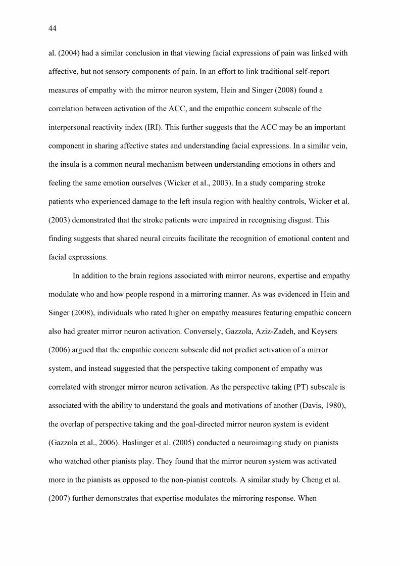
44
al. (2004) had a similar conclusion in that viewing facial expressions of pain was linked with
affective, but not sensory components of pain. In an effort to link traditional self-report
measures of empathy with the mirror neuron system, Hein and Singer (2008) found a
correlation between activation of the ACC, and the empathic concern subscale of the
interpersonal reactivity index (IRI). This further suggests that the ACC may be an important
component in sharing affective states and understanding facial expressions. In a similar vein,
the insula is a common neural mechanism between understanding emotions in others and
feeling the same emotion ourselves (Wicker et al., 2003). In a study comparing stroke
patients who experienced damage to the left insula region with healthy controls, Wicker et al.
(2003) demonstrated that the stroke patients were impaired in recognising disgust. This
finding suggests that shared neural circuits facilitate the recognition of emotional content and
facial expressions.
In addition to the brain regions associated with mirror neurons, expertise and empathy
modulate who and how people respond in a mirroring manner. As was evidenced in Hein and
Singer (2008), individuals who rated higher on empathy measures featuring empathic concern
also had greater mirror neuron activation. Conversely, Gazzola, Aziz-Zadeh, and Keysers
(2006) argued that the empathic concern subscale did not predict activation of a mirror
system, and instead suggested that the perspective taking component of empathy was
correlated with stronger mirror neuron activation. As the perspective taking (PT) subscale is
associated with the ability to understand the goals and motivations of another (Davis, 1980),
the overlap of perspective taking and the goal-directed mirror neuron system is evident
(Gazzola et al., 2006). Haslinger et al. (2005) conducted a neuroimaging study on pianists
who watched other pianists play. They found that the mirror neuron system was activated
more in the pianists as opposed to the non-pianist controls. A similar study by Cheng et al.
(2007) further demonstrates that expertise modulates the mirroring response. When
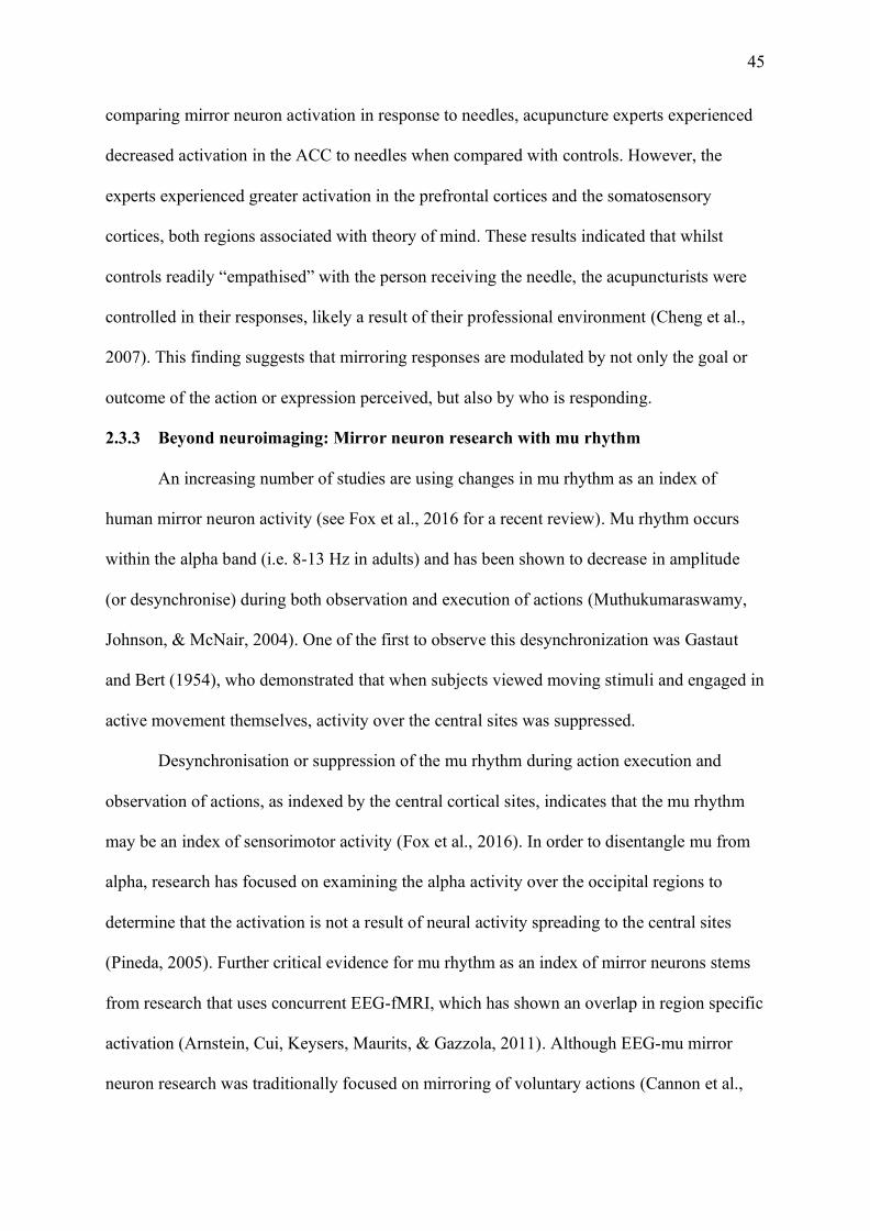
45
comparing mirror neuron activation in response to needles, acupuncture experts experienced
decreased activation in the ACC to needles when compared with controls. However, the
experts experienced greater activation in the prefrontal cortices and the somatosensory
cortices, both regions associated with theory of mind. These results indicated that whilst
controls readily “empathised” with the person receiving the needle, the acupuncturists were
controlled in their responses, likely a result of their professional environment (Cheng et al.,
2007). This finding suggests that mirroring responses are modulated by not only the goal or
outcome of the action or expression perceived, but also by who is responding.
2.3.3 Beyond neuroimaging: Mirror neuron research with mu rhythm
An increasing number of studies are using changes in mu rhythm as an index of
human mirror neuron activity (see Fox et al., 2016 for a recent review). Mu rhythm occurs
within the alpha band (i.e. 8-13 Hz in adults) and has been shown to decrease in amplitude
(or desynchronise) during both observation and execution of actions (Muthukumaraswamy,
Johnson, & McNair, 2004). One of the first to observe this desynchronization was Gastaut
and Bert (1954), who demonstrated that when subjects viewed moving stimuli and engaged in
active movement themselves, activity over the central sites was suppressed.
Desynchronisation or suppression of the mu rhythm during action execution and
observation of actions, as indexed by the central cortical sites, indicates that the mu rhythm
may be an index of sensorimotor activity (Fox et al., 2016). In order to disentangle mu from
alpha, research has focused on examining the alpha activity over the occipital regions to
determine that the activation is not a result of neural activity spreading to the central sites
(Pineda, 2005). Further critical evidence for mu rhythm as an index of mirror neurons stems
from research that uses concurrent EEG-fMRI, which has shown an overlap in region specific
activation (Arnstein, Cui, Keysers, Maurits, & Gazzola, 2011). Although EEG-mu mirror
neuron research was traditionally focused on mirroring of voluntary actions (Cannon et al.,
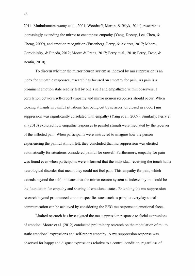
46
2014; Muthukumaraswamy et al., 2004; Woodruff, Martin, & Bilyk, 2011), research is
increasingly extending the mirror to encompass empathy (Yang, Decety, Lee, Chen, &
Cheng, 2009), and emotion recognition (Ensenberg, Perry, & Aviezer, 2017; Moore,
Gorodnitsky, & Pineda, 2012; Moore & Franz, 2017; Perry et al., 2010; Perry, Troje, &
Bentin, 2010).
To discern whether the mirror neuron system as indexed by mu suppression is an
index for empathic responses, research has focused on empathy for pain. As pain is a
prominent emotion state readily felt by one’s self and empathized within observers, a
correlation between self-report empathy and mirror neuron responses should occur. When
looking at hands in painful situations (i.e. being cut by scissors, or closed in a door) mu
suppression was significantly correlated with empathy (Yang et al., 2009). Similarly, Perry et
al. (2010) explored how empathic responses to painful stimuli were mediated by the receiver
of the inflicted pain. When participants were instructed to imagine how the person
experiencing the painful stimuli felt, they concluded that mu suppression was elicited
automatically for situations considered painful for oneself. Furthermore, empathy for pain
was found even when participants were informed that the individual receiving the touch had a
neurological disorder that meant they could not feel pain. This empathy for pain, which
extends beyond the self, indicates that the mirror neuron system as indexed by mu could be
the foundation for empathy and sharing of emotional states. Extending the mu suppression
research beyond pronounced emotion specific states such as pain, to everyday social
communication can be achieved by considering the EEG mu response to emotional faces.
Limited research has investigated the mu suppression response to facial expressions
of emotion. Moore et al. (2012) conducted preliminary research on the modulation of mu to
static emotional expressions and self-report empathy. A mu suppression response was
observed for happy and disgust expressions relative to a control condition, regardless of
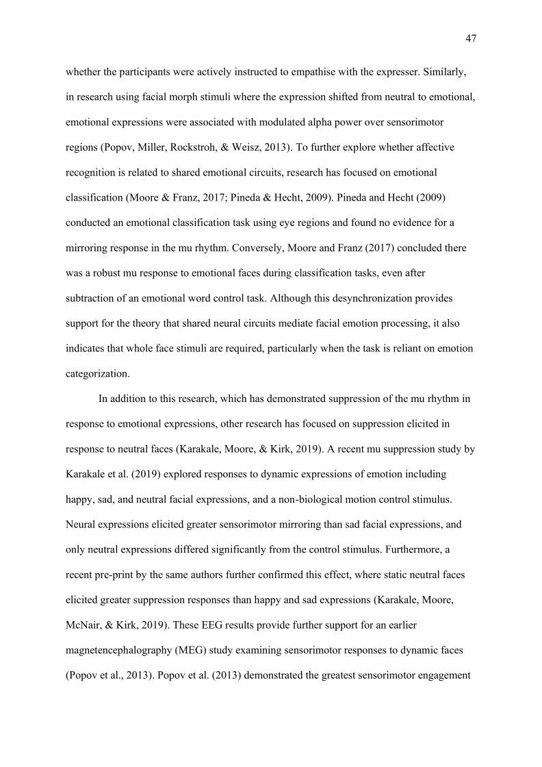
47
whether the participants were actively instructed to empathise with the expresser. Similarly,
in research using facial morph stimuli where the expression shifted from neutral to emotional,
emotional expressions were associated with modulated alpha power over sensorimotor
regions (Popov, Miller, Rockstroh, & Weisz, 2013). To further explore whether affective
recognition is related to shared emotional circuits, research has focused on emotional
classification (Moore & Franz, 2017; Pineda & Hecht, 2009). Pineda and Hecht (2009)
conducted an emotional classification task using eye regions and found no evidence for a
mirroring response in the mu rhythm. Conversely, Moore and Franz (2017) concluded there
was a robust mu response to emotional faces during classification tasks, even after
subtraction of an emotional word control task. Although this desynchronization provides
support for the theory that shared neural circuits mediate facial emotion processing, it also
indicates that whole face stimuli are required, particularly when the task is reliant on emotion
categorization.
In addition to this research, which has demonstrated suppression of the mu rhythm in
response to emotional expressions, other research has focused on suppression elicited in
response to neutral faces (Karakale, Moore, & Kirk, 2019). A recent mu suppression study by
Karakale et al. (2019) explored responses to dynamic expressions of emotion including
happy, sad, and neutral facial expressions, and a non-biological motion control stimulus.
Neural expressions elicited greater sensorimotor mirroring than sad facial expressions, and
only neutral expressions differed significantly from the control stimulus. Furthermore, a
recent pre-print by the same authors further confirmed this effect, where static neutral faces
elicited greater suppression responses than happy and sad expressions (Karakale, Moore,
McNair, & Kirk, 2019). These EEG results provide further support for an earlier
magnetencephalography (MEG) study examining sensorimotor responses to dynamic faces
(Popov et al., 2013). Popov et al. (2013) demonstrated the greatest sensorimotor engagement
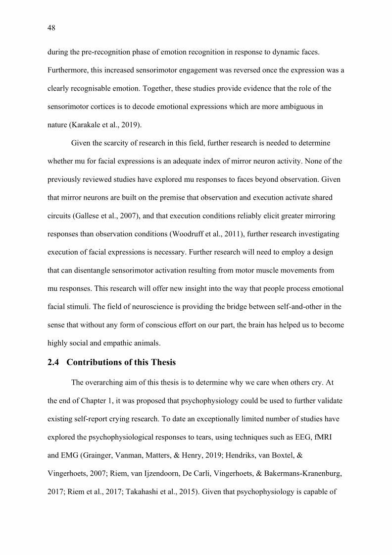
48
during the pre-recognition phase of emotion recognition in response to dynamic faces.
Furthermore, this increased sensorimotor engagement was reversed once the expression was a
clearly recognisable emotion. Together, these studies provide evidence that the role of the
sensorimotor cortices is to decode emotional expressions which are more ambiguous in
nature (Karakale et al., 2019).
Given the scarcity of research in this field, further research is needed to determine
whether mu for facial expressions is an adequate index of mirror neuron activity. None of the
previously reviewed studies have explored mu responses to faces beyond observation. Given
that mirror neurons are built on the premise that observation and execution activate shared
circuits (Gallese et al., 2007), and that execution conditions reliably elicit greater mirroring
responses than observation conditions (Woodruff et al., 2011), further research investigating
execution of facial expressions is necessary. Further research will need to employ a design
that can disentangle sensorimotor activation resulting from motor muscle movements from
mu responses. This research will offer new insight into the way that people process emotional
facial stimuli. The field of neuroscience is providing the bridge between self-and-other in the
sense that without any form of conscious effort on our part, the brain has helped us to become
highly social and empathic animals.
2.4 Contributions of this Thesis
The overarching aim of this thesis is to determine why we care when others cry. At
the end of Chapter 1, it was proposed that psychophysiology could be used to further validate
existing self-report crying research. To date an exceptionally limited number of studies have
explored the psychophysiological responses to tears, using techniques such as EEG, fMRI
and EMG (Grainger, Vanman, Matters, & Henry, 2019; Hendriks, van Boxtel, &
Vingerhoets, 2007; Riem, van Ijzendoorn, De Carli, Vingerhoets, & Bakermans-Kranenburg,
2017; Riem et al., 2017; Takahashi et al., 2015). Given that psychophysiology is capable of
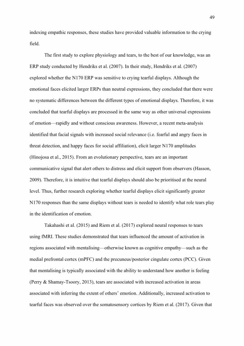
49
indexing empathic responses, these studies have provided valuable information to the crying
field.
The first study to explore physiology and tears, to the best of our knowledge, was an
ERP study conducted by Hendriks et al. (2007). In their study, Hendriks et al. (2007)
explored whether the N170 ERP was sensitive to crying tearful displays. Although the
emotional faces elicited larger ERPs than neutral expressions, they concluded that there were
no systematic differences between the different types of emotional displays. Therefore, it was
concluded that tearful displays are processed in the same way as other universal expressions
of emotion—rapidly and without conscious awareness. However, a recent meta-analysis
identified that facial signals with increased social relevance (i.e. fearful and angry faces in
threat detection, and happy faces for social affiliation), elicit larger N170 amplitudes
(Hinojosa et al., 2015). From an evolutionary perspective, tears are an important
communicative signal that alert others to distress and elicit support from observers (Hasson,
2009). Therefore, it is intuitive that tearful displays should also be prioritised at the neural
level. Thus, further research exploring whether tearful displays elicit significantly greater
N170 responses than the same displays without tears is needed to identify what role tears play
in the identification of emotion.
Takahashi et al. (2015) and Riem et al. (2017) explored neural responses to tears
using fMRI. These studies demonstrated that tears influenced the amount of activation in
regions associated with mentalising—otherwise known as cognitive empathy—such as the
medial prefrontal cortex (mPFC) and the precuneus/posterior cingulate cortex (PCC). Given
that mentalising is typically associated with the ability to understand how another is feeling
(Perry & Shamay-Tsoory, 2013), tears are associated with increased activation in areas
associated with inferring the extent of others’ emotion. Additionally, increased activation to
tearful faces was observed over the somatosensory cortices by Riem et al. (2017). Given that
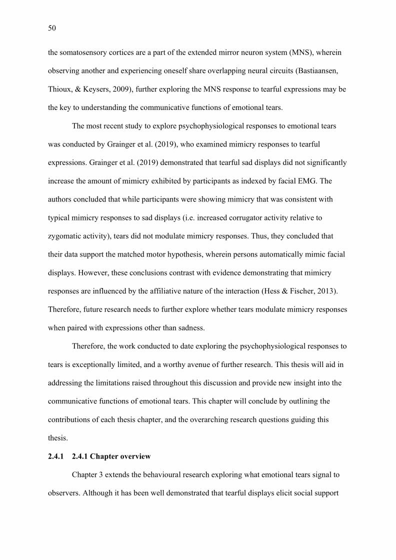
50
the somatosensory cortices are a part of the extended mirror neuron system (MNS), wherein
observing another and experiencing oneself share overlapping neural circuits (Bastiaansen,
Thioux, & Keysers, 2009), further exploring the MNS response to tearful expressions may be
the key to understanding the communicative functions of emotional tears.
The most recent study to explore psychophysiological responses to emotional tears
was conducted by Grainger et al. (2019), who examined mimicry responses to tearful
expressions. Grainger et al. (2019) demonstrated that tearful sad displays did not significantly
increase the amount of mimicry exhibited by participants as indexed by facial EMG. The
authors concluded that while participants were showing mimicry that was consistent with
typical mimicry responses to sad displays (i.e. increased corrugator activity relative to
zygomatic activity), tears did not modulate mimicry responses. Thus, they concluded that
their data support the matched motor hypothesis, wherein persons automatically mimic facial
displays. However, these conclusions contrast with evidence demonstrating that mimicry
responses are influenced by the affiliative nature of the interaction (Hess & Fischer, 2013).
Therefore, future research needs to further explore whether tears modulate mimicry responses
when paired with expressions other than sadness.
Therefore, the work conducted to date exploring the psychophysiological responses to
tears is exceptionally limited, and a worthy avenue of further research. This thesis will aid in
addressing the limitations raised throughout this discussion and provide new insight into the
communicative functions of emotional tears. This chapter will conclude by outlining the
contributions of each thesis chapter, and the overarching research questions guiding this
thesis.
2.4.1 2.4.1 Chapter overview
Chapter 3 extends the behavioural research exploring what emotional tears signal to
observers. Although it has been well demonstrated that tearful displays elicit social support
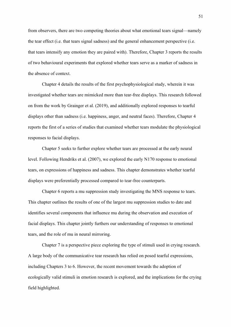
51
from observers, there are two competing theories about what emotional tears signal—namely
the tear effect (i.e. that tears signal sadness) and the general enhancement perspective (i.e.
that tears intensify any emotion they are paired with). Therefore, Chapter 3 reports the results
of two behavioural experiments that explored whether tears serve as a marker of sadness in
the absence of context.
Chapter 4 details the results of the first psychophysiological study, wherein it was
investigated whether tears are mimicked more than tear-free displays. This research followed
on from the work by Grainger et al. (2019), and additionally explored responses to tearful
displays other than sadness (i.e. happiness, anger, and neutral faces). Therefore, Chapter 4
reports the first of a series of studies that examined whether tears modulate the physiological
responses to facial displays.
Chapter 5 seeks to further explore whether tears are processed at the early neural
level. Following Hendriks et al. (2007), we explored the early N170 response to emotional
tears, on expressions of happiness and sadness. This chapter demonstrates whether tearful
displays were preferentially processed compared to tear-free counterparts.
Chapter 6 reports a mu suppression study investigating the MNS response to tears.
This chapter outlines the results of one of the largest mu suppression studies to date and
identifies several components that influence mu during the observation and execution of
facial displays. This chapter jointly furthers our understanding of responses to emotional
tears, and the role of mu in neural mirroring.
Chapter 7 is a perspective piece exploring the type of stimuli used in crying research.
A large body of the communicative tear research has relied on posed tearful expressions,
including Chapters 3 to 6. However, the recent movement towards the adoption of
ecologically valid stimuli in emotion research is explored, and the implications for the crying
field highlighted.

52
Chapter 8 is one of the first empirical investigations exploring the perceptions of
genuineness in tearful displays. These tearful displays include the posed expressions used in
this thesis, and a selection of genuine tearful displays recently adopted in tear research. The
results of three experiments are discussed, and the implications for the crying field are
considered.
Chapter 9 is the concluding chapter of this thesis, where the critical question why we
care when others cry is revisited. We offer several possible answers to this fundamental
question and outline the potential implications of this thesis and what research could explore
moving forward. The thesis concludes with a summary of the communicative functions of
adult emotional tears and highlights the importance that this understanding can afford to our
empathic society as a whole.
2.4.2 2.4.2 Thesis aim
As identified the overarching aim of this thesis is to determine why we care when others cry.
To do this, these three research questions were investigated:
1) Do tears serve as a signal of sadness in the absence of context (Chapter 3)?
2) Do tears modulate physiological responses to facial displays (Chapters 4, 5, and 6)?
3) Do tears increase the authenticity of facial displays (Chapters 7 and 8)?

53
Chapter 3: The Tear Effect: More than just a Marker of Sadness?
3.1 Chapter Overview
Chapter 3 extends the behavioural research exploring what emotional tears signal to
observers. Although it has been well demonstrated that tearful displays elicit social support
from observers, there are two competing theories about what emotional tears signal—namely
the tear effect (i.e. that tears signal sadness) and the general enhancement perspective (i.e.
that tears intensify any emotion they are paired with). Therefore, Chapter 3 reports the results
of two behavioural experiments that explored whether tears serve as a marker of sadness in
the absence of context.
3.2 Publication Status
Under review at Cognition and Emotion, revise and resubmit completed.
3.3 Author Contributions
Krivan - planned and created the experiments, collected the data, conducted the data
analysis, wrote up the manuscript, and completed the revisions from the reviewers.
Caltabiano - provided supervision.
Cottrell - aided in experimental design and provided supervision.
Thomas - provided supervision, contributed to editing and review, and provided the funding
for the experiments.
3.4 Manuscript
3.4.1 Abstract
The function of emotional tears is said to alert others to emotional distress and
signal the need for support. Yet, tears are elicited in moments of extreme joy, in
addition to despair. The majority of research has demonstrated support for the tear
54
effect, wherein tears increase the perception of sadness. However, limited empirical
work has been conducted exploring the general enhancement perspective, which posits
that tears increase the intensity of an emotion. We report the results of two experiments,
which investigated the influence of tears on the identification of sad, happy, and neutral
expressions. Participants were exposed to facial stimuli, half of which featured tears
and half did not. Experiment 1 investigated reaction time responses to these displays, to
determine whether tears were a signal of sadness—in line with the tear effect.
Experiment 2 sought to provide further support for the tear effect by examining the
roles of intensity and valence in the identification of happy and sad tearful displays.
Experiment 1 supported the tear effect, with impaired recognition of happy-tear faces,
and enhanced recognition of neutral-tear faces, while classification of sad expressions
was not influenced by tears. Experiment 2 further demonstrated that the presence of
tears impairs recognition of a happy face. Furthermore, the presence of tears increases
perceived intensity, whilst simultaneously increasing the perception of negative
valence. Thus, we demonstrated support for both the tear effect and the general
enhancement perspective, wherein tears serve as an intense marker of sadness in the
absence of context.
Keywords: adult crying, emotion recognition, reaction time
3.4.2 Introduction
Emotional tears are a universally recognised emotional expression (Gračanin, Bylsma,
& Vingerhoets, 2018). Predictably, it is this universality that has aided in the identification of
the communicative function of emotional tears. Tears have previously been shown to signal
sadness (Provine et al., 2009; Zeifman & Brown, 2011), which in turn reliably elicits help
and support from observers (Balsters et al., 2013; Hendriks et al., 2008). In these studies, it is
demonstrated that tearful expressions communicate distress through sadness and the need for

55
support more effectively than identical tear-free displays. However, these studies have
typically used negative or neutral displays—where there is a specific focus on the perception
of sadness. Therefore, it is not overly surprising that tears signal sadness. For this reason,
there are two related, but competing, theories about what tears signal. The first, the tear effect
postulates that in the absence of social context, tears are perceived as an indicator of sadness
(Provine et al., 2009). However, the second, the general enhancement perspective, asserts
that tears intensify the emotions they are paired with—serving as a “secretory exclamation
point” (Ito et al., 2019; Provine, 2012). Under the general enhancement perspective, tears
would increase the intensity of perceived sadness, but also increase the intensity of perceived
anger for angry displays. Conversely, under the tear effect, tears would increase the
perception of sadness, regardless of the emotion they are paired with.
Of these two competing theories, there is undoubtedly greater support for the tear
effect. Provine et al. (2009) proposed that tears facilitate the recognition of sadness.
Participants rated static images of crying individuals and duplicate images with the tears
removed. Tearful expressions were rated as significantly sadder in appearance than the tear-
free duplicates. In addition, participants perceived the tear-free images as ambiguous in
emotional valence (Provine et al., 2009). Similarly, Zeifman and Brown (2011) demonstrated
that participants perceived faces with tears as sadder than faces without tears, regardless of
target age. Moreover, the greatest effect of this increased sadness was observed for adult
faces, rather than infant and child faces. Takahashi et al. (2015) further demonstrated the tear
effect using sad and neutral expressions and concluded that the magnitude of the tear effect
was greater for neutral expressions. Additional evidence for the tear effect was reported by
Balsters et al. (2013), who found that reaction time responses to sad faces with tears were
significantly faster than to sad tear-free faces. Interestingly, the images were presented briefly
(i.e. 50 ms), and tears were a salient signal, which rapidly attracted attention (Balsters et al.,
56
2013). Together, these studies show that tears signal and facilitate the recognition of sadness.
However, in all these studies the response options were centred around sadness. For example,
in the rating tasks participants were responding with how sad the faces seemed, and in the
reaction time task the response options were sad or not sad. Given the response metrics, it is
not surprising that tears were associated with sadness.
This focus on sadness ratings resulted in the development of the general enhancement
perspective. Proposed by Ito et al. (2019), the general enhancement perspective posits that
tears increase the intensity of all facial expressions. Reed et al. (2015) explored whether the
presence of tears on dynamic posed prototypical displays of happiness, anger, sadness, fearful
and neutral expressions increased emotion specific ratings, in addition to generalised intensity
and valence ratings. Unsurprisingly, sadness specific ratings were greater for all emotions
(except anger) for tearful displays, relative to tear-free expressions. However, tears also
increased the generalised intensity ratings of neutral, happy, angry, and fearful displays, and
in several instances’ emotion specific ratings (e.g. anger rating increased for tearful angry,
happy, and fearful expressions). These results indicate support for a general enhancement
perspective, where tears increase the intensity of emotional displays, rather than just
increasing sadness. Ito et al. (2019) further explored the perception of negative tearful
displays under the general enhancement perspective, by asking participants to rate angry,
fearful, sad, disgusted, and neutral tearful expressions on emotion specific rating scales. As in
Reed et al. (2015), perceived intensity of sadness was increased when the faces featured tears.
Interestingly, the only evidence observed for a general enhancement perspective stemmed
from increased intensity of anger in disgusted expressions with tears. The authors further
explored these ratings in multidimensional space, and the presence of tears resulted in a
tighter clustering of all emotions around sadness (Ito et al., 2019). Thus, both studies
57
concluded that there is a generalised sadness effect when tears are added to emotional
displays.
However, tears are also elicited in response to positive emotions (Miceli &
Castelfranchi, 2003). As previously stated, Reed et al. (2015) examined the effect of tears on
expressions other than sadness. Interestingly, tearful Duchenne smiles were attributed greater
generalised intensity, while tearful non-Duchenne smiles were attributed greater negative
valence (Reed et al., 2015). This result further indicates that tears are an intense marker of
sadness, and that recognising “tears of joy” likely stems from contextual information. This
interpretation is consistent with the dimorphous expression literature, in that when tearful
facial expressions are paired with positive scenarios they are perceived as positive emotions
(Aragón, 2017; Aragón & Bargh, 2018; Aragón & Clark, 2018). Thus, tears serve as a marker
of sadness in the absence of context, for both positive and negative emotional displays.
However, these conclusions stem largely from sadness specific emotion ratings (Ito et
al., 2019; Reed et al., 2015). The virtue of emotion specific ratings is the ability to investigate
whether the presence of tears enhances the intensity of specific emotional expressions (i.e.
greater perceived anger for angry expressions). However, emotion specific ratings increase
the number of comparisons per emotion. Furthermore, the inclusion of a specific rating of
sadness biases results towards a significant tear effect. Interestingly, two studies using
sadness specific ratings have demonstrated that tears do not increase the perception of
sadness for sad displays, but do for other expressions of emotion (Reed et al., 2015; Reed et
al., 2019). Therefore, future research exploring the tear effect and the general enhancement
perspective should use generalised intensity and valence ratings, as in Reed et al. (2015). In
addition, the inclusion of a subjective response measure—such as reaction time (e.g. Balsters
et al. (2013)—will aid in determining the influence of tears on emotion classification.

58
Finally, it is yet to be determined whether the sex of the stimulus influences responses
to tears. In studies exploring crying frequency, females have been found to cry more than
males (Lombardo, Cretser, Lombardo, & Mathis, 1983; Lombardo, Cretser, & Roesch, 2001).
Therefore, one might expect that crying behaviours would be perceived as more acceptable
for female criers due to sex-role stereotypes. Cretser, Lombardo, Lombardo, and Mathis
(1982) demonstrated that females perceived crying by both males and females as appropriate,
whereas males were more likely to perceive female crying as appropriate relative to male
crying. This ‘double standard’ in the perception of male criers likely stems from the sex-role
association linking ‘not-crying’ to masculinity (i.e. holding back the tears). Since this early
work, Fischer et al. (2013) demonstrated that the perception of emotionality and competence
did not differ between males and females in relationship scenarios (i.e. a vignette describing
crying in response to divorce). However, in employment contexts (i.e. a vignette describing
crying after getting fired), males were perceived as more emotional and less competent
relative to females. These effects demonstrated that the damaging effects of crying in the
workplace were harsher for males relative to females. Recently, Stadel et al. (2019)
demonstrated that although ‘willingness to help’ crying persons was strong across multiple
gender dyad combinations, the effect was the lowest in a male-male dyad. Therefore, how the
gender of the participant and the sex of the stimulus influences responses to emotional tears is
yet to be determined.
The present study sought to determine whether tears signal sadness (i.e. in line with
the tear effect) or whether they increase the intensity of emotion (i.e. the general
enhancement perspective). Given that the majority of literature supports a tear effect, we
predicted the presence of tears would increase the perception of sadness for both negative and
positive displays. As incongruent facial features increase reaction time (Calvo, Fernández-
Martín, & Nummenmaa, 2012), we hypothesised that the competing facial features of a smile
59
and tears would impede responses to happy faces. Furthermore, we predicted that the
opposite effect would occur in response to sad facial displays, in line with tears enhancing the
perception of sadness. We also predicted that the presence of tears would decrease
generalised intensity and valence for happy displays and increase intensity and negative
valence for sad displays. Finally, we conducted exploratory analyses to determine whether
sex of stimulus influenced participants responses and as such we did not have any directional
hypotheses for these effects.
3.4.3 Experiment 1
3.4.3.1 Method
3.4.3.1.1 Participants
The sample consisted of 35 undergraduate students (23 females, Mage = 26.1 years,
SDage = 10.5). All participants reported having normal or corrected-to-normal vision. Two
participants who failed to follow experimental instructions were excluded from analysis,
leaving a final sample of 33 participants. Ethical approval was obtained from the James Cook
University Human Research Ethics committee and participants received course credit for
their participation. All participants provided written informed consent in accord with the
Declaration of Helsinki.
We determined sample size following the results of Balsters et al. (2013), who had a
sample size of 30 participants and achieved an effect size of eta squared of .284. Using power
analysis software G*Power (Faul, Erdfelder, Lang, & Buchner, 2007) we calculated a power
analysis for repeated measures, within factors. As the Balsters et al. (2013) study reported a
large effect size, we opted for a conservative effect size of a partial eta squared value of .06,
given that there was no reported literature for responses to happy-tear stimuli. The power
analysis called for a total of 27 participants, and as such we recruited 35 participants to allow
for participant attrition.
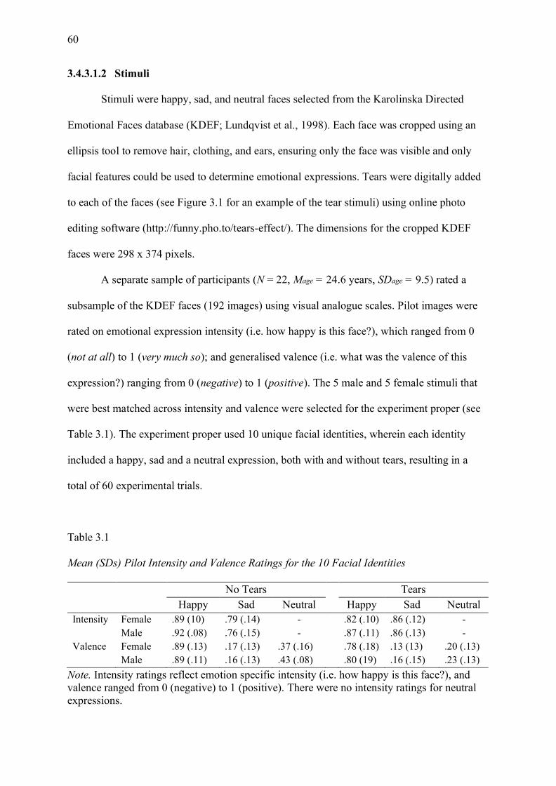
60
3.4.3.1.2 Stimuli
Stimuli were happy, sad, and neutral faces selected from the Karolinska Directed
Emotional Faces database (KDEF; Lundqvist et al., 1998). Each face was cropped using an
ellipsis tool to remove hair, clothing, and ears, ensuring only the face was visible and only
facial features could be used to determine emotional expressions. Tears were digitally added
to each of the faces (see Figure 3.1 for an example of the tear stimuli) using online photo
editing software (http://funny.pho.to/tears-effect/). The dimensions for the cropped KDEF
faces were 298 x 374 pixels.
A separate sample of participants (N = 22, Mage = 24.6 years, SDage = 9.5) rated a
subsample of the KDEF faces (192 images) using visual analogue scales. Pilot images were
rated on emotional expression intensity (i.e. how happy is this face?), which ranged from 0
(not at all) to 1 (very much so); and generalised valence (i.e. what was the valence of this
expression?) ranging from 0 (negative) to 1 (positive). The 5 male and 5 female stimuli that
were best matched across intensity and valence were selected for the experiment proper (see
Table 3.1). The experiment proper used 10 unique facial identities, wherein each identity
included a happy, sad and a neutral expression, both with and without tears, resulting in a
total of 60 experimental trials.
Table 3.1
Mean (SDs) Pilot Intensity and Valence Ratings for the 10 Facial Identities
No Tears Tears Happy Sad Neutral Happy Sad Neutral Intensity Female .89 (10) .79 (.14) - .82 (.10) .86 (.12) - Male .92 (.08) .76 (.15) - .87 (.11) .86 (.13) - Valence Female .89 (.13) .17 (.13) .37 (.16) .78 (.18) .13 (13) .20 (.13) Male .89 (.11) .16 (.13) .43 (.08) .80 (19) .16 (.15) .23 (.13)
Note. Intensity ratings reflect emotion specific intensity (i.e. how happy is this face?), and valence ranged from 0 (negative) to 1 (positive). There were no intensity ratings for neutral expressions.
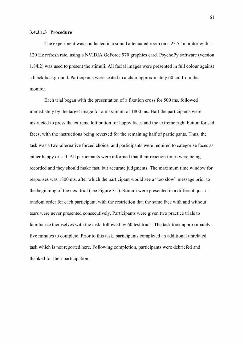
61
3.4.3.1.3 Procedure
The experiment was conducted in a sound attenuated room on a 23.5” monitor with a
120 Hz refresh rate, using a NVIDIA GeForce 970 graphics card. PsychoPy software (version
1.84.2) was used to present the stimuli. All facial images were presented in full colour against
a black background. Participants were seated in a chair approximately 60 cm from the
monitor.
Each trial began with the presentation of a fixation cross for 500 ms, followed
immediately by the target image for a maximum of 1800 ms. Half the participants were
instructed to press the extreme left button for happy faces and the extreme right button for sad
faces, with the instructions being reversed for the remaining half of participants. Thus, the
task was a two-alternative forced choice, and participants were required to categorise faces as
either happy or sad. All participants were informed that their reaction times were being
recorded and they should make fast, but accurate judgments. The maximum time window for
responses was 1800 ms, after which the participant would see a “too slow” message prior to
the beginning of the next trial (see Figure 3.1). Stimuli were presented in a different quasi-
random order for each participant, with the restriction that the same face with and without
tears were never presented consecutively. Participants were given two practice trials to
familiarize themselves with the task, followed by 60 test trials. The task took approximately
five minutes to complete. Prior to this task, participants completed an additional unrelated
task which is not reported here. Following completion, participants were debriefed and
thanked for their participation.
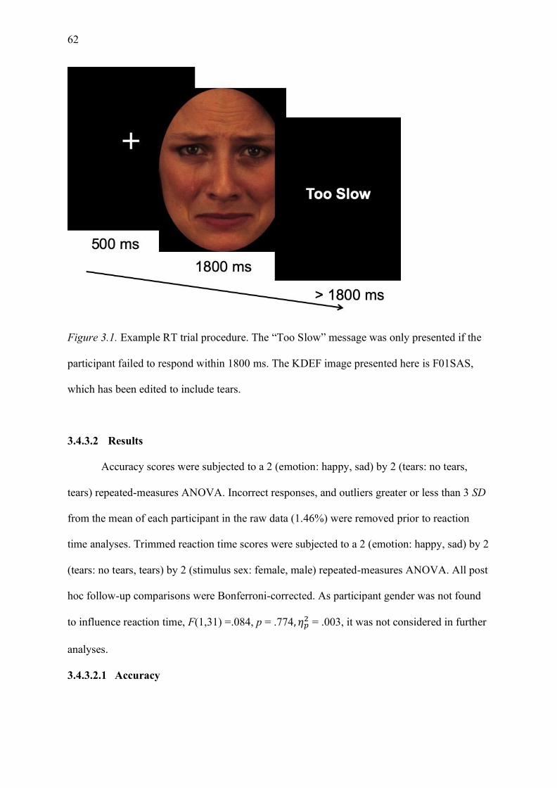
62
Figure 3.1. Example RT trial procedure. The “Too Slow” message was only presented if the
participant failed to respond within 1800 ms. The KDEF image presented here is F01SAS,
which has been edited to include tears.
3.4.3.2 Results
Accuracy scores were subjected to a 2 (emotion: happy, sad) by 2 (tears: no tears,
tears) repeated-measures ANOVA. Incorrect responses, and outliers greater or less than 3 SD
from the mean of each participant in the raw data (1.46%) were removed prior to reaction
time analyses. Trimmed reaction time scores were subjected to a 2 (emotion: happy, sad) by 2
(tears: no tears, tears) by 2 (stimulus sex: female, male) repeated-measures ANOVA. All post
hoc follow-up comparisons were Bonferroni-corrected. As participant gender was not found
to influence reaction time, F(1,31) =.084, p = .774, 𝜂𝑝2 = .003, it was not considered in further
analyses.
3.4.3.2.1 Accuracy

63
Emotions were classified with high accuracy (see Table 3.2). Trials with incorrect responses
(4.24%) and missing responses (0.38%) were pooled as errors for the accuracy analysis.
Table 3.2
Mean (SD’s) Classification Accuracy and Reaction Time across Emotion Categories.
Stimulus
No Tears Tears Accuracy (%) Reaction Time (ms) Accuracy (%) Reaction Time (ms)
Happy 96.06 (7.48) 683.18 (189.97) 89.70 (12.37) 718.90 (215.53) Sad 97.57 (5.61) 714.64 (193.39) 98.18 (5.28) 689.74 (154.72)
Note. Accuracy scores are percentage correct out of 100.
The repeated-measures ANOVA revealed a significant main effect of emotion, F(1,
32) = 13.200, p < .001, 𝜂𝑝2 = .292, where sad faces were classified with significantly greater
accuracy than happy faces. There was also a significant main effect of tears, F(1, 32) = 6.475,
p = .016, 𝜂𝑝2 = .168, where tear-free faces were classified with significantly greater accuracy
than tearful faces. There was a significant interaction between tears and emotion, F(1, 32) =
9.004, p = .005, 𝜂𝑝2 = .220 (see Figure 3.2a). For happy faces, accuracy was significantly
lower when tears were present on a happy face, t(32)= 3.285, p = .002, d = .572. There was
no difference in classification accuracy between sad and sad-tear faces t(32)= -0.494, p =
.625, d = -.086.
64
Figure 3.2. Mean scores for each emotion condition for the a) accuracy and b) response time
data. Error bars are SEM.
3.4.3.2.2 Reaction time
The repeated-measures ANOVA showed no significant main effects of tears, F(1, 32)
=.103, p = .750, 𝜂𝑝2=.003; emotion, F(1, 32) = .020, p = .888, 𝜂𝑝
2=.001; or stimulus sex, F(1,
32) = 3.491, p = .071, 𝜂𝑝2=.098. However, there was a significant interaction between tears
and emotion, F(1, 32) = 8.497, p = .006, 𝜂𝑝2=.210 (see Figure 3.2b). Happy faces were
responded to faster than happy faces with tears, t(32)= -2.287, p = .029, d = -.398; however,
this result was not significant after Bonferroni correction. There was no difference in reaction
time between the sad and sad-tear expressions, t(32)= 1.527, p = .137, d = .266. No other
interactions were significant, F’s < .854, p’s > .362.
3.4.3.2.3 Neutral face analyses
Finally, the neutral faces were analysed separately, to determine whether neutral faces
with and without tears were more perceptually similar to happy or sad faces. Overall, neutral
and neutral-tear expressions were predominantly classified as sad (M = 90.45%); thus, we
treated “sad” as the correct response in the accuracy analysis. Participants were more likely to
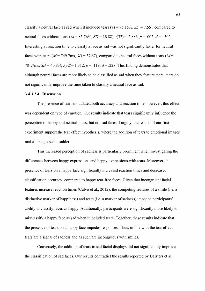
65
classify a neutral face as sad when it included tears (M = 95.15%, SD = 7.55), compared to
neutral faces without tears (M = 85.76%, SD = 18.88), t(32)= -2.886, p = .002, d = -.502.
Interestingly, reaction time to classify a face as sad was not significantly faster for neutral
faces with tears (M = 749.7ms, SD = 37.67), compared to neutral faces without tears (M =
781.7ms, SD = 40.83), t(32)= 1.312, p = .119, d = .228. This finding demonstrates that
although neutral faces are more likely to be classified as sad when they feature tears, tears do
not significantly improve the time taken to classify a neutral face as sad.
3.4.3.2.4 Discussion
The presence of tears modulated both accuracy and reaction time; however, this effect
was dependent on type of emotion. Our results indicate that tears significantly influence the
perception of happy and neutral faces, but not sad faces. Largely, the results of our first
experiment support the tear effect hypothesis, where the addition of tears to emotional images
makes images seem sadder.
This increased perception of sadness is particularly prominent when investigating the
differences between happy expressions and happy expressions with tears. Moreover, the
presence of tears on a happy face significantly increased reaction times and decreased
classification accuracy, compared to happy tear-free faces. Given that incongruent facial
features increase reaction times (Calvo et al., 2012), the competing features of a smile (i.e. a
distinctive marker of happiness) and tears (i.e. a marker of sadness) impeded participants’
ability to classify faces as happy. Additionally, participants were significantly more likely to
misclassify a happy face as sad when it included tears. Together, these results indicate that
the presence of tears on a happy face impedes responses. Thus, in line with the tear effect,
tears are a signal of sadness and as such are incongruous with smiles.
Conversely, the addition of tears to sad facial displays did not significantly improve
the classification of sad faces. Our results contradict the results reported by Balsters et al.
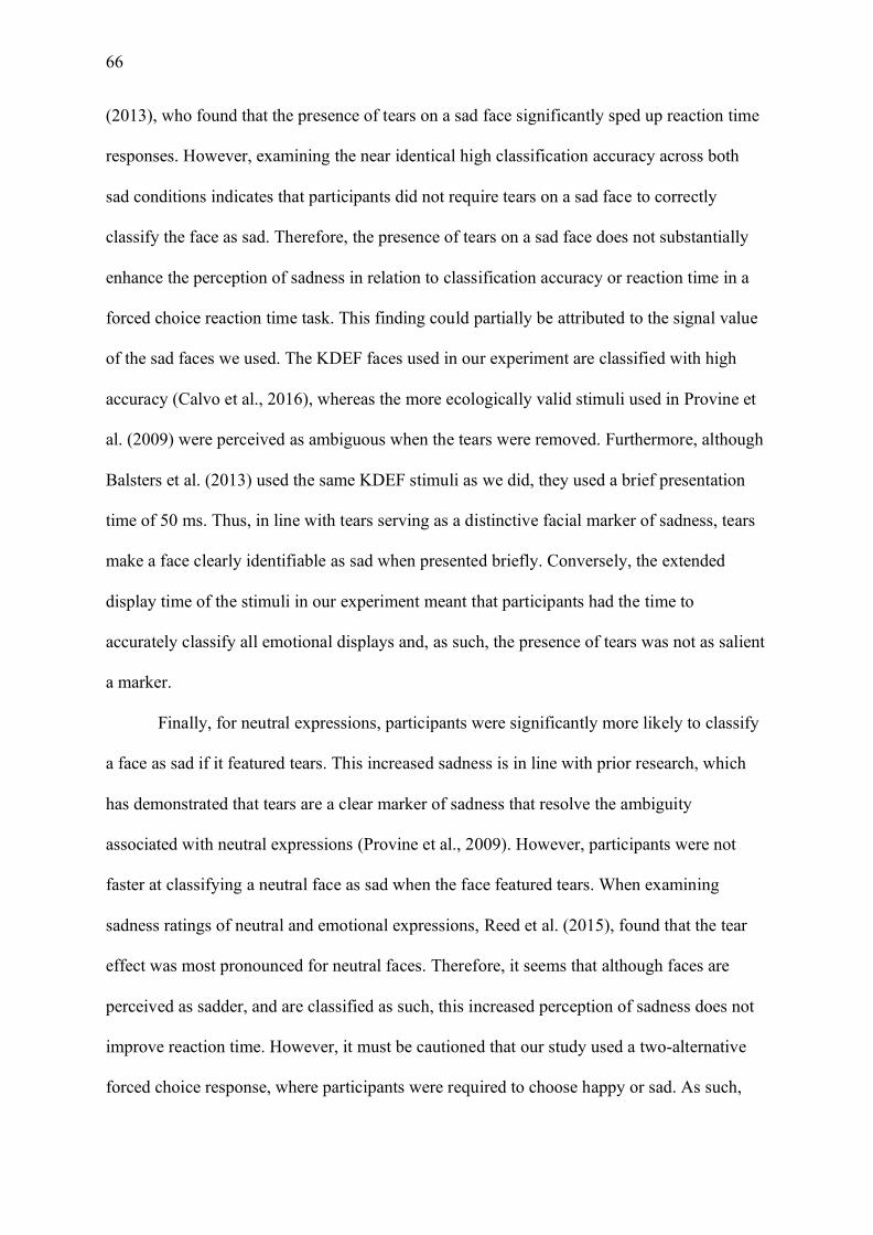
66
(2013), who found that the presence of tears on a sad face significantly sped up reaction time
responses. However, examining the near identical high classification accuracy across both
sad conditions indicates that participants did not require tears on a sad face to correctly
classify the face as sad. Therefore, the presence of tears on a sad face does not substantially
enhance the perception of sadness in relation to classification accuracy or reaction time in a
forced choice reaction time task. This finding could partially be attributed to the signal value
of the sad faces we used. The KDEF faces used in our experiment are classified with high
accuracy (Calvo et al., 2016), whereas the more ecologically valid stimuli used in Provine et
al. (2009) were perceived as ambiguous when the tears were removed. Furthermore, although
Balsters et al. (2013) used the same KDEF stimuli as we did, they used a brief presentation
time of 50 ms. Thus, in line with tears serving as a distinctive facial marker of sadness, tears
make a face clearly identifiable as sad when presented briefly. Conversely, the extended
display time of the stimuli in our experiment meant that participants had the time to
accurately classify all emotional displays and, as such, the presence of tears was not as salient
a marker.
Finally, for neutral expressions, participants were significantly more likely to classify
a face as sad if it featured tears. This increased sadness is in line with prior research, which
has demonstrated that tears are a clear marker of sadness that resolve the ambiguity
associated with neutral expressions (Provine et al., 2009). However, participants were not
faster at classifying a neutral face as sad when the face featured tears. When examining
sadness ratings of neutral and emotional expressions, Reed et al. (2015), found that the tear
effect was most pronounced for neutral faces. Therefore, it seems that although faces are
perceived as sadder, and are classified as such, this increased perception of sadness does not
improve reaction time. However, it must be cautioned that our study used a two-alternative
forced choice response, where participants were required to choose happy or sad. As such,
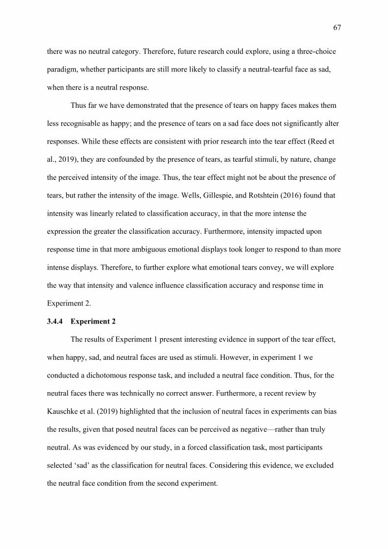
67
there was no neutral category. Therefore, future research could explore, using a three-choice
paradigm, whether participants are still more likely to classify a neutral-tearful face as sad,
when there is a neutral response.
Thus far we have demonstrated that the presence of tears on happy faces makes them
less recognisable as happy; and the presence of tears on a sad face does not significantly alter
responses. While these effects are consistent with prior research into the tear effect (Reed et
al., 2019), they are confounded by the presence of tears, as tearful stimuli, by nature, change
the perceived intensity of the image. Thus, the tear effect might not be about the presence of
tears, but rather the intensity of the image. Wells, Gillespie, and Rotshtein (2016) found that
intensity was linearly related to classification accuracy, in that the more intense the
expression the greater the classification accuracy. Furthermore, intensity impacted upon
response time in that more ambiguous emotional displays took longer to respond to than more
intense displays. Therefore, to further explore what emotional tears convey, we will explore
the way that intensity and valence influence classification accuracy and response time in
Experiment 2.
3.4.4 Experiment 2
The results of Experiment 1 present interesting evidence in support of the tear effect,
when happy, sad, and neutral faces are used as stimuli. However, in experiment 1 we
conducted a dichotomous response task, and included a neutral face condition. Thus, for the
neutral faces there was technically no correct answer. Furthermore, a recent review by
Kauschke et al. (2019) highlighted that the inclusion of neutral faces in experiments can bias
the results, given that posed neutral faces can be perceived as negative—rather than truly
neutral. As was evidenced by our study, in a forced classification task, most participants
selected ‘sad’ as the classification for neutral faces. Considering this evidence, we excluded
the neutral face condition from the second experiment.
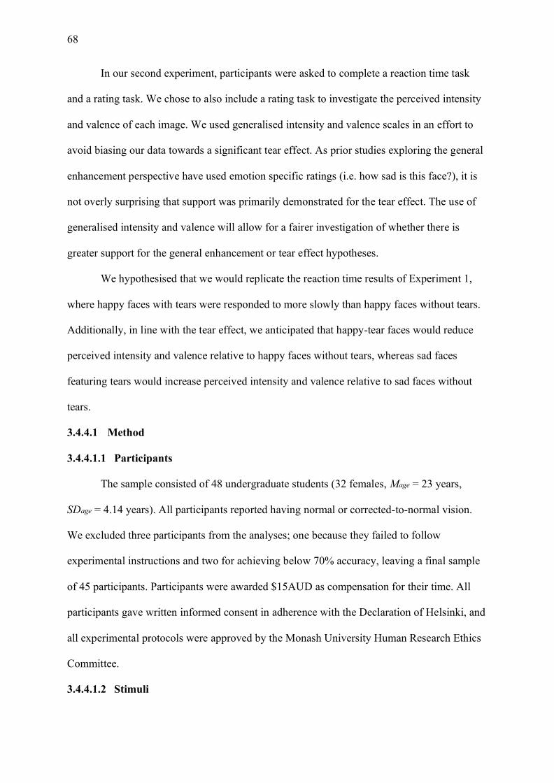
68
In our second experiment, participants were asked to complete a reaction time task
and a rating task. We chose to also include a rating task to investigate the perceived intensity
and valence of each image. We used generalised intensity and valence scales in an effort to
avoid biasing our data towards a significant tear effect. As prior studies exploring the general
enhancement perspective have used emotion specific ratings (i.e. how sad is this face?), it is
not overly surprising that support was primarily demonstrated for the tear effect. The use of
generalised intensity and valence will allow for a fairer investigation of whether there is
greater support for the general enhancement or tear effect hypotheses.
We hypothesised that we would replicate the reaction time results of Experiment 1,
where happy faces with tears were responded to more slowly than happy faces without tears.
Additionally, in line with the tear effect, we anticipated that happy-tear faces would reduce
perceived intensity and valence relative to happy faces without tears, whereas sad faces
featuring tears would increase perceived intensity and valence relative to sad faces without
tears.
3.4.4.1 Method
3.4.4.1.1 Participants
The sample consisted of 48 undergraduate students (32 females, Mage = 23 years,
SDage = 4.14 years). All participants reported having normal or corrected-to-normal vision.
We excluded three participants from the analyses; one because they failed to follow
experimental instructions and two for achieving below 70% accuracy, leaving a final sample
of 45 participants. Participants were awarded $15AUD as compensation for their time. All
participants gave written informed consent in adherence with the Declaration of Helsinki, and
all experimental protocols were approved by the Monash University Human Research Ethics
Committee.
3.4.4.1.2 Stimuli
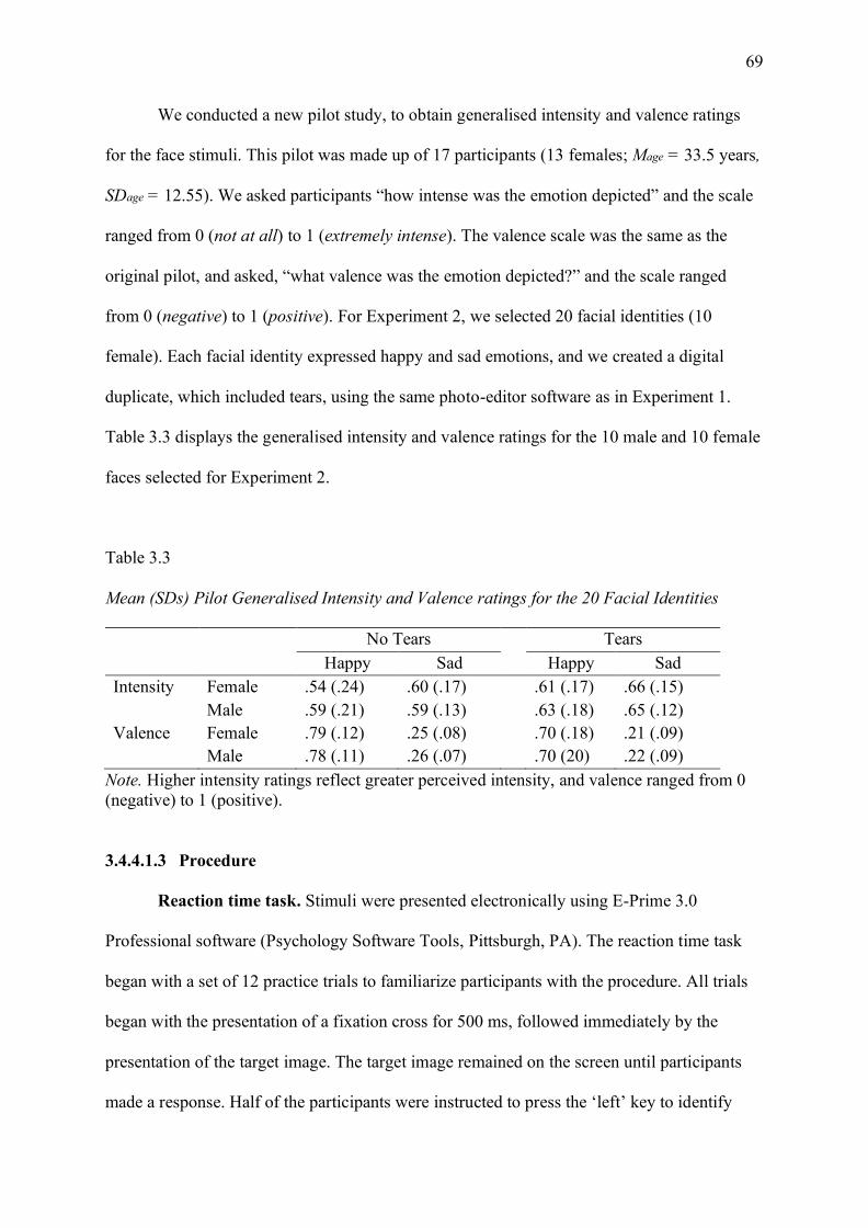
69
We conducted a new pilot study, to obtain generalised intensity and valence ratings
for the face stimuli. This pilot was made up of 17 participants (13 females; Mage = 33.5 years,
SDage = 12.55). We asked participants “how intense was the emotion depicted” and the scale
ranged from 0 (not at all) to 1 (extremely intense). The valence scale was the same as the
original pilot, and asked, “what valence was the emotion depicted?” and the scale ranged
from 0 (negative) to 1 (positive). For Experiment 2, we selected 20 facial identities (10
female). Each facial identity expressed happy and sad emotions, and we created a digital
duplicate, which included tears, using the same photo-editor software as in Experiment 1.
Table 3.3 displays the generalised intensity and valence ratings for the 10 male and 10 female
faces selected for Experiment 2.
Table 3.3
Mean (SDs) Pilot Generalised Intensity and Valence ratings for the 20 Facial Identities
No Tears Tears Happy Sad Happy Sad Intensity Female .54 (.24) .60 (.17) .61 (.17) .66 (.15) Male .59 (.21) .59 (.13) .63 (.18) .65 (.12) Valence Female .79 (.12) .25 (.08) .70 (.18) .21 (.09) Male .78 (.11) .26 (.07) .70 (20) .22 (.09)
Note. Higher intensity ratings reflect greater perceived intensity, and valence ranged from 0 (negative) to 1 (positive).
3.4.4.1.3 Procedure
Reaction time task. Stimuli were presented electronically using E-Prime 3.0
Professional software (Psychology Software Tools, Pittsburgh, PA). The reaction time task
began with a set of 12 practice trials to familiarize participants with the procedure. All trials
began with the presentation of a fixation cross for 500 ms, followed immediately by the
presentation of the target image. The target image remained on the screen until participants
made a response. Half of the participants were instructed to press the ‘left’ key to identify

70
happy expressions, and the ‘right’ key to identify sad expressions, with the buttons on the
Chronos Response Box reversed for the remaining participants. Participants were shown each
target image four times in the reaction time task. This phase of the experiment took
approximately 10 minutes to complete.
Rating task. The rating task began with a series of six practice trials. All trials began
with the presentation of a fixation cross for 500 ms, which was followed by the presentation
of a target image for 3 seconds. Participants were instructed to rate the image they had just
seen on two visual analogue scales. Participants were first asked “what was the intensity of
the emotion depicted” and the scale ranged from 0 (not at all) to 1 (extremely). Next
participants were asked “what was the valence of the emotion depicted” and the scale ranged
from 0 (negative) to 1 (positive). There was no time limit on the rating component of the task.
All images were rated twice. This phase of the experiment took approximately 20 minutes to
complete. After the completion of both tasks’ participants were debriefed and thanked for
their participation. The analyses, with the exception of the exploratory sex effects, were pre-
registered prior to data collection (https://aspredicted.org/blind.php?x=cb79zz).
3.4.4.2 Results
Data were subjected to 2 (emotion: happy, sad) by 2 (tears: no tears, tears) by 2
(stimulus sex: female, male) repeated-measures ANOVAs. When follow-up post-hoc
analyses were conducted, we applied a Bonferroni correction. Table 3.4 denotes the
descriptive statistics for the reaction time and rating tasks, collapsed across stimulus sex. As
in Experiment 1, participant gender was not found to influence reaction time, F(1,43) = .456,
p = .503, 𝜂𝑝2 = .010, or the intensity, F(1,43) = 1.132, p = .293, 𝜂𝑝
2 = .026; or valence, F(1,43)
= 2.396, p = .129, 𝜂𝑝2 = .053; rating data and, as such, it will not be considered further.

71
Table 3.4
Mean (SDs) for Each Emotion Condition for the Reaction Time and Rating Tasks in
Experiment 2.
Reaction Time Task Rating Task Accuracy (%) Reaction Time (ms) Intensity Valence
Happy 96.03 (2.79) 737.43 (224.80) .62 (.19) .77 (.10) Sad 96.97 (3.22) 750.76 (193.78) .56 (.17) .24 (.07) Happy-tear 92.08 (7.52) 818.04 (313.86) .66 (.16) .72 (.14) Sad-tear 97.56 (2.88) 758.90 (208.91) .66 (.16) .18 (.07)
Note. Intensity scores range from 0-1, higher scores = greater intensity. Valence scores range from 0-1 with scores closer to 0 reflecting negative valence, and scores closer to 1 indicating positive valence.
3.4.4.2.1 Reaction time task
Accuracy. The repeated-measures ANOVA revealed a significant main effect of
emotion, F(1,44) = 20.994, p < .001, 𝜂𝑝2 = .323, as participants were more accurate at
classifying sad expressions (M = 97.26, SE = .539), compared to happy expressions (M =
94.06, SE = .539). There was also a significant main effect of tears, F(1,44) = 10.734, p =
.002, 𝜂𝑝2 = .196, where tear-free expressions (M = 96.50, SE = .483) were classified correctly
more often than tearful expressions (M = 94.82, SE = .483). Additionally, there was a
significant tears by emotion interaction, F(1,44) = 12.228, p = .001, 𝜂𝑝2 = .217 (see Figure
3.3a). Happy faces were classified with significantly greater accuracy than happy-tear faces,
t(44)= 3.791, p < .001, d = .565. There was no significant difference in classification
accuracy for the sad and the sad-tear faces, t(32)= -1.099, p = .278, d = -.164.
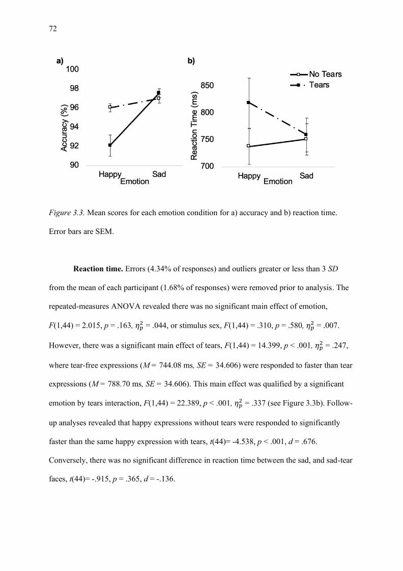
72
Figure 3.3. Mean scores for each emotion condition for a) accuracy and b) reaction time.
Error bars are SEM.
Reaction time. Errors (4.34% of responses) and outliers greater or less than 3 SD
from the mean of each participant (1.68% of responses) were removed prior to analysis. The
repeated-measures ANOVA revealed there was no significant main effect of emotion,
F(1,44) = 2.015, p = .163, 𝜂𝑝2 = .044, or stimulus sex, F(1,44) = .310, p = .580, 𝜂𝑝
2 = .007.
However, there was a significant main effect of tears, F(1,44) = 14.399, p < .001, 𝜂𝑝2 = .247,
where tear-free expressions (M = 744.08 ms, SE = 34.606) were responded to faster than tear
expressions (M = 788.70 ms, SE = 34.606). This main effect was qualified by a significant
emotion by tears interaction, F(1,44) = 22.389, p < .001, 𝜂𝑝2 = .337 (see Figure 3.3b). Follow-
up analyses revealed that happy expressions without tears were responded to significantly
faster than the same happy expression with tears, t(44)= -4.538, p < .001, d = .676.
Conversely, there was no significant difference in reaction time between the sad, and sad-tear
faces, t(44)= -.915, p = .365, d = -.136.

73
There was also a significant emotion by stimulus sex interaction, F(1,44) = 5.786, p =
.020, 𝜂𝑝2 = .116. Follow-up analyses were not significant; however, a trend towards
significance revealed that female happy faces were responded to faster than male happy
faces, t(44)= -1.928, p = .060, d = -.287; while sad male faces were responded to faster than
sad female faces, t(44)= 1.914, p = .062, d = .285.
3.4.4.2.2 Rating task
Intensity ratings. The repeated-measures ANOVA for the intensity rating data
revealed a significant main effect of tears, F(1,44) = 56.760, p < .001, 𝜂𝑝2 = .563, where
tearful expressions (M = .660, SE = .023) were perceived as significantly more intense than
tear-free expressions (M = .586, SE = .023). There was also a significant interaction between
tears and emotion, F(1,44) = 17.185, p < .001, 𝜂𝑝2 = .281 (see Figure 3.4). Specifically, sad-
tear faces were perceived as significantly more intense than sad faces, t(44)= 9.409, p < .001,
d = 1.403 Similarly, happy-tear faces were perceived as significantly more intense than happy
faces, t(44)= -3.048, p = .004, d = .454.
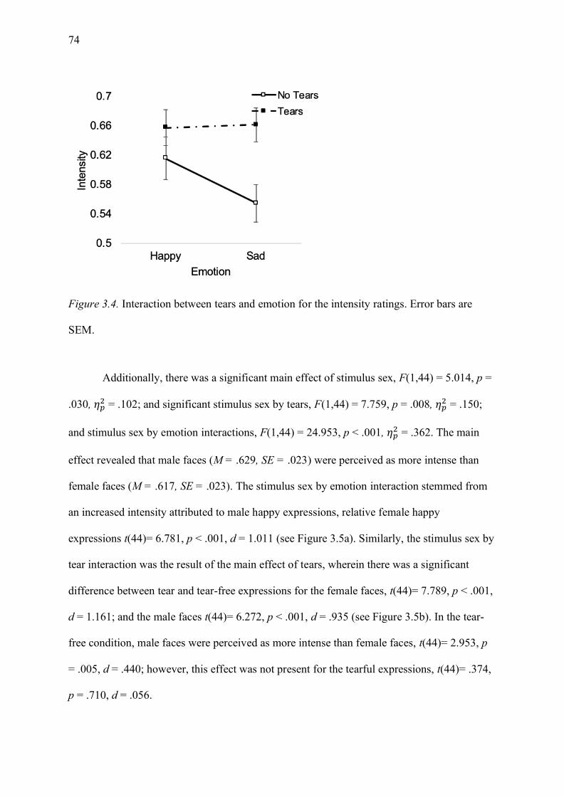
74
Figure 3.4. Interaction between tears and emotion for the intensity ratings. Error bars are
SEM.
Additionally, there was a significant main effect of stimulus sex, F(1,44) = 5.014, p =
.030, 𝜂𝑝2 = .102; and significant stimulus sex by tears, F(1,44) = 7.759, p = .008, 𝜂𝑝
2 = .150;
and stimulus sex by emotion interactions, F(1,44) = 24.953, p < .001, 𝜂𝑝2 = .362. The main
effect revealed that male faces (M = .629, SE = .023) were perceived as more intense than
female faces (M = .617, SE = .023). The stimulus sex by emotion interaction stemmed from
an increased intensity attributed to male happy expressions, relative female happy
expressions t(44)= 6.781, p < .001, d = 1.011 (see Figure 3.5a). Similarly, the stimulus sex by
tear interaction was the result of the main effect of tears, wherein there was a significant
difference between tear and tear-free expressions for the female faces, t(44)= 7.789, p < .001,
d = 1.161; and the male faces t(44)= 6.272, p < .001, d = .935 (see Figure 3.5b). In the tear-
free condition, male faces were perceived as more intense than female faces, t(44)= 2.953, p
= .005, d = .440; however, this effect was not present for the tearful expressions, t(44)= .374,
p = .710, d = .056.
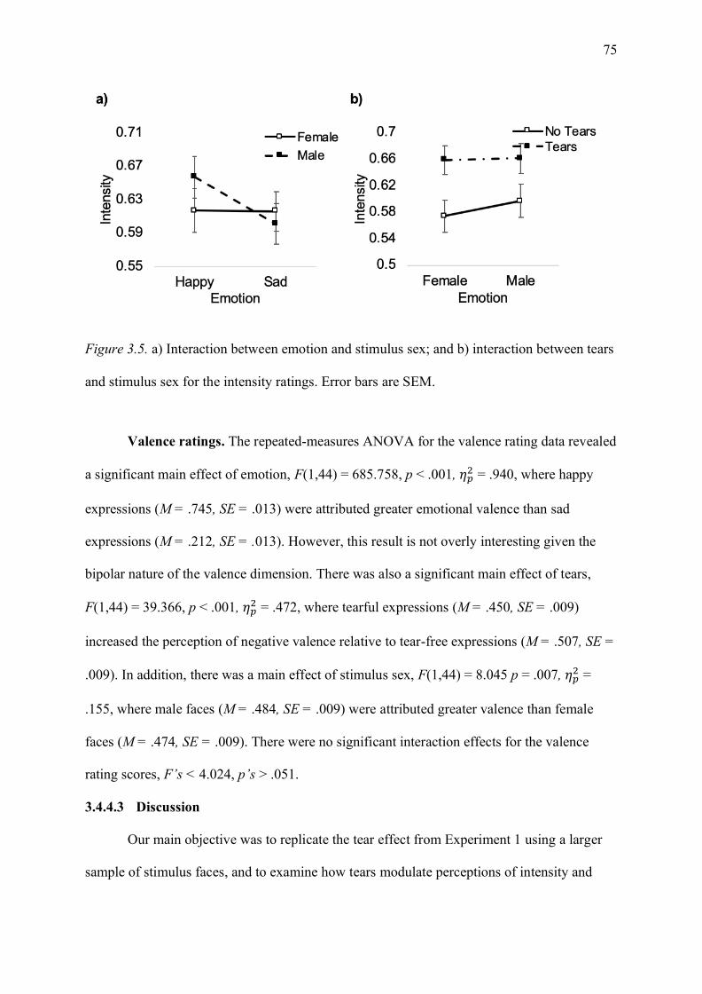
75
Figure 3.5. a) Interaction between emotion and stimulus sex; and b) interaction between tears
and stimulus sex for the intensity ratings. Error bars are SEM.
Valence ratings. The repeated-measures ANOVA for the valence rating data revealed
a significant main effect of emotion, F(1,44) = 685.758, p < .001, 𝜂𝑝2 = .940, where happy
expressions (M = .745, SE = .013) were attributed greater emotional valence than sad
expressions (M = .212, SE = .013). However, this result is not overly interesting given the
bipolar nature of the valence dimension. There was also a significant main effect of tears,
F(1,44) = 39.366, p < .001, 𝜂𝑝2 = .472, where tearful expressions (M = .450, SE = .009)
increased the perception of negative valence relative to tear-free expressions (M = .507, SE =
.009). In addition, there was a main effect of stimulus sex, F(1,44) = 8.045 p = .007, 𝜂𝑝2 =
.155, where male faces (M = .484, SE = .009) were attributed greater valence than female
faces (M = .474, SE = .009). There were no significant interaction effects for the valence
rating scores, F’s < 4.024, p’s > .051.
3.4.4.3 Discussion
Our main objective was to replicate the tear effect from Experiment 1 using a larger
sample of stimulus faces, and to examine how tears modulate perceptions of intensity and

76
valence on happy and sad faces. Our results demonstrate that tears increase the time taken to
classify a face as happy and reduce classification accuracy, whereas reaction time responses
to sad faces are largely not influenced by the presence of tears. Furthermore, these results
support the idea that tears serve as a marker of sadness (Provine et al., 2009), as the presence
of incongruent facial features, such as tears and a smile, increase reaction times to happy-tear
faces. Conversely, our rating data demonstrate support for both the tear effect, and the
general enhancement hypotheses.
Interestingly, while classification accuracy and reaction time to sad faces are not
influenced by the presence of tears, intensity and valence ratings are. In addition to tears
serving as a marker of sadness, tears increase the perceived intensity of happy and sad
expressions. Furthermore, the presence of tears on facial displays decreased valence ratings
(i.e. made ratings more negative). Thus, while tears make all faces seem more intense, their
presence on a face makes a positive expression less positive, and a negative expression more
negative.
Finally, the exploratory sex of stimulus analyses revealed that sex of stimulus largely
did not change the way that participants responded to emotional tears. Moreover, the only
significant stimulus sex by tear interaction was driven by the main effect of tears, wherein
tearful expressions were perceived as more intense than tear-free expressions for male and
female faces. Therefore, when examining reaction time, generalised intensity, and valence
ratings, stimulus sex largely does not change the way that participants respond to tearful
facial displays.
3.4.5 General Discussion
These studies demonstrate the most support for the tear effect, wherein tears—in the
absence of context—symbolise sadness. In Experiment 1, tearful expressions impaired
recognition of happy faces, and enhanced recognition of neutral faces, but made no difference
77
to sad expressions. In Experiment 2, the presence of tears on a happy face once again
impaired recognition, and there were no differences in recognition between sad and sad-tear
expressions. Similarly, in ratings of valence, the addition of tears to a happy face made happy
faces seem less positive, and negative faces seem more negative. However, when examining
the intensity rating data, we demonstrated support for the general enhancement perspective,
wherein all tearful expressions were perceived as more intense than their tear-free
counterparts.
In Experiment 1, we demonstrated that the presence of tears resolved the emotional
ambiguity of neutral faces, where neutral faces were more likely to be classified as sad when
they featured tears. This is in accord with research by Küster (2018) who demonstrated that
tears make a neutral expression more readily recognisable as sad. However, we further
identified that tears did not significantly improve reaction time to neutral expressions. This
contradicts prior studies who have demonstrated that the tear effect is most pronounced for
neutral displays (Provine et al., 2009; Reed et al., 2019). However, in those studies,
participants were instructed to rate the sadness of the tearful displays. Thus, tears may
increase the perception of sadness, however this perception does not improve the time taken
to classify a neutral face as sad.
Furthermore, tearful sad expressions were not responded to significantly faster or
more accurately than sad faces without tears. This finding directly contrasts Balsters et al.
(2013), who reported that sad-tear faces were responded to significantly faster than sad faces
without tears. However, Balsters et al. (2013) presented their stimuli briefly (i.e., 50 ms).
Thus, it seems that the tear effect is more pronounced for sad faces when presentation
durations are shorter. In our experiments, the sad facial expressions both with and without
tears were responded to rapidly and accurately, indicating that participants did not require the
presence of tears to identify a face as sad. This could indicate the occurrence of a ceiling

78
effect, where the sad faces we used were already clearly recognisable as sad (Calvo et al.,
2016), thus we were unable to see an improvement in the classification of the sad-tear faces.
The potential for a ceiling effect for sad and sad-tearful displays is in accord with existing
research investigating blended facial displays. Calvo, Gutiérrez-García, and Del Líbano
(2018) demonstrated that distinctiveness is an important factor when distinguishing truly
happy smiles from blended displays. Moreover, the more distinctive the eye region (i.e.,
angry eyes with smiling mouth), the less likely the display would be judged as happy.
However, for truly happy faces (i.e. happy eyes and smiling mouth) the distinctiveness of the
mouth region meant that the eye region offered no further improvement to recognition
accuracy. Although we used a different paradigm than Calvo et al. (2018), given the high
classification accuracy for sad and sad-tear images in our experiments, it seems that the
presence of tears offered no further improvement for classification accuracy in a dichotomous
response task. Thus, it is possible that the tear effect would be more pronounced for sad faces
when using a multiple response category paradigm.
Previous research into the tear effect and the general enhancement perspective has
favoured comparison of tearful facial expressions with other negative emotions (Ito et al.,
2019), while the influence of tears on happy displays has been largely neglected (Reed et al.,
2015). In the present study, we sought to extend the understanding of how tearful happy
expressions are perceived. Our results support prior work indicating that tears on happy faces
enhance the perception of sadness (Reed et al., 2015). These findings are consistent with the
idea that incongruous facial features result in slower reaction times for happy facial displays
(Calvo et al., 2012). This result further indicates that tears are a distinctive marker of sadness,
and that recognising “tears of joy” likely stems from contextual information. This
interpretation is consistent with the dimorphous expression literature, in that when tearful
facial expressions are paired with positive scenarios they are perceived as positive emotions
79
(Aragón, 2017; Aragón & Bargh, 2018; Aragón & Clark, 2018). Thus, tears when paired with
a happy expression in the absence of context impair one’s ability to classify a face as happy.
The rating data further supports the conclusions drawn from the reaction time results.
Surprisingly, we demonstrated support for both the tear effect and the general enhancement
perspective. Moreover, the presence of tears on both happy and sad faces resulted in greater
perceived intensity. This result supports the general enhancement perspective posed by Ito et
al. (2019), who demonstrated that tears made negative expressions more intensely sad, and
Reed et al. (2015) who demonstrated that Duchenne smiles were perceived as more intense
when they featured tears. Wells et al. (2016) showed that less intense (and as such more
ambiguous) facial displays resulted in longer reaction times. However, in our studies,
responses were slowest for happy-tear expressions despite these faces being intense. This
contradictory finding is clearer when considering the valence rating data. The presence of
tears on sad expressions increased perceived negative valence and decreased perceived
positive valence for happy expressions. Thus, tears make expressions seem more negative.
This reduced positive valence could arguably make a typically distinctive happy face more
ambiguous. The increased ambiguity associated with happy-tear faces is a contributor to the
slower reaction time. As such, we provide further support for Wells et al. (2016); however,
conclude that valence is a larger contributor to reaction time than intensity, at least when
considering tearful displays.
Finally, we conducted exploratory analyses examining whether sex of the stimulus
influenced the way that persons respond to tears. Across the two experiments, we largely
demonstrated that sex of stimulus did not change participants responses to tearful displays.
This null result contradicts other studies which have demonstrated that females receive more
favourable responses than males (Cretser et al., 1982; Fischer et al., 2013; Stadel et al., 2019).
However, the discrepancy in findings could be attributed to two factors. Firstly, Cretser et al.
80
(1982) and Stadel et al. (2019) demonstrated their effects when exploring the relationship
between sex of stimulus and gender of participant. Moreover, male participants were more
likely to hold a ‘double standard’ where male-male dyads are responded to less favourably.
We did not observe participant gender effects in our study, which could have contributed to
our differential results. Secondly, in the prior research crier perception was typically explored
via ratings of ‘need for support’, emotionality, competence, and appropriateness. Thus, it may
be that these factors are more influenced by the sex of the stimulus than generalised intensity
and valence ratings and reaction time measures are. These exploratory sex effects were not
the primary interest of the paper, and we acknowledge that studying sex differences is a
difficult undertaking. Further research could repeat our experiments over a series of studies
with a balanced sample of male and female participants to elucidate whether the effects
reported in prior literature extend to generalised rating and reaction time measures.
A few limitations should be considered when interpreting our results. Firstly, in our
study we were expressly interested in exploring whether tears on happy and sad facial
expressions demonstrated support for the tear effect, or the general enhancement perspective.
This approach resulted in an overarching focus on the perception of sadness—despite our
efforts to use unbiased response metrics. However, tears do not only signal sadness to
observers, as other studies have identified that tears signal powerlessness, appeasement, and
the need for social support (Hendriks et al., 2008; Miceli & Castelfranchi, 2003). Therefore,
further studies could extend the studies reported herein to explore whether the perception of
tearful displays mediates the responses to crying persons. Furthermore, we used posed
standardised facial displays and digitally added tears. This approach allowed for rigorous
control over the experimental stimuli and the subsequent ability to draw conclusions about
the influence of tears. However, tears are touted as an honest expression of emotion and
associated with sincerity (Picó et al., 2020; Vingerhoets, 2013; Zeifman & Brown, 2011).

81
Thus, it is yet to be determined whether the use of posed artificial tears influences participant
responses (Krivan & Thomas, 2020). Further research could use authentic tearful stimuli,
with increased ecological validity, to further explore responses to crying faces.
Our experiments showed that, in the absence of context, tears are a reliable marker of
sadness. Specifically, this marker of sadness is most pronounced for facial displays that are
not already being interpreted as a sad expression (i.e. happy and neutral displays). The
pairing of a distinctive marker of sadness (i.e. tears), with a distinctive marker of joy (i.e.
smiles) results in reduced classification accuracy and slower reaction time to happy
expressions. Furthermore, while the addition of tears makes both positive and negative
emotions more negatively valenced, tears make both facial expressions seem more intense.
Thus, tearful displays are associated with being a marker of negative emotion while eliciting
greater intensity ratings from observers. Overall, our results demonstrate support for both the
general enhancement perspective and the tear effect whereby tears intensify expressions and
in the absence of context, signal sadness.
82
Chapter 4: Unconscious Mimicry of Tearful Expressions
4.1 Chapter Overview
Chapter 3 demonstrated that tears serve as a signal of sadness in the absence of context.
These experiments were a necessary step to determine how tears influenced happy and sad
tearful displays in the absence of context. In the present chapter, I will expand this
behavioural research into the psychophysiological domain, to explore whether the presence of
tears on facial displays modulates mimicry responses. As facial mimicry is related to empathy
and the sharing of affective states, I believed that tears should increase the amount of
mimicry exhibited in response to a face. In Chapter 3, it was concluded that stimulus sex did
not change responses that participants had to tearful displays. In light of this evidence, I have
not explored any stimulus-related sex effects in the following data chapters. I believe this
approach is well justified given the scarcity of research exploring physiological responses to
tears; and the problems with multiple comparisons in psychophysiological research (Luck,
2014). However, I acknowledge, that the potential for sex differences cannot be ruled out
completely, and as such where appropriate I have provided considered recommendations for
further research.
4.2 Publication Status
Manuscript in preparation for submission.
4.3 Author Contributions
Krivan – designed the experiment, created the experimental paradigm, collected the data,
processed the psychophysiological data, conducted the statistical analyses, and drafted the
manuscript.
Cottrell – aided in designing the experiment and provided supervision.
Thomas – aided in revision of drafts and provided supervision.
83
4.4 Manuscript
4.4.1 Abstract
Facial mimicry, or the unconscious matching of another’s expression, is said to
facilitate social communication and increase liking and affiliation. A wealth of research has
demonstrated that happy expressions, and to a lesser extent sad expressions, are mimicked
with a congruent facial display. However, tearful displays have not received this same
attention. Emotional tears elicit increased support and empathy from observers in self report
studies. Given that mimicry falls under the umbrella of empathy, investigating mimicry
responses to tearful displays provides the ability to explore empathic responses to tears that
occur outside conscious awareness. It was predicted that tearful displays would elicit
increased mimicry relative to tear-free displays. Two experimental tasks were conducted, one
where tearful and tear-free stimuli were presented subliminally using forwards and
backwards masking, and the second where the stimuli were presented to conscious awareness
for an extended period. The study failed to demonstrate support for the hypothesis that
mimicry is increased in response to tearful displays. Rather, a trend towards decreased
mimicry in the masked trials was demonstrated, which was not present during the
supraliminal trials. Therefore, persons, for the most part, do not mimic tearful displays. We
conclude with a discussion about why it may not be adaptive to mimic tearful displays.
Keywords: facial mimicry, emotional tears, adult crying, EMG
4.4.2 Introduction
When viewing another’s facial expressions of emotion, people unconsciously match,
or imitate the sender’s expression. Known as facial mimicry, this matching is typically an
automatic, rapid facial reaction, which is difficult to suppress (Dimberg et al., 2000; Dimberg
et al., 2002). A specific type of facial mimicry, termed emotional mimicry, occurs when
someone mimics the emotional expression of another as a means of sharing the emotion with
84
the other person (Hess & Fischer, 2014). As emotions convey our feelings and intentions
(Hess et al., 1999), reciprocating these emotional signals aids in communicating to the sender
that their emotions have been understood. As such, mimicry has been said to foster affiliative
behaviours (Hess & Fischer, 2013) and increase liking amongst conversational partners (Hess
et al., 1999; Van Der Schalk et al., 2011).
There are two competing theories to explain how mimicry occurs. The first suggests
mimicry is an automatic and reflexive pure motor copy, and the second argues for an
affiliative account wherein mimicry is influenced by situational context. The idea of
automatic mimicry as a pure motor copy stems from a perception-behaviour model
(Chartrand & Bargh, 1999). Dubbed the ‘chameleon effect’ or the ‘matched motor
hypothesis’, simply witnessing another’s expression is enough to result in the automatic,
passive process whereby the same expression is performed by oneself (Chartrand & Bargh,
1999; Hess & Fischer, 2014). Thus, motor-matching follows the same concept of mirror
neurons, where the same neurons are activated during the observation and execution of an
action, and the perception-behaviour link occurs as a learned process without awareness or
intention (Hess & Fischer, 2014; Iacoboni, 2005). To this end, facial mimicry through the
lens of motor-matching is a spontaneous, direct imitation of the emotional display (Hess &
Fischer, 2014), regardless of the type of emotion, or the familiarity of the sender (Chartrand
et al., 2005).
By contrast, the affective account argues the exact opposite—that mimicry is more
than pure motor copy. Moreover, whether mimicry occurs, and the magnitude of the mimicry
response is dependent on a variety of factors pertaining to the appropriateness of the response
in social scenarios (Kirkham, Hayes, Pawling, & Tipper, 2015). Mimicry is influenced by the
relationship between the two individuals (Häfner & IJzerman, 2011), and enhanced when the
exchange is positive or communal (Likowski, Mühlberger, Seibt, Pauli, & Weyers, 2008).
85
Furthermore, mimicry is dependent on the type of emotion displayed (Bourgeois & Hess,
2008), and the context in which it is encountered (Seibt et al., 2013). Finally, mimicry is also
influenced by individual differences, such as empathy (Sonnby-Borgstrom, 2002) and the
mood state of the person reciprocating the display (Moody, McIntosh, Mann, & Weisser,
2007). Although the two accounts are inconsistent in relation to the process by which
mimicry occurs, there is consensus that mimicry fosters affiliation and social cohesion (Hess
& Fischer, 2014; Hess et al., 1999), serving as a kind of ‘social glue’ (Lakin et al., 2003).
Where mimicry is facilitated by affiliation, mimicry increases empathy amongst
conversational partners and is a means of sharing another’s perspective (Hess & Fischer,
2013; Seibt et al., 2013). Typically, mimicry has been investigated by examining congruent
facial reactions to happy and sad displays (Hess & Fischer, 2013). For example, a congruent
response to a happy face would be activation of the Zygomaticus Major (ZMaj)—the muscle
responsible for drawing the muscle upwards into a smile (Duchenne 1862/1990). By contrast,
a congruent response to a sad expression is characterized by activation of the Corrugator
Supercilii (CS)—the muscle responsible for drawing the brows together (Dimberg, 1982). As
such, exhibiting a congruent response to a conversational partner is a means of
communicating empathy and understanding (Häfner & IJzerman, 2011). A series of studies
have demonstrated that happy and sad stimuli presented in an affiliative (or even neutral)
context are mimicked more than the same stimuli in a negative context (Likowski et al.,
2008; Likowski, Mühlberger, Seibt, Pauli, & Weyers, 2011). Similarly, mimicry is congruent
and facilitated amongst communal exchange partners (Häfner & IJzerman, 2011) and in-
group members (Bourgeois & Hess, 2008). Evidently, mimicking affiliative emotions has an
increased evolutionary value in increasing social coordination (Lakin et al., 2003). However,
not all emotions are affiliative (Bourgeois & Hess, 2008; Seibt et al., 2013), and as such the
way that we respond to these displays of emotion is reactive (Hess & Fischer, 2013).
86
A reactive emotional response can be congruent, such as frowning at a negative
stimulus (Hess & Fischer, 2014) or incongruent, such as smiling at the disappointment of
another (Seibt et al., 2013). The former is a simple reactive response, where one frowns at a
picture of a negative stimulus (e.g., angry face, a snake; Seibt et al., 2013). The latter, known
as counter-mimicry, is typically elicited in competitive relationships, whereby smiling in
response to a competitor’s disappointment can be a marker of schadenfreude—pleasure
derived from another’s misfortune (Likowski et al., 2008). Further evidence for reactive
responses is demonstrated when mimicry does not occur. Due to the signal value, some
emotions are not mimicked when the context is known (Bourgeois & Hess, 2008; Hinsz &
Tomhave, 1991). For example, anger displays are not affiliative (Knutson, 1996), and if
mimicked, they are likely to result in escalated conflict. As such, non-affiliative displays are
not mimicked or they elicit incongruent muscle responses (Likowski et al., 2011).
The research exploring mimicry responses to sad facial displays has yielded
inconsistent results. Some studies have demonstrated that there is increased CS activity in
response to a sad faces—in line with a reactive response (Harrison, Morgan, & Critchley,
2010; Hess & Blairy, 2001; Lundqvist & Dimberg, 1995). However, other studies have
emphasised that sadness displays are more likely to be mimicked if expressed by a close
partner, or in-group members—in accord with the affiliative account (Bourgeois & Hess,
2008; Häfner & IJzerman, 2011). This mediation of mimicry in response to sad displays
likely stems from the signal value of sad faces and the social cost associated with comforting
a sad person (Bavelas et al., 1986; Bourgeois & Hess, 2008). Evidence for this conclusion
stems from research wherein mimicry is reduced when the social cost to the observer is high
(Johnston, 2002). Under this perspective, mimicry of strangers would not be adaptive given
the increased social cost, whereas mimicry of personally relevant individuals could be means
of strengthening social bonds.
87
In addition to the mixed conclusions demonstrated in sadness mimicry research, there
are also some methodological limitations. Moreover, most studies have used the CS to index
sadness mimicry, despite Duchenne (1862/ 1990), the forefather of facial muscle research,
identifying the Depressor Angulis Oris muscle (i.e., the muscle responsible for drawing the
lips downward) with the expression of sadness. A potential limitation associated with using
the CS to index both sadness and anger, is that anger expressions are characterised by the
brows being lowered, whereas in sadness displays the brows are lowered while the inner
brow is raised (Critchley et al., 2005; Ekman & Friesen, 1978; Ekman & Rosenberg, 2005).
As both of these actions activate the CS (Nicanor & Mohammed, 2001), it is difficult to
determine whether participants are mimicking the display or frowning at the stimulus.
Soussignan et al. (2013) addressed this limitation by indexing sadness mimicry using the
depressor and demonstrated significantly enhanced depressor activity during the viewing of
sad displays. Therefore, sadness mimicry, as indexed by a muscle uniquely associated with
sadness, occurs in the both in the absence of context and with unfamiliar individuals.
To date, most research investigating mimicry of sadness has focused on sad
expressions without tears. Tears, a highly distinctive and uniquely human phenomenon,
reliably elicit help and support from observers (Balsters et al., 2013; Hendriks, Croon, &
Vingerhoets, 2008). Furthermore, tears are thought to be affiliative in that they promote
social bonding (Vingerhoets et al., 2016), serve to reduce aggression from observers (Hasson,
2009), and foster approach behaviours relative to avoidance (Gračanin et al., 2018).
However, most of this research has been self-report, which requires honest responses. This
reliance on honesty is an important limitation to the pro-social responses elicited in tear
research; as it is socially desirable to help others, respondents might seek to conform with
perceived social norms (Mauss & Robinson, 2009), leading to demand characteristics. As
88
facial mimicry is associated with empathy, investigating mimicry responses to tearful
displays would allow for psychophysiological validation of existing self-report research.
A recent study by Grainger et al. (2019) failed to find any differences in EMG activity
between sad faces with tears and sad faces without tears. As such, the authors concluded that
their results offered support for the motor copy hypothesis, in that tearful displays are not
distinct from tear-free sadness, and as such do not elicit increased empathy as evidenced by
mimicry. However, their experiment can be extended in two ways. Firstly, although tearful
displays are associated with the expression of sadness, tears are also elicited in response to
joy and frustration (Miceli & Castelfranchi, 2003). For most negative scenarios that elicit
tears, there is a corresponding positive scenario (e.g., separation and reunion) (Vingerhoets &
Bylsma, 2015). Therefore, perhaps the type of crying expressed by an individual modulates
mimicry. Secondly, Grainger et al. (2019) used the CS to index sadness mimicry. However,
Duchenne (1862/1990) associated the Zygomaticus Minor (ZMin) with weeping. Therefore,
mimicry of tearful displays might only be indexed by the muscle associated with the
expression—the ZMin. As such, the present study will address these limitations by indexing
mimicry using the CS in accord with existing research (Bourgeois & Hess, 2008; Grainger et
al., 2019; Häfner & IJzerman, 2011) and using the ZMin as a specific index of weeping
mimicry. Secondly, the present study included tearful displays associated with joy and
frustration by adding tears to angry and happy expressions.
The present study aimed to investigate whether tears modulate mimicry responses.
Tearful expressions were presented both supraliminally (i.e., to conscious awareness) and
subliminally using forward and backward masking. In accord with tears being a salient
distress signal, it was predicted tears would be attended without conscious awareness,
wherein masked tearful displays would elicit larger mimicry responses than tear-free
displays. Additionally, as tearful expressions are typically associated with pro-social
89
responses and empathy, it was predicted that supraliminal tearful emotional expressions
would elicit significantly greater mimicry than the same images without tears—regardless of
the type of tearing display (i.e. happy-tears, sad-tears, or angry-tears).
4.4.3 Method
4.4.3.1 Participants
The total sample consisted of 35 undergraduate students (23 females, Mage = 26.1,
SDage = 10.5 years). All participants reported having normal or corrected-to-normal vision.
Participants self-reported being right-handed, and participants with facial hair were
prohibited from participation. Ethical approval was obtained from the James Cook University
Human Research Ethics committee, and participants gave written informed consent in accord
with the Declaration of Helsinki.
4.4.3.2 Stimuli
Stimuli were happy, angry, sad, and neutral faces from the Karolinska Directed
Emotional Faces database (KDEF; Lundqvist et al., 1998). Each face was cropped using an
ellipsis tool to remove hair, clothing, and ears to ensure the face only contained the elements
necessary for determining emotional expressions. Tears were digitally added to the images
(running from the eyes down the cheeks) to each of the faces using online photo editor
software (http://funny.pho.to/tears-effect/).
The stimuli were selected from a larger pool of images, which were pilot tested using
an independent group of 22 participants (20 females, Mage = 24.6, SDage = 9.5 years). Pilot
participants rated the images using visual analogue scales on emotional expression intensity,
and valence of expression. For the emotional intensity images, the scales ranged from 0 (not
at all) to 1 (extremely) for happy, sad, and angry expressions. The valence scale ranged from
0 (negative) to 1 (positive). The five best matched male and female faces across stimulus
categories were selected for the main study (see Table 4.1 for a summary of the pilot results).
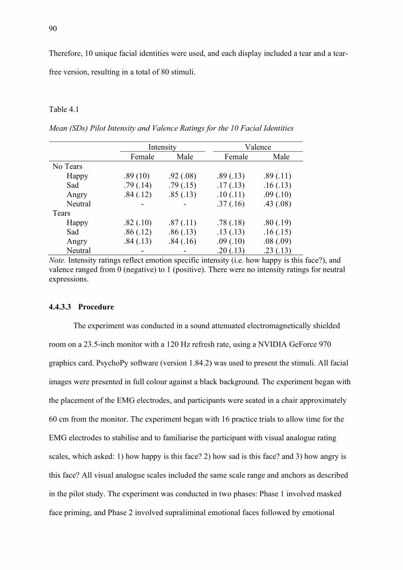
90
Therefore, 10 unique facial identities were used, and each display included a tear and a tear-
free version, resulting in a total of 80 stimuli.
Table 4.1
Mean (SDs) Pilot Intensity and Valence Ratings for the 10 Facial Identities
Intensity Valence Female Male Female Male No Tears Happy .89 (10) .92 (.08) .89 (.13) .89 (.11) Sad .79 (.14) .79 (.15) .17 (.13) .16 (.13) Angry .84 (.12) .85 (.13) .10 (.11) .09 (.10) Neutral - - .37 (.16) .43 (.08) Tears Happy .82 (.10) .87 (.11) .78 (.18) .80 (.19) Sad .86 (.12) .86 (.13) .13 (.13) .16 (.15) Angry .84 (.13) .84 (.16) .09 (.10) .08 (.09) Neutral - - .20 (.13) .23 (.13)
Note. Intensity ratings reflect emotion specific intensity (i.e. how happy is this face?), and valence ranged from 0 (negative) to 1 (positive). There were no intensity ratings for neutral expressions.
4.4.3.3 Procedure
The experiment was conducted in a sound attenuated electromagnetically shielded
room on a 23.5-inch monitor with a 120 Hz refresh rate, using a NVIDIA GeForce 970
graphics card. PsychoPy software (version 1.84.2) was used to present the stimuli. All facial
images were presented in full colour against a black background. The experiment began with
the placement of the EMG electrodes, and participants were seated in a chair approximately
60 cm from the monitor. The experiment began with 16 practice trials to allow time for the
EMG electrodes to stabilise and to familiarise the participant with visual analogue rating
scales, which asked: 1) how happy is this face? 2) how sad is this face? and 3) how angry is
this face? All visual analogue scales included the same scale range and anchors as described
in the pilot study. The experiment was conducted in two phases: Phase 1 involved masked
face priming, and Phase 2 involved supraliminal emotional faces followed by emotional

91
neutral faces. Participants always completed the tasks in this order to avoid viewing the
emotional faces prior to the masked paradigm.
4.4.3.3.1 Phase 1: Masked trials
Each trial followed the same experimental sequence. The trial structure can be seen in
Figure 4.1, where each trial began with the presentation of a fixation cross. Following the
fixation cross, a forward mask of a scrambled face was presented for 500 ms. This face was
followed by presentation of the target emotional face stimuli for 30 ms. The use of 30 ms as a
threshold was chosen as it has been used successfully in other masked priming studies. The
target face was immediately replaced with a backwards mask of the same individual
depicting a neutral expression for 5000 ms. During the presentation of the neutral expression,
participants completed a gender judgement task (i.e. was the face male or female) to ensure
participants maintained attention during the trials. Finally, trials were concluded with an
inter-stimulus interval that varied between 5 and 11 seconds to ensure participants were not
primed to expect a trial, and to also give the electrodes time to stabilize after each motor
response.
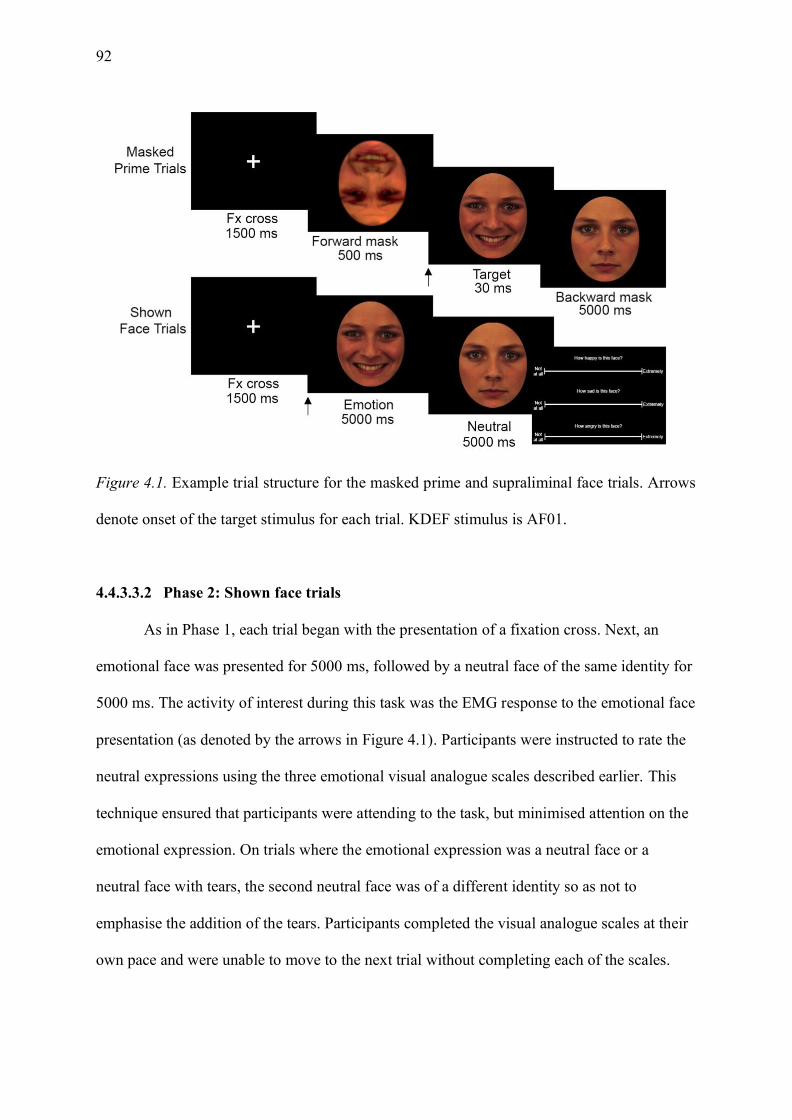
92
Figure 4.1. Example trial structure for the masked prime and supraliminal face trials. Arrows
denote onset of the target stimulus for each trial. KDEF stimulus is AF01.
4.4.3.3.2 Phase 2: Shown face trials
As in Phase 1, each trial began with the presentation of a fixation cross. Next, an
emotional face was presented for 5000 ms, followed by a neutral face of the same identity for
5000 ms. The activity of interest during this task was the EMG response to the emotional face
presentation (as denoted by the arrows in Figure 4.1). Participants were instructed to rate the
neutral expressions using the three emotional visual analogue scales described earlier. This
technique ensured that participants were attending to the task, but minimised attention on the
emotional expression. On trials where the emotional expression was a neutral face or a
neutral face with tears, the second neutral face was of a different identity so as not to
emphasise the addition of the tears. Participants completed the visual analogue scales at their
own pace and were unable to move to the next trial without completing each of the scales.

93
Lastly, participants completed an unrelated five-minute reaction time task where they
made rapid responses to each facial expression (see Chapter 3 for the results of this
experiment). After completion of the experiment, participants were debriefed and questioned
about the purpose of the study to determine whether the target emotional stimulus was
perceived in Phase 1 (masked priming task). None of the participants reported seeing any
target stimuli during the masked priming task.
4.4.3.4 EMG recording and analysis
EMG was recorded using a Biosemi Active-Two amplifier system, using six active
electrodes corresponding to three bipolar montages. Prior to electrode placement, the
participant’s skin was prepared using an alcohol wipe, and was lightly abraded using
electrode paste. The electrodes were affixed (using disposable electrode collars) to the left
side of the participant’s face, over the CS, ZMaj, and ZMin muscle regions. Two additional
electrodes, the common mode sense (CMS) and driven right leg (DRL), were used as
reference and ground electrodes, respectively, which is in accord with the Biosemi electrode
placement manual (https://www.biosemi.com). The EMG signal was continuously recorded
at 2048 Hz, with a 0.1-417 Hz band pass filter. All pre-processing steps were performed
using BrainVision Analyzer 2.0 software (Brain Products, Munich, Germany). EMG data was
band-pass filtered (20-500 Hz, 12 dB/oct) with a 50 Hz notch filter to correct for powerline
noise. These selected recording and filtering techniques are in accord with the published
guidelines for EMG research (Luca, 1997; van Boxtel, 2001).
Continuous EMG was then segmented in epochs from -2000 to 2000 ms after
stimulus onset for each emotional expression. Epochs were subjected to a rectified moving
average with a 125 ms time constant. Epochs of interest were the 2000 ms after stimulus
onset.

94
For the masked prime trials (Phase 1), this 2000 ms epoch was baseline corrected
using the final 500 ms of the fixation cross presentation2. To ensure participants were not
moving during the presentation of the forward mask (which could confound activity during
the presentation of the target stimulus), these trials were scanned for artifacts and trials with
amplitudes ±10 µV were removed. The baseline corrected epochs were scanned for artifacts
and trials with amplitudes ±30 µV were removed. This amplitude cut off was chosen in
accord with published research that details that EMG activation beyond 30 µV is associated
with overt motor movement (Korb, Grandjean, & Scherer, 2010).
For the supraliminal face trials (Phase 2), the epoch of interest (2000 ms following
stimulus onset) was baseline corrected using the final 500 ms of the fixation cross
presentation. The same artifact rejection criteria used for the epochs of interest in Phase 1
were applied here. At least 70% of the data was required for participants to be included in the
analysis (N = 34). All trials from the same condition were averaged. Data was exported
separately for the masked and supraliminal face trials for the 2000 ms time period following
stimulus onset for each of the emotion categories.
4.4.3.5 Data analysis
Data exclusions. To ensure participants maintained attention during the masked
prime task, participants were asked to identify the gender of the neutral backwards-mask
face. Three participants were excluded as they failed to follow experimental procedure during
the task, and a further two participants were removed due to excessive error rates (< 70%
accuracy). The remaining 30 participants responded with high classification accuracy (M =
77.7%, SD = 3.34). One additional participant was excluded due to having excessive artifacts
in the EMG data (failure to retain 70% of trials). Participants who did not meet retention
criteria for the behavioural data were excluded from the psychophysiological masked trial
2This equated to the time period from -1000 ms to -500 ms prior to stimulus onset.
95
data analysis. As participants could not complete Phase 2 without completing the rating
scales, all participants (except for one participant with excessive EMG artifacts) were
retained for the emotional supraliminal face analysis (N = 34).
Data analyses. A series of repeated-measures ANOVAs were conducted to explore
each hypothesis of interest. First, the data during the masked prime trials (Phase 1) was
explored to examine whether different emotions elicited significant corresponding muscle
activity at the pre-attentive level. Second, the degree of mimicry elicited in response to the
supraliminal face stimuli was examined. Separate analyses were conducted for each muscle
group of interest; for the zygomaticus muscles, 2 (muscle: ZMaj, ZMin) by 4 (emotion:
happy, sad, angry, neutral) by 2 (tears: no tears, tears) repeated-measures ANOVAs were
conducted. For the CS, 4 (emotion: happy, sad, angry, neutral) by 2 (tears: no tears, tears)
repeated-measures ANOVAs were conducted. All violations of sphericity were corrected
using Greenhouse-Geisser corrections. All analyses used a set alpha level of .05, however all
follow-up comparisons were Bonferroni-corrected.
4.4.4 Results
4.4.4.1 Physiological responses to masked primes
4.4.4.1.1 ZMaj and ZMin
A 2 x 4 x 2 repeated-measures ANOVA was conducted to investigate whether prime
emotions elicited corresponding muscle activity at the pre-attentive level. The main effect of
muscle approached significance as the ZMaj muscle (M = -.104, SE = .032) was more
relaxed than the ZMin muscle (M = -.072, SE = .032), F(1, 28) = 3.100, p = .089, 𝜂𝑝2 = .100.
No other main effects or interactions were significant, all F’s < 1.339, p’s > .270. The data
for the mimicry responses to masked primes is demonstrated in Table 4.2.

96
Table 4.2
Mean (SDs) Mimicry Responses for the Masked Trials by Emotion and Muscle.
Happy Angry Sad Neutral
Corrugator (CS) No Tear .162 (.490) .169 (.589) .188 (.394) .140 (.460)
Tear .134 (.417) .139 (.474) .071 (.546) .022 (.362)
Zygomaticus Major (ZMaj) No Tear -.064 (.315) -.040 (.418) -.132 (.395) -.157 (.338)
Tear -.093 (.367) -.112 (.333) -.048 (.228) -.186 (.455)
Zygomaticus Minor (ZMin) No Tear -.038 (.320) -.028 (.325) -.093 (.354) -.129 (.236)
Tear -.090 (.335) -.017 (.309) -.005 (.294) -.178 (.452)
4.4.4.1.2 CS
A 4 x 2 repeated-measures ANOVA was conducted to examine the influence of prime
emotion at the pre-attentive level. While no main effects or interactions were significant, the
main effect of tears approached significance, F(1, 28) = 3.211, p = .084, 𝜂𝑝2 = .103; tearful
expressions (M = .092, SE = .071) elicited less mimicry than tear-free expressions (M = .165,
SE = .071).
4.4.4.2 Physiological responses to supraliminal faces
4.4.4.2.1 ZMaj and ZMin
The supraliminal face mimicry at the zygomaticus muscles exhibited a similar pattern
of response as the masked prime analyses wherein no main effects or interactions were
significant, all F’s < 1.288, p’s > .265. The data for the mimicry elicited by each muscle for
each emotion during the supraliminal face trials is presented in Table 4.3.
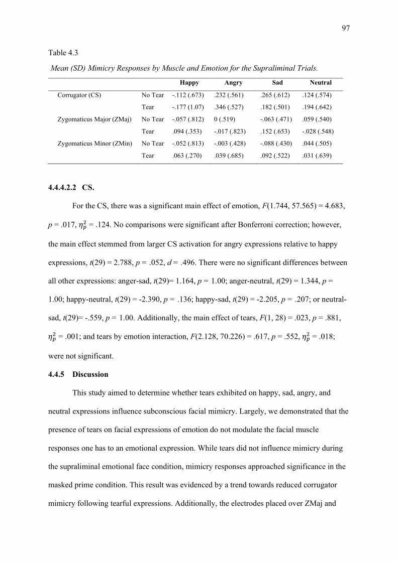
97
Table 4.3
Mean (SD) Mimicry Responses by Muscle and Emotion for the Supraliminal Trials.
Happy Angry Sad Neutral
Corrugator (CS) No Tear -.112 (.673) .232 (.561) .265 (.612) .124 (.574)
Tear -.177 (1.07) .346 (.527) .182 (.501) .194 (.642)
Zygomaticus Major (ZMaj) No Tear -.057 (.812) 0 (.519) -.063 (.471) .059 (.540)
Tear .094 (.353) -.017 (.823) .152 (.653) -.028 (.548)
Zygomaticus Minor (ZMin) No Tear -.052 (.813) -.003 (.428) -.088 (.430) .044 (.505)
Tear .063 (.270) .039 (.685) .092 (.522) .031 (.639)
4.4.4.2.2 CS.
For the CS, there was a significant main effect of emotion, F(1.744, 57.565) = 4.683,
p = .017, 𝜂𝑝2 = .124. No comparisons were significant after Bonferroni correction; however,
the main effect stemmed from larger CS activation for angry expressions relative to happy
expressions, t(29) = 2.788, p = .052, d = .496. There were no significant differences between
all other expressions: anger-sad, t(29)= 1.164, p = 1.00; anger-neutral, t(29) = 1.344, p =
1.00; happy-neutral, t(29) = -2.390, p = .136; happy-sad, t(29) = -2.205, p = .207; or neutral-
sad, t(29)= -.559, p = 1.00. Additionally, the main effect of tears, F(1, 28) = .023, p = .881,
𝜂𝑝2 = .001; and tears by emotion interaction, F(2.128, 70.226) = .617, p = .552, 𝜂𝑝
2 = .018;
were not significant.
4.4.5 Discussion
This study aimed to determine whether tears exhibited on happy, sad, angry, and
neutral expressions influence subconscious facial mimicry. Largely, we demonstrated that the
presence of tears on facial expressions of emotion do not modulate the facial muscle
responses one has to an emotional expression. While tears did not influence mimicry during
the supraliminal emotional face condition, mimicry responses approached significance in the
masked prime condition. This result was evidenced by a trend towards reduced corrugator
mimicry following tearful expressions. Additionally, the electrodes placed over ZMaj and
98
ZMin indexed the same muscular activity, and as such it was not possible to observe any
significant differences over the ZMin for tearful displays. Collectively, these findings
indicate that the tearful expressions used in our experiment do not modulate early facial
mimicry.
While it was not possible to demonstrate that tearful expressions modulated
physiological responses to facial displays presented to conscious awareness, this finding was
in accord with an existing EMG study exploring mimicry of tears (Grainger et al., 2019).
Grainger et al. (2019) found that while corrugator activity was elevated in response to sad
displays, tears did not influence EMG responses when examining expressions of sadness.
Therefore, they concluded that their study provided evidence for the motor matching
hypothesis as the participants matched the target stimuli’s sad expression (i.e. furrowed brow
to a sad stimulus). The motor matching account posits that mimicry occurs as pure motor
copy (Hess & Fischer, 2013), and as such the results of Grainger et al. (2019) indicate that
tearful displays do not significantly change one’s perception of sadness. Therefore,
individuals mimic sad and sad-tearful displays in the same way. Conversely, in our
experiment, when emotional expressions were presented unconsciously, using a masking
paradigm, there was a trend towards reduced corrugator activity following tearful
expressions. This finding indicates that participants frowned less following a masked tearful
display, which does not support a pure motor copy account, given that tears seemed to reduce
participants’ frowning response. Thus, this reduced response to tearful expressions fits with
the affective mimicry account, wherein different expressions mediate mimicry based on their
signal value (Bourgeois & Hess, 2008; Seibt et al., 2013). Moreover, this result could indicate
that: 1) tearful expressions elicit reduced congruent mimicry relative to tear-free expressions;
or 2) that tearful expressions reduce the negative reactivity typically observed to negative
displays.

99
The first possible conclusion demonstrates that a reduced frowning response to tearful
displays—in line with a motor mimicry account—is indicative of reduced mimicry for tearful
displays. Emotional mimicry fosters affiliation between interaction partners, and allows for
the sharing of another’s perspective (Hess & Fischer, 2013). In this way, the reduced
frowning response to tearful faces may signal a desire to not engage in affiliative behaviours
and serves as an indicator to avoid further interaction. However, this account seems unlikely,
given that tearful expressions typically foster approach behaviours rather than avoidance
(Gračanin et al., 2018), and encourage empathic and helping responses from observers
(Hendriks & Vingerhoets, 2006; Hendriks et al., 2008). Therefore, the second possible
explanation, wherein reduced frowning is indicative of reduced negativity and aggression
towards tearful displays is more plausible. Moreover, the reduction of the CS in response to
tearful expressions would indicate that if the frowning response elicited from sad displays is a
marker of a negative response to a stimulus, then tears reduce this negativity. Given that tears
serve to foster support and reduce aggression in observers (Hasson, 2009), this reduced CS
response to tearful displays could be a biological example of the signal value of tears. As
such, tears reduce reactive responses towards negative displays.
A possible explanation for the lack of mimicry responses to supraliminal emotional
expressions observed by Grainger et al. (2019) and herein is that mimicry of tearful displays
may not be adaptive. Firstly, sad facial expressions are affiliative, and as such should result in
congruent responses (Knutson, 1996). However, responding to a sad display comes at a high
cost for the observer (Bavelas et al., 1986; Bourgeois & Hess, 2008) and when social cost is
increased, mimicry is typically decreased (Johnston, 2002). Mimicry signals empathy and
understanding and, as such, fosters a link between interaction partners. However, the mimicry
of tearful displays could result in continued crying (Bourgeois & Hess, 2008; Häfner &
IJzerman, 2011). Although most self-report research has demonstrated favourable helping

100
responses to tearful displays (Hendriks et al., 2008; Lockwood et al., 2013), these helping
responses are typically expressed via down-regulation (Aragón & Clark, 2018). In this way,
the goal of the perceiver is to calm down the tearful individual and aid them in regaining
control over their emotions. Under this perspective, mimicry of tearful displays would be
counterproductive. Finally, previous studies that have evidenced sadness mimicry, have
demonstrated that mimicry of sadness is restricted to in-group members (Bourgeois & Hess,
2008) and close partners (Häfner & IJzerman, 2011). Thus, it is recommended that future
investigations of tearful mimicry use personally relevant stimuli to explore whether the
relationship between the expresser and the observer influences mimicry responses. This
increased personal relevance would aid in determining whether mimicry of tears is reactive or
serves to foster affiliation, as the absence of mimicry amongst close partners would indicate
that mimicry of tearful expressions is not an adaptive response.
One final implication that warrants discussion is the electrode locations used to index
mimicry of sad facial displays. Firstly, the CS is used to index mimicry of both sadness and
anger (Hofelich & Preston, 2012; Likowski et al., 2012; Lundqvist, 1995; Lundqvist &
Dimberg, 1995). Although some studies have demonstrated increased CS activation for sad
displays (Lundqvist, 1995; Lundqvist & Dimberg, 1995), others have demonstrated increased
CS activation for angry displays (Likowski et al., 2012). However, in expressions of sadness
the eyebrows are pulled together and upward, whereas anger is expressed by the eyebrows
pulled together and lowered. Importantly, both actions would be indexed by increased CS
activity (Nicanor & Mohammed, 2001). Therefore, the key to disentangling whether mimicry
is a result of pure motor copy or a reactive response could lie in changing the muscles used to
index sadness. This approach is in accord with seminal research by Duchenne (1862/ 1990),
who argued that the muscle associated with sadness is the depressor anguli oris—the muscle
responsible for drawing the lips downward. Soussignan et al. (2013) adopted this approach
101
and found increased depressor lip activity in response to sad expressions. Thus, the depressor
muscle could be a worthy indicator of sadness mimicry that warrants future investigation.
A similar approach to Soussignan et al. (2013) was adopted in the present study,
wherein the muscle that Duchenne associated with weeping—the ZMin—was used to index
mimicry of tearful expressions. However, it was not possible to distinguish mimicry
responses elicited from the ZMin from the ZMaj. This finding was not entirely unexpected, as
earlier research has demonstrated that both happy and disgust expressions activate the levator
labii muscle (i.e., the muscle responsible for drawing lip upwards—a common marker for
disgust) (Lundqvist, 1995; Lundqvist & Dimberg, 1995). The authors concluded that the
levator was contaminated by cross talk resulting from zygomaticus activation in response to
happy expressions (Lundqvist, 1995; Lundqvist & Dimberg, 1995). As the ZMin lies between
the levator and the ZMaj, it is likely indexing the activity of the larger ZMaj. Therefore,
although Duchenne associated the ZMin with weeping, it is not a reliable muscle to use in
further experimental studies. As such, it is recommended that future studies adopt the use of
the depressor to index mimicry of tearful displays.
It is acknowledged that this study has some limitations. Firstly, a sample of posed
prototypical facial displays was used and these images were digitally modified to include
tears. While the use of prototypical posed displays is in accord with existing research, there is
evidence that the type of facial expressions used influences mimicry. Namely, some studies
have demonstrated that genuine expressions elicit greater mimicry than posed displays
(Krumhuber, Likowski, & Weyers, 2014). This idea has a relevance for tearful displays,
given that posed crying has negative connotations (e.g. crocodile tears). Therefore, further
research using genuine stimuli may aid in determining whether mimicry of tearful displays
occurs. Secondly, no contextual information was provided. As existing research has found
that contextual information can mediate whether emotional faces are mimicked (Hess &
102
Fischer, 2013), the use of context in future experimental paradigms may aid in determining
whether tearful displays elicit increased responses when an individual knows why the
expressor is crying. This recommendation has relevance when exploring mimicry of tearful
joy. In the absence of context, tearful joy is more ambiguous than tear-free smiles (Krivan,
Caltabiano, Cottrell, & Thomas, 2020). However, when the context is known (i.e. tearful
faces paired with victorious scenarios), tearful joy is readily interpreted as positive (Aragón,
2017; Aragón & Clark, 2018), with some believing that tearful joy is the most intense
expression of positive emotion (Aragón & Bargh, 2018; Fernández-Dols & Ruiz-Belda,
1995). This paradigm would allow for the exploration of whether tearful joy displays are
mimicked with corresponding zygomaticus activity when the context is known.
Finally, in this study stimulus sex was not considered, as the use of a single sex
stimulus would reduce the generalisability of results, and a sample of five expressions is
insufficient to accurately index EMG responses. While there are no consistent patterns for
gender effects in facial mimicry research (Seibt, Mühlberger, Likowski, & Weyers, 2015),
the potential for a gender difference for responses to tearful displays cannot be ruled out.
Early crying research demonstrated that there was a significant sex difference in responses to
tearful displays (Lombardo et al., 1983; Lombardo et al., 2001). However, most recent
research has not demonstrated this effect (see Vingerhoets & Bylsma, 2015 for a review). In
saying this, Stadel et al. (2019) demonstrated that self-report ‘willingness to help’ responses
to tearful displays were more common among mixed dyads, than male-male dyads. As such,
further studies investigating tearful displays should explore whether mimicry is dependent on
the gender of the interactional dyad.
In summary, the present study has demonstrated that tearful displays are not
mimicked in a unique way to tear-free expressions. However, a trend towards reduced
mimicry of tearful displays when presented in a masked prime paradigm has been

103
demonstrated. This reduction of corrugator activity in response to tearful displays may
indicate a reduction in aggressive or negative responses to tearful displays or indicate the
desire to disengage from further contact. These conflicting interpretations have highlighted
the importance for future studies to explore sadness mimicry responses to personally relevant
faces, and the use of muscles other than the CS to index sadness (Soussignan et al., 2013).
Overall, the inconclusive results of the present study contribute to the body of literature,
which has demonstrated mixed evidence for mimicry of sad facial displays. However, several
experimental advancements have been suggested herein, to disentangle whether mimicry of
sad facial displays is not adaptive and as such does not occur, or whether mimicry is limited
to personally relevant individuals. Therefore, further exploring mimicry responses to tearful
displays will aid not only in understanding the communicative functions of tears, but also the
communicative functions of affiliative expressions as a whole.

104
Chapter 5: Early Neural Processing of Tearful Faces
5.1 Chapter Overview
Chapter 4 discussed one of the first EMG studies to explore mimicry responses to
tearful displays. The results of the study were inconclusive; however, it seemed that
participants did not mimic tearful displays differently than tear-free expressions. It was
concluded that mimicry of a tearful expression may not be adaptive—particularly if the
observer’s goal is to downregulate the crier. However, this null result does not mean that
tears are not shared at the psychophysiological level. Rather, although we do not mimic
tearful displays, which would encourage continued expression, it is yet to be determined
whether tearful displays are preferentially processed relative to other tear-free displays. This
chapter explores this theory by indexing early neural responses to emotional faces. I
predicted, in line with tears being a salient social signal, that tearful displays would elicit
larger early neural responses compared to tear-free expressions.
5.2 Publication Status
In preparation for submission.
5.3 Author Contributions
Krivan – conceptualised the experiment, created the stimuli, programmed the experimental
procedure, collected the data, pre-processed the EEG data, conducted the statistical analyses
and drafted manuscripts.
Cottrell – aided in conceptualisation and provided supervision.
Thomas – provided supervision and aided in manuscript revision.
5.4 Manuscript
5.4.1 Abstract
Facial expressions are a critical component of social communication. Expressions of
105
joy are readily shared, whereas expressions of sadness are costly to respond to. Despite this
fact, research employing self-report methodologies has identified that tears aid in signalling
distress and elicit greater caregiving responses compared to other tear-free expressions.
Understanding whether these differences also modulate face-specific, event-related potential
(ERP) waveforms will aid in determining whether tears are preferentially processed at the
neural level. Fifty-two participants completed an emotional discrimination task of faces
depicting happy, sad, and neutral expressions both with and without tears. The face specific
N170 ERP was sensitive to the emotional content of the images. Specifically, tearful sadness
elicited significantly larger responses than sad faces without tears, while the inverse result
was observed for happy expressions. Therefore, emotional stimuli requiring rapid
interpretation are preferentially processed at the early neural level. This early neural
processing provides a biological basis for the increased behavioural responses typically
observed in response to sad tearful stimuli.
Keywords: Tears, N170, Social Communication, EEG, Adult Crying
5.4.2 Introduction
The preferential processing of emotionally relevant stimuli is necessary for adaptive
social behaviour (Batty & Taylor, 2003). One method by which emotionally relevant
information is conveyed to others is via the face. As such, facial expressions are a means of
communicating one’s affective state rapidly, without words. Therefore, it has been argued
that the ability to decode facial expressions is an automatic process (Batty & Taylor, 2003;
Rellecke, Sommer, & Schacht, 2012), and that this automaticity is fostered by face specific
neural circuits (Hadjikhani et al., 2009; Haxby et al., 2000, 2002). This notion stems from
neuroimaging research wherein increased hemodynamic activity for facial stimuli is observed
in cortical regions such as the lateral fusiform gyrus (or fusiform face area; FFA), superior
temporal sulcus (STS), and inferior occipital gyri. In addition to this core facial processing
106
network, is an extended subcortical system for the recognition of emotion which encapsulates
the amygdala, insula, and limbic system (Hadjikhani et al., 2009; Haxby et al., 2000). As
such, the ability to rapidly interpret facial expressions is facilitated by a specific face
processing network.
In addition to exploring the physical structures associated with face processing, an
increasing body of research is interested in the temporal, or time-related, processes indexed
via electroencephalography (EEG). EEG is a recording technique that indexes the
synchronised firing of electrical potentials on the surface of the scalp (Brenner et al., 2014).
EEG is capable of indexing activity at the millisecond level, and as such is the preferred
technique for indexing rapid neural changes to facial stimuli. The most reliable index of
facial perception afforded by scalp EEG is the N170 event-related potential (ERP) (Joyce &
Rossion, 2005). Widely associated with facial processing, the N170 is enhanced in response
to facial stimuli relative to non-face objects (Bentin, Allison, Puce, Perez, & McCarthy, 1996;
Sadeh, Zhdanov, Podlipsky, Hendler, & Yovel, 2008). The N170 is typically maximal at
occipitotemporal electrode sites and is present approximately 170 ms after stimulus onset.
The N170 response is generated from face specific regions—identified via neuroimaging—
notably the FFA and STG (Horovitz, Rossion, Skudlarski, & Gore, 2004). As a result, most
of the N170 literature has focused on determining what facial information the N170 is
capable of indexing. There are two broad competing theories; 1) that the N170 represents
structural encoding of facial recognition—where specialised processing such as identity and
emotion are interpreted later, and 2) that the N170 is sensitive to changes in emotional
expression.
Early theories posited that the N170 was responsible for the structural encoding of
facial expressions (Bentin et al., 1996; Bentin & Deouell, 2000; Eimer, 2000, 2000).
Following Bruce and Young’s (1986) facial recognition model, structural encoding is the
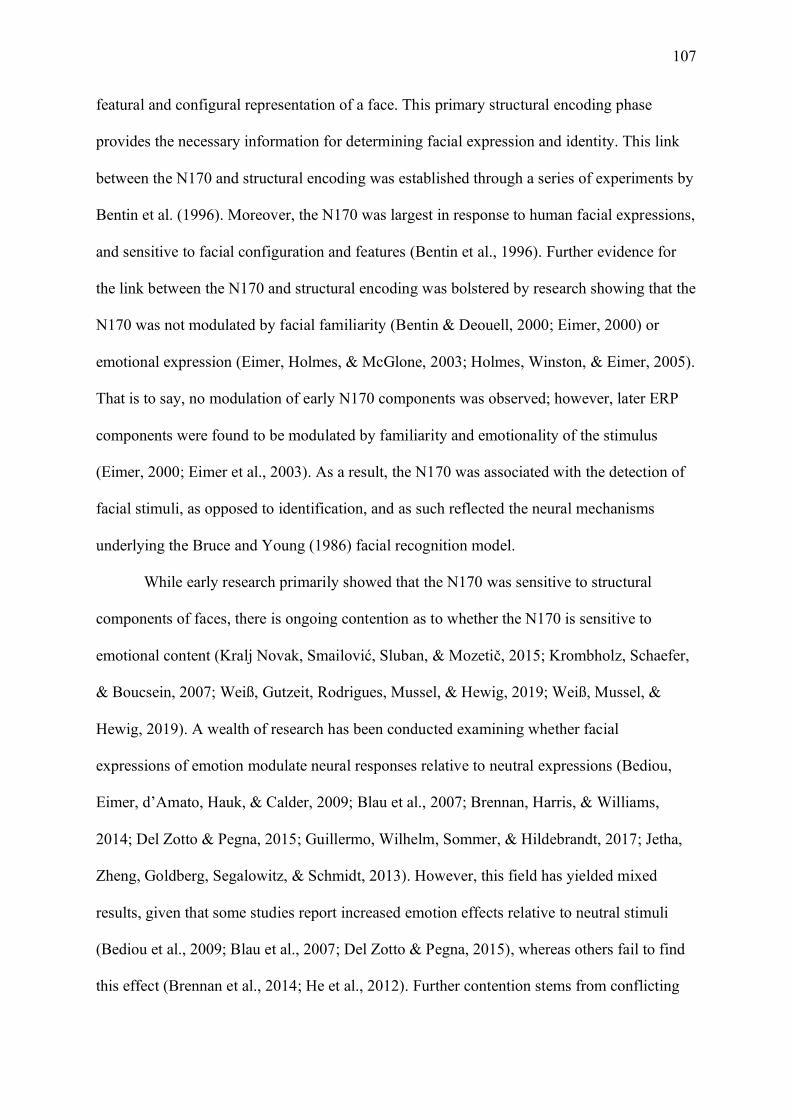
107
featural and configural representation of a face. This primary structural encoding phase
provides the necessary information for determining facial expression and identity. This link
between the N170 and structural encoding was established through a series of experiments by
Bentin et al. (1996). Moreover, the N170 was largest in response to human facial expressions,
and sensitive to facial configuration and features (Bentin et al., 1996). Further evidence for
the link between the N170 and structural encoding was bolstered by research showing that the
N170 was not modulated by facial familiarity (Bentin & Deouell, 2000; Eimer, 2000) or
emotional expression (Eimer, Holmes, & McGlone, 2003; Holmes, Winston, & Eimer, 2005).
That is to say, no modulation of early N170 components was observed; however, later ERP
components were found to be modulated by familiarity and emotionality of the stimulus
(Eimer, 2000; Eimer et al., 2003). As a result, the N170 was associated with the detection of
facial stimuli, as opposed to identification, and as such reflected the neural mechanisms
underlying the Bruce and Young (1986) facial recognition model.
While early research primarily showed that the N170 was sensitive to structural
components of faces, there is ongoing contention as to whether the N170 is sensitive to
emotional content (Kralj Novak, Smailović, Sluban, & Mozetič, 2015; Krombholz, Schaefer,
& Boucsein, 2007; Weiß, Gutzeit, Rodrigues, Mussel, & Hewig, 2019; Weiß, Mussel, &
Hewig, 2019). A wealth of research has been conducted examining whether facial
expressions of emotion modulate neural responses relative to neutral expressions (Bediou,
Eimer, d’Amato, Hauk, & Calder, 2009; Blau et al., 2007; Brennan, Harris, & Williams,
2014; Del Zotto & Pegna, 2015; Guillermo, Wilhelm, Sommer, & Hildebrandt, 2017; Jetha,
Zheng, Goldberg, Segalowitz, & Schmidt, 2013). However, this field has yielded mixed
results, given that some studies report increased emotion effects relative to neutral stimuli
(Bediou et al., 2009; Blau et al., 2007; Del Zotto & Pegna, 2015), whereas others fail to find
this effect (Brennan et al., 2014; He et al., 2012). Further contention stems from conflicting

108
evidence regarding the type of emotion that modulates the N170 response; where some argue
that negative emotions elicit larger N170 amplitudes (Brenner et al., 2014; Jetha et al., 2013),
and others argue for positive displays (Chen et al., 2014; Rossignol et al., 2012). A recent
meta-analysis identified that the N170 was modulated by emotional content, with anger, fear,
and happy expressions exhibiting the largest N170 amplitudes (Hinojosa et al., 2015).
Conversely, sad and disgust expressions were not found to modulate the N170. Thus, the
authors concluded that modulation of the N170 is dependent on the communicative function
of emotional expressions – wherein emotions such as happy, angry, and fear are all
particularly salient signals that are interpreted rapidly relative to sadness and disgust.
Anger, fear, and happy expressions are elicited in social exchanges requiring rapid
interpretation and are typically attributed greater social relevance than disgust and sadness
expressions (Hinojosa et al., 2015). This social relevance stems from the fact that anger and
fear expressions signal danger, and thus rapid interpretation and response is critical to
evolutionarily adaptive behaviour (Tipples et al., 2002). This increased response to
threatening and aversive stimuli is termed the ‘threat hypothesis’, which falls under the
umbrella of a ‘negativity bias’ (Hajcak et al., 2011; Ito et al., 1998; Öhman et al., 2001). The
processing of negative stimuli is said to engage the “low-road” subcortical pathway via the
amygdala, which facilitates rapid responses to threat (LeDoux, 2007; Liddell et al., 2005;
Liddell et al., 2004). As anger and fear both signal potential danger and threat, neural
processes have evolved to ensure their rapid interpretation. However, happy expressions,
while not threatening, also facilitate N170 responses (Chen et al., 2014; Mühlberger et al.,
2009; Tortosa, Lupiáñez, & Ruz, 2013). Moreover, happy expressions have an increased
social relevance as smiles are rapidly exchanged to foster affiliative pro-social behaviours
(Nummenmaa & Calvo, 2015). By contrast, sad and disgust expressions do not modulate the
N170 response (Hinojosa et al., 2015); however, the number of studies including sad or
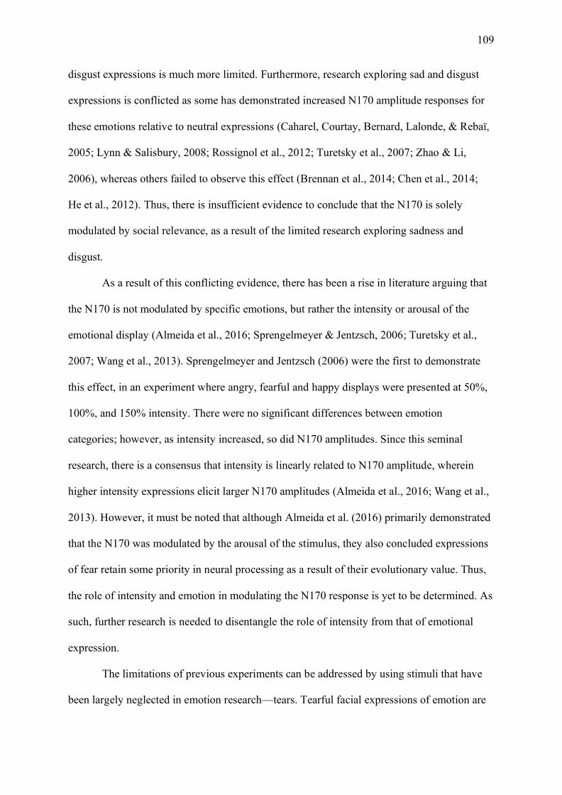
109
disgust expressions is much more limited. Furthermore, research exploring sad and disgust
expressions is conflicted as some has demonstrated increased N170 amplitude responses for
these emotions relative to neutral expressions (Caharel, Courtay, Bernard, Lalonde, & Rebaï,
2005; Lynn & Salisbury, 2008; Rossignol et al., 2012; Turetsky et al., 2007; Zhao & Li,
2006), whereas others failed to observe this effect (Brennan et al., 2014; Chen et al., 2014;
He et al., 2012). Thus, there is insufficient evidence to conclude that the N170 is solely
modulated by social relevance, as a result of the limited research exploring sadness and
disgust.
As a result of this conflicting evidence, there has been a rise in literature arguing that
the N170 is not modulated by specific emotions, but rather the intensity or arousal of the
emotional display (Almeida et al., 2016; Sprengelmeyer & Jentzsch, 2006; Turetsky et al.,
2007; Wang et al., 2013). Sprengelmeyer and Jentzsch (2006) were the first to demonstrate
this effect, in an experiment where angry, fearful and happy displays were presented at 50%,
100%, and 150% intensity. There were no significant differences between emotion
categories; however, as intensity increased, so did N170 amplitudes. Since this seminal
research, there is a consensus that intensity is linearly related to N170 amplitude, wherein
higher intensity expressions elicit larger N170 amplitudes (Almeida et al., 2016; Wang et al.,
2013). However, it must be noted that although Almeida et al. (2016) primarily demonstrated
that the N170 was modulated by the arousal of the stimulus, they also concluded expressions
of fear retain some priority in neural processing as a result of their evolutionary value. Thus,
the role of intensity and emotion in modulating the N170 response is yet to be determined. As
such, further research is needed to disentangle the role of intensity from that of emotional
expression.
The limitations of previous experiments can be addressed by using stimuli that have
been largely neglected in emotion research—tears. Tearful facial expressions of emotion are

110
an inborn biological signal designed to elicit help and support from others (Balsters et al.,
2013; Hendriks et al., 2008). Additionally, behavioural research has demonstrated that tears
are a signal of sadness, which foster approach behaviours relative to avoidance (Gračanin et
al., 2018). The tear effect proposes that in the absence of context a tearful expression will be
perceived as sad (Provine et al., 2009). Thus, tears are a distinctive marker of sadness, which
serve as a salient distress signal. However, it is yet to be determined whether these increased
behavioural responses have a biological basis. Thus far only one study has investigated N170
responses to sad crying faces (Hendriks et al., 2007). Although tearful displays elicited larger
N170 amplitudes relative to neutral expressions, they were not significantly different from
other basic emotional displays (Hendriks et al., 2007). However, Hendriks et al. (2007)
compared sad crying faces with other tear-free expressions. Yet, tears are not only elicited in
response to sadness; tears are also elicited in response to joy (Miceli & Castelfranchi, 2003).
The existing research investigating responses to happy-tear expressions is
exceptionally limited. In the absence of context, happy tearful displays are associated with
increased sadness and increased negative valence (Reed et al., 2015). Thus, tearful happy
expressions reduce the distinctiveness of a typically happy face. As happy expressions
modulate the N170 response (Chen et al., 2014; Rellecke et al., 2012), the inclusion of both
tearful and tear-free stimuli allowed us to investigate how tears modulate the N170 based on
the type of emotion they were paired with. Furthermore, tears are an ideal candidate for
investigating the competing social relevance and intensity hypotheses. Moreover, recent
research has demonstrated that tears increase the overall intensity of a stimulus, regardless of
whether it is positively or negatively valenced (Krivan et al., 2020; Reed et al., 2015). Thus,
tearful stimuli are more intense overall; however, happy faces without tears and sad faces
with tears are particularly distinctive, which increases their social relevance. Therefore, the
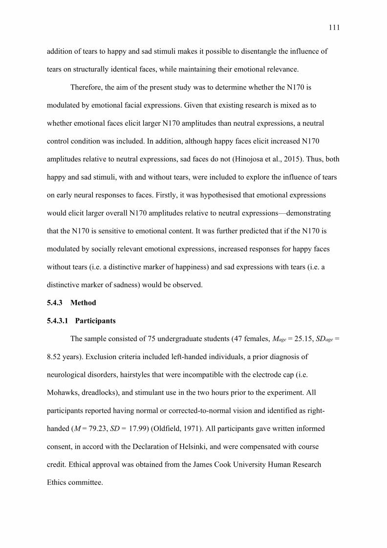
111
addition of tears to happy and sad stimuli makes it possible to disentangle the influence of
tears on structurally identical faces, while maintaining their emotional relevance.
Therefore, the aim of the present study was to determine whether the N170 is
modulated by emotional facial expressions. Given that existing research is mixed as to
whether emotional faces elicit larger N170 amplitudes than neutral expressions, a neutral
control condition was included. In addition, although happy faces elicit increased N170
amplitudes relative to neutral expressions, sad faces do not (Hinojosa et al., 2015). Thus, both
happy and sad stimuli, with and without tears, were included to explore the influence of tears
on early neural responses to faces. Firstly, it was hypothesised that emotional expressions
would elicit larger overall N170 amplitudes relative to neutral expressions—demonstrating
that the N170 is sensitive to emotional content. It was further predicted that if the N170 is
modulated by socially relevant emotional expressions, increased responses for happy faces
without tears (i.e. a distinctive marker of happiness) and sad expressions with tears (i.e. a
distinctive marker of sadness) would be observed.
5.4.3 Method
5.4.3.1 Participants
The sample consisted of 75 undergraduate students (47 females, Mage = 25.15, SDage =
8.52 years). Exclusion criteria included left-handed individuals, a prior diagnosis of
neurological disorders, hairstyles that were incompatible with the electrode cap (i.e.
Mohawks, dreadlocks), and stimulant use in the two hours prior to the experiment. All
participants reported having normal or corrected-to-normal vision and identified as right-
handed (M = 79.23, SD = 17.99) (Oldfield, 1971). All participants gave written informed
consent, in accord with the Declaration of Helsinki, and were compensated with course
credit. Ethical approval was obtained from the James Cook University Human Research
Ethics committee.
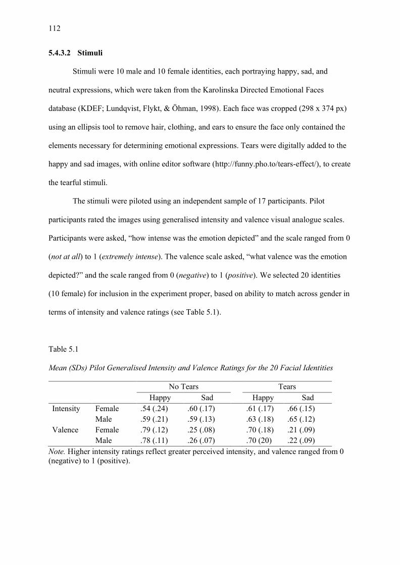
112
5.4.3.2 Stimuli
Stimuli were 10 male and 10 female identities, each portraying happy, sad, and
neutral expressions, which were taken from the Karolinska Directed Emotional Faces
database (KDEF; Lundqvist, Flykt, & Öhman, 1998). Each face was cropped (298 x 374 px)
using an ellipsis tool to remove hair, clothing, and ears to ensure the face only contained the
elements necessary for determining emotional expressions. Tears were digitally added to the
happy and sad images, with online editor software (http://funny.pho.to/tears-effect/), to create
the tearful stimuli.
The stimuli were piloted using an independent sample of 17 participants. Pilot
participants rated the images using generalised intensity and valence visual analogue scales.
Participants were asked, “how intense was the emotion depicted” and the scale ranged from 0
(not at all) to 1 (extremely intense). The valence scale asked, “what valence was the emotion
depicted?” and the scale ranged from 0 (negative) to 1 (positive). We selected 20 identities
(10 female) for inclusion in the experiment proper, based on ability to match across gender in
terms of intensity and valence ratings (see Table 5.1).
Table 5.1
Mean (SDs) Pilot Generalised Intensity and Valence Ratings for the 20 Facial Identities
No Tears Tears Happy Sad Happy Sad Intensity Female .54 (.24) .60 (.17) .61 (.17) .66 (.15) Male .59 (.21) .59 (.13) .63 (.18) .65 (.12) Valence Female .79 (.12) .25 (.08) .70 (.18) .21 (.09) Male .78 (.11) .26 (.07) .70 (20) .22 (.09)
Note. Higher intensity ratings reflect greater perceived intensity, and valence ranged from 0 (negative) to 1 (positive).

113
5.4.3.3 Procedure
The experiment was conducted in a sound attenuated electromagnetically shielded
room on a 24-inch Dell monitor with a 60 Hz refresh rate. PsychoPy software (version
1.91.1) was used to present the stimuli. All facial images were presented in full colour against
a black background.
Each trial followed the same experimental sequence. Participants received instructions
for a delayed match-to-sample task on emotion, where two images were presented
sequentially, and the participants had to report via button press whether the emotions of the
first and second image were the same or different. Prior to the testing phase, 10 practice
trials, using unique faces that were not presented during the actual trials, were given to
familiarise participants with the procedure. Participants were asked to make fast, but
accurate, judgments and indicated they understood it was a reaction time task. An example
trial structure is depicted in Figure 5.1. The intertrial fixation cross was randomly varied in
blocks of 100 ms, between 2500 and 3500 ms, to avoid anticipatory responses. The image
pairs were shown randomly, where every image was shown twice in the first face position,
and on 50% of trials the emotions were the same. The response prompt was shown until a
response was made, with a maximum response time of 2000 ms, after which participants
were informed their response was too slow. The experiment was conducted in 5 blocks of 40
trials (~ 7 minutes), and each block was followed by a 30 second break. After participants
completed this phase of the experiment, they were informed about a second experimental
task; these data were analysed separately (see Chapter 6 for results). After participants
completed both tasks, they were debriefed and thanked for their participation.
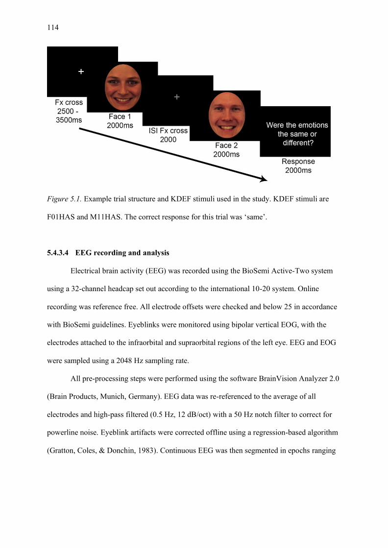
114
Figure 5.1. Example trial structure and KDEF stimuli used in the study. KDEF stimuli are
F01HAS and M11HAS. The correct response for this trial was ‘same’.
5.4.3.4 EEG recording and analysis
Electrical brain activity (EEG) was recorded using the BioSemi Active-Two system
using a 32-channel headcap set out according to the international 10-20 system. Online
recording was reference free. All electrode offsets were checked and below 25 in accordance
with BioSemi guidelines. Eyeblinks were monitored using bipolar vertical EOG, with the
electrodes attached to the infraorbital and supraorbital regions of the left eye. EEG and EOG
were sampled using a 2048 Hz sampling rate.
All pre-processing steps were performed using the software BrainVision Analyzer 2.0
(Brain Products, Munich, Germany). EEG data was re-referenced to the average of all
electrodes and high-pass filtered (0.5 Hz, 12 dB/oct) with a 50 Hz notch filter to correct for
powerline noise. Eyeblink artifacts were corrected offline using a regression-based algorithm
(Gratton, Coles, & Donchin, 1983). Continuous EEG was then segmented in epochs ranging

115
from -100 to 700 ms after stimulus onset3 for each emotion expression, and baseline
corrected in the 100 ms period prior to stimulus onset. EEG epochs were removed from
analyses if the epoch included amplitudes exceeding ±100 µV. Average ERPs were
calculated for each type of facial expression corresponding to ~ 70 trials per participant for
each of the emotion categories. Data were exported using local peak latency and mean area.
The N170 mean amplitude between 175 and 255 ms was averaged, and the local point at
which the peak occurred (±5 time points) was detected.
5.4.3.5 Data exclusions
Four participants from our initial sample were excluded due to technical recording
problems. As at least 70% of each participant’s data had to be retained to be included in
analysis, a further 18 participants were excluded. One further participant, who failed to
respond during the behavioural task, was removed, resulting in a final sample of 52
participants.
5.4.4 Results
5.4.4.1 Behavioural data
Classification accuracy for the image pairs was analysed to ensure participants-
maintained attention throughout the task. Participants performed significantly above chance
when identifying emotional expressions (see Table 5.2), with a mean accuracy of 85.57% (SD
= 6.98). As the task relied on person perception, it is feasible that some participants perceived
the tear and the tear-free emotions as different, despite having the same underlying emotion.
As such, we did not exclude any of the EEG data based on behavioural accuracy, in order to
maintain as much EEG data as possible.
3 EEG triggers were sent after a stimulus was presented for a full frame, rather than at stimulus onset. As a result, our latency data is delayed by 16.67 ms (the time of one frame) from that of existing research which has demonstrated the N170 at ~170ms after onset.

116
Table 5.2
One Sample t-test Results of Accuracy Scores per Emotion Condition during the
Discrimination Task.
Mean SD t p d Happy 89.28 8.30 34.11 <.001 4.730 Sad 82.12 9.44 24.54 <.001 3.403 Happy-tear 86.11 10.62 24.50 <.001 3.399 Sad-tear 84.81 8.46 29.68 <.001 3.116 Neutral (Control) 85.53 8.61 29.76 <.001 4.127
Note. Compared against chance level classification (50% accuracy).
5.4.4.2 ERP data
Grand-averaged ERP waveforms for face stimuli are shown in Figure 5.2. Based on visual
inspection, responses were characterized by an early positive component (P1, peak around
150 ms after stimulus onset), followed by a negative wave (N170, peak around 200 ms after
stimulus onset) bilaterally over occipitotemporal electrode sites (left hemisphere P7/ right
hemisphere P8). Table 5.3 shows the mean amplitude and local peak latency of the N170
response for each of the emotion conditions.
Table 5.3
Mean Amplitudes and Local Peak Latencies (SD) of the N170, for each Emotion at the Left
and Right Occipitotemporal Electrodes.
Left Hemisphere (P7) Right Hemisphere (P8) Amplitude (µV) Latency (ms) Amplitude (µV) Latency (ms) Happy No tears -1.24 (2.78) 209.43 (16.12) -2.45 (3.13) 207.29 (12.88) Tears -1.47 (2.91) 206.12 (19.81) -2.14 (3.06) 206.76 (12.48) Sad No tears -1.54 (2.90) 210.52 (14.27) -2.31 (3.23) 209.61 (14.36) Tears -1.72 (2.79) 211.60 (14.65) -2.71 (3.11) 211.20 (14.71) Neutral No tears -1.19 (2.53) 210.72 (16.24) -2.10 (3.12) 207.23 (12.89)

117
Figure 5.2. Grand-averaged ERPs as a function of facial expression, for left hemisphere
electrode P7 (pictured on left), and right hemisphere electrode P8 (pictured on right). The
waveforms were low pass filtered prior to plotting, with a half-amplitude cut-off of 30 Hz for
clarity of ERP figures.
5.4.4.2.1 N170 mean amplitude
Separate planned contrasts were conducted to compare the neutral condition with the
other emotional expression categories to determine the role of emotional content. Planned
contrasts indicated the neutral condition elicited smaller negative mean amplitude responses
relative to the emotional face conditions at both left hemisphere P7, F(1,51) = 5.580, p =
.022, 𝜂𝑝2 = .099; and right hemisphere P8, F(1,51) = 5.285, p = .026, 𝜂𝑝
2 = .094, electrode
sites. Thus, the neutral condition was excluded from our follow-up analyses to better explore
the electrode by emotion by tear effects at each electrode site.
A 2 (electrode: left hemisphere P7, right hemisphere P8), by 2 (tears: no tear, tears),
by 2 (emotion: happy, sad) repeated-measures ANOVA was conducted to investigate the role
P8

118
of tears in modulating neural responses to emotional faces. A significant main effect of
electrode was observed, with a significantly larger negative mean amplitude at the right
occipitotemporal electrode (P8) compared to the left (P7), F(1,51) = 5.387, p = .024, 𝜂𝑝2 =
.096. There was also a significant main effect of emotion, F(1,51) = 7.498, p = .008, 𝜂𝑝2 =
.128, where sad expressions elicited larger negative mean amplitudes than happy expressions.
Although remaining main effects and all 2-way interactions were non-significant, F’s <
3.904, p’s > .054; there was also a significant electrode by tears by emotion interaction,
F(1,51) = 5.001, p = .030, 𝜂𝑝2 = .089. To follow-up this interaction, separate 2 (tears: tear-
free, tears) by 2 (emotion: happy, sad) repeated-measures ANOVAs were conducted at left
hemisphere electrode site P7 and right hemisphere electrode site P8.
At left hemisphere electrode site P7, there was a main effect of emotion, with sad
expressions eliciting a larger negative mean amplitude relative to happy expressions, F(1,51)
= 5.683, p = .021, 𝜂𝑝2 = .100. Both the main effect of tears, F(1,51) = 3.534, p = .066, 𝜂𝑝
2 =
.06, and the interaction, F(1,51) = .041, p = .840, 𝜂𝑝2 = .001, were non-significant.
At right hemisphere electrode site P8, there were no significant main effects of
emotion, F(1,51) = 3.585, p = .064, 𝜂𝑝2 = .066; or tears, F(1,51) = .166, p = .686, 𝜂𝑝
2 = .003.
However, there was a significant tears by emotion interaction, F(1,51) = 8.260, p = .006, 𝜂𝑝2 =
.139. Follow-up Bonferroni-corrected paired samples t-tests revealed that sad tearful displays
elicited significantly larger negative mean amplitudes compared to sad tear-free displays,
t(51) = 2.346, p = .023, 𝑑 = .325. There was no significant difference between happy tear-free
and happy tearful displays, t(51) = -1.993, p = .052, 𝑑 = -.276.
5.4.4.2.2 N170 peak latency.
To examine whether there were any latency effects, another 2 (electrode: left
hemisphere P7, right hemisphere P8), by 2 (tears: no tear, tears), by 2 (emotion: happy, sad)
repeated-measures ANOVA was conducted on the latency data. There was a significant main
119
effect of emotion, F(1,51) = 11.840, p = .001, 𝜂𝑝2 = .188, whereby happy expressions peaked
earlier than sad expressions. No other main effects or interactions were significant, F’s <
2.730, p’s > .105.
5.4.5 Discussion
The findings demonstrated that the face specific N170 ERP is modulated by the
emotional content depicted in facial displays. Early research identified that the N170 was
involved in the structural encoding of faces (Bentin & Deouell, 2000; Eimer, 2000; Eimer et
al., 2003; Holmes et al., 2005). Rather, we have shown that, overall, emotional expressions
elicit larger N170 amplitudes relative to neutral expressions, which supports an increasing
body of research in the field (Bediou et al., 2009; Blau et al., 2007; Mühlberger et al., 2009).
Additionally, an increased modulation of the N170 to emotional faces was demonstrated at
the occipitotemporal electrode sites, with significantly stronger activation observed over the
right hemisphere (P8), in line with a right hemisphere dominance for emotional processing
(Dimberg & Petterson, 2000; Gainotti, 2012). Finally, we demonstrated that the social
relevance of the facial display, as opposed to the intensity of the stimulus, plays a greater role
in N170 modulation.
Regardless of the type of emotion presented, emotional faces elicited increased
negative N170 amplitudes relative to neutral displays. This increased amplitude response
reveals that emotion processing occurs rapidly, in the same time frame as facial detection.
This finding contradicts earlier studies, which concluded that the N170 ERP reflected the
structural processing of faces (Bentin & Deouell, 2000; Eimer, 2000; Eimer et al., 2003;
Holmes et al., 2005). Rather, it provides support for theoretical models, which state that while
structural encoding occurs independently of expression identification, the two can occur
simultaneously (Bruce & Young, 1986; Eimer & Holmes, 2007). Furthermore, the earlier
studies that concluded that the N170 was sensitive to structural, as opposed to affective,
120
content reached this conclusion because they failed to find significant modulation of emotion
(Eimer et al., 2003; Holmes et al., 2005). However, more recent research has demonstrated
N170 modulation using schematic emotional faces (i.e. line drawings of emotional
expressions) and emojis (i.e. simple graphic symbols) with minor variability between
emotional facial displays (Kralj Novak et al., 2015; Krombholz et al., 2007; Weiß et al.,
2019; Weiß et al., 2019). Critically, Weiß et al. (2019) demonstrated that sad emojis, which
were structurally identical to happy emojis with an inverted smile, elicited larger N170
amplitudes relative to happy and neutral displays. Thus, as in studies using human facial
expressions (Blau et al., 2007; Caharel et al., 2005; Del Zotto & Pegna, 2015), a larger N170
amplitude is observed in response to negative schematic expressions and emojis relative to
positive displays (Krombholz et al., 2007; Weiß et al., 2019; Weiß et al., 2019). Therefore,
the N170 is modulated by the emotional content of a face, even in the absence of human-like
structural features.
In addition to demonstrating an increased modulation of the N170 relative to neutral
expressions, we demonstrated that this modulation was emotion dependent. Existing research
has demonstrated that larger ERPs are elicited in response to facial expressions with greater
social relevance (see Hinojosa et al. (2015) for a review). Specifically, existing research has
shown that facial displays conveying threat-related information, such as fear and anger, elicit
the largest N170 amplitudes (Blau et al., 2007; Jetha et al., 2013). This increased neural
response to threatening facial expressions is in accord with neuroimaging studies identifying
a preferential processing network, including the amygdala, for threatening visual stimuli
(LeDoux, 2007; Liddell et al., 2005; Liddell et al., 2004). However, the meta-analysis by
Hinojosa et al. (2015) also identified that happy facial expressions significantly modulate the
N170 response. Moreover, although some studies have not demonstrated an amplitude
difference between happy, angry, and fearful stimuli (Mühlberger et al., 2009; Rellecke et al.,

121
2012; Tortosa et al., 2013), others have reported that happy expressions elicit larger potentials
overall, when compared to negative displays (Chen et al., 2014; Rossignol et al., 2012).
Therefore, the N170 is not modulated solely by threat-related information. Rather, the N170
is modulated by the social relevance of the stimuli, where certain emotional expressions,
either positive or negative, require rapid social exchange.
To address the theory of social relevance, tearful expressions of sadness and joy were
used. Tears provide a way to address the factor of social relevance, for both positive and
negative emotions, while minimising the structural variation between emotional images. Our
results provide the greatest support for the social relevance hypothesis, as sad tearful displays
and happy tear-free displays elicited the largest potentials overall, relative to neutral
expressions. This increased amplitude response to happy tear-free and sad tearful faces is in
accord with previous behavioural research reflecting an advantage for salient stimuli (Krivan
et al., 2020). Moreover, happy facial expressions are common in rapid social interactions, and
the preferential processing observed in this study and existing research is potentially a result
of their pro-social signal. Although sad tearful displays are negatively valenced, they reliably
elicit support and empathic responses from observers (Balsters et al., 2013; Hendriks et al.,
2008). Additionally, sad tearful displays foster approach behaviours (Gračanin et al., 2018)
and facilitate the recognition of sadness (Ito et al., 2019; Provine et al., 2009). Thus, tears are
a distinctive marker of sadness, akin to smiles being a distinctive marker of joy.
Our support for the social relevance hypothesis contradicts the argument that the
N170 is modulated by the intensity or arousal of an image more so than it is by emotion
(Almeida et al., 2016; Sprengelmeyer & Jentzsch, 2006; Wang et al., 2013). Sprengelmeyer
and Jentzsch (2006) demonstrated that the more intense an emotional stimulus, the greater the
N170 amplitude observed. Similarly, Almeida et al. (2016) demonstrated a linear relationship
between increasing stimulus arousal and N170 amplitudes. However, the results from this
122
study fail to support this theory. Specifically, our results demonstrated that sad-tear and
happy tear-free images elicited significantly larger ERPs than their counterparts. One possible
explanation for the discrepancy between the present findings and earlier intensity research is
the way in which intensity was matched in the present study. Moreover, tearful expressions in
the present study are more intense than tear-free expressions (Krivan et al., 2020). Therefore,
if the intensity account was supported, a main effect of tears would have been observed,
wherein tearful displays elicited greater N170 amplitudes relative to tear-free expressions.
Rather, we have demonstrated the greatest support for the argument that the N170 is
modulated by the communicative criteria of the display—wherein tearful sadness clearly
communicates sadness, and happy tear-free expressions are a distinctive happy expression—
and, as such, are the emotions that elicit larger N170 amplitudes. Therefore, intensity alone
does not modulate the N170, and the greatest support is demonstrated for the social relevance
account.
Furthermore, the interaction between tears and emotion at electrode P8 also
contradicts previous N170 research, which has demonstrated a negativity bias. Moreover, a
negativity bias for N170 amplitude at electrode P7 was observed, where sad faces elicited
larger amplitudes overall when compared to happy expressions. As raised by Hajcak et al.
(2011), this negativity bias is typically difficult to equate. However, the tear stimuli used
herein increased overall perceptions of intensity, while maintaining the social relevance
associated with happy and sad emotional displays. In addition, the inverse of a negativity bias
was demonstrated when examining the latency effects observed at electrode P8, as happy
expressions peaked earlier than sad displays. However, it is important to note that the existing
N170 latency literature is mixed, with some studies demonstrating no latency differences
between facial stimuli (Blau et al., 2007; Guillermo et al., 2017; Herrmann et al., 2002;
Krombholz et al., 2007), and others demonstrating a negative bias (Del Zotto & Pegna, 2015)
123
or a positive advantage (Batty & Taylor, 2003). Thus, there is insufficient evidence to draw
strong conclusions regarding N170 latency modulation from the existing literature.
Nonetheless, the slight processing advantage for positive emotional displays exhibited in our
study might reflect a neural basis for the happy face advantage, which has been widely
observed in behavioural studies (Calvo et al., 2016; Leppänen et al., 2003; Palermo &
Coltheart, 2004), or it might relate to the complexity associated with decoding negative
stimuli (Kuperman, Estes, Brysbaert, & Warriner, 2014; Taylor, 1991).
A limitation that must be addressed is the choice of referencing scheme used. The two
most common referencing systems when examining the N170 are the common average and
the mastoids (Hinojosa et al., 2015). The common average reference has been identified as
the superior choice, given the mastoids proximity to the occipitotemporal electrodes
(Rellecke, Sommer, & Schacht, 2013); as such, the common average is used more frequently
(Hinojosa et al., 2015). However, computing a common average on a small electrode array
(i.e. 32 electrodes) is not ideal (Dien, 1998). For this reason, the ERP data was re-analysed
using the mastoid reference, and the pattern of results was consistent regardless of reference
choice; albeit smaller N170 amplitudes were observed when a mastoid reference was used,
which is consistent with prior research (Rellecke et al., 2013). As a result, it is proposed that
the current conclusion that the emotional content of a facial expression modulates the N170
response more so than intensity is appropriate; however, further research using a larger
electrode array is needed to provide confirmatory evidence.
Furthermore, we did not include an additional control condition wherein participants
completed a task not associated with emotion recognition. Moreover, the current results
contradict an earlier study that examined the way that crying modulates the N170 (Hendriks
et al., 2007). Hendriks et al. (2007) demonstrated that crying faces were distinct from neutral
expressions, although not fundamentally different from other basic emotions. However, their
124
participants were instructed to complete a gender identification task, rather than an emotion-
focused task. The role of directed emotional attention is contentious in determining the
degree to which the N170 is modulated by emotion. Although some studies have
demonstrated that N170 modulation only occurs when attention is focused on affective state
(Eimer et al., 2003), others have demonstrated that emotional modulation occurs in the
absence of conscious awareness (Del Zotto & Pegna, 2015; Pegna, Darque, Berrut, & Khateb,
2011; Pegna et al., 2008), and yields larger effect sizes when indirect tasks are used (Hinojosa
et al., 2015). As such, it is unlikely that the current results stem solely from the choice of
task; however, the results should be further validated by including a task wherein
participants’ attention is directed away from the emotional content of the face.
In conclusion, the present study has demonstrated that the N170 ERP is modulated by
emotional content. Furthermore, this modulation is dependent on the type of emotional
expression presented, in line with the social relevance hypothesis (Hinojosa et al., 2015).
Moreover, although earlier studies primarily demonstrated this social relevance using fearful
and angry displays (Blau et al., 2007; Jetha et al., 2013), we have supported this hypothesis
using a novel tear paradigm. Enhanced N170 responses were observed at right
occipitotemporal electrodes for happy faces without tears and sad faces with tears. This
finding demonstrates that socially relevant stimuli—such as happy expressions, which foster
affiliative behaviours, and tearful expressions, which signal distress—elicit larger neural
responses relative to less salient emotional signals. Additionally, this increased neural
response to tearful expressions provides a biological basis for the enhanced responses
typically observed in behavioural research (Balsters et al., 2013; Hendriks et al., 2008;
Hendriks & Vingerhoets, 2006). As a result, tears serve a clear communicative function,
where the preferential processing of tearful expressions is demonstrated at the early neural
level.
125
Chapter 6: I’ll cry Instead: Mu Suppression Responses to Tearful
Facial Expressions
6.1 Chapter Overview
Thus far in this thesis I have demonstrated that tears signal sadness in the absence of
context, and that tearful displays are facilitated at the neural level. Collectively, the results
thus far demonstrate a consistent picture that responses to tears have a biological basis.
However, thus far I have predominantly focused on what tearful displays communicate, with
a focus on sadness, intensity, and valence. However, numerous studies have demonstrated
that tears also elicit support and empathy from observers (Hareli & Hess, 2012; van de Ven,
Meijs, & Vingerhoets, 2017). It is yet to be determined whether empathic responses to tearful
displays can be indexed using psychophysiology. Specifically, the purpose of this chapter is
to explore the role of the mirror neuron system in responding to tearful faces. Mirror neurons
were chosen to explore empathy and tears, as they are proposed to be the underlying neural
mechanism for empathy in humans (Gallese, 2001).
6.2 Publication Status
Reviews received from Neuropsychologica. Revise and resubmit completed.
6.3 Author Contributions
Krivan – Conceptualization, Methodology, Software, Formal analysis, Investigation, Project
administration, Resources, Software, Visualization, Writing: original draft, review & editing.4
Caltabiano – Supervision
Cottrell – Supervision, Conceptualization.
Thomas – Supervision, Writing: review & editing.
4 These author contributions are according to the Neuropsychologica CRediT statement

126
6.4 Manuscript
6.4.1 Abstract
Tears are a facial expression of emotion that readily elicit empathic responses from
observers. It is currently unknown whether these empathic responses to tears are influenced
by specific neural substrates. The EEG mu rhythm is one method of investigating the human
mirror neuron system, purported to underlie the sharing of affective states and a facilitator of
social cognition. The purpose of this research was to explore the mu response to tearful
expressions of emotion. Sixty-eight participants viewed happy and sad faces, both with and
without tears, in addition to a neutral control condition. Participants first completed an
emotion discrimination task, and then an imitation condition where they were required to
mimic the displayed expression. Mu enhancement was found in response to the
discrimination task, whilst suppression was demonstrated in response to the imitation
condition. Examination of the suppression scores revealed that greater suppression was
observed in response to happy-tear and sad tear-free expressions. Planned contrasts exploring
suppression to neutral faces revealed no significant differences between emotional and
neutral conditions. The mu response to neutral expressions resembled that of the happy-tear
and the sad tear-free conditions, lending support to the idea that ambiguous emotional
expressions require greater sensorimotor engagement. This study provides preliminary
evidence for the role of the mirror neuron system in discerning tearful expressions of emotion
in the absence of context.
Keywords: adult crying, mirror neurons, face perception, EEG, tears
6.4.2 Introduction
Tears are a facial expression of emotion which capture attention. When we witness
another cry, their tears typically elicit strong emotion in ourselves. Tears are a universally
recognised expression of emotion that readily elicit prosocial responses from observers

127
(Hareli & Hess, 2012; van de Ven et al., 2017). These prosocial responses are typically
observed in experimental conditions through the use of self-report measures, where a tearful
individual will elicit greater helping responses relative to the same individual without tears
(Stadel et al., 2019; van de Ven et al., 2017; Vingerhoets et al., 2016; Zickfeld & Schubert,
2018). Despite the wealth of self-report research demonstrating this prosocial behaviour, we
have a limited understanding of why tears elicit this response.
Why we care when others cry
The tear effect is a prominent argument for why we respond so strongly to another’s
crying. The tear effect proposes that individuals with tears are readily perceived as sadder
than the same individual without tears (Provine et al., 2009). As such, the presence of tears on
the face serves as a clear signal of sadness (Balsters et al., 2013). Thus, some have argued
that adult emotional crying serves an evolutionary purpose—where the function of tears is to
elicit helping responses (Balsters et al., 2013; Provine et al., 2009). Further evidence for the
evolutionary role of tears stems from research exploring the physical properties of emotional
tears (e.g. Frey, Desota-Johnson, Hoffman, and McCall (1981); Gelstein et al., (2011), but
see also Gračanin, van Assen, Omrčen, Koraj, and Vingerhoets (2016)). Biologically, the
presence of tears serves as an emotional handicap where the expresser experiences blurred
vision and visibly signals distress, serving to reduce aggression in observers (Hasson, 2009).
Given that adults typically cry around individuals who are close to them (Vingerhoets, van
Geleuken, Van Tilburg, & Van Heck, 1997), tears serve as a signal for help from those who
are witness to the emotional display (Vingerhoets et al., 2016; Zeifman & Brown, 2011).
Recent self-report studies have further demonstrated that tears foster a sense of being moved,
and feeling closely connected to an individual who is expressing this emotion (van de Ven et
al., 2017; Vingerhoets et al., 2016; Zickfeld & Schubert, 2018). Thus, the role of tears
128
appears to be that of a clear signal of sadness, designed to elicit support from observers
through increased connectedness, likely facilitated by empathy.
Empathy is broadly defined as the ability to resonate or share the affective states of
others (Hofelich & Preston, 2012). Such a broad account of empathy encompasses two
overarching components including: empathic concern or ‘I feel as you feel’, and cognitive
empathy ‘I understand how you feel’ (Seibt et al., 2015). Traditionally, empathy has been
measured through the use of self-report inventories (Neumann, Chan, Boyle, Wang, &
Westbury, 2015). Increasingly, researchers are adopting a psychophysiological approach to
validate self-report measures (Larsen et al., 2008), which can be prone to bias and social
desirability (Paulhus & Vazire, 2009). For the field of empathy research, this change in
research focus was facilitated by the discovery of mirror neurons, which are believed to be
the neurological basis of empathy (Gallese, 2001).
Mirror neurons
Mirror neurons are the proposed underlying neural mechanism for empathy in humans
(Gallese, 2001). The role of mirror neurons as a facilitator of empathy stems from the
simulation theory of action understanding (Gallese & Goldman, 1998). The simulation theory
of action understanding posits that observing another individual performing an action will
activate the observer’s own motor system to facilitate understanding (Gallese & Goldman,
1998). Mirror neurons, first discovered through single electrode recording in the motor cortex
of the Macaque monkey, are neurons that respond to both the observation and the execution
of actions (di Pellegrino, Fadiga, Fogassi, Gallese, & Rizzolatti, 1992). The discovery that the
same neurons were involved in both perception and action strengthened the belief that there
was a shared neural circuit for observation of another and experience by oneself (Keysers &
Perrett, 2004). Critically, it was determined that some mirror neurons code not only for the
occurrence of an action, but also the meaning of the action (Iacoboni et al., 2005). This
129
finding indicates that the function of mirror neurons is to facilitate understanding of others
(Oberman & Ramachandran, 2009). Only one study has demonstrated the presence of mirror
neurons at the single cell level in humans (Mukamel et al., 2010); however,
psychophysiological techniques such as electroencephalography (EEG) (Hogeveen,
Chartrand, & Obhi, 2015; Keuken et al., 2011; Peled-Avron, Goldstein, Yellinek, Weissman-
Fogel, & Shamay-Tsoory, 2017; Perry et al., 2010; Pineda & Hecht, 2009; Yang et al., 2009),
and neuroimaging or functional magnetic resonance imaging (fMRI) (Calder et al., 2000;
Kaplan & Iacoboni, 2006; Leslie et al., 2004; Saarela et al., 2006; Wicker et al., 2003) are
increasingly being used to explore the functions of the human mirror neuron system.
Mu suppression
EEG has been used to explore mirroring by measuring the amount of
desynchronization, or suppression, in the mu rhythm. The mu rhythm is typically
characterised as 8-13Hz activity in the alpha band, and is typically maximal over
sensorimotor electrode sites (Hobson & Bishop, 2016; Karakale et al., 2019; Moore et al.,
2012; Schomer, 2007). As observation of biological action is a visual process, there has been
concern as to whether the sensorimotor mu rhythm might be confounded by attentional alpha
(Hobson & Bishop, 2017). To provide a control condition, mu suppression studies are
increasingly analysing the same frequency bands at occipital electrode sites, which allows for
examination of the role of attention to visual stimuli (Hobson & Bishop, 2016; Hoenen,
Schain, & Pause, 2013; Karakale et al., 2019). Although suppression responses originally
extended the research into action understanding (Muthukumaraswamy et al., 2004; Woodruff
& Maaske, 2010; Woodruff et al., 2011), there has since been an expansion to encompass a
greater understanding of the social mind and the mechanisms behind affective sharing.
Relationship with empathy
130
The relationship between mirror neurons and empathy has been predominantly
explored through correlations with traditional self-report measures of empathy (Silas, Levy,
Nielsen, Slade, & Holmes, 2010; Woodruff et al., 2011). The Interpersonal Reactivity Index
(IRI; Davis, 1980) is typically used to examine the relationship between mirror neurons and
empathy, as it has a specific perspective taking (PT) subscale, which targets cognitive
empathy (Joyal, Neveu, Boukhalfi, Jackson, & Renaud, 2018; Woodruff et al., 2011).
However, other studies have demonstrated that mu suppression is also related to the empathic
concern (EC) and personal distress (PD) subscales (Cheng et al., 2008; DiGirolamo, Simon,
Hubley, Kopulsky, & Gutsell, 2019; Perry et al., 2010). A negative correlation between self-
report empathy (indexed by EC and PT subscales) and mu suppression is observed,
demonstrating that as empathy increases, so does the amount of suppression (Woodruff et al.,
2011). However, some studies have failed to find a relationship between suppression and
self-reported empathy (DiGirolamo et al., 2019; Moore et al., 2012), or observed a
relationship in the inverse direction of what is typically expected (Perry et al., 2010;
Woodruff & Klein, 2013). Furthermore, where there are significant correlations reported in
the literature, these are typically a moderate to small effect size (Hobson & Bishop, 2017).
Thus, it remains unclear whether suppression scores are related to traditional self-report
measures of empathy, and further research is required to investigate the relationship between
these two measures.
Facial expressions
The sharing of affective states is a further extension of the role that mirror neurons
play in empathic responding. Research using fMRI has demonstrated that the same regions
are activated during the personal experience of emotion and witnessing another experience
that same emotion for disgust (Wicker et al., 2003) and pain (Cheng, Yang, Lin, Lee, &
Decety, 2008; Saarela et al., 2006; Singer et al., 2004). Furthermore, overlapping facial

131
processing regions are activated when individuals are asked to infer emotions from another’s
face, and when performing a facial movement themselves (Gallese et al., 2007; Niedenthal et
al., 2010). van der Gaag et al. (2007) concluded that increased mirroring responses to
emotional faces were demonstrated in emotion processing regions (such as the inferior frontal
operculum), whereas greater sensorimotor mirroring was demonstrated in response to neutral
expressions. Thus, the research from fMRI studies is inconclusive, in that some studies have
demonstrated increased activation in response to emotion, whereas other studies demonstrate
increased responses to neutral expressions. Recently, this debate about the role of
sensorimotor mirroring has been addressed using EEG and examination of the mu rhythm.
Mu suppression responses to facial expressions and affiliative displays have been
demonstrated in human adults (Ensenberg et al., 2017; Karakale et al., 2019; Moore & Franz,
2017) and infants (Rayson, Bonaiuto, Ferrari, & Murray, 2016), as well as infant neonates
(Ferrari et al., 2012; Vanderwert et al., 2015). Moore et al. (2012) concluded that although
disgust facial expressions elicited a rapid increased mu suppression in the first 500 ms of
viewing, happy facial expressions demonstrated a greater sustained suppressive response
across time. This finding is in line with the emotion literature that details a happy face
advantage when processing emotional facial displays (see Becker and Srinivasan (2014) for a
recent review). Happy facial expressions are typically classified faster and with greater
accuracy in emotion recognition tasks relative to other emotions (Calvo & Lundqvist, 2008;
Leppänen & Hietanen, 2004). Furthermore, smiles are typically mimicked more than other
expressions by conversational partners (Hess & Bourgeois, 2010). A common theory for this
advantage is that happy faces signal affiliative intent and foster approach behaviours
(Davidson, 1992; Stins et al., 2011; van Peer, Rotteveel, Spinhoven, Tollenaar, & Roelofs,
2010). Thus, as mirror neurons are proposed to be a method for sharing others’ affective
states, it is intuitive that there would be greater sharing of affiliative emotions. Research in

132
neonatal rhesus monkeys has demonstrated that mu rhythm desynchronization is sensitive to
affiliative facial gestures, providing evidence for the role of mirror neurons in determining
social relevance (Vanderwert et al., 2015). Thus, the mu rhythm appears to be sensitive to
emotional content depicted in facial displays, as well as the social relevance of the display.
While some studies have demonstrated that emotional faces typically elicit greater
suppression responses (Moore et al., 2012; Moore & Franz, 2017), others have emphasised
the role of the sensorimotor cortex in determining ambiguous displays (Karakale et al., 2019;
van der Gaag et al., 2007). Karakale et al. (2019) in a study examining responses to observing
dynamic facial displays concluded that neutral expressions elicited greater sensorimotor
mirroring when compared to emotional expressions. Relatedly, a magnetoencephalography
(MEG) study by Popov et al. (2013) demonstrated that sensorimotor suppression was
maximal during the pre-recognition period for dynamic emotional expressions. Furthermore,
once the expression reached a point where the emotion was recognisable, this pattern of
responding was reversed. Together, these studies provide evidence that the role of mirror
neurons in the sensorimotor cortices is to facilitate understanding of ambiguous expressions,
as was observed in the fMRI study by van der Gaag et al. (2007).
Thus, it is yet to be determined what type of emotional faces elicit greater modulation
of the mu rhythm. When considering the behavioural research to date exploring perceptions
of tearful expressions, there is consistent evidence that tears serve as a clear marker of
sadness (Balsters et al., 2013; Ito et al., 2019; Reed et al., 2015), and promote approach-
related behaviours relative to avoidance (Gračanin et al., 2018). Thus, if the affiliative
account of mirror neurons is correct, we would anticipate greater mu suppression responses to
sad tearful expressions. However, limited empirical research has examined the way that tears
are perceived on expressions of joy (Reed et al., 2015). In the absence of context, the
presence of tears on a happy face is likely ambiguous, as it could signal ‘tears of joy’ or

133
‘smiling through sadness’. Thus, in keeping with the affiliative account of mirror neurons, we
would anticipate greater mu suppression responses to happy faces without tears, and sad faces
with tears – both clear depictions of approach-related emotion. However, if the ambiguity
account of mirror neurons is correct, we would expect the opposite pattern of results. To date,
research investigating the modulation of the mu rhythm in response to facial expression has
used observation-related tasks (Cooper, Simpson, Till, Simmons, & Puzzo, 2013; Gros,
Panasiti, & Chakrabarti, 2015; Joyal et al., 2018; Moore & Franz, 2017). As the core premise
of mirror neurons is that activation occurs during both observation and execution, our
research seeks to fill this gap through the inclusion of both observation and execution related
facial task conditions.
Current research
We aimed to determine whether the mu rhythm is sensitive to the presence of tears on
facial expressions of emotion. In order to align with the key considerations for mu
suppression research raised by Hobson and Bishop (2017) we used an execution condition to
examine differences between production and perception; a within trial baseline; and
examined alpha suppression at occipital electrode sites. Specifically, we hypothesised that mu
suppression responses would be the most pronounced for the happy and sad-tear faces in
accord with approach behaviours facilitating mirroring. Similarly, as compared to a neutral
face control group, neutral faces would evoke the least suppression relative to other
emotional faces. Finally, we predicted that mu suppression would correlate with self-reported
empathy as assessed by the Interpersonal Reactivity Index (IRI).
6.4.3 Method
6.4.3.1 Participants
The total sample consisted of 74 undergraduate students (47 females, Mage = 25.32,
SDage = 8.53 years). Participants self-identified as right-handed, and handedness was assessed

134
using an extension of the Oldfield Handedness Inventory (M = 79.48, SD = 18.15; Oldfield,
1971). All participants reported being healthy and free from neurological disorders, with
normal or corrected-to-normal vision, and minimized the use of stimulants in the two hours
prior to the experiment. All participants gave written informed consent, in accord with the
Declaration of Helsinki and were compensated with course credit. Ethical approval was
obtained from the James Cook University Human Research Ethics committee. We aimed to
recruit a sample of 60 participants in accord with prior research that recommended this
sample size for achieve adequately powered mu suppression research (Fox et al., 2016;
Hobson & Bishop, 2016).
6.4.3.2 Materials
Stimuli consisted of 10 male and 10 female faces, which portrayed happy, sad, and
neutral facial expressions, taken from the Karolinska Directed Emotional Faces database
(KDEF; Lundqvist, Flykt, & Öhman, 1998). Each face was cropped using an ellipsis tool to
remove hair, clothing, and ears to ensure the face only contained the elements necessary for
determining emotional expression. Tears were digitally added to the happy and sad images,
with online photo editor software (http://funny.pho.to/tears-effect/), to create the happy-tear
and sad-tear stimuli.
The stimuli were selected from a larger pool of 192 images, which were piloted using
an independent group of 22 participants (19 females, Mage = 24.6 years, SDage = 9.49). Pilot
participants rated the images using visual analogue scales on emotional expression intensity,
and valence of the expression. The 10 best-matched male and female faces across stimulus
categories were selected for the main study (see Table 6.1).
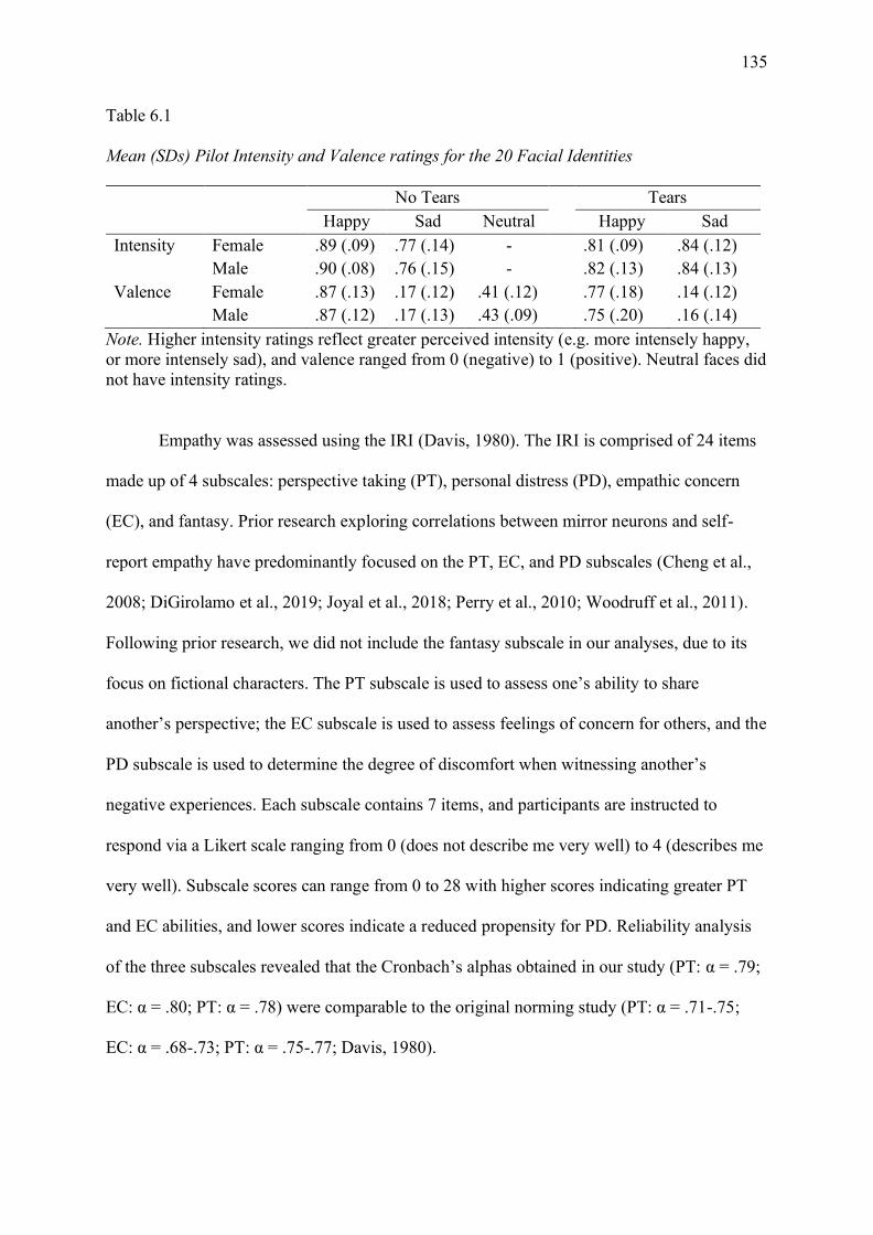
135
Table 6.1
Mean (SDs) Pilot Intensity and Valence ratings for the 20 Facial Identities
No Tears Tears Happy Sad Neutral Happy Sad Intensity Female .89 (.09) .77 (.14) - .81 (.09) .84 (.12) Male .90 (.08) .76 (.15) - .82 (.13) .84 (.13) Valence Female .87 (.13) .17 (.12) .41 (.12) .77 (.18) .14 (.12) Male .87 (.12) .17 (.13) .43 (.09) .75 (.20) .16 (.14)
Note. Higher intensity ratings reflect greater perceived intensity (e.g. more intensely happy, or more intensely sad), and valence ranged from 0 (negative) to 1 (positive). Neutral faces did not have intensity ratings.
Empathy was assessed using the IRI (Davis, 1980). The IRI is comprised of 24 items
made up of 4 subscales: perspective taking (PT), personal distress (PD), empathic concern
(EC), and fantasy. Prior research exploring correlations between mirror neurons and self-
report empathy have predominantly focused on the PT, EC, and PD subscales (Cheng et al.,
2008; DiGirolamo et al., 2019; Joyal et al., 2018; Perry et al., 2010; Woodruff et al., 2011).
Following prior research, we did not include the fantasy subscale in our analyses, due to its
focus on fictional characters. The PT subscale is used to assess one’s ability to share
another’s perspective; the EC subscale is used to assess feelings of concern for others, and the
PD subscale is used to determine the degree of discomfort when witnessing another’s
negative experiences. Each subscale contains 7 items, and participants are instructed to
respond via a Likert scale ranging from 0 (does not describe me very well) to 4 (describes me
very well). Subscale scores can range from 0 to 28 with higher scores indicating greater PT
and EC abilities, and lower scores indicate a reduced propensity for PD. Reliability analysis
of the three subscales revealed that the Cronbach’s alphas obtained in our study (PT: α = .79;
EC: α = .80; PT: α = .78) were comparable to the original norming study (PT: α = .71-.75;
EC: α = .68-.73; PT: α = .75-.77; Davis, 1980).

136
6.4.3.3 Procedure
The experiment was conducted in a sound attenuated electromagnetically shielded
room on a 24-inch Dell monitor. PsychoPy software (version 1.91.1) was used to present the
stimuli. All facial images were presented in full colour against a black background. The
experiment was conducted in two parts to examine both discrimination and execution. The
task design was an extension of van der Gaag et al. (2007) and as in their experiment, the
tasks were always presented in the same order, the discrimination task followed by the
execution task, to ensure that participants were not biased during the discrimination task.
6.4.3.3.1 Discrimination task
Each trial followed the same experimental sequence. Participants received instructions
for a delayed match-to-sample task on emotion (see Figure 6.1a), where images were
presented in pairs and participants had to report via button press whether the emotions of the
first and second image were the same or different. Participants were asked to make fast but
accurate judgments and it was confirmed they understood it was a reaction time task. Only
neural activity during the presentation of the first image was considered here, as it forms the
basis for understanding emotion comprehension, without the motor planning involved in
preparing for the reaction time task. The trial sequence began with the presentation of a
fixation cross, which varied in length of presentation time between 2500 and 3500ms
(fixation varied in blocks of 200 ms; see Figure 6.1a). We implemented a variable fixation
cross length to avoid participants’ anticipating the presentation of an image. We utilised
white fixation crosses to denote between trial fixation, and grey fixation crosses to denote
between image pair fixation. Image pairs were shown randomly, with each image being
shown twice in the first position. After the presentation of the inter-stimulus fixation cross, on
50% of trials the second image presented was a match (i.e. the same emotion) as the first
image. Finally, a response prompt was shown until a response was made, or for 2000 ms;

137
participants who took longer than 2000 ms were informed that their response was too slow.
Prior to the testing phase, 10 practice trials were administered to familiarise participants with
the procedure. Faces used during the practice trials were different to those used in the
experimental trials. The experiment was conducted in blocks of 40 trials (~ 7 minutes), and
each trial was followed by a 30 second break. After the participants completed this phase of
the experiment, they were informed about the execution task.
Figure 6.1. Experimental layout and timing of a) the discrimination task and b) the execution
task. In the execution task the circle was completely green on go trials and completely red on
no-go trials. KDEF stimulus in image is AF01.
6.4.3.3.2 Execution task
Participants were instructed that this task would follow a similar trial procedure to the
previous task and would involve imitation of the emotional faces. The trials adopted an
emotional go/no-go procedure, with participants instructed to reproduce the seen expression,
or to inhibit all responses. Only the neural activity captured during the presentation of the
image was analysed, as it allows for investigation of the neural responses associated with
138
intention to imitate, without the motor movement artifact obtained during expression
production. A go/no-go task was chosen to minimise mimicry during the presentation phase.
Participants were instructed to keep a neutral expression during trials except for during the go
phase. Trials began with a fixation cross, which was presented for between 2500 and 3500 ms
(varied in blocks of 200 ms; see Figure 6.1b). Then, an emotional image was presented for
2000 ms, in randomised order. This image was followed by a grey fixation cross for 2000 ms,
and then either a large green circle on go trials or a large red circle on no-go trials, which
appeared for 3000 ms. As in the discrimination task, every image was shown twice, and 50%
of the trials were go trials.
Once participants had completed both tasks, they completed the IRI. The IRI was
always presented as the last component of the experiment to avoid priming participants about
empathy. After participants had completed the experiment, we conducted a brief interview
asking participants about what they thought the purpose of the study was, and how they
believed they went in the tasks. After the interview, participants were debriefed and thanked
for their participation.
6.4.3.4 EEG recording and analysis
EEG recording took place in an electromagnetically and acoustically shielded room.
The EEG signal was recorded from 32 Ag/AgCl pin-type electrodes mounted in an elastic cap
according to the international 10-20 system, with two additional electrodes placed behind
each ear (i.e., mastoids). Eyeblinks were monitored using bipolar vertical EOG, with the
electrodes attached to the infraorbital and supraorbital regions of the left eye. EEG and EOG
were sampled using a Biosemi Active II system, with a 2048 Hz sampling rate.
All pre-processing steps were performed in Python using the MNE package (Gramfort
et al., 2013). All pre-processing techniques were in accord with the technique outlined in
Oberman, McCleery, Ramachandran, and Pineda (2007), and the script for analysing the data
139
is available on Open Science Framework (https://osf.io/6c3eu/). Data was downsampled to
512 Hz, re-referenced to the average of the left and right mastoids. Next, the data was
bandpass filtered between 0.1–30 Hz. The data was corrected for eyeblink artifacts using
independent component analysis, which is included in the MNE package. The data was
segmented into epochs ranging from 2000 ms pre-stimulus to 2000 ms post-stimulus, leading
to epochs with time length T = 4000 ms. Next the data was inspected for artifacts at the
central and occipital electrode sites of interest (i.e. C3, C4, O1, O2), and segments were
rejected if the EEG signal varied with a Peak to Peak difference of 100μV. Participants were
removed from the analysis if more than 60 trials were rejected (i.e. 30% of the 200 total
trials), in either the discrimination (average trials removed: M = 9.68, SD =14.78) or
execution (M = 9.89, SD =17.03) conditions. The number of trials removed did not differ
between the two tasks, t(74)= -.117, p = .907. For each cleaned segment the integrated power
in the 8–13Hz range was computed using a fast Fourier transform with a 1024 cosine
window. Mu power was calculated at the central electrode sites by computing a ratio score
where the mu power during stimulus presentation was divided by the mu power during the
baseline (i.e. the fixation cross). A ratio was chosen to control for individual differences in
scalp thickness. As ratio data are non-normal a log transform was applied. A log ratio of less
than zero indicates suppression of mu power, whereas positive values indicate enhancement,
and zero values reflect neither suppression nor enhancement. In addition, occipital electrodes
O1 and O2 were analysed as an index of alpha activity.
6.4.4 Results
6.4.4.1 Data exclusions
Of the 74 participants recruited for this study, 1 participant was excluded for failing to
follow task instructions, and a further 5 participants were excluded based on the EEG artifact

140
rejection criteria. Additionally, one participant failed to complete the IRI and as such they
were excluded from the correlational analyses.
6.4.4.2 Behavioural results
The percentage of correctly matched expressions during the discrimination task were
analysed to ensure participants were maintaining attention and that the stimuli were perceived
in a consistent manner. As indicated in Table 6.2, participants performed above chance when
matching emotional expressions, with a mean accuracy of 86.08% (SD = 7.73). We
conducted paired-samples t-tests to explore whether classification accuracy differed between
the tear and tear-free conditions, based on each emotion. Tear-free happy faces were
classified with greater accuracy than happy-tear faces, t(67) = 3.468, p < .001, d = .421,
BF10=27.677. Conversely, sad-tear faces were classified with significantly greater accuracy
than sad faces without tears, t(67) = 3.004, p = .004, d = .364, BF10=7.880. Collectively, these
results demonstrate that happy faces without tears and sad-tear faces are easier to classify
than their counterparts. As the task relied on the participants’ perception about whether the
face pairings were the same emotional expressions or different emotional expressions, we
anticipated some participants would believe the happy-tear condition reflected either tears of
joy or smiling through sadness, or that tearful sadness could be distinct from sadness without
tears. Thus, we did not exclude any individual trials based on accuracy, to maintain as much
EEG data as possible.
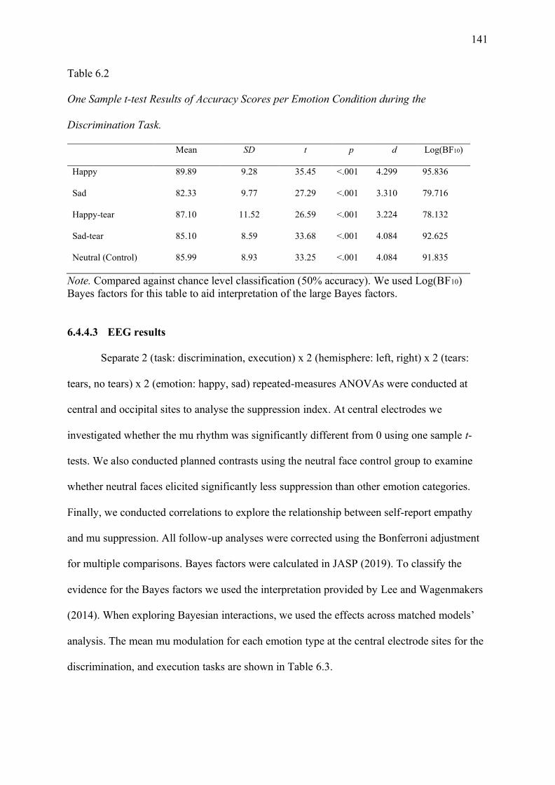
141
Table 6.2
One Sample t-test Results of Accuracy Scores per Emotion Condition during the
Discrimination Task.
Mean SD t p d Log(BF10)
Happy 89.89 9.28 35.45 <.001 4.299 95.836
Sad 82.33 9.77 27.29 <.001 3.310 79.716
Happy-tear 87.10 11.52 26.59 <.001 3.224 78.132
Sad-tear 85.10 8.59 33.68 <.001 4.084 92.625
Neutral (Control) 85.99 8.93 33.25 <.001 4.084 91.835
Note. Compared against chance level classification (50% accuracy). We used Log(BF10) Bayes factors for this table to aid interpretation of the large Bayes factors.
6.4.4.3 EEG results
Separate 2 (task: discrimination, execution) x 2 (hemisphere: left, right) x 2 (tears:
tears, no tears) x 2 (emotion: happy, sad) repeated-measures ANOVAs were conducted at
central and occipital sites to analyse the suppression index. At central electrodes we
investigated whether the mu rhythm was significantly different from 0 using one sample t-
tests. We also conducted planned contrasts using the neutral face control group to examine
whether neutral faces elicited significantly less suppression than other emotion categories.
Finally, we conducted correlations to explore the relationship between self-report empathy
and mu suppression. All follow-up analyses were corrected using the Bonferroni adjustment
for multiple comparisons. Bayes factors were calculated in JASP (2019). To classify the
evidence for the Bayes factors we used the interpretation provided by Lee and Wagenmakers
(2014). When exploring Bayesian interactions, we used the effects across matched models’
analysis. The mean mu modulation for each emotion type at the central electrode sites for the
discrimination, and execution tasks are shown in Table 6.3.

142
Table 6.3
Mean (SD) Mu Modulation at Central Electrodes for each Emotion.
Happy Sad Happy-tear Sad-tear Neutral
Discrimination
Left hemisphere .068 (.376) .075 (.357) .039 (.403) .026 (.401) .076 (.401)
Right hemisphere .105 (.370) .133 (.377) .119 (.410) .074 (.397) .129 (.390)
Averaged hemisphere .087 (.343) .104 (.343) .079 (.376) .050 (.368) .103 (.364)
Execution
Left hemisphere -.092 (.343) -.146 (.384) -.158 (.391) -.123 (.365) -.191 (.311)
Right hemisphere -.092 (.333) -.124 (.356) -.125 (.362) -.120 (.388) -.138 (.302)
Averaged hemisphere -.092 (.311) -.135 (.344) -.141 (.352) -.122 (.355) -.165 (.276)
6.4.4.3.1 Central electrodes
As predicted, we found a significant main effect of task, F(1,67) = 27.309, p < .001,
𝜂𝑝2 = .290, BFincl=7.361e+31, with the execution task eliciting greater mu suppression
compared to the discrimination task. Critically, a task by tears by emotion interaction was
observed, F(1,67) = 4.582, p = .036, 𝜂𝑝2 = .064, BFincl=.628; see Figure 6.2 for a graphical
depiction of this interaction.
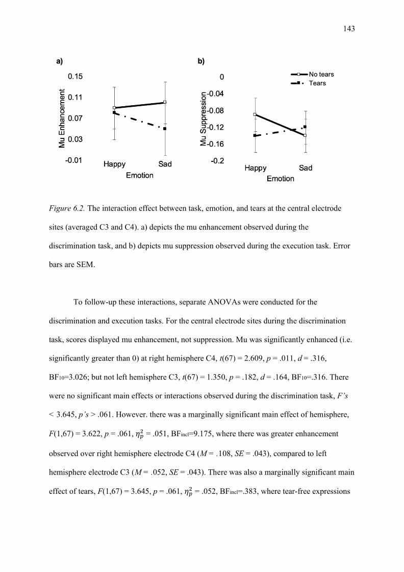
143
Figure 6.2. The interaction effect between task, emotion, and tears at the central electrode
sites (averaged C3 and C4). a) depicts the mu enhancement observed during the
discrimination task, and b) depicts mu suppression observed during the execution task. Error
bars are SEM.
To follow-up these interactions, separate ANOVAs were conducted for the
discrimination and execution tasks. For the central electrode sites during the discrimination
task, scores displayed mu enhancement, not suppression. Mu was significantly enhanced (i.e.
significantly greater than 0) at right hemisphere C4, t(67) = 2.609, p = .011, d = .316,
BF10=3.026; but not left hemisphere C3, t(67) = 1.350, p = .182, d = .164, BF10=.316. There
were no significant main effects or interactions observed during the discrimination task, F’s
< 3.645, p’s > .061. However. there was a marginally significant main effect of hemisphere,
F(1,67) = 3.622, p = .061, 𝜂𝑝2 = .051, BFincl=9.175, where there was greater enhancement
observed over right hemisphere electrode C4 (M = .108, SE = .043), compared to left
hemisphere electrode C3 (M = .052, SE = .043). There was also a marginally significant main
effect of tears, F(1,67) = 3.645, p = .061, 𝜂𝑝2 = .052, BFincl=.383, where tear-free expressions

144
(M = .095, SE = .041) elicited greater enhancement than tearful expressions (M = .064, SE =
.041).
For the execution task, significant suppression was observed over the left C3, t(67) = -
3.724, p < .001, d = -.452, BF10=58.256 and right C4, t(67) = -3.188, p = .002, d = -.387,
BF10=12.776, hemisphere electrodes. There was a marginally significant emotion by tears
interaction effect, F(1,67) = 3.609, p = .062, 𝜂𝑝2 = .051, BFincl=.653 (see Figure 2b). Although
this finding was not significant, we conducted follow-up analyses to test for our emotion
specific hypotheses. Follow-up paired t-tests revealed no significant differences in the
amount of suppression elicited between the happy and happy-tear faces, t(67) = 1.876, p =
.065, d = .228, BF10=.695; or the sad or sad-tear faces, t(67) = -.529, p = .598, d = -.064,
BF10=.152. No other main effects or interactions were significant during the execution task,
F’s < 1.574, p’s > .214.
Planned contrasts were conducted to compare the neutral face control condition to the
four emotional expression conditions at each central electrode site. As prior analyses
indicated that there was only enhancement during the observation task, we did not include
this condition in follow-up analyses. For the execution task, as there were no hemisphere
effects in prior analyses, we averaged across the left and right hemisphere electrodes. The
neutral face condition was not significantly different from any of the emotional categories,
F(1,67) = .2.866, p = .095, 𝜂𝑝2 = .041. Rather, neutral faces seemed to elicit larger suppression
relative to the emotional conditions (see Figure 3). To further investigate the contrast results,
we conducted Bayesian hypothesis testing to determine the degree of similarity between the
neutral control condition and the other emotion conditions. There was moderate support for
the null hypothesis that there was no significant difference between the neutral condition and
the tears-happy (BF01=5.648) and sad tear-free conditions (BF01=4.552). When comparing the
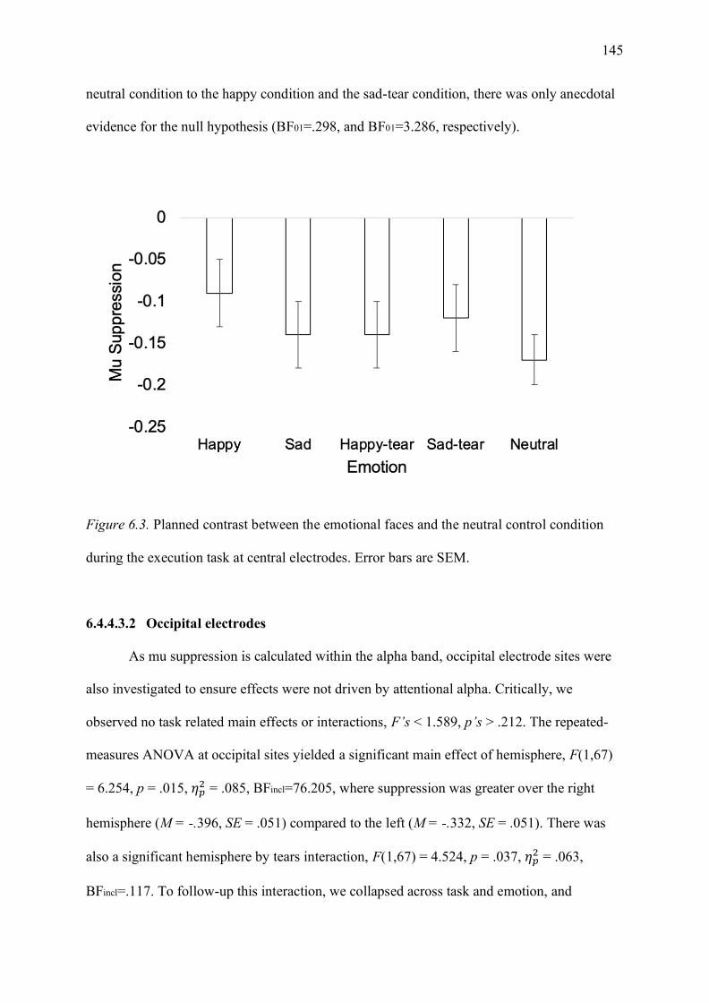
145
neutral condition to the happy condition and the sad-tear condition, there was only anecdotal
evidence for the null hypothesis (BF01=.298, and BF01=3.286, respectively).
Figure 6.3. Planned contrast between the emotional faces and the neutral control condition
during the execution task at central electrodes. Error bars are SEM.
6.4.4.3.2 Occipital electrodes
As mu suppression is calculated within the alpha band, occipital electrode sites were
also investigated to ensure effects were not driven by attentional alpha. Critically, we
observed no task related main effects or interactions, F’s < 1.589, p’s > .212. The repeated-
measures ANOVA at occipital sites yielded a significant main effect of hemisphere, F(1,67)
= 6.254, p = .015, 𝜂𝑝2 = .085, BFincl=76.205, where suppression was greater over the right
hemisphere (M = -.396, SE = .051) compared to the left (M = -.332, SE = .051). There was
also a significant hemisphere by tears interaction, F(1,67) = 4.524, p = .037, 𝜂𝑝2 = .063,
BFincl=.117. To follow-up this interaction, we collapsed across task and emotion, and

146
demonstrated that tearful faces did not yield increased suppression at left, t(67) = -.382, p =
.704, d = .046, BF10=.143 or right hemisphere electrode sites, t(67) = -1.698, p = .094, d = -
.206, BF10=.518.
6.4.4.4 Correlations between central electrodes and self-report empathy
Correlations were conducted to examine whether central electrode suppression was
related to self-report empathy as assessed using the PT, EC, and PD subscales of the IRI. As
in the contrast analyses, we limited our correlations to the execution condition and collapsed
across hemisphere. There were no significant correlations between averaged mu suppression
and the PT: r(66) = .029, p = .818, BF01=6.387; EC: r(66) = .141, p = .255, BF01=3.477; or
the PD: r(66) = .036, p = .772, BF01=6.292, subscales.
6.4.5 Discussion
The current study aimed to determine whether modulation of the mu rhythm is
sensitive to the presence of tears on happy and sad expressions of emotion. Converse to what
we predicted, we observed the greatest mu suppression in response to neutral, happy-tear, and
sad tear-free expressions. This provides support for the ambiguity account proposed by
Karakale et al. (2019), that ambiguous facial expressions require greater sensorimotor
engagement to determine the meaning of the expression. Additionally, we demonstrated that
task-related effects and interactions were only significant at central electrode sites. This
finding indicates that the activity indexed at occipital electrode sites was not sensitive to task-
related differences, providing support that sensorimotor mu is distinct from occipital alpha.
Finally, we failed to observe any significant correlations between mu suppression and self-
report empathy as indexed by the IRI.
Greater sensorimotor activation in response to ambiguous expressions has been noted
in EEG (Karakale et al., 2019) and fMRI (van der Gaag et al., 2007) literature. To isolate the
mu activity involved in motor planning, we conducted a stop-go-task where participants were
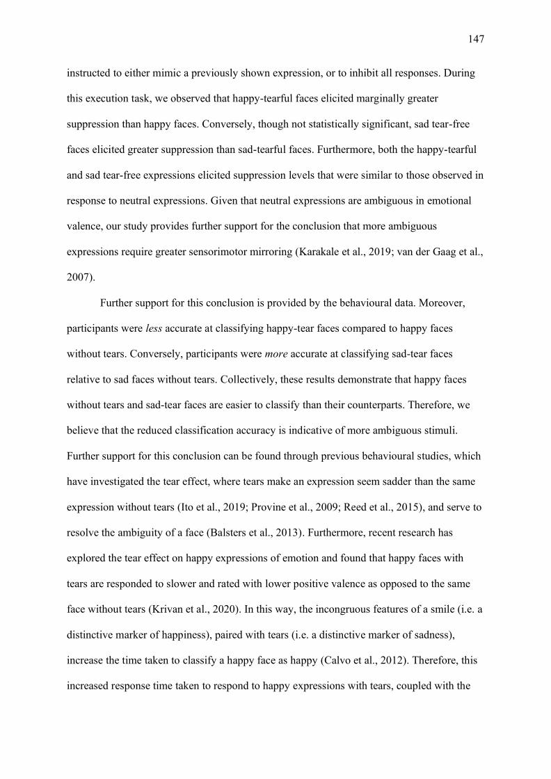
147
instructed to either mimic a previously shown expression, or to inhibit all responses. During
this execution task, we observed that happy-tearful faces elicited marginally greater
suppression than happy faces. Conversely, though not statistically significant, sad tear-free
faces elicited greater suppression than sad-tearful faces. Furthermore, both the happy-tearful
and sad tear-free expressions elicited suppression levels that were similar to those observed in
response to neutral expressions. Given that neutral expressions are ambiguous in emotional
valence, our study provides further support for the conclusion that more ambiguous
expressions require greater sensorimotor mirroring (Karakale et al., 2019; van der Gaag et al.,
2007).
Further support for this conclusion is provided by the behavioural data. Moreover,
participants were less accurate at classifying happy-tear faces compared to happy faces
without tears. Conversely, participants were more accurate at classifying sad-tear faces
relative to sad faces without tears. Collectively, these results demonstrate that happy faces
without tears and sad-tear faces are easier to classify than their counterparts. Therefore, we
believe that the reduced classification accuracy is indicative of more ambiguous stimuli.
Further support for this conclusion can be found through previous behavioural studies, which
have investigated the tear effect, where tears make an expression seem sadder than the same
expression without tears (Ito et al., 2019; Provine et al., 2009; Reed et al., 2015), and serve to
resolve the ambiguity of a face (Balsters et al., 2013). Furthermore, recent research has
explored the tear effect on happy expressions of emotion and found that happy faces with
tears are responded to slower and rated with lower positive valence as opposed to the same
face without tears (Krivan et al., 2020). In this way, the incongruous features of a smile (i.e. a
distinctive marker of happiness), paired with tears (i.e. a distinctive marker of sadness),
increase the time taken to classify a happy face as happy (Calvo et al., 2012). Therefore, this
increased response time taken to respond to happy expressions with tears, coupled with the
148
reduction in positive valence, indicates that happy tearful expressions—in the absence of
context—are more ambiguous than the same tear-free expressions.
Taken together, our results offer the most support for the ambiguity hypothesis, where
more ambiguous emotional expressions require greater sensorimotor engagement. It should
be noted however, that when examining our effect sizes and Bayes factors for comparisons
between our emotion conditions, we typically only provided weak or anecdotal evidence for
our results. Therefore, the effects that we are reporting are quite small, and as such the role of
tears in influencing sensorimotor mirroring responses in static displays is small as well.
However, in our study we used static, posed facial expressions of emotion, and added tears
which were identical on all facial displays. Mirror neurons have primarily been explored
through dynamic biological displays, where the same neurons are activated during
observation and execution of actions (Gallese et al., 1996; Rizzolatti et al., 1996).
Furthermore, several studies have demonstrated that mirror neurons are sensitive to the goal
or the intent of the action performed (di Pellegrino et al., 1992; Iacoboni et al., 2005). Thus,
our use of static facial stimuli and reliance on participants inferring facial movement, is
possibly a contributor to our small effects.
The use of static stimuli and reliance on participants inferring facial movement is also
a potential factor contributing to the enhancement effects observed during the discrimination
task. Although our study is the first to explore mu suppression and the execution of facial
expressions, several other studies have demonstrated mu suppression during observation of
facial stimuli (Cooper et al., 2013; Ensenberg et al., 2017; Gros et al., 2015; Joyal et al.,
2018; Karakale et al., 2019; Moore et al., 2012; Moore & Franz, 2017; Pineda & Hecht,
2009). Of these studies, the majority have used dynamic as opposed to static stimuli facial
stimuli (see Pineda and Hecht (2009) and Moore et al. (2012) for static examples). Although
some studies have demonstrated greater activity in the mirror neuron system during static

149
displays as opposed to dynamic displays (Enticott et al., 2008), others have reported the
opposite (Angelini et al., 2018; Buccino et al., 2001). Therefore, the role of sensorimotor
mirroring in response to both static and dynamic facial displays requires further investigation.
Furthermore, prior research has demonstrated that under some conditions mu is suppressed in
response to faces, whereas, in others, enhancement is elicited (Cooper et al., 2013; Gros et al.,
2015). For example, Gros et al. (2015) demonstrated that faces associated with high reward
conditions elicited suppression, whereas faces associated with low reward elicited
enhancement. Additionally, Cooper et al. (2013) found that observing happy expressions
elicited suppression over left hemisphere central electrodes, but enhancement over the right
hemisphere. Therefore, it does seem that in some instances, enhancement rather than
suppression is demonstrated to facial displays. Thus, further research is needed, using both
dynamic and static displays, to determine under what conditions facial expressions elicit mu
suppression.
An important consideration in mu suppression research is the ability to disentangle
mu from other perceptual processes such as occipital alpha (Hobson & Bishop, 2017). In our
study we observed significant task-related effects at the central electrode sites, but not at
occipital electrode sites. Put differently, regardless of the task condition, alpha was
consistently suppressed relative to baseline for emotional face stimuli. This finding indicates
that the mu suppression observed at central electrode sites is distinct from occipital alpha,
otherwise these effects would also be present at the occipital electrode sites. Additionally, a
hemisphere effect was evidenced at right hemisphere O2, relative to left hemisphere O1, in
line with a right hemisphere advantage in processing emotion (Dimberg & Petterson, 2000;
Gainotti, 2012). Therefore, despite the conflicting results observed in our discrimination and
execution tasks, the activity indexed at the central sites was distinct from that of occipital
alpha.
150
In considering the relationship between mu suppression and empathy, we predicted
that greater mu suppression scores would be correlated with scores on the PT, PD and EC
subscales of the IRI. This hypothesis was not supported as the correlations were not
significant. However, our null results are in line with other research exploring the relationship
between mu suppression and self-reported empathy. Several studies have demonstrated that
there is inconsistent evidence for the relationship between mu suppression and self-report
empathy. Woodruff et al. (2011) demonstrated a weak to moderate negative relationship
between self-reported empathy and mu suppression, whereas others have demonstrated a
positive relationship (Cheng et al., 2008; Yang et al., 2009). Additionally, other high powered
studies have failed to observed a significant relationship between self-report empathy and mu
suppression (DiGirolamo et al., 2019; Perry et al., 2010). These inconsistent findings indicate
that the relationship between the IRI and the mirror-neuron system as indexed by
sensorimotor mu suppression requires further research to determine whether the constructs
are mapping the same types of empathic responses.
There are some limitations and recommendations which we believe could be better
addressed in follow-up studies. Firstly, when investigating the enhancement effects observed
in our study, we explored the mu power of the baseline and stimulus conditions separately
(see Appendix A). We observed—not significant after Bonferroni correction—that the mu
power during the presentation of the fixation cross differed between the two task conditions.
Hobson and Bishop (2017) emphasised the importance of using comparable baseline
techniques across task conditions, and we further demonstrated that even when the baselines
are identical (i.e. a static fixation cross), there is still the potential for baseline differences to
occur.
In addition, we adapted a novel experimental paradigm developed by van der Gaag et
al. (2007), and modified this paradigm for EEG, which allowed for the exploration of
151
observation and execution planning in a single paradigm. This adaptation provides an
important advancement in the mu response to facial expressions research, given that prior
research focuses on observation-based designs. Further research should adopt methodologies
that explore both observation and execution in response to faces, as in the study described
herein. We believe that our paradigm could be further improved through the use of facial
electromyography, which would allow for the exploration of the spontaneous muscle
activation during the viewing of emotional expressions (Dimberg et al., 2000).
Additionally, there were some limitations in the stimuli chosen for our experiment.
We chose to use a standardised set of static emotional displays, and digitally added tears.
This technique ensured that there was as little difference between the tear-free and the tearful
displays as possible. However, static expressions are typically only presented in laboratory
settings, and thus are not as effective as dynamic displays. As mu is suppressed in response to
movement, the use of a dynamic stimulus will aid in strengthening the validity of the present
findings. Finally, it must also be cautioned that the perception of emotional tears as genuine
or false influences the way that people respond to tearful expressions—wherein displays
which are perceived as false do not receive pro-social responses (Krivan & Thomas, 2020;
Picó et al., 2020; Roeyen, Riem, Tončić, & Vingerhoets, 2020). Therefore, the use of
authentic crying stimuli is a worthy avenue of future investigation, which could be the key to
determining the role of empathy in responding to emotional expressions.
Our study aimed to further our understanding of the way that mu is modulated in
response to tearful facial expressions of emotion during both discrimination and execution
conditions. We provide support for the use of mu suppression to index mirror neuron system
involvement in the sharing of facial expressions—though caution that when static posed
displays are used as stimuli this effect is weak and largely influenced by task conditions.
Moreover, the present findings provide evidence of a complex relationship between
152
sensorimotor representation of expressions and the understanding of faces. Primarily, we
have provided further support for the theory that ambiguous emotional expressions require
greater sensorimotor engagement to interpret emotional expression. Furthermore, we have
demonstrated this support through the novel approach of using tearful facial expressions
whilst also demonstrating the tear effect at a psychophysiological level. The representation of
tears at the neural level we have observed in our study provides a psychophysiological basis
for the prosocial responses tears elicit from observers (Hareli & Hess, 2012; van de Ven et
al., 2017). Thus, when it comes to the role of the MNS as indexed by mu, the activation of
our own motor cortex aids in our understanding of others.
153
Chapter 7: Future Directions in Tear Research: Part I
7.1 Chapter Overview
As has been demonstrated in the four previous chapters, tears are a facial feature
which signal sadness in the absence of context and modulate neural processes associated with
emotion recognition. However, these studies have provided evidence that tears function as a
signal using stimuli which are artificial in nature (i.e. static, posed expressions which have
been digitally modified to include tears). One tenet which is yet to be addressed in this thesis
(and for the most part the field of crying as a whole), is whether the type of stimuli used
influence subsequent responses.
Previous studies have demonstrated that tearful displays are more sincere than tear-
free counterparts (Zeifman & Brown, 2011). Additionally, only one study has explored
perceptions of genuineness wherein tearful expressions were perceived to be more genuine
than the same expression without tears (Grainger et al., 2019)5. However, this study was
using stimuli captured in a moment of genuine experience, wherein the tearful expressions
were the genuine stimuli. For this reason, it cannot be assumed that the same perception of
genuineness would apply to a posed stimulus. Therefore, the final aim of this thesis was to
explore whether perceptions of genuineness were dependent on the type of tearful stimulus
used. This chapter details the perspective piece I authored wherein I explore the necessity for
empirical investigation of the type of stimuli used in tear research.
7.2 Publication Status
Published in its entirety.
Krivan, S. J., & Thomas, N. A. (2020). A call for the empirical investigation of tear stimuli.
Frontiers in Psychology, 11, 52. doi:10.3389/fpsyg.2020.00052
5 This study was published after the initial submission of this perspective piece, and as such is not cited.
154
7.3 Author Contributions
Krivan conceptualised and designed the article and wrote the first draft of the manuscript.
Krivan and Thomas contributed to manuscript revision, read and approved the submitted
version.
7.4 Manuscript
7.4.1 Abstract
Emotional crying is a uniquely human behaviour, which typically elicits helping and
empathic responses from observers. However, tears can also be used to deceive. “Crocodile
tears” are insincere tears used to manipulate the observer and foster prosocial responses. The
ability to discriminate between genuine and fabricated emotional displays is critical to social
functioning. When insincere emotional displays are detected, they are most often met with
backlash. Conversely, genuine displays foster prosocial responses. However, the majority of
crying research conducted to date has used posed stimuli featuring artificial tears. As such it
is yet to be determined how the artificial nature of these displays’ impacts person perception.
Throughout this article, we discuss the necessity for empirical investigation of the differences
(or similarities) in responses to posed and genuine tearful expressions. We will explore the
recent adoption of genuine stimuli in emotion research and review the existing research using
tear stimuli. We conclude by offering suggestions and considerations for future advancement
of the emotional crying field through investigation of both posed and genuine tear stimuli.
Keywords: tear effect, face perception, adult crying, emotion, interpersonal communication,
crocodile tears
7.4.2 Introduction
Why do we cry? Emotional crying is a uniquely human display that has fascinated
both scientists and lay people alike; this interest stems from an attempt to determine the
functions of adult emotional tearing. A popular theory is that emotional tears serve a
155
communicative function (Hendriks et al., 2008; Reed et al., 2015; Vingerhoets et al., 2016).
Although tears have been readily touted as an honest signal of emotion (Trimble, 2012;
Vingerhoets, 2013), there is a lack of empirical evidence to justify this perception.
Furthermore, tears reliably elicit empathic responses (Lockwood et al., 2013) and social
support from observers (Vingerhoets et al., 2016), whilst also signalling appeasement, which
serves to reduce aggression (Hasson, 2009). While the presence of tears on a face can signal
the need for social support, tears are also used to manipulate and deceive.
The accurate detection of emotional deception is critical to social functioning. Fake
tears, or ‘crocodile tears’ are an insincere tearing display that evokers use to elicit sympathy
and support from observers. How evokers produce these insincere tears is not yet known.
Crocodile tears are typically associated with the disingenuous tears of celebrities and
politicians (Manusov & Harvey, 2011), and conveying fabricated remorse during criminal
court proceedings (ten Brinke, MacDonald, Porter, & O'Connor, 2012). Alexander (2003)
found that insincere narcissistic crying appears empty and orchestrated, and that witnessing
this tearful display results in feeling uneasy and unmoved in a therapeutic environment. As
such, insincere emotional displays elicit negative responses (Hideg & van Kleef, 2017) and
reduced trust (Krumhuber et al., 2007). However, crocodile tears could also be elicited via
deep acting where the evoker draws on previous experience in an effort to feel the emotion
they are displaying (Lu et al., 2019). As such, tears elicited in this manner are driven by
genuine feeling, however, are acted and physically effortful by nature. Given that tears are
known to increase perceptions of remorse during apologies (Hornsey et al., 2019), and
remorse is an important factor in sentencing and parole hearings (Bandes, 2016), further
research exploring how we distinguish between sincere and crocodile tears is needed.
Despite the negative connotations associated with insincere emotion, most crying
research has used standardised or posed faces featuring artificial tears. Although these studies

156
have demonstrated that tears are responded to favourably (Balsters et al., 2013; Lockwood et
al., 2013), how the artificial nature of these displays impact person perception is yet to be
determined. We call for the empirical investigation of the perception of both posed and
genuine emotional tear displays. We first discuss the movement towards the adoption of
genuine and ecologically valid stimuli in emotion research. Then, we explore research
utilising images of crying faces and highlight the advancements achieved and potential
implications for the posed face methodology. Furthermore, we discuss recommendations for
future research that highlight the perceptual differences between posed and genuine tearful
displays. Finally, we conclude it is necessary to explore perceptions of genuine and
disingenuous crying and believe posed and genuine stimuli can aid in this investigation.
7.4.3 Genuine emotional displays
The recent movement towards using genuine expressions has predominantly stemmed
from human ability to determine the genuineness of emotional displays (McLellan, Johnston,
Dalrymple-Alford, & Porter, 2010). Primarily, genuine expression research has investigated
the difference between Duchenne and non-Duchenne smiles (Duchenne, 1862/1990; Ekman,
Davidson, & Friesen, 1990) . Smiles are characterised by the activation of the zygomaticus
major (i.e., the muscle responsible for drawing the corners of the mouth upwards), while
Duchenne smiles feature both zygomaticus and orbicularis oculi activation (i.e., the muscle
associated with the crinkling of the eyes). Duchenne smiles are reliably judged as more
intensely happy (Leppänen & Hietanen, 2007), and are mimicked more than non-Duchenne
smiles (Krumhuber et al., 2014). Additionally, when mimicry is constrained, people are less
accurate at recognising emotional expressions (Oberman, Winkielman, & Ramachandran,
2007) and they display a reduced ability to discriminate between posed and genuine smiles
(Rychlowska et al., 2014).
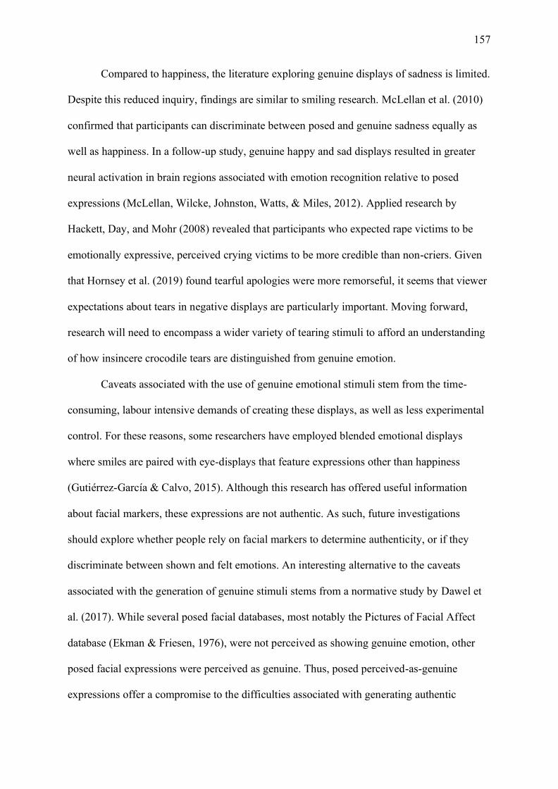
157
Compared to happiness, the literature exploring genuine displays of sadness is limited.
Despite this reduced inquiry, findings are similar to smiling research. McLellan et al. (2010)
confirmed that participants can discriminate between posed and genuine sadness equally as
well as happiness. In a follow-up study, genuine happy and sad displays resulted in greater
neural activation in brain regions associated with emotion recognition relative to posed
expressions (McLellan, Wilcke, Johnston, Watts, & Miles, 2012). Applied research by
Hackett, Day, and Mohr (2008) revealed that participants who expected rape victims to be
emotionally expressive, perceived crying victims to be more credible than non-criers. Given
that Hornsey et al. (2019) found tearful apologies were more remorseful, it seems that viewer
expectations about tears in negative displays are particularly important. Moving forward,
research will need to encompass a wider variety of tearing stimuli to afford an understanding
of how insincere crocodile tears are distinguished from genuine emotion.
Caveats associated with the use of genuine emotional stimuli stem from the time-
consuming, labour intensive demands of creating these displays, as well as less experimental
control. For these reasons, some researchers have employed blended emotional displays
where smiles are paired with eye-displays that feature expressions other than happiness
(Gutiérrez-García & Calvo, 2015). Although this research has offered useful information
about facial markers, these expressions are not authentic. As such, future investigations
should explore whether people rely on facial markers to determine authenticity, or if they
discriminate between shown and felt emotions. An interesting alternative to the caveats
associated with the generation of genuine stimuli stems from a normative study by Dawel et
al. (2017). While several posed facial databases, most notably the Pictures of Facial Affect
database (Ekman & Friesen, 1976), were not perceived as showing genuine emotion, other
posed facial expressions were perceived as genuine. Thus, posed perceived-as-genuine
expressions offer a compromise to the difficulties associated with generating authentic

158
stimuli, while allowing additional control. This advancement is particularly important for tear
research, as it is currently unknown whether posed-tearful displays are perceived as
perceptually genuine. As such, it is important that future investigations explore whether there
are differences (or similarities) between the posed expressions typically used in crying
research and genuine tearful stimuli.
7.4.4 The artificial tear
Most existing research investigating the perception of emotional tearing uses posed
facial expressions that feature artificial tears, added using eyedrops or digital enhancement
(Ito et al., 2019; Reed et al., 2015). These artificial images have been used to explore how the
presence of tears influences the perception of sadness (Hendriks et al., 2007; Ito et al., 2019),
and the degree of helping behaviours elicited (Balsters et al., 2013; Hendriks & Vingerhoets,
2006; Lockwood et al., 2013). When images with visible tears are perceived as significantly
sadder than the same image without tears it is referred to as the tear effect (Provine et al.,
2009).
In exploring perceptions of sadness, tears are typically added to sad and neutral faces,
and various measures (e.g., reaction time, rating scales, and electroencephalography) are
employed to examine how tears are perceived (Balsters et al., 2013; Hendriks et al., 2007).
Balsters et al. (2013) demonstrated that even when brief presentations of tearful sad and
neutral faces are shown, participants correctly categorise perceived sadness faster for sad
expressions with tears, relative to sad and neutral tear-free expressions. However,
contradictory evidence has been demonstrated when exploring the affective ratings of
Duchenne smiles featuring tears. Reed et al. (2015) demonstrated that a tearful Duchenne
smile was perceived as more intense than the tear-free counterpart. Furthermore, a trend
towards increased happiness ratings was observed for the tearful Duchenne face. Thus, it is
possible that Duchenne smiles signify genuine joy when they are accompanied by tears, akin
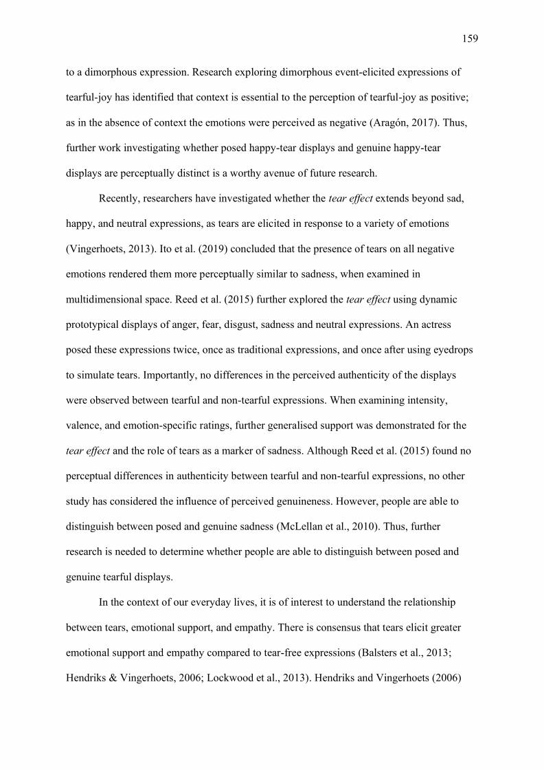
159
to a dimorphous expression. Research exploring dimorphous event-elicited expressions of
tearful-joy has identified that context is essential to the perception of tearful-joy as positive;
as in the absence of context the emotions were perceived as negative (Aragón, 2017). Thus,
further work investigating whether posed happy-tear displays and genuine happy-tear
displays are perceptually distinct is a worthy avenue of future research.
Recently, researchers have investigated whether the tear effect extends beyond sad,
happy, and neutral expressions, as tears are elicited in response to a variety of emotions
(Vingerhoets, 2013). Ito et al. (2019) concluded that the presence of tears on all negative
emotions rendered them more perceptually similar to sadness, when examined in
multidimensional space. Reed et al. (2015) further explored the tear effect using dynamic
prototypical displays of anger, fear, disgust, sadness and neutral expressions. An actress
posed these expressions twice, once as traditional expressions, and once after using eyedrops
to simulate tears. Importantly, no differences in the perceived authenticity of the displays
were observed between tearful and non-tearful expressions. When examining intensity,
valence, and emotion-specific ratings, further generalised support was demonstrated for the
tear effect and the role of tears as a marker of sadness. Although Reed et al. (2015) found no
perceptual differences in authenticity between tearful and non-tearful expressions, no other
study has considered the influence of perceived genuineness. However, people are able to
distinguish between posed and genuine sadness (McLellan et al., 2010). Thus, further
research is needed to determine whether people are able to distinguish between posed and
genuine tearful displays.
In the context of our everyday lives, it is of interest to understand the relationship
between tears, emotional support, and empathy. There is consensus that tears elicit greater
emotional support and empathy compared to tear-free expressions (Balsters et al., 2013;
Hendriks & Vingerhoets, 2006; Lockwood et al., 2013). Hendriks and Vingerhoets (2006)
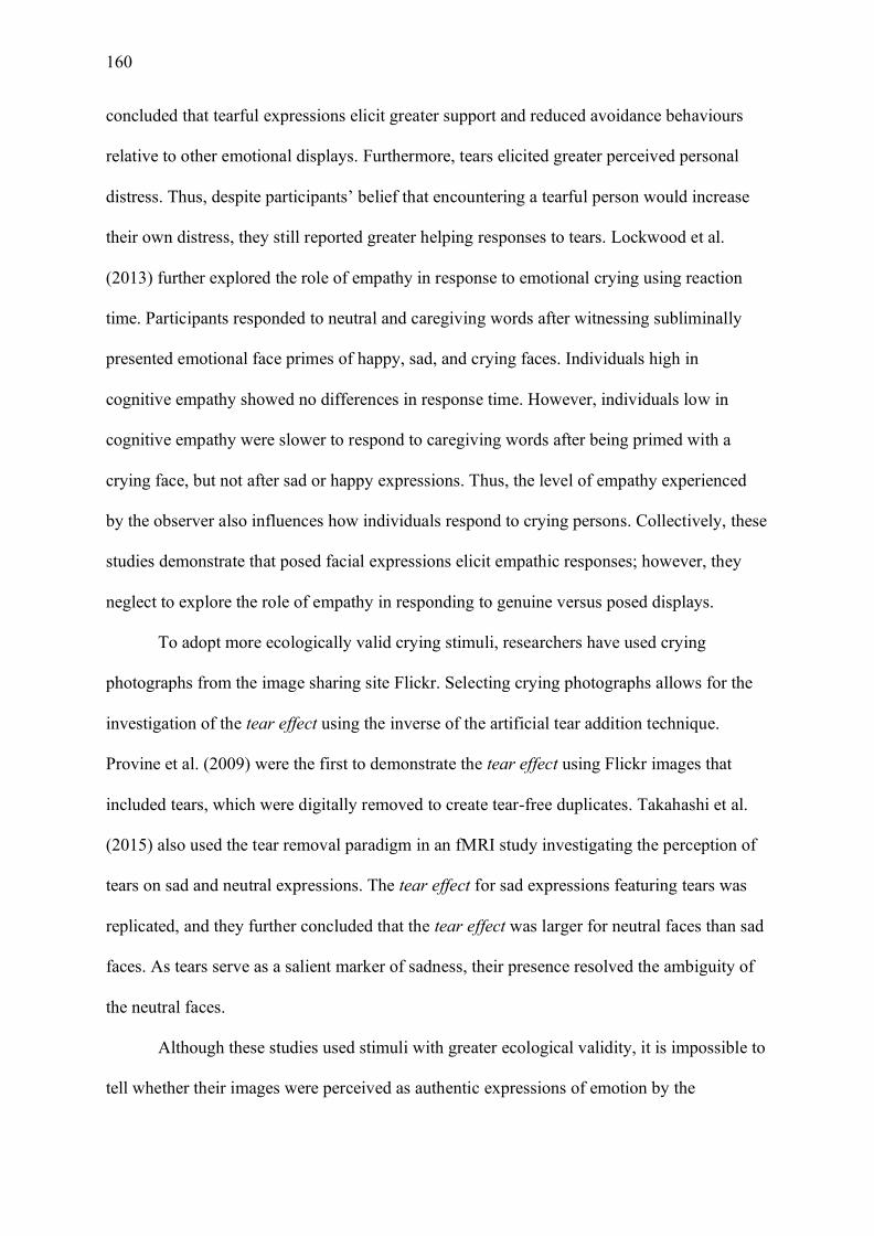
160
concluded that tearful expressions elicit greater support and reduced avoidance behaviours
relative to other emotional displays. Furthermore, tears elicited greater perceived personal
distress. Thus, despite participants’ belief that encountering a tearful person would increase
their own distress, they still reported greater helping responses to tears. Lockwood et al.
(2013) further explored the role of empathy in response to emotional crying using reaction
time. Participants responded to neutral and caregiving words after witnessing subliminally
presented emotional face primes of happy, sad, and crying faces. Individuals high in
cognitive empathy showed no differences in response time. However, individuals low in
cognitive empathy were slower to respond to caregiving words after being primed with a
crying face, but not after sad or happy expressions. Thus, the level of empathy experienced
by the observer also influences how individuals respond to crying persons. Collectively, these
studies demonstrate that posed facial expressions elicit empathic responses; however, they
neglect to explore the role of empathy in responding to genuine versus posed displays.
To adopt more ecologically valid crying stimuli, researchers have used crying
photographs from the image sharing site Flickr. Selecting crying photographs allows for the
investigation of the tear effect using the inverse of the artificial tear addition technique.
Provine et al. (2009) were the first to demonstrate the tear effect using Flickr images that
included tears, which were digitally removed to create tear-free duplicates. Takahashi et al.
(2015) also used the tear removal paradigm in an fMRI study investigating the perception of
tears on sad and neutral expressions. The tear effect for sad expressions featuring tears was
replicated, and they further concluded that the tear effect was larger for neutral faces than sad
faces. As tears serve as a salient marker of sadness, their presence resolved the ambiguity of
the neutral faces.
Although these studies used stimuli with greater ecological validity, it is impossible to
tell whether their images were perceived as authentic expressions of emotion by the

161
participants. As Flickr is a website where people primarily upload their own images to share
with friends and followers, images shared to the platform are likely posed and self-selected
by the individual to present themselves in a positive manner (Angus & Thelwall, 2010;
Malinen, 2010). Thus, posed datasets allowed for the investigation of the perception of tears
with rigorous control (Balsters et al., 2013; Lockwood et al., 2013); and stimuli with greater
ecological validity have replicated these effects (Provine et al., 2009; Takahashi et al., 2015);
however, the need for research using genuine tearful expressions remains.
7.4.5 The genuine tear
Recently, researchers have begun to use photographic stimuli featuring emotional
tearing, which were captured in a moment of genuine emotional experience. These images
were captured during the Museum of Modern Art, Artist is Present exhibit, where nearly
1000 people sat with Marina Abramović and cried during the experience. As these tears were
elicited in a moment of genuine emotion, these studies have investigated the perceived
warmth and competence of the crying persons (van de Ven et al., 2017; Zickfeld & Schubert,
2018; Zickfeld et al., 2018), as well as the perceived social-connectedness and willingness to
provide help to crying persons (Stadel et al., 2019; Vingerhoets et al., 2016). The original
study by van de Ven et al. (2017) concluded that tearful individuals were perceived as
warmer, though less competent, than tear-free individuals. Two replications of this study also
determined that tearful individuals were perceived as warmer; however, neither study
replicated the reduced competence effect when using a larger sample of target crying faces
(Zickfeld & Schubert, 2018; Zickfeld et al., 2018). Zickfeld et al. (2018) concluded that the
competence effect from the original study was likely target specific, and thus the presence of
tears is unlikely to alter perceptions of competence.
Importantly, the work conducted using genuine tear stimuli has also replicated the
findings that emotional tears elicit support and willingness to help. Vingerhoets et al. (2016)
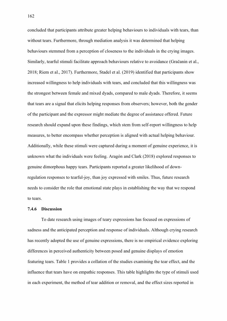
162
concluded that participants attribute greater helping behaviours to individuals with tears, than
without tears. Furthermore, through mediation analysis it was determined that helping
behaviours stemmed from a perception of closeness to the individuals in the crying images.
Similarly, tearful stimuli facilitate approach behaviours relative to avoidance (Gračanin et al.,
2018; Riem et al., 2017). Furthermore, Stadel et al. (2019) identified that participants show
increased willingness to help individuals with tears, and concluded that this willingness was
the strongest between female and mixed dyads, compared to male dyads. Therefore, it seems
that tears are a signal that elicits helping responses from observers; however, both the gender
of the participant and the expressor might mediate the degree of assistance offered. Future
research should expand upon these findings, which stem from self-report willingness to help
measures, to better encompass whether perception is aligned with actual helping behaviour.
Additionally, while these stimuli were captured during a moment of genuine experience, it is
unknown what the individuals were feeling. Aragón and Clark (2018) explored responses to
genuine dimorphous happy tears. Participants reported a greater likelihood of down-
regulation responses to tearful-joy, than joy expressed with smiles. Thus, future research
needs to consider the role that emotional state plays in establishing the way that we respond
to tears.
7.4.6 Discussion
To date research using images of teary expressions has focused on expressions of
sadness and the anticipated perception and response of individuals. Although crying research
has recently adopted the use of genuine expressions, there is no empirical evidence exploring
differences in perceived authenticity between posed and genuine displays of emotion
featuring tears. Table 1 provides a collation of the studies examining the tear effect, and the
influence that tears have on empathic responses. This table highlights the type of stimuli used
in each experiment, the method of tear addition or removal, and the effect sizes reported in

163
the published literature. It must be noted, that the type of task, the number of identities used,
and the gender of the stimuli varied widely across these studies. This variability further
highlights the need for empirical studies using both posed and genuine tearful expressions.
This empirical investigation will assist with better understanding the perceptual differences
between tear stimuli and aid in our understanding of how we discriminate genuine and posed
emotion.
Table 7.1
A Comparison of the Effect Sizes Reported in Published Studies Examining Tears
Authors Stimulus type Tear method Effect size
Faster reaction time to tearful images Balsters et al. (2013) KDEF Digitally added 𝜂2 = .284† Gračanin et al. (2018) MoMA Digitally removed 𝜂2
𝑝 = .26†
Riem et al. (2017) MoMA Digitally removed 𝜂2𝑝 = .69 †
Greater perceived sadness for tearful images Provine et al. (2009) Flickr tear images Digitally removed 𝜂2= .26† Takahashi et al. (2015) Flickr tear images Digitally removed 𝜂2
𝑝 = .793*
Reed et al. (2015) Female actress using FACS Eye-drops d = .22 Ito et al. (2019) TFEID Digitally added 𝜂2
𝑝 = .073
van de Ven et al. (2017) MoMA Digitally removed 𝜂2
𝑝 = .15†
Zickfeld et al. (2018) MoMA Digitally removed d = .86 Greater willingness to help / greater perceived support for tearful images
Balsters et al. (2013) KDEF Digitally added 𝜂2 = .375† Vingerhoets et al. (2016) MoMA Digitally removed d = .85 – 1.32 Zickfeld and Schubert (2018) MoMA Digitally removed dS = .70 – .82
Note. KDEF – Karolinska Directed Emotional Faces; MoMA – Genuine tear expressions captured during Museum of Modern Art Performance; TFEID – Taiwanese Facial Expression Image Database; Flickr tear images – images of tearful individuals found on Flickr (unknown if genuine or posed). Effect sizes are reported as in the published papers. *denotes that original paper did not report effect size, and thus it was estimated from main effect of tears. †denotes effect size from main effect.
Furthermore, as the majority of the work conducted to date has used posed
expressions, there has been limited focus on the other facial responses that accompany
emotional tears, including blotchy faces and bloodshot eyes (Provine, Cabrera, Brocato, &
Krosnowski, 2011; Provine, Nave-Blodgett, & Cabrera, 2013). Küster (2018) explored the
164
influence of tears and pupil size on the perception of sadness using digital avatars. While both
the presence of tears and smaller pupil sizes increased perceived sadness, there was no
interaction effect between tears and pupil size. The inverse consideration of the extreme
features accompanying emotional crying is the perceptual and affective differences between
tearing up and crying uncontrollably (i.e., ugly crying). Research using vignettes has
demonstrated that the intensity of tears moderates observer reactions, where in some
scenarios just tearing up may elicit more positive responses than weeping (Wong, Steinfeldt,
LaFollette, & Tsao, 2011). Thus, further work in this field should explore the relationship
between the intensity of the tears and observer responses. It may be that assistance for
emotional crying is curvilinear, where there is an optimum level of tearing that elicits helping
responses from observers.
Finally, the adoption of investigative techniques like psychophysiology may offer
insight into the perceptions of tears to further corroborate the results from self-report studies.
Recently, mirror neurons have been proposed as a mechanism for sharing others emotional
states, with ‘feeling’ and ‘perceiving’ emotion sharing neural substrates (Singer et al., 2004;
Wicker et al., 2003). Similarly, facial mimicry studies have identified that when participants’
ability to mimic is impaired, they show reduced emotion recognition abilities (Oberman et al.,
2007; Rychlowska et al., 2014). In addition, examination of other physiological techniques,
such as eye-tracking and galvanic skin response, may yield fruitful information about the
features that individuals attend to in decoding an emotional face, and the degree of arousal
that tearful expressions elicit. Analysis of the arousal response may assist in determining the
motivation for the helping behaviours as a metric of personal distress. Furthermore, the
inclusion of psychophysiological metrics allows for greater certainty in the true nature of the
self-report responses.

165
In this paper, we have reviewed recent work using facial expressions as a means of
investigating inter-individual functions of crying. Reviewing these studies has revealed that
the use of posed expressions has afforded an understanding of the communicative functions
of emotional tears by employing rigorously controlled stimuli between conditions. In
addition, the use of genuine expressions of emotion in more recent crying research has
replicated findings that both posed and genuine expressions of emotion are effective at
eliciting support and attention. However, whether posed tearful expressions are being treated
as perceptually authentic, or if their staged nature is impacting upon person perception is yet
to be determined. Thus, to continue advancing the understanding about the interpersonal
functions of human emotional tearing, we need to adopt an approach that better explores how
we perceive both genuine and non-genuine crying expressions. This advancement needs to
encompass a greater range of tearing stimuli to allow for the exploration of the physiological
effects that accompany emotional tearing. This research will provide a basis for
understanding the type of emotional tears we respond to. People can distinguish between
posed and genuine emotions, yet tears have not received this same inquiry. Determining how
we distinguish between posed and genuine tearful expressions, will aid in further
understanding the functions of this uniquely human phenomenon.
166
Chapter 8: Future Directions in Tear Research: Part II
8.1 Chapter Overview
Following the perspective piece outlined in the previous chapter, I conducted three
experiments exploring the perceptions of genuineness for three types of tearful stimuli. The
first stimuli are tears that were elicited during a moment of genuine experience. These
genuine tearful displays are the most authentic tear stimuli presently used in crying research.
Secondly, I used the Karolinska Directed Emotional Faces (KDEF) displays, which I have
used throughout my thesis, and digitally modified these images to include tears. This
procedure allowed for the exploration of how my KDEF tear stimuli are perceived relative to
a genuine tearful stimulus. Finally, I also included a sample of posed faces, wherein the
individuals were instructed to pose as though they were crying. I included this third sample of
images, to explore whether posed crying (with artificial tears added digitally) was perceived
as more genuine than posed sadness (with artificial tears digitally added). The following
chapter details the results of three experiments exploring the perceptions of genuineness
relating to tearful individuals.
8.2 Publication Status
Manuscript in preparation for publication.
8.3 Author Contributions
Krivan – designed the experiments, created the experiments, collected the data, conducted
the data analyses, wrote the first and revised drafts of the manuscript.
Thomas – aided in experimental design, provided funding for the experiments, supervised the
research, and aided in editing the manuscript.
8.4 Manuscript
8.4.1 Abstract
167
Numerous studies have demonstrated that tearful expressions elicit increased support,
aid and succour from observers relative to tear-free expressions. Although there is consensus
that tears are responded to favourably, there is a limited understanding of why tears are such
an effective communicative display. One proposed facilitator of these favourable responses is
that tears are perceived as an honest expression of emotion, yet there have been limited
empirical tests of this assumption. This study sought to address this gap by examining
whether participants are sensitive to the genuineness of tearful stimuli. We conducted a series
of experiments examining whether participants could distinguish between shown and felt
emotion, and whether responses to genuine word primes were facilitated by genuine tearful
displays. The results demonstrate that participants are sensitive to the genuineness of
emotional tears. Furthermore, tears increased the perceived genuineness of posed displays.
Finally, we did not observe any influence of facial prime on responses to positive and
negative words associated with genuineness. Collectively, these results demonstrate that
observers are sensitive to the genuineness of emotional tears and highlights the importance of
experimental stimuli in tear research.
8.4.2 Introduction
Emotional tears are a universally recognised expression of emotion that command
attention from observers (Vingerhoets, 2013). This attention is typically manifested as pro-
social responses from observers. Tearful displays are known to elicit greater empathy,
emotional support, and helping responses, relative to tear-free expressions (Hendriks et al.,
2008; Vingerhoets et al., 2016). Furthermore, tears foster approach, rather than avoidance,
behaviours and signal appeasement to reduce aggression (Gračanin et al., 2018; Hasson,
2009). As such, tearful displays have been touted as an honest distress signal (Trimble, 2012;
Vingerhoets, 2013), despite the lack of empirical evidence for this conclusion (see Krivan
and Thomas (2020) for a review of this perspective). However, tears can also be used to
168
manipulate and deceive. Crocodile tears are an insincere crying display that are designed to
elicit pro-social responses from observers. For example, a person may feign sadness through
a tearful display either to elicit sympathy from observers or to convey remorse (ten Brinke et
al., 2012). As such, crocodile tears are typically associated with the disingenuous tears of
politicians and celebrities (Manusov & Harvey, 2011). Thus, the ability to distinguish
between posed and genuine tears is integral to avoiding manipulation from insincere criers.
Evidently, the accurate decoding of facial expressions is critical to social functioning.
Typically, facial expressions are investigated as a tool of social communication wherein
facial emotion is a means of signalling affective state. However, not all communicated facial
expressions are actually felt by the expressor. In these instances, expressions are posed or
feigned by an expressor, and as such convey little information about affective state (Douglas,
Porter, & Johnston, 2012). Posed displays can be used to conceal true emotions (Gutiérrez-
García & Calvo, 2015), fake genuine experience (Hess & Kleck, 1990), and even to deceive
and manipulate observers (Krumhuber & Manstead, 2009; McLellan et al., 2010). As such,
posed expressions are deliberate, purposeful displays that intentionally convey emotion
(Ekman & Friesen, 1982; Hess & Kleck, 1990; Motley & Camden, 1988). By contrast,
genuine emotional displays are spontaneous and congruent to one’s affective state (Dawel,
Palermo, O’Kearney, & McKone, 2015; McLellan et al., 2010). As such, genuine emotional
displays can be considered “event-elicited” (Dawel et al., 2017), “spontaneous”, or “emotion-
induced” (Hess & Kleck, 1990). The ability to accurately decode whether a facial expression
is genuine or posed has ramifications for effective social communication. For example, aiding
an individual crying crocodile tears could result in being manipulated by a deceptive
individual. A further example of ineffective social communication would be reciprocating a
smile that was used to mask anger (Gosselin, Beaupré, & Boissonneault, 2002). As a result,
169
researchers have increasingly become interested in how we discriminate genuine from posed
emotion.
One method that is frequently used to determine the authenticity of facial displays is
physical facial markers. Within the Facial Action Coding System, muscular facial movements
are separated into distinct action units (AU: Ekman & Friesen, 1978). These action units
quantify muscular activation and are typically associated with specific emotional expressions.
For example, a distinctive marker of happiness is the smile (Becker & Srinivasan, 2014;
Gutiérrez-García & Calvo, 2015), where the lips are pulled upwards towards the ears, known
as AU 12. However, genuine enjoyment features an additional physical marker (Frank,
Ekman, & Friesen, 1993). Known as the Duchenne smile, genuine enjoyment pairs AU 12
with the crinkling of the crow’s feet around the eyes (AU 6: Ekman, Davidson, & Friesen,
1990). Although not as well researched as genuine enjoyment, genuine sadness also has
distinctive facial markers, which are signalled through AU’s 1 and 4, wherein the eyebrows
are drawn upwards and together (Ekman, 2003; Ekman & Rosenberg, 2005). These AU of
genuine enjoyment and sadness are argued to be markers of authenticity as they are difficult
to pose (Mehu, Mortillaro, Bänziger, & Scherer, 2012), and are often present in genuine but
not posed emotion (Ekman, 2003; Ekman, Friesen, & O'Sullivan, 1988).
The ability to distinguish between posed and genuine emotion is typically investigated
using tasks where participants determine whether the faces are showing (i.e. does the face
look sad) or feeling (i.e. was the person feeling sad) a particular emotion (McLellan et al.,
2010; McLellan et al., 2012; Namba, Kabir, Miyatani, & Nakao, 2018). In studies of
genuineness, superior performance is typically observed in response to happy facial displays
(Dawel et al., 2015; McLellan et al., 2010). However, the limited research investigating
discrimination of sad facial expressions has shown that when participants are expressly told
to focus on the affective state of the stimuli, genuine sadness is discriminated equally as well
170
as genuine happiness (McLellan et al., 2010; Namba et al., 2018). Furthermore, McLellan et
al. (2012) used neuroimaging to demonstrate that genuine displays of happiness and sadness
elicit significantly greater activation of brain regions associated with emotion perception and
intentional attribution. As such, the ability to discriminate genuine from posed emotion is
greatest for happiness and requires a focus on affective state for sadness.
A shortcoming of the existing literature must be acknowledged. Namely, the majority
of work discussed thus far has relied on a stimulus set developed by McLellan et al. (2010).
These stimuli feature 17 females who were informed that the purpose of the experiment was
to develop a stimulus set of both genuine and posed displays. Although genuine expressions
were elicited via emotion-inducing films, potentially the stimuli were biased by demand
characteristics. Participants’ knowledge of the task, combined with the explicit instruction to
“look into the camera as much as possible”, potentially inhibited truly spontaneous facial
reactions. Furthermore, a normative study by Dawel et al. (2017) concluded that, with the
exception of happiness and disgust, the genuine stimuli from the McLellan stimulus set were
not perceived as genuine. Rather, angry, fearful, and sad expressions were found to be
ambiguous in perceived genuineness. As such, further research should strive to adopt a
stimulus set comprised of males and females, where emotion is captured in a moment of
genuine experience.
Although early research into the communicative functions of emotional tears relied on
posed stimuli and the use of artificial tears (Balsters et al., 2013; Fischer et al., 2013;
Hendriks & Vingerhoets, 2006), recent research has adopted the use of a genuine stimulus set
(Stadel et al., 2019; van de Ven et al., 2017; Vingerhoets et al., 2016). These tearful displays
were captured in a moment of genuine experience, and feature males and females of different
ages and cultures. As such, these genuine tearful displays make for a rich dataset for use in
crying research. However, it is yet to be established whether these crying displays are

171
perceived as genuine by observers. As demonstrated by Dawel et al. (2017), not all genuine
displays are perceived as genuine by observers. Thus, establishing that genuine tears are
perceived as genuine by observers is critical to understanding the communicative functions of
tears. Moreover, it may be that the favourable responses typically elicited in response to tears
only occur if a perceiver believes that the tears are an honest and sincere expression of
emotion. Recent research by Roeyen et al. (2020) provided empirical evidence for this
conclusion wherein perceiving tears as crocodile tears reduced the typical positive
characteristics associated with crying persons (i.e. warmth and sincerity). Furthermore, this
damaging effect occurred regardless of whether the tearful stimuli were genuine or fake.
Therefore, establishing whether this genuine stimulus set is perceived as genuine by
observers is a critical first step in determining whether individuals are sensitive to the
differences between posed and genuine tears.
The aim of the present research was to evaluate whether genuine tears can be
discriminated from posed tearful expressions. Specifically, we sought to validate that tearful
displays captured in a moment of genuine experience are perceived as genuine by observers.
Furthermore, we predicted that if people can distinguish between posed and genuine tearful
displays, that genuine faces would be perceived to be ‘feeling’ emotion more than posed
displays.
8.4.3 Pilot experiment
8.4.3.1 Method
8.4.3.1.1 Participants
A total of 110 participants were recruited via Amazon Mechanical Turk. Exclusions
were based on participants failing the understanding check detailed in the procedure (N =
13), and participants who repeatedly selected the same ratings on each scale regardless of the
images presented (N = 5). These exclusions resulted in a final sample of 92 participants,

172
ranging from 19 to 73 years of age (Mage = 33.99, SD = 11.31). Fifty-six of the participants
were male, and participants were primarily located in America (77.17%). Participants were
awarded a small monetary payment as compensation for their time (US$.80). All participants
gave informed consent prior to commencing the study. All experiments were approved by the
Monash University human research ethics committee and all procedures were carried out in
accord with the declaration of Helsinki.
8.4.3.1.2 Stimuli
Stimuli were images of male and female faces, with varying degrees of
“genuineness”. All stimuli were cropped to ensure faces filled most of the frame and were
464 × 619 pixels. Posed sad facial expressions consisted of six male and six female faces
taken from the KDEF database expressing the emotion sadness. Posed crying expressions
were captured as still frames from a YouTube clip, where the persons in the video were asked
to show what it looks like when they cry. As these stimuli had never been used in research
before, we used a slightly larger sample of male faces, six females and eight males, to
account for the fact that females are typically more expressive than males (Briton & Hall,
1995; Brody & Hall, 2008) to ensure we could choose the best faces for the experiment
proper. For both sets of posed facial displays, tears were digitally added using online photo
editor software (http://funny.pho.to/tears-effect/), to create the sad-tear stimuli. Genuine
tearful facial expressions were selected from a series of crying images, where tears were shed
in a moment of genuine emotion. These tearful stimuli have been used in previous crying
research as effective stimuli (Vingerhoets et al., 2016). Six male and six female faces were
included, and tears were digitally removed from these images using Photoshop.
8.4.3.1.3 Procedure
The survey was created using Qualtrics. Participants were given detailed instructions
to ensure they understood the terminology used throughout the survey (see Appendix B).

173
These instructions were matched as closely as possible to a stimuli norming study conducted
by Dawel et al. (2017) investigating perceptions of genuineness. After participants had read
the instructions, they were presented with an understanding check to ensure they had read and
understood the instructions (see Appendix C). We used the same understanding check as the
Dawel et al. (2017) study. Incorrect answers were linked back to the instructions screen.
Participants who failed to answer these questions correctly after three repetitions were
prohibited from completing the survey.
Participants were presented with an emotional face with the questions and scales
below. Each rating scale ranged from -100 to +100. For genuine expressions scale anchors
were -100 (completely fake), 0 (can’t tell), and +100 (completely genuine). Intensity and
valence scale anchors were: -100 (not at all intense) through to +100 (extremely intense); and
-100 (negative), 0 (neutral), and +100 (positive). After completing the ratings, participants
selected which of the emotion categories the facial expressions represented. The presentation
of the images was randomised for each participant.
After rating the emotional faces, participants were asked to classify whether a person
would be perceived as genuine or fake based on a series of synonyms and antonyms
associated with genuineness (i.e. trustworthy/untrustworthy, truthful/untruthful, honest/
dishonest, and sincere/insincere). For example, what kind of person would you associate
trustworthy with? Genuine (correct answer) or fake (incorrect answer). Participants were then
asked to provide basic demographic details and thanked for their participation in the survey.
This pilot, and the two experiments reported in this study were pre-registered prior to data
collection (https://aspredicted.org/blind.php?x=mh9wi4).
8.4.3.2 Results and discussion
We conducted a series of 3 (face type: genuine, KDEF, YouTube) by 2 (tears: no tears, tears)
repeated-measures ANOVAs for each of the rating variables. Where the assumption of

174
sphericity was violated, we corrected using the Greenhouse-Geisser (GG) correction. All
follow-up comparisons were Bonferroni corrected. Table 8.1 shows the average ratings for
each stimulus on the genuineness, intensity, and valence scales, as well as the percentage of
stimuli that were classified as sad.
Table 8.1
Means (SDs) for Each Rating Scale and the Emotion Classification Task.
Genuineness Intensity Valence Emotion (%)
Genuine No Tear 34.02 (28.59) 13.75 (29.92) -.27 (31.37) 41.67 (24.71)
Tear 32.16 (32.45) 22.20 (29.80) -14.90 (41.41) 81.97 (21.01)
KDEF No Tear .12 (43.17) 13.16 (33.45) -15.38 (38.15) 71.83 (28.22)
Tear 8.66 (43.62) 23.29 (31.44) -20.00 (43.01) 87.95 (20.20)
YouTube No Tear 19.30 (31.00) 15.32 (28.05) -3.64 (33.63) 48.37 (26.39)
Tear 16.25 (38.31) 22.48 (30.36) -16.03 (43.79) 84.01 (23.01)
Note. Genuineness, intensity, and valence ratings ranged from -100 to +100. Emotion reflects the percentage of responses classified as sad.
8.4.3.2.1 Genuineness ratings
The repeated-measures ANOVA for the genuineness rating data revealed a significant
main effect of face type, F(1.303,118.583) = 51.004, p < .001, 𝜂𝑝2 = .359. Post hoc
comparisons revealed that the posed KDEF faces were perceived as significantly less genuine
than the genuine faces, t(91) = 9.925, p < .001, d = 1.035, and the YouTube faces, t(91) =
7.100, p < .001, d = .740. Furthermore, the YouTube faces were perceived as significantly
less genuine than the genuine faces, t(91) = 8.423, p < .001, d = .878. In addition, there was a
significant face type by tear interaction, F(1.751,159.302) = 10.740, p < .001, 𝜂𝑝2 = .106 (see
Figure 8.1). Follow-up paired-samples t-tests revealed that tearful KDEF faces were
perceived as significantly more genuine than the tear-free faces, t(91) = 2.810, p = .006, d =
.293. There were no significant differences between the tear and the no-tear conditions for the

175
genuine, t(91) = .726, p = .470, d = .076; or the YouTube faces, t(91) = .992, p = .324, d =
.103.
We also conducted a series of one sample t-tests against the mid-point (i.e. “can’t
tell”) of our visual analogue scale. Neither the KDEF faces with tears, t(91) = 2.904, p =
.060, d = .199, nor without tears, t(91) = .027, p = .978, d = .003 were able to be readily
classified as genuine or not-genuine. The genuine faces both with tears, t(91) = 9.505, p <
.001, d = .991, and without tears, t(91) = 11.412, p < .001, d = 1.190, and the YouTube faces
with tears, t(91) = 4.068, p < .001, d = .424, and without tears, t(91) = 5.973, p < .001, d =
.623 were all perceived as genuine.
Figure 8.1. Tears by Face Type interaction effects for the genuineness ratings. Error bars are
SEM. Higher scores indicate greater perceived genuineness.
8.4.3.2.2 Intensity ratings
The repeated-measures ANOVA for the intensity rating data revealed a significant
main effect of tears, F(1,91) = 37.328, p < .001, 𝜂𝑝2 = .291, where tear expressions were

176
perceived as significantly more intense than tear-free expressions. No other main effects or
interactions were significant F’s < .930 , p’s > .397.
8.4.3.2.3 Valence ratings
The repeated-measures ANOVA for the valence rating data revealed a significant
main effect of face type, F(1.578,143.623) = 35.36, p < .001, 𝜂𝑝2 = .280, where KDEF faces
were attributed greater negative emotional valence than genuine faces, t(91) = 7.748, p <
.001, d = .808, and YouTube faces, t(92) = 7.700, p < .001, d = .803. The YouTube faces
were also perceived as more negatively valenced than the genuine faces, t(91) = 2.242, p =
.036, d = .265. There was also a significant main effect of tears, F(1,91) = 49.81, p < .001, 𝜂𝑝2
= .354, where tearful faces were attributed significantly greater negative valence ratings
relative to tear-free expressions. In addition, there was a significant face type by tears
interaction, F(1.844,167.791) = 22.34, p < .001, 𝜂𝑝2 = .197 (see Figure 8.2). Follow-up
comparisons revealed that the tearful expressions were perceived as more negative for the
genuine faces, t(91) = 7.812, p < .001, d = .814, the YouTube faces, t(91) = 7.053, p < .001,
d = .735, and the KDEF faces, t(91) = 2.878, p = .005, d = .300, relative to the tear-free
images.
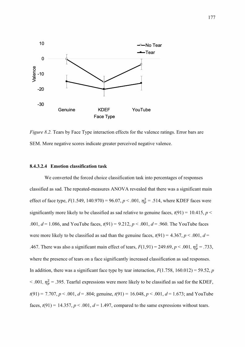
177
Figure 8.2. Tears by Face Type interaction effects for the valence ratings. Error bars are
SEM. More negative scores indicate greater perceived negative valence.
8.4.3.2.4 Emotion classification task
We converted the forced choice classification task into percentages of responses
classified as sad. The repeated-measures ANOVA revealed that there was a significant main
effect of face type, F(1.549, 140.970) = 96.07, p < .001, 𝜂𝑝2 = .514, where KDEF faces were
significantly more likely to be classified as sad relative to genuine faces, t(91) = 10.415, p <
.001, d = 1.086, and YouTube faces, t(91) = 9.212, p < .001, d = .960. The YouTube faces
were more likely to be classified as sad than the genuine faces, t(91) = 4.367, p < .001, d =
.467. There was also a significant main effect of tears, F(1,91) = 249.69, p < .001, 𝜂𝑝2 = .733,
where the presence of tears on a face significantly increased classification as sad responses.
In addition, there was a significant face type by tear interaction, F(1.758, 160.012) = 59.52, p
< .001, 𝜂𝑝2 = .395. Tearful expressions were more likely to be classified as sad for the KDEF,
t(91) = 7.707, p < .001, d = .804; genuine, t(91) = 16.048, p < .001, d = 1.673; and YouTube
faces, t(91) = 14.357, p < .001, d = 1.497, compared to the same expressions without tears.
178
8.4.4 Experiment 1
8.4.4.1 Method
8.4.4.1.1 Participants
Thirty-seven participants (26 females) elected to participate in this study. Participants
ranged in age from 19 to 46 (Mage = 23.46, SDage = 5.12 years). All participants had normal
or corrected-to-normal vision, and 34 were right-handed. Two participants reported during
debriefing that they knew the experiment was investigating perceptions of genuineness, and
as such they were excluded from analyses. Participants were awarded a monetary payment of
$15.00 AUD as compensation for their time. Participants gave written informed consent prior
to participation in the experiment in accord with the declaration of Helsinki.
We determined sample size following the McLellan et al. (2010) study, which
included a sample of 24 participants. We used G*Power (Faul et al., 2007) to estimate the
sample size for a repeated-measures, within-participants design. The smallest reported effect
size in McLellan et al. was 𝜂𝑝2 = .128; however, this effect size was across multiple emotions
and we focused solely on sadness. For this reason, we set a conservative effect size (𝜂𝑝2 =
.06); the power analysis called for a total of 27 participants. As such, we recruited 37
participants to allow for participant exclusions.
8.4.4.1.2 Stimuli
Facial stimuli were the images described in the pilot experiment. For these three
stimulus categories (i.e. Genuine; Posed KDEF; Posed YouTube), we selected four male and
four female facial identities. We selected the images that provided the best representation of
the overall ratings for genuineness, valence, and intensity from the pilot study.
8.4.4.1.3 Procedure
The emotional state recognition task was conducted using e-Prime Professional e-
Studio 2.0.10.356 software. Participants were informed that they would be presented with
179
separate blocks to judge whether the emotion was shown or was felt by the target individual.
Participant instructions were as follows:
“Your job is to decide whether or not the faces are SHOWING emotion and whether
or not they are FEELING emotion. For instance, sometimes when people smile, it
does not mean they are actually happy, and sometimes when people are sad, they may
not have the most intense expression of sadness, but they are feeling the emotion.”
These instructions were slightly adapted from the original study by McLellan et al.
(2010) to include an additional statement used by Dawel et al. (2017). We chose to add the
additional statement regarding intensity to ensure participants were not solely relying on the
intensity of the stimulus to make their judgements. Participants received two practice blocks
(one show and one felt), which included 12 trials each. None of the identities used in the
practice trials were repeated in the experimental trials. The practice trials had the same trial
procedure as the experimental trials. Each block began with the word “SHOW” or “FELT”.
Trials began with the presentation of a fixation cross, varied in length between 500 and 1000
ms (in blocks of 100ms), to prevent anticipatory responses. Participants were then presented
with an emotional face, which was visible until they made a response. Half the participants
were instructed to use the far-left button to denote “yes” responses and the far-right button to
denote “no” responses, while this response mapping was reversed for remaining participants.
The order of the show and felt tasks was counterbalanced between participants and faces in
each block were displayed in a unique random order for each participant. There were 48 trials
per experimental block, for a total of 96 trials, which took approximately 20 minutes to
complete.
8.4.4.2 Results and discussion
As in McLellan et al. (2010) we calculated the percentage of “yes” responses per
condition separately for tear and tear-free images (see Table 8.2). If participants were able to

180
discriminate between genuine and posed displays, it was expected that posed displays would
be perceived to be showing but not feeling emotion, whereas genuine displays should be
perceived as both showing and feeling emotion. As demonstrated in Table 8.2, genuine faces
were more likely to be judged as feeling compared to showing emotion. When a genuine
tearful face was shown, participants believed the tearful expressions were both showing and
feeling the depicted emotion. However, participants were less likely to report that the face
was showing and feeling emotion when tears were removed from the genuinely sad faces.
Conversely, for the KDEF posed expressions, participants were significantly more likely to
state that the faces were showing emotion, relative to feeling emotion. Furthermore, tear
images were significantly more likely to be judged as both showing and feeling emotion,
relative to tear-free images. Similarly, the YouTube faces with tears were significantly more
likely to be judged as both showing and feeling emotion, relative to tear-free expressions.
Although tearful YouTube faces were significantly more likely to be perceived as showing
emotion relative to feeling emotion, tear-free images showed no significant difference
between the judgement conditions.

181
Table 8.2
Percentage of YES Responses by Judgement Condition and Face Type.
Judgement condition
Face type Show (% yes) Feel (% yes)
KDEF Tear-free 76a 48b
Tears 93b 75a
YouTube Tear-free 55a 50a
Tears 95b 77c
Genuine Tear-free 40a 56c
Tears 85b 88b
Note. Different subscripts within a row indicate significant difference in the percentage of yes responses between the show and the feel conditions for each face type. Different subscripts within a column indicate significant differences in the percentage of yes responses between the tear and the tear-free judgements for each face type. No comparisons were made across face types in this table. Level of significance was corrected for multiple comparisons, paired t-tests, p = .004).
To further explore this data, we used a non-parametric signal detection analysis. The
data for each participant was converted into hits (H) and false alarms (FA). Hits were defined
as correctly responding yes to a genuine expression, while false alarms were defined as
responding yes to posed expressions. As outlined in Snodgrass and Corwin (1988), hits and
false alarms were corrected according to the following formulas:
H = (number of hits + .05) / (trials + 1)
FA= (number of false alarms + .05) / (trials + 1)
The sensitivity (𝐴′) equation for when hits ≥ false alarms:
𝐴′ = 0.5 + [(𝐻 − 𝐹𝐴)(1 + 𝐻 − 𝐹𝐴)]/[(4𝐻(1 − 𝐹𝐴)]
When false alarms ≥ hits the equation is modified to:
𝐴′ = 0.5 − [(𝐹𝐴 − 𝐻)(1 + 𝐹𝐴 − 𝐻)]/[(4𝐹𝐴(1 − 𝐻)]
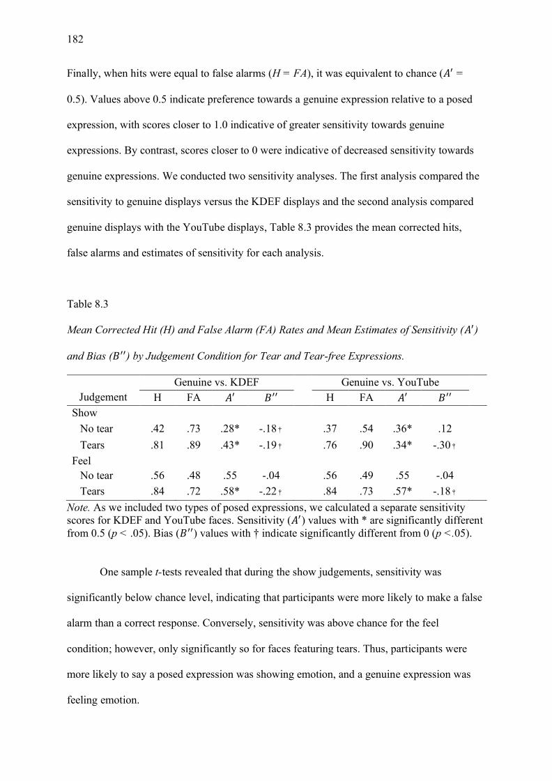
182
Finally, when hits were equal to false alarms (H = FA), it was equivalent to chance (𝐴′ =
0.5). Values above 0.5 indicate preference towards a genuine expression relative to a posed
expression, with scores closer to 1.0 indicative of greater sensitivity towards genuine
expressions. By contrast, scores closer to 0 were indicative of decreased sensitivity towards
genuine expressions. We conducted two sensitivity analyses. The first analysis compared the
sensitivity to genuine displays versus the KDEF displays and the second analysis compared
genuine displays with the YouTube displays, Table 8.3 provides the mean corrected hits,
false alarms and estimates of sensitivity for each analysis.
Table 8.3
Mean Corrected Hit (H) and False Alarm (FA) Rates and Mean Estimates of Sensitivity (𝐴′)
and Bias (𝐵′′) by Judgement Condition for Tear and Tear-free Expressions.
Judgement
Genuine vs. KDEF Genuine vs. YouTube H FA 𝐴′ 𝐵′′ H FA 𝐴′ 𝐵′′
Show No tear .42 .73 .28* -.18† .37 .54 .36* .12 Tears .81 .89 .43* -.19† .76 .90 .34* -.30†
Feel No tear .56 .48 .55 -.04 .56 .49 .55 -.04 Tears .84 .72 .58* -.22† .84 .73 .57* -.18†
Note. As we included two types of posed expressions, we calculated a separate sensitivity scores for KDEF and YouTube faces. Sensitivity (𝐴′) values with * are significantly different from 0.5 (p < .05). Bias (𝐵′′) values with † indicate significantly different from 0 (p <.05).
One sample t-tests revealed that during the show judgements, sensitivity was
significantly below chance level, indicating that participants were more likely to make a false
alarm than a correct response. Conversely, sensitivity was above chance for the feel
condition; however, only significantly so for faces featuring tears. Thus, participants were
more likely to say a posed expression was showing emotion, and a genuine expression was
feeling emotion.

183
We also calculated a measure of bias (𝐵′′), to examine whether participants were
biased towards yes responses. As with the sensitivity measure, the equation for when H > FA
was:
𝐵′′ = [𝐻(1 − 𝐻) − 𝐹𝐴(1 − 𝐹𝐴)]/[𝐻(1 − 𝐻) + 𝐹𝐴(1 − 𝐹𝐴)]
When FA > H, the H and FA values were replaced as follows:
𝐵′′ = [𝐹𝐴(1 − 𝐹𝐴) − 𝐻(1 − 𝐻)]/[𝐹𝐴(1 − 𝐹𝐴) + 𝐻(1 − 𝐻)]
Possible values ranged from -1 to +1, with positive values indicating a bias towards
“NO”, and negative values indicating a bias towards “YES”; 0 values indicate a neutral
criterion (see Table 8.3). During the show condition, 𝐵′′ scores showed that participants were
significantly biased towards responding with “yes” (i.e., “YES” for showing) for both tear-
free and tearful expressions in the first sensitivity analysis (genuine faces versus KDEF), but
only tearful expressions in the second sensitivity analysis (genuine versus YouTube). During
the feel condition, participants demonstrated no bias (i.e. a neutral criterion was used) when
judging tear-free expressions. By contrast, the presence of tears on a facial display
significantly biased the participants towards responding “yes” during the feel task. Thus, the
presence of tears significantly biased participants towards a “yes” response, demonstrating
that tears make an expression look sadder and increased the perception that the face felt sad.
We conducted two separate ANOVAs for each of the separate sensitivity indices to
examine the influence the type of posed expression has on sensitivity to genuineness. The
ANOVAs were comprised of a 2 (judgement: show, felt) by 2 (tears: tear-free, tears)
repeated-measures ANOVAs to further explore the differences between the tear and tear-free
conditions. The first analysis compared sensitivity between genuine and KDEF faces, which
revealed a significant main effect of judgment, F(1, 34) = 26.021, p < .001, 𝜂𝑝2=.434. False
alarms were more likely during the show condition relative to the feel condition; participants
performed worse during the show condition, as evidenced by the increased number of false
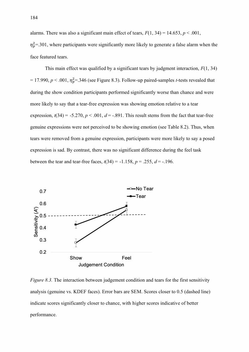
184
alarms. There was also a significant main effect of tears, F(1, 34) = 14.653, p < .001,
𝜂𝑝2=.301, where participants were significantly more likely to generate a false alarm when the
face featured tears.
This main effect was qualified by a significant tears by judgment interaction, F(1, 34)
= 17.990, p < .001, 𝜂𝑝2=.346 (see Figure 8.3). Follow-up paired-samples t-tests revealed that
during the show condition participants performed significantly worse than chance and were
more likely to say that a tear-free expression was showing emotion relative to a tear
expression, t(34) = -5.270, p < .001, d = -.891. This result stems from the fact that tear-free
genuine expressions were not perceived to be showing emotion (see Table 8.2). Thus, when
tears were removed from a genuine expression, participants were more likely to say a posed
expression is sad. By contrast, there was no significant difference during the feel task
between the tear and tear-free faces, t(34) = -1.158, p = .255, d = -.196.
Figure 8.3. The interaction between judgement condition and tears for the first sensitivity
analysis (genuine vs. KDEF faces). Error bars are SEM. Scores closer to 0.5 (dashed line)
indicate scores significantly closer to chance, with higher scores indicative of better
performance.

185
The second analysis, which examined sensitivity between genuine and YouTube
faces, revealed a significant main effect of judgment, F(1, 34) = 36.896, p < .001, 𝜂𝑝2=.520,
where, as in the first sensitivity analysis, sensitivity was worse for the show task relative to
the feel task. As indicated in Figure 8.4, sensitivity was below chance for the show
judgements, and above chance for the feel judgements. Neither the main effect of tears, F(1,
34) = .060, p = .808, 𝜂𝑝2=.002, nor the judgment by tears interaction, F(1, 34) = 2.332, p =
.136, 𝜂𝑝2=.064, were significant.
Figure 8.4. A graphical depiction of the second sensitivity analysis (genuine faces versus
YouTube faces). Error bars are SEM. Scores closer to 0.5 (dashed line) indicate scores
significantly closer to chance, with higher scores indicative of better performance.
8.4.5 Experiment 2
Experiment 1 allowed us to investigate whether people can discriminate between
posed and genuine emotional displays of sadness. We confirmed that participants can
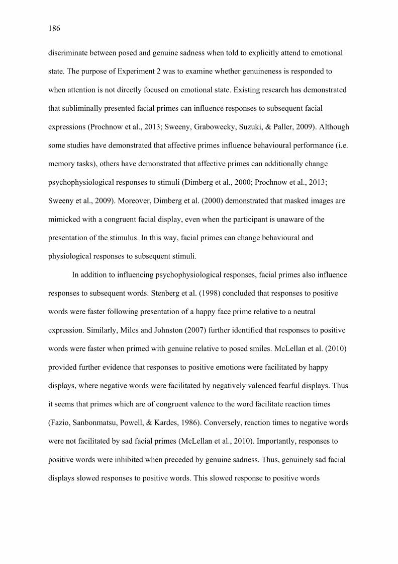
186
discriminate between posed and genuine sadness when told to explicitly attend to emotional
state. The purpose of Experiment 2 was to examine whether genuineness is responded to
when attention is not directly focused on emotional state. Existing research has demonstrated
that subliminally presented facial primes can influence responses to subsequent facial
expressions (Prochnow et al., 2013; Sweeny, Grabowecky, Suzuki, & Paller, 2009). Although
some studies have demonstrated that affective primes influence behavioural performance (i.e.
memory tasks), others have demonstrated that affective primes can additionally change
psychophysiological responses to stimuli (Dimberg et al., 2000; Prochnow et al., 2013;
Sweeny et al., 2009). Moreover, Dimberg et al. (2000) demonstrated that masked images are
mimicked with a congruent facial display, even when the participant is unaware of the
presentation of the stimulus. In this way, facial primes can change behavioural and
physiological responses to subsequent stimuli.
In addition to influencing psychophysiological responses, facial primes also influence
responses to subsequent words. Stenberg et al. (1998) concluded that responses to positive
words were faster following presentation of a happy face prime relative to a neutral
expression. Similarly, Miles and Johnston (2007) further identified that responses to positive
words were faster when primed with genuine relative to posed smiles. McLellan et al. (2010)
provided further evidence that responses to positive emotions were facilitated by happy
displays, where negative words were facilitated by negatively valenced fearful displays. Thus
it seems that primes which are of congruent valence to the word facilitate reaction times
(Fazio, Sanbonmatsu, Powell, & Kardes, 1986). Conversely, reaction times to negative words
were not facilitated by sad facial primes (McLellan et al., 2010). Importantly, responses to
positive words were inhibited when preceded by genuine sadness. Thus, genuinely sad facial
displays slowed responses to positive words. This slowed response to positive words

187
following sad displays indicates that priming with sad expressions inhibits the classification
of positive words.
With this in mind, we sought to further explore the way that a sad facial prime
influences subsequent response to words. Given that tears are perceived to be an honest
signal of emotion (Trimble, 2012; Vingerhoets, 2013), we predicted responses to words
associated with genuineness would be facilitated when priming with a genuine tearful
expression. By contrast, tears can also be used to manipulate and deceive. Namely, crocodile
tears are renowned as the disingenuous crying of politicians and celebrities (Manusov &
Harvey, 2011), or the false tearing displays that are used to garner sympathy during court
proceedings (ten Brinke et al., 2012). As such, we predicted that posed artificial tear displays
would facilitate responses to negative words that are associated with being disingenuous.
8.4.5.1 Method
8.4.5.1.1 Participants
Experiment 1 participants also completed Experiment 2 within the same testing
session.
8.4.5.1.2 Stimuli
We used the same stimuli in Experiments 1 and 2. For the word categorisation task,
we used words typically associated with genuineness (i.e. trustworthy, sincere, honest,
truthful) and their antonyms (i.e. untrustworthy, insincere, dishonest, untruthful). These
words were found to be clearly identifiable as positive or negative using a likableness scale
(>5 for positive and <1 for negative, on a 7-point scale, with higher scores indicating greater
perceived likeableness) (Anderson, 1968; Chandler, 2018). Furthermore, we piloted the
words to ensure that genuine and disingenuous words would be associated with a genuine or
a fake person, respectively (see Dawel et al. (2017) for an overview of these terms in prior
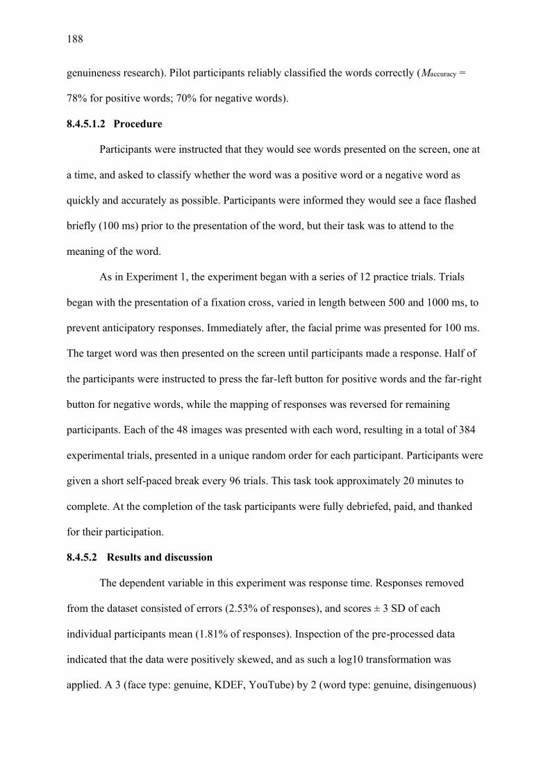
188
genuineness research). Pilot participants reliably classified the words correctly (Maccuracy =
78% for positive words; 70% for negative words).
8.4.5.1.2 Procedure
Participants were instructed that they would see words presented on the screen, one at
a time, and asked to classify whether the word was a positive word or a negative word as
quickly and accurately as possible. Participants were informed they would see a face flashed
briefly (100 ms) prior to the presentation of the word, but their task was to attend to the
meaning of the word.
As in Experiment 1, the experiment began with a series of 12 practice trials. Trials
began with the presentation of a fixation cross, varied in length between 500 and 1000 ms, to
prevent anticipatory responses. Immediately after, the facial prime was presented for 100 ms.
The target word was then presented on the screen until participants made a response. Half of
the participants were instructed to press the far-left button for positive words and the far-right
button for negative words, while the mapping of responses was reversed for remaining
participants. Each of the 48 images was presented with each word, resulting in a total of 384
experimental trials, presented in a unique random order for each participant. Participants were
given a short self-paced break every 96 trials. This task took approximately 20 minutes to
complete. At the completion of the task participants were fully debriefed, paid, and thanked
for their participation.
8.4.5.2 Results and discussion
The dependent variable in this experiment was response time. Responses removed
from the dataset consisted of errors (2.53% of responses), and scores ± 3 SD of each
individual participants mean (1.81% of responses). Inspection of the pre-processed data
indicated that the data were positively skewed, and as such a log10 transformation was
applied. A 3 (face type: genuine, KDEF, YouTube) by 2 (word type: genuine, disingenuous)

189
by 2 (tears: no tears, tears) was conducted on the log transformed data, however, means
reported in Figure 8.5 reflect response time in milliseconds for ease of interpretation.
Bonferroni adjustment was used to correct for follow-up comparisons.
There was a significant main effect of face type, F(2, 68) = 6.294, p = .003, 𝜂𝑝2=.156
where YouTube faces were responded to significantly faster than the KDEF faces, t = 4.060,
p < .001, d = .686. There was no difference between the genuine faces and the YouTube
faces, t = 1.998, p = .161, d = .338, or the genuine faces and the KDEF faces, t = 1.370, p =
.539, d = .232. There was also a significant main effect of word type, F(1, 34) = 12.326, p =
.001, 𝜂𝑝2=.266, where genuine words were responded to significantly faster than disingenuous
words. An interaction between face type and tears was marginally significant, F(1, 34) =
3.031, p = .055, 𝜂𝑝2=.082 (see Figure 8.5). Follow-up comparisons using paired-samples t-
tests revealed that there were no significant differences between the tear and the tear-free
conditions for the genuine faces, t(34) = .765, p = .449, d = .129; or the YouTube faces, t(34)
= -.488, p = .629, d = -.083; however participants were significantly faster at responding to
KDEF faces with tears, t(34) = 2.554, p = .015, d = .432. This indicates that participants were
faster to respond to both genuine and disingenuous words when preceded by a KDEF face
edited to include tears, as opposed to the original tear-free stimuli. There was no evidence
supporting the hypotheses that genuine faces would enhance responses to genuine words, or
190
that posed expressions would enhance responses to disingenuous words.
Figure 8.5. Mean reaction time to categorise words as a function of expression type and tear
presence. Error bars are SEM.
8.4.6 General Discussion
Our research provides a novel investigation of how genuine tearful expressions of
emotion are distinguished from posed, artificial displays. Our results demonstrate that
participants are sensitive to the differences between posed and genuine sadness, which further
supports existing research (McLellan et al., 2010). We have expanded on this existing
literature by showing that participants were sensitive to the differences between posed and
genuine tearful images. Firstly, we demonstrated, using rating scales, that genuine tearful
displays were perceived as significantly more genuine than posed tearful displays.
Additionally, participants were more likely to say posed, relative to genuine, expressions
showed emotion, and that genuine expressions were feeling emotion rather than posing.
Furthermore, the presence of tears biased judgements; regardless of whether emotions were
genuine or posed, as participants were more likely to indicate that tearful expressions were
191
both showing and feeling sadness. Finally, we were unable to demonstrate that tearful prime
expressions mediated responses to words.
Overall, participants were more likely to say that tearful expressions both showed and
felt emotion. Thus, in the absence of context, tears served as a marker of sadness. This
finding provides further support for the tear effect in that tears resolve emotional ambiguity
and signal sadness (Provine et al., 2009). Furthermore, our sensitivity analyses clearly show
that posed displays showed emotion more so than genuine expressions. This finding is
consistent with previous work, which has found that posed expressions are easier to identify
as a specific emotion relative to spontaneous genuine expressions (Calvo & Nummenmaa,
2016; Namba et al., 2018). This ease of identification stems from the exaggerated nature of
posed expressions (Gosselin, Kirouac, & Doré, 1995), and also highlights the ambiguity
associated with spontaneous facial displays (Motley & Camden, 1988). As such, when
participants are instructed to judge whether a face looks sad, posed facial expressions are
more likely to be classified as sad, relative to genuine expressions.
By contrast, when asking participants to judge whether the person in the image felt sad,
participants were more likely to classify genuine expressions as feeling sadness, relative to
posed expressions. Thus, we provided further support for the idea that participants are
sensitive to the genuineness of sad facial displays (Dawel et al., 2015; McLellan et al., 2010;
Namba et al., 2018). Furthermore, participants’ sensitivity to the affective state of tearful
faces was significantly above chance level, which was not the case for tear-free expressions.
This sensitivity indicates that the presence of tears on an image significantly aided
participants in distinguishing genuine from posed emotion. As identified by Niedenthal and
Brauer (2012), humans are sensitive to signals high in emotional salience and credibility.
Existing research has identified that tearful displays are a distinctive marker of sadness
(Krivan et al., 2020) and are believed to be an honest signal of emotion (Trimble, 2012;
192
Vingerhoets, 2013), which increase the sincerity of sad facial displays (Picó et al., 2020;
Zeifman & Brown, 2011). We found that tears aid in identifying genuine from posed
emotion. As such, our data provide empirical evidence that genuine tearful expressions are an
honest signal of emotion.
Contrary to our hypothesis, we were unable to demonstrate that genuine and posed
expressions facilitated responses to genuine and disingenuous words, respectively. In this
way, we have demonstrated further support for McLellan et al. (2010), wherein sad facial
primes do not influence classification of positive and negative words. However, this
conclusion does not support the conclusion of Stenberg et al. (1998), wherein a sad facial
prime facilitated the processing of negative words. Rather, in our study, responses were
fastest to genuine (i.e. positive) words overall. Prior research has demonstrated a positivity
advantage, where words of a positive valence are responded to more quickly than words of a
negative valence (Bayer & Schacht, 2014; Fazio et al., 1986; McLellan et al., 2010; Miles &
Johnston, 2007; Stenberg et al., 1998). In some previous studies, faster responses to positive
words were facilitated by a prime of congruent valence (Fazio et al., 1986; McLellan et al.,
2010), whereas in others there is a general positivity advantage (Stenberg et al., 1998).
However, in the present study, all stimuli were negatively valenced. Therefore, we
demonstrated faster classification of positive words associated with genuineness, compared to
negative words associated disingenuousness—without the inclusion of a congruent facial
prime. This superior responding to genuine words that are positively valenced provides
support for the positivity advantage. Much like how happy faces are facilitated in facial
recognition tasks (Calvo et al., 2016; Leppänen et al., 2003; Palermo & Coltheart, 2004),
positive words are also preferentially processed compared to negative words (Bayer &
Schacht, 2014; Feyereisen et al., 1986; Stenberg et al., 1998). In this way, the preferential
processing and retention of positive, as opposed to negative, information (Matlin & Stang,
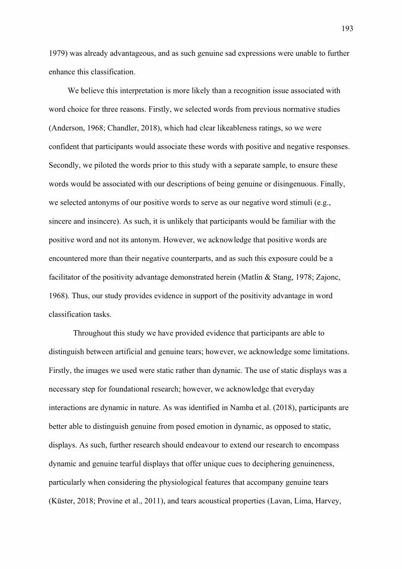
193
1979) was already advantageous, and as such genuine sad expressions were unable to further
enhance this classification.
We believe this interpretation is more likely than a recognition issue associated with
word choice for three reasons. Firstly, we selected words from previous normative studies
(Anderson, 1968; Chandler, 2018), which had clear likeableness ratings, so we were
confident that participants would associate these words with positive and negative responses.
Secondly, we piloted the words prior to this study with a separate sample, to ensure these
words would be associated with our descriptions of being genuine or disingenuous. Finally,
we selected antonyms of our positive words to serve as our negative word stimuli (e.g.,
sincere and insincere). As such, it is unlikely that participants would be familiar with the
positive word and not its antonym. However, we acknowledge that positive words are
encountered more than their negative counterparts, and as such this exposure could be a
facilitator of the positivity advantage demonstrated herein (Matlin & Stang, 1978; Zajonc,
1968). Thus, our study provides evidence in support of the positivity advantage in word
classification tasks.
Throughout this study we have provided evidence that participants are able to
distinguish between artificial and genuine tears; however, we acknowledge some limitations.
Firstly, the images we used were static rather than dynamic. The use of static displays was a
necessary step for foundational research; however, we acknowledge that everyday
interactions are dynamic in nature. As was identified in Namba et al. (2018), participants are
better able to distinguish genuine from posed emotion in dynamic, as opposed to static,
displays. As such, further research should endeavour to extend our research to encompass
dynamic and genuine tearful displays that offer unique cues to deciphering genuineness,
particularly when considering the physiological features that accompany genuine tears
(Küster, 2018; Provine et al., 2011), and tears acoustical properties (Lavan, Lima, Harvey,

194
Scott, & McGettigan, 2015). Furthermore, a recent study by Roeyen et al. (2020)
demonstrated that if tears are perceived as false or insincere (i.e. crocodile tears) by an
observer, this opinion has a damaging effect on crier perception. Interestingly, this effect held
regardless of whether the emotional tears were genuine. Therefore, further research exploring
not only the type of stimuli used, but also how the stimuli are perceived is needed to better
understand the communicative functions of tears.
In conclusion, the present studies provide evidence that genuine tearful expressions of
emotion are distinct from posed, artificial tear displays. By asking participants to determine
whether the facial stimuli were showing and feeling sadness we demonstrated that
participants were more likely to attribute showing emotion to a posed expression, in line with
posed displays being clearly recognisable (Calvo & Nummenmaa, 2016; Namba et al., 2018).
By contrast, when asking participants to determine whether the person in the sad facial
displays were feeling emotion, participants were significantly more likely to select a genuine
expression felt emotion. Furthermore, the presence of tears facilitated the differentiation
between posed and genuine displays. Therefore, the present research supports previous
findings that persons are sensitive to genuine displays of sadness and provides important
empirical evidence that individuals are sensitive to tearful displays.

195
Chapter 9: Why Do We Care When Others Cry?
9.1 Summary
As I have demonstrated throughout this thesis, tears demand attention. This thesis
provides a valuable addition to the literature exploring the communicative functions of
emotional tears. A series of self-report behavioural studies had demonstrated that tears elicit
empathy, aid, and succour from observers (Balsters et al., 2013; Hendriks & Vingerhoets,
2006; Hendriks et al., 2008; Vingerhoets et al., 2016; Zickfeld & Schubert, 2018), and the
data from this body of work demonstrates that these empathic responses have a physiological
basis. The overarching aim of this thesis was to determine why we care when others cry. To
achieve this aim, three questions were investigated: 1) do tears serve as a signal of sadness in
the absence of context (Chapter 3)?; 2) do tears modulate physiological responses to facial
displays (Chapters 4, 5, & 6)?; and 3) do tears increase the authenticity of facial displays
(Chapter 8)? These three questions will be addressed sequentially, while outlining the
findings. The outstanding research questions pertaining to each of these aims will also be
addressed and potential avenues for future research will be highlighted. Finally, the thesis
will conclude by outlining why the continued investigation of tears is important to better
understand the development of empathy and prosocial behaviour in society as a whole.
9.2 Tears as a Signal
9.2.1 Findings and implications
As explored in Chapter 1, tears serve a unique signalling function (Hasson, 2009).
Following the tear effect, tears serve as a signal of sadness in the absence of context (Provine
et al., 2009). This signal of sadness is effective at communicating distress, which in turn
elicits help and succour from observers. However, prior to this thesis, limited work had
explored whether the tear effect is evident in expressions other than sadness (Ito et al., 2019;
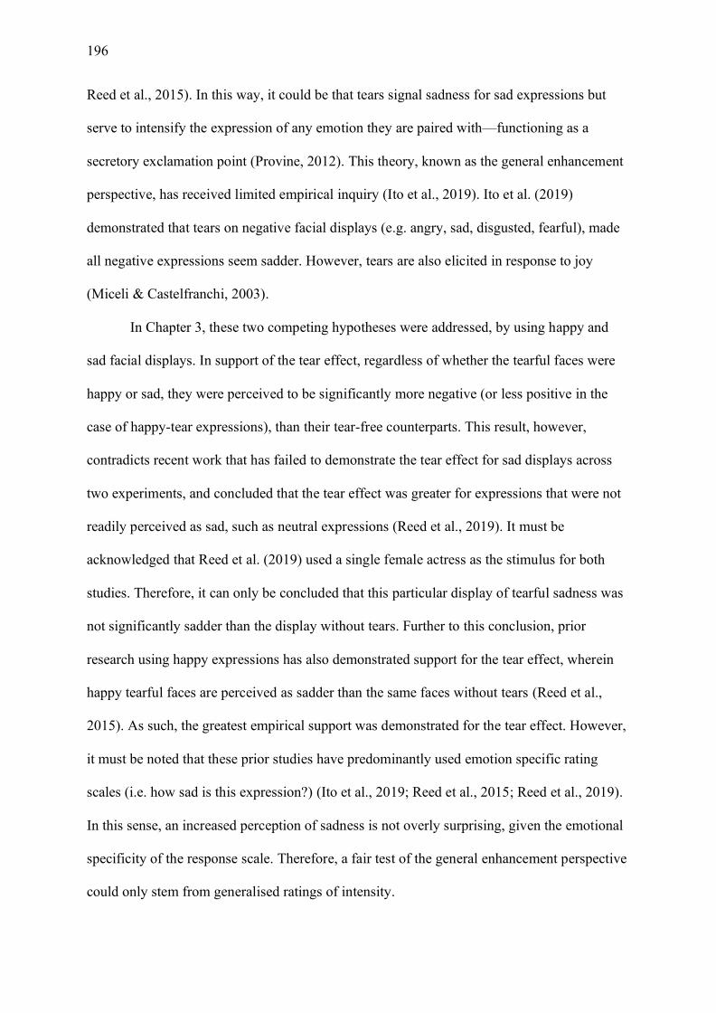
196
Reed et al., 2015). In this way, it could be that tears signal sadness for sad expressions but
serve to intensify the expression of any emotion they are paired with—functioning as a
secretory exclamation point (Provine, 2012). This theory, known as the general enhancement
perspective, has received limited empirical inquiry (Ito et al., 2019). Ito et al. (2019)
demonstrated that tears on negative facial displays (e.g. angry, sad, disgusted, fearful), made
all negative expressions seem sadder. However, tears are also elicited in response to joy
(Miceli & Castelfranchi, 2003).
In Chapter 3, these two competing hypotheses were addressed, by using happy and
sad facial displays. In support of the tear effect, regardless of whether the tearful faces were
happy or sad, they were perceived to be significantly more negative (or less positive in the
case of happy-tear expressions), than their tear-free counterparts. This result, however,
contradicts recent work that has failed to demonstrate the tear effect for sad displays across
two experiments, and concluded that the tear effect was greater for expressions that were not
readily perceived as sad, such as neutral expressions (Reed et al., 2019). It must be
acknowledged that Reed et al. (2019) used a single female actress as the stimulus for both
studies. Therefore, it can only be concluded that this particular display of tearful sadness was
not significantly sadder than the display without tears. Further to this conclusion, prior
research using happy expressions has also demonstrated support for the tear effect, wherein
happy tearful faces are perceived as sadder than the same faces without tears (Reed et al.,
2015). As such, the greatest empirical support was demonstrated for the tear effect. However,
it must be noted that these prior studies have predominantly used emotion specific rating
scales (i.e. how sad is this expression?) (Ito et al., 2019; Reed et al., 2015; Reed et al., 2019).
In this sense, an increased perception of sadness is not overly surprising, given the emotional
specificity of the response scale. Therefore, a fair test of the general enhancement perspective
could only stem from generalised ratings of intensity.

197
In addition to the increased perception of negative valence demonstrated in Chapter 3,
there was also an increase in generalised intensity for tearful expressions. This finding
demonstrates that tears make both happy and sad expressions seem more intense—in support
of the general enhancement perspective. The finding that tearful displays are more intense
than tear-free counterparts is not overly surprising, given that intense emotions are typically
associated with profound feelings and are elicited in response to substantial life influences
(Sonnemans & Frijda, 1994). One theoretical model—termed here the helplessness6 model—
argues that tears are shed when one feels helpless (Frijda, 1986; Miceli & Castelfranchi,
2003). Under the helplessness model, tears occur once an individual has exhausted all
avenues and is unable to alleviate or remedy a situation. In this way, tears are associated with
feeling overwhelmed by a situation or emotion. The helplessness model is validated by a
series of self-report studies which have demonstrated that tearful individuals are perceived as
more helpless than tear-free individuals (Vingerhoets et al., 2016). Additionally, tears are
typically associated with substantial and important relational life events (e.g. death, birth,
reunion)7, wherein the expression of overwhelming emotion is appropriate (Fischer et al.,
2013; Vingerhoets, 2013). Thus, it may be that tears are inherently perceived as intense as a
result of their association with extreme life events. Consequently, tears would be associated
with intense feelings and, in the absence of context, these feelings are interpreted as largely
negative.
Contrary to expectations, tearful-sad expressions were not responded to significantly
faster than sad expressions without tears. Exploring reaction time data with response
accuracy measures indicated that responses to sad expressions were at ceiling level, and as
such the addition of tears offered no further improvement to the classification of sad faces.
6 Sometimes termed the powerlessness model 7Although these experiences are typically rare and, as such, tears are usually elicited in response to more mundane life experiences (e.g. frustration, disappointment, failure), it is the case that tears are typically associated with substantial events.
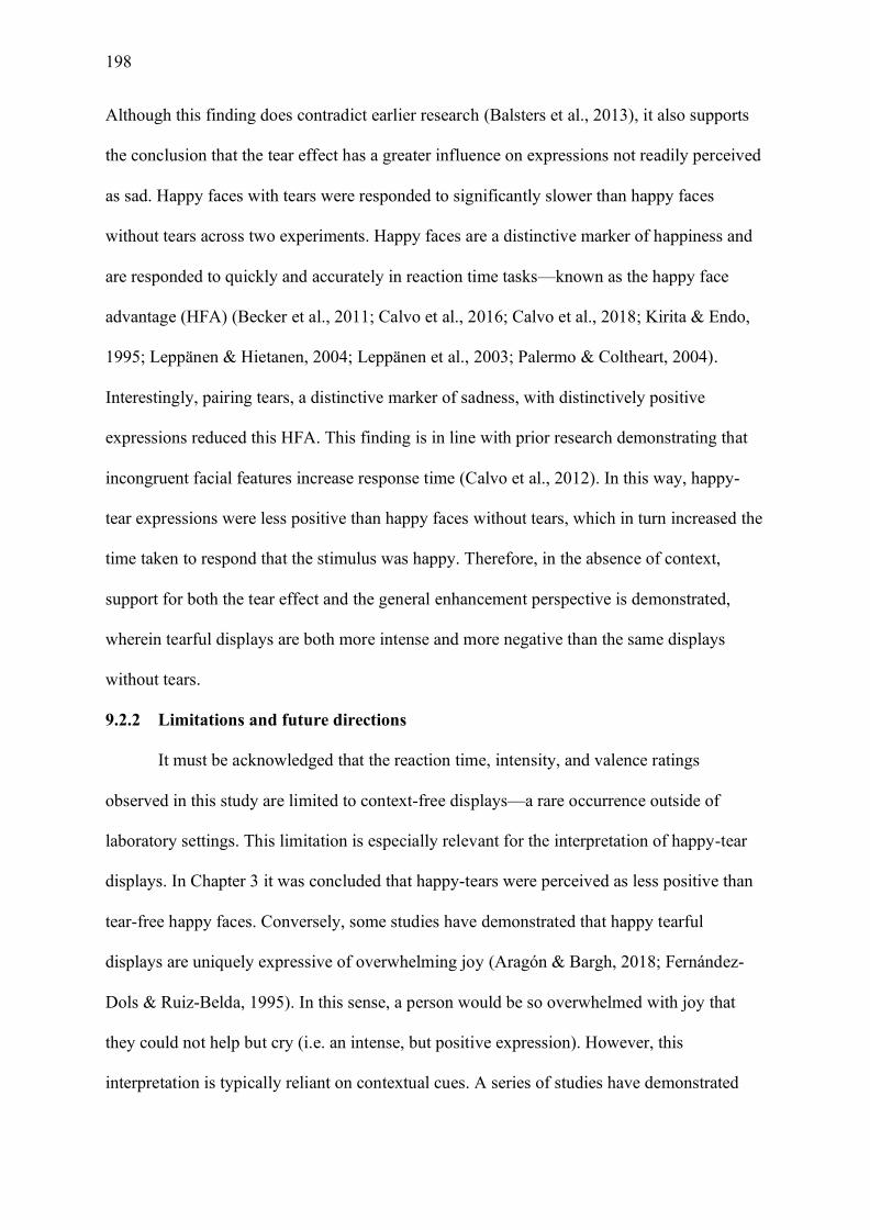
198
Although this finding does contradict earlier research (Balsters et al., 2013), it also supports
the conclusion that the tear effect has a greater influence on expressions not readily perceived
as sad. Happy faces with tears were responded to significantly slower than happy faces
without tears across two experiments. Happy faces are a distinctive marker of happiness and
are responded to quickly and accurately in reaction time tasks—known as the happy face
advantage (HFA) (Becker et al., 2011; Calvo et al., 2016; Calvo et al., 2018; Kirita & Endo,
1995; Leppänen & Hietanen, 2004; Leppänen et al., 2003; Palermo & Coltheart, 2004).
Interestingly, pairing tears, a distinctive marker of sadness, with distinctively positive
expressions reduced this HFA. This finding is in line with prior research demonstrating that
incongruent facial features increase response time (Calvo et al., 2012). In this way, happy-
tear expressions were less positive than happy faces without tears, which in turn increased the
time taken to respond that the stimulus was happy. Therefore, in the absence of context,
support for both the tear effect and the general enhancement perspective is demonstrated,
wherein tearful displays are both more intense and more negative than the same displays
without tears.
9.2.2 Limitations and future directions
It must be acknowledged that the reaction time, intensity, and valence ratings
observed in this study are limited to context-free displays—a rare occurrence outside of
laboratory settings. This limitation is especially relevant for the interpretation of happy-tear
displays. In Chapter 3 it was concluded that happy-tears were perceived as less positive than
tear-free happy faces. Conversely, some studies have demonstrated that happy tearful
displays are uniquely expressive of overwhelming joy (Aragón & Bargh, 2018; Fernández-
Dols & Ruiz-Belda, 1995). In this sense, a person would be so overwhelmed with joy that
they could not help but cry (i.e. an intense, but positive expression). However, this
interpretation is typically reliant on contextual cues. A series of studies have demonstrated
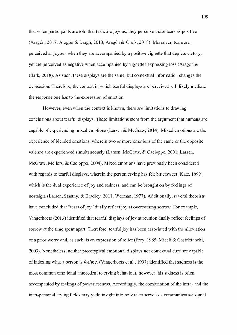
199
that when participants are told that tears are joyous, they perceive those tears as positive
(Aragón, 2017; Aragón & Bargh, 2018; Aragón & Clark, 2018). Moreover, tears are
perceived as joyous when they are accompanied by a positive vignette that depicts victory,
yet are perceived as negative when accompanied by vignettes expressing loss (Aragón &
Clark, 2018). As such, these displays are the same, but contextual information changes the
expression. Therefore, the context in which tearful displays are perceived will likely mediate
the response one has to the expression of emotion.
However, even when the context is known, there are limitations to drawing
conclusions about tearful displays. These limitations stem from the argument that humans are
capable of experiencing mixed emotions (Larsen & McGraw, 2014). Mixed emotions are the
experience of blended emotions, wherein two or more emotions of the same or the opposite
valence are experienced simultaneously (Larsen, McGraw, & Cacioppo, 2001; Larsen,
McGraw, Mellers, & Cacioppo, 2004). Mixed emotions have previously been considered
with regards to tearful displays, wherein the person crying has felt bittersweet (Katz, 1999),
which is the dual experience of joy and sadness, and can be brought on by feelings of
nostalgia (Larsen, Stastny, & Bradley, 2011; Werman, 1977). Additionally, several theorists
have concluded that “tears of joy” dually reflect joy at overcoming sorrow. For example,
Vingerhoets (2013) identified that tearful displays of joy at reunion dually reflect feelings of
sorrow at the time spent apart. Therefore, tearful joy has been associated with the alleviation
of a prior worry and, as such, is an expression of relief (Frey, 1985; Miceli & Castelfranchi,
2003). Nonetheless, neither prototypical emotional displays nor contextual cues are capable
of indexing what a person is feeling. (Vingerhoets et al., 1997) identified that sadness is the
most common emotional antecedent to crying behaviour, however this sadness is often
accompanied by feelings of powerlessness. Accordingly, the combination of the intra- and the
inter-personal crying fields may yield insight into how tears serve as a communicative signal.
200
A final limitation to the theory that tears serve as a signal is that it is difficult to
explain why we cry when we are alone. International studies of adult crying behaviours
revealed that we predominantly cry when alone (37% of responses) or with one person
present (29%)—most often a partner (Vingerhoets, 2013). Therefore, if the purpose of crying
is to elicit help and support from those witness to the display, why would we prefer to cry
when alone? This criticism is not restricted to tearful displays. The Behavioural Ecology
View (BECV) asserts that facial expressions are communicative signals rather than
expressions of emotion (Fridlund, 1991, 1994, 2017)—yet we make a myriad of faces when
alone (Ekman, 1972; Ekman et al., 1990). Under the BECV, the faces made when alone are
known as implicit sociality (Fridlund, 1994). The assumption of implicit sociality states that
being physically alone does not mean we are psychologically alone (Fridlund, 1991, 2017).
Implicit sociality can stem from anticipating social interaction and rehearsing an encounter,
or imaging the continued presence of individuals after the conclusion of an encounter
(Fridlund, 2017). Similarly, we may treat inanimate objects, pets, or even treat ourselves as
social others. Some have argued that private displays are the purest form of expression, as
they are free from social demands and display rules (Ekman et al., 1990).
Implicit sociality can also be used to explain private weeping. Firstly, although
weeping is responsible for eliciting comfort from observers, this need for comfort may stem
from helplessness (Miceli & Castelfranchi, 2003). For the weeper, weeping typically occurs
once one has exhausted all methods available to them and feels as though they are unable to
remedy their situation. For the expresser, tears as a signal of helplessness, signal that the crier
needs help (Miceli & Castelfranchi, 2003). While death, loss, and birth are all commonly
reported antecedents to crying behaviour, these events are rare occurrences throughout one’s
life (Vingerhoets & Cornelius, 2001). In this way, tears are more often produced in response
to mundane life sufferings (e.g. frustration, disappointment, failure) (Miceli & Castelfranchi,
201
2003). Additionally, many of the antecedents to crying are social; for example, loss,
separation, conflict and even reunion (Vingerhoets & Bylsma, 2015). Hence, reimagining or
replaying events and interactions when alone can result in feeling implicitly social, while free
from potentially negative social responses. Thus, tears shed alone are implicitly social and
can function as an expressive signal regardless of whether one is alone, or in the presence of
others.
9.2.3 Conclusions
Despite these limitations, this thesis has provided empirical evidence that tears function
as a biological signal. However, unlike a strict BECV view, tears are capable of signalling
emotion, namely sadness (at least in the absence of context). Therefore, crying signals
feelings of distress and sadness to those around us, which in turn solicits comfort, empathy,
and succour from observers in behavioural tasks (Hendriks et al., 2008). As identified in this
thesis, tears modulate our psychophysiological responses to facial displays. As such, tears are
a unique signal, which are preferentially processed at the psychophysiological level.
9.3 Psychophysiological Responses to Tears
9.3.1 Findings and implications
The second component of this thesis was to examine whether tears, as a salient
biological signal, are preferentially processed at the psychophysiological level. This question
was examined through a series of psychophysiological studies using facial EMG and EEG, as
these techniques offer the ability to explore the rapid, automatic responses one has to tearful
displays.
Chapter 4 details the results of the first psychophysiological study I conducted, which
revealed that tears largely did not influence facial mimicry responses as evidenced by facial
EMG. Using a masked priming paradigm, a trend towards a reduction in corrugator activity
following the presentation of tearful expressions was demonstrated. The corrugator is largely
202
associated with the production of negative emotion and is the muscle responsible for drawing
the eyebrows together into a frown (Dimberg et al., 2000; Ekman & Rosenberg, 2005).
Although participants exhibited less mimicry in response to a tearful expression, this finding
could indicate that they frowned less in response to tearful stimuli. Thus, this reduced
frowning response could be a physiological expression of reduced aggression in response to
tearful displays. This conclusion supports earlier literature showing that tears reduce
aggression in observers (Hasson, 2009; Hendriks et al., 2008; Kottler, 1996). Unfortunately,
this trend towards significance observed during the masked prime stage was not present when
participants were shown emotional expressions for an extended period of time (i.e., 5
seconds). Therefore, it is difficult to conclude what role tears play in inducing mimicry in
observers given the disparity between the subliminally and supraliminally presented tearful
displays.
However, the null results observed in the supraliminal presentation phase are in
accord with a recent study demonstrating no difference in mimicry responses to sad-tearful
displays (Grainger et al., 2019). Furthermore, the literature has generally demonstrated that
mimicry is increased in affiliative scenarios (Likowski et al., 2008; Likowski et al., 2011). In
the case of sad facial displays, mimicry is typically limited to in-group members (Bourgeois
& Hess, 2008) or close partners (Häfner & IJzerman, 2011). Thus, it is possible that mimicry
of tearful displays is also limited to in-group members or close partners, rather than strangers.
This conclusion is justified given that if tears are shared with a close other, it is usually a
partner (Vingerhoets, 2013). From an evolutionary perspective, mimicry of tears might be
selective wherein mimicry only occurs for displays that are personally relevant to the
individual.
Conversely, it could simply be that mimicry of distress and sadness is not adaptive.
Sadness is considered a high cost emotion, wherein responding to a sad display (i.e.

203
providing comfort and succour) comes at a cost to the observer (Bavelas et al., 1986;
Bourgeois & Hess, 2008). When mimicking a social display comes at a cost to the mimicker,
mimicry is reduced (Johnston, 2002). In the case of tears, although mimicry could encourage
the continued expression of emotion, it also signals understanding and fosters affiliation
between interactional partners (Bourgeois & Hess, 2008). However, research exploring self-
report emotion regulation responses to tearful joy and sadness has indicated that tears
increase the likelihood of a down-regulation response (Aragón & Clark, 2018). Down-
regulation responses are used to calm down and comfort individuals, and to aid the individual
in regaining control over their emotions. Therefore, if the goal of an interactional partner is to
down-regulate a crying individual, mimicking that display would be counterproductive.
Consequently, future research exploring mimicry of tearful displays must better account for
the social factors that influence mimicry responses: the personal connection between
interaction partners; the context in which crying occurs; and the subsequent interactional
goals of the perceiver.
While tears were not found to modulate mimicry responses, they were found to
modulate early face-related neural responses. The results presented in Chapter 5
demonstrated that the N170 event-related potential (ERP) was modulated by emotional
content, which contradicts earlier research that suggested the N170 was only involved in
early structural encoding (Bentin et al., 1996; Bentin & Deouell, 2000; Eimer, 2000, 2000).
Furthermore, the N170 was found to be modulated by the type of emotion depicted, providing
support for a recent meta-analysis, which concluded that the N170 is preferentially modulated
by stimuli that require rapid interpretation (Hinojosa et al., 2015). As such, expressions that
facilitate survival are prioritised and these expressions have increased social relevance. In
Chapter 5, it was revealed that sad tearful displays and happy displays without tears were
preferentially processed. As has been outlined, tearful displays are a distinctive signal of

204
sadness—wherein tears aid individuals in identifying sadness. Additionally, tearful displays
foster approach behaviours (Gračanin et al., 2018) and helping responses from observers
(Hendriks et al., 2008). In this way, tearful displays are preferentially processed at the early
neural level, which provides a biological basis for the pro-social responses observed in both
Chapter 3 and earlier work. Furthermore, in accord with existing literature, the N170 was also
modulated by happy displays without tears. This preferential processing of happy faces can
be attributed to the affiliative nature of smiling facial displays and their role in fostering pro-
social behaviour (Nummenmaa & Calvo, 2015). Evidently, displays that are more socially
relevant elicit larger N170 ERP responses. Therefore, this study provides
psychophysiological evidence for the conclusions drawn in Chapter 3, as tears foster the
detection of sadness and increase the social relevance of sad faces.
Finally, in Chapter 6 I explored whether the mirror neuron system is sensitive to tears.
Mirror neurons are believed to be a critical component in the sharing of affective states, and
the way that we as humans experience empathy (Carr et al., 2003; Gallese, 2001). In this
way, exploring whether the mu rhythm is sensitive to tearful expressions of emotion allows
for physiological validation of the existing self-report research. I demonstrated that
expressions that were more perceptually ambiguous: happy-tears, tear-free sadness, and
neutral faces elicited increased mu suppression responses. Emotionally ambiguous
expressions elicit increased suppression responses as the sensorimotor system works harder
to activate these representations. This increased response to ambiguous displays has been
previously reported in both the EEG (Karakale et al., 2019; Karakale et al., 2019) and fMRI
(van der Gaag et al., 2007) literature. Additionally, this study is the first to provide evidence
that the mu rhythm is influenced by the presence of tears on a face. The increased activation
observed in response to more ambiguous expressions is the inverse of the results reported in
Chapters 3 and 5. Moreover, the reduced suppression response to clearly distinctive displays
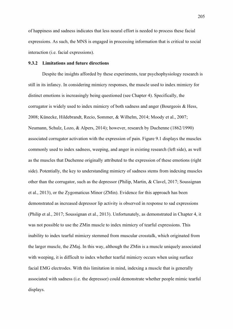
205
of happiness and sadness indicates that less neural effort is needed to process these facial
expressions. As such, the MNS is engaged in processing information that is critical to social
interaction (i.e. facial expressions).
9.3.2 Limitations and future directions
Despite the insights afforded by these experiments, tear psychophysiology research is
still in its infancy. In considering mimicry responses, the muscle used to index mimicry for
distinct emotions is increasingly being questioned (see Chapter 4). Specifically, the
corrugator is widely used to index mimicry of both sadness and anger (Bourgeois & Hess,
2008; Künecke, Hildebrandt, Recio, Sommer, & Wilhelm, 2014; Moody et al., 2007;
Neumann, Schulz, Lozo, & Alpers, 2014); however, research by Duchenne (1862/1990)
associated corrugator activation with the expression of pain. Figure 9.1 displays the muscles
commonly used to index sadness, weeping, and anger in existing research (left side), as well
as the muscles that Duchenne originally attributed to the expression of these emotions (right
side). Potentially, the key to understanding mimicry of sadness stems from indexing muscles
other than the corrugator, such as the depressor (Philip, Martin, & Clavel, 2017; Soussignan
et al., 2013), or the Zygomaticus Minor (ZMin). Evidence for this approach has been
demonstrated as increased depressor lip activity is observed in response to sad expressions
(Philip et al., 2017; Soussignan et al., 2013). Unfortunately, as demonstrated in Chapter 4, it
was not possible to use the ZMin muscle to index mimicry of tearful expressions. This
inability to index tearful mimicry stemmed from muscular crosstalk, which originated from
the larger muscle, the ZMaj. In this way, although the ZMin is a muscle uniquely associated
with weeping, it is difficult to index whether tearful mimicry occurs when using surface
facial EMG electrodes. With this limitation in mind, indexing a muscle that is generally
associated with sadness (i.e. the depressor) could demonstrate whether people mimic tearful
displays.
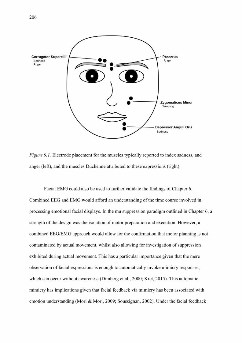
206
Figure 9.1. Electrode placement for the muscles typically reported to index sadness, and
anger (left), and the muscles Duchenne attributed to these expressions (right).
Facial EMG could also be used to further validate the findings of Chapter 6.
Combined EEG and EMG would afford an understanding of the time course involved in
processing emotional facial displays. In the mu suppression paradigm outlined in Chapter 6, a
strength of the design was the isolation of motor preparation and execution. However, a
combined EEG/EMG approach would allow for the confirmation that motor planning is not
contaminated by actual movement, whilst also allowing for investigation of suppression
exhibited during actual movement. This has a particular importance given that the mere
observation of facial expressions is enough to automatically invoke mimicry responses,
which can occur without awareness (Dimberg et al., 2000; Kret, 2015). This automatic
mimicry has implications given that facial feedback via mimicry has been associated with
emotion understanding (Mori & Mori, 2009; Soussignan, 2002). Under the facial feedback
207
view, mimicry facilitates motor resonance. Therefore, the concurrent measurement of EEG
and EMG would allow for an understanding of how mu suppression is influenced by
unconscious motor movement. Evidence for the efficacy of this approach has been
demonstrated in existing action-related finger tapping studies (Muthukumaraswamy et al.,
2004; Woodruff & Maaske, 2010; Woodruff et al., 2011). Concurrent EEG-EMG was used to
confirm that motor activity was not present in the observation-based trials
(Muthukumaraswamy et al., 2004). Thus, in the same vein, EEG and facial EMG would
allow the disentangling of the time course of the way we process emotional displays—from
mimicry to mirroring.
9.3.3 Conclusions
Thus far, it is evident that tears serve as a distinctive signal of sadness, and that this
signal is preferentially processed at the physiological level. Therefore, tearful expressions—
particularly those of sadness—are interpreted rapidly. Moreover, humans appear to be
biologically hard-wired to respond to socially relevant information. This thesis has provided
both behavioural and psychophysiological evidence to demonstrate that tears enhance the
social relevance of sad faces. However, what is it about tears that make them such an
effective signal? One explanation is that tears are perceived as an honest expression of
emotion and this authenticity affords meaning to the display (Trimble, 2012; Vingerhoets,
2013). Namely, tearful persons are believed to be sincere (Picó et al., 2020; Zeifman &
Brown, 2011). Presumably, this sincerity fosters feelings of warmth and social connectedness
to the tearful person (Fischer et al., 2013; Ven de Ven et al., 2017; Zickfeld & Schubert,
2018; Zickfeld et al., 2018). Yet, experimental studies have predominantly used posed faces
with artificial tears—either elicited via eyedrops or digitally added to a photograph (Balsters
et al., 2013; Fischer et al., 2013; Hendriks & Vingerhoets, 2006; Hendriks et al., 2007; Ito et
al., 2019; Lockwood et al., 2013; Reed et al., 2015; Švegar, Fiamengo, Grundler, & Kardum,
208
2016). Recently, tear researchers have preferentially adopted the use of tearful stimuli
wherein the tears were shed in a moment of true emotion (Gračanin et al., 2018; Picó et al.,
2020; van de Ven et al., 2017; Vingerhoets et al., 2016). However, prior to this thesis, limited
empirical work had explored the perceptions genuine and posed stimuli together—a
necessary comparison to elucidate why tears are such an effective signal.
9.4 The Type of Stimuli used in Crying Research
9.4.1 Findings and implications
Chapter 7 explored what was proposed as a critical component of future crying
research—an empirical investigation of the stimuli used in crying research. As described in
Chapter 8, three experiments were conducted to explore the perception of genuineness for
three different types of sad stimuli. Unsurprisingly, these experiments demonstrated that
genuine-tearful displays were perceived as more genuine than posed faces with artificial tears
that were added digitally. Additionally, posed KDEF faces with tears were perceived as
significantly more genuine than posed KDEF faces without tears. To further explore whether
participants were sensitive to the genuineness of tearful sadness, an experiment was
conducted using a paradigm that has previously been used in genuineness research (McLellan
et al., 2010; Miles & Johnston, 2007; Namba et al., 2018). When participants were asked if
faces were showing emotion, participants were significantly more likely to state that a posed
KDEF display was showing sadness relative to a genuine display. By contrast, when
participants were asked to judge whether the face was feeling emotion, participants displayed
greater sensitivity to genuine displays. Therefore, my research has provided the first
empirical evidence that persons are sensitive to the genuineness of sad tearful displays.
Undoubtedly, the findings from Chapter 8 have implications for the work discussed in
Chapters 3, 4, 5, and 6, given that these chapters all relied on the use of posed stimuli. The
primary concern for these results is that tearful KDEF faces were perceived as more genuine

209
than posed KDEF faces without tears. This concern stems from an inability to definitively
conclude whether this perception of genuineness influenced results. However, several points
must be noted. Firstly, the magnitude of the ‘increased perception of genuineness’ effect was
quite small. As such, it is unlikely that the increase in perceived genuineness solely facilitated
the behavioural and physiological responses obtained in earlier chapters. Rather, tears as a
distinctive signal of sadness remains a more likely explanation. It is reasonable to conclude
that genuine signals would be more effective than disingenuous ones. Moreover, it is intuitive
that genuine expressions would be responded to more favourably (e.g. enhanced ratings,
improved response time, and greater ERP responses).
Evidence for this conclusion can be drawn from research exploring perceptions of
Duchenne and non-Duchenne smiles. In the absence of context, both tears and smiles serve as
distinctive markers of sadness and joy, respectively. Genuine Duchenne smiles are perceived
as more intensely happy, more pleasant, and signal greater affiliation than posed smiles
(Leppänen & Hietanen, 2007). In addition, these behavioural responses seem to have a
physiological basis wherein Duchenne smiles are mimicked more often and elicit greater
neural responses relative to posed smiles (Krumhuber et al., 2014; McLellan et al., 2012). It
appears that genuine expressions foster increased pro-social responses from observers,
wherein genuineness, at least in the case of smiles, is responded to favourably. Therefore, it is
plausible that tearful displays that are perceived as genuine might elicit increased behavioural
and physiological responses. However, how the perception of tearful genuineness modulates
early physiological responses is yet to be determined.
Given that persons are sensitive to the difference between genuine and posed tearful
sadness, this sensitivity provides empirical support for the theory that tears are an honest
signal of emotion (Trimble, 2012; Vingerhoets, 2013). Tears are associated with sincerity,
credibility, and honesty (Picó et al., 2020; Trimble, 2012; Zeifman & Brown, 2011), and it is

210
known that humans are sensitive to signals that are high in salience and credibility
(Niedenthal & Brauer, 2012). It makes sense that humans are particularly sensitive to social
signals—like tears—which biologically function to elicit support from observers. This
sensitivity may stem from the increased cost associated with providing this support, as well
as a desire to not be manipulated by an inauthentic expressor (Bavelas et al., 1986).
Manipulative tears, known as crocodile tears, are an insincere tearing display designed to
elicit support from observers. Although crocodile tears are qualitatively different from the
posed tears used in crying research, the understanding that humans are capable of
distinguishing posed from genuine emotion has obvious implications for the field of crocodile
tear research. Very limited research has been conducted exploring how humans distinguish
between genuine and crocodile tears (Roeyen et al., 2020). Roeyen et al. (2020) recently
demonstrated that the greatest damage to a crier’s image stemmed from the perception that a
crying display is fake. Specifically, the perception that tears were false resulted in increased
ratings of manipulativeness, and decreased warmth and perceived reliability of tearful
individuals. Critically, this damaging perception was present, regardless of whether the tears
were genuine or fake. Therefore, further research should explore what specifically makes
tears seem genuine, which will aid in determining how persons distinguish between crocodile
tears and genuine tears. This distinction will not only aid in understanding tearful displays,
but also has the potential to influence fields such as criminal justice and law enforcement,
wherein the accurate detection of insincere emotion is paramount.
9.4.2 Limitations and future directions
As the studies presented in Chapter 8 were some of the first to explore perceptions of
genuineness in crying research, there is a great deal of future work that needs to be
conducted. The next section will outline limitations associated with the use of genuine
displays, and what can be done to overcome these caveats in future research.

211
Firstly, the genuine stimuli used in this research, and that of prior studies, were
facilitated by a set of tearful displays that were captured in a moment of genuine experience.
One limitation of these naturalistic photographs is that we are unable to know what the
individual was feeling that lead them to weep. In this way, it is impossible to control for
feeling across this stimulus set. However, the creation of a genuine tearful stimulus set, where
feeling is known, is also not without limitations. Namely, developing a stimulus set of
genuine expressions is a time consuming and labour-intensive project, which subsequently
results in lower experimental control over the stimuli than that of posed standardised displays
(Dawel et al., 2017; Krivan & Thomas, 2020). An alternative to these caveats was offered by
Dawel et al. (2017), as some posed stimuli are perceived as genuine. This ‘perceived as
genuine’ effect was observed across emotions and stimulus sets. In this sense, posed stimuli
that are perceived as genuine offer an alternative to genuine tearful expressions. The research
conducted in Chapter 8 has demonstrated that this ‘posed perceived as genuine’ effect is also
the case for some posed tearful displays. Therefore, it would be advantageous to conduct a
wider-scale investigation of the stimuli used in tear research to examine which posed tearful
displays are perceived as genuine. This investigation would offer the opportunity for future
research to use tearful displays that are perceived as genuine, while affording additional
experimental control over the stimuli.
Alternatively, genuine expressions of sadness that have been modified to include
artificial tears could also offer interesting conclusions. Genuine displays of sadness are likely
more readily available than genuine tearful displays, and as such would afford additional
experimental control over the type of tears that are used. This control would allow for the
investigation of how different levels of tearing are perceived (e.g., from tearing up to
hysterical crying). Vignette studies have demonstrated that in some scenarios, tearing up is
responded to more favourably than weeping (Wong et al., 2011). Therefore, it seems that
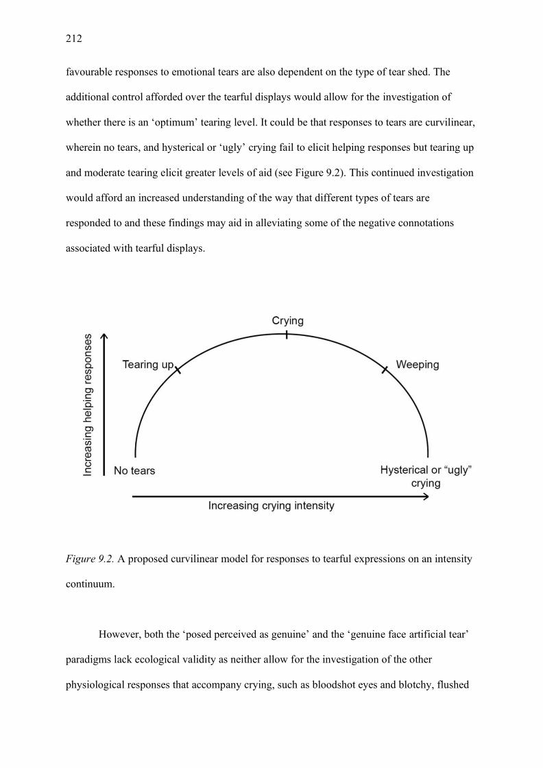
212
favourable responses to emotional tears are also dependent on the type of tear shed. The
additional control afforded over the tearful displays would allow for the investigation of
whether there is an ‘optimum’ tearing level. It could be that responses to tears are curvilinear,
wherein no tears, and hysterical or ‘ugly’ crying fail to elicit helping responses but tearing up
and moderate tearing elicit greater levels of aid (see Figure 9.2). This continued investigation
would afford an increased understanding of the way that different types of tears are
responded to and these findings may aid in alleviating some of the negative connotations
associated with tearful displays.
Figure 9.2. A proposed curvilinear model for responses to tearful expressions on an intensity
continuum.
However, both the ‘posed perceived as genuine’ and the ‘genuine face artificial tear’
paradigms lack ecological validity as neither allow for the investigation of the other
physiological responses that accompany crying, such as bloodshot eyes and blotchy, flushed
213
faces (Provine et al., 2011; Provine et al., 2013). These physiological responses are an
involuntary response to emotional tears. Although tears can be feigned, it is yet to be
determined whether these feigned tears are also accompanied by additional physiological
responses. In addition, the conceptualisation that tearing occurs on a continuum (see Figure
9.2), suggests that perhaps increasingly intense tears elicit increased physiological responses.
For example, ‘tearing up’ may elicit slight sclera reddening, while ‘hysterical or ugly crying’
may elicit extremely puffy, bloodshot eyes and a blotchy, red, flushed face. Therefore, these
accompanying physiological features may be that the key to understanding responses to
tearful displays, and the ability to distinguish genuine emotion. Arguably, the continued
investigation of tearful displays using ecologically valid stimuli are likely to yield interesting
insights into how humans perceive and subsequently respond to tearful displays.
9.4.3 Conclusions
Undoubtedly, the work conducted in this thesis is a preliminary investigation into the
differences between posed and genuinely tearful stimuli. In saying this, the results of these
experiments provide a strong rationale for continued investigation. The promising results
obtained herein provide interesting avenues for further research, and the potential use of
stimuli that overcome the caveats associated with genuine displays. Additionally, this
research has afforded a preliminary insight into how individuals discriminate between posed
and genuine emotional displays. Thus, this research provides the opportunity to explore not
only displays that are associated with pro-social behaviour, but also how humans are able to
distinguish manipulative behaviours from genuine expressions.
9.5 The Big Picture
Tears are undoubtedly mysterious. They signal sadness, distress, and sorrow, as well
as feelings of helplessness and powerlessness, which in turn reduces aggression in observers
and fosters pro-social responses. Tears are also attended to without conscious awareness, as
214
tears are preferentially processed at the physiological level. All these points, offered jointly
throughout this thesis and through existing literature, point to why tearful displays have
persisted through evolution. Although Darwin (1872/1979) originally argued that tearful
displays were purposeless, it seems that their purpose is, in part, their effectiveness as a social
signal.
As identified, tears are a multifaceted signal. It is this diversity in what a tearful
display expresses that has likely contributed to the lack of tear research relative to other basic
emotional expressions. Tearful displays are universally recognisable; however, it would be
counterintuitive to assume that they solely signal sadness. Moving forward, the field of
crying research is vast and largely unexplored. Much like the necessity for exploring mixed
emotions, it is likely that tearful displays would benefit from less bounded inquiry. Research
using ecologically valid and authentic stimuli is at the forefront of emotion research, and
pioneering tear research is being conducted exploring tearful genuineness. Tears are an
honest signal, which means that they reliably elicit succour and comfort from observers.
However, tears can also be used to deceive. Therefore, the exploration of how we, as humans,
are capable of distinguishing between genuine tearful displays and insincere crocodile tears
will give us a greater understanding of the human condition. As has been evidenced
throughout this thesis, tears demand attention from observers. I argue that tears should also
demand attention from emotion researchers. The continued investigation of how tears are
expressed, perceived, and responded to has the unique potential to provide insight into the
development of empathy and prosocial behaviour in society as a whole.

215
References
Achaibou, A., Pourtois, G., Schwartz, S., & Vuilleumier, P. (2008). Simultaneous recording
of EEG and facial muscle reactions during spontaneous emotional mimicry.
Neuropsychologia, 46(4), 1104-1113. doi:10.1016/j.neuropsychologia.2007.10.019
Adolphs, R. (2009). The social brain: Neural basis of social knowledge. Annual Review of
Psychology, 60, 693-716. doi:10.1146/annurev.psych.60.110707.163514
Adolphs, R., Tranel, D., & Damasio, A. R. (2003). Dissociable neural systems for
recognizing emotions. Brain and Cognition, 52(1), 61-69. doi:10.1016/S0278-
2626(03)00009-5
Adolphs, R., Tranel, D., Hamann, S., Young, A. W., Calder, A. J., Phelps, E. A., . . .
Damasio, A. R. (1999). Recognition of facial emotion in nine individuals with
bilateral amygdala damage. Neuropsychologia, 37(10), 1111-1117.
doi:10.1016/S0028-3932(99)00039-1
Alexander, T. (2003). Narcissism and the experience of crying. British Journal of
Psychotherapy, 20(1), 27-38. doi:10.1111/j.1752-0118.2003.tb00112.x
Almeida, P. R., Ferreira-Santos, F., Chaves, P. L., Paiva, T. O., Barbosa, F., & Marques-
Teixeira, J. (2016). Perceived arousal of facial expressions of emotion modulates the
N170, regardless of emotional category: Time domain and time–frequency dynamics.
International Journal of Psychophysiology, 99, 48-56.
doi:10.1016/j.ijpsycho.2015.11.017
Anderson, N. H. (1968). Likableness ratings of 555 personality-trait words. Journal of
Personality and Social Psychology, 9(3), 272-279. doi:10.1037/h0025907
Angelini, M., Fabbri-Destro, M., Lopomo, N. F., Gobbo, M., Rizzolatti, G., & Avanzini, P.
(2018). Perspective-dependent reactivity of sensorimotor mu rhythm in alpha and beta
216
ranges during action observation: an EEG study. Scientific Reports, 8(1), 12429.
doi:10.1038/s41598-018-30912-w
Angus, E., & Thelwall, M. (2010). Motivations for image publishing and tagging on Flickr.
Paper presented at the 14th International Conference on Electronic Publishing,
Helsinki, Finland.
Anisfeld, M. (1996). Only tongue protrusion modeling is matched by neonates.
Developmental Review, 16(2), 149-161. doi:10.1006/drev.1996.0006
Aragón, O. R. (2017). “Tears of joy” and “tears and joy?” personal accounts of dimorphous
and mixed expressions of emotion. Motivation and Emotion, 41(3), 370-392.
doi:10.1007/s11031-017-9606-x
Aragón, O. R., & Bargh, J. A. (2018). “So happy I could shout!” and “So happy I could cry!”
Dimorphous expressions represent and communicate motivational aspects of positive
emotions. Cognition and Emotion, 32(2), 286-302.
doi:10.1080/02699931.2017.1301388
Aragón, O. R., & Clark, M. S. (2018). “Tears of joy” & “smiles of joy” prompt distinct
patterns of interpersonal emotion regulation. Cognition and Emotion, 32(5), 913-940.
doi:10.1080/02699931.2017.1360253
Arnstein, D., Cui, F., Keysers, C., Maurits, N. M., & Gazzola, V. (2011). μ-Suppression
during action observation and execution correlates with BOLD in dorsal premotor,
inferior parietal, and SI cortices. Journal of Neuroscience, 31(40), 14243-14249.
doi:10.1523/JNEUROSCI.0963-11.2011
Baird, A. D., Scheffer, I. E., & Wilson, S. J. (2011). Mirror neuron system involvement in
empathy: a critical look at the evidence. Social Neuroscience, 6(4), 327-335.
doi:10.1080/17470919.2010.547085

217
Balconi, M., & Canavesio, Y. (2016). Is empathy necessary to comprehend the emotional
faces? The empathic effect on attentional mechanisms (eye movements), cortical
correlates (N200 event-related potentials) and facial behaviour (electromyography) in
face processing. Cognition and Emotion, 30(2), 210-224.
doi:10.1080/02699931.2014.993306
Balsters, M. J. H., Krahmer, E. J., Swerts, M. G. J., & Vingerhoets, A. J. J. M. (2013).
Emotional tears facilitate the recognition of sadness and the perceived need for social
support. Evolutionary Psychology, 11(1), 148-158.
doi:10.1177/147470491301100114
Bandes, S. A. (2016). Remorse and criminal justice. Emotion Review, 8(1), 14-19.
doi:10.1177/1754073915601222
Bastiaansen, J. A. C. J., Thioux, M., & Keysers, C. (2009). Evidence for mirror systems in
emotions. Philosophical Transactions of the Royal Society of London: Biological
Sciences, 364(1528), 2391-2404. doi:10.1098/rstb.2009.0058
Batson, C. D. (2009). These things called empathy: eight distinct but related phenomena. In J.
Decety & W. Ickes (Eds.), The social neuroscience of empathy (pp. 3-15). Cambridge,
MA: The MIT Press.
Batty, M., & Taylor, M. J. (2003). Early processing of the six basic facial emotional
expressions. Cognitive Brain Research, 17(3), 613-620. doi:10.1016/S0926-
6410(03)00174-5
Baumeister, R. F., Bratslavsky, E., Finkenauer, C., & Vohs, K. D. (2001). Bad is stronger
than good. Review of General Psychology, 5(4), 323-370. doi:10.1037/1089-
2680.5.4.323

218
Bavelas, J., Black, A., Lemery, C. R., & Mullett, J. (1986). "I show how you feel". Motor
mimicry as a communicative act. Journal of Personality and Social Psychology,
50(2), 322-329. doi:10.1037/0022-3514.50.2.322
Bayer, M., & Schacht, A. (2014). Event-related brain responses to emotional words, pictures,
and faces – a cross-domain comparison. Frontiers in Psychology, 5, 1106.
doi:10.3389/fpsyg.2014.01106
Becker, D. V., Anderson, U. S., Mortensen, C. R., Neufeld, S. L., & Neel, R. (2011). The
face in the crowd effect unconfounded: Happy faces, not angry faces, are more
efficiently detected in single- and multiple-target visual search tasks. Journal of
Experimental Psychology: General, 140(4), 637-659. doi:10.1037/a0024060
Becker, D. V., & Srinivasan, N. (2014). The vividness of the happy face. Current Directions
in Psychological Science, 23(3), 189-194. doi:10.1177/0963721414533702
Bediou, B., Eimer, M., d’Amato, T., Hauk, O., & Calder, A. J. (2009). In the eye of the
beholder: Individual differences in reward-drive modulate early frontocentral ERPs to
angry faces. Neuropsychologia, 47(3), 825-834.
doi:10.1016/j.neuropsychologia.2008.12.012
Bentin, S., Allison, T., Puce, A., Perez, E., & McCarthy, G. (1996). Electrophysiological
studies of face perception in humans. Journal of Cognitive Neuroscience, 8(6), 551–
565. doi:10.1162/jocn.1996.8.6.551
Bentin, S., & Deouell, L. Y. (2000). Structural encoding and identification in face processing:
ERP evidence for separate mechanisms. Cognitive Neuropsychology, 17(1-3), 35-54.
doi:10.1080/026432900380472
Bentsianov, B., & Blitzer, A. (2004). Facial anatomy. Clinics in Dermatology, 22(1), 3-13.
doi:10.1016/j.clindermatol.2003.11.011
219
Blair, R. J. R., Morris, J. S., Frith, C. D., Perrett, D. I., & Dolan, R. J. (1999). Dissociable
neural responses to facial expressions of sadness and anger. Brain, 122(5), 883-893.
doi:10.1093/brain/122.5.883
Blau, V. C., Maurer, U., Tottenham, N., & McCandliss, B. D. (2007). The face-specific N170
component is modulated by emotional facial expression. Behavioral and Brain
Functions, 3(1), 7. doi:10.1186/1744-9081-3-7
Blechert, J., Sheppes, G., Di Tella, C., Williams, H., & Gross, J. J. (2012). See what you
think, reappraisal modulated behavioural and neural responses to social stimuli.
Psychological Science, 23, 346-353. doi:10.1177/0956797612438559
Botvinick, M., Jha, A. P., Bylsma, L. M., Fabian, S. A., Solomon, P. E., & Prkachin, K. M.
(2005). Viewing facial expressions of pain engages cortical areas involved in the
direct experience of pain. NeuroImage, 25(1), 312-319.
doi:10.1016/j.neuroimage.2004.11.043
Bourgeois, P., & Hess, U. (2008). The impact of social context on mimicry. Biological
Psychology, 77(3), 343-352. doi:10.1016/j.biopsycho.2007.11.008
Breiter, H. C., Etcoff, N. L., Whalen, P. J., Kennedy, W. A., Rauch, S. L., Buckner, R. L., . . .
Rosen, B. R. (1996). Response and habituation of the human Amygdala during visual
processing of facial expression. Neuron, 17(5), 875-887. doi:10.1016/S0896-
6273(00)80219-6
Brennan, A. N., Harris, A. W. F., & Williams, L. M. (2014). Neural processing of facial
expressions of emotion in first onset psychosis. Psychiatry Research, 219(3), 477-
485. doi:10.1016/j.psychres.2014.06.017
Brenner, C. A., Rumak, S. P., Burns, A. M. N., & Kieffaber, P. D. (2014). The role of
encoding and attention in facial emotion memory: An EEG investigation.

220
International Journal of Psychophysiology, 93(3), 398-410.
doi:10.1016/j.ijpsycho.2014.06.006
Briton, N. J., & Hall, J. A. (1995). Beliefs about female and male nonverbal communication.
Sex Roles, 32, 79-90. doi:10.1007%2FBF01544758
Brody, L. R., & Hall, J. A. (2008). Gender and emotion in context. In M. Lewis, J. M.
Haviland-Jones, & L. F. Barrett (Eds.), Handbook of emotions (Vol. 3, pp. 395-408).
New York, NY: The Guilford Press.
Bruce, V., & Young, A. (1986). Understanding face recognition. British Journal of
Psychology, 77, 305-327. doi:10.1111/j.2044-8295.1986.tb02199.x
Buccino, G., Binkofski, F., Fink, G. R., Fadiga, L., Fogassi, L., Gallese, V., . . . Freund, H. J.
(2001). Action observation activates premotor and parietal areas in a somatotopic
manner: an fMRI study. European Journal of Neuroscience, 13(2), 400-404.
doi:10.1111/j.1460-9568.2001.01385.x
Cacioppo, J. T., Petty, R. E., Losch, M. E., & Kim, H. S. (1986). Electromyographic activity
over facial muscle regions can differentiate the valence and intensity of affective
reactions. Journal of Personality and Social Psychology, 50(2), 260-268.
doi:10.1037//0022-3514.50.2.260
Cacioppo, J. T., Tassinary, L. G., & Berntson, G. G. (2007). Psychophysiological science:
Interdisciplinary approaches to classic questions about the mind. In J. T. Cacioppo, L.
G. Tassinary, & G. G. Berntson (Eds.), Handbook of psychophysiology (3rd ed., pp. 1-
16). Cambridge, England: Cambridge University Press.
Caharel, S., Courtay, N., Bernard, C., Lalonde, R., & Rebaï, M. (2005). Familiarity and
emotional expression influence an early stage of face processing: An
electrophysiological study. Brain and Cognition, 59(1), 96-100.
doi:10.1016/j.bandc.2005.05.005

221
Calder, A. J., Keane, J., Manes, F., Antoun, N., & Young, A. W. (2000). Impaired
recognition and experience of disgust following brain injury. Nature Neuroscience,
3(11), 1077-1078. doi:10.1038/80586
Calder, A. J., Young, A. W., Rowland, D., Perrett, D. I., Hodges, J. R., & Etcoff, N. L.
(1996). Facial emotion recognition after bilateral amygdala damage: Differentially
severe impairment of fear. Cognitive Neuropsychology, 13(5), 699-745.
doi:10.1080/026432996381890
Calvo, M. G., Avero, P., Fernández-Martín, A., & Recio, G. (2016). Recognition thresholds
for static and dynamic emotional faces. Emotion, 16(8), 1186-1200.
doi:10.1037/emo0000192
Calvo, M. G., Avero, P., & Lundqvist, D. (2006). Facilitated detection of angry faces: Initial
orienting and processing efficiency. Cognition and Emotion, 20(6), 785-811.
doi:10.1080/02699930500465224
Calvo, M. G., Fernández-Martín, A., & Nummenmaa, L. (2012). Perceptual, categorical, and
affective processing of ambiguous smiling facial expressions. Cognition, 125(3), 373-
393. doi:10.1016/j.cognition.2012.07.021
Calvo, M. G., Gutiérrez-García, A., & Del Líbano, M. (2018). What makes a smiling face
look happy? Visual saliency, distinctiveness, and affect. Psychological Research,
82(2), 296-309. doi:10.1007/s00426-016-0829-3
Calvo, M. G., & Lundqvist, D. (2008). Facial expressions of emotion (KDEF): identification
under different display-duration conditions. Behavior Research Methods, 40(1), 109-
115. doi:10.3758/BRM.40.1.109
Calvo, M. G., & Marrero, H. (2009). Visual search of emotional faces: The role of affective
content and featural distinctiveness. Cognition and Emotion, 23(4), 782-806.
doi:10.1080/02699930802151654

222
Calvo, M. G., & Nummenmaa, L. (2008). Detection of emotional faces: Salient physical
features guide effective visual search. Journal of Experimental Psychology: General,
137(3), 471– 494. doi:10.1037/a0012771
Calvo, M. G., & Nummenmaa, L. (2016). Perceptual and affective mechanisms in facial
expression recognition: An integrative review. Cognition and Emotion, 30(6), 1081-
1106. doi:10.1080/02699931.2015.1049124
Cannon, E. N., Yoo, K. H., Vanderwert, R. E., Ferrari, P. F., Woodward, A. L., & Fox, N. A.
(2014). Action experience, more than observation, influences mu rhythm
desynchronization. PLoS ONE, 9(3), e92002. doi:10.1371/journal.pone.0092002
Carr, L., Iacoboni, M., Dubeau, M. C., Mazziotta, J. C., & Lenzi, G. L. (2003). Neural
mechanisms of empathy in humans: a relay from neural systems for imitation to
limbic areas. Proceedings of the National Academy of Sciences of the United States of
America, 100(9), 5497-5502. doi:10.1073/pnas.0935845100
Ceccarini, F., & Caudek, C. (2013). Anger superiority effect: The importance of dynamic
emotional facial expressions. Visual Cognition, 21(4), 498-540.
doi:10.1080/13506285.2013.807901
Chandler, J. (2018). Likeableness and meaningfulness ratings of 555 (+487) person-
descriptive words. Journal of Research in Personality, 72, 50-57.
doi:10.1016/j.jrp.2016.07.005
Chartrand, T. L., & Bargh, J. A. (1999). The chameleon effect: the perception-behavior link
and social interaction. Journal of Personality and Social Psychology, 76(6), 893-910.
doi:10.1037//0022-3514.76.6.893
Chartrand, T. L., Maddux, W. W., & Lakin, J. L. (2005). Beyond the perception-behavior
link: the ubiquitous utility and motivational moderators of non- conscious mimicry. In

223
R. R. Hassin, J. S. Uleman, & J. A. Bargh (Eds.), The new unconscious. (pp. 334-
361). New York, NY: Oxford University Press.
Chen, J., Ma, W., Zhang, Y., Wu, X., Wei, D., Liu, G., . . . Zhang, Z. (2014). Distinct facial
processing related negative cognitive bias in first-episode and recurrent major
depression: Evidence from the N170 ERP component. PLoS ONE, 9(10), e109176.
doi:10.1371/journal.pone.0109176
Cheng, Y., Chen, C., Lin, C.-P., Chou, K.-H., & Decety, J. (2010). Love hurts: An fMRI
study. NeuroImage, 51(2), 923-929. doi:10.1016/j.neuroimage.2010.02.047
Cheng, Y., Lee, P.-L., Yang, C.-Y., Lin, C.-P., Hung, D., & Decety, J. (2008). Gender
differences in the mu rhythm of the human mirror-neuron system. PLoS ONE, 3(5),
e2113. doi:10.1371/journal.pone.0002113
Cheng, Y., Lin, C.-P., Liu, H.-L., Hsu, Y.-Y., Lim, K.-E., Hung, D., & Decety, J. (2007).
Expertise modulates the perception of pain in others. Current Biology, 17(19), 1708-
1713. doi:10.1016/j.cub.2007.09.020
Cheng, Y., Yang, C.-Y., Lin, C.-P., Lee, P.-L., & Decety, J. (2008). The perception of pain in
others suppresses somatosensory oscillations: A magnetoencephalography study.
NeuroImage, 40(4), 1833-1840. doi:10.1016/j.neuroimage.2008.01.064
Cooper, N., Simpson, A., Till, A., Simmons, K., & Puzzo, I. (2013). Beta event-related
desynchronization as an index of individual differences in processing human facial
expression: further investigations of autistic traits in typically developing adults.
Frontiers in Human Neuroscience, 7(159). doi:10.3389/fnhum.2013.00159
Cretser, G., Lombardo, W., Lombardo, B., & Mathis, S. (1982). Reactions to men and
women who cry: A study of sex differences in percieved societal attitudes versus
personal attitudes. Perceptual and Motor Skills, 55(2), 479-486.
doi:10.2466/pms.1982.55.2.479
224
Critchley, H. D., Rotshtein, P., Nagai, Y., O'Doherty, J., Mathias, C. J., & Dolan, R. J.
(2005). Activity in the human brain predicting differential heart rate responses to
emotional facial expressions. NeuroImage, 24(3), 751-762.
doi:10.1016/j.neuroimage.2004.10.013
Crivelli, C., Carrera, P., & Fernández-Dols, J.-M. (2015). Are smiles a sign of happiness?
Spontaneous expressions of judo winners. Evolution and Human Behavior, 36(1), 52-
58. doi:10.1016/j.evolhumbehav.2014.08.009
Darwin, C. (1979). The expression of emotions in man and animals. London: Julian
Friedmann. (Original work published 1872)
Davidson, R. J. (1992). Emotion and affective style: Hemispheric substrates. Psychological
Science, 3(1), 39-43. doi:10.1111/j.1467-9280.1992.tb00254.x
Davis, M. H. (1980). A multidimensional approach to individual differences in empathy.
Journal of Personality and Social Psychology, 10, 85.
Davis, M. H. (1983). Measuring individual differences in empathy: Evidence for a
multidimensional approach. Journal of Personality and Social Psychology, 44(1),
113-126. doi:10.1037/0022-3514.44.1.113
Dawel, A., Palermo, R., O’Kearney, R., & McKone, E. (2015). Children can discriminate the
authenticity of happy but not sad or fearful facial expressions, and use an immature
intensity-only strategy. Frontiers in Psychology, 6, 462.
doi:10.3389/fpsyg.2015.00462
Dawel, A., Wright, L., Irons, J., Dumbleton, R., Palermo, R., O’Kearney, R., & McKone, E.
(2017). Perceived emotion genuineness: normative ratings for popular facial
expression stimuli and the development of perceived-as-genuine and perceived-as-
fake sets. Behavior Research Methods, 49(4), 1539-1562. doi:10.3758/s13428-016-
0813-2

225
de Lange, F. P., Spronk, M., Willems, R. M., Toni, I., & Bekkering, H. (2008).
Complementary systems for understanding action intentions. Current Biology, 18(6),
454-457. doi:10.1016/j.cub.2008.02.057
de Vignemont, F., & Singer, T. (2006). The empathic brain: how, when and why? Trends in
Cognitive Sciences, 10(10), 435-441. doi:10.1016/j.tics.2006.08.008
Decety, J., & Jackson, P. L. (2004). The functional architecture of human empathy.
Behavioral and Cognitive Neuroscience Reviews, 3(2), 71-100.
doi:10.1177/1534582304267187
Decety, J., & Lamm, C. (2006). Human empathy through the lens of social neuroscience. The
Scientific World Journal, 6, 1146-1163. doi:10.1100/tsw.2006.221
Del Giudice, M., Manera, V., & Keysers, C. (2009). Programmed to learn? The ontogeny of
mirror neurons. Developmental Science, 12(2), 350-363. doi:10.1111/j.1467-
7687.2008.00783.x
Del Zotto, M., & Pegna, A. J. (2015). Processing of masked and unmasked emotional faces
under different attentional conditions: an electrophysiological investigation. Frontiers
in Psychology, 6(1691). doi:10.3389/fpsyg.2015.01691
Detre, J. A., & Floyd, T. F. (2001). Functional MRI and its applications to the clinical
neurosciences. The Neuroscientist, 7(1), 64-79. doi:10.1177/107385840100700110
di Pellegrino, G., Fadiga, L., Fogassi, L., Gallese, V., & Rizzolatti, G. (1992). Understanding
motor events: a neurophysiological study. Experimental Brain Research, 91(1), 176-
180. doi:10.1007/BF00230027
Dien, J. (1998). Issues in the application of the average reference: Review, critiques, and
recommendations. Behavior Research Methods, 30(1), 34-43.
doi:10.3758/BF03209414
226
DiGirolamo, M. A., Simon, J. C., Hubley, K. M., Kopulsky, A., & Gutsell, J. N. (2019).
Clarifying the relationship between trait empathy and action-based resonance indexed
by EEG mu-rhythm suppression. Neuropsychologia, 133, 107172.
doi:10.1016/j.neuropsychologia.2019.107172
Dimberg, U. (1982). Facial reactions to facial expressions. Psychophysiology, 19(6), 643-
647. doi:10.1111/j.1469-8986.1982.tb02516.x
Dimberg, U., & Öhman, A. (1996). Behold the wrath: Psychophysiological responses to
facial stimuli. Motivation and Emotion, 20(2), 149-182. doi:10.1007/BF02253869
Dimberg, U., & Petterson, M. (2000). Facial reactions to happy and angry facial expressions:
Evidence for right hemisphere dominance. Psychophysiology, 37(5), 693-696.
doi:10.1017/S0048577200990759
Dimberg, U., Thunberg, M., & Elmehed, K. (2000). Unconscious facial reactions to
emotional facial expressions. Psychological Science, 11(1), 86-89. doi:10.1111/1467-
9280.00221
Dimberg, U., Thunberg, M., & Grunedal, S. (2002). Facial reactions to emotional stimuli:
Automatically controlled emotional responses. Cognition and Emotion, 16(4), 449-
471. doi:10.1080/02699930143000356
Douglas, K. M., Porter, R. J., & Johnston, L. (2012). Sensitivity to posed and genuine facial
expressions of emotion in severe depression. Psychiatry Research, 196(1), 72-78.
doi:10.1016/j.psychres.2011.10.019
Dovidio, J. F. (1991). The empathy-altruism hypothesis: Paradigm and promise.
Psychological Inquiry, 2(2), 126-128. doi:10.1207/s15327965pli0202_4
Du, S., & Martinez, A. M. (2011). The resolution of facial expressions of emotion. Journal of
Vision, 11(13), 24. doi:10.1167/11.13.24
227
Duchenne de Boulogne, G.-B. (1990). The mechanism of human facial expression (A.
Cuthbertson, Trans.). New York, NY: Cambridge University Press. (Original work
published 1862).
Dunbar, R. I. M., & Shultz, S. (2007). Evolution in the social brain. Science, 317(5843),
1344-1347. doi:10.1126/science.1145463
Eastwood, J. D., Smilek, D., & Merikle, P. M. (2001). Differential attentional guidance by
unattended faces expressing positive and negative emotion. Perception and
Psychophysics, 63(6), 1004-1013. doi:10.3758/BF03194519
Eimer, M. (2000). Event-related brain potentials distinguish processing stages involved in
face perception and recognition. Clinical Neurophysiology, 111, 694-705.
doi:10.1016/s1388-2457(99)00285-0
Eimer, M. (2000). The face-specific N170 component reflects late stages in the structural
encoding of faces. NeuroReport, 11(10), 2319–2324. doi:10.1097/00001756-
200007140-00050
Eimer, M., & Holmes, A. (2007). Event-related brain potential correlates of emotional face
processing. Neuropsychologia, 45(1), 15-31.
doi:10.1016/j.neuropsychologia.2006.04.022
Eimer, M., Holmes, A., & McGlone, F. P. (2003). The role of spatial attention in the
processing of facial expression: An ERP study of rapid brain responses to six basic
emotions. Cognitive, Affective, & Behavioral Neuroscience, 3(2), 97-110.
doi:10.3758/CABN.3.2.97
Eisenberg, N., & Fabes, R. A. Empathy: Conceptualization, measurement, and relation to
prosocial behavior. Motivation and Emotion, 14(2), 131-149.
doi:10.1007/BF00991640

228
Eisenberger, N. I., Lieberman, M. D., & Williams, K. D. (2003). Does rejection hurt? An
fMRI study of social exclusion. Science, 302(5643), 290-292.
doi:10.1126/science.1089134
Ekman, P. (1972). Universals and cultural differences in facial expressions of emotion. In J.
Cole (Ed.), Nebraska Symposium on Motivation (pp. 207-282). Lincoln, NB:
University of Nebraska Press.
Ekman, P. (1992). An argument for basic emotions. Cognition and Emotion, 6(3-4), 169-200.
doi:10.1080/02699939208411068
Ekman, P. (2003). Darwin, deception, and facial expression. Annals of the New York
Academy of Sciences, 1000, 205-221. doi:10.1196/annals.1280.010
Ekman, P., Davidson, R., & Friesen, W. (1990). The Duchenne smile: Emotional expression
and brain physiology: II. Journal of Personality and Social Psychology, 58(2), 342-
353. doi:10.1037/0022-3514.58.2.342
Ekman, P., & Friesen, W. (1969). The repertoire of nonverbal behavior: Categories, origins,
usage, and coding. Semiotica, 1(1), 49-98. doi:10.1515/9783110880021.57
Ekman, P., & Friesen, W. (1978). Facial action coding system. Palo Alto, CA: Consulting
Psychologist Press.
Ekman, P., Friesen, W., & O'Sullivan, M. (1988). Smiles when lying. Journal of Personality
and Social Psychology, 54(3), 414-420. doi:10.1037/0022-3514.54.3.414
Ekman, P., & Friesen, W. V. (1971). Constants across cultures in the face and emotion.
Journal of Personality and Social Psychology, 17(2), 124-129. doi:10.1037/h0030377
Ekman, P., & Friesen, W. V. (1976). Pictures of Facial Affect. Palo Alto: Consulting
Psychologists Press.
Ekman, P., & Friesen, W. V. (1978). Facial Action Coding System: Consulting Psychologists
Press.
229
Ekman, P., & Friesen, W. V. (1982). Felt, false, and miserable smiles. Journal of Nonverbal
Behavior, 6(4), 238-252. doi:10.1007/BF00987191
Ekman, P., Friesen, W. V., O'Sullivan, M., Chan, A., Diacoyanni-Tarlatzis, I., Heider, K., . . .
Tzavaras, A. (1987). Universals and cultural differences in the judgments of facial
expressions of emotion. Journal of Personality and Social Psychology, 53(4), 712-
717. doi:10.1037/0022-3514.53.4.712
Ekman, P., & Rosenberg, E. L. (2005). What the face reveals: Basic and applied studies of
spontaneous expression using the facial action coding system (FACS) (2nd ed.). New
York, NY: Oxford University Press.
Ekman, P., Sorenson, E. R., & Friesen, W. V. (1969). Pan-cultural elements in facial displays
of emotions. Science, 164, 86-88. doi:10.1126/science.164.3875.86
Ensenberg, N. S., Perry, A., & Aviezer, H. (2017). Are you looking at me? Mu suppression
modulation by facial expression direction. Cognitive, Affective, & Behavioral
Neuroscience, 17(1), 174-184. doi:10.3758/s13415-016-0470-z
Enticott, P. G., Johnston, P. J., Herring, S. E., Hoy, K. E., & Fitzgerald, P. B. (2008). Mirror
neuron activation is associated with facial emotion processing. Neuropsychologia,
46(11), 2851-2854. doi:10.1016/j.neuropsychologia.2008.04.022
Fan, Y., & Han, S. (2008). Temporal dynamic of neural mechanisms involved in empathy for
pain: An event-related brain potential study. Neuropsychologia, 46(1), 160–173.
doi:10.1016/j.neuropsychologia.2007.07.023
Farley, S. D. (2014). Nonverbal reactions to an attractive stranger: The role of mimicry in
communicating preferred social distance. Journal of Nonverbal Behavior, 38(2), 195-
208. doi:10.1007/s10919-014-0174-4

230
Faul, F., Erdfelder, E., Lang, A.-G., & Buchner, A. (2007). G*Power 3: A flexible statistical
power analysis program for the social, behavioral, and biomedical sciences. Behavior
Research Methods, 39, 175-191. doi:10.3758/bf03193146
Fazio, R. H., Sanbonmatsu, D. M., Powell, M., & Kardes, F. R. (1986). On the automatic
activation of attitudes. Journal of Personality and Social Psychology, 50(2), 229-238.
doi:10.1037/0022-3514.50.2.229
Fernández-Dols, J.-M., & Ruiz-Belda, M.-A. (1995). Are smiles a sign of happiness? Gold
medal winners at the Olympic Games. Journal of Personality and Social Psychology,
69(6), 1113-1119. doi:10.1037/0022-3514.69.6.1113
Fernández-Dols, J.-M., & Ruiz-Belda, M. A. (1995). Expression of emotion versus
expressions of emotions: Everyday conceptions about spontaneous facial behavior. In
J. A. Russell, J.-M. Fernández-Dols, A. S. R. Manstead, & J. C. Wellenkamp (Eds.),
Everyday conceptions of emotion (pp. 505-522). Dordrecht, NL: Kluwer Academic
Press.
Fernández-Dols, J.-M., Sánchez, F., Carrera, P., & Ruiz-Belda, M. A. (1997). Are
spontaneous expressions and emotions linked? An experimental test of coherence.
Journal of Nonverbal Behavior, 21, 163-177. doi:10.1023/A:1024917530100
Ferrari, P. F., Gallese, V., Rizzolatti, G., & Fogassi, L. (2003). Mirror neurons responding to
the observation of ingestive and communicative mouth actions in the monkey ventral
premotor cortex. European Journal of Neuroscience, 17(8), 1703-1714.
doi:10.1046/j.1460-9568.2003.02601.x
Ferrari, P. F., Vanderwert, R. E., Paukner, A., Bower, S., Suomi, S. J., & Fox, N. A. (2012).
Distinct EEG amplitude suppression to facial gestures as evidence for a mirror
mechanism in newborn monkeys. Journal of Cognitive Neuroscience, 24(5), 1165-
1172. doi:10.1162/jocn_a_00198

231
Ferrari, P. F., Visalberghi, E., Paukner, A., Fogassi, L., Ruggiero, A., & Suomi, S. J. (2006).
Neonatal imitation in rhesus macaques. PLOS Biology, 4(9), e302.
doi:10.1371/journal.pbio.0040302
Feyereisen, P., Malet, C., & Martin, Y. (1986). Is the faster processing of expressions of
happiness modality-specific? In H. D. Ellis, M. A. Jeeves, F. Newcombe, & A. Young
(Eds.), Aspects of Face Processing (pp. 349-355). Boston: Martinus Nijhoff.
Fischer, A. H., Eagly, A. H., & Oosterwijk, S. (2013). The meaning of tears: Which sex
seems emotional depends on the social context. European Journal of Social
Psychology, 43(6), 505-515. doi:10.1002/ejsp.1974
Fiske, S., Cuddy, A., & Glick, P. (2007). Universal dimensions of social cognition: Warmth
and competence. Trends in Cognitive Sciences, 11, 77-83.
doi:10.1016/j.tics.2006.11.005
Fogassi, L. (2011). The mirror neuron system: How cognitive functions emerge from motor
organization. Journal of Economic Behavior & Organization, 77(1), 66-75.
doi:10.1016/j.jebo.2010.04.009
Fox, E., Lester, V., Russo, R., Bowles, R. J., Pichler, A., & Dutton, K. (2000). Facial
expressions of emotion: Are angry faces detected more efficiently? Cognition and
Emotion, 14(1), 61-92. doi:10.1080/026999300378996
Fox, N. A., Bakermans-Kranenburg, M. J., Yoo, K. H., Bowman, L. C., Cannon, E. N.,
Vanderwert, R. E., . . . van IJzendoorn, M. H. (2016). Assessing human mirror
activity with EEG mu rhythm: A meta-analysis. Psychological Bulletin, 142(3), 291-
313. doi:10.1037/bul0000031
Frank, M. G., Ekman, P., & Friesen, W. V. (1993). Behavioral markers and recognizability of
the smile of enjoyment. Journal of Personality and Social Psychology, 64(1), 83-93.
doi:10.1037/0022-3514.64.1.83

232
Frey, W. H. (1985). Crying: The mystery of tears. Minneapolis, MN: Winston Press.
Frey, W. H., Desota-Johnson, D., Hoffman, C., & McCall, J. T. (1981). Effect of stimulus on
the chemical composition of human tears. American Journal of Ophthalmology,
92(4), 559-567. doi:10.1016/0002-9394(81)90651-6
Fridlund, A. J. (1991). Sociality of solitary smiling: Potentiation by an implicit audience.
Journal of Personality and Social Psychology, 60, 229-240. doi:10.1037/0022-
3514.60.2.229
Fridlund, A. J. (1994). Human facial expression: An evolutionary view. San Diego, CA:
Academic Press.
Fridlund, A. J. (2017). The behavioral ecology view of facial displays, 25 years later. In J.-M.
Fernández-Dols & J. A. Russell (Eds.), The science of facial expression (pp. 77-92).
New York, NY: Oxford University Press.
Fridlund, A. J., Kenworthy, K. G., & Jaffey, A. K. (1992). Audience effects in affective
imagery: Replication and extension to dysphoric imagery. Journal of Nonverbal
Behavior, 16(3), 191-212. doi:10.1007/BF00988034
Fridlund, A. J., Sabini, J. P., Hedlund, L. E., Schaut, J. A., Shenker, J. I., & Knauer, M. J.
(1990). Audience effects on solitary faces during imagery: Displaying to the people in
your head. Journal of Nonverbal Behavior, 14(2), 113-137. doi:10.1007/BF01670438
Frijda, N. H. (1986). The emotions. Cambridge: Cambridge University Press.
Frith, C. D. (2007). The social brain? Philosophical Transactions of the Royal Society B:
Biological Sciences, 362(1480), 671. doi:10.1098/rstb.2006.2003
Gainotti, G. (2012). Unconscious processing of emotions and the right hemisphere.
Neuropsychologia, 50(2), 205-218. doi:10.1016/j.neuropsychologia.2011.12.005
Gallese, V. (2001). The 'shared manifold' hypothesis. From mirror neurons to empathy.
Journal of Consciousness Studies, 8(5-6), 33-50. doi:10.1159/000072786
233
Gallese, V., Eagle, M. N., & Migone, P. (2007). Intentional attunement: mirror neurons and
the neural underpinnings of interpersonal relations. Journal of the American
Psychoanalytic Association, 55(1), 131-175. doi:10.1177/00030651070550010601
Gallese, V., Fadiga, L., Fogassi, L., & Rizzolatti, G. (1996). Action recognition in the
premotor cortex. Brain, 119, 593-609. doi:10.1093/brain/119.2.593
Gallese, V., & Goldman, A. (1998). Mirror neurons and the simulation theory of mind-
reading. Trends in Cognitive Sciences, 2(12), 493-501. doi:10.1016/s1364-
6613(98)01262-5
Gallese, V., Keysers, C., & Rizzolatti, G. (2004). A unifying view of the basis of social
cognition. Trends in Cognitive Sciences, 8(9), 396-403.
doi:10.1016/j.tics.2004.07.002
Gastaut, H. J., & Bert, J. (1954). EEG changes during cinematographic presentation; moving
picture activation of the EEG. Electroencephalography and Clinical
Neurophysiology, 6(3), 433-444. doi:10.1016/0013-4694(54)90058-9
Gazzola, V., Aziz-Zadeh, L., & Keysers, C. (2006). Empathy and the somatotopic auditory
mirror system in humans. Current Biology, 16(18), 1824-1829.
doi:10.1016/j.cub.2006.07.072
Gazzola, V., Rizzolatti, G., Wicker, B., & Keysers, C. (2007). The anthropomorphic brain:
the mirror neuron system responds to human and robotic actions. NeuroImage, 35(4),
1674-1684. doi:10.1016/j.neuroimage.2007.02.003
Gelstein, S., Yeshurun, Y., Rozenkrantz, L., Shushan, S., Frumin, I., Roth, Y., & Sobel, N.
(2011). Human tears contain a chemosignal. Science, 331, 226-230.
doi:10.1126/science.1198331

234
Ghuman, A. S., Brunet, N. M., Li, Y., Konecky, R. O., Pyles, J. A., Walls, S. A., . . .
Richardson, R. M. (2014). Dynamic encoding of face information in the human
fusiform gyrus. Nature Communications, 5. doi:10.1038/ncomms6672
Goense, J., Bohraus, Y., & Logothetis, N. K. (2016). fMRI at high spatial resolution:
Implications for BOLD-models. Frontiers in Computational Neuroscience, 10, 66.
doi:10.3389/fncom.2016.00066
Gosselin, P., Beaupré, M., & Boissonneault, A. (2002). Perception of genuine and masking
smiles in children and adults: Sensitivity to traces of anger. The Journal of Genetic
Psychology, 163(1), 58-71. doi:10.1080/00221320209597968
Gosselin, P., Kirouac, G., & Doré, F. Y. (1995). Components and recognition of facial
expression in the communication of emotion by actors. Journal of Personality and
Social Psychology, 68(1), 83-96. doi:10.1037//0022-3514.68.1.83
Goubert, L., Craig, K. D., Vervoort, T., Morley, S., Sullivan, M. J. L., Williams, d. C. A. C., .
. . Crombez, G. (2005). Facing others in pain: The effects of empathy. Pain, 118(3),
285-288. doi:10.1016/j.pain.2005.10.025
Gračanin, A., Bylsma, L. M., & Vingerhoets, A. J. J. M. (2018). Why only humans shed
emotional tears. Human Nature, 29(2), 104-133. doi:10.1007/s12110-018-9312-8
Gračanin, A., Krahmer, E., Rinck, M., & Vingerhoets, A. J. J. M. (2018). The effects of tears
on approach–avoidance tendencies in observers. Evolutionary Psychology, 16(3),
1474704918791058. doi:10.1177/1474704918791058
Gračanin, A., van Assen, M. A. L. M., Omrčen, V., Koraj, I., & Vingerhoets, A. J. J. M.
(2016). Chemosignalling effects of human tears revisited: Does exposure to female
tears decrease males’ perception of female sexual attractiveness? Cognition and
Emotion, 31(1), 139-150. doi:10.1080/02699931.2016.1151402
235
Grainger, S. A., Vanman, E. J., Matters, G., & Henry, J. D. (2019). The influence of tears on
older and younger adults’ perceptions of sadness. Psychology and Aging, 34(5), 665-
673. doi:10.1037/pag0000373
Gramfort, A., Luessi, M., Larson, D., Engemann, D., Strohmeier, D., Brodbeck, C., . . .
Hämäläinen, M. (2013). MEG and EEG data analysis with MNE-Python. Frontiers in
Neuroscience, 7. doi:10.3389/fnins.2013.00267
Gratton, G., Coles, M. G. H., & Donchin, E. (1983). A new method for offline removal of
ocular artifact. Electroencephalography and Clinical Neurophysiology, 55(4), 468-
484. doi:10.1016/0013-4694(83)90135-9
Gros, I. T., Panasiti, M. S., & Chakrabarti, B. (2015). The plasticity of the mirror system:
How reward learning modulates cortical motor simulation of others.
Neuropsychologia, 70, 255-262. doi:10.1016/j.neuropsychologia.2015.02.033
Guillermo, R., Wilhelm, O., Sommer, W., & Hildebrandt, A. (2017). Are event-related
potentials to dynamic facial expressions of emotion related to individual differences
in the accuracy of processing facial expressions and identity? Cognitive, Affective, &
Behavioral Neuroscience, 17(2), 364-380. doi:10.3758/s13415-016-0484-6
Gutiérrez-García, A., & Calvo, M. G. (2015). Discrimination thresholds for smiles in genuine
versus blended facial expressions. Cogent Psychology, 2(1), 1064586.
doi:10.1080/23311908.2015.1064586
Hackett, L., Day, A., & Mohr, P. (2008). Expectancy violation and perceptions of rape victim
credibility. Legal and Criminological Psychology, 13(2), 323-334.
doi:10.1348/135532507X228458
Hadjikhani, N., Kveraga, K., Naik, P., & Ahlfors, S. P. (2009). Early (M170) activation of
face-specific cortex by face-like objects. NeuroReport, 20(4), 403-407.
doi:10.1097/WNR.0b013e328325a8e1
236
Häfner, M., & IJzerman, H. (2011). The face of love: Spontaneous accommodation as social
emotion regulation. Personality and Social Psychology Bulletin, 37(12), 1551-1563.
doi:10.1177/0146167211415629
Hager, J. C., & Ekman, P. (1979). Long-distance of transmission of facial affect signals.
Ethology and Sociobiology, 1(1), 77-82. doi:10.1016/0162-3095(79)90007-4
Hajcak, G., Weinberg, A., MacNamara, A., & Foti, D. (2011). ERPs and the study of
emotion. In E. S. Kappenman & S. J. Luck (Eds.), The oxford handbook of event-
related potential components (pp. 441–472): Oxford University Press.
Hansen, C. H., & Hansen, R. D. (1988). Finding the face in the crowd: An anger superiority
effect. Journal of Personality and Social Psychology, 54(6), 917-924.
doi:10.1037/0022-3514.54.6.917
Hareli, S., & Hess, U. (2012). The social signal value of emotions. Cognition and Emotion,
26(3), 385-389. doi:10.1080/02699931.2012.665029
Harrison, N. A., Morgan, R., & Critchley, H. D. (2010). From facial mimicry to emotional
empathy: A role for norepinephrine ? Social Neuroscience, 5(4), 393-400.
doi:10.1080/17470911003656330
Haslinger, B., Erhard, P., Altenmüller, E., Schroeder, U., Boecker, H., & Ceballos-Baumann,
A. O. (2005). Transmodal sensorimotor networks during action observation in
professional pianists. Journal of Cognitive Neuroscience, 17(2), 282-293.
doi:10.1162/0898929053124893
Hasson, O. (1997). Towards a general theory of biological signaling. Journal of Theoretical
Biology, 185(2), 139-156. doi:10.1006/jtbi.1996.0258
Hasson, O. (2009). Emotional tears as biological signals. Evolutionary Psychology, 7(3), 363-
370. doi:10.1177/147470490900700302

237
Hatfield, E., Bensman, L., Thornton, P. D., & Rapson, R. L. (2014). New perspectives on
emotional contagion: A review of classic and recent research on facial mimicry and
contagion. Interpersona: An International Journal on Personal Relationships, 8(2),
159-179. doi:10.5964/ijpr.v8i2.163
Hatfield, E., Cacioppo, J. T., & Rapson, R. L. (1994). Emotional contagion. New York, NY:
Cambridge University Press.
Hatfield, E., Rapson, R. L., & Le, Y. L. (2009). Emotional contagion and empathy. In J.
Decety & W. Ickes (Eds.), The social neuroscience of empathy (pp. 19-30).
Cambridge, MA: The MIT Press.
Haxby, J. V., Hoffman, E. A., & Gobbini, M. I. (2000). The distributed human neural system
for face perception. Trends in Cognitive Sciences, 4(6), 223-233. doi:10.1016/S1364-
6613(00)01482-0
Haxby, J. V., Hoffman, E. A., & Gobbini, M. I. (2002). Human neural systems for face
recognition and social communication. Biological Psychiatry, 51(1), 59-67.
doi:10.1016/S0006-3223(01)01330-0
He, W., Chai, H., Chen, W., Zhang, J., Xu, Y., Zhu, J., & Wang, W. (2012). Facial emotion
triggered cerebral potentials in treatment-resistant depression and borderline
personality disorder patients of both genders. Progress in Neuro-
Psychopharmacology and Biological Psychiatry, 37(1), 121-127.
doi:10.1016/j.pnpbp.2011.12.003
Hebb, D. O. (1949). The organization of behavior. New York: John Wiley & Sons.
Hein, G., & Singer, T. (2008). I feel how you feel but not always: The empathic brain and its
modulation. Current Opinion in Neurobiology, 18(2), 153-158.
doi:10.1016/j.conb.2008.07.012

238
Hendriks, M., Croon, M. A., & Vingerhoets, A. J. (2008). Social reactions to adult crying:
The help-soliciting function of tears. The Journal of Social Psychology, 148(1), 22-
42. doi:10.3200/SOCP.148.1.22-42
Hendriks, M., & Vingerhoets, A. J. J. M. (2006). Social messages of crying faces: Their
influence on anticipated person perception, emotions and behavioural responses.
Cognition and Emotion, 20(6), 878-886. doi:10.1080/02699930500450218
Hendriks, M. C., Croon, M. A., & Vingerhoets, A. J. J. M. (2008). Social reactions to adult
crying: The help-soliciting function of tears. The Journal of Social Psychology,
148(1), 22-42. doi:10.3200/SOCP.148.1.22-42
Hendriks, M. C., van Boxtel, G. J., & Vingerhoets, A. J. J. M. (2007). An event-related
potential study on the early processing of crying faces. NeuroReport, 18(7), 631-634.
doi:10.1097/WNR.0b013e3280bad8c7
Hendriks, M. C., & Vingerhoets, A. J. J. M. (2006). Social messages of crying faces: Their
influence on anticipated person perception, emotions and behavioural responses.
Cognition and Emotion, 20(6), 878-886. doi:10.1080/02699930500450218
Hennenlotter, A., Schroeder, U., Erhard, P., Castrop, F., Haslinger, B., Stoecker, D., . . .
Ceballos-Baumann, A. O. (2005). A common neural basis for receptive and
expressive communication of pleasant facial affect. NeuroImage, 26(2), 581-591.
doi:10.1016/j.neuroimage.2005.01.057
Herrmann, M. J., Aranda, D., Ellgring, H., Mueller, T. J., Strik, W. K., Heidrich, A., &
Fallgatter, A. J. (2002). Face-specific event-related potential in humans is independent
from facial expression. International Journal of Psychophysiology, 45(3), 241-244.
doi:10.1016/S0167-8760(02)00033-8
239
Hess, U., Banse, R., & Kappas, A. (1995). The intensity of facial expression is determined by
underlying affective state and social situation. Journal of Personality and Social
Psychology, 69, 280-288. doi:10.1037/0022-3514.69.2.280
Hess, U., & Blairy, S. (2001). Facial mimicry and emotional contagion to dynamic emotional
facial expressions and their influence on decoding accuracy. International Journal of
Psychophysiology, 40(2), 129-141. doi:10.1016/S0167-8760(00)00161-6
Hess, U., & Bourgeois, P. (2010). You smile--I smile: emotion expression in social
interaction. Biological Psychology, 84(3), 514-520.
doi:10.1016/j.biopsycho.2009.11.001
Hess, U., & Fischer, A. (2013). Emotional mimicry as social regulation. Personality and
Social Psychology Review, 17(2), 142-157. doi:10.1177/1088868312472607
Hess, U., & Fischer, A. (2014). Emotional mimicry: Why and when we mimic emotions.
Social and Personality Psychology Compass, 8(2), 45-57. doi:10.1111/spc3.12083
Hess, U., & Kleck, R. E. (1990). Differentiating emotion elicited and deliberate emotional
facial expressions. European Journal of Social Psychology, 20(5), 369-385.
doi:10.1002/ejsp.2420200502
Hess, U., Philippot, P., & Blairy, S. (1999). Mimicry: Facts and fiction. In P. Philippot, R.
Feldman, & E. Coats (Eds.), The social context of nonverbal behavior (pp. 213-241).
Cambridge, England: Cambridge University Press.
Hess, U., & Thibault, P. (2009). Darwin and emotional expression. American Psychologist,
64(2), 120-128. doi:10.1037/a0013386
Heyes, C. (2010). Mesmerising mirror neurons. NeuroImage, 51(2), 789-791.
doi:10.1016/j.neuroimage.2010.02.034

240
Hideg, I., & van Kleef, G. A. (2017). When expressions of fake emotions elicit negative
reactions: The role of observers' dialectical thinking. Journal of Organizational
Behavior, 38(8), 1196-1212. doi:10.1002/job.2196
Hinojosa, J. A., Mercado, F., & Carretié, L. (2015). N170 sensitivity to facial expression: A
meta-analysis. Neuroscience & Biobehavioral Reviews, 55, 498-509.
doi:10.1016/j.neubiorev.2015.06.002
Hinsz, V. B., & Tomhave, J. A. (1991). Smile and (half) the world smiles with you, frown
and you frown alone. Personality and Social Psychology Bulletin, 17(5), 586-592.
doi:10.1177/0146167291175014
Hobson, H. M., & Bishop, D. V. M. (2016). Mu suppression – A good measure of the human
mirror neuron system? Cortex, 82, 290-310. doi:10.1016/j.cortex.2016.03.019
Hobson, H. M., & Bishop, D. V. M. (2017). The interpretation of mu suppression as an index
of mirror neuron activity: past, present and future. Royal Society Open Science, 4(3).
doi:10.1098/rsos.160662
Hoenen, M., Schain, C., & Pause, B. M. (2013). Down-modulation of mu-activity through
empathic top-down processes. Social Neuroscience, 8(5), 515-524.
doi:10.1080/17470919.2013.833550.
Hofelich, A. J., & Preston, S. D. (2012). The meaning in empathy: Distinguishing conceptual
encoding from facial mimicry, trait empathy, and attention to emotion. Cognition and
Emotion, 26(1), 119-128. doi:10.1080/02699931.2011.559192
Hogeveen, J., Chartrand, T. L., & Obhi, S. S. (2015). Social mimicry enhances mu-
suppression during action observation. Cerebral Cortex, 25(8), 2076-2082.
doi:10.1093/cercor/bhu016
241
Holmes, A., Winston, J. S., & Eimer, M. (2005). The role of spatial frequency information
for ERP components sensitive to faces and emotional facial expression. Cognitive
Brain Research, 25(2), 508-520. doi:10.1016/j.cogbrainres.2005.08.003
Hornsey, M. J., Wohl, M. J. A., Harris, E. A., Okimoto, T. G., Thai, M., & Wenzel, M.
(2019). Embodied remorse: Physical displays of remorse increase positive responses
to public apologies, but have negligible effects on forgiveness. Journal of Personality
and Social Psychology. doi:10.1037/pspi0000208
Horovitz, S. G., Rossion, B., Skudlarski, P., & Gore, J. C. (2004). Parametric design and
correlational analyses help integrating fMRI and electrophysiological data during face
processing. NeuroImage, 22(4), 1587-1595. doi:10.1016/j.neuroimage.2004.04.018
Horstmann, G. (2003). What do facial expressions convey: Feeling states, behavioral
intentions, or actions requests? Emotion, 3(2), 150-166. doi:10.1037/1528-
3542.3.2.150
Horstmann, G., & Bauland, A. (2006). Search asymmetries with real faces: Testing the anger-
superiority effect. Emotion, 6(2), 193-207. doi:10.1037/1528-3542.6.2.193
Iacoboni, M. (2005). Neural mechanisms of imitation. Current Opinion in Neurobiolology,
15(6), 632-637. doi:10.1016/j.conb.2005.10.010
Iacoboni, M. (2009). Mirroring people: The new science of empathy and how we connect
with others. New York, NY: Farrar, Straus and Giroux.
Iacoboni, M., Molnar-Szakacs, I., Gallese, V., Buccino, G., Mazziotta, J. C., & Rizzolatti, G.
(2005). Grasping the intentions of others with one's own mirror neuron system. PLOS
Biology, 3(3), e79. doi:10.1371/journal.pbio.0030079
Ito, K., Ong, C. W., & Kitada, R. (2019). Emotional tears communicate sadness but not
excessive emotions without other contextual knowledge. Frontiers in Psychology, 10,
878. doi:10.3389/fpsyg.2019.00878
242
Ito, T. A., Larsen, J. T., Smith, N., & Cacioppo, J. T. (1998). Negative information weighs
more heavily on the brain: The negativity bias in evaluative categorizations. Journal
of Personality and Social Psychology, 75(4), 887-900. doi:10.1037//0022-
3514.75.4.887
Izard, C. E. (1971). The face of emotion. New York, NY: Appleton Century Crofts.
Jabbi, M., & Keysers, C. (2008). Inferior frontal gyrus activity triggers anterior insula
response to emotional facial expressions. Emotion, 8(6), 775-780.
doi:10.1037/a0014194
Jackson, P. L., Brunet, E., Meltzoff, A. N., & Decety, J. (2006). Empathy examined through
the neural mechanisms involved in imagining how I feel versus how you feel pain.
Neuropsychologia, 44(5), 752-761. doi:10.1016/j.neuropsychologia.2005.07.015
Jackson, P. L., Meltzoff, A. N., & Decety, J. (2005). How do we perceive the pain of others?
A window into the neural processes involved in empathy. NeuroImage, 24(3), 771-
779. doi:10.1016/j.neuroimage.2004.09.006
Jakobs, E., Manstead, A. S. R., & Fischer, A. H. (1999). Social motives and emotional
feelings as determinants of facial displays: The case of smiling. Personality and
Social Psychology Bulletin, 25, 250-270. doi:10.1177/0146167299025004003
Jakobs, E., Manstead, A. S. R., & Fischer, A. H. (1999). Social motives, emotional feelings,
and smiling. Cognition and Emotion, 13(4), 321-345. doi:10.1080/026999399379212
Jakobs, E., Manstead, A. S. R., & Fischer, A. H. (2001). Social context effects on facial
activity in a negative emotional setting. Emotion, 1(1), 51-69. doi:10.1037//1528-
3542.1.1.51
Jankowiak-Siuda, K., Rymarczyk, K., & Grabowska, A. (2011). How we empathize with
others: A neurobiological perspective. Medical Science Monitor, 17(1), 18-28.
doi:10.12659/MSM.881324
243
JASP. (2019). JASP (Version 0.11.1).
Jetha, M. K., Zheng, X., Goldberg, J. O., Segalowitz, S. J., & Schmidt, L. A. (2013). Shyness
and emotional face processing in schizophrenia: An ERP study. Biological
Psychology, 94(3), 562-574. doi:10.1016/j.biopsycho.2013.10.001
Jiang, Y. I., Li, W., Recio, G., Liu, Y., Luo, W., Zhang, D., & Sun, D. (2014). Time pressure
inhibits dynamic advantage in the classification of facial expressions of emotion.
PLoS ONE, e100162. doi:10.1371/journal.pone.0100162
Johnston, L. (2002). Behavioral mimicry and stigmatization. Social Cognition, 20, 18-34.
doi:10.1521/soco.20.1.18.20944
Johnston, L., Miles, L. K., & Macrae, C. N. (2010). Why are you smiling at me? Social
functions of enjoyment and non-enjoyment smiles. British Journal of Social
Psychology, 49, 107-127. doi:10.1348/ 014466609X412476
Joyal, C. C., Neveu, S.-M., Boukhalfi, T., Jackson, P. L., & Renaud, P. (2018). Suppression
of sensorimotor alpha power associated with pain expressed by an avatar: A
preliminary EEG study. Frontiers in Human Neuroscience, 12, 273.
doi:10.3389/fnhum.2018.00273
Joyce, C., & Rossion, B. (2005). The face-sensitive N170 and VPP components manifest the
same brain processes: The effect of reference electrode site (Vol. 116).
Kämpfe, N., Penzhorn, J., Schikora, J., Dünzl, J., & Schneidenbach, J. (2009). Empathy and
social desirability: a comparison of delinquent and non-delinquent participants using
direct and indirect measures. Psychology, Crime & Law, 15(1), 1-17.
doi:10.1080/10683160802010640
Kanwisher, N., McDermott, J., & Chun, M. M. (1997). The Fusiform Face Area: A module in
human extrastriate cortex specialized for face perception. The Journal of
Neuroscience, 17(11), 4302-4311. doi:10.1523/JNEUROSCI.17-11-04302.1997

244
Kaplan, J. T., & Iacoboni, M. (2006). Getting a grip on other minds: Mirror neurons,
intention understanding, and cognitive empathy. Social Neuroscience, 1(3-4), 175-
183. doi:10.1080/17470910600985605
Karakale, O., Moore, M. R., & Kirk, I. J. (2019). Mental simulation of facial expressions: Mu
suppression to the viewing of dynamic neutral face videos. Frontiers in Human
Neuroscience, 13, 34. doi:10.3389/fnhum.2019.00034
Karakale, O., Moore, M. R., McNair, N., & Kirk, I. J. (2019). Increased sensorimotor activity
during categorisation of ambiguous emotional expressions. bioRxiv, 717553.
doi:10.1101/717553
Katz, J. (1999). How emotions work. Chicago, IL: Chicago University Press.
Kauschke, C., Bahn, D., Vesker, M., & Schwarzer, G. (2019). The role of emotional valence
for the processing of facial and verbal stimuli-positivity or negativity bias? Frontiers
in Psychology, 10, 1654-1654. doi:10.3389/fpsyg.2019.01654
Keuken, M. C., Hardie, A., Dorn, B. T., Dev, S., Paulus, M. P., Jonas, K. J., . . . Pineda, J. A.
(2011). The role of the left inferior frontal gyrus in social perception: An rTMS study.
Brain Research, 1383, 196-205. doi:10.1016/j.brainres.2011.01.073
Keysers, C. (2011). The empathic brain: How the discovery of mirror neurons changes our
understanding of human nature. US: Social Brain Press.
Keysers, C., & Gazzola, V. (2006). Towards a unifying neural theory of social cognition. In
S. Anders, G. Ende, M. Junghofer, J. Kissler, & D. Wildgruber (Eds.), Progress in
Brain Research (Vol. 156, pp. 379-401): Elsevier.
Keysers, C., & Gazzola, V. (2009). Expanding the mirror: vicarious activity for actions,
emotions, and sensations. Current Opinion in Neurobiology, 19(6), 666-671.
doi:10.1016/j.conb.2009.10.006
245
Keysers, C., & Perrett, D. I. (2004). Demystifying social cognition: a Hebbian perspective.
Trends in Cognitive Sciences, 8(11), 501-507. doi:10.1016/j.tics.2004.09.005
Killgore, W. D. S., & Yurgelun-Todd, D. A. (2004). Activation of the amygdala and anterior
cingulate during nonconscious processing of sad versus happy faces. NeuroImage,
21(4), 1215-1223. doi:10.1016/j.neuroimage.2003.12.033
Kim, M. J., Loucks, R. A., Neta, M., Davis, F. C., Oler, J. A., Mazzulla, E. C., & Whalen, P.
J. (2010). Behind the mask: the influence of mask-type on amygdala response to
fearful faces. Social Cognitive and Affective Neuroscience, 5(4), 363-368.
doi:10.1093/scan/nsq014
Kirita, T., & Endo, M. (1995). Happy face advantage in recognizing facial expressions. Acta
Psychologica, 89(2), 149-163. doi:10.1016/0001-6918(94)00021-8
Kirkham, A. J., Hayes, A. E., Pawling, R., & Tipper, S. P. (2015). Facial mimicry and
emotion consistency: Influences of memory and context. PLoS ONE, 10(12),
e0145731. doi:10.1371/journal.pone.0145731
Knutson, B. (1996). Facial expressions of emotion influence interpersonal trait inferences.
Journal of Nonverbal Behavior, 20(3), 165-182. doi:10.1007/BF02281954
Korb, S., Grandjean, D., & Scherer, K. R. (2010). Timing and voluntary suppression of facial
mimicry to smiling faces in a Go/NoGo task—An EMG study. Biological
Psychology, 85(2), 347-349. doi:10.1016/j.biopsycho.2010.07.012
Kottler, J. A. (1996). The language of tears. San Francisco, CA: Jossey-Bass.
Kottler, J. A., & Montgomery, M. J. (2001). Theories of crying. In A. J. Vingerhoets & R. R.
Cornelius (Eds.), Adult crying: A biopsychosocial approach. Hove, England:
Brunner-Routledge.
Kralj Novak, P., Smailović, J., Sluban, B., & Mozetič, I. (2015). Sentiment of emojis. PLoS
ONE, 10(12), e0144296. doi:10.1371/journal.pone.0144296

246
Kraut, R. E. (1982). Social presence, facial feedback, and emotion. Journal of Personality
and Social Psychology, 42(5), 853-863. doi:10.1037/0022-3514.42.5.853
Kret, M. E. (2015). Emotional expressions beyond facial muscle actions. A call for studying
autonomic signals and their impact on social perception. Frontiers in Psychology, 6.
doi:10.3389/fpsyg.2015.00711
Krivan, S. J., Caltabiano, N., Cottrell, D., & Thomas, N. A. (2020). The tear effect: More
than just a marker of sadness? Manuscript submitted for publication.
Krivan, S. J., & Thomas, N. A. (2020). A call for the empirical investigation of tear stimuli.
Frontiers in Psychology, 11, 52. doi:10.3389/fpsyg.2020.00052
Krombholz, A., Schaefer, F., & Boucsein, W. (2007). Modification of N170 by different
emotional expression of schematic faces. Biological Psychology, 76(3), 156-162.
doi:10.1016/j.biopsycho.2007.07.004
Krumhuber, E., & Manstead, A. (2009). Can Duchenne smiles be feigned? New evidence on
felt and false smiles. Emotion, 9, 807-820. doi:10.1037/a0017844
Krumhuber, E., Manstead, A. S. R., Cosker, D., Marshal, D., Rosin, P. L., & Kappas, A.
(2007). Facial dynamics as indicators of trustworthiness and cooperative behavior.
Emotion, 7(4), 730-735. doi:10.1037/1528-3542.7.4.730
Krumhuber, E. G., Likowski, K. U., & Weyers, P. (2014). Facial mimicry of spontaneous and
deliberate Duchenne and Non-Duchenne smiles. Journal of Nonverbal Behavior,
38(1), 1-11. doi:10.1007/s10919-013-0167-8
Künecke, J., Hildebrandt, A., Recio, G., Sommer, W., & Wilhelm, O. (2014). Facial EMG
responses to emotional expressions are related to emotion perception ability. PLoS
ONE, 9(1), e84053. doi:10.1371/journal.pone.0084053

247
Kuperman, V., Estes, Z., Brysbaert, M., & Warriner, A. B. (2014). Emotion and language:
valence and arousal affect word recognition. Journal of Experimental Psychology:
General, 143(3), 1065-1081. doi:10.1037/a0035669
Küster, D. (2018). Social effects of tears and small pupils are mediated by felt sadness: An
evolutionary view. Evolutionary Psychology, 16(1), 1474704918761104.
doi:10.1177/1474704918761104
LaBarbera, J. D., Izard, C. E., Vietze, P., & Parisi, S. A. (1976). Four- and six-month-old
infants' visual responses to joy, anger, and neutral expressions. Child Development,
47(2), 535-538. doi:10.2307/1128816
Lahav, A., Saltzman, E., & Schlaug, G. (2007). Action representation of sound: audiomotor
recognition network while listening to newly acquired actions. The Journal of
Neuroscience, 27(2), 308-314. doi:10.1523/jneurosci.4822-06.2007
Lakin, J. L., & Chartrand, T. L. (2003). Using nonconscious behavioral mimicry to create
affiliation and rapport. Psychological Science, 14(4), 334-339. doi:10.1111/1467-
9280.14481
Lakin, J. L., Jefferis, V. E., Cheng, C. M., & Chartrand, T. L. (2003). The chameleon effect
as social glue: Evidence for the evolutionary significance of nonconscious mimicry.
Journal of Nonverbal Behavior, 27(3), 145-162. doi:10.1023/A:1025389814290
Lamm, C., Decety, J., & Singer, T. (2011). Meta-analytic evidence for common and distinct
neural networks associated with directly experienced pain and empathy for pain.
NeuroImage, 54(3), 2492-2502. doi:10.1016/j.neuroimage.2010.10.014
Larsen, J. T., Bertson, G. G., Poehlemann, K. M., Ito, T. A., & Cacioppo, J. T. (2008). The
psychophysiology of emotion. In M. Lewis, J. Haviland-Jones, & L. Feldman Barrett
(Eds.), Handbook of emotions (3rd ed., pp. 180-195). New York, NY: The Guilford
Press.

248
Larsen, J. T., & McGraw, A. P. (2014). The case for mixed emotions. Social and Personality
Psychology Compass, 8(6), 263-274. doi:10.1111/spc3.12108
Larsen, J. T., McGraw, A. P., & Cacioppo, J. T. (2001). Can people feel happy and sad at the
same time? Journal of Personality and Social Psychology, 81(4), 684-696.
doi:10.1037/0022-3514.81.4.684
Larsen, J. T., McGraw, A. P., Mellers, B. A., & Cacioppo, J. T. (2004). The agony of victory
and thrill of defeat: Mixed emotional reactions to disappointing wins and relieving
losses. Psychological Science, 15(5), 325-330. doi:10.1111/j.0956-7976.2004.00677.x
Larsen, J. T., Stastny, B. J., & Bradley, J. (2011). It's a bittersweet symphony:
Simultaneously mixed emotional responses to music with conflicting cues. Emotion,
11(6), 1469-1473. doi:10.1037/a0024081
Larsen, R. J., & Diener, E. (1992). Promises and problems with the circumplex model of
emotion. In M. S. Clark (Ed.), Review of personality and social psychology: Emotion
(Vol. 13, pp. 25-59). Newbury Park, CA.: Sage.
Lavan, N., Lima, C. F., Harvey, H., Scott, S. K., & McGettigan, C. (2015). I thought that I
heard you laughing: Contextual facial expressions modulate the perception of
authentic laughter and crying. Cognition and Emotion, 29(5), 935-944.
doi:10.1080/02699931.2014.957656
LeDoux, J. (2007). The amygdala. Current Biology, 17(20), R868-R874.
doi:10.1016/j.cub.2007.08.005
Lee, M. D., & Wagenmakers, E.-J. (2014). Bayesian cognitive modeling: A practical course.
Cambridge: Cambridge University Press.
Lehr, D. J., Bergum, B. O., & Standing, T. E. (1966). Response latency as a function of
stimulus affect and presentation order. Perceptual and Motor Skills, 23, 1111-1116.
doi:10.2466/pms.1966.23.3f.1111

249
Leppänen, J. M., & Hietanen, J. K. (2004). Positive facial expressions are recognized faster
than negative facial expressions, but why? Psychological Research, 69(1), 22-29.
doi:10.1007/s00426-003-0157-2
Leppänen, J. M., & Hietanen, J. K. (2007). Is there more in a happy face than just a big
smile? Visual Cognition, 15(4), 468-490. doi:10.1080/13506280600765333
Leppänen, J. M., Tenhunen, M., & Hietanen, J. K. (2003). Faster choice-reaction times to
positive than to negative facial expressions. Journal of Psychophysiology, 17(3), 113-
123. doi:10.1027//0269-8803.17.3.113
Leslie, K. R., Johnson-Frey, S. H., & Grafton, S. T. (2004). Functional imaging of face and
hand imitation: towards a motor theory of empathy. NeuroImage, 21(2), 601-607.
doi:10.1016/j.neuroimage.2003.09.038
Levenson, R. W., & Ruef, A. M. (1992). Empathy: a physiological substrate. Journal of
Personality and Social Psychology, 63(2), 234-246. doi:10.1037/0022-3514.63.2.234
Liddell, B. J., Brown, K. J., Kemp, A. H., Barton, M. J., Das, P., Peduto, A., . . . Williams, L.
M. (2005). A direct brainstem-amygdala-cortical 'alarm' system for subliminal signals
of fear. NeuroImage, 24(1), 235-243. doi:10.1016/j.neuroimage.2004.08.016
Liddell, B. J., Williams, L. M., Rathjen, J., Shevrin, H., & Gordon, E. (2004). A temporal
dissociation of subliminal versus supraliminal fear perception: An event-related
potential study. Journal of Cognitive Neuroscience, 16(3), 479-486.
doi:10.1162/089892904322926809
Likowski, K. U., Mühlberger, A., Gerdes, A. B. M., Wieser, M. J., Pauli, P., & Weyers, P.
(2012). Facial mimicry and the mirror neuron system: simultaneous acquisition of
facial electromyography and functional magnetic resonance imaging. Frontiers in
Human Neuroscience, 6, 214. doi:10.3389/fnhum.2012.00214
250
Likowski, K. U., Mühlberger, A., Seibt, B., Pauli, P., & Weyers, P. (2008). Modulation of
facial mimicry by attitudes. Journal of Experimental Social Psychology, 44, 1065-
1072. doi:10.1016/j.jesp.2007.10.007
Likowski, K. U., Mühlberger, A., Seibt, B., Pauli, P., & Weyers, P. (2011). Processes
underlying congruent and incongruent facial reactions to emotional facial expressions.
Emotion, 11(3), 457-467. doi:10.1037/a0023162
Lipp, O. V., Price, S. M., & Tellegen, C. L. (2009). Emotional faces in neutral crowds:
Detecting displays of anger, happiness, and sadness on schematic and photographic
images of faces. Motivation and Emotion, 33(3), 249. doi:10.1007/s11031-009-9136-
2
Lipps, T. (1903). Einfühlung, inner Nachahmung, und Organ-empfindungen. Archiv für die
gesamte Psychologie, 1, 184-204.
LoBue, V. (2009). More than just another face in the crowd: superior detection of threatening
facial expressions in children and adults. Developmental Science, 12(2), 305-313.
doi:10.1111/j.1467-7687.2008.00767.x
Lockwood, P., Millings, A., Hepper, E., & Rowe, A. C. (2013). If I cry, do you care?
Individual differences in empathy moderate the facilitation of caregiving words after
exposure to crying faces. Journal of Individual Differences, 34(1), 41-47.
doi:10.1027/1614-0001/a000098
Lombardo, W. K., Cretser, G., Lombardo, B., & Mathis, S. (1983). Fer cryin' out loud —
There is a sex difference. Sex Roles, 9(9), 987-995. doi:10.1007/BF00290058
Lombardo, W. K., Cretser, G. A., & Roesch, S. C. (2001). For crying out loud—the
differences persist into the '90s. Sex Roles, 45(7-8), 529-547.
doi:10.1023/A:1014862714833

251
Lu, Y., Wu, W., Mei, G., Zhao, S., Zhou, H., Li, D., & Pan, D. (2019). Surface acting or deep
acting, who need more effortful? A study on emotional labor using functional near-
infrared spectroscopy. Frontiers in Human Neuroscience, 13, 151.
doi:10.3389/fnhum.2019.00151
Luca, C. J. D. (1997). The use of surface electromyography in biomechanics. Journal of
Applied Biomechanics, 13(2), 135-163. doi:10.1123/jab.13.2.135
Lundqvist, D., Flykt, A., & Öhman, A. (1998). The Karolinska Directed Emotional Faces -
KDEF. from Psychology section, Karolinska Institutet
Lundqvist, L.-O. (1995). Facial EMG reactions to facial expressions: A case of facial
emotional contagion? Scandinavian Journal of Psychology, 36(2), 130-141.
doi:10.1111/j.1467-9450.1995.tb00974.x
Lundqvist, L.-O., & Dimberg, U. (1995). Facial expressions are contagious. Journal of
Psychophysiology, 9(3), 203-211.
Lynn, S. K., & Salisbury, D. F. (2008). Attenuated modulation of the N170 ERP by facial
expressions in schizophrenia. Clinical EEG and Neuroscience, 39(2), 108-111.
doi:10.1177/155005940803900218
Lyyra, P., Hietanen, J. K., & Astikainen, P. (2014). Anger superiority effect for change
detection and change blindness. Consciousness and Cognition, 30, 1-12.
doi:10.1016/j.concog.2014.07.013
Malinen, S. (2010, May 9-15). Photo exhibition or online community? The role of social
interaction in Flickr. Paper presented at the 2010 Fifth International Conference on
Internet and Web Applications and Services, Barcelona.
Manusov, V., & Harvey, J. (2011). Bumps and tears on the road to the presidency: Media
framing of key nonverbal events in the 2008 democratic election. Western Journal of
Communication, 75(3), 282-303. doi:10.1080/10570314.2011.571650

252
Martin, J., Rychlowska, M., Wood, A., & Niedenthal, P. (2017). Smiles as multipurpose
social signals. Trends in Cognitive Sciences, 21(11), 864-877.
doi:10.1016/j.tics.2017.08.007
Matlin, M. W. (2017). Pollyanna principle. In R. F. Pohl (Ed.), Cognitive Illusions: Intriguing
Phenomena in Judgement, Thinking and Memory (pp. 315-333). New York, NY:
Routledge.
Matlin, M. W., & Stang, D. J. (1978). The Pollyanna principle: selectivity in language,
memory, and thought. Cambridge, MA: Schenkman Publishing Co.
Matlin, M. W., & Stang, D. J. (1979). The Pollyanna principle. Cambridge, MA: Shenkman.
Matsumoto, D., & Ekman, P. (1989). American-Japanese cultural differences in intensity
ratings of facial expressions of emotion. Motivation and Emotion, 13(2), 143-157.
doi:10.1007/BF00992959
Mauss, I. B., & Robinson, M. D. (2009). Measures of emotion: A review. Cognition and
Emotion, 23(2), 209-237. doi:10.1080/02699930802204677
McLellan, T., Johnston, L., Dalrymple-Alford, J., & Porter, R. (2010). Sensitivity to genuine
versus posed emotion specified in facial displays. Cognition and Emotion, 24(8),
1277-1292. doi:10.1080/02699930903306181
McLellan, T. L., Wilcke, J. C., Johnston, L., Watts, R., & Miles, L. K. (2012). Sensitivity to
posed and genuine displays of happiness and sadness: A fMRI study. Neuroscience
Letters, 531(2), 149-154. doi:10.1016/j.neulet.2012.10.039
Mehu, M., Grammer, K., & Dunbar, R. I. M. (2007). Smiles when sharing. Evolution and
Human Behavior, 28(6), 415-422. doi:10.1016/j.evolhumbehav.2007.05.010
Mehu, M., Mortillaro, M., Bänziger, T., & Scherer, K. R. (2012). Reliable facial muscle
activation enhances recognizability and credibility of emotional expression. 12, 701-
715. doi:10.1037/a0026717
253
Meltzoff, A. N., & Moore, M. K. (1977). Imitation of facial and manual gestures by human
neonates. Science, 198(4312), 75-78. doi:10.1126/science.897687
Miceli, M., & Castelfranchi, C. (2003). Crying: Discussing its basic reasons and uses. New
Ideas in Psychology, 21(3), 247-273. doi:10.1016/j.newideapsych.2003.09.001
Miles, L. K., & Johnston, L. (2007). Detecting happiness: Perceiver sensitivity to enjoyment
and non-enjoyment smiles. Journal of Nonverbal Behavior, 31(4), 259-275.
doi:10.1007/s10919-007-0036-4
Molnar-Szakacs, I., & Overy, K. (2006). Music and mirror neurons: from motion to
’e’motion. Social Cognitive and Affective Neuroscience, 1(3), 235-241.
doi:10.1093/scan/nsl029
Moody, E. J., McIntosh, D. N., Mann, L. J., & Weisser, K. R. (2007). More than mere
mimicry? The influence of emotion of rapid facial reactions to faces. Emotion, 7(2),
447-457. doi:10.1037/1528-3542.7.2.447
Moore, A., Gorodnitsky, I., & Pineda, J. (2012). EEG mu component responses to viewing
emotional faces. Behavioural Brain Research, 226(1), 309-316.
doi:10.1016/j.bbr.2011.07.048
Moore, M. R., & Franz, E. A. (2017). Mu rhythm suppression is associated with the
classification of emotion in faces. Cognitive, Affective, & Behavioral Neuroscience,
17(1), 224-234. doi:10.3758/s13415-016-0476-6
Mori, K., & Mori, H. (2009). Another test of the passive facial feedback hypothesis: When
your face smiles, you feel happy. Perceptual and Motor Skills, 109, 76-78.
doi:10.2466/PMS.109.1.76-78
Motley, M. T., & Camden, C. T. (1988). Facial expression of emotion: A comparison of
posed expressions versus spontaneous expressions in an interpersonal communication
254
setting. Western Journal of Speech Communication, 52(1), 1-22.
doi:10.1080/10570318809389622
Mühlberger, A., Wieser, M. J., Herrmann, M. J., Weyers, P., Tröger, C., & Pauli, P. (2009).
Early cortical processing of natural and artificial emotional faces differs between
lower and higher socially anxious persons. Journal of Neural Transmission, 116(6),
735-746. doi:10.1007/s00702-008-0108-6
Mukamel, R., Ekstrom, A. D., Kaplan, J., Iacoboni, M., & Fried, I. (2010). Single-neuron
responses in humans during execution and observation of actions. Current Biology,
20(8), 750-756. doi:10.1016/j.cub.2010.02.045
Murata, A., Saito, H., Schug, J., Ogawa, K., & Kameda, T. (2016). Spontaneous facial
mimicry is enhanced by the goal of inferring emotional states: Evidence for
moderation of "automatic" mimicry by higher cognitive processes. PLoS ONE, 11(4).
doi:10.1371/journal.pone.0153128
Muthukumaraswamy, S. D., Johnson, B., & McNair, N. A. (2004). Mu rhythm modulation
during observation of an object-directed grasp. Cognitive Brain Research, 19(2), 195-
201. doi:10.1016/j.cogbrainres.2003.12.001
Namba, S., Kabir, R. S., Miyatani, M., & Nakao, T. (2018). Dynamic displays enhance the
ability to discriminate genuine and posed facial expressions of emotion. Frontiers in
Psychology, 9, 672. doi:10.3389/fpsyg.2018.00672
Neal, D. T., & Chartrand, T. L. (2011). Embodied emotion perception: Amplifying and
dampening facial feedback modulates emotion perception accuracy. Social
Psychological and Personality Science, 2(6), 673-678.
doi:10.1177/1948550611406138
Neumann, D., Chan, R., Boyle, G. J., Wang, Y., & Westbury, R. (2015). Self-report,
behavioral, and neuroscientific approaches to measuring empathy: a multidisciplinary
255
perspective. In G. J. Boyle & D. H. Saklofske (Eds.), Measurement of Personality and
Social Psychological Constructs (pp. 257-289). Chennai: Elsevier.
Neumann, D. L., & Westbury, H. R. (2011). The psychophysiological measurement of
empathy. In D. J. Scapaletti (Ed.), Psychology of Empathy (pp. 119-142). New York,
NY: Nova Science Publishers Inc.
Neumann, R., Schulz, S. M., Lozo, L., & Alpers, G. W. (2014). Automatic facial responses to
near-threshold presented facial displays of emotion: Imitation or evaluation?
Biological Psychology, 96, 144-149. doi:10.1016/j.biopsycho.2013.12.009
Nicanor, G. I., & Mohammed, M. E. (2001). The corrugator supercilii muscle revisited.
Aesthetic Surgery Journal, 21(3), 209-214. doi:10.1067/maj.2001.116055
Niedenthal, P., & Brauer, M. (2012). Social functionality of human emotion. Annual Review
of Psychology, 63(1), 259-285. doi:10.1146/annurev.psych.121208.131605
Niedenthal, P. M., Mermillod, M., Maringer, M., & Hess, U. (2010). The Simulation of
Smiles (SIMS) model: Embodied simulation and the meaning of facial expression.
Behavioral and Brain Sciences, 33(6), 417-433. doi:10.1017/S0140525X10000865
Nummenmaa, L., & Calvo, M. G. (2015). Dissociation between recognition and detection
advantage for facial expressions: A meta-analysis. Emotion, 15(2), 243-256.
doi:10.1037/emo0000042
Oberman, L., McCleery, J., Ramachandran, V. S., & Pineda, J. A. (2007). EEG evidence for
mirror neuron activity during the observation of human and robot actions: Toward an
analysis of the human qualities of interactive robots. Neurocomputing, 70(13), 2194-
2203. doi:10.1016/j.neucom.2006.02.024
Oberman, L., & Ramachandran, V. S. (2009). Reflections on the mirror neuron system: Their
evolutionary functions beyond motor representation. In J. A. Pineda (Ed.), Mirror
Neuron Systems (pp. 39-59). New York, NY: Humana Press.

256
Oberman, L. M., Winkielman, P., & Ramachandran, V. S. (2007). Face to face: Blocking
facial mimicry can selectively impair recognition of emotional expressions. Social
Neuroscience, 2(3-4), 167-178. doi:10.1080/17470910701391943
Öhman, A., Lundqvist, D., & Esteves, F. (2001). The face in the crowd revisited: A threat
advantage with schematic stimuli. Journal of Personality and Social Psychology, 80,
381-396. doi:10.1037/0022-3514.80.3.381
Oldfield, R. C. (1971). The assessment and analysis of handedness: The Edinburgh inventory.
Neuropsychologia, 9(1), 97-113. doi:10.1016/0028-3932(71)90067-4
Palermo, R., & Coltheart, M. (2004). Photographs of facial expression: Accuracy, response
times, and ratings of intensity. Behavior Research Methods, Instruments, &
Computers, 36(4), 634-638. doi:10.3758/bf03206544
Parkinson, B. (2005). Do facial movements express emotions or communicate motives?
Personality and Social Psychology Review, 9(4), 278-311.
doi:10.1207/s15327957pspr0904_1
Paulhus, D. L., & Vazire, S. (2009). The self-report method. In R. W. Robins, R. C. Fraley, &
R. F. Krueger (Eds.), Handbook of research methods in personality psychology (pp.
224-239). New York, NY: The Guilford Press.
Peeters, G. (1971). The positive-negative asymmetry: On cognitive consistency and positivity
bias. European Journal of Social Psychology, 1(4), 455-474.
doi:10.1002/ejsp.2420010405
Pegna, A., Darque, A., Berrut, C., & Khateb, A. (2011). Early ERP modulation for task-
irrelevant subliminal faces. Frontiers in Psychology, 2(88).
doi:10.3389/fpsyg.2011.00088
257
Pegna, A. J., Landis, T., & Khateb, A. (2008). Electrophysiological evidence for early non-
conscious processing of fearful facial expressions. International Journal of
Psychophysiology, 70(2), 127-136. doi:10.1016/j.ijpsycho.2008.08.007
Peled-Avron, L., Goldstein, P., Yellinek, S., Weissman-Fogel, I., & Shamay-Tsoory, S. G.
(2017). Empathy during consoling touch is modulated by mu-rhythm: An EEG study.
Neuropsychologia, 116, 68-74. doi:10.1016/j.neuropsychologia.2017.04.026
Perry, A., Bentin, S., Bartal, I. B.-A., Lamm, C., & Decety, J. (2010). “Feeling” the pain of
those who are different from us: Modulation of EEG in the mu/alpha range.
Cognitive, Affective, & Behavioral Neuroscience, 10(4), 493-504.
doi:10.3758/cabn.10.4.493
Perry, A., & Shamay-Tsoory, S. G. (2013). Understanding emotional and cognitive empathy:
A neuropsychological perspective. In S. Baron-Cohen, M. Lombardo, & H. Tager-
Flusberg (Eds.), Understanding Other Minds: Perspectives from Developmental
Social Neuroscience. (pp. 178-194). London: Oxford University Press.
Perry, A., Troje, N. F., & Bentin, S. (2010). Exploring motor system contributions to the
perception of social information: Evidence from EEG activity in the mu/alpha
frequency range. Social Neuroscience, 5(3), 272-284.
doi:10.1080/17470910903395767
Pessoa, L., Japee, S., Sturman, D., & Ungerleider, L. G. (2006). Target visibility and visual
awareness modulate amygdala responses to fearful faces. Cerebral Cortex, 16, 366–
375. doi:10.1093/cercor/bhi115
Philip, L., Martin, J.-C., & Clavel, C. (2017). Suppression of facial mimicry of negative facial
expressions in an incongruent context. Journal of Psychophysiology, 1-12.
doi:10.1027/0269-8803/a000191

258
Picó, A., Gračanin, A., Gadea, M., Boeren, A., Aliño, M., & Vingerhoets, A. J. J. M. (2020).
How visible tears affect observers’ judgements and behavioral intentions: Sincerity,
remorse, and punishment. Journal of Nonverbal Behavior. doi:10.1007/s10919-019-
00328-9
Pineda, J. A. (2005). The functional significance of mu rhythms: Translating “seeing” and
“hearing” into “doing”. Brain Research Reviews, 50(1), 57-68.
doi:10.1016/j.brainresrev.2005.04.005
Pineda, J. A., & Hecht, E. (2009). Mirroring and mu rhythm involvement in social cognition:
Are there dissociable subcomponents of theory of mind? Biological Psychology,
80(3), 306-314. doi:10.1016/j.biopsycho.2008.11.003
Pinkham, A. E., Griffin, M., Baron, R., Sasson, N. J., & Gur, R. C. (2010). The face in the
crowd effect: Anger superiority when using real faces and multiple identities.
Emotion, 10(1), 141-146. doi:10.1037/a0017387
Popov, T., Miller, G. A., Rockstroh, B., & Weisz, N. (2013). Modulation of α Power and
Functional Connectivity during Facial Affect Recognition. The Journal of
Neuroscience, 33(14), 6018-6026. doi:10.1523/jneurosci.2763-12.2013
Preis, M. A., Kröner-Herwig, B., Schmidt-Samoa, C., Dechent, P., & Barke, A. (2015).
Neural correlates of empathy with pain show habituation effects. An fMRI study.
PLoS ONE, 10(8), e0137056. doi:10.1371/journal.pone.0137056
Preston, S. D., & de Waal, F. B. (2002). Empathy: Its ultimate and proximate bases.
Behavioural Brain Science, 25(1), 1-20. doi:10.1017/s0140525x02000018
Preston, S. D., & Hofelich, A. J. (2012). The many faces of empathy: Parsing empathic
phenomena through a proximate, dynamic-systems view of representing the other in
the self. Emotion Review, 4(1), 24-33. doi:10.1177/1754073911421378
259
Prkachin, G. C. (2003). The effects of orientation on detection and identification of facial
expressions of emotion. British Journal of Psychology, 94(1), 45-62.
doi:10.1348/000712603762842093
Prochnow, D., Kossack, H., Brunheim, S., Müller, K., Wittsack, H. J., Markowitsch, H. J., &
Seitz, R. J. (2013). Processing of subliminal facial expressions of emotion: A
behavioral and fMRI study. Social Neuroscience, 8(5), 448-461.
doi:10.1080/17470919.2013.812536
Provine, R. R. (2012). Curious behavior. Cambridge, MA: The Belknap Press.
Provine, R. R., Cabrera, M. O., Brocato, N. W., & Krosnowski, K. A. (2011). When the
whites of the eyes are red: A uniquely human cue. Ethology, 117(5), 395-399.
doi:10.1111/j.1439-0310.2011.01888.x
Provine, R. R., Krosnowski, K. A., & Brocato, N. W. (2009). Tearing: Breakthrough in
human emotional signaling. Evolutionary Psychology, 7(1), 52-56.
doi:10.1177/147470490900700107
Provine, R. R., Nave-Blodgett, J., & Cabrera, M. O. (2013). The emotional eye: Red sclera as
a uniquely human cue of emotion. Ethology, 119(11), 993-998. doi:10.1111/eth.12144
Psychology Software Tools Inc. (2019). E-Prime 2.0 Professional. Retrieved from
https://www.pstnet.com
Rankin, K. P., Kramer, J. H., & Miller, B. L. (2005). Patterns of cognitive and emotional
empathy in frontotemporal lobar degeneration. Cognitive and Behavioral Neurology,
18(1), 28-36. doi:10.1097/01.wnn.0000152225.05377.ab
Rayson, H., Bonaiuto, J. J., Ferrari, P. F., & Murray, L. (2016). Mu desynchronization during
observation and execution of facial expressions in 30-month-old children.
Developmental Cognitive Neuroscience, 19, 279-287. doi:10.1016/j.dcn.2016.05.003

260
Reed, L. I., & DeScioli, P. (2017). The communicative function of sad facial expressions.
Evolutionary Psychology, 15(1), 1-9. doi:10.1177/1474704917700418
Reed, L. I., Deutchman, P., & Schmidt, K. L. (2015). Effects of tearing on the perception of
facial expressions of emotion. Evolutionary Psychology, 13(4), 1-5.
doi:10.1177/1474704915613915
Reed, L. I., Matari, Y., Wu, M., & Janaswamy, R. (2019). Emotional tears: An honest signal
of trustworthiness increasing prosocial behavior? Evolutionary Psychology, 17(3),
1474704919872421. doi:10.1177/1474704919872421
Reik, T. (1948). Listening with the third ear: The inner experience of a psychoanalyst. New
York, NY: Farrar, Straus and Giroux.
Rellecke, J., Sommer, W., & Schacht, A. (2012). Does processing of emotional facial
expressions depend on intention? Time-resolved evidence from event-related brain
potentials. Biological Psychology, 90(1), 23-32. doi:10.1016/j.biopsycho.2012.02.002
Rellecke, J., Sommer, W., & Schacht, A. (2013). Emotion effects on the N170: A question of
reference? Brain Topography, 26(1), 62-71. doi:10.1007/s10548-012-0261-y
Riem, M. M. E., van Ijzendoorn, M. H., De Carli, P., Vingerhoets, A. J. J. M., & Bakermans-
Kranenburg, M. J. (2017). As tears go by: Baby tears trigger more brain activity than
adult tears in nulliparous women. Social Neuroscience, 12(6), 633-636.
doi:10.1080/17470919.2016.1247012
Riem, M. M. E., van Ijzendoorn, M. H., De Carli, P., Vingerhoets, A. J. J. M., & Bakermans-
Kranenburg, M. J. (2017). Behavioral and neural responses to infant and adult tears:
The impact of maternal love withdrawal. Emotion, 17(6), 1021–1029.
doi:10.1037/emo0000288
261
Rigoulot, S., D’Hondt, F., Defoort-Dhellemmes, S., Despretz, P., & Honoré, J. (2011).
Fearful faces impact in peripheral vision: Behavioural and neural evidence.
Neuropsychologia, 49, 2013-2021. doi:10.1016/j.neuropsychologia.2011.03.031
Rizzolatti, G., & Craighero, L. (2004). The mirror-neuron system. Annual Review of
Neuroscience, 27(1), 169-192. doi:10.1146/annurev.neuro.27.070203.144230
Rizzolatti, G., Fadiga, L., Fogassi, L., & Gallese, V. (1996). Premotor cortex and the
recognition of motor actions. Cognitive Brain Research, 3, 131-141.
doi:10.1016/0926-6410(95)00038-0
Roeyen, I., Riem, M., Tončić, M., & Vingerhoets, A. J. J. M. (2020). The damaging effects of
perceived crocodile tears for a crier’s image. Frontiers in Psychology, 11.
doi:10.3389/fpsyg.2020.00172
Rossignol, M., Campanella, S., Maurage, P., Heeren, A., Falbo, L., & Philippot, P. (2012).
Enhanced perceptual responses during visual processing of facial stimuli in young
socially anxious individuals. Neuroscience Letters, 526(1), 68-73.
doi:10.1016/j.neulet.2012.07.045
Rozin, P., & Royzman, E. B. (2001). Negativity bias, negativity dominance, and contagion.
Personality and Social Psychology Review, 5(4), 296-320.
doi:10.1207/S15327957PSPR0504_2
Russell, J. A. (1980). A circumplex model of affect. Journal of Personality and Social
Psychology, 39(6), 1161-1178. doi:10.1037/h0077714
Russell, J. A., & Barrett, L. F. (1999). Core affect, prototypical emotional episodes, and other
things called emotion: Dissecting the elephant. Journal of Personality and Social
Psychology, 76(5), 805-819. doi:10.1037//0022-3514.76.5.805

262
Rychlowska, M., Cañadas, E., Wood, A., Krumhuber, E. G., Fischer, A., & Niedenthal, P. M.
(2014). Blocking mimicry makes true and false smiles look the same. PLoS ONE,
9(3), e90876. doi:10.1371/journal.pone.0090876
Rymarczyk, K., Biele, C., Grabowska, A., & Majczynski, H. (2011). EMG activity in
response to static and dynamic facial expressions. International Journal of
Psychophysiology, 79(2), 330-333. doi:10.1016/j.ijpsycho.2010.11.001
Saarela, M. V., Hlushchuk, Y., Williams, A. C. d. C., Schürmann, M., Kalso, E., & Hari, R.
(2006). The compassionate brain: Humans detect intensity of pain from another's face.
Cerebral Cortex, 17(1), 230-237. doi:10.1093/cercor/bhj141
Sadeh, B., Zhdanov, A., Podlipsky, I., Hendler, T., & Yovel, G. (2008). The validity of the
face-selective ERP N170 component during simultaneous recording with functional
MRI. NeuroImage, 42(2), 778-786. doi:10.1016/j.neuroimage.2008.04.168
Sakkalis, V. (2011). Review of advanced techniques for the estimation of brain connectivity
measured with EEG/MEG. Computers in Biology and Medicine, 41(12), 1110-1117.
doi:10.1016/j.compbiomed.2011.06.020
Scheler, M. (1970). The nature of sympathy. Hamden, CT: Archon Books. 1913
Schlosberg, H. (1952). The discrimination of facial expressions in terms of two dimensions.
Journal of Experimental Psychology, 44(4), 229-237. doi:10.1037/h0055778
Schomer, D. L. (2007). The normal EEG in an adult. In A. S. Blum & S. B. Rutkove (Eds.),
The Clinical Neurophysiology Primer (pp. 57-71). Totowa, NJ: Humana Press.
Scott-Phillips, T. C. (2008). On the correct application of animal signalling theory to human
communicaiton. In A. D. M. Smith, K. Smith, & R. Ferrer i Cancho (Eds.), The
Evolution of Language (pp. 275-282). Singapore: World Scientific Publishing.
Seibt, B., Mühlberger, A., Likowski, K. U., & Weyers, P. (2015). Facial mimicry in its social
setting. Frontiers in Psychology, 6, 1122. doi:10.3389/fpsyg.2015.01122

263
Seibt, B., Weyers, P., Likowski, K. U., Pauli, P., Mühlberger, A., & Hess, U. (2013).
Subliminal interdependence priming modulates congruent and incongruent facial
reactions to emotional displays. Social Cognition, 31(5), 613-631.
doi:10.1521/soco.2013.31.5.613
Shamay-Tsoory, S. G. (2011). The neural bases for empathy. The Neuroscientist, 17(1), 18-
24. doi:10.1177/1073858410379268
Silas, J., Levy, J. P., Nielsen, M. K., Slade, L., & Holmes, A. (2010). Sex and individual
differences in induced and evoked EEG measures of action observation.
Neuropsychologia, 48(9), 2417-2426. doi:10.1016/j.neuropsychologia.2010.03.004
Sims, T. B., Van Reekum, C. M., Johnstone, T., & Chakrabarti, B. (2012). How reward
modulates mimicry: EMG evidence of greater facial mimicry of more rewarding
happy faces. Psychophysiology, 49(7), 998-1004. doi:10.1111/j.1469-
8986.2012.01377.x
Singer, T., Seymour, B., O'Doherty, J., Kaube, H., Dolan, R. J., & Frith, C. D. (2004).
Empathy for pain involves the affective but not sensory components of pain. Science,
303(5661), 1157-1162. doi:10.1126/science.1093535
Smith, A. (1976). A theory of moral sentiments. Oxford, UK: Clarendon Press. (Original
work published 1759)
Snodgrass, J. G., & Corwin, J. (1988). Pragmatics of measuring recognition memory:
Applications to dementia and amnesia. Journal of Experimental Psychology: General,
117(1), 34-50. doi:10.1037/0096-3445.117.1.34
Sonnby-Borgstrom, M. (2002). Automatic mimicry reactions as related to differences in
emotional empathy. Scandinavian Journal of Psychology, 43(5). doi:10.1111/1467-
9450.00312
264
Sonnemans, J., & Frijda, N. H. (1994). The structure of subjective emotional intensity.
Cognition and Emotion, 8(4), 329-350. doi:10.1080/02699939408408945
Soussignan, R. (2002). Duchenne smile, emotional experience, and autonomic reactivity: A
test of the facial feedback hypothesis. Emotion, 2(1), 52-74. doi:10.1037/1528-
3542.2.1.52
Soussignan, R., Chadwick, M., Philip, L., Conty, L., Dezecache, G., & Grèzes, J. (2013).
Self-relevance appraisal of gaze direction and dynamic facial expressions: Effects on
facial electromyographic and autonomic reactions. Emotion, 13(2), 330-337.
doi:10.1037/a0029892
Sprengelmeyer, R., & Jentzsch, I. (2006). Event related potentials and the perception of
intensity in facial expressions. Neuropsychologia, 44(14), 2899-2906.
doi:10.1016/j.neuropsychologia.2006.06.020
Stadel, M., Daniels, J. K., Warrens, M. J., & Jeronimus, B. F. (2019). The gender-specific
impact of emotional tears. Motivation and Emotion, 43(4), 696-704.
doi:10.1007/s11031-019-09771-z
Stenberg, G., Wiking, S., & Dahl, M. (1998). Judging words at face value: Interference in
word processing reveals automatic processing of affective facial expressions.
Cognition and Emotion, 12, 755-782. doi:10.1080/026999398379420
Stins, J. F., Roelofs, K., Villan, J., Kooijman, K., Hagenaars, M. A., & Beek, P. J. (2011).
Walk to me when I smile, step back when I’m angry: emotional faces modulate
whole-body approach–avoidance behaviors. Experimental Brain Research, 212(4),
603-611. doi:10.1007/s00221-011-2767-z
Strack, F., Martin, L. L., & Stepper, S. (1988). Inhibiting and facilitating conditions of the
human smile: a nonobtrusive test of the facial feedback hypothesis. Journal of
Personality and Social Psychology, 54(5), 768-777. doi:10.1037//0022-3514.54.5.768
265
Švegar, D., Fiamengo, N., Grundler, M., & Kardum, I. (2016). The effects of facial
expressions and tears on the speed of sex recognition. International Journal of
Psychology, 53(1), 49-57. doi:10.1002/ijop.12256
Sweeny, T. D., Grabowecky, M., Suzuki, S., & Paller, K. A. (2009). Long-lasting effects of
subliminal affective priming from facial expressions. Consciousness and Cognition,
18(4), 929-938. doi:10.1016/j.concog.2009.07.011
Takahashi, H. K., Kitada, R., Sasaki, A. T., Kawamichi, H., Okazaki, S., Kochiyama, T., &
Sadato, N. (2015). Brain networks of affective mentalizing revealed by the tear effect:
The integrative role of the medial prefrontal cortex and precuneus. Neuroscience
Research, 101, 32-43. doi:10.1016/j.neures.2015.07.005
Taylor, S. E. (1991). Asymmetrical effects of positive and negative events: The mobilization-
minimization hypothesis. Psychological Bulletin, 110(1), 67-85. doi:10.1037/0033-
2909.110.1.67
ten Brinke, L., MacDonald, S., Porter, S., & O'Connor, B. (2012). Crocodile tears: Facial,
verbal and body language behaviours associated with genuine and fabricated remorse.
Law and Human Behavior, 36(1), 51-59. doi:10.1037/h0093950
Thioux, M., Gazzola, V., & Keysers, C. (2008). Action understanding: How, what and why.
Current Biology, 18(10), 431-434. doi:10.1016/j.cub.2008.03.018
Tipples, J., Atkinson, A. P., & Young, A. W. (2002). The eyebrow frown: A salient social
signal. Emotion, 2(3), 288-296. doi:10.1037//1528-3542.2.3.288
Titchener, E. B. (1909). Lectures on the experimental psychology of the thought processes.
New York, NY: Macmillan.
Tomkins, S. S. (1962). Affect, imagery, and consciousness (Vol. 1). New York, NY:
Springer.
266
Tortosa, M. I., Lupiáñez, J., & Ruz, M. (2013). Race, emotion and trust: An ERP study. Brain
Research, 1494, 44-55. doi:10.1016/j.brainres.2012.11.037
Trimble, M. (2012). Why humans like to cry: Tragedy, evolution, and the brain. Oxford, UK:
Oxford University Press.
Turetsky, B. I., Kohler, C. G., Indersmitten, T., Bhati, M. T., Charbonnier, D., & Gur, R. C.
(2007). Facial emotion recognition in schizophrenia: When and why does it go awry?
Schizophrenia Research, 94(1), 253-263. doi:10.1016/j.schres.2007.05.001
Uddin, L. Q., Iacoboni, M., Lange, C., & Keenan, J. P. (2007). The self and social cognition:
the role of cortical midline structures and mirror neurons. Trends in Cognitive
Sciences, 11(4), 153-157. doi:10.1016/j.tics.2007.01.001
van Baaren, R. B., Decety, J., Dijksterhuis, A., van der Leij, A., & van Leeuwen, M. (2009).
Being imitated: Consequences of nonconsciously showing empathy. In J. Decety &
W. Ickes (Eds.), The social neuroscience of empathy (pp. 31-42). Cambridge, MA:
The MIT Press.
van Baaren, R. B., Holland, R. W., Kawakami, K., & van Knippenberg, A. (2004). Mimicry
and prosocial behavior. Psychological Science, 15(1), 71-74. doi:10.1111/j.0963-
7214.2004.01501012.x
Van Baaren, R. B., Maddux, W. W., Chartrand, T. L., de Bouter, C., & van Knippenberg, A.
(2003). It takes two to mimic: Behavioural consequences of self-construals. Journal
of Personality and Social Psychology, 84, 1093-1102. doi:10.1037/0022-
3514.84.5.1093
van Boxtel, A. (2001). Optimal signal bandwidth for the recording of surface EMG activity
of facial, jaw, oral, and neck muscles. Psychophysiology, 38(1), 22-34.
doi:10.1111/1469-8986.3810022
267
van de Ven, N., Meijs, M. H. J., & Vingerhoets, A. J. J. M. (2017). What emotional tears
convey: Tearful individuals are seen as warmer, but also as less competent. British
Journal of Social Psychology, 56(1), 146-160. doi:10.1111/bjso.12162
van der Gaag, C., Minderaa, R. B., & Keysers, C. (2007). Facial expressions: What the mirror
neuron system can and cannot tell us. Social Neuroscience, 2(3/4), 179-222.
doi:10.1080/17470910701376878
Van Der Schalk, J., Fischer, A., Doosje, B., Wigboldus, D., Hawk, S., Rotteveel, M., & Hess,
U. (2011). Convergent and divergent responses to emotional displays of ingroup and
outgroup. Emotion, 11(2), 286-298. doi:10.1037/a0022582
van Peer, J. M., Rotteveel, M., Spinhoven, P., Tollenaar, M. S., & Roelofs, K. (2010). Affect-
congruent approach and withdrawal movements of happy and angry faces facilitate
affective categorisation. Cognition and Emotion, 24(5), 863-875.
doi:10.1080/02699930902935485
Vanderwert, R. E., Simpson, E. A., Paukner, A., Suomi, S. J., Fox, N. A., & Ferrari, P. F.
(2015). Early social experience affects neural activity to affiliative facial gestures in
newborn nonhuman primates. Developmental Neuroscience, 37(3), 243-252.
doi:10.1159/000381538
Ven de Ven, N., Meijs, M. H. J., & Vingerhoets, A. (2017). What emotional tears convey:
Tearful individuals are seen as warmer, but also as less competent. British Journal of
Social Psychology, 56(1), 146-160. doi:doi:10.1111/bjso.12162
Vingerhoets, A. J. J. M. (2013). Why only humans weep: Unravelling the mysteries of tears.
Oxford, England: Oxford University Press.
Vingerhoets, A. J. J. M., & Bylsma, L. M. (2015). The riddle of human emotional crying: A
challenge for emotion researchers. Emotion Review. doi:10.1177/1754073915586226

268
Vingerhoets, A. J. J. M., & Cornelius, R. R. (2001). Adult crying: A biopsychosocial
approach: Brunner-Routledge.
Vingerhoets, A. J. J. M., van de Ven, N., & Velden, Y. (2016). The social impact of
emotional tears. Motivation and Emotion, 40(3), 455–463. doi:10.1007/s11031-016-
9543-0
Vingerhoets, A. J. J. M., van Geleuken, A. J. M. L., Van Tilburg, M. A. L., & Van Heck, G.
L. (1997). The psychological context of crying: Towards a model of adult crying. In
A. J. J. M. Vingerhoets, F. van Bussel, & A. Boelhouwer (Eds.), The (non) expression
of emotions in health and disease (pp. 323-336). Tilburg, the Netherlands: Tilburg
University Press.
Vischer, R. (1994). On the optical sense of form: a contribution to aesthetics. In H. F.
Mallgrave & E. Ikonomou (Eds.), Empathy, form, and space : problems in German
aesthetics, 1873-1893. Santa Monica, CA: University of Chicago Press. (Reprinted
from: 1873).
Wang, Y., Liu, F., Li, R., Yang, Y., Liu, T., & Chen, H. (2013). Two-stage processing in
automatic detection of emotional intensity: a scalp event-related potential study.
NeuroReport, 24(14), 818–821. doi:10.1097/WNR.0b013e328364d59d
Watson, D., & Tellegen, A. (1985). Toward a consensual structure of mood. Psychological
Bulletin, 98, 219-235. doi:10.1037//0033-2909.98.2.219
Weiß, M., Gutzeit, J., Rodrigues, J., Mussel, P., & Hewig, J. (2019). Do emojis influence
social interactions? Neural and behavioral responses to affective emojis in bargaining
situations. Psychophysiology, 56(4), e13321. doi:10.1111/psyp.13321
Weiß, M., Mussel, P., & Hewig, J. (2019). The value of a real face: Differences between
affective faces and emojis in neural processing and their social influence on decision-
making. Social Neuroscience, 1-14. doi:10.1080/17470919.2019.1675758
269
Wells, L. J., Gillespie, S. M., & Rotshtein, P. (2016). Identification of emotional facial
expressions: Effects of expression, intensity, and sex on eye gaze. PLoS ONE, 11(12),
e0168307. doi:10.1371/journal.pone.0168307
Werman, D. S. (1977). Normal and pathological nostalgia. Journal of the American
Psychoanalytic Association, 25, 387-398. doi:10.1177/000306517702500205
Whalen, P. J., Rauch, S. L., Etcoff, N. L., McInerney, S. C., Lee, M. B., & Jenike, M. A.
(1998). Masked presentations of emotional facial expressions modulate Amygdala
activity without explicit knowledge. The Journal of Neuroscience, 18(1), 411.
doi:10.1523/JNEUROSCI.18-01-00411.1998
White, M. (1995). Preattentive analysis of facial expressions of emotion. Cognition and
Emotion, 9, 439-460. doi:10.1080/02699939508408975
Wicker, B., Keysers, C., Plailly, J., Royet, J.-P., Gallese, V., & Rizzolatti, G. (2003). Both of
us disgusted in my insula: The common neural basis of seeing and feeling disgust.
Neuron, 40(3), 655-664. doi:10.1016/S0896-6273(03)00679-2
Williams, L. M., Liddell, B. J., Kemp, A. H., Bryant, R. A., Meares, R. A., Peduto, A. S., &
Gordon, E. (2006). Amygdala-prefrontal dissociation of subliminal and supraliminal
fear. Human Brain Mapping, 27(8), 652-661. doi:10.1002/hbm.20208
Williams, M., Moss, S., Bradshaw, J., & Mattingley, J. (2005). Look at me, I'm smiling:
Visual search for threatening and nonthreatening facial expressions. Visual Cognition,
12(1), 29-50. doi:10.1080/13506280444000193
Wolf, N. S., Gales, M. E., Shane, E., & Shane, M. (2001). The developmental trajectory from
amodal perception to empathy and communication: The role of mirror neurons in this
process. Psychoanalytic Inquiry, 21(1), 94-112. doi:10.1080/07351692109348925

270
Wong, Y. J., Steinfeldt, J. A., LaFollette, J. R., & Tsao, S.-C. (2011). Men's tears: Football
players' evaluations of crying behavior. Psychology of Men and Masculinity, 12(4),
297-310. doi:10.1037/a0020576
Woodruff, C. C., & Klein, S. (2013). Attentional distraction, μ-suppression and empathic
perspective-taking. Experimental Brain Research, 229(4), 507-515.
doi:10.1007/s00221-013-3612-3
Woodruff, C. C., & Maaske, S. (2010). Action execution engages human mirror neuron
system more than action observation. NeuroReport, 21(6), 432-435.
doi:10.1097/WNR.0b013e3283385910
Woodruff, C. C., Martin, T., & Bilyk, N. (2011). Differences in self- and other-induced mu
suppression are correlated with empathic abilities. Brain Research, 1405, 69-76.
doi:10.1016/j.brainres.2011.05.046
Wundt, W. (1897). Outline of psychology (C. Hubbard, Trans.). Germany: Wilhelm
Englemann.
Wyczesany, M., & Ligeza, T. S. (2015). Towards a constructionist approach to emotions:
verification of the three-dimensional model of affect with EEG-independent
component analysis. Experimental Brain Research, 233(3), 723-733.
doi:10.1007/s00221-014-4149-9
Yang, C.-Y., Decety, J., Lee, S., Chen, C., & Cheng, Y. (2009). Gender differences in the mu
rhythm during empathy for pain: An electroencephalographic study. Brain Research,
1251, 176-184. doi:10.1016/j.brainres.2008.11.062
Yik, M. S. M., Russell, J. A., & Feldman Barrett, L. (1999). Structure of self-reported current
affect: Integration and beyond. Journal of Personality and Social Psychology, 77(3),
600-619. doi:10.1037/0022-3514.77.3.600

271
Young, R. D., & Fry, M. (1966). Some are laughing; some are not - why? Psychological
Reports, 18, 747-754. doi:10.2466/pr0.1966.18.3.747
Zahavi, A. (1975). Mate selection—a selection for a handicap. Journal of Theoretical
Biology, 53(1), 205-214. doi:10.1016/0022-5193(75)90111-3
Zahavi, A. (1977). The cost of honesty (further remarks on the handicap principle). Journal
of Theoretical Biology, 67(3), 603-605. doi:10.1016/0022-5193(77)90061-3
Zahavi, D. (2012). Comment: Basic empathy and complex empathy. Emotion Review, 4(1),
81-82. doi:10.1177/1754073911421387
Zajonc, R. B. (1968). Attitudinal effects of mere exposure. Journal of Personality and Social
Psychology, 9(2), 1-27. doi:10.1037/h0025848
Zeifman, D. M., & Brown, S. A. (2011). Age-related changes in the signal value of tears.
Evolutionary Psychology, 9(3), 313-324. doi:10.1177/147470491100900304
Zeman, J., & Garber, J. (1996). Display rules for anger, sadness, and pain: It depends on who
is watching. Child Development, 67(3), 957-973. doi:10.1111/j.1467-
8624.1996.tb01776.x
Zhang, D., Wang, L., Luo, Y., & Luo, Y. (2012). Individual differences in detecting rapidly
presented fearful faces. PLoS ONE, 7, e49517. doi:10.1371/journal.pone.0049517
Zhao, L., & Li, J. (2006). Visual mismatch negativity elicited by facial expressions under
non-attentional condition. Neuroscience Letters, 410(2), 126-131.
doi:10.1016/j.neulet.2006.09.081
Zickfeld, J. H., & Schubert, T. W. (2018). Warm and touching tears: tearful individuals are
perceived as warmer because we assume they feel moved and touched. Cognition and
Emotion, 32(8), 1691-1699. doi:10.1080/02699931.2018.1430556

272
Zickfeld, J. H., van de Ven, N., Schubert, T. W., & Vingerhoets, A. J. J. M. (2018). Are
tearful individuals perceived as less competent? Probably not. Comprehensive Results
in Social Psychology, 3(2), 119-139. doi:10.1080/23743603.2018.1514254

273
Appendix A
Baseline vs. stimulus analyses for Chapter 6
A 2 (task: observation, execution) by 2 (hemisphere: left right) by 2 (emotion: happy
sad) by 2 (tears: no tears, tears) by 2 (segment: baseline, stimulus) repeated measures
ANOVA was conducted. We were interested in exploring whether there were any segment
related effects. We observed a significant task by segment interaction, F(1,67) = 17.680, p <
.001, 𝜂𝑝2 = .209 (See Table 1 for a summary of the marginal means). Analyses of the simple
main effects revealed no significant difference between the observation and execution tasks
during the baseline, F(1,67) = 4.297, p = .042, or the stimulus segments, F(1,67) = 0.653, p =
.653. No other segment effects were significant, F’s < 3.816, p’s > .055
Table 1.
Summary of the Marginal Means (SE’s) for the Baseline and Stimulus Segments across Task
Conditions
Observation Execution
Baseline 6.538e -7 (1.058 e-7) 7.912e -7 (1.058 e-7)
Stimulus 7.081e -7 (1.058 e-7) 6.652e -7 (1.058 e-7)

274
Appendix B
The detailed instructions given to the Chapter 8 pilot participants were as follows:
All the expressions you will see were photographed in laboratories8, but some of them
are genuine and some are faked. In genuine expressions, emotions were induced by showing
people video clips, pictures or sounds, or by asking them to remember an emotional event.
An example of a genuine expression is when somebody smiles and they really feel happy,
like when they get a present or see something funny. An example of a faked expression is
when somebody smiles for a school photo, without feeling any emotion.
You will see each image at the top of the page. After you have studied the expression you
will rate it on three scales:
The genuineness scale will ask you to rate how genuine the emotion depicted was
ranging from completely fake to completely genuine. Sometimes people show facial
expressions of emotion they genuinely feel, and sometimes they display expressions that are
faked or posed (e.g. to be polite or because they are acting). An example of a genuine
expression is when someone smiles and they really feel happy, like when they get a present
or see something funny. An example of a faked expression is when somebody smiles for a
school photo, without feeling any emotion. Your task is to decide whether faces are showing
genuinely felt expressions or faked/posed/acted expressions. We want you to ignore the
strength of the expressions when you rate how genuine or fake each expression is. For
example, an expression of sadness may be very subtle but be completely genuinely felt. Such
an expression should be rated as completely genuine. On the other hand, an expression of
sadness may be very strong but be completely faked/posed/acted. Such an expression should
be rated as completely faked.
8 This point was not actually true, as the genuine expressions we have used were captured during a moment of genuine experience, rather than in a laboratory. We chose to keep this wording the same as the Dawel et al., (2017) study to minimise the potential for participants to look for subtle differences between the photographs.
275
The intensity scale will ask you to rate the strength of the emotional expression,
ranging from not at all to extremely intense.
The valence scale will ask you what the perceived valence of the emotion was,
ranging from negative to positive. By valence we mean whether the emotion is positive (e.g.
happy) or whether the emotion is negative (e.g. angry/ sad).
Move the slider to the point on the scale which best reflects your perception of the
presented expression.
The last question is multiple choice and asks what emotion is being depicted? You
need to select which of the seven emotions (Happy/ Sad/ Angry/ Neutral/ Fear/ Surprise/
Disgust) are being expressed.
276
Appendix C
The two questions which were used as an attention check in Chapter 8 were as follows:
Q1: An example of a genuine expression is: a) when somebody smiles and they really
feel happy (e.g. like when they get a present they like) (correct answer), or b) When
somebody smiles without feeling any emotion (e.g., for a school photo) (incorrect answer), or
c) I don’t understand what you mean by genuine (incorrect answer); and
Q2: An example of a fake expression is: (a) When someone shows a fearful
expression without feeling any emotion, or when feeling a different emotion to fear (e.g., a
parent playing “tigers” with their child might pretend a fearful expression, but feel no
emotion or feel happy playing with their child) (correct answer), b) When somebody shows a
fearful expression and they really feel afraid (e.g., when watching a scary film or hearing a
creepy noise in the dark) (incorrect answer), or c) I don’t understand what you mean by fake
(incorrect answer).