il4 promotes phagocytosis of murine leukemia cells
TRANSCRIPT

IL4 promotes phagocytosis of murine leukemia cells counteracted by CD47 upregulation
by Pablo Peña-Martínez, Ramprasad Ramakrishnan, Carl Högberg, Caroline Jansson, David Gisselsson Nord, and Marcus Järås
Haematologica 2021 [Epub ahead of print]
Citation: Pablo Peña-Martínez, Ramprasad Ramakrishnan, Carl Högberg, Caroline Jansson, David Gisselsson Nord, and Marcus Järås. IL4 promotes phagocytosis of murine leukemia cells counteracted by CD47 upregulation. Haematologica. 2021; 106:xxxdoi:10.3324/haematol.2020.270421
Publisher's Disclaimer.E-publishing ahead of print is increasingly important for the rapid dissemination of science.Haematologica is, therefore, E-publishing PDF files of an early version of manuscripts thathave completed a regular peer review and have been accepted for publication. E-publishingof this PDF file has been approved by the authors. After having E-published Ahead of Print,manuscripts will then undergo technical and English editing, typesetting, proof correction andbe presented for the authors' final approval; the final version of the manuscript will thenappear in print on a regular issue of the journal. All legal disclaimers that apply to thejournal also pertain to this production process.

1
IL4 promotes phagocytosis of murine leukemia cells counteracted
by CD47 upregulation
Pablo Peña-Martínez1, Ramprasad Ramakrishnan1, Carl Högberg1, Caroline Jansson1, David
Gisselsson Nord1, and Marcus Järås1
1Division of Clinical Genetics, Department of Laboratory Medicine, Lund University, Lund,
Sweden.
The authors declare no conflict of interest.
Running title: IL4 enhances phagocytosis of murine leukemia cells.
Corresponding author email: Marcus Järås, [email protected], telephone: +46 222 69
96.
Data sharing statement: Raw data and normalized gene expression data are available in the
Gene Expression Omnibus database under accession number GSE155048.
Abstract word count: 252.
Main text word count: 3039. Figures: 5. Tables: 0. Supplementary files: 2.
Acknowledgments: The authors would like to thank Dr Benjamin Ebert, Brigham and
Women’s Hospital, Boston, for sharing the dsRed+ MLL-AF9 leukemia cells. We thank Dr
James Mulloy, University of Cincinnati, Cincinnati, for sharing the MA9:16 cells. We thank
the following granting agencies for their support: the Swedish Cancer Society, the Swedish
Childhood Cancer Foundation, the Swedish Research Council, the Crafoord Foundation, the
Royal Physiographic Society in Lund, and the Medical Faculty of Lund University.
Contributions: PPM, RR, CH and CJ performed research, PPM and MJ performed data
analysis and wrote the manuscript, and all other authors contributed with valuable comments.

2
ABSTRACT
Cytokines are key regulators of tumor immune surveillance by controlling immune cell
activity. Here, we investigated whether interleukin 4 (IL4) has antileukemic activity via
immune-mediated mechanisms in an in vivo murine model of acute myeloid leukemia driven
by the MLL–AF9 fusion gene. Although IL4 strongly inhibited leukemia development in
immunocompetent mice, the effect was diminished in immune-deficient recipient mice,
demonstrating that the antileukemic effect of IL4 in vivo is dependent on the host immune
system. Using flow cytometric analysis and immunohistochemistry, we revealed that the
antileukemic effect of IL4 coincided with an expansion of F4/80+ macrophages in the bone
marrow and spleen. To elucidate whether this macrophage expansion was responsible of the
antileukemic effect, we depleted macrophages in vivo with clodronate liposomes.
Macrophage depletion eliminated the antileukemic effect of IL4, showing that macrophages
mediated the IL4-induced killing of leukemia cells. In addition, IL4 enhanced murine
macrophage-mediated phagocytosis of leukemia cells in vitro. Global transcriptomic analysis
of macrophages revealed an enrichment of signatures associated with alternatively activated
macrophages and increased phagocytosis upon IL4 stimulation. Notably, IL4 concurrently
induced Stat6-dependent upregulation of CD47 on leukemia cells, which suppressed
macrophage activity. Consistent with this finding, combining CD47 blockade with IL4
stimulation enhanced macrophage-mediated phagocytosis of leukemia cells. Thus, IL4 has
two counteracting roles in regulating phagocytosis in mice; enhancing macrophage-mediated
killing of leukemia cells, but also inducing CD47 expression that protects target cells from
excessive phagocytosis. Taken together, our data suggests that combined strategies that
activate macrophages and block CD47 have therapeutic potential in AML.

3
INTRODUCTION
Acute myeloid leukemia (AML) is a fatal disease characterized by an accumulation of
myeloid blasts in the bone marrow (BM). For AML to develop, the malignant cells must
escape tumor immune surveillance. Several evasion mechanisms have been described in
AML, mainly associated with suppression of natural killer (NK) cells and macrophages.1-3
Suppression of NK cells is mediated by secretion of ligands from the leukemic blasts and
through direct cell–cell interactions with leukemic cells.4 An absence of NKG2D ligands on
leukemia stem cells mediates their immune evasion.5 The main inhibitory signal to
macrophages is CD47, which is upregulated on AML cells and protects them from
phagocytosis.2 Paradoxically, tumor associated macrophages (TAMs) in AML also contribute
to immune suppression.6, 7 Whereas interleukin (IL)2 and IL15 promote restoration of NK
cell function in AML,8 anti-CD47 blocking antibodies can rescue macrophage function.9
Whether cytokine treatment can restore and boost macrophage-mediated antileukemic
activity is currently unclear.
In a syngeneic murine AML model, we previously found that IL4 exerts antileukemic
activity by inducing Stat6-dependent apoptosis of AML cells.10 Elevated IL4 levels in mice
eradicate AML cells in both the spleen and BM, resulting in increased survival. Under
physiological conditions, IL4 is a pleiotropic cytokine that regulates several immunological
processes, such as B cell class switching, T helper cell maturation, alternative activation of
macrophages, and activation of NK cells.11, 12 IL4 can bind to the IL4 receptor (IL4R) type I
receptor complex, a heterodimer of the IL4R alpha (IL4RA) and IL2 receptor subunit gamma
(IL2RG) chains, or to the IL4R type II receptor complex, a dimer of IL4RA and IL13RA1.13
Whether immune cells also mediate the antileukemic activity of IL4 has not been previously
explored.

4
In this study, we show that IL4 regulates phagocytosis by enhancing macrophage-
mediated killing of AML cells and increasing CD47 expression on leukemia cells that
inhibits macrophages. Combined blocking of CD47 and IL4 stimulation enhanced
macrophage-mediated killing of AML cells. Hence, our data suggest that combined strategies
that activate macrophages and block CD47 have therapeutic potential in AML.
METHODS
Murine leukemia model
All animal experiments were conducted according to the protocol approved by the Animal
Care and Use Committee of the Lund/Malmö Ethical Committee. MLL–AF9 leukemias were
generated in a dsRed C57BL/6 transgenic background (6051; Jackson Laboratory, Bar
Harbour, NY, USA), as previously described.14 The MLL–AF9 leukemia was serially
propagated in sublethally irradiated (600 cGy) C57BL/6 recipient mice and leukemia stem
cells were enriched as previously described.15 All experiments involving murine leukemia
cells were performed using tertiary or quaternary transplanted leukemia cells. As
immunodeficient murine recipients, sublethally irradiated (250 cGy) NOD/SCID and
NOD.Cg-PrkdcscidIl2rgtm1Wjl/SzJl (NSG) mice were used (in-house breeding). All mice used
in experiments were age- and sex-matched.
In vivo depletion of macrophages
To deplete macrophages in mice transplanted with retrovirally transduced leukemia cells, we
used intraperitoneal (i.p.) injection of 200 μL of clodronate liposomes (5 mg/mL; Liposoma
B.V., Amsterdam, The Netherlands). Controls were injected with phosphate-buffered saline
(PBS). We administered the first injection of clodronate liposomes one day before injections
of leukemia cells and repeated the procedure every tenth day. All mice in the survival

5
experiments were sacrificed based on at least one of the following criteria: immobility,
hunched back, hind leg paralysis, or dehydration.
Phagocytosis assay
For mouse phagocytosis assays, c-Kit+ dsRed+ murine MLL–AF9 leukemia cells were added
to macrophage cultures in a 2:1 ratio. After 18 hours, the cells were stained with a BV421–
F4/80 antibody (BioLegend, San Diego, CA, USA), and the percentage of F4/80+dsRed+ cells
was determined by FACS analysis.
For the CD47 blocking experiments, we incubated c-Kit+ dsRed+ murine MLL–AF9
leukemia cells for 30 minutes with an anti-CD47 antibody or rat IgG2a isotype control (30
μg/mL; BioXCell, Lebanon, NH, USA), before co-culture with macrophages for 1 hour at
37°C. The percentage of F4/80+dsRed+ cells was determined by flow cytometry as described
above.
For human phagocytosis assays, we labeled human leukemia cell lines with the
PKH67 green fluorescent cell dye according to manufacturer’s instructions (Sigma-Aldrich,
Darmstadt, Germany) and stained macrophages with the PKH26 red fluorescent cell dye
(Sigma-Aldrich). AML cells were mixed with human macrophages in a 2:1 ratio and
incubated for either 2 hours (Mono Mac 6 cells) or 18 hours (MA9:16 cells). The percentage
of PKH26+ PKH67+ macrophages was determined by FACS.
RNA sequencing analysis
Global gene expression profiling was performed on sorted F4/80+ spleen cells from mice
transplanted with IL4-overexpressing leukemia cells and non-transplanted irradiated controls.
Cells were collected 12 days after irradiation. In addition, RNA sequencing was performed

6
on macrophages produced in vitro by stimulating murine monocytes for 7 days with murine
(m)CSF1 (25 ng/mL) and mIL4 (20 ng/mL) or only mCSF1.
Raw data and normalized gene expression data are available in the Gene Expression
Omnibus database under accession number GSE155048.
RESULTS
The antileukemic activity of IL4 in vivo is predominantly mediated via immune cells
To characterize whether immune cells contribute to the previously described antileukemic
effects of IL4 in vivo,10 we used a murine AML model driven by the MLL–AF9 (KMT2A-
MLLT3) fusion gene.14 The leukemia cells were generated in a dsRed+ transgenic
background, allowing for convenient tracking of leukemia cells upon serial
transplantations.16, 17 Serial passaging of leukemia cells in mice did not alter IL4RA
expression on AML blasts (Figure S1A). Consistent with previous results,10 we confirmed
that elevated IL4 levels mediated by retroviral expression in c-Kit+ AML cells transplanted
into mice (IL4 group) resulted in strong in vivo antileukemic activity. The IL4 group showed
prolonged survival compared to controls and were almost devoid of leukemia cells in the BM
and spleen at the time of sacrifice (Figure 1A and B, Figure S1B).
To address whether the antileukemic activity of IL4 in vivo was immune mediated, we
used two strains of immunodeficient recipient mice: NOD/SCID mice, which lack T and B
cells and have decreased activity of both NK cells and macrophages,18 and NSG mice, which
additionally lack NK cells.19 In NOD/SCID animals, the antileukemic effect of IL4 was
reduced, and we observed increased levels of leukemia cells in the BM and spleens compared
to immunocompetent mice (Figure 1C, Figure S1C). These findings suggest that immune
cells at least partially mediate the antileukemic effect of IL4. To further characterize the
antileukemic effect of IL4, we used the NSG mouse strain, which lacks a functional IL4

7
receptor type I complex because of deficiency in the Il2rg gene. Of note, in NSG mice, the
antileukemic effect of IL4 was abolished, and survival was even shorter than in controls, with
high levels of leukemia cells in both the BM and spleens at the time of sacrifice (Figure 1D,
Figure S1D). These findings suggest that the antileukemic effect of IL4 in vivo depends on
immune cells expressing the IL4 receptor type I complex.
IL4 expands macrophages in vivo
To identify the type of immune cell that mediates the IL4-induced antileukemic effects, we
analyzed the hematopoietic compartment in mice receiving IL4-secreting AML cells. At day
19 post transplantation, we detected no IL4-induced alterations in blood cell lineages by flow
cytometry (Figure 2A). Moreover, at this time-point, we detected no circulating leukemia
cells in the blood of mice in the IL4 group (Figure 2B). In contrast, at day 27 post
transplantation, the white blood cell, red blood cell, and platelet counts in the IL4 group were
reduced compared to controls that had not been injected with leukemia cells (Figure 2C,
Figures S2A and B). Of note, at the time of sacrifice, when the mice had succumbed to
disease (Figure 1B), F4/80+ macrophages showed significant expansion in the BM (on
average, 2.4% versus 1%; P < 0.001) and spleens (on average, 7.9% versus 1.3%; P <
0.0001) of IL4 mice (Figure 2D, Figures S2C and D). We confirmed this IL4-induced
increase in the proportion of macrophages by immunohistochemistry (Figure 2E, Figure
S2E). We also confirmed IL4RA expression on the F4/80+ cells from both groups of mice,
supporting that IL4 receptor signaling may directly stimulate macrophages in this model
(Figure S2F). Hematoxylin staining of sections revealed extramedullary hematopoiesis in the
spleens of the IL4 mice, as indicated by a marked increase in megakaryocytes and altered
spleen architecture with increased red pulp and decreased white pulp (Figure S2G). In
addition to a reduction in leukemia cells, the decrease in circulating white blood cells,

8
increased extramedullary hematopoiesis, and hypocellular BM indicated that the elevated IL4
levels resulted in BM failure in these animals. By contrast, in NSG mice, the IL4 group
exhibited high levels of leukemia cells in the bone marrow, similar to the MIG control group
(Figure S2H).
IL4 stimulation increases murine macrophage-mediated phagocytosis of leukemia cells
To assess whether the IL4-induced expansion of macrophages in vivo was responsible for the
antileukemic activity of IL4, we depleted macrophages by intraperitoneal injections of
clodronate liposomes,20, 21 followed by injection of IL4-secreting AML cells (Figure 3A).
Efficient depletion of macrophages was observed in the spleen but not in the BM (Figure 3B).
Consistent with the macrophage depletion, we found a proportional increase of leukemia cells
in the spleen of these mice (on average, 33% versus 6%; P < 0.05), but not in the bone
marrow (Figure 3C). In contrast, depletion of macrophages had no effect on the level of
leukemia cells in the MIG control group (Figures S3A and B). These findings suggest that
macrophages mediate the IL4-induced killing of leukemia cells.
Because macrophages kill cells by phagocytosis, we next assessed whether IL4
stimulation results in increased macrophage-mediated phagocytosis of leukemia cells in
culture. Murine monocytes isolated from BM were differentiated into macrophages for 7 days
by supplementation of the culture medium with CSF1 (Figure 3D). The addition of IL4 to the
medium resulted in increased phagocytosis of leukemia cells, as evident by macrophage
acquisition of dsRed fluorescence (Figures 3E and F). Consistent with a more activated state,
the IL4-stimulated macrophages had an increased volume and were less irregular than
unstimulated cells, as evaluated using phase holograph imaging (Figure S4).
In contrast to its effect on murine macrophages, human IL4 is well known to
differentiate human monocytes into anti-inflammatory macrophages.22 To assess how human

9
IL4 affects phagocytosis of leukemia cells, human macrophages were stimulated with IL4
before mixing with AML cell lines. In line with a differential role of IL4 in mice and humans,
IL4 suppressed human macrophage-mediated phagocytosis of the AML cells (Figure 3G–I).
IL4 induces polarization of macrophages
To investigate how IL4 affects the global gene expression of macrophages, we performed
RNA sequencing of murine macrophages generated in vitro with or without IL4 stimulation.
In addition, we performed RNA sequencing on sorted dsRed negative F4/80+ macrophages
from mice transplanted with IL4-expressing leukemia cells and macrophages from leukemic
control mice. In agreement with a described role for IL4 in promoting macrophage
polarization, IL4 induced the expression of several genes associated with alternative
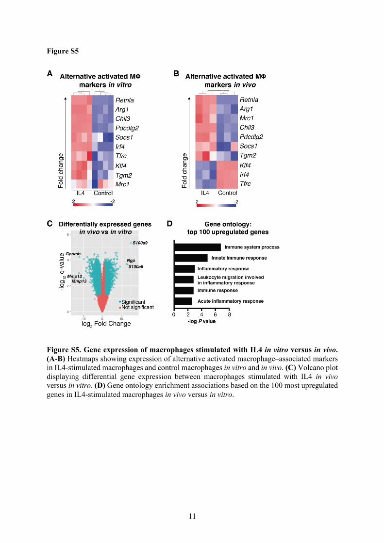
activation of macrophages, including Arg1, Chil3, and Retnla (Figure 4A, Figure S5A and
B),22, 23 which were among the most differentially upregulated genes (Tables S1 and S2). Of
note, IL4 also induced strong upregulation in vivo of the chemokine Ccl24, a biomarker for
macrophages that originate from monocytes rather than tissue-resident macrophages (Figure
4A).24 Moreover, the IL4-induced macrophages showed downregulation of genes such as
Cd68, which is associated with TAMs (Figure 4B),25 indicating that IL4 differentiates
macrophages into a phenotype that is distinct from TAMs.
We next performed gene set enrichment analysis to identify gene expression
signatures enriched in the IL4-induced macrophages in vivo. In accordance with increased
phagocytosis of macrophages stimulated with IL4 in vitro, we found an enrichment of
phagocytosis signatures in macrophages harvested from mice in the IL4 group (Figure 4C).
Moreover, IL4 stimulation resulted in enrichment of genes associated with major
histocompatibility complex (MHC) proteins (Figure 4C). To determine the influence of the in
vivo microenvironment, we compared the gene expression profiles of IL4-stimulated

10
macrophages generated in vitro versus in vivo (Table S3). Macrophages generated in vivo
exhibited a preferential upregulation of several markers associated with inflammation and
immune activation (Figures S5C and D). Altogether, the gene expression data suggest that
IL4 stimulation leads to an expansion of monocyte-derived macrophages with increased
phagocytic activity.
IL4 upregulates CD47 in a Stat6-dependent manner
We next searched for IL4-induced mechanisms in leukemia cells that might affect their
interactions with macrophages. Interestingly, the macrophage-inhibitory protein CD47 was
upregulated on leukemia cells in the IL4 group compared to controls at the time of sacrifice
(Figure 5A). Consistent with this finding, IL4 induced the expression of CD47 in leukemia
cells in a dose-dependent manner, showing that IL4 activates signaling that induces CD47
expression (Figure 5B). Moreover, in RNA sequencing data that we previously generated,10
Cd47 was upregulated in both c-Kit+ AML cells and normal c-Kit+ BM cells stimulated with
IL4 (Figure 5C).
We next explored the mechanistic basis of the IL4-induced upregulation of CD47.
Because STAT6 is a critical downstream mediator of IL4R-signaling, we used CRISPR/Cas9
genetic engineering to knock out Stat6 in Cas9-expressing MLL–AF9 AML cells using Stat6
sgRNAs that we previously characterized.10 Stat6 disruption hindered the IL4-induced
upregulation of CD47 (Figure 5D), demonstrating that IL4 upregulates CD47 in a STAT6-
dependent manner. Thus, in addition to activating murine macrophages, we identified a
previously unknown role of IL4 in protecting cells from phagocytosis via CD47 upregulation.
11
Combined IL4 treatment and CD47 blockade results in enhanced macrophage-mediated
phagocytosis of AML cells
Because CD47 protects cells from phagocytosis, we next evaluated whether the IL4-induced
upregulation of CD47 on AML cells counteracts enhanced phagocytosis by IL4-stimluated
macrophages. Consistent with this hypothesis, AML cells pre-treated for 24 hours with IL4
and washed before co-culture with macrophages were partially resistant to phagocytosis
(Figure 5E). To overcome the inhibitory signal provided by increased CD47 expression, we
used an α-CD47 blocking antibody. Combined blocking of CD47 on AML cells and IL4
stimulation of macrophages resulted in enhanced phagocytosis of AML cells (Figure 5F).
These findings show that IL4 has a dual role in murine phagocytosis by directly activating
macrophages and enhancing their phagocytic activity, while also inducing CD47 expression
that counteracts phagocytosis in target cells.
DISCUSSION
Distinct types of macrophages control tumor development. Whereas TAMs promote tumor
development by suppressing the immune system, other types of macrophages achieve tumor
immune surveillance through phagocytosis of malignant cells.26-29 We found that IL4 has
antileukemic effects in mice, predominantly mediated by alternatively activated macrophages
that normally play a key role in tissue repair and immune regulation.30, 31 The observed
expansion of alternatively activated macrophages is consistent with findings showing that
IL4, via the IL4 receptor type I complex, directly promotes the outgrowth of macrophages
beyond homeostatic levels in the setting of nematode infections.32 However, nematode
infections trigger the expansion of tissue residual macrophages.32 In contrast, the IL4-induced
macrophages with antileukemic activity showed higher expression of Ccl24, Mrc1, and
Pdcd1lg2, suggesting that they are of monocytic origin, from either the BM or peripheral

12
blood.24 Among hematopoietic cells, only macrophages showed increased numbers following
enforced expression of IL4 in vivo. IL4 also boosted the phagocytic activity of murine
monocyte-derived macrophages in vitro, suggesting that IL4 acts directly on the
monocytes/macrophages that mediate the antileukemic effect. Moreover, consistent with their
increased phagocytic activity, the IL4-induced macrophages were functionally and
molecularly distinct from TAMs, which are classically associated to an alternatively activated
phenotype.25 Furthermore, the IL4-induced macrophages were functionally distinct from
AML-associated macrophages, which polarize into a leukemia-supportive state that
accelerates disease development.3 The reason why IL4 induced stronger macrophage
activation in vivo compared to in vitro could be related to interactions with other immune
cells or the AML blasts, resulting in enhanced phagocytic activity. Of note, the macrophages
were dependent on IL4 for their anti-leukemic activity as depletion of macrophages in the
MIG control group did not affect the leukemia burden.
Constitutive expression of IL4 in mice has not been linked previously to anti-cancer
activity, but it has been associated with excessive phagocytosis resulting in decreased blood
cell counts, extramedullary hematopoiesis, and increased mortality.33, 34 We found that IL4
induced potent antileukemic activity, with some mice surviving long-term without signs of
disease or tolerability concerns, while other mice eventually had to be sacrificed despite very
low levels of leukemia cells in their BM and spleens. The low blood cell counts and
expansion of megakaryocytes in the spleen indicated extramedullary hematopoiesis and
suggests that elevated IL4 levels induced macrophage activation with excessive phagocytosis.
This pattern resembles that of hemophagocytic lymphohistiocytosis (HLH), a disease
characterized by aberrantly activated macrophages.35 Hence, we speculate that the cause of
death of non-leukemic mice in the IL4 group was due to the HLH-like symptoms. Of note,
the leukemic cells were selectively depleted, indicating that the IL4-induced macrophages
13
preferentially attacked them. The reason is unclear but could be related to altered expression
of genes by leukemia cells that regulate macrophages, such as MHC class I molecules or
calreticulin.36, 37
In addition to IL4 boosting macrophage-mediated phagocytosis, stimulation of AML
cells with IL4 induced STAT6-dependent upregulation of CD47, revealing a previously
unrecognized mechanism that regulates CD47 expression and thereby protects cells from
phagocytosis. This mechanism could possibly have evolved to protect endogenous cells from
phagocytosis in areas where high IL4 levels activate macrophages to fight invading
pathogens. Consistent with these findings, a super-enhancer region with binding sites for
STAT6 has been shown to regulate CD47 expression,38 providing a putative mechanistic
basis for how CD47 is upregulated via the IL4/STAT6 pathway. Given that combined IL4
stimulation and CD47 inhibition enhanced macrophage-mediated phagocytosis of AML cells,
our data suggest therapeutic potential for strategies that combine direct activation of
macrophages with blocking of inhibitory signals to macrophages. Because IL4 has opposing
effects in murine and human macrophages, we speculate that other cytokines that activate
human macrophages may also upregulate CD47 or other ‘don’t eat me’ signals on target
cells. Identifying these mechanisms may translate into new therapeutic opportunities in AML
and possibly other cancer types.
In summary, here we show that IL4 has a potent in vivo antileukemic effect in mice
by promoting macrophage-mediated phagocytosis of AML cells. IL4 stimulation induced
CD47 upregulation in a STAT6–dependent manner, and combined IL4 stimulation with
CD47 blockade further enhanced macrophage-mediated phagocytosis of AML cells. These
findings deepen our understanding of how IL4 regulates murine macrophages and suggest
that strategies to combine macrophage activation with CD47 inhibition should be explored
further as a therapeutic approach in cancer.
14
REFERENCES
1. Costello RT, Sivori S, Marcenaro E, et al. Defective expression and function of
natural killer cell-triggering receptors in patients with acute myeloid leukemia. Blood.
2002;99(10):3661-3667.
2. Jaiswal S, Jamieson CH, Pang WW, et al. CD47 is upregulated on circulating
hematopoietic stem cells and leukemia cells to avoid phagocytosis. Cell. 2009;138(2):271-
285.
3. Al-Matary YS, Botezatu L, Opalka B, et al. Acute myeloid leukemia cells polarize
macrophages towards a leukemia supporting state in a Growth factor independence 1
dependent manner. Haematologica. 2016;101(10):1216-1227.
4. Carlsten M, Järås M. Natural Killer Cells in Myeloid Malignancies: Immune
Surveillance, NK Cell Dysfunction, and Pharmacological Opportunities to Bolster the
Endogenous NK Cells. Front Immunol. 2019;10:2357.
5. Paczulla AM, Rothfelder K, Raffel S, et al. Absence of NKG2D ligands defines
leukaemia stem cells and mediates their immune evasion. Nature. 2019;572(7768):254-259.
6. Petty AJ, Yang Y. Tumor-Associated Macrophages in Hematologic Malignancies:
New Insights and Targeted Therapies. Cells. 2019;8(12):1526.
7. Li Y, You MJ, Yang Y, Hu D, Tian C. The Role of Tumor-Associated Macrophages
in Leukemia. Acta haematologica. 2020;143(2):112-117.
8. Wrangle JM, Patterson A, Johnson CB, et al. IL-2 and Beyond in Cancer
Immunotherapy. J Interferon Cytokine Res. 2018;38(2):45-68.
9. Majeti R, Chao MP, Alizadeh AA, et al. CD47 is an adverse prognostic factor and
therapeutic antibody target on human acute myeloid leukemia stem cells. Cell.
2009;138(2):286-299.

15
10. Peña-Martínez P, Eriksson M, Ramakrishnan R, et al. Interleukin 4 induces apoptosis
of acute myeloid leukemia cells in a Stat6-dependent manner. Leukemia. 2018;32(3):588-
596.
11. Li Z, Chen L, Qin Z. Paradoxical roles of IL-4 in tumor immunity. Cell Mol
Immunol. 2009;6(6):415-422.
12. Kiniwa T, Enomoto Y, Terazawa N, et al. NK cells activated by Interleukin-4 in
cooperation with Interleukin-15 exhibit distinctive characteristics. Proc Natl Acad Sci U S A.
2016;113(36):10139-10144.
13. Paul WE. History of interleukin-4. Cytokine. 2015;75(1):3-7.
14. Krivtsov AV, Twomey D, Feng Z, et al. Transformation from committed progenitor
to leukaemia stem cell initiated by MLL-AF9. Nature. 2006;442(7104):818-822.
15. Eriksson M, Peña-Martínez P, Ramakrishnan R, et al. Agonistic targeting of
TLR1/TLR2 induces p38 MAPK-dependent apoptosis and NFkappaB-dependent
differentiation of AML cells. Blood Adv. 2017;1(23):2046-2057.
16. Miller PG, Al-Shahrour F, Hartwell KA, et al. In Vivo RNAi screening identifies a
leukemia-specific dependence on integrin beta 3 signaling. Cancer Cell. 2013;24(1):45-58.
17. Järås M, Miller PG, Chu LP, et al. Csnk1a1 inhibition has p53-dependent therapeutic
efficacy in acute myeloid leukemia. J Exp Med. 2014;211(4):605-612.
18. Piganelli JD, Martin T, Haskins K. Splenic macrophages from the NOD mouse are
defective in the ability to present antigen. Diabetes. 1998;47(8):1212-1218.
19. Ito M, Hiramatsu H, Kobayashi K, et al. NOD/SCID/γcnull mouse: an excellent
recipient mouse model for engraftment of human cells. Blood. 2002;100(9):3175.
20. Claassen I, Van Rooijen N, Claassen E. A new method for removal of mononuclear
phagocytes from heterogeneous cell populations in vitro, using the liposome-mediated
macrophage 'suicide' technique. J Immunol Methods. 1990;134(2):153-161.
16
21. Qian Q, Jutila MA, Van Rooijen N, Cutler JE. Elimination of mouse splenic
macrophages correlates with increased susceptibility to experimental disseminated
candidiasis. J Immunol. 1994;152(10):5000-5008.
22. Loke Pn, Nair MG, Parkinson J, Guiliano D, Blaxter M, Allen JE. IL-4 dependent
alternatively-activated macrophages have a distinctive in vivo gene expression phenotype.
BMC Immunol. 2002;3:7.
23. Martinez FO, Helming L, Milde R, et al. Genetic programs expressed in resting and
IL-4 alternatively activated mouse and human macrophages: similarities and differences.
Blood. 2013;121(9):e57-e69.
24. Gundra UM, Girgis NM, Ruckerl D, et al. Alternatively activated macrophages
derived from monocytes and tissue macrophages are phenotypically and functionally distinct.
Blood. 2014;123(20):e110.
25. Haas L, Obenauf AC. Allies or Enemies-The Multifaceted Role of Myeloid Cells in
the Tumor Microenvironment. Front Immunol. 2019;10:2746.
26. Chen Y, Zhang X. Pivotal regulators of tissue homeostasis and cancer: macrophages.
Exp Hematol Oncol. 2017;6(23-23).
27. Feng M, Chen JY, Weissman-Tsukamoto R, et al. Macrophages eat cancer cells using
their own calreticulin as a guide: Roles of TLR and Btk. Proc Natl Acad Sci U S A.
2015;112(7):2145-2150.
28. Jaiswal S, Chao MP, Majeti R, Weissman IL. Macrophages as mediators of tumor
immunosurveillance. Trends Immunol. 2010;31(6):212-219.
29. Loyher P-L, Hamon P, Laviron M, et al. Macrophages of distinct origins contribute to
tumor development in the lung. J Exp Med. 2018;215(10):2536-2553.
30. Gordon S, Martinez FO. Alternative activation of macrophages: mechanism and
functions. Immunity. 2010;32(5):593-604.
17
31. Sica A, Mantovani A. Macrophage plasticity and polarization: in vivo veritas. J Clin
Invest. 2012;122(3):787-795.
32. Jenkins SJ, Ruckerl D, Thomas GD, et al. IL-4 directly signals tissue-resident
macrophages to proliferate beyond homeostatic levels controlled by CSF-1. J Exp Med.
2013;210(11):2477.
33. Erb KJ, Rüger B, von Brevern M, Ryffel B, Schimpl A, Rivett K. Constitutive
Expression of Interleukin (IL)-4 In Vivo Causes Autoimmune-type Disorders in Mice. J Exp
Med. 1997;185(2):329-339.
34. Milner JD, Orekov T, Ward JM, et al. Sustained IL-4 exposure leads to a novel
pathway for hemophagocytosis, inflammation, and tissue macrophage accumulation. Blood.
2010;116(14):2476-2483.
35. La Rosée P, Horne A, Hines M, et al. Recommendations for the management of
hemophagocytic lymphohistiocytosis in adults. Blood. 2019;133(23):2465-2477.
36. Barkal AA, Weiskopf K, Kao KS, et al. Engagement of MHC class I by the inhibitory
receptor LILRB1 suppresses macrophages and is a target of cancer immunotherapy. Nat
Immunol. 2018;19(1):76-84.
37. Feng M, Marjon KD, Zhu F, et al. Programmed cell removal by calreticulin in tissue
homeostasis and cancer. Nat Commun. 2018;9(1):3194.
38. Betancur PA, Abraham BJ, Yiu YY, et al. A CD47-associated super-enhancer links
pro-inflammatory signalling to CD47 upregulation in breast cancer. Nat Commun.
2017;8:14802.

18
FIGURE LEGENDS
Figure 1. IL4 has antileukemic activity in a microenvironment-dependent manner. (A)
dsRed+ c-Kit+ MLL–AF9 AML cells were transduced with retroviral vectors coexpressing
GFP and a murine IL4 cDNA (MIG–IL4) or an empty control vector (MIG). Two days later,
sorted GFP+ AML cells were transplanted into sublethally irradiated mice. (B)
Transplantation of 10 000 leukemia cells into C57BL/6 mice. Kaplan–Meier survival curves
(9 mice per group, pooled from two independent experiments), and percentage of leukemia
(dsRed+) cells in the BM of mice at the time of sacrifice. (C) Transplantation of 30 000
leukemia cells into NOD/SCID mice. Kaplan–Meier survival curves (n = 6 mice per group)
and percentage of leukemia cells in the BM of mice at the time of sacrifice. (D)
Transplantation of 30 000 leukemia cells into NSG mice. Kaplan–Meier survival curves (n =
14 mice per group, pooled from two independent experiments), and percentage of leukemia
cells in the BM of mice at the time of sacrifice. BM, bone marrow. ***P < 0.001; ****P <
0.0001.
Figure 2. IL4 stimulation increases the frequency of macrophages in vivo. C57BL/6 mice
were transplanted with 30 000 sorted GFP+ MLL–AF9 AML cells 2 days post transduction
with retroviral vectors coexpressing GFP and a mIL4 cDNA (MIG–IL4) or a control vector
(MIG). (A) Percentages of blood cell populations within dsRed– cells 19 days after
transplantation (n = 3). (B) Percentage of leukemia (dsRed+) cells in the peripheral blood on
day 19 after transplantation (n = 3). (C) WBC counts at days 12 and 27 for MIG–IL4 and
non-transplanted irradiated control mice (IL4 group, n = 4; controls, n = 3). (D) Percentage of
F4/80+ cells within dsRed- cells in BM and spleens of mice at the time of sacrifice (controls,
n = 4; IL4 group, n = 5). (E) Representative immunohistochemistry staining of F4/80+ cells in
BM (40×; scale bar, 20 µm) and spleens (10×; scale bar, 100 µm). BM, bone marrow; N.D.,

19
not detected; PB, peripheral blood; WBC, white blood cell. **P < 0.01; ***P < 0.001; ****P
< 0.0001.
Figure 3. IL4 stimulation causes macrophage-mediated depletion of leukemia cells in
vivo. (A) C57BL/6 mice were transplanted with 30 000 sorted GFP+ MLL–AF9 AML cells
transduced with retroviral vectors expressing a mIL4 cDNA (MIG–IL4) or GFP only (MIG;
data presented in Supplementary Figure S2). One day prior to transplantation, mice received
intraperitoneal (i.p.) injections of clodronate liposomes (MΦdep group; n = 4) or PBS as
control (n = 5). Every tenth day, new i.p. injections were performed. (B) Percentage of F4/80+
cells and (C) leukemia cells in BM and spleens at the time of sacrifice in the IL4 group. (D)
Monocytes were isolated from mouse BM and differentiated into macrophages in culture with
mCSF1 (25 ng/mL) and mIL4 (20 ng/mL) for 7 days, and then MLL–AF9 dsRed+ AML cells
were co-cultured with the macrophages. (E) Representative flow cytometry contour plots
showing dsRed+ cells within F4/80+ cells in freshly mixed cultures (0 hours) and after 18
hours of co-culture with macrophages and dsRed+ leukemia cells. (F) Phagocytosis assay
with dsRed+ AML cells and murine macrophages (n = 3). The percentage of dsRed+ cells
within F4/80+ cells is presented. (G) CD14+ cells were isolated from human blood and
differentiated into macrophages in culture with human (h)CSF1 (25 ng/mL) and hIL4 (20
ng/mL) for 7 days and then co-cultured with membrane-stained AML cell lines. (H)
Phagocytosis assay with PKH67+ MA9:16 cells and PKH26+ human macrophages (n = 4).
The percentage of PKH67+ cells within PKH26+ cells is presented. (I) Phagocytosis assay
with PKH67+ Mono Mac 6 cells and PKH26+ human macrophages (n = 5). BM, bone
marrow; MM6, Mono Mac 6; MΦ, macrophage. **P < 0.01; ***P < 0.001; ****P < 0.0001.

20
Figure 4. IL4 expands macrophages enriched for gene expression signatures associated
with alternative activation of macrophages and phagocytosis. RNA sequencing was
performed on murine macrophages generated from monocytes in vitro, and on sorted dsRed-
F4/80+ macrophages from mice in the IL4 and control groups. (A) Volcano plots displaying
differential gene expression between IL4-stimulated macrophages and control macrophages
in vitro (left plot), and macrophages from mice in the IL4 or control group (right plot). The y-
axis corresponds to the –log10(q-value) and the x-axis to the log2 of the gene expression fold
change. Green dots represent significant differentially expressed genes with a q-value < 0.05
and fold change > 2.0. (B) Heatmap showing expression of genes associated with
upregulation in TAMs. IL4-stimulated macrophages and control macrophages were harvested
from mice. (C) Gene set enrichment analysis revealed enrichment of phagocytosis and MHC
protein complex signatures in macrophages harvested from mice. FDR, false discovery rate;
GO: gene ontology; MΦ, macrophage; NES, normalized enrichment score; TAM: tumor-
associated macrophage.
Figure 5. Combined IL4 stimulation and CD47 blockade result in enhanced
macrophage-mediated phagocytosis of AML cells. (A) Representative histograms showing
CD47 expression on AML cells in BM and spleens of mice transplanted with dsRed+
leukemia cells transduced with the MIG–IL4 or control (MIG) vectors. (B) CD47 expression
on AML cells following IL4 stimulation for 24 hours. (C) Cd47 expression shown as FPKM
values of normalized reads from RNA sequencing data of c-Kit+ dsRed+ leukemia cells and c-
Kit+ normal BM cells stimulated with IL4 for 18 hours. Data are presented as box and
whiskers diagrams; the line indicates median, box limits are first and third quartiles, and bars
indicate maximum and minimum values. (D) CD47 expression measured by flow cytometry
after 24 hours of stimulation with mIL4 (100 ng/mL) in cells transduced with lentiviral

21
vectors expressing Stat6 or control sgRNAs. (E) Phagocytosis assay with macrophages
derived from murine BM monocytes stimulated with mCSF1 (25 ng/mL) and mIL4 (20
ng/mL) for 7 days. The AML cells were treated with mIL4 (100 ng/mL) or no IL4 (control)
for 24 hours prior to co-culture (n = 3). Phagocytosis is presented as the percentage of dsRed+
cells within F4/80+ cells. (F) Phagocytosis assay with mouse BM monocyte-derived
macrophages stimulated for 7 days with mCSF1 (25 ng/mL) and mIL4 (20 ng/mL) or mCSF1
only (n = 3). AML cells were cultured for 1 hour with a blocking anti-CD47 antibody or
corresponding isotype control and then mixed with the macrophages. FPKM, fragments per
kilobase million; gMFI, geometric mean fluorescence intensity; NBM, normal bone marrow.
*P < 0.05; **P < 0.01; ***P < 0.001; ****P < 0.0001.


1
Supplementary information for:
IL4 promotes phagocytosis of murine leukemia cells counteracted by CD47 upregulation
Pablo Peña-Martínez1, Ramprasad Ramakrishnan1, Carl Högberg1, Caroline Jansson1, David
Gisselsson Nord1, and Marcus Järås1
1Division of Clinical Genetics, Department of Laboratory Medicine, Lund University, Lund,
Sweden

2
SUPPLEMENTARY METHODS
Murine leukemia model
To enrich for leukemia stem cells, femurs from leukemic mice were crushed, red blood cells
were lysed using NH4Cl solution (STEMCELL Technologies, Vancouver, Canada), and c-Kit+
cells were enriched by CD117 MicroBeads in MACS® Cell Separation Columns according to
the manufacturer’s instructions (Miltenyi Biotec, Bergisch Gladbach, Germany), as previously
described.1 Except for propagation of leukemia cells, all experiments involving murine AML
cells were initiated using c-Kit+ BM cells. dsRed+ c-Kit+ MLL–AF9 leukemia cell cultures were
grown in serum-free expansion medium (SFEM; Stemspan, STEMCELL Technologies)
supplemented with 1% penicillin/streptomycin, 20 ng/mL murine (m)IL3, 25 ng/mL stem cell
factor (mSCF), and 20 ng/mL human (h)IL6 (PeproTech, Rocky Hill, NJ, USA).
Viral vector generation and production
The murine stem cell virus gammaretroviral vector coexpressing a mIL4 cDNA and GFP
(green fluorescent protein) connected with an internal ribosome entry site (MIG–IL4), and
Stat6 and control single-guided RNA (sgRNA) vectors were previously generated.2 The Cas9-
expressing leukemia cells were generated as previously described.2 Viral vectors were
produced using standard protocols in 293T cells. Gammaretroviral vectors were pseudotyped
with an ecotropic envelope and lentiviral vectors with a vesicular stomatitis virus G envelope.
For transduction experiments, SFEM was supplemented with mIL3 (40 ng/mL), mSCF (50
ng/mL), and hIL6 (40 ng/mL) and mixed with the viral vectors. Transduction was performed
by spinoculation at 600 ×g for 1 hour at 32°C, and 24 hours after transduction, the medium was
replaced with fresh SFEM supplemented with cytokines.
3
Flow cytometric analysis and cell sorting
Flow cytometric analyses were performed using a LSRFortessa™ flow cytometer (BD
Biosciences, San Jose, CA, USA), and cell sorting was performed using a FACSAria™ II cell
sorter (BD Biosciences). To analyze cell populations in mice transplanted with leukemia cells
transduced with retroviral vectors, we stained blood and BM cells using APC–CD3, PE/Cy7–
CD4, BV510–CD8, APC/Cy7–CD19, and BV421–NK1.1 antibodies for lymphoid lineage
stains, and APC–Ly6g, PE/Cy7–CD11c, BV421–CD115, and APC/Cy7–F4/80 antibodies for
myeloid lineage stains (all from BioLegend, San Diego, CA, USA). Before flow cytometric
cell sorting, we stained F4/80+ cells with a BV421–F4/80 antibody (BioLegend). Staining of
CD47 was achieved using an AF647–CD47 antibody (BioLegend). Staining of IL4RA was
performed using a BV421–CD124 antibody (BD Biosciences).
Macrophage differentiation
To isolate both human and mouse monocytes, we used MACS® Cell Separation Columns with
monocyte isolation kits according to the manufacturer’s instructions (Miltenyi Biotec).
Monocytes from mouse BM were enriched by negative selection, whereas we used CD14+
selection to isolate human monocytes from the peripheral blood of healthy donors. Isolated
monocytes were differentiated into macrophages in Roswell Park Memorial Institute (Gibco,
Thermo Scientific, Waltham, MA, USA) medium supplemented with 10% heat-inactivated
fetal bovine serum, 1% penicillin/streptomycin, and 25 ng/mL murine or human colony-
stimulating factor 1 (CSF1) for 7 days, and 20 ng/mL of murine or human IL4 for 7 days
(cytokines from PeproTech). Half of the culture medium was replaced every 2-3 days.
4
Immunohistochemistry
Organs harvested from mice were fixed in 4% paraformaldehyde for 48 hours and stored in
70% ethanol. Formalin-fixed, paraffin-embedded tissue sections (4 µm) were dried on
positively charged slides for 15 minutes at 60°C. The slides were then deparaffinized in xylene
and hydrated in graded ethanol solutions. Endogenous peroxidase was blocked for 20 minutes
with 1% H2O2 (Sigma-Aldrich) diluted in PBS pH 7.4 (Applichem, Darmstadt, Germany).
Heat-induced epitope retrieval was performed by using target retrieval solution, pH 9.0
(Agilent DAKO, Santa Clara, CA, USA), and 0.2% Triton X-100 (Sigma-Aldrich) in a
decloaking chamber (Biocare Medical, Pacheco, CA, USA) at 95°C for 20 minutes. Sections
were incubated for 60 minutes with a rabbit anti-mouse F4/80 primary antibody (Thermo
Fisher Scientific) in a 1:200 dilution in PBS containing 5% normal goat serum (Jackson
Immuno Research, Ely, UK). Staining was obtained by using a horseradish peroxidase–
conjugated anti-rabbit polyclonal antibody (AH Diagnostics, Tilst, Denmark) for 30 minutes,
followed by incubation with the liquid DAB+ Substrate Chromogen System (Agilent DAKO)
for 5 minutes, and counterstaining with Mayer’s Hematoxylin (Histolab, Askim, Sweden) for
30 seconds. All incubations were performed at room temperature, and sections were washed
three times with PBS after each incubation. Slides were mounted with Faramount Mounting
Medium, Aqueous (Agilent DAKO). Images were acquired in an Olympus BX43 (Olympus,
Waltham, MA, USA) with the Cellsens software (Olympus).
Phase holographic imaging
For morphologic analysis, a total of 5 000 macrophages were seeded per well in a 24-well plate
and placed in a Holomonitor® M4 (Phase Holographic Imaging AB, Lund, Sweden). The
microscope was located in an incubator at 37°C and 5% CO2. Cells were allowed to attach for
one hour, and then images were acquired and analyzed with the software Hstudio™ (Phase

5
Holographic Imaging AB). Individual cells were measured for volume and irregularity, a
parameter based on the roundness of the cell.
RNA sequencing analysis
We used QIAshredder and RNeasy Microkit (QIAgen, Hilden, Germany) to extract RNA and
validated RNA quality using a 2100 Bioanalyzer (Agilent Technologies, Inc).
To prepare RNA libraries from mouse cells, we used the TruSeq RNA sample prep kit
v2 (Illumina, San Diego, Ca, USA) and performed sequencing in a NextSeq 500 Desktop
Sequencer (Illumina) with the NextSeq 500/550 Mid Output v2 kit, 150 cycles (Illumina). The
sequenced reads were aligned to the mm10 reference mouse genome using TopHat 2.0.13. For
statistical analysis, differential gene expression, and visualization of the RNA sequencing data,
we used Qlucore Omics Explorer 3.0 (Qlucore, Lund, Sweden). Gene set enrichment analysis
(GSEA)3 was performed with pre-ranked gene lists, based on the GSEA guidelines for RNA
sequencing data, followed by pairwise t-tests for comparisons between groups.

6
Figure S1
Figure S1. IL4 has antileukemic activity in a microenvironment-dependent manner. (A) Expression of IL4RA on dsRed+ c-Kit+ MLL–AF9 AML from serial propagations. (B–D) Percentage of leukemia (dsRed+) cells in the spleens of mice at the time of sacrifice. ****P < 0.0001.

7
Figure S2
Figure S2. IL4 stimulation in vivo induces expansion of macrophages. (A–B) RBC and platelet count at days 12 and 27 for MIG–IL4 and non-transplanted irradiated mice as controls (controls, n = 3; MIG–IL4, n = 4). (C) Percentage of BM and spleen cell populations within the non-dsRed fraction at the time of sacrifice. (D) Representative contour plots of F4/80+ cells in BM and spleen. (E) Count of F4/80+ cells on IHC slides of BM per 20x magnification field (n = 3). (F) Expression of IL4RA on F4/80+ cells harvested from mice at the time of sacrifice. (G) Representative hematoxylin and eosin (H&E) staining of BM and spleens (10 ×; scale bar, 100 µm). Arrowheads indicate megakaryocytes. (H) Representative H&E staining of BM from
8
NSG mice (10 ×; scale bar, 100 µm). BM, bone marrow; RBC, red blood cell. *P < 0.05, **P < 0.01; ***P < 0.001; ****P < 0.0001.

9
Figure S3
Figure S3. Depletion of macrophages in vivo did not affect leukemia levels in mice receiving non–IL4-expressing leukemia cells. C57BL/6 mice were transplanted with 30 000 sorted GFP+ MLL–AF9 AML cells transduced with the MIG control vector. One day prior to transplantation, mice received intraperitoneal (i.p.) injections of clodronate liposomes (MΦdep group) or PBS as control. Every tenth day, new i.p. injections were performed. (A) F4/80+ cells at the time of sacrifice in BM and spleens of mice receiving clodronate liposomes. (B) Percentage of leukemia cells in BM and spleen of mice receiving clodronate liposomes. MΦ, macrophage. **P < 0.01.

10
Figure S4
Figure S4. IL4 changes the morphology of macrophages. Monocytes were isolated from mouse BM and differentiated into macrophages in culture with mCSF1 (25 ng/mL) and mIL4 (20 ng/mL) or mCSF1 only (control) for 7 days and then analyzed using holograph imaging. (A) Cell volume and (B) irregularity of control (n = 60) and IL4-stimulated (n = 90) cells. ***P < 0.001; ****P < 0.0001.
11
Figure S5
Figure S5. Gene expression of macrophages stimulated with IL4 in vitro versus in vivo. (A-B) Heatmaps showing expression of alternative activated macrophage–associated markers in IL4-stimulated macrophages and control macrophages in vitro and in vivo. (C) Volcano plot displaying differential gene expression between macrophages stimulated with IL4 in vivo versus in vitro. (D) Gene ontology enrichment associations based on the 100 most upregulated genes in IL4-stimulated macrophages in vivo versus in vitro.

12
SUPPLEMENTARY TABLES
Table S1. Differentially expressed genes in IL4 macrophages versus control macrophages
generated in vitro (provided as Excel files).
Table S2. Differentially expressed genes in IL4 macrophages versus control macrophages
generated in vivo (provided as Excel files).
Table S3. Differentially expressed genes in IL4 macrophages generated in vivo versus in vitro
(provided as Excel files).

13
REFERENCES
1. Eriksson M, Peña-Martínez P, Ramakrishnan R, et al. Agonistic targeting of TLR1/TLR2 induces p38 MAPK-dependent apoptosis and NFkappaB-dependent differentiation of AML cells. Blood advances. 2017;1(23):2046-2057. 2. Peña-Martínez P, Eriksson M, Ramakrishnan R, et al. Interleukin 4 induces apoptosis of acute myeloid leukemia cells in a Stat6-dependent manner. Leukemia. 2018;32(3):588-596. 3. Subramanian A, Tamayo P, Mootha VK, et al. Gene set enrichment analysis: A knowledge-based approach for interpreting genome-wide expression profiles. Proc Natl Acad Sci U S A. 2005;102(43):15545-15550.